Note: Descriptions are shown in the official language in which they were submitted.
CA 02753871 2011-08-29
WO 2010/104854 PCT/US2010/026665
IMMUNOMODULATORY THERAPEUTIC AGENTS
Rights in the Invention
This invention was made with support from the United States government, from
the National Institutes of Health Contract No. NOl 15435, which was awarded by
the
National Institute of Allergy and Infectious Diseases of the National
Institutes of Health,
and the United States Government has rights in this invention
Reference to Related Applications
This application claims priority to United States Provisional Application No.
61/158,526 entitled "Therapeutic Activity of Immunomodulatory Agents" filed
March 9,
2009, the entirety of which is hereby incorporated by reference.
Background
1. Field of the Invention
This invention is directed to components isolated from biologic fluids (blood,
serum, or exudates), to methods for isolating such components from natural
sources, to
methods for utilizing these components to maintain and enhance normal function
of the
human body, and to method for treating diseases and disorders comprising
administration
of therapeutically effective amounts of the isolated components of either
natural or
synthesized forms.
2. Description of the Background
In healthy multi-cellular organisms, homeostasis is impeccably maintained
through
a tightly regulated balance of up and down regulation of various cellular
functions. The
loss of this homeostatic balance at the cellular level results in chronic
disease. These
cellular regulatory activities also result in interaction between cell and
interaction between
systems. These interactions result in an intricate relationship between cells
within each of
the body's organs as well as interactions between each of these
organs/systems. This
interaction takes place by several forms of communication such as cytokines,
enzymes,
and circulating cells.
Any significant insult to the body causes the release of a group of peptides
which
have profound immunomodulatory effects. These peptides may fall into the
broadly
described category of cytokines (nonantibody proteins released as a response
to a specific
1
CA 02753871 2011-08-29
WO 2010/104854 PCT/US2010/026665
stimulus and which act as intercellular mediators), and they may act as
enzymes. In either
case, they trigger a pathway which slowly moves the body toward a response
with one or
more of the following characteristics:
1) A rapid decrease of pain and swelling with a persistent analgesic response;
2) A stimulation of the adaptive immune response;
3) Delayed stimulation of the breakdown and resorption of Fibrin deposits;
4) Stimulation of surveillance cells of the immune system (NK cells, T Killer
Lymphocytes); or
5) A shift of the baseline status of the organism to an anti-inflammatory
state.
While many cytokines have been identified and their roles in specific
responses
partially described, a limited knowledge of this aspect of cellular regulation
is yet
available. This is a very active area of research as demonstrated by the
contradictory
established activity for many of the cytokines. Fully unraveling the intricate
intercellular
communication carried out through cytokines will necessitate years of
additional research.
The integumentary system comprises the single greatest defense against
pathogens
in all higher forms of life. At the time when an organism is most vulnerable
to exposure to
a pathologic insult due to an interruption of the integumentary system, the
organism
utilizes this immune system up-regulation to protect itself from any potential
infectious
processes. In the cytokine/immune cell cascade, the type of modification which
occurs
through these peptides enhances the ability of the organism to recognize and
respond to
the plethora of pathologic insults likely to occur when the skin is breached,
while strongly
suppressing the inflammation this type of stimulation usually causes. This
includes the
activation of immune system cells and the release of cytokines which as a
therapeutic can
enhance the organism's ability to recognize and respond to other pathologic
insults,
including acute and chronic bacterial infections, acute and chronic viral
infections,
parasitic diseases, and even neoplastic processes. While skin represents the
most important
barrier to infection, the innate immune system enables a rapid response to the
myriad of
attacks resulting from a breach in the integument. The innate immune system
recognizes
and eradicates pathogens and harmful foreign molecules and has a role in the
surveillance
and rejection of tumors (Auf et al., 2001; Bacha et al, 2004; Gorelik et al.,
1995; Wu and
Pruett, 1999).
Besides stimulating the innate immune system, this response also facilitates
wound
and tissue healing through resorption of fibrin. These deposits are
exacerbating factors in
many chronic diseases, resulting in the deactivation of many of the healing
mechanisms
2
CA 02753871 2011-08-29
WO 2010/104854 PCT/US2010/026665
and blocking the nutritional support of the damaged cells. This effect is seen
in chronic
wounds as well as the plaques of multiple sclerosis, the neurofibrillary
tangles of
Alzheimer's disease, the plaques of Atherosclerosis, the tissue changes of
autoimmune
diseases, and the fibrin deposits around cancer cells which act as a shield
protecting the
tumor from the immune system.
This response also facilitates wound healing and immunomodulation decreases
the
effects of inappropriate antibody expression by stimulating the production of
T-killer
lymphocytes. These cells seek out and destroy B-Cells which are
inappropriately
producing auto-antibodies. By eliminating the production of auto-antibodies,
the attack on
the body is stopped and the inflammation these antibodies produce is
eliminated,
decreasing the symptoms and often the most significant cause of autoimmune
disease.
These peptides also enhance the ability of an organism to recognize and
destroy cells
manifesting abnormal protein on their cell wall. As all cancer cells express
abnormal cells
on their cell membrane, these T-killer lymphocytes are essential in the
recognition and
destruction of all types of cancer.
In addition to this increased surveillance, these peptides have a strong anti-
inflammatory activity. Through stimulation of TH-2 cytokines, these peptides
suppress
the inflammatory response that otherwise would be expected in acute injury. In
addition,
they appear to be even more helpful in blocking inflammation from chronic
processes.
This anti-inflammatory effect enhances the healing of both acute and chronic
injuries.
In chronic disease processes, the deposition of fibrin in the extra-vascular
space is
an integral part of the progression of many diseases. Mobilizing these chronic
fibrin
deposits and preventing their deposition have now become targets for
therapeutics, but no
successful therapeutics which work through these mechanisms have previously
been
identified. Preventing fibrin deposits from occurring in response to an acute
pathologic
process prevents the transition of these acute processes into chronic disease.
Fibrinogen is a bivalent protein composed of six polypeptide chains, two each
of
the Aa, B13 and y chains. These chains are linked together near their Amino
termini by
disulfide bonds, leaving their carboxy termini exposed to the action of
thrombin. In
response to injury to a blood vessel wall, thrombin is activated and cleaves
fibrinogen to
produces a fibrin monomer and two peptides each of fibrinopeptides A and B.
These
fibrin monomers then bind to each other to form a loose scaffolding for clot
formation.
The released fibrinopeptides have been characterized, with some species
dependant
3
CA 02753871 2011-08-29
WO 2010/104854 PCT/US2010/026665
activities, but they have previously not been identified as modulators of the
immune
system. They also have the therapeutic benefits of acutely decreasing vascular
permeability and triggering a delayed stimulation of the resorption of fibrin
from the
intimal and extra vascular spaces, benefits which have not been previously
identified or
recognized. Throughout the systems of the body, any activity which produces a
change in
our homeostasis also stimulates an opposite process to allow for the reversal
of that
change. One therefore would expect a process that causes a blood vessel to
leak to also
result in a process of correcting that leak. In the case of Fibrin production,
this is attained
by two mechanisms: 1) the production of a clot to seal the area of leakage,
and 2) the
release of molecules which decrease the overall vascular permeability to
prevent the
migration of these molecules into areas where they are not needed and can be
harmful. In
addition, the presence of fibrin in the extravascular space and subintimal
space are harmful
on a long term basis but necessary acutely. Fibrinopeptide A produces a
delayed
resorption of these fibrin deposits to prevent the problems associated with
their chronic
presence. When this resorption fails, chronic disease result. The cross
species activity of
these peptides is known, but the mechanism of this interspecies activity has
not been
previously explained. While Fibrinopeptide B possesses little to no homology
from
species to species, the terminal sequence of Fibrinopeptide A has significant
homology
through most mammals, likely accounting for most of this cross species
activity. In
addition, a portion of C3 bears considerable homology from one mammal to
another.
The deposition of fibrin into the intima of blood vessels in coronary artery
disease
and into the extra-vascular space in many other diseases results in the
progression of these
diseases. Mounting data on the regulation of fibrin indicates that this fibrin
deposition is a
major part of many chronic disease processes. This is due not only to the
impairment of
function caused by the physical barrier fibrin forms, but also the pro-
inflammatory activity
of fibrin in these spaces. In addition, the presence of fibrin in these spaces
suppresses the
activity of some cells which are essential for healing. One example of this is
the ability of
extra-vascular fibrin to inactivate the regenerative activity of Swann Cells.
Over the last
several years researchers have been able to demonstrate the benefit of removal
of extra-
vascular fibrin in many of these disease processes. These studies demonstrate
the
proinflammatory activity of fibrin as well as the impairment of normal
cellular/organ
function. This impairment is a major component of the pathologic process of
many
4
CA 02753871 2011-08-29
WO 2010/104854 PCT/US2010/026665
diseases, including but not limited to Multiple Sclerosis, Rheumatoid
Arthritis, Peripheral
nerve crush injury, Alzheimer's Disease, macular degeneration, chronic wounds
and
Atherosclerosis. In these studies extra-vascular fibrin was an important
target for new
therapeutics.
Over the last several years Thl and Th2 cytokines have also become a prominent
focus in the study of the immunologic/inflammatory response. Initially, Th2
cytokines
were viewed as anti-inflammatory and Thl cytokines were viewed as pro-
inflammatory.
This generalization does not fully describe the complex and intricate
interaction between
these opposite ends of the inflammatory spectrum. Alternatively, the
inflammatory
response has also been characterized into active and passive components, and
further still,
into portions active in the innate and adaptive immune system. None of these
divisions
truly describes the response seen in vivo, as the systemic response nearly
always
incorporates a combination of ThI and Th2 activities, innate and adaptive, and
active and
passive immunologic/inflammatory responses.
This combination activity also occurs in an organism's response to these
peptides.
These peptides cause a spectrum of activities in the cytokine cascade that
could be viewed
as either stimulatory or suppressive, but the net result is a stimulation of
the immune
system, stimulation of the removal of fibrin from the extra-vascular space,
and suppression
of inflammation.
Many different proteins and peptides circulating in the bloodstream are well
recognized for their effects on the local inflammatory processes in humans as
well as in
experimental animal models. These proteins and peptides include a variety of
cytokines
and chemokines. These substances produce a feedback loop that regulates the
extent of
the local inflammatory response. In most chronic diseases this control over
the
inflammatory response is inadequate, allowing localized inflammation to result
in the
destruction of healthy tissues. Fibrinopeptide A has an anti-inflammatory
affect in a
variety of disease models, thus decreasing local tissue destruction and
ameliorating the
disease state. These models are herein described, demonstrating the anti-
inflammatory
effect of Fibrinopeptide A. The mechanism of action of this anti-inflammatory
response is
demonstrated the specific ability of fibrinopeptide A to produce the shift in
the cytokine
panel from a predominantly Thl response to a predominantly Th2 response
through the
production of specific anti-inflammatory cytokines. In addition to this
cytokine shift,
fibrinopeptide A has the ability to decrease vascular permeability. This
decrease in the
vascular permeability has the effect of maintaining the plasma proteins (such
as fibrin)
CA 02753871 2011-08-29
WO 2010/104854 PCT/US2010/026665
within the blood vessels, preventing their pro-inflammatory activity in the
extra-vascular
space. Although Fibrin in particular has been implicated as a pro-inflammatory
molecule
in the extra vascular space, Fibrinopeptide A has the ability to greatly
reduce the migration
of fibrin from the blood stream into the extra-vascular space, and to expedite
the removal
of fibrin from this space. This anti-inflammatory activity (or at least
prevention of a pro-
inflammatory activity) has profound implications in the treatment of chronic
inflammatory
conditions.
In 1978, Ruhenstroth-bauer et. al. demonstrated the anti-inflammatory activity
of
Fibrinopeptides A and B. In their research, Ruhenstroth-bauer and associates
sought an
understanding of the specific cause of inflammation in response to a
pathologic challenge.
They first demonstrated a shift in the acute phase proteins released after a
pathologic
insult. They then isolated an anti-inflammatory activity of the proteins
produced by this
shift. U.S. Patent 4215109 describes the process of further isolating this
anti-inflammatory
activity first to fibrinogen, and then further testing confirmed
Fibrinopeptides A and B to
be the source of this biologic anti-inflammatory response, a response that
could have a
beneficial effect in almost all pathologic processes. They demonstrated this
benefit in a
carrageenin-induced rat paw edema model, demonstrating the benefit of
increased
fibrinogen injected intra-peritoneally, then isolating this beneficial
activity to
fibrinopeptide A and B. However, their research failed to describe the
mechanism of
action of this anti-inflammatory activity or even isolate this activity to a
specific peptide.
They did not utilize their research to produce a therapeutic.
This same group (Scherer et. al, 1981) subsequently demonstrated this anti-
inflammatory effect in another disease model. Improvement in the course of
Experimental
Allergic Encephalomyelitis (EAE) in guinea pigs and rats (a disease model for
Multiple
Sclerosis (MS)) by daily intraperitoneal injections with human fibrinopeptides
A and B
was demonstrated. These injections produced significant amelioration of the
disease state
in the treated animals as compared to controls. Improvements in the clinical
neurological
signs of the disease were evident in that the number, the severity and the
duration of
pareses were diminished in treated animals. Furthermore, the inflammatory
alterations of
vasopermeability associated with extravasation of plasma proteins and edema of
the
neuroparenchyma were significantly less pronounced in the fibrinopeptide-
treated animals.
Finally, a significantly higher titre of circulating immune complexes was
observed in the
serum of these animals, demonstrating that treatment with fibrinopeptides A
and B did not
alter the specific immune response to the antigenic challenge. They therefore
concluded
6
CA 02753871 2011-08-29
WO 2010/104854 PCT/US2010/026665
this anti-inflammatory response is not at the expense of immunosuppression. No
differences in anti-basic protein and anti-brain antibody production were
observed. The
characteristic cellular infiltrates of EAE also showed no significant
qualitative or
quantitative differences between fibrinopeptide-treated animals and the saline-
treated
controls, (Scherer et. al. 1979) but the inflammation typically identified in
conjunction
with these findings was much less pronounced.
Shortly after these studies were completed, Marusic et. al. demonstrated
similar
findings with the induction of any type of peritonitis. As these peptides are
mildly acidic,
the research by Scherer et. al. was likely viewed as a false positive, and
there does not
appear to be further published research from anyone into this pathway. Fibrin
deposition
is a significant contributing factor (Adams et al. 2004) in the development of
plaques in
MS, and these deposits form as a consequence of the leaky vasculature. As
described
above, Scherer et. al. demonstrated that this vascular leakage was ameliorated
by treatment
of Fibrinopeptides A and B.
Decreased vascular permeability also slightly reduces the migration of immune
complexes, and slows the deposition of fibrin in the extra-vascular space. In
addition,
fibrin has the ability to regulate Schwann cell differentiation by maintaining
Swann cells
in a nonmyelinating state. Fibrin induces phosphorylation of ERK1/2 and
production of
p75 NGF low affinity receptor in Schwann cells which maintains them in a
nonmyelinating state, suppresses fibronectin production, and prevents
synthesis of myelin
proteins. (Akassoglou, et. al., 2002). In many chronic neurologic diseases
this continued
presence of Fibrin in the extra-vascular space is implicated in the
persistence of the
disease process and progression of neurologic symptoms. These include the
presence of
fibrin in Multiple Sclerosis Plaques, Alzheimer's disease neurofibrillary
tangles, Basil
Ganglion lesions in Parkinson's disease, Peripheral nerve lesions in Chronic
Inflammatory
Demyelinating Polyneuropathy, and many others. These fibrin depositions are
also an
important part of the pathologic process outside of the neurological system.
This is
demonstrated by the essential role of fibrin in the development of
atherosclerotic heart
disease, chronic wounds, Hypertension, Cancer (creating a barrier around the
cancer cells),
macular degeneration, Autoimmune diseases, and many others.
Anti-Allergenic/Anti-Anaphylaxis Activity have also been observed. Masuda et.
al. (2001) demonstrated that a Fibrinopeptide A fragment (the same fragment
identified as
the object of this invention) deglycosylates mouse antibody IgE. This
deglycosylated IgE
no longer had the ability to stimulate histamine release from Mast Cells, thus
preventing
7
CA 02753871 2011-08-29
WO 2010/104854 PCT/US2010/026665
an anaphylactic/allergic response. This deglycosylation however did not affect
the ability
of IgE to interact with the antigen, bind with mast cells and other
immunologic cells, or
alter the ability of these cells to perform all of their other normal
activities resulting from
IgE antigen/antibody complex attachment to their membrane receptor. They
further
demonstrated that the synthetic form of the peptide sequence also
deglycosylated IgE in a
similar fashion. Once this deglycosylation had occurred, they were no longer
able to
induce mouse systemic anaphylaxis (Masuda et.al. 2001). However, Masuda and
associates failed to elucidate the mechanism of this deglycosylation. They
also did not
evaluate any further effect the deglycosylated IgE may have on the immunologic
response.
The modification of the systemic effect of cytokines through glycosylation and
deglycosylation is well established. This deglycosylation of IgE may be a
direct effect of
the Fibrinopeptide A on IgE, or it may be an additional effect of the
Immunomodulation.
This deglycosylation and subsequent lack of histamine response may partially
explain the
decreased permeability of blood vessels seen after administration of
Fibrinopeptide A. A
lack of histamine response may result in a decrease in the fibrin deposition
at the sight of
insult. As this deglycosylation does not appear to suppress the immune system
in any
other way and does not even alter the ability of IgE to attach to an antigen,
the beneficial
effect comes with no detrimental effect on an organism's immunologic response.
Rather,
this deglycosylation just controls the detrimental acquired hyperactivity
which causes
allergic reactions.
The ability of Fibrinopeptide A to decrease the severity of injury in a burn
model
has been demonstrated. (Wormser, Uri Patent 10/790888). In this patent
fibrinopeptide A
in conjunction with Histone peptides was found to prevent injury from thermal
and
chemical burns and speed healing of burns that had already occurred. They
postulate this
healing occurs primarily due to the anti-inflammatory effect. To demonstrate
this they
pretreated with the exudates from burns of other animals of the same species,
and then
isolated the fraction of the exudates responsible for this benefit. They found
that a fraction
containing only Fibrinopeptide A had tremendous protective effect in
decreasing the
severity of burn. They did not postulate a mechanism of action for this
protective effect.
This work indicates the mechanism of this effect through the decreased
permeability of
blood vessels which prevents the damage that the extravasation of the plasma
proteins
causes, including the release of lysosomal enzymes and the production of
superoxide
anions.
8
CA 02753871 2011-08-29
WO 2010/104854 PCT/US2010/026665
The ability of fibrinopeptide B (and possibly A) to facilitate wound healing
has
also been demonstrated by the ability of Fibrinopeptide B to enhance migration
of
fibroblasts, monocytes and neutrophils into the area of injury, without
stimulating the
release of lysosomal enzymes or the production of superoxide anion from these
neutrophils (Senior. et.al., 1986). The release of these substances in
response to the chemo
attractant activity of fibrin toward neutrophils is postulated to result in
the demyelination
seen in Multiple Sclerosis. In addition, Gray et. al. (1990) demonstrated the
ability of the
fibrinogen alpha and beta chains to stimulate the replication of fibroblasts,
and this activity
was significantly enhanced by the addition of thrombin to a fibrinogen
containing solution,
strongly suggesting this activity is associated with the products of this
cleavage.
These finding have not been recognized as sufficient to develop a therapeutic
containing them, as demonstrated by the lack of information and research
continued in this
area after the discovery of these peptides decades ago. This lack of ongoing
research is at
least partially due to the failure of all of these researchers to recognize
the
immunomodulatory and anti-inflammatory ability of these peptides, enhancing
the
immune system while decreasing the inflammatory response. This
immunomodulation
aids in the prevention of chronic infection, promotes a much healthier Th2
environment,
and stimulates migration of cells necessary in wound healing, while preventing
the
mechanisms of damage which slow healing and lead to chronic wounds.
The growth of new blood vessels is a complicated multifactorial process
involving
cells of several different types. When vascular injury interrupts the flow of
blood, the
healing process necessitates restoration of blood flow to the healing cells.
This process
begins with the degradation and absorption of injured cells and thrombus
coupled with
migration of fibroblasts to fill the injury defect. Then vascular cells
differentiate to form
tubules which eventually mature into blood vessels. Angiogenesis is essential
to the
normal physiological processes of wound healing.
Fibrin accumulates around leaky blood vessels in solid tumors (Brown, et.al.
1988). Fibrin has also been shown to polymerize at the host-tumor interface to
form fibrin
networks that can promote tumor angiogenesis by supporting the adhesion,
migration,
proliferation and differentiation of endothelial cells (Bootle-Wilbraham
et.al. 2001). As
this fibrin network thickens this promotion of angiogenesis is lost and the
fibrin network
becomes a barrier to adhesion, migration, proliferation, and differentiation
of these
endothelial cells. This barrier also prevents immune cells from recognizing
and
eliminating tumor cells. By only utilizing the activated fragments
fibrinopeptides A and
9
CA 02753871 2011-08-29
WO 2010/104854 PCT/US2010/026665
B, the resulting cytokine cascade has these beneficial angiogenic activities
without
"protecting" tumor cells from immunogenic attack. This effect is partially
explained
through extrapolation of the available data on Interleukin 1 B and the effect
it has on
inflammatory and immune cells. The migration and differentiation of cells in a
wound bed
are greatly affected by this cytokine and the presence of certain other immune
cells
(macrophages and lymphocytes). IL-I B enhances migration of these cells into
the wound
bed, thus producing an environment which is more conducive to angiogenesis. In
addition, the elevation of IL-10 in response to this activated fragment of
Fibrinopeptide A
and other direct effects of fibrinopeptide A on the inflammatory cascade offer
an enhanced
ability of lymphocytes, monocytes, macrophages, and monocytes to migrate into
these
areas without the release of lysosomal enzymes but still with the ability to
attack cells and
other foreign bodies.
The deposition of Fibrin in the walls of blood vessels occurs in many
disorders of
the vascular system. The ability of Fibrinopeptide A to mobilize these fibrin
depositions
and to prevent further deposition has far reaching implications in all
vascular disease.
These include, but are in no way limited to, improvements in Coronary Artery
Disease,
Macular Degeneration, Claudication, and Atherosclerosis. In many additional
diseases
this process enhances blood flow, improving the outcome in most chronic
diseases. This
absorption of these fibrin deposits creates an environment which is more
conducive to the
angiogenic process when an injury to any tissue occurs. This is best
exemplified in the
diabetic foot ulcer, in which the chronic fibrin deposition in the macro and
micro
vasculature greatly impedes blood flow and prevent tissues from healing due to
the lack of
circulation to the affected area.
No previous studies document the ability of Fibrinopeptide A to stimulate
angiogenesis and, thus, a healthy vascular environment. In fact, Staton et.al.
(US Pat.
Application Publication No. 20040039157) demonstrated that one fragment of
Fibrinopeptide A produced during cleavage by plasmin actually had a quite
prominent
anti-angiogenic activity. Thompson et.al (1992) also isolated the angiogenic
activity of
fibrinogen to fragment E, a portion of fibrinogen at the central knot released
by plasmin,
but failed to recognize the potential therapeutic effect of Fibrinopeptide A,
a byproduct
which should have been present in the solution he demonstrated to have the
therapeutic
activity, as Thrombin was initially used in the solution to breakdown
Fibrinogen.
CA 02753871 2011-08-29
WO 2010/104854 PCT/US2010/026665
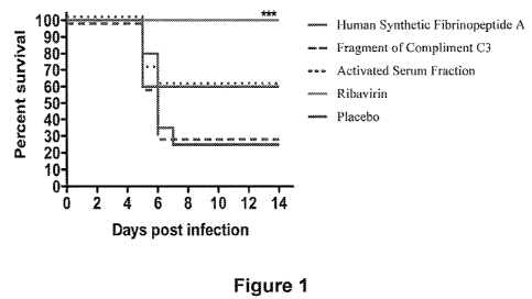
No published studies establish Fibrinopeptide A and/or B as an antiviral or
antibiotic. An increased survivability of mice treated with Fibrinopeptide A
and then
given Ponto Toro Virus was observed. Two different forms of Fibrinopeptide A
were
utilized: 1) a filtered serum fraction of goat serum calculated to contain
approximately 3
mg of Fibrinopeptide A (also containing goat Fibrinopeptide B and a fragment
of
Complement C3), and 2) synthetic Fibrinopeptide A. While these substances did
not
perform as well as a direct anti-viral (an expected outcome), the results did
demonstrate
improved survival of the treated animals when compared to the placebo group.
(See Figure
1). In this study several criteria were analyzed. These included Liver,
spleen, and serum
virus titers; Serum alanine aminotransferase (ALT) determinations; livers and
lungs were
scores for hepatic icterus on day 3 of infection; daily weight measurement;
Mean Day to
Death; and overall survivability. The two groups treated with test articles
containing
fibrinopeptide A performed identically. In these treatment groups 60% of the
mice lived,
while in the placebo group only 25% survived. This improvement was
statistically
significant for each of the fibrinopeptide A groups independently (P value =
0.03), and
when these groups are combined to calculate the overall improvement with
Fibrinopeptide
A the statistical significance improved (P value = 0.015). This increased
survivability
occurred even though there was no observable difference in any of the other
disease
criteria evaluated, indicating no change in the ability of the virus to cause
disease, but
rather an increased ability of the organism to fight off a life threatening
infection
following one dose of Fibrinopeptide A. No measure of inflammation or fibrin
deposition
was performed in this study. A difference exists between Fibrinopeptide A and
the
Ribavirin control, this difference was not statistically significant (P value
= 0.08).
These results demonstrate the potential therapeutic value of Fibrinopeptide A
in the
infectious disease arena, with the ability to augment healing and decrease the
duration of
symptoms. As the body's normal response to infectious diseases results in a
very pro-
inflammatory state, even after the infection is cured this state often causes
persistent
symptoms. Fibrinopeptide A has the ability to alleviate these symptoms and to
therefore
shorten the symptomatic phase of the disease without blocking the body's
ability to fight
off the infection. This effect also may be due to the ability of
Fibrinopeptide A to
mobilize proteins out of the extra-vascular space.
Given the delay in the shift of the cytokine panel and based on the
anticipated
effects of this demonstrable shift, the enhanced immunity produced by
fibrinopeptide A
has a much stronger effect on the adaptive immune response than on the innate
immune
11
CA 02753871 2011-08-29
WO 2010/104854 PCT/US2010/026665
response. This difference accounts for lack of improvement in all of the other
measures of
disease while still greatly enhancing survivability.
A second study was conducted to test these substances against Influenza A
HIN1.
In this study the control was low dose Ribavirin. All of the mice died in
every group,
suggesting a more severe infection than anticipated. This again demonstrates
the lack of
time to develop a true adaptive response which would have been enhanced by the
presence
of Fibrinopeptide A.
The anti-neoplastic activity of the peptides of this invention in treating
neoplastic
disease are due to three different mechanisms of action: 1) increased
surveillance by the
immune system to eliminate neoplastic cells, 2) preventing or eliminating the
deposition of
fibrin around cancer cells, and 3) decreasing the swelling around tumor cell
clusters and
the symptoms this swelling causes.
Primarily through the activity of IL-113 the immune system production of T-
killer
lymphocytes, NK lymphocytes, and B cells is increased. This differentiation
allows the
organism's immune system to seek out and destroy cancer cells based on the
abnormal
proteins manifest on their cell membranes. These peptides therefore have the
ability to
treat even those cancers that respond poorly to chemotherapy. While this
mechanism of
action will require time to attack and remove cancers that have already
spread, this type of
stimulation can also prevent cancer from ever developing.
Increased plasma fibrinogen levels or secretion of fibrinogen by the malignant
tumor cells themselves cause the deposition of fibrinogen or fibrin into the
extracellular
matrix of the malignant tumor tissues, and these factors have the effect as
part of the
extracellular matrix to promote proliferation, invasion and metastasis of the
malignant
tumor cells (Rybarczyk et.al. 2000). The ability of these peptides to prevent
the migration
of fibrin into the matrix surrounding the tumor cells will therefore have the
effect of
eliminating this protection of cancer cells from the host's immune system and
facilitate
recognition and elimination of cancer cells by the host. In addition to this
effect, the
stimulation of the host immune system by these peptides enhances the ability
of the
immune system to destroy these cells.
The anti-inflammatory activity described above decreases the symptoms of
metastatic cancer, as many of these symptoms are due to the inflammation the
metastasis
cause. In addition, the symptoms commonly caused by chemotherapy are partially
due to
the inflammatory effect of these medications and the cellular destruction
these medications
cause.
12
CA 02753871 2011-08-29
WO 2010/104854 PCT/US2010/026665
From the above description, these peptides have the ability to treat auto-
immune
disease by: 1) decreasing the inflammatory response to an auto-immune antibody
attack,
2) decreasing the fibrin depositions which lead to the progression of auto-
immune disease
pathology, and 3) destroying the B-cells which produce auto-antibodies through
the
production of T-killer lymphocytes which seek out and destroy cells producing
auto-
antibodies. The loss of the ability to perform this surveillance function is
ultimately
responsible for the development of autoimmune disease. These peptides have the
ability to
restore this function. While IL-1 P has been implicated in progression of the
destructive
process of some diseases, this low level stimulation does not seem to have
these effects or
the presence of IL- 10 stimulation mediates/prevents these effects.
Buckheit (WO/2006/116381) demonstrated that a serum fraction from goats
treated
with cancer cell lysates has an anti-neoplastic activity toward that
particular cancer. While
this was initially thought to be secondary to antibody formation in goats,
they
subsequently demonstrated that the serum fraction from these animals depleted
of the
large proteins (including immunoglobulin) still contain this anti-neoplastic
activity. They
also demonstrated the ability of a serum fraction from a goat pretreated with
cancer cell
lysates from one type of cancer to treat a different type of cancer. They
postulate that this
effect is related to antibody fragments.
Immunization or vaccination involves exposing a patient to inactivated
pathogenic
antigens in order to stimulate an immune response to that specific pathogen.
This active
type of immunity typically provides long term protection against that specific
disease.
Extensive attempts to establish active immunity toward several common viruses
have
proven futile to date, and this has led to research into the utilization of
passive immunity to
treat these diseases. This type of therapy utilizes neutralizing antibodies
produced by one
or many patients or animals to treat infection in another patient or animal.
Historically,
passive immunity has been utilized to treat a variety of diseases. For many
decades,
immuno-compromised patients have been given pooled IgG to enhance their
immunity.
With the increase incidence of blood born infection in our population and the
ability to
produce monoclonal antibodies, this therapy has fallen out of favor for the
treatment of
general mild immune system dysfunction. Pooled antibody preparations are only
rarely
used now to boost the immune system in times of increased exposure, and to
stop the
attack of autoimmune disease.
13
CA 02753871 2011-08-29
WO 2010/104854 PCT/US2010/026665
Despite these factors, passive immunity has continued to receive attention as
a
possible therapeutic for certain viral and bacterial infections. However, the
serum from
individuals or animals with established immunity might also contain the virus
or bacteria,
thus, transfer of serum could result in an infection as well. In an effort to
develop new
anti-viral and antibiotic drugs to specific diseases, this hyper-immune serum
has been
evaluated for therapeutic potential to humans afflicted with these diseases.
These
approaches carry the obvious difficulties of the occurrence of
hypersensitivity reactions
and the potential for additional infection, but they have demonstrable
efficacy. The most
simplistic form of this type of therapy is performed by simply exposing a host
animal to a
particular pathogen and then extracting blood from the animal and injecting
the serum
fraction containing the antibodies into the patient.
Karpas (U.S. Patent No. 4,863,730) utilized a preparation containing a high
titer of
heterologous human neutralizing antibodies obtained from the plasma of HIV
positive
patients to treat HIV. While this method proved beneficial in decreasing
viremia and
delaying onset of AIDS, clinical application and large scale production are
exceptionally
problematic.
Davis (WO 97/02839, WO 01/60156, 02/07760, and US 2002/006022) utilized a
method involving inoculation of goats with viral lysates (HIV) or bacterial
lysates
(Staphylococcus, Steptococcus, E.coli) and then injecting the serum obtained
from these
hyper-immune goats to treat HIV infected patients. His method and success
utilizing this
method to treat HIV and other infections have been widely publicized
(Washington Post,
April 9, 2000; Dateline Houston television broadcast, Sept 18, 1998; etc.). In
his process
he utilizes standard extraction and purification methods including ammonium
sulfate
precipitation followed by a filtration process (dialysis or gel filtration)
after allowing the
blood to initially clot.
Gelder and associates (U.S. Patents # 6043347, 6258599, 6335017, and 6670181)
also developed a method utilizing hyper-immune goats to produce neutralizing
antibodies
which are hypothesized to recognize certain viral epitopes. They utilize
antigens that fail
to trigger the production of neutralizing antibodies in humans but are handled
appropriately by goats. Gelder complicated the process described by Davis by
injecting
purifying proteins from HIV-1 MN and HIV-2 NZ into goats, and then augmenting
the
immunity with synthetic peptides from regions known to contain highly
conserved HIV
epitopes. This method has lead to production of a medication (HRG214) which is
currently in clinical trials for the treatment of HIV. The manufacturer also
claims that its
14
CA 02753871 2011-08-29
WO 2010/104854 PCT/US2010/026665
serum prepared from animals exposed to one virus through their process is
beneficial in
the treatment of other types of viral diseases. (See Vironyx web site). In
addition,
depleting the serum of large proteins (including removal of all full
antibodies) does not
eliminate the benefit but does enhance the safety of the preparation. It is
postulated that
this benefit is derived from the presence in the remaining serum fraction of
antibody
fragments (particularly the Feb fragment). In the information accompanying
this research,
it is stated that it is best to remove all proteins greater than 30 kD in
size, essentially
eliminating all of the antibodies and fragments that result from the treatment
of the goats.
Dalgleish (WO 03/004 049, WO 03/064472) recognized that the activity of some
of these formulas could not be fully explained by the activity of neutralizing
antibodies.
He therefore postulated that the anti-inflammatory activity of these
preparations may be
dependent upon anti-HLA and/or anti-FAS antibodies. He demonstrated that these
anti-
bodies have an anti-inflammatory effect, preventing an over-stimulation of the
immune
system by viral epitopes resembling normal human HLA. Dalglish and associates
demonstrated that the serum fraction enriched with these anti-HLA and/or anti-
FAS
antibodies are useful in the treatment of a wide variety of diseases with
inappropriately
high HLA levels such as chronic infections (both viral and bacterial),
tropical cancers
(lung, pancreas, liver, bowel, lymph nodes and skin cancers are specified),
and other
diseases with high HLA levels such as Diabetes and Multiple Sclerosis. In his
research,
Dalglish and associates did not utilize hyper-immune goats (no treatment of
the goats with
antigen prior to removal of the blood).
Tolett (WO 04/033665), also describes the therapeutic benefit of a
heterologous
serum mixture for treatment of HIV using the filtered, but otherwise
unpurified, serum or
plasma of HIV-exposed animals. The serum or plasma mixture is simply an
unprocessed
mixture of serum from various animals that has not undergone any purification
process.
Ansley (U.S. Pat. No. 5,219,578) uses a similar preparation process to prepare
an
IgG serum fraction, although in this patent, no prior stimulation of the
goat's immune
system is undertaken. The serum of these pathogen nave goats was removed and
processed, and then utilizes to prevent and treat a variety of veterinary
diseases. These
diseases include equine lower respiratory disease (ELRD) caused by a variety
of
opportunistic organisms, ovine foot rot in sheep and lambs caused by various
serotypes of
B. nodosus, and bovine respiratory disease. Ansley demonstrated that the non-
immunized
goat serum induces non-specific activation of the immune system in the treated
animal,
resulting in a remarkable therapeutic effect.
CA 02753871 2011-08-29
WO 2010/104854 PCT/US2010/026665
Hamm et.al. demonstrated the ability of a caprine serum fraction to treat
equine
lower respiratory infection.
Thacker (U.S. Pat. No. 7,358,044) demonstrated that a serum fraction
containing
low molecular weight peptides could be used to stimulate the immune system,
greatly
improving the survival rate in animals lethally challenged with a variety of
pathogens. In
this studies, serum from pathogen naive animals was used in the preparation of
the
medication.. This patent also references studies in which a fraction of
caprine serum,
substantially free of immunoglobulins, could confer significant protection to
chickens
challenged with a lethal dose of Pasteurella multocida when the caprine serum
fraction
was administered 24 hours prior to the bacterial challenge. Similar results
were found in
mice given a lethal challenged with Salmonella typhimurium.
Buckheit (U.S. Pat. App 2006/0292162) demonstrated the serum or plasma from
animals inoculated with lysates from viruses, bacteria, or cancers cells has
the ability to
treat the disease from which the lysates were prepared. This therapeutic
effect is greatest
in the serum fraction which is essentially free of all antibodies and large
proteins.
In addition to these studies demonstrating the benefit of serum fractions,
several
studies have been completed exploring the use of neutralizing monoclonal
antibodies. The
results of these studies have proven disappointing. (see, e.g., Burton D R et
al. Science
(1994) 266: 1024-1027; Trkola A. et al. J. Virol. (1996) 70: 1100-1108; Conley
A J. et al.
Proc. Natl. Acad. Sci. USA (1994) 91: 3348-3352;). Although these antibodies
seemed to
have a significant benefit in vitro, no clear benefit could be demonstrated in
vivo (Stiehm,
1995). In general, heterologous antibody mixtures (produced from raw serum and
therefore containing the active peptides of this invention) seem to be
markedly more
beneficial than monoclonal antibodies, again suggesting an alternative
mechanism of
action to the antibodies alone. These mixtures are also felt to be more
beneficial in the
prevention of disease than the treatment of disease (Montefiori, 2001).
Summary of the Invention
As embodied and broadly described herein, the present invention is directed to
pharmaceutical compositions, dietary supplements of these composition, and
method for
the preparation of a biologically active fraction of mammalian serum from
animal blood
and isolated and manufactured peptides therefrom to modulate the immune system
and
enhance the immune response under a variety of conditions. In addition, the
invention
includes the synthetic forms of these peptides, and the invention includes and
derivations
16
CA 02753871 2011-08-29
WO 2010/104854 PCT/US2010/026665
and modifications of these peptides that enhance these therapeutic and
prophylactic
benefits.
One embodiment of the invention is directed to an agent comprising a peptide
containing a sequence identified in SEQ ID NOs. 1-5, 7-9, 11-13, 15, 16, or 20-
22, a
sequence of Fibrinopeptide A, a sequence of a region of Fibrinopeptide A that
is
substantially homologous between species of mammals that produce
Fibrinopeptide A, a
sequence of Compliment C3, or any of the foregoing sequences also containing
one or
more conservative amino acid substitutions, wherein the agent contains
substantially no
detectable Fibrinopeptide B. Preferably the agent further comprising a
pharmaceutically
acceptable carrier such as, for example, water, oil, edible oil, fatty acids,
lipids,
polysaccharides, cellulose, glycerin, glycol, and combinations thereof. A
preferable edible
oil includes, for example, lemon oil, peppermint oil, or grape seed oil.
Preferred agents
are formulated for oral, transmucosal, parenteral, lymphatic, or intravenous
administration
such that the biologically active form of the agent is released into a system
of a patient at a
physiologically effective concentration. Also preferred is an agent which is a
dietary
supplement and agents which are purified from biological sources or
synthetically
manufactured.
Another embodiment of the invention is directed to a pharmaceutical
composition
comprising Fibrinopeptide A or a fragment thereof, and a pharmaceutically
acceptable
carrier, wherein the Fibrinopeptide A or fragment thereof is at a
therapeutically effective
amount. Preferably the therapeutically effective amount is from 0.1 mg to 500
mg. Also
preferred is the composition wherein the therapeutically effective
concentration prevents
deposition and stimulates resorption of fibrin within the extravascular
spaces, such as is
associated with coronary artery disease, and subintimal spaces in a patient.
Preferably the
composition is nontoxic at the therapeutically effective concentration and
substantially
free of detectable Fibrinopeptide B. The composition may caontain
Fibrinopeptide A or
fragment thereof that are derived from a human or non-human, but preferably
mammalian
sequence of Fibrinopeptide A. Mammals that express the non-human sequence of
Fibrinopeptide A include an equine, a feline, a canine, a bovine, a caprine,
an ovine, and a
murine.
Another embodiment of the invention is directed to a method for treating or
preventing a disorder of a patient comprising: providing a pharmaceutical
composition
comprising Fibrinopeptide A or a fragment thereof, and not Fibrinopeptide B,
and a
pharmaceutically acceptable carrier, wherein the Fibrinopeptide A or fragment
thereof, is
17
CA 02753871 2011-08-29
WO 2010/104854 PCT/US2010/026665
derived from a mammal that is not a human; and administering a dose of the
composition
to the patient, wherein administration is transmucosal such that the
Fibrinopeptide A or
fragment thereof achieves a therapeutically effective level within the
lymphatic system of
the patient within 5 minutes of administration. Preferably the patient is a
human, and also
preferably the disorder is vascular inflammation or coronary artery disease.
The preferred
single dosage of the composition contains from 0.1 mg to 10 mg of active
ingredient, and
preferred administration comprises an initial administration and subsequently,
both oral
and transmucosal, and a continued administration, and the continued
administration is not
repeated for an interval of at least 7 days. Preferably the Fibrinopeptide A
or fragment
thereof stimulates the patient's cells to release cytokines IL 10, IL-10, and
not IL-l, IL-4 or
TNFd. Another preferred aspect is for the activity of Fibrinopeptide B of the
patient to be
suppressed, such as, for example, by the administration of a Fibrinopeptide B
binding
agent.
Another embodiment of the invention is directed to a method of preventing
deposition of fibrin and absorbing fibrin deposited within blood vessels of a
patient,
comprising: providing a pharmaceutical composition that comprises
Fibrinopeptide A or a
fragment thereof and a pharmaceutically acceptable carrier; and administering
the
composition to a patient such that the Fibrinopeptide A or fragment thereof is
at a
therapeutically effective level is achieved in the lymphatic system of the
patient.
Preferably the patient is a human and the Fibrinopeptide A or fragment thereof
is derived
from a mammalian sequence of Fibrinopeptide A that is not a human.
Administration of
the composition is preferably directly to the lymphatic system by transmucosal
administration, and comprises an initial administration and subsequently, a
continued
administration, and the continued administration is no more than once a week.
Another embodiment of the invention is directed to a fraction of serum of a
mammal wherein the fraction contains multiple components, is clarified of
particulates,
and substantially all components are within a molecular weight range of from
about 1,200
Daltons to about 1,700 Daltons. Preferably the mammal is an equine, a feline,
a canine, a
bovine, a caprine, an ovine, or a murine.
Another embodiment of the invention is directed to an agent comprising a
peptide
containing a sequence selected from the group consisting of SEQ ID NOs. 6, 10,
14, and
17-19, a sequence of Fibrinopeptide B, and a sequence of a region of
Fibrinopeptide B that
is substantially homologous between species of mammals that produce
Fibrinopeptide B,
18
CA 02753871 2011-08-29
WO 2010/104854 PCT/US2010/026665
wherein the agent contains substantially no detectable Fibrinopeptide A.
Preferably the
agent further comprising a pharmaceutically acceptable carrier such as, for
example,
water, oil, edible oil, fatty acids, lipids, polysaccharides, cellulose,
glycerin, glycol, and
combinations thereof. A preferable edible oil includes, for example, lemon
oil,
peppermint oil, or grape seed oil. Preferred agents are formulated for oral,
transmucosal,
parenteral, lymphatic, or intravenous administration such that the
biologically active form
of the agent is released into a system of a patient at a physiologically
effective
concentration. Also preferred is an agent which is a dietary supplement and
agents which
are purified from biological sources or synthetically manufactured.
Another embodiment of the invention is directed to a method for treating or
preventing a disorder of a patient comprising: providing a pharmaceutical
composition
comprising Fibrinopeptide B or a fragment thereof, wherein the composition
contains
substantially no detectable Fibrinopeptide A, and a pharmaceutically
acceptable carrier,
wherein the Fibrinopeptide B or fragment thereof, is derived from a mammal
that is not a
human; and administering a dose of the composition to the patient, wherein
administration
is transmucosal such that the Fibrinopeptide B or fragment thereof achieves a
therapeutically effective level within the lymphatic system of the patient
within 5 minutes
of administration. Preferably the patient is a human, and the disorder is an
auto-immune
disorder, such as, for example, arthritis, Crohn's disease, Coeliac disease,
diabetes mellitus
type 1, Grave's disease, idiopathic thrombocytopenic purpura, psoriasis,
scleroderma,
systemic lupus erythematosus, or ulcerative colitis, or the disorder is a
immunoregulatory
disorder, such as, for example, an overactive immune system. The preferred
single dose
contains from 0.1 mg to 10 mg of active ingredient.
Another embodiment of the invention is directed to a fraction of serum of a
mammal wherein the fraction contains multiple components, is clarified of
particulates,
and substantially all components are within a molecular weight range of from
about 800
Daltons to about 2,300 Daltons. Preferably, the mammal is selected from the
group
consisting of an equine, a feline, a canine, a bovine, a caprine, an ovine,
and a murine.
Other embodiments and advantages of the invention are set forth in part in the
description, which follows, and in part, may be obvious from this description,
or may be
learned from the practice of the invention.
19
CA 02753871 2011-08-29
WO 2010/104854 PCT/US2010/026665
Description of the Figures
Figure 1 Effect of Fibrinopeptide A in a natural and synthetic form on the
survivability of mice to Ponto Toro infection. Activated Serum Fraction
contains Goat Fibrinopeptides A and B as well as the Fragment of
Compliment C3.
Figure 2 Effect of PEGylated and non-PEGylated synthetic Fibrinopeptide A in
an
acute Experimental Allergic Encephalomyelitis mouse model.
Figure 3 HPLC reading of the Bovine serum fraction embodiment of the
invention.
Peaks at 21.73 and 22.84 were both identified as SERIM A; Peaks at 22.59
and 23.28 were identified as SERIM B; the small peak at 20.13 seconds
was identified as SERIM C.
Figure 4 HPLC reading of the Equine serum fraction embodiment of the
invention.
Peaks at 21.32 and 18.30 were identified as SERIM A; peaks at 14.56 and
23.53 are SERIM B; the peak at 11.62 and 11.84 are SERIM C
Figure 5 HPLC reading of the caprine serum fraction embodiment of the
invention.
The horse serum fraction contains the highest relative amount of Equine
SERIM A (peak at 17.86). Other peptides were not identified in this
specimen, but review of the protein databases shows no sequencing
information for the other two SERIMs in the equine database.
Figure 6 HPLC reading of the Human serum fraction embodiment of the invention.
As can be seen, the human sample contains many more peptides than the
animal samples. However, samples still correlate with the majority of the
peptide mass. Peaks at 29.46 and 20.96 both correspond to Peptide A.
Peaks at 25.27 and 30.41 correspond to peptide B, and the peak at 19.16
corresponds to peptide C.
Description of the Invention
The human body has an amazing ability to heal following a severe traumatic
injury. In considering the differences in response to injury between those
suffering from
severe trauma and those suffering minor injuries, three differences stand out:
1) Those
involved in severe trauma have a markedly enhanced immune system response; 2)
There is
a relative minimization of swelling in the early stages of injury for those
experiencing
severe trauma; and 3) Severe Trauma creates a numbing effect, decreasing the
pain felt by
severe trauma patients when compared to those suffering more minor injury. The
medical
literature and the current medical paradigm attribute these findings to the
"stress response"
CA 02753871 2011-08-29
WO 2010/104854 PCT/US2010/026665
and the release of endogenous endorphins as part of this response. A group of
peptides
released during these types of injuries has been surprisingly discovered that
are
responsible for many of the benefits of this stress response. When these
peptides are
utilized in chronic diseases this response has tremendous benefits to the
patient.
Herein are identified a number of cytokine activities of peptides, some of
which
have been previously identified as molecules but the cytokine activity has not
been
otherwise shown. In addition, certain molecules are characterized herein that
were not
previously identified as biologically active substances. While the sequences
of certain
peptides may be established, cleavages of these proteins and the releasing of
biologically
active peptides have not been previously described. These peptides fall in two
classes: 1)
those released as part of the clotting cascade, and 2) those released as part
of the
complement system. Many of the peptides released as part of the clotting
cascade have
been identified, but the cytokine mechanism of action has not been previously
described or
recognized. The peptides of the complement system have not been previously
described
as cleavages from the parent proteins, and their activity as cytokines also
has not been
previously described. The description herein discloses that these peptides are
released in
response to a break in the integument. Most any pathologic insult severe
enough to cause
damage to the walls of blood vessels will produce a similar release of these
peptides.
During the initiation of the clotting cascade, many small peptides are
released in
the activation of the proteins which form the framework of a blood clot. These
degradation products have always been considered relatively inactive peptides
although
some minor activities outside of the clotting cascade have been attributed to
them. These
peptides are present in the bloodstream just long enough to be further
recycled, with half
lives of only minutes. However, given the complex interaction between the
various
systems in other physiologic processes, degradation products from the clotting
cascade
have the ability to up-regulate the immune system, as the need for clotting
typically
coincides with exposure to pathogens. Also, cytokine activity usually occurs
at very low
doses. The ability of small volumes of these peptides to have a profound
effect even with
their very short half lives in the body is surprising. Although unexpected,
this may be due
to a need for an up regulation of the immune system when there is a breech in
the walls of
a blood vessel or of the integument.
Peptides of the invention have this ability to block the deposition of fibrin
and
associated material. This is a direct effect or part of the result of the
immunomodulation
and stimulation of the cytokine cascade, but the fact that Fibrinopeptide A
either directly
21
CA 02753871 2011-08-29
WO 2010/104854 PCT/US2010/026665
or indirectly results in the regulation of Fibrin deposition presents a major
breakthrough in
the management of both acute and chronic diseases. In addition, the complement
system
is activated in response to this same type of insult and the subsequent
exposure to
infectious agents. A previously unidentified peptide is released from the C3
protein of the
complement cascade, and contributes to this immunomodulatory activity.
One embodiment of the invention is directed to an agent comprising a peptide
containing a sequence identified in SEQ ID NOs. 1-5, 7-9, 11-13, 15, 16, or 20-
22. Also
included are peptides that comprise the sequence of Fibrinopeptide A or
Compliment C3,
and a sequence of a region of Fibrinopeptide A or Compliment C3 that is
substantially
homologous between species of mammals that produce Fibrinopeptide A or
Compliment
C3, respectively. The sequence may be derived from human or non-human sources.
The
invention is also directed to a sequence that contains one or more
conservative amino acid
substitutions of any of the aforesaid sequences. Preferably, the agent
contains
substantially no detectable Fibrinopeptide B. Preferably the agent further
comprising a
pharmaceutically acceptable carrier such as, for example, water, oil, edible
oil, fatty acids,
lipids, polysaccharides, cellulose, glycerin, glycol, and combinations
thereof, and any of a
number of conventionally used carriers such are disclosed in WO/010757
entitled
"Pharmaceutical Composition" by J. Arch and N. Bowring (which is incorporated
by
reference). Preferable edible oil includes, for example, lemon oil, peppermint
oil, or grape
seed oil, or other natural oils and fatty acids derived from plants. Preferred
agents are
formulated for oral, transmucosal, parenteral, lymphatic, or intravenous
administration
such that the biologically active form of the agent is released into a system
of a patient at a
physiologically effective concentration. Also preferred is an agent which is
purified from
biological sources or synthetically manufactured, including both the peptide
sequences
themselves. The invention also includes nucleic acid sequences that encode
these
peptides.
Another embodiment of the invention is the agent described above and herein,
that
is a dietary supplement. The agents of the invention are safe for human and
animal
ingestion, and non-toxic at all effective dosages, and contain no endogenous
endotoxin or
other harmful materials or contaminants. Administration as a dietary
supplement can be
as the agent in a pure form, preferably transmucosally and more preferably
suspended in a
fatty acid, saccharide or polysaccharide, oil, or other carrier substance
(e.g. as a liquid, gel,
paste, powder, tablet, or pill) for immediate absorption by the mucosa of the
mouth, such
22
CA 02753871 2011-08-29
WO 2010/104854 PCT/US2010/026665
as under the tongue. As a dietary supplement, the agent can be administered to
a patient
or in association with other ingredients such as in a beverage or food
product.
Another embodiment of the invention is directed to a pharmaceutical
composition
comprising Fibrinopeptide A or a fragment thereof, and a pharmaceutically
acceptable
carrier, wherein the Fibrinopeptide A or fragment thereof is at a
therapeutically effective
amount. Preferably the therapeutically effective amount is from 0.1 mg to 500
mg. Also
preferred is the composition wherein the therapeutically effective
concentration prevents
deposition and stimulates resorption of fibrin within the extravascular
spaces, such as is
associated with coronary artery disease, and subintimal spaces in a patient.
Preferably the
composition is nontoxic at the therapeutically effective concentration and
substantially
free of detectable Fibrinopeptide B. The composition may caontain
Fibrinopeptide A or
fragment thereof that are derived from a human or non-human, but preferably
mammalian
sequence of Fibrinopeptide A. Mammals that express the non-human sequence of
Fibrinopeptide A include an equine, a feline, a canine, a bovine, a caprine,
an ovine, and a
murine.
Another embodiment of the invention is directed to a method for treating or
preventing a disorder of a patient comprising: providing a pharmaceutical
composition
comprising Fibrinopeptide A or Compliment C3, or a fragment of either, and a
pharmaceutically acceptable carrier. Preferably the composition does not
contain a
detectable amount of Fibrinopeptide B, and the Fibrinopeptide A or Compliment
C3,
fragment of either, is derived from a mammal that is not a human; and
administering a
dose of the composition to the patient, wherein administration is transmucosal
such that
the Fibrinopeptide A or Compliment C3, fragment of either, achieves a
therapeutically
effective level within the lymphatic system of the patient within 5 minutes of
administration. Preferably the patient is a human, and also preferably the
disorder is
vascular inflammation or coronary artery disease. The preferred single dosage
of the
composition contains from 0.1 mg to 10 mg of active ingredient, more
preferably from 0.1
to 5 mg, and more preferably less than 1 mg. The administration may be on a
periodic
basis, and preferred administration comprises an initial administration of a
single effective
dose for a series of days, and a subsequently administered dose administered
once every
other day, more preferably once every few days, and more preferably once a
week or even
less frequently. Administration for all doses is preferably oral and
transmucosal, such as
under the tongue. Preferably the Fibrinopeptide A or fragment thereof
stimulates the
patient's cells to release cytokines IL1p, IL-10, and not IL-1, IL-4 or TNF&.
Another
23
CA 02753871 2011-08-29
WO 2010/104854 PCT/US2010/026665
preferred aspect is for the activity of Fibrinopeptide B of the patient to be
suppressed, such
as, for example, by the administration of a Fibrinopeptide B binding agent.
Binding
agents include ligands, antibodies, or antibody fragments that are specific
for
Fibrinopeptide B, and, preferably, are non-toxic and include one or more
substances (e.g.
liquids or chemicals) that render the Fibrinopeptide relatively B inactive as
compared with
the activity of Fibrinopeptide A.
Another embodiment of the invention is directed to a method of preventing
deposition of fibrin and also absorbing fibrin deposited within blood vessels
and other
areas of the body of a patient. These methods comprise: providing a
pharmaceutical
composition that comprises Fibrinopeptide A or Compliment C3, or a fragment of
either,
and a pharmaceutically acceptable carrier; and administering the composition
to a patient
such that the Fibrinopeptide A or Compliment C3, or fragment of either, is at
a
therapeutically effective level is achieved in the lymphatic system of the
patient.
Preferably the patient is a human and the Fibrinopeptide A or Compliment C3,
or fragment
of either, is derived from a mammalian sequence of the same molecule that is
not a
human. Administration of the composition is preferably directly to the
lymphatic system
by transmucosal administration, and comprises an initial administration and
subsequently,
a continued administration, and the continued administration is no more than
once every
few days such as once a week or even once a month.
Another embodiment of the invention is directed to a fraction of serum of a
mammal wherein the fraction contains multiple components, is clarified of
particulates,
and substantially all components are within a defined molecular weight range.
Methods to
fractionate serum by molecular weight are well known and include dialysis with
molecular
weight cut-off membranes, centrifugation, and salt fractionation. The
molecular weight
range is preferably less than 3,000 Daltons, more preferably from about5800
Daltons to
about 2,500 Daltons, more preferably from about 1,000 Daltons to about 2,000
Daltons,
more preferably from about 1,200 Daltons to about 1,800 Daltons, and more
preferably
from about 1,400 Daltons to about 1,800 Daltons. Preferably the mammal is an
equine
(horse), a canine (dog), a feline (cat), a bovine (e.g. cow, cattle, or bull),
a caprine (goat),
an ovine (sheep or lamb), or a murine (mouse), or may be any suitable mammal
that
produces Fibrinopeptite A or Compliment C3.
24
CA 02753871 2011-08-29
WO 2010/104854 PCT/US2010/026665
Another embodiment of the invention is directed to an agent comprising a
peptide
containing a sequence of SEQ ID NOs. 6, 10, 14, or 17-19, a sequence of
Fibrinopeptide
B, or a sequence of a region of Fibrinopeptide B that is substantially
homologous between
species of mammals that produce Fibrinopeptide B. Preferably the agent
contains
substantially no detectable amounts of Fibrinopeptide A. Preferably the agent
further
comprising a pharmaceutically acceptable carrier such as, for example, water,
oil, edible
oil, fatty acids, lipids, polysaccharides, cellulose, glycerin, glycol, and
combinations
thereof, or another conventional carrier such as is disclosed in WO/010757
entitled
"Pharmaceutical Composition" by J. Arch and N. Bowring (which is incorporated
by
reference). A preferable edible oil includes, for example, lemon oil,
peppermint oil, or
grape seed oil, or anyother vegetable or fruit oil or fatty acid, or a plant
oil,
polysaccharide, or fatty acid. Preferred agents are formulated for oral,
transmucosal,
parenteral, lymphatic, or intravenous administration such that the
biologically active form
of the agent is released into a system of a patient at a physiologically
effective
concentration. Preferred administration is oral, under the tongue. Also
preferred is an
agent which is purified from biological sources or synthetically manufactured.
The
invention also includes nucleic acid sequences that encode these peptides.
Another embodiment of the invention is the agent described above and herein,
that
is a dietary supplement. The agents of the invention are safe for human and
animal
ingestion, and non-toxic at all effective dosages, and contain no endogenous
endotoxin or
other harmful materials or contaminants. Administration as a dietary
supplement can be
as the agent in a pure form, preferably transmucosally and more preferably
suspended in a
fatty acid, saccharide or polysaccharide, oil, or other carrier substance
(e.g. as a liquid, gel,
paste, powder, tablet, or pill) for immediate absorption by the mucosa of the
mouth, such
as under the tongue. As a dietary supplement, the agent can be administered to
a patient
or in association with other ingredients such as in a beverage or food
product.
Another embodiment of the invention is directed to a method for treating or
preventing a disorder of a patient comprising: providing a pharmaceutical
composition
comprising Fibrinopeptide B or a fragment thereof, wherein the composition
contains
substantially no detectable Fibrinopeptide A, and a pharmaceutically
acceptable carrier,
wherein the Fibrinopeptide B or fragment thereof, is derived from a mammal
that is not a
human; and administering a dose of the composition to the patient, wherein
administration
is transmucosal such that the Fibrinopeptide B or fragment thereof achieves a
therapeutically effective level within the lymphatic system of the patient
within 5 minutes
CA 02753871 2011-08-29
WO 2010/104854 PCT/US2010/026665
of administration. Preferably the patient is a human, and the disorder is an
auto-immune
disorder, such as, for example, arthritis, Crohn's disease, Coeliac disease,
diabetes mellitus
type 1, Grave's disease, idiopathic thrombocytopenic purpura, psoriasis,
scleroderma,
systemic lupus erythematosus, or ulcerative colitis, or the disorder is a
immunoregulatory
disorder, such as, for example, an overactive immune system. The preferred
single dose
contains from 0.1 mg to 10 mg of active ingredient, or more preferably from
0.1 mg to 5
mg, or more preferably from 0.1 mg to 1 mg.
Another embodiment of the invention is directed to a fraction of serum of a
mammal wherein the fraction contains multiple components, is clarified of
particulates in
a manner conventionally know, and substantially all components are within a
molecular
weight range of from about 800 Daltons to about 2,700 Daltons. Preferably the
components are within the molecular weight range of from about 1,000 Daltons
to about
2,500 Daltons, and more preferably from about 1,200 Daltons to about 1,800
Daltons.
Preferably, the mammal is selected from the group consisting of an equine, a
canine, a
feline, a bovine, a caprine, an ovine, and a murine, but my be any suitable
mammal that
produces Fibrinopeptide B.
Fibrinopeptide A, natural or synthetic, regulates the Fibrin deposition in the
extra-
vascular space (both deposition of fibrin in this space and mobilization of
fibrin deposits
from this space) and thereby both control the progress of disease and
ameliorate symptoms
which result from this deposition. Accordingly, the invention is also directed
to
Fibrinopeptide A, natural or synthetic, to regulate the Fibrin deposition in
the sub intimal
space (both deposition of fibrin in this space and mobilization of fibrin
deposits from this
space) and thereby both control the progress of disease and ameliorate
symptoms which
result from this deposition.
A combination of Fibrinopeptide A and B has been utilized in therapeutic
studies.
But these studies have not differentiated the activity of one peptide from the
other. In
addition, most of the existing published research uses species-specific
fibrinopeptides,
thereby failing to demonstrate the cross species benefit. The activities of
Fibrinopeptide A
as an immunomodulator are shown herein. Also shown herein are the high
interspecies
homologous regions at the C terminus of the peptide.
Fibrinopeptides A and B act primarily on the immunologically non-specific
phase
of EAE development by reducing the severity of vascular permeability
alterations through
a pronounced direct anti-inflammatory response. This response ameliorates the
acute
inflammatory response in a disease process. This type of response is therefore
not
26
CA 02753871 2011-08-29
WO 2010/104854 PCT/US2010/026665
expected to greatly decrease the initial symptoms of an autoimmune attack, but
over time
to stop the attack and enhance the healing from the attack.
Fibrinopeptide A regulates both the deposition and resorption of fibrin, and
extra-
vascular fibrin. The processes described for obtaining the serum by any of the
above
methods produces a serum rich in the peptides of this invention. Considering
no inhibitors
of clotting are utilized in the majority of these preparations, coagulation
naturally occurs
immediately following removal of the blood from the donor animal or patient,
releasing
some or all of the peptides which are the object of this patent. Once
released, these
peptides undergo further natural processing to create the active peptide
fragments. Since
the filtration methods described in these patents should not eliminate these
small peptides
from the serum, and given the established efficacy of Fibrinopeptides A and B,
these
peptides are responsible for part or all of the therapeutic effect seen with
all of these
preparations.
Many chronic diseases exhibit the presence of fibrin deposits as an important
pathologic part of disease progression. Preventing these deposits and
eliminating the
existing deposits represent important targets for therapeutics in these
diseases. As
evaluated herein, the benefits of these same peptides released as a response
to traumatic
injury was significant data demonstrating the ability of these peptides to
prevent or slow
the deposition of fibrin and stimulate the resorption of fibrin. These
peptides are released
in a staggered fashion in the activation of fibrin. Cleavage activates
Fibrinopeptide A,
which inhibits the deposition of fibrin, followed by Fibrinopeptide B, which
promotes the
deposition of fibrin. The combination however results in wound repair.
Utilizing mass spectrometry, a group of small peptides were isolated from
serum
after filtration of the sample to maintain only those substances that were
preferably less
than 3kD in size. The vast majority of these peptides of the invention are by-
products of
the clotting cascade, although not previously utilized as therapeutic agents.
The
therapeutic activity of these peptides falls into three categories: 1)
regulation of fibrin
deposition and resorption of existing fibrin deposits; 2) modulation of the
immune system
from the passive mode seen in chronic disease to an active surveillance mode;
and 3) anti-
inflammatory activity.
The regulation of the deposition of fibrin into the extra-vascular space is
recognized as an important potential target for therapeutics for a variety of
diseases,
including Lupus, Multiple Sclerosis, Atherosclerosis, Rheumatoid Arthritis,
and
Alzheimer's disease. In these (as well as many other diseases) the deposition
of fibrin into
27
CA 02753871 2011-08-29
WO 2010/104854 PCT/US2010/026665
the extra-vascular space is an important event in the progression of disease.
While this
fibrin deposition may not be the cause of a specific disease, the process
started at the time
of this deposition of fibrin is an essential pathologic element in the
progression and tissue
destruction caused by these diseases. This deposition also blocks the
mechanisms the
body usually utilizes to heal injured tissues. The ability of these peptides
to block this
deposition of fibrin has never been recognized as a potential therapeutic
modality. In
addition, the chronic deposition of fibrin is well established to prevent the
normal healing
of tissues, due to a cascade effect the presence of fibrin causes in these
tissues. Peptides of
the invention also trigger the resorption of these fibrin deposits, allowing
the natural
healing processes to resume in chronic diseases.
The immune cascade triggered by the injection of peptides of the invention
demonstrates this type of combination Th1/Th2 response, as represented in the
data
demonstrating a consistent elevation of Interleukins lB and 10, and
inconsistent elevation
of Interleukins 13, 5, 6, and 8 and TNF-a. These interleukins originate from
immune cells
(macrophages, monocytes and lymphocytes).
The anti-inflammatory response of peptides, Fibrinopeptide A and B, was first
identified in a publication from 1978 (Ruhenstroth-bauer, et. al. U.S. Patent
No.
4,215,109). There is, however, a profound lack of any subsequent publication
looking at
this activity and specifically the lack of additional published studies in the
Experimental
Allergic (Autoimmune) Encephalomyelitis models. In addition, the authors did
not
identify the mechanism of an anti-inflammatory response, and did not identify
the
immune-stimulatory capacity of these peptides. As shown herein, this anti-
inflammatory
response is due in part to the ability of peptides of the invention to
stimulate the release of
the Th2 cytokines IL-10. While this response is meaningful in acute disease,
the acute
disease models are not designed to show the more significant benefit of this
response in
chronic diseases. With just Fibrinopeptide A, three study arms were run
utilizing various
doses of PEGylated Fibrinopeptide A. This modification was found to decrease
any
measurable activity of Fibrinopeptide A, demonstrating the importance of the
degradation
of Fibrinopeptide A in activation of this peptide.
In addition to this anti-inflammatory property of Fibrinopeptides A and B, the
cascade initiated by these peptides increases the production of the pro-
inflammatory
cytokine IL-1B. IL-1B is an important mediator of the inflammatory response,
and is
involved in a variety of cellular activities, including cell proliferation,
differentiation, and
28
CA 02753871 2011-08-29
WO 2010/104854 PCT/US2010/026665
apoptosis (stimulated cell death). IL-I stimulates thymocyte proliferation and
differentiation, possibly by inducing IL-2 release, although elevation of IL-2
has not been
demonstrated in our studies. IL-1 also stimulates B-cell maturation and
proliferation,
triggers the release of fibroblast growth factor and collagenase from synovial
cells (a
stimulator of other T and B lymphocytes). IL-1 has been identified as an
endogenous
pyrogen due to its ability to stimulate the release of prostaglandin. While
the increase seen
in IL-113 from these peptides does not seem to be sufficient to induce a
pyrogenic
response, the overall effect on the immune system is quite profound. The lack
of
pyrogenicity may also be in part due to the anti-inflammatory activity of IL-
10 which is
simultaneously stimulated.
As the integument is the first line of defense against infectious challenges,
an
immunomodulatory cascade occurs in response to any breech of that integument.
The
release of these peptides, in response to this type of breach, comprises at
least a significant
portion of this immunomodulatory activity. In the process of identifying the
active
ingredient of this immunomodulatory activity, the bioactive forms of these
peptides are
actually fragments of the previously describe peptides, and these fragments
are much more
active than the full peptide. To activate fibrin, Fibrinopeptides A and B are
cleaved from
the carboxyl termini of the fibrinogen subunits Aa and B(3. Fibrinogen A and B
then
undergo further physiologic activation steps to become the potent
immunomodulators.
While many minor effects of these peptides have been observed, none are viewed
as
significant or as viable therapeutic options disclosed herein.
In addition to the release of the above clotting factors in response to a
breach in the
integument or as a response to an infectious insult, an immune cascade begins
which
heightens the ability of the immune system to seek out and destroy abnormal
cells. This
immune system stimulation can be broken down into two aspects, the innate and
adaptive
response. With introduction of abnormal cells into the body, the innate immune
system
responds rapidly to ward off the insult. One portion of this response is the
activation of
the complement cascade which activates a system to attack and destroy
infectious
organisms. In conjunction with this, a previously unidentified fragment of
complement
C3 protein is consistently present in the serum fractions from all mammals
tested,
suggestion the participation of this molecule in the immunomodulatory activity
of these
serum fractions. This protein causes a generalized stimulation of both the
innate and
adaptive portions of the immune system.
29
CA 02753871 2011-08-29
WO 2010/104854 PCT/US2010/026665
No previous indication of the cleavage site releasing this peptide has been
previously identified, nor does the literature acknowledge that this fragment
has any
biological activity. This fragment is removed from the amine terminus of the
portion of the
C3 protein remaining after peptides C3 a-g have been removed. The remaining
fragment
is called Complement C3 alpha' chain fragment 2. This fragment composes amino
acids
1321-1663 of the Complement C3 protein, the remaining protein after C3f is
cleaved and
participates in the complement cascade. Following the cleavage of C3f (amino
acids
1304-1320), it is demonstrated herein that additional enzymatic activity
occurs. Cleavage
of this peptide is species dependant, with an apparent homologous sequence at
the amine
terminus, but with differences at the carboxy terminus. The substitutions at
the carboxy
terminus have changed the cleavage site in humans compared to other mammals,
but this
change does not affect the activity of the molecule. In human serum, this
peptide
constitutes the sequence 1321-1336 of the C3 protein, with a sequence of
SEETKENEGFTVTAEGK (SEQ ID NO. 16). In other species identified, this sequence
contains a substitution of Arginine for Glycine at the 1329 position, with
addition
substitution at the 1333 position resulting in a change in the cleavage site.
These
substitutions therefore result in cleavage of a truncated peptide with the
sequence
SEETKENERFTV (SEQ ID NO. 7) in most other mammals. The homology between
species varies slightly and therefore the sequence numbering varies between
species. This
alters the amino acid numbering of the location of this peptide. However, in
each species
this peptide is released as the next segment after the cleavage of complement
C3f (C3g is
located further toward the amine terminus than sequence C3f). With the minor
exceptions
described above, this latter sequence has strong homology in all of the
species in which
the full sequence for Complement C3 protein has been identified. In either
case, this
additional cleavage separates off a fragment containing significant immune
system
stimulation. This stimulation enhances the activity of both the innate and
adaptive
immune system allowing for a greatly enhanced activity.
While unrestrained, chronic stimulation of the immune system carries a
significant
risk of long term side effects as demonstrated with many of the other immune
system
stimulators, when this molecule is used in conjunction with the anti-
inflammatory activity
of activated fragment of Fibrinopeptide A the combination greatly enhances the
immune
system and the anti-inflammatory activity of Fibrinopeptide A essentially
completely
controls expected side effects. In addition to these findings, a synthetic
peptide composed
CA 02753871 2011-08-29
WO 2010/104854 PCT/US2010/026665
of the sequence constituting the amine terminus of imf-C3 and the carboxy
terminus of af-
FA has this dual action.
The strength of the data supporting the utility of this fragment is enhanced
by the
fact that a known substitution at amino acid 1320 of Arginine with Glutamine
results in a
C3 hypocomplementemia (C3 allotype C3'F02') (Watanabe et al., 1993). This
substitution which would be expected to alter the cleavage site causes a
significant
alteration in the immune system, resulting in a poor response to pathologic
challenges, and
demonstrating the essential presence of this molecule for normal immune
response. This
data also can be extrapolated to demonstrate the ability of this molecule to
stimulate the
innate immune system.
As each of the other protein fragments of Complement C3 is involved in the
complement cascade and this peptide apparently is not. As part of the
therapeutic serum
fractions, this molecule has been shown herein to have a tremendous benefit in
a variety of
pathologic processes through the stimulation of the immune system. Utilizing
the
traditional nomenclature of the complement cascade, this protein would be
Complement
C3h. Since it does not participate in the complement cascade, this protein is
referred to as
immunomodulatory fragment of C3 (imf-C3), clarifying the type of activity of
this
peptide, which lies outside of the complement cascade.
The invention comprises obtaining a therapeutic component of serum or
synthesizing the active peptides in the serum component through the process of
de novo
synthesis or fermentation, and utilizing these peptides to treat a host of
infectious,
inflammatory, neoplastic and autoimmune conditions. One embodiment of the
invention
is directed to a therapeutic modality which utilizes physiologically activated
degradation
products from the clotting cascade to activate a response in the immune
system. In
addition to the specific peptides described, similarly derived or synthesized
peptides from
the serum of other animals not specifically described in this patent are
inclusive, as these
peptides contain enough homology and similar characteristics to indicate that
homologous
peptides from other animals also will have the same therapeutic activity.
As embodied and broadly described herein, the present invention is directed to
pharmaceutical compositions and methods for the preparation of a biologically
active
fraction of mammalian serum from animal blood to modulate the immune system,
enhance
the immune response, suppress the inflammatory reaction, and reduce the
chronic
deposition of extra-vascular fibrin of any other mammal under a variety of
conditions.
The biologically active fraction prepared according to the methods of the
invention
31
CA 02753871 2011-08-29
WO 2010/104854 PCT/US2010/026665
isolates peptides that provide for extensive new therapies. One embodiment of
the
invention is a method for providing the peptide, for example, by producing a
biological
active fraction of blood serum comprising the steps of: (i) withdrawal of
blood from an
animal; (ii) isolation of serum from said blood; and (iii) isolating a
fraction containing
these peptides. It is further preferred that the animal be a mammal, although
the same
characteristics are found in the serum of other animals such as a fowl.
Preferred methods
to produce a serum fraction containing these peptides include but are not
limited to ultra
filtration, HPLC separation, other forms of chromatography, tangential flow
filtration,
dialysis, centrifugation, electrophoresis, and others.
While raw serum allows for the therapeutic benefit, it is preferred that the
serum is
filtered to limit the other molecules given to the receiving animal. Ideally
this results in
elimination of any molecules larger than 6 kD, and preferably molecules larger
than 3 kD.
In a preferred embodiment of the method of the present invention the blood is
arterial
and/or venous blood. In a further preferred embodiment, the method further
comprises the
step of incubating said blood with thrombin, either physiologically or by the
addition of
thrombin in vitro. The method preferably comprises the step of lyophilization
of said
serum and the serum fraction is frozen and stored at -80 C until near the time
of usage.
Alternatively, the lyophilized serum fraction can be suspended in an
appropriate solution
and administered orally as a dietary supplement to improve the normal function
of the
immune system.
Treatment
1) Another aspect of the present invention is the biologically active serum
fraction
which is producible according to the method of the present invention, or
prepared
in any way as to include the serum fraction of the present invention.
2) A further aspect of the present invention is the utilization of this serum
fraction as
a pharmaceutical preparation comprising this biologically active serum
fraction
according to the present invention combined with any of a variety of
additional
pharmaceutical grade additives to facilitate the utilization of the receiving
animal.
These additives may include fillers, carriers, binders, adsorbents,
preservatives,
diluents, etc. In a preferred embodiment of the pharmaceutical preparation of
the
present invention the preparation is formulated in a solution for subcutaneous
or
intramuscular injection. Other embodiments include the use of the invention as
a
topical preparation such as a gel, lotion or patch, a sublingual solution or
preparation, a suppository, a lozenge, capsule or tablet, or the like.
32
CA 02753871 2011-08-29
WO 2010/104854 PCT/US2010/026665
3) Another aspect of the present invention is the use of the biologically
active serum
fraction of the present invention or of a pharmaceutical preparation of the
present
invention for the production of a medication for the treatment of acute
bacterial
infection in human and veterinary use.
4) Another aspect of the present invention is the use of the biologically
active serum
fraction of the present invention or of a pharmaceutical preparation of the
present
invention for the production of a medication for the treatment of chronic
bacterial
infection in human and veterinary use.
5) Another aspect of the present invention is the use of the biologically
active serum
fraction of the present invention or of a pharmaceutical preparation of the
present
invention for the production of a medication for prophylaxis against bacterial
infection in human and veterinary use.
6) Another aspect of the present invention is the use of the biologically
active serum
fraction of the present invention or of a pharmaceutical preparation of the
present
invention for the production of a medication for the treatment of acute viral
infection in human and veterinary use.
7) Another aspect of the present invention is the use of the biologically
active serum
fraction of the present invention or of a pharmaceutical preparation of the
present
invention for the production of a medication for the treatment of chronic
viral
infection such as HIV, HCV, HBV, HSV, HPV, etc., in human and veterinary use.
8) Another aspect of the present invention is the use of the biologically
active serum
fraction of the present invention or of a pharmaceutical preparation of the
present
invention for the production of a medication for the prophylaxis against viral
infections such as those listed above in human and veterinary use.
9) Another aspect of the present invention is the use of the biologically
active serum
fraction of the present invention or of a pharmaceutical preparation of the
present
invention for the production of a medication for the treatment of parasitic
diseases.
10) Another aspect of the present invention is the use of the biologically
active serum
fraction of the present invention or of a pharmaceutical preparation of the
present
invention for the production of a medication for the prophylaxis against
parasitic
diseases in human and veterinary use.
11) Another aspect of the present invention is the use of the biologically
active serum
fraction of the present invention or of a pharmaceutical preparation of the
present
invention for the production of a medication for the treatment of Autoimmune
33
CA 02753871 2011-08-29
WO 2010/104854 PCT/US2010/026665
diseases including Rheumatoid arthritis, Systemic Lupus Erythematosus,
Sceraderma, Mixed Connective Tissue Disease, Sjogren's disease, Psoriasis,
Ankylosing Spondylitis and Reactive Arthritis, Behcet's Syndrome, Vasculitis,
Sarcoidosis, Polyserositis, Amyloidosis, Chrohn's Disease, Ulcerative Colitis,
etc.,
in human and veterinary use.
12) Another aspect of the present invention is the use of the biologically
active serum
fraction of the present invention or of a pharmaceutical preparation of the
present
invention for the production of a medication for the treatment of Neurologic
disorders including Demyelinating Diseases (Multiple Sclerosis, etc.),
Degenerative Diseases (Alzheimer's Disease, Parkinson's Disease, etc.),
Neuropathies (Diabetic, idiopathic, Toxic, etc.) and other chronic nerve pain
(RSD,
etc.) in human and veterinary use.
13) Another aspect of the present invention is the use of the biologically
active serum
fraction of the present invention or of a pharmaceutical preparation of the
present
invention for the production of a medication for the treatment of Neoplastic
Diseases including Carcinomas, Sarcomas, Leukemias, and Lymphomas in human
and veterinary use.
14) Another aspect of the present invention is the use of the biologically
active serum
fraction of the present invention or of a pharmaceutical preparation of the
present
invention for the production of a medication for the treatment of inflammatory
conditions of the musculoskeletal system in human and veterinary use.
15) Another aspect of the present invention is the use of the biologically
active serum
fraction of the present invention or of a pharmaceutical preparation of the
present
invention for the production of a medication for the treatment of Hyperimmune
conditions such as mitigating the process of anaphylaxis and decreasing the
intensity of seasonal allergies in human and veterinary use.
16) Another aspect of the present invention is the use of the biologically
active serum
fraction of the present invention or of a pharmaceutical preparation of the
present
invention for the production of a medication for the treatment of chronic
wounds
including chronic pressure ulcers, diabetic foot ulcers, etc in human and
veterinary
use.
34
CA 02753871 2011-08-29
WO 2010/104854 PCT/US2010/026665
17) Another aspect of the present invention is the use of the biologically
active serum
fraction of the present invention or of a pharmaceutical preparation of the
present
invention for the production of a medication for the treatment of other forms
of
chronic pain in human and veterinary use.
18) A further aspect of the present invention is the utilization of this serum
fraction as
a dietary supplement preparation comprising this biologically active serum
fraction
according to the present invention combined with any of a variety of
additional
food grade additives to facilitate the utilization of the receiving animal.
These
additives may include fillers, carriers, binders, adsorbents, preservatives,
diluents,
etc.
In a further preferred embodiment of the use of the present invention the
medication is produced synthetically through the synthesis of these peptides.
These
synthetic peptides have the same biologic activity as the filtered fraction of
mammalian
serum. In this embodiment the invention is directed to the isolation and
manufacture of
the peptides comprising the sequence of SEQ ID NO 1-21 as well as conservative
substitutions and modifications thereof. In a further embodiment of this
invention these
synthetic peptides are utilized for treatments 1-18 delineated above.
In a further preferred embodiment of the present invention, the medication is
any
peptide with the following characteristics of the activated fragments: 1) a N-
terminus
comprising 8 to 20 amino acids, 2) this portion typically contains a greater
than average
number of acidic amino acids, 3) a C-terminus containing the sequence FLAEGGGV
SEQ
ID NO 22), a homologous sequence, or a portion of this C-terminus comprising
the
sequence GGV (SEQ ID NO 21), and 4) an expected Arginine missing from the C-
terminus when compared to cataloged peptides for fibrinopeptide A. This
terminal
sequence is highly conserved in mammalian species and is believed to be the
active
portion of the peptide.
Another embodiment of the present invention is a peptide sharing these
characteristics or homologous structure to these three peptides possesses the
same biologic
activity as the invention, whether obtained from natural or synthetic sources.
Preferably,
these peptides represent conservative amino acid substitutions of one or more
of the amino
acids of Fibrinopeptide A or a fragment containing a conservative sequence
thereof.
Conservative substitutions are defined as those amino acid replacements that
preserve the
structure and functional properties of protein.
CA 02753871 2011-08-29
WO 2010/104854 PCT/US2010/026665
In this embodiment the peptide obtained from the fibrinogen alpha chain from
any
animal homologous to the amino acid sequence of the fibrinopeptide A in the
homosapien
sequencing data is included.
In a further embodiment of this invention peptides possessing these
characteristics
are utilized for treatments 1-18 delineated above.
Another embodiment of the invention is the process of removing blood from a
patient, performing any purification/filtration process isolating a peptide
with the above
characteristics and then administering the peptide as an autologous injection
to produce
the biologic activity of the invention.
In this embodiment processing the serum may occur over a short time and the
serum may be reinjected immediately, or the serum may be drawn in bulk and
then small
portions of the processed peptide containing product may be given at intervals
over a
prolonged timeframe.
In this embodiment the processing for autologous injection may occur by any of
a
variety of methods including Ultra filtration, HPLC separation, other forms of
chromatography, tangential flow filtration, dialysis, centrifugation,
electrophoresis, and
many others.
In this embodiment the blood drawn is immediately placed in a centrifuge, the
serum is separated and then processed or stored frozen until processing of the
serum
occurs. Dosage aliquots are stored frozen until immediately prior to
injection.
In a further embodiment of this invention peptides thus processed for
autologous
injection are utilized for treatments 1-18 as delineated above.
Another embodiment of the invention is the production of antibodies or
antibody
fragments that are specifically reactive against peptides of the invention.
Another embodiment of the invention is directed to nucleic acid sequences, and
sequences that hybridize thereto, that encode the peptides of the invention.
The following examples illustrate embodiments of the invention, but should not
be
viewed as limiting the scope of the invention.
Examples
This invention is the product of a review of the available literature,
analysis of
mass spectrum data from ultra filtered fractions of human, bovine, feline,
equine, and
caprine serum, followed by establishing the fractions and the synthetic
peptides as
possessing the stated immunomodulation. The process of removing blood from an
animal
or human the clotting process is initiated unless a clot inhibitor is
utilized. The peptides
36
CA 02753871 2011-08-29
WO 2010/104854 PCT/US2010/026665
found in this ultrafiltered product are predominantly byproducts of the
clotting cascade. It
was surprising to find that the C-terminal Arginine in these previously
defined peptides
had been removed. This activity is due to the presence of Carboxypeptidase B.
The
presence of this enzyme in the bloodstream physiologically activates many
peptides. The
removal of the carboxy terminus arginine by this enzyme from fibrinopeptides A
and B
and imf-C3 is incidental, as this enzyme is present in the serum and performs
this Arginine
cleavage on a constant basis for molecules containing a carboxy-terminus
Arginine. This
removal activates these peptide into potent immunomodulators. The amount of
the
peptides that had the Arginine still attached was so minimal in the animal
samples that it
was hard to find the full peptide in the mass spectrometry data from any of
the animals
(See Figures 1, 2, 3 and 4). The only peptide that was isolated from all four
mammal
specimens was SEQ ID No 12, but the presence of this peptide in the bovine and
equine
specimen was minimal and may be associated with cross contamination. In the
human
sample (processed the same way) the amount of the peptide with the terminal
Arginine
still attached was far greater (See figure 4), indicating the process of
removal of this
Arginine by Carboxypeptidase B in human serum is a far less efficient process
than occurs
in animals.
In addition to the activated fragments of Fibrinogen described above, each of
the
samples except the equine sample contained the previously unidentified
fragment of C3
complement described above. This fragment lies at the N-terminus of the
complement
C3c alpha' chain fragment 2, comprising 12-17 amino acids, depending on the
species.
Searching in three different data bases, this C3 complement does not appear to
be
sequenced in the horse, possibly accounting for the lack of identification of
this peptide in
that sample. In the other species analyzed, this fragment has considerable
homology,
especially of the Amine Terminus. The homologous segment in the human C3
compliment fragment also has not been identified as being cleaved from C3c
alpha' chain
fragment 2. This human C3 alpha chain fragment has a strongly homologous
sequence at
the N terminus, with the substitution of only one peptide through the first
twelve amino
acids. The data indicates that a higher quantity of this peptide in the
caprine and bovine
serum fraction analysis, possibly accounting for the preferential use of
caprine serum in
much of the available data. The activity of this molecule may account for the
utilization of
the goat as the primary source for animal serum in the serum fractions
currently being
used for therapeutic, as the presence of this peptide in the goat and cow
appears to be
significantly higher than was found in other species. The MASCOT search
database used
37
CA 02753871 2011-08-29
WO 2010/104854 PCT/US2010/026665
in conjunction with the Mass spectrometry results identified this peptide in
the Bovine and
Caprine samples as a sequence hit, but only identified it as a possible
sequence hit in the
other samples. The shorter goat and cow peptide was also found to have greater
activity
than the longer human naturally occurring peptide.
Although the fibrinopeptide B fragments seen in the samples from the various
animals do not have any significant homology (a characteristic of both
fibrinopeptide A
and the described fragment of C3 compliment) fibrinopeptide B may still be an
important
part of some of the therapeutic benefits. This lack of any significant
homology indicates
the therapeutic benefit is most likely species specific, limiting the ability
to use animal
models to document benefit of human peptides. In fact, review of the
sequencing
information from various mammals shows that this area of the b chain of
fibrinogen to
have little homology even between closely related species (orangutan markedly
different
sequence from human fibrinopeptide B).
Once these peptides had been identified, a comparative analysis was performed
to
evaluate similarities in the serum fractions. Tremendous homology was found in
the
carboxy termini of peptide A and in peptide C, but no significant homology in
peptide B
between the species. Most of the interspecies activity is likely to reside in
Peptides A and
C.
As the data on anti-infective activity in serum fractions seemed the
strongest,
animal models for Ponto Toro and Influenza A HINI were used for these two
viruses
using three different specimens: 1) synthetic human activated fibrinopeptide
A, 2)
synthetic animal peptide imf-C3; and 3) a filtered lyophilized serum fraction
from goat
tested and found to contain all three peptides. While these substances did not
perform as
well as a direct anti-viral, the results did demonstrate improved survival of
the treated
animals when compared to the placebo group.
In this study several criteria were analyzed. These included liver, spleen,
and
serum virus titers; Serum alanine aminotransferase (ALT) determinations;
livers and lungs
were scored for hepatic icterus on day 3 of infection; daily weight
measurement; Mean
Day to Death; and overall survivability. The two groups treated with test
articles
containing Fibrinopeptide A performed identically. In these treatment groups
60% of the
mice lived, while in the placebo group only 25% survived. This improvement was
statistically significant for each of the Fibrinopeptide A treated groups
independently (P
value = 0.03), and when these groups are combined to calculate the overall
improvement
with Fibrinopeptide A the statistical significance improved (P value = 0.015).
This
38
CA 02753871 2011-08-29
WO 2010/104854 PCT/US2010/026665
increased survivability occurred even though there was no observable
difference in any of
the other disease criteria evaluated, suggesting no change in the ability of
the virus to
cause disease, but rather an increased ability of the organism to fight off a
life threatening
infection following one dose of peptide A. No measure of inflammation or
fibrin
deposition was performed in this study. While a difference exists between
peptide A and
the Ribavirin control, this difference was not statistically significant (P
value = 0.08).
These results demonstrate the therapeutic value of these peptides
(particularly
Fibrinopeptide A) in the infectious disease arena, with the ability to augment
healing and
decrease the duration of symptoms. As the body's normal response to infectious
diseases
results in a very pro-inflammatory state, even after the infection is removed
this
inflammatory state often causes persistent symptoms. Fibrinopeptide A has the
ability to
alleviate these symptoms and to therefore shorten the symptomatic phase of the
disease
without blocking the body's ability to fight off the infection. This effect
may also be
partially due to the ability of fibrinopeptide A to mobilize proteins
(especially fibrin) out
of the extra-vascular space.
In addition to this Ponto Toro study, a study was conducted to test these
substances
against Influenza A HINI. In this study the control was low dose Ribavirin.
All of the
mice died in every group, indicating a more severe infection than anticipated.
Cytokine panels were evaluated from healthy volunteers after administration of
these substances to more fully delineate the mechanism of action and
therapeutic activity.
In an effort to document the effect of these peptides, a filtered serum sample
containing peptides of the formulation of this invention (see HPLC tracing of
the goat
filtration fraction, Figure 5) was administered to a healthy volunteer,
initially utilizing the
preparation from a goat. A cytokine 12 panel was obtained immediately prior to
this
administration, and then at intervals following administration (15 minutes, 1
hour, and 3
hours following administration; see Table 1). Based on published studies, a
shift in the
cytokine panel during these intervals was expected, but far less effect was
observed.
Another much higher dose of an autologous preparation was administered to the
same
healthy volunteer 5 weeks later. Note in the last column of Table I the marked
difference
in the initial levels of Interleukins 10, 13, and 10. In addition, there is a
slight increase in
the level of Interleukin 2 receptor. Given the subtle changes in the first set
of data, the
subtle shift in the cytokine panel initially seen continued to escalate over
an extended
period of time.
39
CA 02753871 2011-08-29
WO 2010/104854 PCT/US2010/026665
Table 1: Cytokine 12 Panel following administration of Caprine peptides of
this
invention
TEST Pre 15 min 1 hour 3 hours 5 weeks
IL-2 nl:0-12 0 0 0 0 0
IL-2R nl:0-1033 364 420 377 416 593
IL-12 nl:0-6 0 0 0 0 0
IFN-y nl:0-5 0 0 0 0 0
IL-4 nl:0-5 1 1 1 1 0
IL-5 nl:0-5 0 0 0 0 0
IL-10 nl:0-18 4 7 6 8 19
IL-13 nl:0-5 4 7 5 5 30
IL-1B nl:0-36 7 12 7 9 32
IL-6 nI:0-5 0 0 0 0 0
IL-8 nl:0-5 0 0 0 0 0
TNF-a nl:0-22 0 0 0 0 0
*Above timed tests were obtained immediately before injection with a caprine
serum fraction containing the
peptides of Figure 5, and then at 15 minutes, 1 hour, and 3 hours after
injection. The last set of data (5
weeks) was from a separate test on the same human subject and was prior to any
intervention.
Table 2: Timed Cytokine 12 Panels following administration of an
autologous preparation of peptides of Figure 6.
TEST Pre 15 min 1 hour 3 hours 6hours 12hours 24hours
TNF-a (0-22) <5 <5 <5 <5 <5 6 <5
INF -y (0-5) <5 <5 <5 <5 <5 <5 <5
IL-5 (0-5) <5 <5 <5 <5 <5 <5 <5
IL-13 (0-5) 30H 28 H 28H 30H 30H 24 H 23 H
IL-12 (0-6) <5 <5 <5 <5 <5 <5 <5
IL-4 (0-5) <5 <5 <5 <5 <5 <5 <5
IL-10 (0-18) 19 H 15 18 13 19 H 28 H 13
IL-1 (0-36) 32 28 24 24 23 50 H 28
IL-6 (0-5) <5 <5 <5 <5 <5 <5 <5
IL-8 (0-5) <5 <5 <5 <5 <5 <5 <5
IL-2 (0-12) <5 <5 <5 <5 <5 <5 <5
IL-2R (0-1033) 593 587 539 513 547 591 537
The second test was designed to look at the cytokine panel extending out only
for
24 hours. In this second set of data, (Table 2) note the shift in these same
cytokines
occurring in the 12 hour test. Again, in the 24 hour period evaluated, the
shift is quite
subtle. In general, these shifts would not be expected to produce rapid
effects in the
disease states of individuals, and they therefore do not fully explain the
mechanism of
action of this group of peptides. They do demonstrate the bioactivity of these
peptides and
as the response is consistent with clinic benefit, they validate use of the
peptides.
CA 02753871 2011-08-29
WO 2010/104854 PCT/US2010/026665
Note not only the same shift described above with the first set of Cytokine 12
profiles, but also the prominent difference between the initial values over
five weeks after
the initial evaluation was done. This demonstrates a prominent shift in the
cytokine panel
that persists for several weeks after the initial dose was given. This shift
is most notable in
the levels of IL-13 and IL-10, Th2 cytokines with a very prominent anti-
inflammatory
activity. In addition, note that the levels of IL-1 B are considerably higher
both at the pre
dose value and a spike which occurs 12 hours after injection. IL-1B is
considered a
proinflammatory cytokine due to the stimulation of immune system cells
following
injection. In addition, at 12 hours there is a mild rise in the level of TNF-
a, also
potentially demonstrating an enhancement of the immune system as a significant
part of
the mechanism of action.
After getting back the results of the Ponto Toro test and seeing no results
from imf-
C3, a healthy volunteer was given synthetic imf-C3 and cytokine panels were
followed for
24 hours. These cytokine panels did not demonstrate in any changes over the 24
hour time
frame. Giving just Fibrinopeptide A does not result in an elevation of
Interleukin-13, and
just giving Fibrinopeptide A alone may result in long term stimulation of a
pro-
inflammatory response, which does not occur when giving the blend of these
peptides.
Peptide was suspended in an appropriate medium and a healthy volunteer took
the
preparation daily for two weeks, drawing blood for a cytokine 12 panel every
five days.
Due to the size of the molecules absorption from the mucosal route seemed
unlikely, and
the rapid digestion of peptides in the stomach would eliminate their efficacy
in a true oral
route. This same healthy volunteer had lab work drawn right before receiving
an IV dose
of synthetic Fibrinopeptide A. The IV dose was expected to have a more
profound
therapeutic effect, since treatment was simulating a normal intravascular
biologic process.
It was surprisingly discovered that the initial sample drawn prior to IV
injection of
synthetic Fibrinopeptide-A demonstrated a more significant increase in the
Interleukin-10
and Interleukin-lb levels than seen in any of the previous tests. (Table 3).
41
CA 02753871 2011-08-29
WO 2010/104854 PCT/US2010/026665
Table 3: Cytokine 12 Panels timed after an IV dose of
50 mg of Synthetic Fibrinopeptide A
TEST Pre 10 Hours 14 hours 60 hours 30 days
IL-2 nI:0-12 <5 <5 <5 <5 <5
IL-2R nI:0-1033 550 475 492 425 546
IL-12 nI:0-6 <5 <5 <5 <5 <5
IFN-y nI:0-5 <5 <5 <5 <5 <5
IL-4 nI:0-5 <5 <5 <5 <5 <5
IL-5 nI:0-5 <5 <5 <5 <5 <5
IL-10 nI:0-18 96 87 87 74 22
IL-13 n1:0-5 N/A N/A N/A N/A <5
IL-10 nI:0-36 52 52 54 54 <5
IL-6 nI:0-5 13 11 10 7 <5
IL-8 nI: 0-5 <5 <5 <5 <5 <5
TNF-a nl:0-22 <5 <5 <5 <5 <5
Note the elevation in the initial lab test of the IL-l0, IL-Ib, and IL-6
tests, occurring prior to
the IV dose and 4 weeks after oral dosing.
In addition to these elevations, a mild elevation of Interleukin-6 was also
observed.
Interleukin-6 is a pro-inflammatory cytokine and is a very strong stimulator
of the innate
immune system. Equally surprising, the IV administration of a dose 10 times
stronger
than the subcutaneous dosage given previously led to no appreciable response.
The most
significant of these tests is the blood drawn one month after the IV dose was
given. In this
test the marked elevation of at least IL-10 and IL-1(3, which had been present
in each of
the other dosages given by any route, was observed. This data, especially when
taken
together with the Experimental Autoimmune Encephalomyelitis date, discussed
below,
strongly indicates the importance of this molecule in the lymphatic system as
a primary
site of activity. In the blood stream these molecules are rapidly degradated
and perhaps
they do not even have the ability to stimulate the cells or molecules
necessary to begin the
process of resorption of fibrin and stimulation of the immune system. This
location for
primary activity also explains the stimulation of the adaptive immune system
over the
innate immune response, as the lymphatic system is more involved in the
adaptive
response, while the vascular system is more involved in the innate response.
The response
to orally administered Fibrinopeptide A also solidifies this location of
activity as
transmucosal absorption occurs almost exclusively through the lymphatic route,
occurs
rapidly for those molecules which are absorbed, and the oral submucosal region
(especially the sublingual region) has an extensive lymphatic drainage with
rapid access to
the cells which would produce this type of response while at least partially
being shielded
42
CA 02753871 2011-08-29
WO 2010/104854 PCT/US2010/026665
from the enzymes that inactivate the peptides of this invention. Since chronic
fibrin
deposition in the extravascular space is always pathologic and pro-
inflammatory, it is not
surprising that patients have a mechanism triggered by this deposition to aid
in the
removal of these substances. This patent demonstrates that this mechanism
occurs through
the release of Fibrinopeptide A in conjunction with the initiation of this
deposition,
triggering the delayed resorption of these fibrin deposits. These results also
demonstrate
that a significant portion of the activity of fibrinopeptide A occurs at least
3 days after
treatment with fibrinopeptide A, an acceptable timeframe to assure the clot
has matured
and is infiltrated with fibroblasts before the fibrin removal begins. The
persistent activity
of a single dose of Fibrinopeptide A, elevating the levels of these cytokines
for over a
month, demonstrates the importance of complete removal of this pro-
inflammatory protein
from the extravascular space. By signaling the need for removal from the
extravascular
space, the same process should occur for similar chronic deposits in the
subintimal space.
Table 5: Oral Daily dosing with 3 mg Fibrinopeptide A
(Limited prior exposure to this peptide)
TEST Pre 1 Week Off I Week
IL-2 nI:0-12 <5 <5 6
IL-2R nI:0-1033 513 423 147
IL-12 nI:0-6 <5 <5 6
IFN-y nI:0-5 <5 <5 5
IL-4 n1: 0-5 <5 <5 <5
IL-5 n1: 0-5 <5 <5 5
IL-10 nI:0-18 24 137 28
IL-13 n1:0-5 <5 <5 8
IL-1(3 nI:0-36 <5 30 <5
IL-6 n1: 0-5 <5 <5 5
IL-8 n1: 0-5 <5 <5 8
TNF-a n1:0-22 <5 <5 <5
The efficacy of oral (sublingual) Fibrinopeptide A was further demonstrated in
two
patients as displayed in Tables 5 and 6. One of these patients was the same
patient
previously utilized, while the other was relatively naYve to any previous
Fibrinopeptide A
treatment. The patient had taken two doses orally approximately seven weeks
prior to this
treatment, no lab work had been obtained with this prior dosing. This patient
took one
preparation of Fibrinopeptide A orally daily for one week, and then did not
take any
further Fibrinopeptide A and blood was checked again after the patient had not
taken any
Fibrinopeptide A for one week.
43
CA 02753871 2011-08-29
WO 2010/104854 PCT/US2010/026665
Note the response in the IL-10 and IL-lb in this patient similar to the other
patients. However, this patient return back toward baseline values much more
rapidly than
the other patients, accentuating the variability expected in this type of
response from one
patient to another. This also validates the efficacy of oral administration to
achieve an anti-
inflammatory/immunomodulatory benefit.
The other patient had received Fibrinopeptide A one month prior beginning oral
dosing with Fibrinopeptide A. This initial cytokine panel shows the same
changes seen in
previous studies with Fibrinopeptide A and again results in a significant
change in the
Cytokine panel for well over a month. However, as shown in Table 6, the
Cytokine panel
had return to almost normal at the conclusion of the study. This again
elucidates the lack
of effect of Fibrinopeptide A when given IV and the importance of utilizing a
lymphatic
route when treating with Fibrinopeptide A, and probably the others as well.
Table 6: Cytokine 12 panels following oral administration
Pre and 1 2 weeks
TEST month 1 Week 2 weeks 2 Weeks off back on FPA
after IV Use FPA (different
carrier)
IL-2 n1: 0-12 <5 <5 7 7 7
IL-2R nl:0- 546 504 254 255 268
1033
IL-12 n1:0-6 <5 <5 6 6 6
IFN-y n1:0-5 <5 <5 5 <5 5
IL-4 nl: 0-5 <5 <5 <5 <5 <5
IL-5 n 1: 0-5 <5 <5 5 6 6
IL-10 nl. 8 22 141 183 230 204
IL-13 n1:0-5 <5 <5 6 8 9
11-10 n1.36 <5 30 83 37 48
IL-6 n1:0-5 <5 <5 77 66 63
IL-8 n1: 0-5 5 <5 8 8 8
TNF-a n1. - <5 <5 <5 <5 <5
The data in table 6 further illustrates the up regulation of all aspects of
the immune
system in response to this persistent exposure to synthetic activated
fibrinopeptide A
through a lymphatic administration. Note the stimulation of the adaptive
immune system
through the marked elevation of IL-10 (a finding consistently demonstrated in
the other
cytokine panels), and also now a significant elevation of IL-6, a very strong
stimulator of
the innate immune system.
44
CA 02753871 2011-08-29
WO 2010/104854 PCT/US2010/026665
Another study was conducted to determine how this molecule could be
structurally
enhanced to maximize the benefit. In this study, several peptides were
identified whose
activity was markedly improved by protecting them from degradation. This was
typically
accomplished through the addition of a long molecule to the inactive portion
of the
peptide. The carboxy terminus of activated Fibrinopeptide A was considered to
be the
active portion of the peptide, the carboxy terminus was PEGylate (addition of
Poly
Ethylene Glycol). This molecule was used to perform an EAE study. This study
was
designed to mimic the study done by Ruhenstroth-Bauer in 1981 utilizing two of
these
peptides, and showing some albeit mild benefit. The synthetic PEGylated
peptide was
expected to perform considerably better than the non-PEGylated peptide.
Synthetic non-
PEGylated peptide A was used as a positive control to compare the PEGylated
verses non-
PEGylated peptides. The medications were administered subcutaneously, due to
this route
having some efficacy in the cytokine profiles.
The PEGylated peptide had little or no response. As demonstrated from the
graph
(Figure 2), none of the test article treated groups showed significant
differences in EAE
development from the vehicle-treated mice. Test article 2 (PEGylated peptide)
dosed
daily, every 3 days and weekly never even approached statistically significant
benefit.
During the first 22 days of the study EAE development in mice treated with
test
article 1 (daily non-PEGylated peptide A) was very similar to the vehicle-
treated mice.
Then, mice in this group started recovering, while the vehicle-treated mice
showed
worsening of disease. This difference in disease severity between these groups
did not
reach statistical significance (p<0.1). Disease worsened in test article ]-
treated mice on
days 27 and 28 and became very similar to disease severity to the mice in the
vehicle-
treated group. It is not clear if this difference in disease severity was
serendipitous or a
result of some efficacy of non-PEGylated peptide A, but as it did not reach
statistical
significance.
First, since the PEGylated peptide had no activity, this indicated an
additional
cleavage was necessary to fully activate the peptide. PEGylation may have
prevented
migration to the site of greatest benefit - the lymphatic or at least
extravascular
compartment. PEGylating the peptide increased its size from approximately 1500
kD to
greater than 30,000 kD, a size difference that would definitely be expected to
prevent it
from easily crossing into the extravascular space.
Secondly, this study demonstrated the effect of this peptide does not block
the
initial attack of an autoimmune disorder. The first dose was given 24 hours
prior to
CA 02753871 2011-08-29
WO 2010/104854 PCT/US2010/026665
induction of EAE. Symptoms were expected to be worse with administration of
the non-
PEGylated peptide, as the production of auto-antibodies would initial be
increased. The
long term benefits are still expected to improve all disorders of this type as
the IL-lb will
increase the surveillance and elimination of B-Cells producing auto-
antibodies. The subtle
non-statistically significant improvement seen from days 22 - 26 could be due
to the
decrease in inflammation and fibrin deposition in the extravascular space.
This study also demonstrated the possibility of enhancing peptide activity by
shortening the size to allow migration into the extravascular space,
indicating that the
cleavage/degradation products of the peptides will also produce activity.
Currently the medical and scientific communities have adopted a belief that if
a
little of a given substance is good, a lot of that substance is usually
better. The type of
changes seen in the cytokine panel may be inadequate to qualify the
utilization of these
peptides as a therapeutic. However, in nature the harsh adjustments in
homeostasis
associated with most medications do not exist. These peptides through this
type of gentle
correction have the ability to restore the normal functional state of the
immune system by
shifting the immunologic state back from a permissive to an active response.
This change
has the ability to enhance the body's surveillance against infectious
diseases, and
malignant tumor cells, eliminate cells producing auto-antibodies, stop the
harmful aspects
of the mechanisms of inflammation, stimulate the absorption and elimination of
harmful
molecules deposited outside of the vascular lumen, and decreases the chronic
stimulation
of sensory neurons which result in chronic pain.
The mechanism of action involves, at least: Enhanced Immunity; Decreased
Inflammatory Response; Prevention of deposition and stimulation of resorption
of Fibrin
in the extravascular and subintimal spaces
Enhanced Adaptive Immunity
The mammalian immune system is divided into two types of immune response.
The innate immune system is described as protective against acute insult,
protecting the
organism until the adaptive immune system can take over. The activity of the
peptides
activated fragment of Fibrinopeptide A (af-FA), activated fragment of
Fibrinopeptide B
(af-FB), and immunomodulatory fragment of C3 Complement (imf-C3) indicates
utilization of a different division of the immune response more along the
lines of an active
verses a permissive immune response. Under this concept, the active immune
response
enhances the organism's capacity to recognize, seek out and destroy any
pathogen or cell
containing foreign or abnormal characteristics and destroy these cells through
the
46
CA 02753871 2011-08-29
WO 2010/104854 PCT/US2010/026665
stimulation of various cytotoxic and phagocytic cells, and through the
stimulation of B and
T cells. This partially accomplished through a cytokine cascade, initiated by
the release of
cytokines that are generally accepted as proinflammatory. However, when
stimulated
through administration of these peptides, the response seen clinically is
actually an anti-
inflammatory/immune stimulatory response. This response is due to the ability
of these
peptides to greatly enhance the localized and destructive ability of these
cells while
mitigating the damage done to the cells surrounding the foreign object or
pathologic cells.
This mechanism allows for a much more localized and direct activity against
any pathogen
by greatly enhancing the immune system's ability to destroy pathogenic insults
while
minimizing the systemic and even localized destructive reaction. Through the
localized
activity of NK lymphocytes, macrophages and T lymphocytes, the organism is
able to
mount a very aggressive attack on pathogens through a multifaceted reaction
without the
tissue destruction which usually accompanies these types of reactions.
The timed cytokine panels (Tables 1 and 6) provide insight into an explanation
for
this activity. The immediate and persistent elevation of cytokine IL-1(3
demonstrates the
initiation of a cytokine cascade immediately after injection. This indicates a
receptor on
the surface of monocytes and/or macrophages, as these are the primary source
of IL-1 ^
production. IL-1 ^ increases the presence of adhesion factors, enabling
transmigration of
Neutrophils and other leukocytes to the site of infection without stimulating
the release of
cytotoxic substances from these cells. This process carries a tremendous
benefit when
localized at the source of a breach of the integument. On a systemic basis the
increased
surveillance this process stimulates augments the body's ability to seek out
and destroy
any pathogens, abnormal cells, or even deposit of abnormal proteins. While IL-
1(3 has
previously been implicated in harmful neuro-inflammation, recent evidence
contradicts
this theory and demonstrates a definite benefit of the stimulation of IL1(3 in
brain disease.
(Shaftel, 2008)
Decreased Inflammatory Response
In addition to the immediate rise in IL-10, there is a rapid elevation in the
expression of Interleukin-l0 (IL-10). IL-10 is recognized as a pleotropic
cytokine, with
predominant anti-inflammatory effects. IL-10 is produced primarily by
monocytes, again
indicating a mode of action with primary effect on a monocyte receptor. IL-10
down-
regulates the expression of Thl cytokines, explaining the profound and rapid
anti-
inflammatory activity of these peptides. This anti-inflammatory activity in
the presence of
47
CA 02753871 2011-08-29
WO 2010/104854 PCT/US2010/026665
activation of T killer lymphocytes, NK cells and Neutrophils explains the
decreased
symptoms experienced by an organism while enhancing the ability of the
organism to
eliminate any pathogenic challenges. IL-10 also enhances B cell survival,
proliferation,
and antibody production. In the presence of IL-10, the potential for this to
result in auto-
immune disease is eliminated by the increase in T-Killer lymphocytes
stimulated by IL-
1(.i, eliminating cells producing auto-antibodies.
In addition, IL- 10 counteracts the inflammatory effect of mast cells,
mitigating the
effect these cells have in hypersensitivity reactions. IL-10 also plays a
significant role in
the differentiation and function of the T regulatory cell, which plays an
important role in
the direction of the immune responses and tolerance. IL-10 has been shown to
stimulate
angiogenesis, an important part of wound healing (Dace et al 2008).
Working in conjunction with IL-10, IL-I 3 levels inconsistently rise following
an
injection of these peptides. IL-13 is produced by activated T lymphocytes that
inhibit
inflammatory cytokine production induced by bacterial endotoxin. It also
stimulates
gamma-interferon production by natural killer cells, enhancing the effect of
interleukin-2.
IL-I 3 is best known for induction of reactive airway disease, but despite the
roll of IL-13
in the activation of the immune system by these peptides, no hypersensitivity
reactions
were seen from this activation. To the contrary, all of the available
literature supports the
use of these peptides in the treatment of the hypersensitivity reactions. This
is likely due to
the combination activation of IL-10 in conjunction with this IL-13 up
regulation, and is
strong evidence of the role of IL-13 in the bodies attempt to stop an asthma
attack rather
than the presence of IL-13 being causative. Whether this response is important
in the
activity of these molecules will require additional research. It may also be
that the
presence of the combination causes this rise while limiting the rise of some
of the other
proinflammatory cytokines seen with the oral administration of Fibrinopeptide
A in Table
6.
Prevention of deposition and stimulation of resorption of Fibrin in the
extravascular and
subintimal spaces
Fibrinopeptide A has the ability to stimulate the absorption of Fibrin from
the
extra-vascular space. Several studies imply the ability of fibrinopeptide A to
mobilize the
already deposited fibrin. In two of these studies, Ancrod was utilized to
increase the
uptake and metabolism of fibrinogen from the blood stream. In both of these
studies, it
was concluded that producing a hypofibrinogenemic state increased the body's
resorption
48
CA 02753871 2011-08-29
WO 2010/104854 PCT/US2010/026665
of fibrin from the extra-vascular state. This method of inducing
hypofibrinogenemia has
the side effect of releasing Fibrinopeptide A. Ancrod, like Thrombin, cleaves
the Arg-Gly
bond, releasing Fibrinopeptide A from the Aa chain of Fibrinogen.
Fibrinopeptide A is
then further activated by removal of the terminal Arginine. Unlike Thrombin,
Ancrod
does not cleave the Arg- Gly bond connecting Fibrinopeptide B to the Bp chain
of
Fibrinogen. This highly specific activity releases Fibrinopeptide A and
results in rapid
uptake of the remaining Fibrinogen fragment (desAA-fibrin monomer) by the
liver,
resulting in hypofibrinogenemia.
Ancrod is now a generally accepted way of experimentally producing
hypofibrinogenemia in animal models (trade name VIPRINEX ). The most recent of
these indications was for the treatment of acute ischemic stroke. The data
indicates that a
great deal of the therapeutic benefit of this treatment is produced by simply
giving
Fibrinopeptide A, markedly reducing the swelling, decreasing vascular leakage,
and
encouraging fibrinolysis without increasing the risk of Intracranial
Hemorrhage or other
coagulation disorder.
In addition, this hypofibrinogenemic state is wrongly theorized to be
responsible
for a wide range of therapeutic effects. This includes significant improvement
in the
symptoms of Lupus Erythematosus (Cole et. al. 1990) in which Ancrod treatment
markedly slowed the progression of renal disease and procoagulant activity,
resulting in
marked improvement in survival with Ancrod therapy. In another study, Ancrod
therapy
was utilized to treat Glomerulonephritis (Kim, et.al. 1988). They evaluated
the functional,
immunogenic and histopathologic effects of Ancrod fibrinolysis in acute
glomerulonephritis. Their findings for short term improvement (14 day study)
demonstrated improvement in all three areas investigated. They also
demonstrated an
increase toward normal in C3 and C4, a decrease in serum Igs, a decrease in
Gamma
Globulin and anti-dsDNA antibody, and a decrease in glomerular C3 and Ig
deposits,
suggesting an improvement in immunologic factors in patients with Lupus
nephritis. The
histopathologic results from this study demonstrated the prevention of further
glomerular
sclerosis in these patients.
Similar findings where recently demonstrated in Alzheimer's disease by Paul
et. al.
in 2009 at Rockefeller University. In their study they used a transgenic mouse
model of
Alzheimer's disease and identified fibrin deposition to be an important
participant in the
development of b-amyloid neurofibrillary tangle pathology and blood-brain
barrier
49
CA 02753871 2011-08-29
WO 2010/104854 PCT/US2010/026665
permeability. Utilizing three experimental models they demonstrated this
causative
activity: 1) Mice with genetically decreased functional plasminogen have
increased
neurovascular damage, while mice with genetically decreased functional
fibrinogen have
decreased blood-brain barrier damage; 2) Treatment of Alzheimer's Disease mice
with a
plasmin inhibitor exacerbates pathology, while removal of fibrinogen with
ancrod
treatment slows progression of inflammation surrounding (i-amyloid lesions;
and 3)
Pretreatment with ancrod slowed pathologic progression from plasmin
inhibition. These
studies implicate fibrin in the neuroinflammatory process of Alzheimer's
disease. While
the primary cause of Alzheimer's disease still appears to be R-amyloid
protein, the disease
does not seem to progress without the deposition of fibrin this protein
induces. By
slowing or blocking the process of fibrin deposition, or stimulating the
resorption of
deposited fibrin, Alzheimer's disease progression can be stopped and the
symptoms
ameliorated.
In all of these studies, Ancrod was utilized to produce hypofibrinogenemia.
The
researchers postulate hypofibrinogenemia is responsible for these positive
effects, as well
as their side effects. The data demonstrates the positive results can be
obtained by only
utilizing Fibrinopeptide A, without the side effects expected from this type
of marked
compromise of the coagulation cascade. In each of the referenced studies, the
investigators failed to recognize the ability of the released fibrinopeptide A
to stimulate
the reuptake of deposited fibrin, prevent further deposition of fibrin, and
markedly
attenuate the inflammatory response in both the acute and chronic phases of
these
diseases.
This data demonstrates the ability of Fibrinopeptide A to improve these
diseases by
slowing the deposition of Fibrin in and stimulating the resorption of Fibrin
from the extra-
vascular and subintimal spaces. One mechanism is the activation of Tissue
Plasminogen
Activator, Urokinase Plasminogen Activator, or the inhibition of Tissue
Plasminogen
Activator Inhibitor. In addition Protein C plays an important role. As the
overall activity
favors the resorption of fibrin deposition, activation of Urokinase
plasminogen activator is
the mechanism to explain the benefit seen in the removal of fibrin deposition
of chronic
disease. In addition, Fibrinopeptide A has the ability to slow the migration
of all serum
proteins from the vascular space into the extra-vascular space. This effect is
most likely
due to the ability of these peptides to prevent the release of pro-
inflammatory molecules
from vacuoles in the leukocytes migrating into this space. This likely occurs
through IL-
CA 02753871 2011-08-29
WO 2010/104854 PCT/US2010/026665
10, an Interleukin involved in the anti-inflammatory mechanism documented in
this
patent.
These peptides have the ability to control the deposition of fibrin in the
extravascular and subintimal spaces. While this activity has never been
identified, this
activity is intuitive in that the production of harmful deposits also triggers
the mechanism
by which the body should remove them. While this activity is delayed as are
most of the
activities of these peptides, the initiation of a mechanism to remove fibrin
from the
extravascular space is stimulated by the release of these peptides. The
deposition of fibrin
beneath the intima of blood vessels in vascular disease and the deposition of
fibrin into the
extra-vascular space in many other diseases results in the progression and
exacerbation of
these diseases. Over the last several years a great deal of research has
focused on the
regulation of fibrin, as mounting data suggests this deposition is a major
part of many
chronic disease processes. The discovery of therapeutics with the ability to
regulate fibrin
is now the impetus for extensive research, but the ability to regulate fibrin
deposition has
been elusive. The need for removal of these fibrin deposits is demonstrated by
the
impairment of function caused by the physical barrier fibrinogen forms, and by
the pro-
inflammatory activity of fibrin in these spaces. In addition, the presence of
fibrin in these
spaces has now been shown to suppress the activity of some cells which are
essential for
healing. One example of this is the ability of extra-vascular fibrin to
inactivate the
regenerative activity of Swann Cells. Over the last several years researchers
have been
able to demonstrate the benefit of removal of extra-vascular fibrin in many of
these
disease processes. These studies demonstrate the proinflammatory activity of
fibrin as
well as the impairment of normal cellular/organ function in their presence.
This
impairment is a major component of the pathologic process of many diseases,
including
but not limited to Multiple Sclerosis, Rheumatoid Arthritis, peripheral nerve
crush injury,
Alzheimer's Disease, Macular Degeneration, and Atherosclerosis. In these
studies the
researchers recognize extra-vascular fibrin as an important target for new
therapeutics.
These peptides have the ability to regulate both the deposition and resorption
of fibrin, and
are therefore a new treatment option for a wide variety of diseases.
af-FA, af-FB, and imf-C3 therefore initiate a complex interaction between
cytokines and immune system cells that allows the patient's immune system to
recognize
and respond to the source of most chronic disease by enhancing the ability of
the patient's
immune system to better recognize foreign proteins. At the same time, the
antibody
response involved in autoimmune disease is decreased through the increased
surveillance
51
CA 02753871 2011-08-29
WO 2010/104854 PCT/US2010/026665
and elimination of B cells producing auto-antibodies, and through the potent
anti-
inflammatory effect. The treatment of autoimmune disease through this
treatment also
increases the evidence that most chronic illnesses are related to dysfunction
of the immune
system.
Therapeutic Functions - Filtered Product containing af-FA, af-FB and imf-C3
= Treatment of infectious diseases
Viruses, Chronic (HIV, HBV, HPV, HSV, etc)
All chronic viral conditions result from the body's inability to recognize and
eliminate a foreign substance associated with the virus. This is likely at
least in part
through a desensitization of the immune system to the proteins of that virus.
Through the
mechanism of action of this therapeutic, the enhancement of the immune system
allows
for the recognition and elimination of any virus, eliminating the permissive
tendency to
tolerate the presence of viruses which are not causing acute symptoms. This
enhanced
ability to recognize, seek out and destroy virus allows for the detection and
elimination of
all types of viral infection, even encapsulated viruses.
Acquired Immune Deficiency Syndrome (AIDS)
AIDS is a disease in which a virus HIV infects and destroys cells of the
immune
system and can be life threatening when a specific type of T-lymphocytes
called CD4
lymphocytes level drops to below 200/mcl. At this level the body looses
cellular
(acquired) immunity. This places the host at risk for a variety of diseases
which are
normally prevented by this portion of the immune system. Patients suffering
from this
syndrome suffer the symptoms of the opportunistic diseases, but HIV infection
is
otherwise asymptomatic except a mild flu like illness shortly after initial
infection. As the
virus hides in immune and other cells, the body gradually adopts a permissive
approach to
the proteins manifest on the surface of these cells. When the virus begins to
replicate more
aggressively, the body fails to recognize and attack the abnormal proteins
manifest on
these cells, or even to attack the virus itself after it is released from
these cells. The
process of replication depends on reverse transcription of the viral RNA, so
the western
medicine approach centers on preventing this activity of the HIV viral RNA.
While this
approach has been successful at slowing disease progression, no medications
have been
found to date which destroy the virus and cure the disease.
The curative ability of these peptides when given to patients with HIV/AIDS
has
not been established. The mechanism of action of peptides of the invention
does indicate
several benefits to the HIV infected population, and the potential curative
ability of these
52
CA 02753871 2011-08-29
WO 2010/104854 PCT/US2010/026665
peptides. One of the prominent activities of these peptides is the conversion
of the immune
system from a permissive state back to an aggressive or active state. This
allows the
patient's own immune system to seek out and attack cells infected by this
virus through T-
Killer lymphocytes. In addition, the cytokine up-regulation stimulated by
these peptides is
expected to directly enhance the production of T-reg cells, including CD4
cells. These
changes allow the immune system to seek out and destroy the virus and cells
which have
the virus hiding inside. The anti-inflammatory activity also diminishes the
symptoms of
opportunistic infections and augments the immunologic response to these
infections.
Acute (Influenza, Ponta Toro, HAV, etc.)
In addition to this benefit in chronic infection, this peptide greatly
decreases the
symptoms and severity of acute infection by decreasing the inflammatory and
reactive
response within tissues while enhancing the ability of the protective immune
system to
combat the virus. af-FA, af-FB and imf-C3 improve survival of animals in a
variety of
acute viral disease models.
Bacteria
Bacterial infection represents a severe insult to the system. In the presence
of this
most emergent form of insult to the system af-FA, af-FB, and imf-C3 enhance
the
activities of the immune system which are most crucial to the elimination of
the bacterial
insult, while controlling the symptoms which result from increased
inflammation. When a
patient experiences a breach of the integument a significant exposure to
bacteria occurs.
Despite this exposure, patients rarely develop an infection at the site and
even less
frequently develop a systemic infection. At least part of this protective
response occurs
due to the release of Fibrinopeptides A and B into the bloodstream as
fibrinogen is
activated to seal the breech in the system. A similar benefit occurs when af-
FA and af-FB
are given to a patient that has been exposed to an acute infection. This
effect is greater
when the medication is administered prior to the exposure. However, whether
administration is at the time of exposure or after the exposure, both are
still beneficial in
the process of eliminating the infection. This stimulation occurs through
several different
immune cells, including T cells, B cells and macrophages. T40 cells play a
particularly
important roll.
Parasites
In the same way af-FA and af-FB and imf-C3 enhances the immune response to
viruses and bacteria it enhances the ability of the body to respond to
parasitic diseases
whether chronic or acute.
53
CA 02753871 2011-08-29
WO 2010/104854 PCT/US2010/026665
Spirochetes are particularly difficult to treat, but with the immune system
modulation produced by af-FA, af-FB and imf-C3 even these organisms are
recognized
and destroyed
Fungus and yeast infections are typically considered opportunistic, but with
af-FA,
af-FB, and imf-C3 the enhanced surveillance a patient experiences virtually
eliminates the
potential for this type of infection. For those suffering from this type of
infection, the
peptides enhance the ability of the immune system to respond and eradicate the
infection.
Treatment for Cancer
Cancer is a broad term describing a myriad of diseases with some common
features: 1) Loss of cellular regulation; 2) abnormal replication; and 3)
destruction of
adjacent tissues through either infiltration or compression. These diseases
can be caused
by a variety of factors as well, including infection, radiation, exposure to
toxins, and
genetic predisposition. Once cells develop cancerous features, they are
typically destroyed
by the organism. When this induced apoptosis fails, cancer develops. The
western
medicine approach entails the use of radiation and chemotherapy to destroy
cancerous
cells, but these approaches are wrought with the difficulties of negative side
effects.
Through enhanced surveillance and destruction of cells with abnormal proteins
on
their surface (a common feature of all cancer cells), these peptides have the
ability to
prevent and even treat cancer without all of the negative side effects of
traditional western
medicine cancer treatments. In addition to this direct benefit on the
destruction of cancer,
these peptides have the ability to ameliorate the symptoms of chemotherapy and
radiation,
as they decrease the inflammation the patients have in response to these
therapeutic
modalities.
Not only does this enhanced surveillance help the immune system recognize
foreign bodies, it also enhances the process of apoptosis in eliminating any
abnormal cells.
Cancer cells are known to possess abnormal proteins on their surface. The
failure of the
immune system to recognize these proteins and trigger apoptosis in these cells
allows
cancer to progress. These peptides stimulate this process, allowing the
patient's immune
system to eliminate abnormal cells. Currently several different autologous
vaccines are
utilized to treat cancer. The current method theorizes that processing and
then re-injecting
cancer cells is responsible for the therapeutic benefit by allowing the body
to recognize
these abnormal proteins and then attack them. Given the method of processing
of most of
these vaccines, most probably still contain large quantities of the peptides
of this invention
and the exposure to the processed cell together with the ability of the immune
system to
54
CA 02753871 2011-08-29
WO 2010/104854 PCT/US2010/026665
more completely recognize these abnormal cells leads to the therapeutic
benefit. The
peptides of this invention in and of them selves have this same ability.
In addition to this benefit, these peptides decrease the leakage of blood
vessels, and
this benefit decreases the deposition of fibrin around tumors, making them
more
susceptible to the attack of the immune system.
IL-lb is preferably used as an adjunct to cancer treatment in an effort to
minimize
the insult to the immune system of some chemotherapeutic agents. As these
peptides
stimulate the release of this cytokine, the benefit in this therapeutic
indication is obvious.
In addition, the anti-inflammatory activity of these peptides greatly improves
the
tolerability of chemotherapy and radiation therapy, decreasing the pain and
suffering
associated with these treatments.
As the side effects are very minimal and the immune system is not suppressed,
this
option for treating cancers of all varieties holds many advantages over
conventional
treatment. In addition, the lack of destruction of other cells in the body
eliminates the need
to tolerate the very negative side effects of chemotherapy.
Treatment for Coronary Artery Disease (CAD)
Coronary Artery Disease (and vascular disease of all types) develops when the
intima of the blood vessel wall is injured, or when the lipids in the
bloodstream are high
enough that they begin to deposit in the subintimal space. Either injury or
lipid deposition
results in a progressive condition leading eventually to severe narrowing of
the blood
vessel. In these lesions, lipids represent approximately thirty percent of the
lesion, while
the other seventy percent contains the deposition of fibrin, iron, and other
proteins.
Traditional Western medicine utilizes therapeutics with a powerful lipid
lowering
potential, but these also carry considerable risk of side effects. Besides
those with severe
side effects, many patients taking these medications do not feel well and have
muscle pain
with minimal exercise or even at rest. This type of treatment has been shown
to reduce the
risk of Coronary Artery Disease events.
While controlling this deposition of lipids has always been and will continue
to be
an important aspect of CAD treatment, the ability of these peptides to
mobilize the fibrin
deposits in the wall of the blood vessel will have a greater effect on the
long term health of
patients suffering from this disease than simply trying to slow the deposition
of lipids. By
simple math it is easy to see the benefit of treating the problem occupying
70% of the
lesion over addressing the problem occupying thirty percent. The lipid portion
of the
plaque is also protected by a fibrinous cap. These peptides enhance the
ability of the body
CA 02753871 2011-08-29
WO 2010/104854 PCT/US2010/026665
to take up this lipid portion of the plaque by triggering the resorption of
this protective
fibrinous cap. The ability of these peptides to stop inflammation and enhance
the
resorption of fibrin and iron from the subintimal and extravascular spaces has
obvious
therapeutic benefits for all vascular diseases.
Treatment for Osteoarthritis
Many painful conditions are just the result of degeneration of normal tissues
through the aging process. While this process in and of its self does not
cause pain, these
broken down tissues do stimulate the inflammatory cascade which result in
chronic pain
and stiffness. Osteoarthritis is an example of this type of chronic painful
condition. The
initial process seems to be overuse or injury, but the subsequent inflammation
also
contributes to the degenerative process. While these peptides cannot restore
the
degenerated tissue, they do stop the secondary inflammatory process. By
blocking this
process, the amount of pain and the speed of continued degeneration are both
decreased
dramatically.
Treatment for autoimmune disease (Rheumatoid Arthritis, Lupus Erythematosus,
Scleroderma. etc.)
Each of the auto-immune diseases has specific symptoms of only that disease.
Auto-immune disease begins with a genetic component in which the HLA haplotype
response to a pathogen results in the production of an antibody which attacks
not only the
pathogen, but also a constituent part of the patient's body. This happens in
all individuals,
but when the patient's immune system looses the ability to eliminate B-cells
with this
activity, the result is a persistent stimulation and perpetuation of these
cells as the
antibodies seek out and destroy the "pathogenic insult", in this case the
patient's own
tissue. As these antibodies attach to the patient's tissues, they trigger an
inflammatory
cascade which results initially in tissue destruction, and then in fibrin and
iron deposition
which further perpetuate the inflammatory response. This creates a vicious
cycle of
destruction of tissue, pain, and progression of disease pathogenesis.
These peptides ameliorate the health of patients with autoimmune disease by:
1)
enhancing the body's mechanism of seeking out and destroying cells which
produce auto-
antibodies, 2) decreasing the local inflammation responsible for many of the
acute
symptoms, 3) blocking the deposition and stimulating the resorption of harmful
extravascular fibrin and iron deposits, a consequence of the bodies reaction
to the auto-
antibodies, and 4) stimulating the absorption of fibrin from the blood vessel
intima which
causes the venous insufficiency. These activities all result from the ability
of these peptides
56
CA 02753871 2011-08-29
WO 2010/104854 PCT/US2010/026665
to stimulate the release of IL- 1B, IL-10, and IL- 13 from macrophages and
monocytes, and
through the stimulation of removal of harmful extravascular substances.
Treatment for Multiple Sclerosis
Multiple Sclerosis is a disease with many characteristics leading up to the
progression of neurologic induced disability. In this disease, the initial
event appears to be
an autoimmune attack of the myelin in the central nervous system. As these
areas of
injury progress, acute inflammation around these areas of autoimmune attack
causes
varying degrees of neurologic improvement, which initially resolve as the
inflammation
diminishes. With each "MS attack", the lesions progress, the nerve injury
becomes more
extensive, and the deposition of fibrin and iron into the space surrounding
the lesion
becomes more extensive. As these deposits advance, they create a more
extensive
inflammatory response, stop the ability of these areas to heal, and therefore
cause the
disease to further progress. This process also progresses along the venous
drainage of the
affected area, causing an increase in venous pressure. In addition, fibrin
deposition in MS
causes a fibrin cuff around blood vessels in many disease processes, including
MS. This
fibrin cuff slows blood flow, decreases perfusion of nutrients, and results in
vascular
congestion. The resulting venous insufficiency and its role in the progression
of MS is
now the source of considerable debate in the scientific literature and among
neurologists.
Placing stents in these veins at areas of occlusion is a treatment under
investigation in
progressive MS, with preliminary data from Europe and Canada showing
substantial
improvement in most patients. While the investigators claim this improvement
proves that
MS is a vascular disease rather than auto-immune, their data reinforces the
multifactorial
nature of this very complicated disease.
These peptides have the ability to treat multiple sclerosis through a reverse
of this
cascade. It does this by: 1) enhancing the body's mechanism of seeking out and
destroying
cells which produce auto-antibodies, 2) decreasing the local inflammation
responsible for
many of the neurologic symptoms, 3) blocking the deposition and stimulating
the
resorption of harmful extravascular fibrin and iron deposited due to the
increase in
vascular leakage of these substances into the extravascular space, and 4)
stimulating the
absorption of subintimal fibrin, the cause of the venous insufficiency in
progressive MS.
These activities all result from the ability of these peptides to stimulate
the release of IL-
1B, IL-I 0, and IL-13 from macrophages and monocytes, and through the
stimulation of
removal of harmful extravascular substances. Each of these activities is
discussed at length
in the background sections above.
57
CA 02753871 2011-08-29
WO 2010/104854 PCT/US2010/026665
Treatment for Alzheimer's Disease
The cause of Alzheimer's Disease remains elusive, but once again appears to be
a
multifactorial problem. Current consensus attributes the onset to the
deposition of (3
amyloid protein in the connective tissue between neurons. This then stimulates
the
deposition of fibrin and iron, resulting in an effect somewhat like that
described for MS,
although predominantly in the memory centers as this seems to be the location
most
susceptible to the deposition of (3-amyloid protein. Once this protein is
deposited, the
Blood-Brain Barrier (BBB) becomes leaky, allowing the body to encase the R-
amyloid
protein in a fibrin network and forming neuritic plaques. This suggests the
body's
recognition of this protein as a harmful substance in this location. These
fibrin deposits
then increase the localized inflammatory response, leading to the progression
of the
disease. Cortes-Cantoneli et.al.(2009) demonstrated this causative effect of
fibrin
deposition through a series of experiments designed to first increase fibrin
deposition, and
then to reduce fibrin deposition. Many other researchers have also
demonstrated the role
of inflammation in the development of Alzheimer's Disease. In addition, the
vascular
changes of Alzheimer's Disease either cause or are a result of these abnormal
protein
deposits.
While much of the expected benefit in Alzheimer's Disease is speculative,
these
peptides should have a tremendous effect by mobilizing the protein deposits,
eliminating
the inflammation, and enhancing/improving blood flow to and from the damaged
tissues.
Treatment for chronic wounds
af-FA, af-FB, and imf-C3 greatly enhance healing in chronic wounds, this
occurs
through a variety of activities and through the direct stimulation of
fibroblasts by these
peptides as well as the enhanced angiogenesis stimulated by the cytokine
cascade. In
addition, most chronic wounds have a chronic low grade infection. af-FA, af-
FB, and imf-
C3 stimulate the immune system to recognize and eliminate this chronic
infection,
speeding healing. Use as a treatment for hypersensitivity reactions
The ability of af-FA to deglycosolate and thereby inactivate IgE is
established. In
addition to this activity, these peptides trigger the release of IL-10 and IL-
13. IL-10
undoubtedly plays a prominent role in this process. Even in using animal serum
for the
procurement of these peptides and then injecting fragments containing larger
proteins,
there does not seem to be any anaphylactic potential in these serum fractions.
Treatment for chronic inflammatory conditions
58
CA 02753871 2011-08-29
WO 2010/104854 PCT/US2010/026665
The profound anti-inflammatory action of af-FA, of--FB, and imf-C3 results in
the
amelioration of all types of chronic inflammation. Patients with inflammation
secondary
to infection, autoimmune disorders, and degenerative disease will experience a
decrease in
their pain symptoms with administration of these peptides.
Treatment for chronic pain
Shortly after a significant injury occurs, patients report a period of
relatively less
pain. The response seen in patients to an injection of these peptides
indicates that this
cytokine expression modification also causes a change in nerve function,
decreasing the
sensitivity of pain fibers. The shift from a ThI (pro-inflammatory) to a Th2
(anti-
inflammatory) state also plays a significant role in the treatment of chronic
pain.
Treatment for Diabetic Ulcers
Diabetic wounds occur as a result of two different processes. The first is the
development of chronic arterial insufficiency in the small arterioles and
capillaries. The
overlying tissue does not get sufficient blood flow to sustain life, and
therefore breaks
down and ulcerates. The bed is then open but, due to ongoing difficulty with
poor
circulation, the base of the wound bed still does not have sufficient blood
flow to promote
healing. The ulcer therefore becomes a chronic wound, and eventually will get
infected
and necessitate amputation. The other type of diabetic ulcer results from
diabetic
neuropathy. In this type of wound the patient does not have sufficient feeling
in the
affected body part to recognize a consistent inappropriate source of pressure
(e.g. - poorly
fitting shoes or a foreign body in the shoe). This creates a pressure ulcer,
but the lack of
proper innervation and the poor circulation both prevent proper healing.
The peptides found in these serum fractions help in this healing process
through
several effects: 1) they open up the blood vessels through the effect on both
lipid and fibin
deposition that is causing the poor blood flow; 2) they decrease the
inflammatory changes
around the nerves and promote the remyelinization of nerve cells; 3) they
break down the
fibrinous layer which forms at the base of these wounds; 4) they promote an
anti-
inflammatory environment which promotes healing; 5) they stimulate the immune
system
to address the infectious component of these chronic wounds; 6) they stimulate
the
replication and migration of fibroblasts; 7) they stimulate the
differentiation of vascular
cells promoting angiogenesis; and 8) they promote the migration of macrophages
into the
wound bed to enhance elimination of any substances that may slow healing.
These effects
transform the site of the wound from one that suppresses the body's ability to
heal to an
environment that promotes rapid healing.
59
CA 02753871 2011-08-29
WO 2010/104854 PCT/US2010/026665
Reflex Sympathetic Dystrophy
Reflex Sympathetic Dystrophy (RSD), also called Complex Regional Pain
Syndrome (CRPS), is a neurologic disorder of the Peripheral Nervous System.
The key
symptom of RSD is continuous, intense pain out of proportion to the severity
of the injury,
which gets worse rather than better over time. RSD most often affects one of
the arms,
legs, hands, or feet. Often the pain spreads to include the entire arm or leg.
Typical
features include dramatic changes in the color and temperature of the skin
over the
affected limb or body part, accompanied by intense burning pain, skin
sensitivity,
sweating, and swelling. The cause of RSD remains unclear. In some cases the
sympathetic nervous system plays an important role in sustaining the pain.
Another theory
suggests RSD is caused by a triggering of the immune response, which leads to
the
characteristic inflammatory symptoms of redness, warmth, and swelling in the
affected
area. Because there is no cure for RSD, current accepted treatment is aimed at
just
relieving painful symptoms.
While the cause of RSD is unclear, the benefit of these peptides is
established.
They decrease pain experienced by RSD patients due to improvements in both the
immune
system and a decrease in the inflammation of nerve cells. After taking these
peptides,
patients experience an almost immediate relief of many of their painful
symptoms, a
benefit which persists over time. The response seen in patients to an
injection of these
peptides indicates that this cytokine expression modification also causes a
change in nerve
function, decreasing the sensitivity of pain fibers. The shift from a Thl (pro-
inflammatory) to a Th2 (anti-inflammatory) state likely also plays a
significant role in the
treatment of chronic pain.
Use as a treatment for neurologic disorders including seizures. Parkinson's
disease, and
even schizophrenia.
af-FA, af-FB and imf-C3 decrease the activity in stimulated nerve cells and
decrease the inflammation around these cells. This response not only has a
profound
effect on pain nerves, but it also plays an important role in the treatment of
seizure,
Parkinson's disease, Multiple Sclerosis, and even schizophrenia.
Prevention of Disease
This brief summary discusses only a few of the many diseases in which these
peptides have tremendous therapeutic benefit. However, perhaps the greatest
use of the
peptides is in the prevention of disease. Taken regularly, the activity of
these peptides has
essentially an anti-aging benefit. When taken as a whole, the benefits of
taking these
CA 02753871 2011-08-29
WO 2010/104854 PCT/US2010/026665
peptides diametrically oppose the established aging process. This is perhaps
best
illustrated in the ability of Peptide A to block the damage to cells and
tissues by thermal
and chemical burns. After administration of one of these peptides
subcutaneously (Peptide
A), the amount of chemical or thermal insult required to attain the same
degree of burn
increased substantially. This demonstrates the protective effect of these
peptides.
Furthermore, the role of fibrin deposition in the aging process is well
established. The
ability to prevent these deposits will therefore greatly slow the aging
process. The
immune response is also changed to be more like the immune response of a
child, without
losing the acquired immunity present in adults. This change in the functioning
of the
immune system prevents these changes of aging and even reverses the existing
aging
process to some degree.
Examples of implementation of the invention
Natural Serum Fractions containing af-FA, af-FB and imf-C3: Many serum
fractions containing these peptides are already being produced and test for
the treatment of
disease. However, these peptides have not been recognized as the active
component of
these preparations.
Synthetic af-FA, af-FB, and imf-C3: The natural forms of these peptides have
not
been previously identified in any established therapeutic, but may be the
active ingredient
in some therapeutics. A synthetic form of Fibrinopeptide A in humans is also
readily
available for laboratory use, but a form acceptable for animal or human use is
not readily
available. In addition, the removal of the terminal Arginine to activate the
molecule has
not been identified and is only available through custom synthesis. As this
activation is
important for the therapeutic effect, and as the carboxypeptidase B activity
is limited in
humans to a greater degree than in other mammals, a custom synthesis is
preferred.
To establish the therapeutic activity and compare unactivated (with terminal
Arginine still attached) af-FA, af-FB, and imf-C3 with the active form,
comparison testing
using the naturally obtained peptide as well as synthetic forms of the peptide
must be
performed. From preliminary data, the synthetic form of af-FA, af-FB, and imf-
C3 is
comparable to the natural form as far as bioactivity. In addition, the
activated form
(terminal Arginine removed) is much more bioactive than the unactivated form.
This data
strongly supports the benefit of synthetic af-FA, af-FB, and imf-C3 as a
therapeutic for
many disease processes including the above delineated processes.
61
CA 02753871 2011-08-29
WO 2010/104854 PCT/US2010/026665
Synthetic product with similar characteristic: In addition to using the exact
sequence, the structural homology of this peptide from different species
indicates that any
peptide with similar structure will be biologically active. While any form of
peptide with
these structural similarities is likely to contain the same biological
activity, the naturally
derived sequences are expected to be the safest and most active.
Autologous vaccine: In addition to the therapeutic activity of the natural and
synthetic forms of both animal and human af-FA of--FB and imf-C3, this data
indicates a
patient's own blood can be utilized as the source of obtaining these peptides.
The
patient's blood is obtained in a manner similar to a routine blood draw for
analysis, run
through a simple process to encourage the release and activation of the
patient's own af-
FA and af-FB, and then these peptides are filtered and reinjected into the
patient. This
process eliminates all of the complications associated with a foreign protein
of any type,
and can be utilized to treat many different disease processes, including those
above.
Immunization for prevention of disease: The mechanism of action of af-FA, af-
FB, and imf-C3 establishes the potential for the utilization of these
molecules as a method
to prevent disease. In patients with a known exposure to a pathogen, this
therapeutic
enhances the body's ability to eliminate the illness before the organism
becomes
symptomatic.
Utilization as a vaccine adjunct: The enhanced B cell life and increased
activity
toward foreign molecules also indicates the potential for this therapeutic to
be utilized as
an adjunct to current vaccinations, and allows vaccine molecules to be
presented in an
environment that augments the organism's response.
Table 7 - Sequence ID information
= SEQ ID NO. 1: ADSGEGDFLAEGGGVR (Human Fibrinopeptide A).
= SEQ ID NO.2: ADSGEGDFLAEGGGV (Human Fibrinopeptide A activated by
removal of the terminal Arginine).
= SEQ ID NO. 3: EDGSDPPSGDFLTEGGGVR (Bovine sequence analogous to
human Fibrinopeptide A in location and sequencing in the Fibrinogen Alpha
Chain, also a portion of the AAI02565 protein).
= SEQ ID NO. 4: EDGSDPPSGDFLTEGGGV (Bovine sequence Fibrinogen Alpha
Chain activated by removal of the terminal Arginine).
= SEQ ID NO. 5: EDGSDPPSGDFLTEGGGV with hydration or other modification.
62
CA 02753871 2011-08-29
WO 2010/104854 PCT/US2010/026665
= SEQ ID NO. 6: TDYDEGQDDRPKVGLGA with a sulfate attached (a portion of
the Fibrinogen Beta Chain Sequence).
= SEQ ID NO. 7: SEETKENERFTV (portion of bovine protein AAI12453 from
Complement C3 protein and from caprine Complement C3 protein).
= SEQ ID NO. 8: TEEGEFLHEGGGVR (homologous sequence of equine
fibrinogen alpha chain to Fibrinopeptide A).
= SEQ ID NO. 9: TEEGEFLHEGGGV (homologous sequence of equine fibrinogen
alpha activated by removal of the terminal Arginine).
= SEQ ID NO. 10: DHEEEDGRTKVTFDA (Portion of the equine fibrinogen beta
chain).
= SEQ ID NO. 11: ADDSDPVGGEFLAEGGGVR (Caprine Fibrinopeptide A).
= SEQ ID NO. 12: ADDSDPVGGEFLAEGGGV (Caprine Fibrinopeptide A
activated by removal of the terminal Arginine).
= SEQ ID NO. 13: DDSDPVGGEFLAEGGG (Caprine Fibrinopeptide A
degradation product much more prominent than any other degradation product
besides SEQ ID NO 12).
= SEQ ID NO. 14: GYLDYDEVDDNRAKLPLDA with a sulfate group attached to
tyrosine (portion of the Caprine Beta Chain).
= SEQ ID NO. 15: SEETKENEGFTV (human homologous sequence to
Complement C3 protein identified in both the caprine and the bovine samples)
= SEQ ID NO. 16: SEETKENEGFTVTAEGK (sequence cleaved in vivo in human
specimens)
= SEQ ID NO. 17: GVNDNEEGFFSAR (human Fibrinopeptide B)
= SEQ ID NO. 18: GVNDNEEGFFSA (human Fibrinopeptide B activated by the
removal of the terminal Arginine)
= SEQ ID NO. 19: NDNEEGFFSA (Active fragment of Fibrinopeptide B found in
human serum samples)
= SEQ ID NO. 20: SEETKENE....FLAEGGGV (Spliced product of Amine
terminus of SEQ ID NO. 15 and SEQ ID NO. 21)
= SEQ ID NO. 21: GGV (the minimum sequence necessary to produce the activity
of
Fibrinopeptide A as demonstrated herein)
= SEQ ID NO 22: FLAEGGGV (interspecies conserved region)
63
CA 02753871 2011-08-29
WO 2010/104854 PCT/US2010/026665
Other embodiments and uses of the invention will be apparent to those skilled
in
the art from consideration of the specification and practice of the invention
disclosed
herein. All references cited herein, including all publications, U.S. and
foreign patents and
patent applications, are specifically and entirely incorporated by reference.
Furthermore,
the term "comprising" includes the terms "consisting of' and "consisting
essentially of."
It is intended that the specification and examples be considered exemplary
only with the
true scope and spirit of the invention indicated by the following claims.
64