Note: Descriptions are shown in the official language in which they were submitted.
CA 02753873 2011-08-29
-1-
DESCRIPTION
METHOD FOR DETECTING SUBSTANCE IN BIOLOGICAL SAMPLE
TECHNICAL FIELD
[0001] The present invention relates to a method for qualitatively and/or
quantitatively
detecting a substance in a biological sample. In particular, the method of the
present
invention can detect even a trace amount of substance that is present in a
biological sample
and that cannot be readily detected in usual manner.
BACKGROUND ART
[0002] Method for detecting a substance to be detected utilizing a carrier on
which a protein
that can specifically binds to the substance is immobilized by hydrophobic
bonding, covalent
bonding, etc.
In order to detect a substance in a biological sample, a method utilizing a
substance
that specifically binds to the former substance (substance to be detected) has
been widely
used. As the substances that specifically bind to the substance to be
detected, for example,
antibodies or other proteins are usually used. These substances can be
immobilized on a
carrier such as a microplate, microbeads, or a sensor chip. For example,
hydrophobic
bonding and covalent bonding are known as general means for the
immobilization.
[0003] In the "hydrophobic bonding", a carrier and a protein that specifically
binds to a
substance to be detected (hereinafter, may be referred to as "specific
protein") are bound to
each other by interaction between the hydrophobic surface of the carrier and
the hydrophobic
moiety of the specific protein. This is convenient from the point of not
needing specific
reagents. However, such binding is usually weak. In the case where the
hydrophobic
bonding is applied to, for example, enzyme-linked immunosorbent assay (ELISA),
the
protein is detached from the carrier during, for example, a washing procedure
after binding in
many cases. Furthermore, in the case where a specific protein is bound to a
carrier by
hydrophobic bonding, the function of the protein may be lost completely or
partially in many
cases.
CA 02753873 2011-08-29
2-
[0004] The "covalent bonding" utilizes interaction between functional groups
(e.g., amino
groups) of a specific protein and functional groups (e.g., carboxyl group)
provided on the
surface of the carrier, and is strong. However, after a specific protein is
bound to a carrier
by covalent bonding, the function of the protein is lost completely or
partially in many cases,
like the hydrophobic bonding.
[0005] In addition to the hydrophobic bonding and the covalent bonding, known
is a
method for fusing a plurality of histidine molecules to terminals of protein
molecules and
binding the fusion protein having the histidine tags to, for example, a base
plate, such as a
protein chip, having a surface provided with nickel. The interaction between
the histidine
tags and nickel ions is, however, not very strong, and nickel ions are known
to non-
specifically bind to a variety of biological molecules.
[0006] In such a specific binding assay system using a solid phase on which a
protein that
specifically binds to a substance to be detected is bound by hydrophobic
bonding or covalent
bonding, non-specific binding, which causes background signals and thus should
be reduced,
is generally a severe problem. In order to solve this problem, the following
methods have
been proposed for example: a method of adding an extract of a bacterium
component to a
reagent for detection (JP No. S59-99257 A (1984)); a method of adding a
culture component
of host cells containing a vector of the same species as that used in
production of a
recombinant protein capable of specifically binding to a substance to be
detected and the
vector not containing the gene encoding the protein to a sample (JP No. H8-
43392 A (1996));
and a method of heat-treating an aqueous extract from cells of the same
species as that
producing a recombinant protein capable of specifically binding to a substance
to be detected,
and the cell not containing this protein, and then adding water-soluble
fraction of the heated
aqueous extract to a sample (JP No.2004-301646 A). These methods show some
effects on
inhibition of non-specific binding.
[0007] Detection of substance utilizing avidin-biotin binding
Avidin is a glycoprotein derived from egg white and extremely strongly binds
to
biotin (vitamin H). The interaction between avidin and biotin is one of the
strongest non-
CA 02753873 2011-08-29
3-
covalent bonds (Green, (1975), Adv Protein Chem, 29: 85-133). On the other
hand,
streptavidin is an avidin-like protein derived from Streptomyces avidinii and
also strongly
binds to biotin. The interaction of (strept)avidin-biotin, because of its high
acting force, has
been widely applied, for example, for detection of antigens and antibodies in
the fields of
molecular biology and biochemistry (Green, (1990), Methods Enzymol, 184: 51-
67).
[0008] A method has been proposed for binding a protein to a carrier by the
biotin-binding
ability of avidin or streptavidin. That is, the method involves binding
(strept)avidin to a
base plate such as a microplate by covalent bonding or hydrophobic bonding and
further
binding to a biotinylated protein to immobilize the protein.
[0009] A technology of immobilizing base plate-biotin-avidin-biotin-desired
protein in this
order is also reported in which an avidin protein is bound to a base plate
provided with biotin
by avidin-biotin binding and then a biotinylated desired protein thereto at
another biotin
pocket of the avidin (JP No. H4-236353 A (1992)). A substance to be detected
can be
detected using a plate on which a specific protein is immobilized by such a
method.
[0010] The assay utilizing the avidin-biotin binding has also a big problem
with a large
background signal, like the assay using a solid phase on which a protein that
specifically
binds to a substance to be detected is bound by, for example, hydrophobic
bonding or
covalent bonding. Countermeasures have been proposed for solving this problem
are, for
example: a method in which a sample is put into contact with a solid phase to
which
inactivated (strept)avidin is bound and then contact with a solid phase to
which active
(strept)avidin is bound (JP No. H8-114590 A (1996)); a method in which a
biotinylated
substance is bound to a avidin-bound solid phase, and then this is put into
contact with a
conjugate of polyethylene glycol and biotin (JP No. H11-211727 A (1999)); and
a method in
which a biotin-containing solution is put into contact with a solid phase (JP
No. 2002-48794
A), in addition to the above-mentioned methods for preventing non-specific
binding.
Unfortunately, all the methods exhibit insufficient practical advantages.
[0011] In the past, fusion proteins have been produced using avidin or
streptavidin in order
to label a protein or to use as a diagnostic marker or a targeted cell-
specific factor (Airenne et
CA 02753873 2011-08-29
4-
al., (1999), Biomol Eng, 16: 87-92). Application of these fusion proteins, in
particular,
fusion proteins of avidin or streptavidin and antibodies such as scFv, Fab
fragment, or IgG to
agents for specifically targeting, for example, cancer cells has been studied.
In addition, an
idea of a column is described on which scFv is immobilized through avidin-
biotin binding
using a fusion protein between streptavidin and scFv (Kiprivanov et al.,
(1995), Hum Antib
Hybrid, 6: 93-101; Dubel et al., (1995), J Immunol Methods, 178: 201-209).
However, no
example is known on immobilization using a biotin-binding protein to detect a
substance to
be detected in a biological sample. There is a report on an example in which
ELISA is
performed by immobilizing a streptavidin fusion protein on a biotinylated
plate and using an
antibody for the fusion protein as the antigen (W02002/046395), but this
merely
demonstrates that a streptavidin fusion protein can be immobilized on a
carrier without losing
its activity.
CITATION LIST
PATENT DOCUMENT
[0012] Patent Document 1: JP No. S59-99257 A
Patent Document 2: JP No. H8-43392 A
Patent Document 3: JP No. 2004-301646 A
Patent Document 4: JP No. H8-114590 A
Patent Document 5: JP No. H11-211727 A
Patent Document 6: JP No. 2002-48794 A
Patent Document 7: W02002/046395
Patent Document 8: W02002/072817
NON-PATENT DOCUMENT
[0013] Non-Patent Document 1: Green, (1975), Adv Protein Chem, 29: 85-133
Non-Patent Document 2: Green, (1990), Methods Enzymol, 184: 51-67
Non-Patent Document 3: Airenne et al., (1999), Biomol Eng, 16: 87-92
Non-Patent Document 4: Kiprivanov et al., (1995), Hum Antib Hybrid, 6: 93-101
Non-Patent Document 5: Dubel et al., (1995), J Immunol Methods, 178: 201-209
CA 02753873 2011-08-29
5-
Non-Patent Document 6: Takakura et al., (2009), FEBS J, 276: 1383-1397
SUMMARY OF INVENTION
TECHNICAL PROBLEM
[0014] It is an object of the present invention to provide a method for
qualitatively and/or
quantitatively detecting a substance in a biological sample.
[0015] Specifically, it is an object of the present invention to provide a
method for
qualitatively and/or quantitatively detecting a trace amount of substance that
is present in a
biological sample while reducing the background signal level.
SOLUTION TO PROBLEM
[0016] The inventors have diligently studied and, as a result, have developed
a system in
which a fusion protein between a protein that specifically binds to a
substance to be detected
and a biotin-binding protein is immobilized on a carrier through binding
between biotin and
the biotin-binding protein. Furthermore, the inventors have discovered that,
in particular, in
order to detect a trace amount of substance that is present in a biological
sample in this
system, a countermeasure against the background signal is very important and
have arrived at
the present invention through a measure for reducing the background signal
level.
Specifically, a reduction in nonspecific binding was significant after a cell
homogenate
extract and a biotin-binding protein were added to a biological sample.
Alternatively,
addition of a cell homogenate extract prepared from cells genetically
engineered to bind a
biotin-binding protein, instead of the biotin-binding protein, achieved
substantially the same
effect.
[0017] In addition, the present inventors have discovered that, in the system
in which a
fusion protein between a protein that specifically binds to a substance to be
detected and a
biotin-binding protein is immobilized on a carrier, a lower concentration of
substance can be
stably measured, with a reduced background signal level, by putting (blocking)
the biotin-
binding protein into contact with the carrier before the addition of a
biological sample.
[0018] Based on the knowledge described above, the present invention provides
a method
of high-sensitive detection with reduced non-specific binding in a system in
which a
CA 02753873 2011-08-29
.6-
substance that specifically detects a substance to be detected is immobilized
on a carrier
through avidin-biotin binding.
[0019] The present invention includes the following nonlimiting embodiments.
[0020] [Embodiment 1]
A method for detecting a substance in a biological sample, which comprises:
1) providing a carrier on which biotin is bound and providing a fusion protein
between a protein that specifically binds to a substance to be detected and a
biotin-binding
protein;
2) binding the fusion protein to the carrier provided in step 1) through
binding
between biotin and the biotin-binding protein to produce a fusion protein-
bound carrier;
3) mixing
(a) a biological sample, and
(b-i) a cell homogenate extract prepared from cells of the same species as the
host
cells used for expressing the fusion protein in step 1), and a biotin-binding
protein, or
(b-ii) a cell homogenate extract prepared from cells of the same species as
the host
cells used for expressing the fusion protein in step 1), and genetically
engineered to express a
biotin-binding protein,
and adding the mixture to the fusion protein-bound carrier produced in step
2); and
4) detecting a substance that has bound to the protein that specifically binds
to a
substance to be detected in the fusion protein.
[0021] [Embodiment 2]
A method for detecting a substance in a biological sample, which comprises:
1) providing a carrier on which biotin is bound and providing a fusion protein
between a protein that specifically binds to a substance to be detected and a
biotin-binding
protein;
2) binding the fusion protein to the carrier provided in step 1) through
binding
between biotin and the biotin-binding protein to produce a fusion protein-
bound carrier;
3) putting a biotin-binding protein into contact with the fusion protein-bound
carrier
CA 02753873 2011-08-29
-7-
produced in step 2) to block the carrier;
4) after the blocking step in step 3), adding a biological sample to the
fusion protein-
bound carrier; and
5) detecting a substance to be detected that has bound to the protein that
specifically
binds to the substance in the fusion protein.
[0022] [Embodiment 3]
A method for detecting a substance in a biological sample, which comprises:
1) providing a carrier on which biotin is bound and providing a fusion protein
between a protein that specifically binds to a substance to be detected and a
biotin-binding
protein;
2) binding the fusion protein to the carrier provided in step 1) through
binding
between biotin and the biotin-binding protein to produce a fusion protein-
bound carrier;
3) putting a biotin-binding protein into contact with the fusion protein-bound
carrier
produced in step 2) to block the carrier;
4) after the blocking step in step 3), mixing
(a) a biological sample, and
(b-i) a cell homogenate extract prepared from cells of the same species as the
host
cells used for expressing the fusion protein in step 1), and a biotin-binding
protein, or
(b-ii) a cell homogenate extract prepared from cells of the same species as
the host
cells used for expressing the fusion protein in step 1), and genetically
engineered to express a
biotin-binding protein,
and adding the mixture to the fusion protein-bound carrier; and
5) detecting a substance to be detected that has bound to the protein that
specifically
binds to the substance in the fusion protein.
[0023] [Embodiment 4]
The method according to Embodiment 1 or 3, wherein step 3(b-i) in Embodiment 1
or step 4(b-i) in Embodiment 3 comprises adding a cell homogenate extract
extracted from
cells comprising any vector, as the cell homogenate extract.
CA 02753873 2011-08-29
8-
[0024] [Embodiment 5]
The method according to any one of Embodiments 1 to 4, wherein the biotin-
binding
protein is tamavidin or a variant thereof.
[0025] [Embodiment 6]
The method according to any one of Embodiments 1 to 5, wherein the biological
sample is selected from the group consisting of blood, serum, cerebrospinal
fluid, saliva,
sweat, urine, tear, lymph fluid, and mother's milk.
[0026] [Embodiment 7]
A carrier for detecting a substance in a biological sample, wherein
a fusion protein between a protein that specifically binds to a substance to
be
detected and a biotin-binding protein is bound to a carrier by binding between
biotin and the
biotin-binding protein, wherein
the fusion protein is bound to the carrier by a method comprising:
1) providing a carrier on which biotin is bound and providing a fusion protein
between a protein that specifically binds to a substance to be detected and a
biotin-binding
protein;
2) binding the fusion protein to the carrier provided in step 1) through
binding
between biotin and the biotin-binding protein to produce a fusion protein-
bound carrier; and
3) putting a biotin-binding protein into contact with the fusion protein-bound
carrier
produced in step 2) to block the carrier.
[0027] [Embodiment 8]
A kit for detecting a substance in a biological sample, which comprises:
A) a carrier on which a fusion protein between a protein that specifically
binds to a
substance to be detected and a biotin-binding protein is bound by binding
between biotin and
the biotin-binding protein; and
an agent for diluting a biological sample, comprising
B-i) a cell homogenate extract prepared from cells of the same species as the
host
cells used for expressing the fusion protein in step A), and a biotin-binding
protein, or
CA 02753873 2011-08-29
9.
B-ii) a cell homogenate extract prepared from cells of the same species as the
host
cells used for expressing the fusion protein in step A), and genetically
engineered to express a
biotin-binding protein; and/or
C) a blocking agent containing a biotin-binding protein.
ADVANTAGEOUS EFFECTS OF INVENTION
[0028] The method of the present invention enables high-sensitivity and stable
detection,
with a reduced background signal level, of a substance to be detected in a
biological sample.
In particular, the method of the present invention enables detection of a
trace amount of
substance which is present in a biological sample and, usually, cannot be
readily detected.
BRIEF DESCRIPTION OF THE DRAWINGS
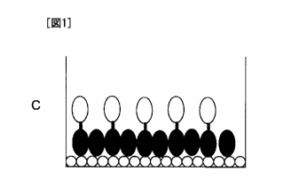
[0029] Fig. 1 includes schematic diagrams illustrating blocking by a biotin-
binding protein.
The open circles represent biotin, the closed ellipses represent a biotin-
binding protein, and
the open ellipses represent a protein that specifically binds to a substance
to be detected:
Fig. 1A: without blocking by a biotin-binding protein and with immobilization
of a fusion
protein; Fig. 1B: without blocking by a biotin-binding protein and without
immobilization of
a fusion protein; Fig. 1C: with blocking by a biotin-binding protein and with
immobilization
of a fusion protein; and Fig. 1D: with blocking by a biotin-binding protein
and without
immobilization of a fusion protein.
Fig. 2 illustrates detection by Western blotting of a fusion protein between
SITH-1
and TM2 expressed in E. coli. E. coli containing a vector alone was used as a
control.
Fig. 3 shows the effect of the addition of an E. coli homogenate extract to
serum on
non-specific binding and the blocking effect of tamavidin 2. Fig. 3-1 shows
the result of
blocking by BSA (only), that is, the result obtained by subtracting the
measured value in a
biotinylated plate (0.5% BSA Blocking) on which nothing is immobilized from
the measured
value in a SITH-1-TM2 fusion protein-immobilized plate (0.5% BSA Blocking).
Fig. 3-2
shows the result of blocking by BSA and TM2, that is, the result obtained by
subtracting the
measured value in a biotinylated plate (50 ttg/mL TM2/0.5% BSA Blocking) on
which
nothing is immobilized from the measurement value in a SITH-1-TM2 fusion
protein-
CA 02753873 2011-08-29
- 10-
immobilized plate (50 g/mLTM2/0.5% BSA Blocking).
Fig. 4 illustrates detection of a fusion protein between Harpin and TM2
expressed in
E. coli in lane 2 by Western blotting. E. coli containing a vector alone was
used (lane 1) as
a control.
EMBODIMENTS OF INVENTION
[0030] I. Method of detection of the present invention (Embodiment 1)
The method of detection (Embodiment 1) according to the present invention
relates
to a method for detecting a substance in a biological sample and comprises:
1) providing a carrier on which biotin is bound and providing a fusion protein
between a protein that specifically binds to a substance to be detected and a
biotin-binding
protein;
2) binding the fusion protein to the carrier provided in step 1) through
binding
between biotin and the biotin-binding protein to produce a fusion protein-
bound carrier;
3) mixing
(a) a biological sample, and
(b-i) a cell homogenate extract prepared from cells of the same species as the
host
cells used for expressing the fusion protein in step 1), and a biotin-binding
protein, or
(b-ii) a cell homogenate extract prepared from cells of the same species as
the host
cells used for expressing the fusion protein in step 1), and genetically
engineered to express a
biotin-binding protein,
and adding the mixture to the fusion protein-bound carrier produced in step
2); and
4) detecting a substance that has bound to the protein that specifically binds
to a
substance to be detected in the fusion protein.
[0031] Substance to be detected in biological sample
The present invention relates to a method for detecting a substance in a
biological
sample.
[0032] Any biological sample that is expected to contain a substance as a
detection target
can be used in the present invention without limitation. Examples of the
sample include
CA 02753873 2011-08-29
-11-
cells and tissues collected from organisms and fragments thereof, for example,
humors, more
preferably, blood, serum, cerebrospinal fluid, saliva, sweat, urine, tear,
lymph fluid, and
mother's milk.
[0033] These humors may be used after dilution as needed. The dilution rate
is, but not
limited to, generally in the range of about 2 to about 10000 fold, preferably
about 100 to
1000 fold. The diluent may be any buffer solution, which may contain any
proper blocking
agent. Preferred blocking agents have high inhibitory effect on nonspecific
binding, and
can be selected from blocking agents well-known to persons skilled in the art,
such as BSA
and casein. Note that in Embodiment 2 of the present invention, the blocking
agent is a
biotin-binding protein. This will be described below.
[0034] The substance to be detected in the present invention is any substance
that is desired
to be detected or measured in a biological sample, and preferred examples
thereof include
proteins such as antibodies and antigens and their fragments, peptides,
nucleic acids,
carbohydrates, and glycolipids.
[0035] The present invention enables measurement of a trace amount of
substance that is
present in a biological sample and cannot be readily detected or accurately
determined
quantitatively by conventional methods. For example, if the substance to be
detected is an
antibody exhibiting a low antibody titer in a serum (e.g., a low antibody
titer not detectable at
1000-fold dilution, but detectable at 100-fold dilution with difficulty), a
low dilution rate of
the serum is required. As a result, non-specific binding derived from serum
components
inevitably increases. Thus, no known method enables detection or determination
of quantity
for the antibody. In contrast, the method of the present invention can readily
detect and
accurately determine quantity of the antibody.
[0036] Nonlimiting examples of the substance to be detected in the present
invention
include antibodies against a small protein encoded by the intermediate
transcript of HHV-6
(SITH-1). Other examples include antibodies against other antigens of herpes
viruses,
antibodies against virus-related antigens derived from, for example,
cytomegaloviruses,
hepatitis viruses, HIVs, HTLVs, measles viruses, and influenza viruses,
antibodies against
CA 02753873 2011-08-29
12-
bacterium-related antigens derived from, for example, Helicobacter pylori, and
antibodies
against fungi-related antigens.
[0037] SITH-1 based on Description in PCT/JP2008/67300 and U.S. Provisional
Application No. 61/102441
(1) SITH-1 Protein and Nucleic Acid
The structures and functions of the SITH-1 protein and a nucleic acid are
disclosed
in PCT/JP2008/67300, and the entity thereof is incorporated therein.
[0038] The SITH-1 is a factor involving latent infection with herpes viruses,
and more
particularly, a protein specifically expressed during latent infection with
herpes viruses. The
term "specifically expressed during latent infection with herpes viruses"
therein refers to
specific expression of genes or gene products derived from herpes viruses
during latent
infection (not productive infection) with herpes viruses in hosts infected
with herpes viruses.
[0039] Examples of the SITH-1 protein and the nucleic acid include (a) a
protein which has
an amino acid sequence of SEQ ID NO: 1 and a nucleic acid encoding the
protein.
[0040] The SITH-1 protein having the amino acid sequence of SEQ ID NO: 1, as
described
in Reference Example below, was isolated and identified as a protein that is
specifically
expressed during latent infection with human herpes viruses 6 (HHV-6). The
SITH-1
protein is a protein having the amino acid sequence of SEQ ID NO: 1, composed
of 159
amino acids, and having a molecular mass of about 17.5 kDa.
[0041] The SITH-1 protein is encoded by the nucleic acid of the SITH-1 gene.
The cDNA
of this SITH-1 gene, as shown in SEQ ID NO: 3, has a size of 1795 base pairs
(about
1.79 kbp), the nucleotide sequence from the 954th to 956th being the
initiation codon (Kozak
ATG), while the nucleotide sequence from 1431st to 1433rd being the
termination codon
(TAA). Accordingly, the SITH-1 nucleic acid has a nucleotide sequence from
954th to
1430th as an open reading frame (ORF) in the nucleotide sequence of SEQ ID NO:
3, the
ORF having a size of 477 base pairs (about 0.48 kbp). In the cDNA of the SITH-
1, the
nucleotide sequence representing the ORF region is shown in SEQ ID NO: 2. The
nucleotide sequence of SEQ ID NO: 2 includes three bases of the stop codon.
CA 02753873 2011-08-29
- 13 -
[0042] The SITH-1 nucleic acid is always expressed in the cytoplasm of a cell
latent-
infected with HHV-6, but not in a productively infected cell. The nucleic acid
encoding the
SITH-1 protein is encoded by a DNA that is a complementary strand of the HHV-6
latent
infection specific gene (H6LT), which has been reported to date, and its
expression is
enhanced in the intermediate stage of the latent infection with HHV-6. These
facts
demonstrate that the SITH-1 protein is a protein that is specifically
expressed during latent
infection with HHV-6.
[0043] The SITH-1 protein binds to a host protein, CAML (calcium-modulating
cyclophilin
ligand, Accesion #: U18242) to increase the calcium concentration in the glial
cells. The
CAML is a protein that is known to be abundantly present in the brain and
lymphocytes in
the host living organism and increase the intracellular calcium concentration.
It is
considered that an increase in intracellular calcium concentration due to
expression of the
SITH-1 protein probably leads to activation of overall signaling in the latent-
infected cells,
and thus contributes to efficient reactivation of HHV-6.
[0044] It is known that the glial cells in the brain are latent-infected with
HHV-6. When
HHV-6 during the latent infection or at the intermediate stage which is a
latent infection state
with high activity expresses the SITH-1, the calcium concentration seems to
increase in the
glial cells. It is believed that an increase in intracellular calcium
concentration in the brain
is wedded to psychiatric disorders such as mood disorders (Riken Annual Report
2003).
[0045] The SITH-1 protein has a function that maintains activity to bind to
the host protein,
CAML to increase the intracellular calcium concentration. Furthermore,
expression of the
SITH-1 protein in the glial cells, in which this protein seems to be most
strongly expressed,
in the brain can induce psychiatric disorders. Accordingly, the SITH-1 protein
is believed to
be expressed during the latent infection with herpes viruses or at the initial
stage of
reactivation of the herpes viruses to cause the host to have any psychiatric
disorder.
[0046] (2) Antibody against SITH-1
The antibody against the SITH-1 can be prepared as a polyclonal antibody or a
monoclonal antibody from the SITH-1 protein, its variant, or their partial
peptides as antigen
CA 02753873 2011-08-29
- 14-
by a known process. Examples of the known process are described in documents
such as
Harlow et al., "Antibodies: A laboratory manual (Cold Spring Harbor
Laboratory, New York
(1988))" and Iwasaki et al., "Monoclonal Antibody: Hybridoma and ELISA,
Kodansha
(1991)". The resulting antibody can be used for detection and determination of
the SITH-1
protein.
[0047] The term "antibody" refers to immunoglobulins (IgA, IgD, IgE, IgG, IgM,
and Fab
fragments, F(ab')2 fragments, and Fc fragments thereof). Examples of the
antibody include,
but not limited to, polyclonal antibodies, monoclonal antibodies, single-
stranded antibodies,
antiidiotype antibodies, and humanized antibodies.
[0048] The term "antibody recognizing the SITH-1 protein" includes complete
molecules
and antibody fragments specifically attachable to the SITH-1 protein (for
example, Fab and
F(ab')2 fragments). Fab, F(ab')2, and other fragments of the SITH-1 antibody
can be used
according to the method disclosed in the present specification or any known
method. Such
fragments can be typically produced by cleavage by proteolysis using an
enzyme, e.g., papain
(yielding a Fab fragment) or pepsin (yielding an F(ab')2 fragment).
[0049] It is believed that patients having mood disorders and individuals
having potential
mood disorders exhibit increased expression levels of the SITH-1 protein and
thus increased
SITH-1 antibody titers. In one embodiment of the present invention, detection
of the SITH-
1 antibody in a biological sample enables identification of patients having
mood disorders
and individuals having potential mood disorders.
[0050] Carrier on which fusion protein between protein specifically binding to
substance to
be detected and biotin-binding protein is bound (steps 1) and 2) of Example 1)
The method of detection according to the present invention uses a carrier on
which a
fusion protein between a protein that specifically binds to a substance to be
detected and a
biotin-binding protein is bound by binding between biotin and the biotin-
binding protein.
[0051] The carrier of the present invention can be produced by a method
including:
1) providing a carrier on which biotin is bound and providing a fusion protein
between a protein that specifically binds to a substance to be detected and a
biotin-binding
CA 02753873 2011-08-29
15 -
protein; and
2) binding the fusion protein to the carrier provided in step 1) through
binding
between biotin and the biotin-binding protein to produce a fusion protein-
bound carrier.
[0052] Biotin-bound carrier
"Biotin" is a generic name of D-[(+)-cis-hexahydro-2-oxo-1H-thieno-(3,4)-
imidazole-4-valeric acid]. It is one of water-soluble vitamin categorized into
a vitamin B
group, and is also referred to as vitamin B7, vitamin H, or coenzyme R. Biotin
very strongly
binds to avidin, one of the glycoproteins contained in egg white, so that its
absorption is
precluded. Thus, large dose of uncooked egg white may cause biotin deficiency
disease in
some cases.
[0053] The term "biotin" throughout the specification includes iminobiotin
(Hofmann et al.,
(1980), Proc Natl Acad Sci USA, 77: 4666-4668), desthiobiotin (Hirsch et al.,
(2002), Anal
Biochem, 308: 343-357), and biotin analogs such as biocytin and biotin
sulfoxide, in addition
to the biotin described above.
[0054] Systems using avidin (biotin-binding protein)/biotin complexes are
widely used in
the fields of tissue immunology, DNA analysis, and clinical assay. In the
method for
binding a protein to a carrier according to the present invention, a fusion
protein composed of
a biotin-binding protein and a desired protein is bound to a carrier by avidin-
biotin binding.
The method of the present invention ensures more efficient action of the
protein, without
impairing the function thereof, than conventional binding by avidin-biotin
binding.
[0055] Examples of materials for the solid carrier include, but not limited
to, cellulose,
Teflon (registered trademark), nitrocellulose, agarose, dextran, chitosan,
polystyrene,
polyacrylamide, polyesters, polycarbonates, polyamides, polypropylene, nylons,
polydivinylidene difluoride, latex, silica, glass, glass fiber, gold,
platinum, silver, copper,
iron, stainless steel, ferrite, silicon wafer, polyethylene,
polyethyleneimine, poly(lactic acid),
resin, polysaccharides, proteins such as albumin, carbon, and combination
thereof.
Preferred materials have a certain level of strength, a stable composition,
and reduced non-
specific binding.
CA 02753873 2011-08-29
16-
[0056] Examples of the form of the solid carrier include, but not limited to,
beads, magnetic
beads, thin films, microtubes, filters, plates, microplates, carbon nanotubes,
and sensor chips.
Flat solid carriers such as thin films and plates may be provided with pits,
grooves, or filter
bottoms, as is known in the art.
[0057] In an embodiment of the invention, beads may have a spherical diameter
in the
range of about 25 nm to about 1 mm. In a preferred embodiment, the beads may
have a
diameter in the range of about 50 nm to about 10 tm. The size of the beads may
be selected
depending on the specific application. Since some bacterial spores have a size
of an order
of about 1 m, preferred beads for capturing such spores have a diameter
larger than 1 m.
[0058] Without any limitation, for example, in the case that high sensitivity
for detection is
desired, beads as described above can preferably be used as the solid carrier
in the view point
that the beads provide high contacting frequency between the target substance
to be detected
and the substance that specifically binds to the target substance, and that
cleaning operation
thereof is easy.
[0059] An exemplary method of binding biotin to the carrier involves use of a
biotinylation
reagent. Examples of the biotinylation reagent include, but not limited to, EZ-
Link
(registered trademark) Sulfo-NHS-Biotin (the length of the linker: 13.5
angstroms, the
reactive group: primary amine, hereinafter the same order), EZ-Link
(registered trademark)
Sulfo-NHS-LC-Biotin (22.4 angstroms, primary amine), EZ-Link (registered
trademark)
Sulfo-NHS-LCLC-Biotin (30.5 angstroms, primary amine), EZ-Link (registered
trademark)
PFP-Biotin (9.6 angstroms, amine), EZ-Link (registered trademark) Maleimide-
PEO2-Biotin
(29.1 angstroms, thiol group), EZ-Link (registered trademark) Biotin-PEO2
Amine
(20.4 angstroms, carboxyl group), EZ-Link (registered trademark) Biotin-PEO3-
LC Amine
(22.9 angstroms, carboxyl group), EZ-Link (registered trademark) Biotin-
Hydrazide
(15.7 angstroms, aldehyde group), EZ-Link (registered trademark) Biotin-LC-
Hydrazide
(24.7 angstroms, aldehyde group), and EZ-Link (registered trademark) NHS-
Iminobiotin
(13.5 angstroms, primary amine), which are available from PIERCE.
[0060] Using these biotinylation reagent, biotin can be bound to a desired
carrier such a
CA 02753873 2011-08-29
17_
microplate, microbeads, or a sensor chip by any known process. For example,
various
carriers having functional groups, such as amino, carboxyl, thiol, tosyl,
epoxy, and maleimide
groups, and activated ester (for example, magnetic beads, Sepharose beads,
agarose beads,
latex beads, and microtiter plates) can be used. For example, in the case of
the use of a
biotinylation reagent containing NHS ester, the reagent may be dissolved in an
organic
solvent such as dimethyl sulfoxide (DMSO) or phosphate buffer of pH 7 to 9,
and then may
be added to an immobilization carrier having amino groups to bind biotin
thereto. In the
case of the use of a biotinylation reagent containing amino groups, the
carboxyl groups on the
immobilization carrier may be converted to activated ester using carbodiimide
such as 1-
ethyl-3-(3-dimethylaminopropyl) carbodiimide hydrochloride (EDC), followed by
addition of
a biotinylation reagent dissolved in buffer solution (pH: about 5) to bind
biotin to the carrier.
The biotinylated immobilization carrier is preferably blocked with BSA after
inactivation of
unreacted functional groups.
[0061] Commercially available biotinylated carriers can also be used. Examples
of the
biotinylated microplates include, but not limited to, Reacti-Bind TM Biotin
Coated Polystyrene
Plates (PIERCE). Examples of the biotinylated microbeads include, but not
limited to,
magnetic beads, such as BioMag Biotin (available from Polysciences), magnetic
nanobeads,
such as nanomag (registered trademark)-D biotin and nanomag (registered
trademark)-silica
biotin available from Corefront, polystyrene microbeads, such as Beadlyte
(registered
trademark) Biotin Beads (available from Upstate), agarose, such as Biotin
Agarose and 2-
iminobiotin-Agarose available from Sigma, and highly cross-linked agarose,
such as Biotin-
Sepharose (available from Biosearch Technologies, Inc.).
[0062] The length of the linker binding the carrier to biotin is preferably at
least 5
angstroms, more preferably at least 13.5 angstroms, more preferably at least
22.4 angstroms,
and most preferably at least 30.5 angstroms.
[0063] Protein specifically binds to substance to be detected
The protein that specifically binds to a substance to be detected (hereinafter
may be
referred to as "specific protein") is not particularly limited In one
embodiment of the
CA 02753873 2011-08-29
18_
present invention, for example, one of the antigen and antibody, ligand such
as hormone and
receptor, lectin and saccharide, or complementary bindings of nucleic acid in
a sample to be
tested is selectively analyzed by means of the ability to form a specific
complex with the
other, but the specific protein is not limited thereto.
[0064] More specifically, examples of the protein include, but not limited to,
antibodies,
antigenic proteins, lectins, peptides, protein A, protein G, protein L,
receptors, and enzymatic
proteins. Examples of the antibody include antibody fragments containing
antigen-binding
sites, such as scFv and Fab, in addition to IgG. Examples of the antigenic
protein include
proteins derived from viruses such as hepatitis B and C viruses, HIVs,
influenza viruses, and
herpes viruses; proteins derived from bacteria such as Helicobacter pylori;
tumor markers
such as CEA and PSA; and sex hormones. The lectin is a saccharide-binding
protein, and
examples thereof include monosaccharide specific lectins such as mannose
specific lectin,
Ga1NAc specific lectin, GIcNAc specific lectin, fucose specific lectin, and
sialic acid specific
lectin, and oligosaccharide specific lectins. Furthermore, the examples
include DNA/RNA
binding proteins. Examples of the peptide include those composed of 2 to 100
amino acids,
preferably 4 to 50 amino acids, and more preferably 6 to 30 amino acids.
[0065] Furthermore, in the present invention, examples of the protein that
specifically binds
to a substance to be detected include, but not limited to, SITH-1 proteins.
[0066] Biotin-binding protein
The present invention includes a method of immobilizing a protein that
specifically
binds to a substance to be detected to a carrier by binding between biotin and
a biotin-binding
protein. In the present invention, "biotin and a biotin-binding protein" may
be referred to as
"avidin-biotin binding" in some cases.
[0067] Any protein that strongly binds to biotin can be preferably used as the
biotin-binding
protein, and examples thereof include avidin, streptavidin, neutravidin, AVR
protein
(Biochem. J., (2002), 363: 609-617), bradavidin (J. Biol. Chem., (2005), 280:
13250-13255),
rhizavidin (Biochem. J., (2007), 405: 397-405), tamavidin (W02002/072817), and
variants
thereof. The dissociation constant (KD) with biotin is preferably 10-6 M or
less, more
CA 02753873 2011-08-29
- 19-
preferably 10-8 M or less, more preferably 10-10 M or less. Note that the
biotin-binding
protein that is added to a substance to be tested and the biotin-binding
protein that is used for
blocking the carrier will be described below.
[0068] Particularly preferred biotin-binding proteins are tamavidin and
variants thereof,
which can be highly expressed in E. coli. Tamavidin is a biotin-binding
protein discovered
in an edible mushroom, Pleurotus cornucopiae (W02002/072817, Takakura et al.,
(2009),
FEBS J, 276: 1383-1397). An example of the variants of tamavidin is tamavidin
exhibiting
high binding capability and low non-specific binding characteristics
(PCT/JP2009/64302).
[0069] The term "tamavidin" in the present invention refers to tamavidin 1,
tamavidin 2, or
a variant thereof. Specifically, tamavidin of the present invention may be
typically a protein
having the amino acid sequence of SEQ ID NO: 5 or SEQ ID NO: 7, or a protein
encoded by
a nucleic acid having the nucleotide sequence of SEQ ID NO: 4 or SEQ ID NO: 6.
Alternatively, tamavidin of the present invention may be a protein that is a
variant of a
protein having the amino acid sequence of SEQ ID NO: 5 or SEQ ID NO: 7 or a
protein
encoded by a nucleic acid having the nucleotide sequence of SEQ ID NO: 4 or
SEQ ID NO:
6 and having biotin binding capability similar to that of tamavidin 1 or 2 or
high binding
capability and low non-specific binding characteristics. Throughout the
specification,
tamavidin 1, tamavidin 2, and variants thereof may be collectively referred to
as tamavidin.
[0070] The variant of tamavidin 1 or 2 may be a protein having an amino acid
sequence
comprising one or more deletion, substitution, insertion, and/or addition of
one or more
amino acids in the amino acid sequence of SEQ ID NO: 5 or 7 and having biotin
binding
capability similar to that of tamavidin 1 or 2. The substitution may be
conservative
substitution. The conservative substitution refers to replacement of a
specific amino acid
residue with any residue having similar physicochemical features. Nonlimiting
examples of
the conservative substitution include substitutions between amino acid
residues containing
aliphatic groups, such as mutual substitution between Ile, Val, Leu, and Ala;
and substitutions
between polar residues, such as mutual substitution between Lys and Arg,
between Glu and
Asp, and between Gln and Asn.
CA 02753873 2011-08-29
-20-
[0071] The variant by deletion, substitution, insertion, and/or addition of an
amino acid or
amino acids can be produced by a known technique such as site-specific
mutagenesis (e.g.,
see Nucleic Acid Research, Vol. 10, No. 20, pp. 6487-6500, 1982, which is
incorporated
herein in its entirety) to a DNA encoding a wild-type protein. Throughout the
specification,
the term "one or more amino acids" herein refers to an amino acid or amino
acids that can be
deleted, substituted, inserted, and/or added by preferably site-specific
mutagenesis. In
addition, the term "one or more amino acids" herein may refer to one or more
amino acids.
The "one or several amino acids" refers to, but not limited to, 50 or less,
preferably 40 or
less, 30 or less, 20 or less, 10 or less, 8 or less, 5 or less, or 3 or less
amino acids. The
variant of tamavidin 1 or 2 may also be a protein having an amino acid
sequence sharing an
identity of 60% or more, preferably 65% or more, 70% or more, 75% or more, 80%
or more,
85% or more, 90% or more, 95% or more, 96% or more, 97% or more, 98% or more,
or 99%
or more, more preferably 99.3% or more with that of SEQ ID NO: 5 or SEQ ID NO:
7 and
having biotin binding capability similar to that of tamavidin 1 or 2 or high
binding capability
and low non-specific binding characteristics.
[0072] The percent identity between two amino acid sequences may be determined
by
visual inspection and mathematical calculation. Alternatively, the percent
identity between
two protein sequences may be determined through comparison of sequence
information using
a GAP computer program available from the University of Wisconsin Genetics
Computer
Group (UWGCG) based on the algorithm by Needleman, S. B. and Wunsch, C. D. (J.
Mol.
Biol., 48: 443-453, 1970). Preferred default parameters of the GAP program
include: (1)
scoring matrix: blosum62 described in Henikoff, S. and Henikoff, J. G., (Proc.
Natl. Acad.
Sci. USA, 89: 10915-10919, 1992); (2) 12 gap weights; (3) 4 gap length
weights; and (4) no
penalty for terminal gaps.
[0073] Any other program used by persons skilled in the art may also be used
for
comparison of the sequences. The percent identity can be determined by, for
example,
comparison with the sequence information using a BLAST program described in
Altschul et.
al., (Nucl. Acids. Res., 25, pp. 3389-3402, 1997). This program is available
from the
CA 02753873 2011-08-29
-21-
websites of National Center for Biotechnology Information (NCBI) or DNA Data
Bank of
Japan (DDBJ) on the Internet. The conditions (parameters) for identity search
by the
BLAST program is described in detail on these sites. Although these parameters
can be
partly modified if necessary, search is generally carried out using the
default values.
Alternatively, the percent identity between two amino acid sequences may be
determined
using a program such as genetic information processing software GENETYX Ver. 7
(available from GENETYX CORPORATION) or FASTA algorithm, wherein search may be
carried out using the default values.
[0074] The percent identity between two nucleotide sequences can be determined
by visual
inspection and mathematical calculation. Preferably, such comparison is
carried out through
comparison of sequence information using a computer program. A particularly
preferred
computer program is a version 10.0 program "GAP", Wisconsin package of
Genetics
Computer Group (GCG, Madison, Wisconsin) (Devereux, et al., 1984, Nucl. Acids
Res., 12:
387). The use of the "GAP" program enables comparison between two amino acid
sequences and comparison between a nucleotide sequence and an amino acid
sequence, in
addition to comparison of two nucleotide sequences.
[0075] The "biotin-binding protein" constituting a fusion protein to be
immobilized to a
carrier is used for preparing a fusion protein-bound carrier by binding the
fusion protein to
the carrier through binding between biotin and the biotin-binding protein.
Accordingly, it is
preferred that the biotin binding activity of a variant of tamavidin 1 or 2 be
not significantly
decreased compared to that in the case of forming the fusion proteins using
these wild-types.
[0076] Accordingly, nonlimitingly, in the variant of tamavidin 1, preferably,
N14, S18, Y34,
S36, S78, W82, W98, W110, and D118 in the amino acid sequence of SEQ ID NO: 5
are not
modified. Note that the notation, for example, Y34 indicates the 34th tyrosine
residue of the
amino acid sequence of SEQ ID NO: 5. Alternatively, in the modification of
these amino
acid, the amino acid is preferably replaced with one having a similar property
or structure.
For example, asparagine (N14) is replaced with glutamine (Q) or aspartic acid
(D), preferably
aspartic acid; serine (S18, S36, or S78) is replaced with threonine (T) or
tyrosine (Y),
CA 02753873 2011-08-29
.22-
preferably threonine; tyrosine (Y34) is replaced with serine (S), threonine
(T), or
phenylalanine (F), preferably phenylalanine; tryptophan (W82, W98, or W110) is
replaced
with phenylalanine (F); and aspartic acid (D118) is replaced with glutamic
acid (E) or
asparagine (N), preferably asparagine.
[0077] In the variant of tamavidin 2, preferably, four tryptophan residues
(W69, W80, W96,
and W108) in the amino acid sequence of SEQ ID NO: 7 are not modified.
Alternatively,
when these amino acid residues are modified, the amino acid is preferably
replaced with one
having a similar property or structure, for example, phenylalanine(F). In
addition, it is
desirable that amino acid residues (N14, S18, Y34, S36, S76, T78, and D116)
that probably
interact directly with biotin are also not modified. Alternatively, in the
modification of
these amino acid residues, the amino acid is preferably replaced with one
having a similar
property or structure in order to maintain the binding with biotin. For
example, asparagine
(N14) is replaced with glutamine (Q) or aspartic acid (D), preferably aspartic
acid; aspartic
acid (D40) is replaced with asparagine (N); serine (S18, S36, or S76) is
replaced with
threonine (T) or tyrosine (Y), preferably threonine; tyrosine (Y34) is
replaced with serine (S),
threonine (T), or phenylalanine (F), preferably phenylalanine; threonine (T78)
is replaced
with serine (S) or tyrosine (Y), preferably serine; and aspartic acid (D116)
is replaced with
glutamic acid (E) or asparagine (N), preferably asparagine.
[0078] Preferred variants of tamavidin in the present invention include the
following
variants (PCT/JP2009/64302).
[0079] The tamavidin variant is a modified biotin-binding protein that has the
amino acid
sequence of SEQ ID NO: 7 or an amino acid sequence comprising one to several
amino acid
mutations in this sequence or having an identity of at least 80% with this
sequence and that
shows biotin binding activity, wherein one or more residues selected from the
group
consisting of:
1) the arginine residue at the 104th site of SEQ ID NO: 7;
2) the lysine residue at the 141st site of SEQ ID NO: 7;
3) the lysine residue at the 26th site of SEQ ID NO: 7; and
CA 02753873 2011-08-29
-23-
4) the lysine residue at the 73rd site of SEQ ID NO: 7;
are replaced with acidic or neutral amino acid residues.
[0080] More preferably, the modified biotin-binding protein is selected from
the group
consisting of:
a modified biotin-binding protein (R104E-K141E) in which the arginine residue
at
the 104th site is replaced with a glutamic acid residue, and the lysine
residue at the 141st site
is replaced with a glutamic acid residue, in SEQ ID NO: 7;
a modified biotin-binding protein (D40N-R104E) in which the aspartic acid
residue
at the 40th site is replaced with a asparagine residue, and the arginine
residue at the 104th site
is replaced with a glutamic acid residue, in SEQ ID NO: 7;
a modified biotin-binding protein (D40N-K141E) in which the aspartic acid
residue
at the 40th site is replaced with a asparagine residue, and the lysine residue
at the 141st site is
replaced with a glutamic acid residue, in SEQ ID NO: 7; and
a modified biotin-binding protein (D40N-R104E-K141E) in which the aspartic
acid
residue at the 40th site is replaced with a asparagine residue, the arginine
residue at the 104th
site is replaced with a glutamic acid residue, and the lysine residue at the
141st site is
replaced with a glutamic acid residue, in SEQ ID NO: 7.
[0081] Fusion protein between protein specifically binding to substance to be
detected and-
biotin-binding protein
In the present invention, a fusion protein between a protein that specifically
binds to
a substance to be detected and a biotin-binding protein (hereinafter may be
referred to as
"biotin-binding protein fusion protein" or "fusion protein") is immobilized on
a carrier.
[0082] The method for preparing the biotin-binding protein fusion protein is
not particularly
limited. For example, the fusion protein may be expressed by a known genetic
engineering
technique. For example, the fusion protein can be obtained by expressing a
gene encoding a
fusion protein between a biotin-binding protein and a desired protein using an
expression
system such as E. coli. Note that tamavidin and variants thereof are preferred
to achieve a
high expression in E. coli.
CA 02753873 2011-08-29
.24-
[0083] In the biotin-binding protein fusion protein, a biotin-binding protein
and a desired
protein may be directly bound to each other or may be bound via a linker, but
are preferably
bound via an amino acid linker. The length of the linker may be at least one
amino acid, but
is preferably at least five amino acids and more preferably at least six amino
acids. In order
to enhance the binding strength between biotin immobilized on a carrier and
tamavidin, the
length of the linker is preferably at least ten amino acids, more preferably
at least 12 amino
acids, at least 15 amino acids, at least 18 amino acids, and most preferably
at least 25 amino
acids. Probably, such linkers also increase the activity of the tamavidin
fusion protein.
The amino acids constituting the linker are not particularly limited, and the
linker is
preferably made of repetition of neutral amino acids such as glycine, serine,
or alanine.
Nonlimiting examples thereof include GGGGS, GGSGG, GASAG, GSGAA, GSGSA,
GGGGSG, GGGSGGS, GGSGGGGS, AAAAGSGAA, GGGGSGGGGSGGGGS, and
GGGGSGGGGSGGGGSGGGGSGGGGS (SEQ ID NOs: 8 to 18).
[0084] The biotin-binding protein may be bound to either the N-terminal or the
C-terminal
of a desired protein. In the expression of a desired protein, for example, if
the periplasmic
space is more suitable than the cytoplasm of E. coli, a leader sequence for
targeting the
periplasm may be used. Examples of the leader sequence include, but not
limited to, PelB
(Lei et al., (1987), J Bacteriol, 169: 4379-4383) and OmpA (Gentry-Weeks et
al., (1992), J
Bacteriol, 174: 7729-7742).
[0085] In the case of a biotin-binding protein fusion protein obtained from a
soluble
fraction, the soluble fraction, without purifying the crude protein extract,
may be put into
direct contact with a biotinylated carrier to bind the fusion protein to the
biotinylated carrier.
Through sufficiently washing the carrier, the purification and the
immobilization to the
carrier of the fusion protein can be simultaneously achieved. Alternatively,
the fusion
protein may be bound to the biotinylated carrier after purification using a
column to which a
biotin analog such as iminobiotin Hofmann et al., (1980), Proc Natl Acad Sci
USA, 77: 4666-
4668) is bound.
[0086] Alternatively, a tag for purification may be further added to the N-
terminal or the C-
CA 02753873 2011-08-29
_25_
terminal of the biotin-binding protein fusion protein. Examples of the tag
include, but not
limited to, a c-myc epitope tag (Munro and Pelham, (1986), Cell, 46: 291-300),
a histidine
tag (Hochuli et al., (1988), Bio/Technol, 6: 1321-1325; Smith et al., (1988),
J Biol Chem,
263: 7211-7215), a Halo tag (Los and Wood, (2007), Methods Mol Biol, 356: 195-
208), a
Flag tag (Einhauer and Jungbauer, (2001), J Biochem Biophys Methods, 49: 455-
465), and
combinations thereof.
[0087] In the case of a biotin-binding protein fusion protein obtained from an
insoluble
fraction, a known process employed, for example, the protein is once
solubilized using a
chaotropic salt such as urea or guanidine hydrochloride, and then refolding of
the protein is
enhanced with gradually removing the chaotropic salt by, for example, dialysis
(Sano and
Cantor, (1991), Bio/Technology, 9: 1378-1381; Sano et al., (1992), Proc Nat]
Acad Sci USA,
89: 1534-1538).
[0088] In the case of a desired protein expressed in an insoluble fraction in
E. coli, for
example, a maltose-binding protein (Bach et al., (2001), J Mol Biol, 312: 79-
93), thioredoxin
(Jurado et al., (2006), J Mol Biol, 357: 49-61), glutathione S-transferase
(Tudyka and Skerra,
(1997), Protein Sci, 6: 2180-2187), or a chaperon such as one described in
Ideno et al.,
(2004), Appl Microbiol Biotechnol, 64: 99-105 may be coexpressed, or a three-
component
fusion protein composed of the fusion protein and a chaperon may be produced.
The
maltose-binding protein, the thioredoxin, and the glutathione S-transferase
can also be used
as tags for purification.
[0089] The fusion protein may be expressed by any other known expression
system, such as
insect cells, plant cells, mammalian cells, yeast cells, Bacillus subtilis
cells, or a cell-free
expression system. In particular, when the protein to be fused is expressed in
plant cells (for
example, plant lectin), it is preferred that the fusion protein is also
expressed in a plant cell
expression system. A suitable expression system will be apparent to those
skilled in the art
in consideration of properties of the protein to be fused.
[0090] Binding of fusion protein to carrier
In the present invention, a protein can be bound to a carrier through avidin-
biotin
CA 02753873 2011-08-29
26-
binding by providing a biotin-bound carrier and a biotin-binding protein
fusion protein and
putting them into contact with each other.
[0091] A crude cell homogenate extract containing a biotin-binding protein
fusion protein is
prepared in a total protein content of, but is not limited to, 0.1 mg/mL to 5
mg/mL, preferably
0.2 mg/mL to 2 mg/mL. This extract is put into contact with a carrier to which
biotin is
bound at 10 C to 40 C, preferably 20 C to 30 C, for 5 min to 2 hr, preferably
30 min to 1 hr.
Alternatively, a purified biotin-binding protein fusion protein in a
concentration of 0.1 p.g/mL
to 5 g/mL may be put into contact with a carrier to which biotin is bound.
[0092] Addition of biological sample to carrier (step 3 of Embodiment 1)
The method of Embodiment 1 of the present invention includes, after the
providing
of a fusion protein-bound carrier, the following step 3):
mixing
(a) a biological sample, and
(b-i) a cell homogenate extract prepared from cells of the same species as the
host
cells used for expressing the fusion protein in step 1), and a biotin-binding
protein, or
(b-ii) a cell homogenate extract prepared from cells of the same species as
the host
cells used for expressing the fusion protein in step 1), and genetically
engineered to express a
biotin-binding protein,
and adding the mixture to the fusion protein-bound carrier produced in step
2).
[0093] In a method of detection using a carrier, generally known approaches
for reducing
non-specific binding which causes background signals are, for example,
addition of an
extract of a bacterium component to a reagent for detection (JP No. S59-99257
A (1984));
addition of a culture component of host cells to a sample, where a vector of
the same species
as that used in production of a recombinant protein capable of specifically
binding to a
substance to be detected and containing no gene encoding the protein (JP No.
H8-43392 A
(1996)); heat treatment of an aqueous extract from cells of the same species
as that producing
a recombinant protein capable of specifically binding to a substance to be
detected and not
containing this protein, and addition of its water-soluble fraction to a
sample (JP No. 2004-
CA 02753873 2011-08-29
-27-
301646 A).
[0094] The present inventors had attempted to apply the above-mentioned
methods to a
system of fusion protein, but sufficient effect has was not obtained. However,
the present
inventors have diligently studied and, as a result, have arrived at a method
of obtaining a
noticeable effect. Specifically, the present inventors have discovered that it
is preferable to
put a substance to be detected into contact with a carrier in the presence of
both a cell
homogenate extract and a biotin-binding protein.
[0095] In one embodiment, a cell homogenate extract prepared from cells of the
same
species as the host cells used for expressing the fusion protein, biotin-
binding protein, and a
biological sample are mixed, and the mixture may be added to a carrier. When a
biotin-
binding protein and a cell homogenate extract are added to a sample to be
tested, either one
may be added first, or the both may be simultaneously added. Alternatively,
after mixing
the biotin-binding protein and the cell homogenate extract, the sample to be
tested may be
added thereto.
[0096] In another embodiment, a cell homogenate extract prepared from cells of
the same
species as the host cells used for expressing the fusion protein and
genetically engineered to
express a biotin-binding protein and a sample to be tested are mixed, and the
mixture may be
added to a carrier. Specifically, a recombinant protein is expressed in cells
by integrating
the biotin-binding protein into an expression vector, and the homogenate
extract from the
cells may be used.
[0097] In the case of a serum sample to be tested, the serum is usually used
after dilution in
a cell homogenate extract into 10 to 10000-fold, preferably 100 to 1000-fold,
more preferably
100 to 500-fold.
[0098] Cells from which cell homogenate extracts are derived, such as E. coli
cells, yeast
cells, mammalian cells, insect cells, and plant cells, can be used without
limitation.
Preferred cells are the same species as that of host cells used to express a
fusion protein.
For example, in the case of preparation of a fusion protein using E. coli, it
is preferred that
the cell homogenate extract be also prepared using E. coli. In the case of
expression of a
CA 02753873 2011-08-29
_28_
fusion protein in a cell-free system, the cell homogenate extract used can be
used as it is or as
a suspension in a desired buffer solution.
[0099] More preferably, the cells are derived from different species from the
organism from
which the biological sample is derived. For example, for a biological sample
derived from
human being, a cell homogenate extract is preferably prepared from cells
derived from an
organism other than human beings. However, for example, for a fusion protein
expressed in
mammalian cells, a cell line obtained from specific cancer cells is generally
used. In such a
case, the content of the human cell line and the content of (for example)
human cells from
which the biological sample are derived are substantially highly different
from each other,
even if the cell homogenate extract from a cell line derived from human being
is used; hence,
the effect of the present invention can be obtained.
[0100] The cell used for preparation of the cell homogenate extract may
contain any vector,
preferably an empty vector. The empty vector is of the same species as that of
the vector
used in expression of a protein that specifically binds to substance to be
detected or a fusion
protein thereof and does not contain genes that encode these proteins. The
empty vector
may further contain any nucleic acid. Alternatively, the empty vector may be
any known
vector that is different from the vector used in expression of a protein that
specifically binds
to substance to be detected or a fusion protein thereof.
[0101] Any cell homogenate extract derived from cells can be used without
limitation.
For example, a protein component, a carbohydrate component, or a lipid
component of cells,
or a mixture thereof may be used. Preferably, a soluble extract of cells can
be used.
[0102] The cell homogenate extract can be prepared by a variety of processes
without
limitation. Usually, the cell homogenate extract can be prepared as a soluble
components by
homogenizing or solubilizing cells cultured in a proper culture medium through
a physical
means such as, ultrasonic application, a chemical means using a surfactant, or
an enzymatic
treatment, and performing centrifugation or filtration. In order to prolong
the storage life,
preferably the clear liquid prepared by centrifugation or filtration is mixed
with, for example,
a protease inhibitor, or is heated, for example, in an autoclave to suppress
or deactivate
CA 02753873 2011-08-29
-29-
various enzymes derived from cells. The concentration of the cell homogenate
extract can
be varied depending on the extent of the occurring non-specific reaction and
can be set in a
range sufficient to absorb the non-specific reaction.
[0103] As a nonlimiting specific example of the method for preparing a cell
homogenate
extract, in the case of E. coli cells, E. coli cells (which may contain a
vector optionally
containing a gene encoding a biotin-binding protein) are inoculated into an LB
culture
medium containing an antibiotic; shake culture is performed at 25 C to 37 C
until the
absorbance at OD 600 reaches 0.25 to 1, preferably 0.4 to 0.6; 0.1 mM to 5 mM,
preferably
0.5 mM to 1 mM IPTG is added thereto; and then shake culture is performed at
25 C to 37 C
for 2 to 24 hr, preferably 4 to 16 hr. The bacterial cells are recovered by
centrifugation from
the culture solution, are suspended in a desired buffer solution, and are
homogenized. After
centrifugation of the homogenized solution, the supernatant is collected as a
crude E. coli
extract.
[0104] The biological sample and cell homogenate extract can be added to a
carrier by any
procedure. The biological sample, however, must come into contact with the
cell
homogenate extract before the contact with the carrier. In other words, as
long as the
biological sample is put into sufficient contact with the cell homogenate
extract, the
component derived from the cell homogenate extract is not necessarily added to
the carrier
finally together with the biological sample. For example, a carrier to which
the cell
homogenate extract component is bound is prepared, and a treated biological
sample may be
added thereto. Specific examples include an embodiment in which a biological
sample is
passed through a cell homogenate extract component column.
[0105] When a biological sample is mixed with a crude cell homogenate extract,
the sample
is reacted with, but not limited to, a crude cell homogenate extract that has
been prepared
with a desired buffer (which may contain, for example, BSA, casein, or a
commercially
available blocking agent) into a total protein concentration of 0.05 to 5
mg/mL, preferably
0.5 to 5 mg/mL, at 10 C to 30 C, preferably, 20 C to 30 C for 30 min to 4 hr,
preferably 1 to
2 hr. For a biological sample of serum, the serum is preferably diluted with,
but not limited
CA 02753873 2011-08-29
-30-
to, the crude cell homogenate extract to 100 to 1000-fold.
[0106] Addition of biotin-binding protein to sample to be tested
The present invention is characterized in that a mixture of a biological
sample, a cell
homogenate extract prepared from cells of the same species as the host cells
used for
expressing a fusion protein, and a biotin-binding protein is added to a
carrier (step b-i), or a
mixture of a sample to be tested and a cell homogenate extract prepared from
cells of the
same species as the host cells used for expressing a fusion protein and
genetically engineered
to express a biotin-binding protein is added to a carrier (step b-ii).
[0107] In the method of the present invention, the background signal level can
be finally
suppressed by adding a biotin-binding protein to a sample to be tested.
[0108] Such a biotin-binding protein may be the same as or different from the
biotin-
binding protein constituting a fusion protein. Either the wild type or a
variant may be used.
The biotin-binding activity of the variant may be equivalent, higher, or lower
compared to
that of the wild type.
[0109] In an embodiment of the addition, powder of a biotin-binding protein
(which may be
a naturally occurring protein or may be expressed by genetic engineering) may
be added to
the sample directly or after its dissolution in a proper liquid. For example,
in another
embodiment, a mixture of a sample and a cell homogenate extract may be treated
with a
carrier to which the biotin-binding protein is immobilized (for example, the
mixture is passed
through a column) (step b-i), instead of direct addition of the biotin-binding
protein to the
sample.
[0110] In the case of addition of the biotin-binding protein to a crude cell
homogenate
extract, the final concentration of the added biotin-binding protein is, but
not limited to, 1 to
500 [ug/mL, preferably 10 to 100 g/mL. The concentration of the biotin-
binding protein
that is genetically engineered to be expressed in the cells may also
substantially be the same
as above, but any other concentration is acceptable.
[0111] Alternatively, a gene encoding a biotin-binding protein is introduced
into host cells
and is expressed, and a cell extract containing a biotin-binding protein
obtained by
CA 02753873 2011-08-29
31-
homogenizing the host cells may be used (step b-ii). In this case, the biotin-
binding protein
may be expressed in a desired host by a method well known to persons skilled
in the art. In
the case where a fusion protein between a protein that specifically binds to a
substance to be
detected and a biotin-binding protein is expressed by genetic engineering, the
cell
homogenate extract is preferably derived from the same species as that of the
host.
[0112] In the case of the host being E. coli, a gene encoding the biotin-
binding protein is
incorporated into an expression vector and is introduced into E. coli, and E.
coli is cultivated
while expression of protein is induced. Conditions for induction, such as an
expression
vector and host E. coli strains, culture medium component, IPTG concentration,
and
cultivation temperature can be appropriately determined.
[0113] Method of detection of substance to be detected (step 4 of Embodiment
1)
The method of detection according to the present invention detects a substance
bound to the protein that specifically binds to a substance to be detected in
a fusion protein.
[0114] Persons skilled in the art can appropriately select the method for
detecting substance
to be detected based on the properties of a desired protein. Preferred
examples of such a
method include immunoassays such as enzyme-linked immunosorbent assay (ELISA,
including sandwich ELISA)) and radioimmunoassay (RIA); and other assays such
as nucleic
acid hybridization assay and surface plasmon resonance. After a sample to be
tested is
reacted with a substance that specifically binds and interacts with a
substance to be detected
and is immobilized by avidin-biotin binding, and the substance is detected.
[0115] In the immunoassay, for example, of a substance to be detected of an
antibody, an
antigen is immobilized and is reacted with the antibody present in a sample to
be tested, and
the reaction product is detected by any method well known to persons skilled
in the art. For
example, in the case where the sample to be tested is one derived from human
being, the
human antibody bound to the antigen is detected using an anti-human antibody.
In this
procedure, the anti-human antibody is labeled with a phosphor, enzyme, or
radioisotope, and
the fluorescence intensity, enzyme activity, or radiation dose is finally
measured to indirectly
measure or determine the amount of the antibody. When the substance to be
determined is
CA 02753873 2011-08-29
.32-
an antigen, an antibody against one site (epitope) of the antigen is
immobilized and is reacted
with the antigen present in a sample to be tested, and an antibody against
another epitope of
the antigen is further reacted with the antigen. In this procedure, this
secondary antibody
against the other epitope is labeled as described above to indirectly measure
the amount of
the antigen. Alternatively, in the nucleic acid hybridization assay, a nucleic
acid composed
of several tens of nucleotides to several hundreds or several thousands
nucleotides and
having a sequence region complementary to a nucleic acid to be determined is
immobilized
by avidin-biotin binding. This is reacted with a sample to be tested
containing a nucleic
acid labeled with a phosphor or radioisotope, and the amount of the phosphor
or isotope is
measured.
[0116] Any labeling well known to persons skilled in the art may be employed.
Alternatively, commercially available phosphor- or enzyme-labeled anti-human
antibodies
may be used. Examples of the phosphor labeling include labeling using, for
example,
fluorescein or rhodamine, and labeling using a fluorescent protein such as a
green fluorescent
protein (GFP). Enzymes used for enzyme labeling are, but not limited to,
peroxidase,
alkaline phosphatase, luciferase, and glucose oxidase. Substrates for
measurement using
these enzymes are commercially available. For example, TBA and substrates for
chemiluminescence can be used for peroxidase. Examples of the radioisotope
include
iodine (175I and 121I), carbon (14C), sulfur (35S), and tritium (3H), and
phosphorus (32P) for
nucleic acids.
[0117] The amount of an antigen or an antibody present in the biological
sample (specimen)
can be readily calculated by comparison with the amount present in a standard
preparation
(e.g., a standard sample of a healthy subject or a typical patient in the case
of clinical
samples) using a linear regression computer algorithm. Such assay for
detecting an antigen
or an antibody, for example, ELISA is disclosed in lacobelli et al., Breast
Cancer Research
and Treatment, 11: 19-30 (1988).
[0118] For example, in the case that the amount of the substance to be
detected, e.g.,
antibody, in a biological sample is small (the case of a low antibody titer)
or that the number
CA 02753873 2011-08-29
- 33-
of non-specific binds due to a biological sample, such as serum, per se is
large, the effect of
background signals due to non-specific binding would be noticeable.
Accordingly, the
substance to be detected can be more precisely determined by appropriately
subtracting the
background signals from the measured value. Those skilled in the art can
appropriately
determine the background signal to be subtracted depending on each
experimental system.
[0119] For example, an example of such an embodiment is a "Human/Monkey anti-
type I
and type II Collagen IgG Antibody assay kit" (manufactured by Chondrex), which
detects an
antibody against collagen present in serum. In this kit, the background value
(measured
value of the secondary antibody only without addition of serum) is subtracted
from the
measured value of a sample.
[0120] In the case where the number of non-specific binds due to a biological
sample per se
is large, such as in case of serum, , in order to subtract the background due
to the non-specific
reaction, as described in Example 2, the following embodiment is effective.
The measured
value in the section where an SITH-1 antigen (protein that specifically binds
to the substance
to be detected) is not immobilized (however, in the section, blocking with BSA
or the like has
been operated, and the serum (biological sample) containing the anti-SITH-1
antibody (the
substance to be detected in the biological sample) has been added, as in the
section where the
SITH-1 antigen is immobilized) is subtracted from the measured value at the
carrier (plate)
on which a fusion protein between the SITH-1 antigen and tamavidin is
immobilized.
Alternatively, as described in Example 2 or 3, the amount may be calculated by
subtracting
the measured value in a carrier where only tamavidin is immobilized (bound) is
subtracted
from the measured value in a carrier (plate) where a fusion protein between an
SITH-1
antigen and tamavidin is immobilized.
[0121] In another example of the embodiment for subtracting non-specific
reaction specific
to sample per se, as the above-mentioned "Human/Monkey anti-type I and type II
Collagen
IgG Antibody assay kit", the measured value at a well on which collagen is not
immobilized
may be subtracted from the measured value of a serum collagen antibody at a
well of a
microtiter plate on which collagen is immobilized. Furthermore, a kit of a
similar
CA 02753873 2011-08-29
-34-
embodiment is "Virus antibody EIA "Seiken"Herpes IgM" (manufactured by Denka
Seiken)
that is used for detecting an anti-simplex herpes virus IgM antibody in serum
or plasma.
[0122] Preferably, the amount can be more precisely determined by subtracting
the
measured value in the section where an arbitrary protein of which antibody is
not possessed
by an organism (e.g., human being in the case of SITH-1) from which the sample
is derived
is immobilized (a non-limiting example of the immobilized protein is GFP, when
the
organism is a mammalian). The method of immobilization is not particularly
limited, and,
preferably, a fusion protein with a biotin-binding protein is produced and is
immobilized on a
biotinylated carrier by avidin-biotin binding.
[0123] Persons skilled in the art can appropriately design and select the
method of
calculation as in above based on the properties of a biological sample and the
characteristics
of an antibody used.
[0124] The method of detection according to the present invention can
specifically detect an
antibody having a low antibody titer in, preferably, serum.
[0125] The effect of Embodiment 1 of the present invention can be apparent by,
for
example, comparing the results of TM2/pTrc99A/BL21 cell homogenate extract
(closed
triangle) in Fig. 3-1 with the results of PBS (open square) or pTrc99A/BL21
cell homogenate
extract (closed circle) in Example 2. Furthermore, the effect can be also
apparent by
comparing the S/N ratio of the crude extract of BL2 introduced with
TM2/pTrc99A with the
S/N ratio of PBS or the crude cell homogenate extract of BL2 introduced with
pTrc99A
shown in Table 3 in Example 3.
[0126] II. Method of detection of the present invention (Embodiment 2)
The method of detection (Embodiment 2) according to the present invention
relates
to a method for detecting a substance in a biological sample and comprises:
1) providing a carrier on which biotin is bound and providing a fusion protein
between a protein that specifically binds to a substance to be detected and a
biotin-binding
protein;
2) binding the fusion protein to the carrier provided in step 1) through
binding
CA 02753873 2011-08-29
- 35
between biotin and the biotin-binding protein to produce a fusion protein-
bound carrier;
3) putting a biotin-binding protein into contact with the fusion protein-bound
carrier
produced in step 2) to block the carrier;
4) after the blocking step in step 3), adding a biological sample to the
fusion protein-
bound carrier; and
5) detecting a substance to be detected that has bound to the protein that
specifically
binds to the substance in the fusion protein.
[0127] In Embodiment 2 of the present invention, the method preparing the
fusion protein-
bound carrier in steps 1 and 2 is the same as that in Embodiment 1. In
addition, the last step
of detecting a substance to be detected that has been bound to a protein
specifically binding
to a substance to be detected in the fusion protein (step 4 of Embodiment 1
and step 5 of
Embodiment 2) is the same as that in Embodiment 1.
[0128] The method of Embodiment 2 is characterized by a step of blocking a
carrier (step 3
of Embodiment 2) before addition of a biological sample to the carrier.
[0129] In the present invention, background signals can be further suppressed
by putting a
biotin-binding protein into contact with a fusion protein-bound carrier before
the addition of
a biological sample to the carrier. It is believed that the biotin-binding
protein binds to the
biotin moiety of the fusion protein in a free state without binding on the
carrier surface (Fig.
1), though not necessarily restricted by a theory.
[0130] The biotin-binding protein that will be put into contact with the
binding carrier may
be the same as or different from the biotin-binding protein constituting the
fusion protein, and
may be either a wild type or a variant. The biotin-binding activity of the
variant may be
equivalent, higher or lower compared to that of the wild type, but is
preferably equivalent or
lower. The variant may be, for example, but not limited to, a protein that has
a biotin-
binding activity lower than that of the wild-type protein and has a
dissociation constant (KD)
with biotin of 10-5 (M) or less. Such a biotin-binding protein can be produced
by chemical
modification (US-A-5051356), but a method of modifying an amino acid sequence
is most
preferred (Qureshi et al., (2002), J. Biol. Chem., 276: 46422-46428).
CA 02753873 2011-08-29
36 -
[0131] In the present invention, the biotin-binding protein may be put into
contact with the
binding carrier by any method without particular limitation. That is, the
biotin-binding
protein may be added to a carrier directly or in a form of an appropriate
solution.
Furthermore, a blocking agent well-known to persons skilled in the art, such
as BSA or
casein, may be simultaneously used.
[0132] In a nonlimiting example, a biotin-binding protein is added to a
desired buffer (e.g.,
TBS or PBS containing 0.02% to 1%, preferably 0.1% to 0.5%, of Tween-20 or
Triton) in a
final concentration of 1 to 500 1g/mL, preferably 10 to 100 g/mL. BSA or
casein may be
further added thereto in a final concentration of 1 to 50 g/mL, preferably 5
to 10 g/mL.
This blocking solution is put into contact with a carrier to which a biotin-
binding protein
fusion protein is bound by avidin-biotin binding at 10 C to 40 C, preferably
20 C to 30 C,
for 10 min to 2 hr, preferably 30 min to 1 hr.
[0133] The effect of Embodiment 2 of the present invention can be apparent by,
for
example, in Example 2, comparing the value at antibody being 0 with the value
in
pTrc99A/BL21 cell homogenate extract (closed circle) shown in Figs. 3-1 and 3-
2. At
antibody being 0, no antibody is present; hence, the fluorescence would be 0
if non-specific
binding does not occur. Here, the comparison of the values at antibody being 0
shown by
closed circles in Figs. 3-1 and 3-2 confirms that the fluorescence value is
reduced to almost a
half in the case of blocking of a carrier with TM2 and is apparent that the
non-specific
binding is considerably reduced. In Fig. 3-2 showing the result of blocking of
a carrier with
TM2, the graph overall shifts below compared to that in Fig. 3-1. Furthermore,
the effect
can be apparent by comparing the value in "blocking by BSA" and the value in
"blocking by
BSA and TM2" for the results of pTrc99A/BL21 cell homogenate extract in Table
2 of
Example 2.
[0134] III. Method of detection of the present invention (Embodiment 3)
The method of detection (Embodiment 3) according to the present invention
relates
to a method for detecting a substance in a biological sample and comprises:
1) providing a carrier on which biotin is bound and providing a fusion protein
CA 02753873 2011-08-29
-37-
between a protein that specifically binds to a substance to be detected and a
biotin-binding
protein;
2) binding the fusion protein to the carrier provided in step 1) through
binding
between biotin and the biotin-binding protein to produce a fusion protein-
bound carrier;
3) putting a biotin-binding protein into contact with the fusion protein-bound
carrier
produced in step 2) to block the carrier;
4) after the blocking step in step 3), mixing
(a) a biological sample, and
(b-i) a cell homogenate extract prepared from cells of the same species as the
host
cells used for expressing the fusion protein in step 1), and a biotin-binding
protein, or
(b-ii) a cell homogenate extract prepared from cells of the same species as
the host
cells used for expressing the fusion protein in step 1), and genetically
engineered to express a
biotin-binding protein,
and adding the mixture to the fusion protein-bound carrier; and
5) detecting a substance to be detected that has bound to the protein that
specifically
binds to the substance in the fusion protein.
[0135] Embodiment 3 is a combination of Embodiment 1 and Embodiment 2.
Specifically, the addition of a cell homogenate extract and a biotin-binding
protein in
Embodiment 1 and the blocking of a carrier with a biotin-binding protein in
Embodiment 2
are performed.
[0136] The effect of Embodiment 3 of the present invention can be apparent by,
for
example, comparing the results of TM2/pTrc99A/BL21 cell homogenate extract
(closed
triangle) in Fig. 3-2 with other results in Figs. 3-1 and 3-2, for example,
the results of PBS
(open square) in Example 2. The effect can also be apparent by comparing the
S/N ratio of
the crude extract of BL21 introduced with TM2/pTrc99A with other results, for
example, the
S/N ratio of PBS, E. coli (BL21), or the crude cell homogenate extract of BL21
introduced
with pTrc99A shown in Table 5 in Example 4.
[0137] IV. Fusion protein-bound carrier
CA 02753873 2011-08-29
-38-
The present invention also provides a carrier for detecting a substance in a
biological
sample. The carrier of the present invention is characterized by a fusion
protein between a
protein that specifically binds to a substance to be detected and a biotin-
binding protein is
bound to a carrier by binding between biotin and the biotin-binding protein,
wherein the
fusion protein is bound to the carrier by a method comprising:
1) providing a carrier on which biotin is bound and providing a fusion protein
between a protein that specifically binds to a substance to be detected and a
biotin-binding
protein;
2) binding the fusion protein to the carrier provided in step 1) through
binding
between biotin and the biotin-binding protein to produce a fusion protein-
bound carrier; and
3) putting a biotin-binding protein into contact with the fusion protein-bound
carrier
produced in step 2) to block the carrier. The carrier of the present invention
is blocked with
a biotin-binding protein before the addition of a biological sample.
[0138] Accordingly, in the carrier of the present invention, preferably, a
fusion protein
between a protein that specifically binds to a substance to be detected and a
biotin-binding
protein is bound to a carrier by avidin-biotin binding, and a biotin-binding
protein is bound to
unreacted biotin on the carrier by avidin-biotin binding.
[0139] VII. Kit
The present invention also provides a kit for detecting a substance in a
biological
sample. The kit of the present invention comprises:
A) a carrier on which a fusion protein between a protein that specifically
binds to a
substance to be detected and a biotin-binding protein is bound by binding
between biotin and
the biotin-binding protein; and
an agent for diluting a biological sample, comprising
B-i) a cell homogenate extract prepared from cells of the same species as the
host
cells used for expressing the fusion protein in step A), and a biotin-binding
protein, or
B-ii) a cell homogenate extract prepared from cells of the same species as the
host
cells used for expressing the fusion protein in step A), and genetically
engineered to express a
CA 02753873 2011-08-29
-39-
biotin-binding protein; and/or
C) a blocking agent containing a biotin-binding protein.
[0140] The agent for diluting a biological sample may be a cell homogenate
extract (and
biotin-binding protein) itself or may be an agent for diluting a cell
homogenate extract
together with a biological sample and may contain an appropriate buffer and a
solvent such as
a commercially available cell diluent or serum diluent.
EXAMPLES
[0141] The present invention will now be described specifically by way of the
following
examples, which are not intended to limit the technical scope of the
invention. Based on the
descriptions of the specification, modifications and changes will be apparent
to those skilled
in the art, and such modifications and changes fall within the technical scope
of the
invention.
[0142] In Examples 1 and 2, a fusion protein between a human herpes virus 6
(HHV-6)-
derived SITH-1 protein and tamavidin 2 was expressed in E. coli, and an E.
coli crude extract
obtained by sonication was directly reacted with a biotinylated microplate to
thereby bind the
fusion protein to the microplate through tamavidin-biotin binding. The
resulting SITH-1
plate was reacted with human serum diluted with an E. coli crude extract
(supplemented with
rabbit anti-SITH-1 antibody; in this test, commercially available human sera
supplemented
with serial dilutions of rabbit anti-SITH-1 antibody (antiserum) were used as
pseudo-clinical
specimens) to measure the titer of anti-SITH-1 antibody contained in the human
serum.
[0143] Example 1: SITH-1 gene and construction of vector for expression of
fusion protein
of tamavidin 2
In this example, a gene encoding a fusion protein having tamavidin 2 (TM2)
located
at the C-terminal side of the SITH-1 via a linker was designed.
[0144] 1-1. Design of primer
In order to construct a SITH1-TM2 fusion gene, first, primers for fusion of
SITH-1
and TM2 genes via a linker (5xlinker: GGGGSGGGGSGGGGSGGGGSGGGGS) (SEQ ID
NO: 18) were designed.
CA 02753873 2011-08-29
-40-
[0145] That is, a primer consisting of a DNA sequence encoding a C-terminal
region of
SITH-1 at the 5'-side, a linker at the center, and an N-terminal region of TM2
at the 3'-side
(SITH1C-5xlink-TM2N-F)(SEQ ID NO: 19) and a primer consisting of a DNA
sequence
encoding the N-terminal region of TM2 at the 5'-side, the linker, and the C-
terminal region
of SITH-1 at the 3'-side in the reverse direction (SITHIC-5xlink-TM2N-R) (SEQ
ID NO:
20) were designed.
[0146] Then, a primer consisting of a 5' region including an N-terminal region
of SITH-1
and an EcoRl restriction enzyme cleavage site (CCATGG) located upstream
thereof (SITH1
5' EcoRI-F (SEQ ID NO: 21)) and a primer consisting of a sequence encoding a
3' region of
the TM2 gene and a BamHI restriction enzyme cleavage site (GGATCC) located
downstream
thereof (TM2CtermBam) (SEQ ID NO: 22) were designed. The primers for
construction of
a fusion gene between SITH-1 and TM2 are summarized in Table 1.
[0147] [Table 1]
Primers for construction of a fusion gene between SITH-1 and TM2
Name Sequence Length
SITH1 5' EcoRI-F AAAGAATTCGGATATGAAGAAAAAGTGTC 29 mer
SITH1C-5xlink-TM2N-F CCGAAAACTGAAATGAATGTGggtggcggtggcagc 117 mer
ggtggcggtggcagcggtggcggtggcagcggtggcggtggcagcgg
tggcggtggcagcTCAGACGTTCAATCTTCACTC
SITHIC-5xlink-TM2N-R GAGTGAAGATTGAACGTCTGAgctgccaccgccacc 117 mer
gctgccaccgccaccgctgccaccgccaccgctgccaccgccaccgct
gccaccgccaccCACATTCATTTCAGTTTTCGG
TM2CtermBam TTTGGATCCTTACTTCAACCTCGGTGCG 28 mer
The restriction enzyme recognition sites are underlined. The linker sequences
are indicated
in small letters.
[0148] 1-2. PCR
In order to construct a SITH1-TM2 fusion gene, PCR was performed in two steps.
[0149] In the first step of PCR, a plasmid obtained by integrating the SITH-1
gene (ORF)
CA 02753873 2011-08-29
-41-
(SEQ ID NO: 2) into FLAG Expression Vector (SIGMA) was used as a template to
amplify a
SITH-1 region with the primers SITH1 5' EcoRI-F and SITHIC-5xlink-TM2N-R, and
a
plasmid obtained by integrating the TM2 gene into vector pTrc99A
(W02002/072817) was
used as a template to amplify a TM2 region with the primers SITHIC-5xlink-TM2N-
F and
TM2CtermBam.
[0150] PCR was accomplished using a GeneAmp PCR System 9600 (PERKIN ELMER) in
20 tL reaction solution containing template DNA (500 ng), 10 x ExTaq buffer (2
L,
TaKaRa), 2.5 mM dNTP (1.6 L), primers (20 pmoles each), and 5 U/ L ExTaq (0.1
L)
under reaction conditions: 96 C for 3 min, (95 C for 1 min, 60 C for 1 min, 72
C for 2 min)
x 20 cycles, and 72 C for 6 min. As a result, a PCR product of 579 bp was
obtained for the
SITH-1 region, while a PCR product of 528 bp was obtained for the TM2 region.
These
PCR products were fractionated in a TAE buffer using agarose gel
electrophoresis. The gel
was cut out for each PCR product, and the products were collected using a
QIAEX II gel
extraction kit (QIAGEN). The extraction was performed according to the manual
attached
to the kit.
[0151] The two PCR products were used as templates to perform the second step
of PCR
using the primers SITH1 5' EcoRI-F and TM2CtermBam. The reaction was
accomplished
using a GeneAmp PCR System 9600 (PERKIN ELMER) in 20 tL reaction solution
containing template DNA (500 ng), 10 x Pyrobest buffer (2 L, TaKaRa), 2.5 mM
dNTP
(1.6 p.L), primers (20 pmoles each), and 5 U/ L Pyrobest DNA polymerase (0.1
p,L) under
reaction conditions: 96 C for 3 min, (95 C for 1 min, 60 C for 1 min, 72 C for
2 min) x
20 cycles, and 72 C for 6 min. As a result, a PCR product of 990 bp was
obtained.
[0152] 1-3. Cloning
The SITH1-TM2 fusion gene obtained by PCR was cloned into vector pCR4Blunt
TOPO (Invitrogen).
[0153] Specifically, first, ligation reaction was performed according to the
manual attached
to the vector kit. The DNA was introduced into E. coli TB1 by electroporation,
and the
plasmid DNA was extracted in a routine manner (Sambrook et al., 1989,
Molecular Cloning,
CA 02753873 2011-08-29
-42-
A laboratory manual, 2nd edition). Each plasmid for which the presence of an
insert was
confirmed was analyzed with M13 primers (TaKaRa) and an ABI PRISM fluorescent
sequencer (Model 310 Genetic Analyzer, Perkin Elmer) to determine its
nucleotide sequence,
which was then confirmed to have no mutation in comparison with the sequence
of the
designed gene. The plasmid carrying the fusion gene was double-digested with
EcoRl and
BamHI, and agarose gel electrophoresis and purification were performed in the
same manner
as described above to collect a DNA fragment containing the fusion gene. This
fragment
was ligated into E. coli expression vector pTrc99A (Pharmacia), which had been
digested
with EcoRl and BamHI, using a Ligation Kit (TaKaRa). In the manner described
above, a
vector for SITH1-TM2 fusion protein expression, SITH1-TM2/pTrc99A, was
completed.
The ligation product was transformed into E. coli TB1, and the plasmid DNA was
extracted
and was further transformed into E. coli BL21. The resulting E. coli colonies
were each
used as a template to amplify an insert gene region by PCR with SITH1 5' EcoRI-
F and
TM2CtermBam to thereby confirm the presence or absence of the insert gene.
[0154] In the manner described above, a vector SITH1-TM2/pTrc99A for
expression of the
fusion protein between SITH1 and TM2 was completed. The nucleotide sequence
encoding
SITH1-TM2 in the expression vector SITH1-TM2/pTrc99A is shown in SEQ ID NO:
23, and
the amino acid sequence encoded thereby is shown in SEQ ID NO: 24.
[0155] In the 5' side of SITH1-TM2, two amino acid residues are added
downstream of the
translation initiation methionine (nucleotides 4-9 in SEQ ID NO: 23, amino
acid residues 2-3
in SEQ ID NO: 24) because the EcoRl site was used for integration into
pTrc99A. These
amino acid residues are followed by the SITH-1 sequence (nucleotides 10-483 in
SEQ ID
NO: 23, amino acid residues 4-161 in SEQ ID NO: 24), 5xlinker (nucleotides 484-
558 in
SEQ ID NO: 23, amino acid residues 162-186 in SEQ ID NO: 24), and the TM2
sequence
(excluding Met) (nucleotides 559-981 in SEQ ID NO: 23, amino acid residues 187-
326 in
SEQ ID NO: 24).
[0156] 1-4. E. coli expression
E. coli BL21 carrying SITH1-TM2/pTrc99A was inoculated into LB medium
CA 02753873 2011-08-29
-43-
(50 mL) containing an antibiotic, ampicillin, (final concentration: 100 g/mL)
and cultured
with shaking at 30 C until the absorbance at OD600 reached 0.5. Then, 1 mM
IPTG was
added thereto, and shake culture was performed at 30 C for an additional 5 hr.
The cultured
solution (50 mL) was centrifuged to collect the cells. The cells were
suspended in 3 mL of
0.1 M HEPES/KOH (pH 7.4) and then homogenized by ultrasonication. The
homogenate
was centrifuged (15000 rpm), and the resulting supernatant was used as an E.
coli crude
extract. To confirm the expression of the TM2-fused SITH-1 protein, proteins
contained in
each crude extract were fractionated by SDS-PAGE and analyzed by Western
blotting.
[0157] For detection of SITH-1, rabbit anti-SITH-1 antibody (i.e., anti-SITH-1
antibody
(antiserum) prepared from rabbits immunized with a purified product of SITH-1
expressed in
E. coli cells) and alkaline phosphatase-labeled anti-rabbit IgG antibody (BIO
RAD) were
each diluted 1000-fold and used. The results are shown in Fig. 2. A band of
approximately 35 kDa was detected from the TM2-fused SITH-1-expressing E. coli
cells.
This size was substantially equal to the molecular weight (34.8 kDa) of TM2-
fused SITH-1.
It should be noted that E. coli crude extracts were also prepared in the same
manner as shown
above from E. coli BL21 carrying pTrc99A or TM2/pTrc99A (Takakura et al.,
(2009), FEBS
J., 276: 1383-1397) after being cultured in the presence of IPTG.
[0158] Example 2: ELISA detection of anti-SITH-1 antibody in human serum
The TM2-fused SITH-1-expressing E. coli crude extract obtained in Example 1
was
diluted with 0.1 M HEPES/KOH (pH 7.4) into a total soluble protein
concentration of 2
mg/mL, 100 L of which was then added to each well in a biotin plate
(manufactured by
Sumitomo Bakelite Co., Ltd.). The plate was allowed to stand at room
temperature for 1 hr
to immobilize the TM2-fused SITH-1 protein onto the biotin plate through
tamavidin-biotin
binding. Then, each well in the plate was washed with 0.1% Tween 20-containing
TBS
buffer (TBST) three times, followed by addition of a 5 g/mL BSA/TBST solution
or 5
g/mL purified TM2/5 g/mL BSA/TBST solution in a volume of 250 L per well.
The
plate was allowed to stand at room temperature for 1 hr to block each well.
Then, each well
was washed with TBST three times.
CA 02753873 2011-08-29
-44-
[0159] Next, human serum (Human Serum pool, Cosmo-Bio, Inc.) was diluted 100-
fold
with PBS or with the pTrc99A-carrying E. coli crude extract (5 mg total
soluble protein/mL)
or the TM2/pTrc99A-carrying E. coli crude extract (5 mg total soluble
protein/mL) prepared
in Example 1-4. To the resulting solution, rabbit anti-SITH-1 antibody
(antiserum) was
added in serial dilution to give a volume ratio of 0.002, 0.001, 0.0005, or
0.00025. These
human serum dilutions were added in 100 [tL volumes to the plate on which the
TM2-fused
SITH-1 was immobilized, followed by incubation at room temperature for 1 hr.
[0160] It is expected that the antibody titer in serum of the rabbit anti-SITH-
1 antibody
(antiserum) is about 50 times higher compared to the SITH-1 antibody titer in
serum of a
patient with a typical depression (mood disorder). Accordingly, pseudo sample
for serum of
a patient with a typical depression can be prepared by mixing 1/50 volume of
rabbit
anti-SITH-1 antibody (antiserum) to a commercially available human serum (of a
healthy
person). Accordingly, the dilution rates of the antibody as discussed above
provide samples
similar to those when serum of depression patients are used after about 10 to
80 folds
dilutions.
[01611 Asa control, a biotin plate onto which nothing was bound was
categorized into
sections of two types blocking treatment, i.e., a section blocked with only
BSA described
above and a section blocked with BSA and TM2. In the section blocked with a
solution
containing tamavidin, since a part of the tamavidin in the blocking solution
specifically binds
to biotin on the plate, it is believed that the same state as a tamavidin-
immobilized plate can
be obtained. Then, human serum was diluted 100-fold with the same PBS solution
or the
same pTrc99A-carrying or TM2/pTrc99A-carrying E. coli crude extract (5 mg
total soluble
protein/mL) as used above, followed by addition of serially diluted rabbit
anti-SITH-1
antibody. The resulting dilutions were added in 100 L volumes to the control
plate and
incubated at room temperature for 1 hr. The rabbit anti-SITH-1 antibody was
reacted with
TM2-fused SITH-1 in the human serum, and then the plate was washed with TBST
three
times.
[0162] Then, to detect each of the rabbit anti-SITH-1 antibody bound to SITH-1
CA 02753873 2011-08-29
-45-
immobilized on the plate through tamavidin and the human IgG in serum which is
presumed
to be bound non-specifically in each well, a mixture of horseradish peroxidase-
labeled goat
anti-rabbit IgG antibody and peroxidase-labeled goat anti-human IgG antibody,
each of
which was diluted 5000-fold with TBST, was added in a volume of 100 tL per
well, and the
plate was left to stand for 1 hr at room temperature. Then, each well was
washed with
TBST three times, and peroxidase activity was detected. The activity was
measured as
follows: To each well, SuperSignal ELISA Pico Chemiluminescent Substrate
(PIERCE) was
added in a volume of 100 L and allowed to stand for 5 min at room
temperature, followed
by measuring the luminescence intensity with a plate reader Infinite M200
(TECAN).
[0163] It should be noted that the data also include a luminescence intensity
value measured
at each concentration of rabbit anti-SITH-1 antibody for the control section
sample (i.e., the
section wherein the TM2 plate on which TM2-fused SITH-1 was not immobilized,
but which
was blocked with BSA or with both BSA and TM2 and treated with human serum
containing
anti-SITH-1 antibody at each concentration). The value of the control section
was
subtracted from the luminescence intensity value of each TM2-fused SITH-1-
immobilized
section. The resulting value was defined as the detected amount of anti-SITH-1
antibody
contained in the serum.
[0164] Fig. 3 shows the effect of addition of E. coli homogenate extract to
serum and the
effect of blocking by TM2 on non-specific binding. In Fig. 3, open squares
indicate the
section where serum was diluted with PBS 100 fold, closed circles indicate the
section where
serum was diluted with an E. coli homogenate extract having only an expression
vector 100
fold, and closed triangles indicate the section where serum was diluted with a
TM2
expressing E. coli homogenate extract 100 fold. The vertical axis
(luminescence) of each
graph represents the amount of the detected anti-SITH-1 antibody while the
lateral axis
represents the dilution ratio of the serially diluted anti-SITH-1 antibody.
[0165] Fig. 3-1 shows the results of detection of the anti-SITH-1 antibody
amount in serum
without blocking by a biotin-binding protein (TM2), where open squares
indicate human
serum diluted with PBS, closed circles indicate human serum diluted with a
pTrc99A-
CA 02753873 2011-08-29
-46-
carrying E. coli crude extract, and closed triangles indicate serum diluted
with a
TM2/pTrc99A-carrying E. coli crude extract.
[0166] Fig. 3-2 shows the results of detection of the anti-SITH-1 antibody
amount in serum
without blocking by a biotin-binding protein (TM2), where open squares
indicate human
serum diluted with PBS, closed circles indicate human serum diluted with a
pTrc99A-
carrying E. coli crude extract, and closed triangles indicate serum diluted
with a
TM2/pTrc99A-carrying E. coli crude extract.
[0167] Further, the S/N ratio was calculated by the equation shown below to
compare the
effects of blocking and each serum dilution.
[0168] S/N ratio = Detected amount of anti-SITH-1 antibody in a section
containing
anti-SITH-1 antibody at each concentration/Detected amount of anti-SITH-1
antibody in a
section free from anti-SITH-1 antibody
The equation shows that the detection sensitivity is higher when S/N ratio is
larger.
[0169] Table 2 shows the calculated results of S/N ratio in each section.
[0170] [Table 2]
Blocking by BSA
Dilution ratio of anti- PBS pTrc99A/BL21 cell TM2/pTrc99A/BL21 cell
SITH-1 antibody homogenate extract homogenate extract
0.002 1.6 3.8 6.0
0.001 1.7 3.6 4.7
0.0005 1.5 2.6 1.6
0.00025 1.3 2.2 0.9
0 1.0 1.0 1.0
Blocking by BSA and TM2
Dilution ratio of anti- PBS pTrc99A/BL21 cell TM2/pTrc99A/BL21 cell
SITH-1 antibody homogenate extract homogenate extract
0.002 1.7 5.0 14.3
0.001 1.6 4.7 14.1
0.0005 1.5 3.6 10.9
0.00025 1.3 2.9 5.5
0 1.0 1.0 1.0
The S/N ratio in each section is normalized by the value at a dilution ratio
of the anti-SITH-1
antibody of 0 (the case where the anti-SITH-1 antibody is not added, S/N=1).
CA 02753873 2011-08-29
-47-
[0171] The anti-SITH-1 antibody contained in the serum showed non-specific
binding in
most of the serum sections diluted with PBS, and a high level of luminescence
intensity was
also detected even in the sections free from anti-SITH-1 antibody. In
contrast, in the serum
sections diluted with the pTrc99A-carrying E. coli crude extract or with the
TM2/pTrc99A-
carrying E. coli crude extract, or in the serum sections diluted with the
pTrc99A-carrying E.
coli crude extract, luminescence intensity was significantly low in sections
without addition
of the SITH-1 antibody, and non-specific binding was dramatically reduced. The
non-
specific binding in the section diluted with the TM2/pTrc99A-carrying E. coli
crude extract
was particularly low (Figs. 3-1 and 3-2). In this case, those blocked with TM2
together with
BSA (BSA+TM2) showed higher quatitativity in antibody detection in, in
particular, the low
concentration (dilution ratio: 0.0005 or low) range than those blocked with
BSA only (Fig. 3-
2).
[0172] Furthermore, it was revealed that in the section to which the pTrc99A-
carrying E.
coli crude extract was added and in the section to which the TM2/pTrc99A-
carrying E. coli
crude extract was added, the S/N ratio was increased by blocking the BSA
containing TM2 to
allow detection of the anti-SITH-1 antibody with high sensitivity (Table 2).
In particular, in
the section to which the TM2/pTrc99A-carrying E. coli crude extract was added,
the increase
in S/N ratio was significant.
[0173] These results indicated that non-specific binding originating from the
human serum
is reduced by blocking a TM2-fused SITH-1-immobilized plate with a solution
containing
TM2 and diluting human serum with a TM2 expressing E. coli crude extract,
thereby
enabling the sensitive, stable, and quantitative detection of anti-SITH-1
antibody even at a
very low concentration.
[0174] Example 3: ELISA detection of anti-SITH-1 antibody in human serum using-
magnetic beads
In this example, various serum diluents were studied for their effect on the
detection
of SITH-1 antibody at very low concentrations in the system using biotinylated
magnetic
beads on which TM2-fused SITH-1 was immobilized.
CA 02753873 2011-08-29
.48-
[0175] 3-1. Preparation of TM2-fused SITH-1-immobilized magnetic beads
To 1 mL (30 mg beads/mL) of magnetic beads, Dynabeads M-270 Amine,
(DynalBiotech), 1 mL of 10 mM NHS-Lc-Lc-Biotin (PIERCE) was added. By end-over-
end mixing at room temperature for 30 min, biotin was covalently bonded to the
magnetic
beads through reaction between amino groups and NHS active ester groups. Then,
the
magnetic beads was washed twice with a 0.1% BSA/0.01% Tween 20/PBS solution,
and
finally suspended in PBS buffer. The resulting magnetic beads (30 mg beads/mL
PBS) were
used as biotinylated magnetic beads.
[0176] TM2-fused SITH-1 was expressed in E. coli cells in the same manner as
shown in
Example 1-4 above, and the E. coli crude extract thus prepared was diluted
with 0.1 M
HEPES/KOH (pH 7.4) to give a total soluble protein concentration of 5 mg/mL,
to which the
biotinylated magnetic beads were then added. By end-over-end mixing at room
temperature
for 1 hr, TM2-fused SITH-1 was immobilized on the magnetic beads through
tamavidin-
biotin binding. Then, the magnetic beads were washed three times with 0.2%
Tween
20-containing TBS buffer (TTBS).
[0177] In addition, a crude extract of TM2-expressing E. coli cells (Takakura
et al., (2009),
FEBS J, 276: 1383-1397) was diluted with 0.1 M HEPES/KOH (pH 7.4) to give a
total
soluble protein concentration of 5 mg/mL, to which the biotinylated magnetic
beads were
then added. By end-over-end mixing at room temperature for 1 hr, TM2 was
immobilized
through tamavidin-biotin binding. After washing in the same manner as shown
above, the
TM2-immobilized magnetic beads thus completed (30 mg beads/mL PBS) were used
for
subtraction of non-specific binding inherent to serum samples.
[0178] 3-2. ELISA analysis using magnetic beads
To commercially available human serum (Human Serum pool, Cosmo-Bio), the
same rabbit anti-SITH-1 antibody as used in Example 2 was added in a 1/50
volume to
prepare SITH-1 antibody-containing human serum. On the other hand, human serum
free
from SITH-1 antibody was used as a control.
[0179] The human serum or rabbit SITH-1 antibody-containing human serum was
diluted
CA 02753873 2011-08-29
-49-
1000-fold with PBS or with the pTrc99A vector product-expressing E. coli crude
extract or
the TM2-expressing E. coli crude extract, each of which had been adjusted with
0.1 M
HEPES/KOH (pH 7.4) to give a total soluble protein concentration of 5 mg/mL.
To the
resulting solution, BSA was further added at a final concentration of 2%
(w/v). In Example
2 described above, human serum was diluted 100-fold and rabbit anti-SITH-1
antibody was
then added at a volume ratio of 0.00025 to 0.002 relative to the diluted
serum. However, in
this Example 3-2, rabbit anti-SITH-1 antibody was added in a 1150 volume (at a
volume ratio
of 0.02) to human serum and then diluted 1000-fold. Thus, the dilution factor
of the rabbit
SITH-1 antibody is 1/50000, which is the same as that of depression patient's
serum diluted
about 1000-fold.
[0180] To the serum dilutions thus prepared (1 mL each), the TM2-fused SITH-1-
immobilized magnetic beads or the TM2-immobilized magnetic beads were added in
10 L
volumes and reacted by end-over-end mixing for 1 hr at room temperature,
followed by
washing three times with TBST. Horseradish peroxidase-labeled goat anti-rabbit
IgG
antibody (for detection of the rabbit anti-SITH-1 antibody bound to the SITH-1
antigen
immobilized on the magnetic beads) and peroxidase-labeled goat anti-human IgG
antibody
(for detection of human IgG bound non-specifically to the magnetic beads) were
each diluted
5000-fold with 2% BSA-containing TBST and then mixed. This peroxidase-labeled
secondary antibody mixture was added in 1000 L volumes to the magnetic beads
and mixed
end-over-end for 1 hr at room temperature. Then, the beads were further washed
three
times with TBST, and 100 L of detection reagent (1 step ELISA ultraTMB,
PIERCE) was
added and reacted at room temperature for 1 min. One hundred microliter of 2 M
sulfuric
acid was added to stop the reaction, and the degree of color development
(absorbance at
450 nm wavelength, A450) was measured with a plate reader Infinite M200
(TECAN). The
measurement was performed in duplicate for each treatment section to determine
a mean
value.
[0181] For use as data, the A450 value measured for each serum diluent (PBS,
the pTrc99A
vector product-expressing E. coli crude extract, or the TM2-expressing E. coli
crude extract)
CA 02753873 2011-08-29
-50-
in the TM2-fused SITH-1-immobilized magnetic beads was processed by
subtraction of the
A450 value measured for the corresponding serum diluent in the TM2-immobilized
magnetic
beads. The results obtained are shown in Table 3.
[0182] [Table 3]
Table 3. Effect of serum diluents on SITH-1 antibody detection in human serum
Serum diluent A450 S/N ratio
Serum containing SITH-1 Serum free from SITH-1
antibody (S) antibody (N)
PBS 0.64 0.25 2.6
pTrc99A-carrying 0.14 0.04 3.5
BL21 crude extract
TM2/pTrc99A-carrying 0.22 0.02 11.0
BL21 crude extract
[0183] As shown in Table 3, the A450 value of SITH-1 antibody in human serum
(S) was
highest in the case of using PBS as a serum diluent, while the A450 value in
serum free from
SITH-1 antibody (N) was also very high, thus indicating that the S/N ratio
(i.e., the ratio of
the degree of color development in human serum containing rabbit anti-SITH-1
antibody to
the degree of color development in serum free from rabbit anti-SITH-1
antibody) was lowest
in PBS.
[0184] In contrast, when the TM2-expressing E. coli crude extract was used as
a serum
diluent, the A450 value in serum free from SITH-1 antibody was lowest, and
hence
non-specific binding could be greatly suppressed. In addition, the S/N ratio
was highest
when the TM2-expressing E. coli crude extract was used as a serum diluent, and
the value
thereof was larger than 10. This indicated that in the use of the TM2-fused
SITH-1-
immobilized magnetic beads, the addition of the TM2-expressing E. coli crude
extract to
human serum leads to a reduction in non-specific binding and high-sensitive
detection of
CA 02753873 2011-08-29
51 -
anti-SITH-1 antibody.
[0185] If depression (mood disorder) is diagnosed by quantitative
determination of the
SITH-1 antibody, the human serum containing rabbit anti-SITH-1 antibody and
the human
serum free from rabbit anti-SITH-1 antibody used in this specification can be
recognized as
serum of a depression (mood disorder) patient and serum of a healthy person,
respectively.
As shown in Table 3 above, the value of depression patient (S) is 0.64, and
the value of
healthy person (N) is 0.25 in the case of using PBS as a serum diluent, while
the value of
depression patient (S) is 0.22, and the value of healthy person (N) is 0.02 in
the case of using
the TM2/pTrc99A-carrying BL21 crude extract as a serum diluent. It is
predicted that in a
large number of clinical samples (serum), some of the sera from healthy
persons show high
non-specific binding, and, in contrast, some of the sera from depression
patients do not
clearly show a high SITH-1 antibody titer. Accordingly, as a method for
detecting these
differences, it is desirable that the non-specific binding (N) be low as much
as possible and
that the specific binding (S) can be certainly detected. From these points of
view, the S/N
ratio in this example is an effective index for comparing effects of each
serum diluent for
example.
[0186] Example 4: ELISA detection of anti-Harpin antibody in human serum using
fusion
protein between Harpin and tamavidin
In this Example, a fusion protein between Harpin, which is a protein derived
from a
plant pathogenic bacteria (hrpZpss protein, Takakura et al., 2004, Physiol.
Mol. Plant Pathol.,
64: 83-89), and tamavidin 2 (TM2) was expressed in E. coli cells and was
immobilized on a
biotinylated microplate by tamavidin-biotin binding. The resulting Harpin
plate was
blocked with tamavidin 2 and was then reacted with serum (human serum
containing a rabbit
anti-Harpin antibody) diluted with each E. coli crude extract. Subsequently,
the anti-Harpin
antibody was detected using peroxidase-labeled goat anti-human IgG antibody
and
horseradish peroxidase-labeled goat anti-rabbit IgG antibody. The details will
be described
below.
[0187] 4-1. Construction of vector for expression of fusion protein of Harpin
and tamavidin
CA 02753873 2011-08-29
52 -
2 and E. coli expression
A gene encoding a fusion protein having a TM2 sequence located at the C-
terminal
side of Harpin via a linker was constructed by PCR. In order to construct a
Harpin-TM2
fusion gene, primers for linking Harpin and TM2 genes via a linker (5xlinker:
GGGGSGGGGSGGGGSGGGGSGGGGS) (SEQ ID NO: 18) were designed. The designed
primers are summarized in Table 4.
[0188] [Table 4]
Table 4. Primers for production of a gene encoding a fusion protein between
Harpin and TM2
Primer Name Sequence
HarpinNtermEcoR1-F AAA GAA TTC atg cag agt ctc agt ctt as
HarpinC-5xlink-TM2N-R GTG AAG ATT GAA CGT CTG Agc tgc cac cgc cac cgc tgc
cac cgc cac cgc tgc cac cgc cac cgc tgc cac cgc cac cgc tgc cac
cgc cac cGG CTG CAG CCT GAT TGC GGG
HarpinC-5xlink-TM2N-F CCC GCA ATC AGG CTG CAG CCg gtg gcg gtg gca gcg gtg
gcg gtg gca gcg gtg gcg gtg gca gcg gtg gcg gtg gca gcg gtg gcg
gtg gca gcT CAG ACG TTC AAT CTT CAC
[0189] As shown in Table 4, a primer consisting of a DNA sequence encoding a C-
terminal
region of Harpin at the 5'-side, a linker at the center, and an N-terminal
region of TM2 at the
3'-side (HarpinC-5xlink-TM2N-F, Table 4, SEQ ID NO: 27) and a primer
consisting of a
DNA sequence encoding the N-terminal region of TM2 at the 5'-side, the linker
at the center,
and the C-terminal region of Harpin at the 3'-side in the reverse direction
(HarpinC-5xlink-
TM2N-R, Table 4, SEQ ID NO: 26) were designed. Furthermore, a primer for the N-
terminal region of Harpin, HarpinNtermEcoRl-F (including EcoRI cleavage
sequence at the
5' end, Table 4, SEQ ID NO: 25) was designed.
[0190] In order to construct a Harpin-TM2 fusion gene, PCR was performed in
two steps.
In the first step of PCR, a Harpin region was amplified with the primers
HarpinNtermEcoRl-
CA 02753873 2011-08-29
-53-
F and HarpinC-5xlink-TM2N-R using a plasmid (Takakura et al., 2004, Physiol.
Mol. Plant
Pathol., 64: 83-89) in which a Harpin gene (ORF) (SEQ ID NO: 28) (encoded
amino acid
sequence: SEQ ID NO: 29) was integrated in pCR2.1 Vector (Invitrogen) as a
template.
Separately, a TM2 region was amplified with the primers HarpinC-5xlink-TM2N-F
and
TM2CtermBam using a plasmid obtained by integrating the TM2 gene into vector
pTrc99A
(W02002/072817) as a template.
[0191] PCR was accomplished using a GeneAmp PCR System 9600 (PERKIN ELMER) in
20 tL reaction solution containing template DNA (100 ng), 10 x ExTaq buffer (2
L,
TaKaRa), 2.5 mM dNTP (1.6 L), primers (20 pmoles each), and 5 U/ L ExTaq (0.1
L)
under the reaction conditions: 25 cycles of 94 C, 60 C, and 72 C for 30 sec
each. As a
result, a PCR product of about 1 kbp was obtained for the Harpin region, while
a PCR
product of about 0.5 kbp was obtained for the TM2 region. These PCR products
were
fractionated in a TAE buffer using agarose gel electrophoresis. The gel was
cut out for each
PCR product, and the products were collected using a QIAEX II gel extraction
kit
(QIAGEN). The extraction was performed according to the manual attached to the
kit.
[0192] The two PCR products were used as templates to perform the second step
of PCR
using the primers HarpinNtermEcoRl-F and TM2CtermBam. The reaction was
accomplished using a GeneAmp PCR System 9600 (PERKIN ELMER) in 20 tL reaction
solution containing template DNA (100 ng), 10 x Pyrobest buffer (2 L,
TaKaRa), 2.5 mM
dNTP (1.6 p,L), primers (20 pmoles each), and 5 U/ L Pyrobest DNA polymerase
(0.1 [LL)
under the reaction conditions: (94 C for 30 sec, 60 C for 1 min, 72 C for 1.5
min) x 25
cycles. As a result, a PCR product of about 1.5 kbp was obtained.
[0193] The Harpin-TM2 fusion gene obtained by PCR was cloned into vector
pCR4Blunt
TOPO (Invitrogen). After confirmation of the nucleotide sequence, the plasmid
carrying the
fusion gene was double-digested with EcoRl and BamHl, and agarose gel
electrophoresis and
purification were performed in the same manner as described above to collect a
DNA
fragment containing the fusion gene. This fragment was ligated into E. coli
expression
vector pTrc99A (Pharmacia), which had been digested with EcoRI and BamHI,
using a
CA 02753873 2011-08-29
_54-
Ligation Kit (TaKaRa). The ligation product was transformed into E. coli BL21
(DE3), and
the nucleotide sequence was confirmed.
[0194] As described above, a vector Harpin-TM2/pTrc99A for expression of the
fusion
protein between Harpin and TM2 was completed. The nucleotide sequence encoding
Harpin-TM2 is shown in SEQ ID NO: 30, and the amino acid sequence encoded
thereby is
shown in SEQ ID NO: 31.
[0195] E. coli BL21 (DE3) carrying Harpin-TM2/pTrc99A was inoculated into LB
medium
(50 mL) containing an antibiotic, ampicillin, (final concentration: 100 g/mL)
and cultured
with shaking at 30 C until the absorbance at OD600 reached 0.5. Then, 1 mM
IPTG was
added thereto, and shake culture was performed at 30 C for an additional 5 hr.
The cultured
solution (50 mL) was centrifuged to collect the cells. The cells were
suspended in 3 mL of
0.1 M HEPES/KOH (pH 7.4) and then homogenized by ultrasonication. The
homogenate
was centrifuged (15000 rpm), and the resulting supernatant was used as an E.
coli crude
extract. To confirm the expression of the TM2-fused Harpin protein, proteins
contained in
each crude extract were fractionated by SDS-PAGE and analyzed by Western
blotting.
[0196] For detection of Harpin, rabbit anti-Harpin-1 antibody (Takakura et
al., 2004,
Physiol. Mol. Plant Pathol., 64: 83-89) and alkaline phosphatase-labeled anti-
rabbit IgG
antibody (BIO RAD) were each diluted 1000-fold and used.
[0197] The results are shown in Fig. 4. In the E. coli homogenate extract
having only an
expression vector not containing an insert, no specific band was detected
(Fig. 4, lane 1). In
contrast, in the TM2-fused Harpin-expressing E. coli, a band migrating at
approximately
50 kDa was detected (Fig. 4, lane 2). This size was substantially equal to the
molecular
weight (51.4 kDa) of TM2-fused Harpin.
[0198] 4-2. ELISA detection of anti-Harpin antibody in human serum
The TM2-fused Harpin-expressing E. coli crude extract was diluted with 0.1 M
HEPES/KOH (pH 7.4) into a total soluble protein concentration of 1 mg/mL, 100
L of
which was then added to each well in a biotin plate (Sumitomo Bakelite Co.,
Ltd.). The
plate was allowed to stand at room temperature for 1 hr for immobilization of
the TM2-fused
CA 02753873 2011-08-29
- 55 -
Harpin through tamavidin-biotin binding. Then, each well in the plate was
washed with
0.1% Tween 20-containing TBS buffer (TBST) three times, followed by addition
of a 50
g/mL purified TM2 (W02002/072817)/0.5% BSA/TBST solution in a volume of 250 L
per well. The plate was allowed to stand at room temperature for 1 hr to block
each well.
Then, each well was washed with TBST three times.
[0199] Next, human serum (Human Serum pool, Cosmo-Bio, Inc.) was diluted 100-
fold
with PBS or with the E. coli crude extract (5 mg total soluble protein/mL 0.1
M
HEPES/KOH (pH 7.4)), the pTrc99A vector product-expressing E. coli crude
extract (5 mg
total soluble protein/mL 0.1 M HEPES/KOH (pH 7.4)), or the TM2-expressing E.
coli crude
extract (5 mg total soluble protein/mL 0.1 M HEPES/KOH (pH 7.4)). After
addition of 1%
BSA to the resulting solution, rabbit anti-Harpin antibody was added in a
volume ratio of
1/500 thereto to prepare anti- Harpin antibody containing serum. Serum free
from Harpin
antibody was used as a control.
[0200] These sera (100 L each) were added to the TM2-fused Harpin-immobilized
plate
and were allowed to stand for reaction at room temperature for 1 hr, followed
by washing
with TBST three times. A horseradish peroxidase-labeled goat anti-rabbit IgG
antibody (for
detection of the rabbit anti-Harpin antibody bound to the Harpin antigen) and
a peroxidase-
labeled goat anti-human IgG antibody (for detection of human IgG bound non-
specifically to
the wells) were each diluted 5000-fold with 1% BSA-containing TBST and then
mixed.
This peroxidase-labeled secondary antibody mixture was added in 100 L volumes
to the
wells. The plate was allowed to stand for a reaction at room temperature for 1
hour. Then,
the plate was further washed with TBST three times, and 100 tL detection
reagent (1 step
ELISA ultraTMB, PIERCE) was added and reacted at room temperature for 5 min.
One
hundred microliter of 2 M sulfuric acid was added to stop the reaction, and
the degree of
color development (absorbance at 450 nm wavelength, A450) was measured with a
plate
reader Infinite M200 (TECAN). The measurement was performed in triplicate for
each
treatment section to determine a mean value. In this example, the
concentration of the
antibody was relatively high, and the system was relatively low in non-
specific binding.
CA 02753873 2011-08-29
- 56-
Consequently, subtraction of the measured value at the section where the
antigen was not
immobilized was unnecessary.
[0201] Table 5 shows the results.
[0202] [Table 5]
Table 5. Effect of serum diluents on Harpin antibody detection in human serum
Serum diluent A450 S/N ratio
Serum containing Harpin Serum free from Harpin
antibody (S) antibody (N)
PBS 2.26 0.23 9.7
E. coli (BL21) crude 2.13 0.19 11.3
extract
pTrc99A-carrying 2.03 0.15 13.4
BL21 crude extract
TM2/pTrc99A-carrying 2.16 0.12 16.8
BL21 crude extract
[0203] As shown in Table 5, the A450 values (S) of Harpin antibody in human
serum did
not differ significantly among the serum diluents, whereas the values (N) in
serum free from
Harpin'antibody as the control decreased in the order of the TM2/pTrc99A-
carrying BL21
crude extract, the pTrc99A-carrying BL21 crude extract, the E. coli (BL21)
crude extract, and
PBS. This indicates that the TM2/pTrc99A-carrying BL21 crude extract could
suppress
non-specific binding in the highest degree. Regarding the S/N ratio (i.e., the
degree of color
development in human serum containing rabbit anti-Harpin antibody/the degree
of color
development in serum free from rabbit anti-Harpin antibody), the case of using
the tamavidin
2-expressing E. coli extract (TM2/pTrc99A-carrying BL21 crude extract) as the
serum
diluent showed the highest value.
[0204] These results indicated that the dilution of human serum with an E.
coli extract that
CA 02753873 2011-08-29
-57-
has expressed tamavidin 2 suppresses non-specific binding to provide high-
sensitive
detection.
SEQUENCE LISTING FREE TEXT
[0205] SEQ ID NO: 1: amino acid sequence of SITH-1
[0206] SEQ ID NO: 2: nucleotide sequence of SITH-1 ORF
[0207] SEQ ID NO: 3: nucleotide sequence of SITH-1 cDNA
[0208] SEQ ID NO: 4: nucleotide sequence of tamavidin 1
[0209] SEQ ID NO: 5: amino acid sequence of tamavidin 1
[0210] SEQ ID NO: 6: nucleotide sequence of tamavidin 2
[0211] SEQ ID NO: 7: amino acid sequence of tamavidin 2
[0212] SEQ ID NO: 8: example of linker sequence
[0213] SEQ ID NO: 9: example of linker sequence
[0214] SEQ ID NO: 10: example of linker sequence
[0215] SEQ ID NO: 11: example of linker sequence
[0216] SEQ ID NO: 12: example of linker sequence
[0217] SEQ ID NO: 13: example of linker sequence
[0218] SEQ ID NO: 14: example of linker sequence
[0219] SEQ ID NO: 15: example of linker sequence
[0220] SEQ ID NO: 16: example of linker sequence
[0221] SEQ ID NO: 17: example of linker sequence
[0222] SEQ ID NO: 18: example of linker sequence (used in Example)
[0223] SEQ ID NO: 19: nucleotide sequence of primer SITHIC-5xlink-TM2N-F
[0224] SEQ ID NO: 20: nucleotide sequence of primer SITHIC-5xlink-TM2N-R
[0225] SEQ ID NO: 21: nucleotide sequence of primer SITH1 5' EcoRI-F
[0226] SEQ ID NO: 22: nucleotide sequence of primer TM2CtermBam
[0227] SEQ ID NO: 23: nucleotide sequence of SITH-1-TM2
[0228] SEQ ID NO: 24: amino acid sequence of SITH-1-TM2
[0229] SEQ ID NO: 25: nucleotide sequence of primer HarpinNtermEcoRl-F
CA 02753873 2011-08-29
-58-
[0230] SEQ ID NO: 26: nucleotide sequence of primer HarpinC-5xlink-TM2N-R
[0231] SEQ ID NO: 27: nucleotide sequence of primer HarpinC-5xlink-TM2N-F
[0232] SEQ ID NO: 28: nucleotide sequence of Harpin gene (ORF)
[0233] SEQ ID NO: 29: amino acid sequence encoded by Harpin gene (ORF)
[0234] SEQ ID NO: 30: nucleotide sequence of Harpin-TM2
[0235] SEQ ID NO: 31: amino acid sequence of Harpin-TM2