Note: Descriptions are shown in the official language in which they were submitted.
CA 02754072 2016-08-18
64371-1124
METHODS AND PRODUCTS FOR IN VIVO ENZYME PROFILING
RELATED APPLICATIONS
This application claims the benefit under 35 U.S.C. 119(e) of U.S.
provisional
.. application serial number US 61/156,660, entitled "METHODS AND PRODUCTS FOR
IN
VIVO ENZYME PROFILING" filed on March 2, 2009.
GOVERNMENT SUPPORT
This invention was made with government support under Grant No. 5-R01-
.. CA124427-03 awarded by the NH-I. The government has certain rights in this
invention.
FIELD OF THE INVENTION
The present invention relates to methods and products associated with in vivo
enzyme
profiling. Some aspects of the present invention relate to profiling enzymatic
reaction
.. products. In particular, the invention relates to methods of in vivo
processing of exogenous
molecules followed by detection of signature molecules as representative of
the presence or
absence of active enzymes associated with disease or conditions. The invention
also relates
to products, kits, and databases for use in the methods of the invention.
BACKGROUND OF THE INVENTION
Dysregulation of proteases in cancer has important consequences in cell
signaling and
helps drive cancer cell proliferation, invasion, angiogenesis, avoidance of
apoptosis, and
metastasis. Currently, in vivo analysis of proteases (and other enzymes such
as glycosidase,
esterase, etc.) activity is limited to biopsy or local fluorescent techniques,
which are hindered
.. by their invasiveness or low multiplexing potential, respectively.
SUMMARY OF THE INVENTION
The invention in some aspects is a=method involving administering to a subject
a pro-
diagnostic reagent, wherein the pro-diagnostic reagent comprises a modular
structure having
a carrier domain linked to a signature producing domain , wherein the
signature producing
domain is capable of producing a signature molecule in the subject;
identifying a biological
sample for detection of the signature molecule, wherein the biological sample
is at a site
remote from the production of the signature molecule; and, subjecting the
biological sample
to an analysis method in order to detect the presence of the signature
molecule, wherein the
presence of the signature molecule in the biological sample is indicative of a
biological
predictor molecule within the subject.
1
CA 02754072 2011-08-31
WO 2010/101628
PCT/US2010/000633
In one embodiment the signature producing domain comprises an enzyme
susceptible
domain linked to a signature molecule, wherein the biological predictor
molecule is an
enzyme, wherein the enzyme susceptible domain is susceptible to modification
by the
enzyme in the subject, and wherein the presence of the signature molecule in
the biological
sample is indicative of an active enzyme within the subject.
In another embodiment the signature producing domain comprises an active
signature
producing agent, wherein the active signature producing agent is capable of
modifying the
biological predictor molecule to produce the signature molecule in the
subject. The active
signature producing agent may be an enzyme, such as a protease or a
glycosidase.
In other embodiments the pro-diagnostic reagent further comprises an
implantable
microdelivery device that houses the modular structure. The implantable
microdelivery
device in some embodiments is an implantable capsule with a semi-permeable
membrane that
encapsulates the modular structure. In other embodiments the implantable
microdelivery
device is a chip having the modular structure attached thereto. In yet other
embodiments the
implantable microdelivery device is a sustained-release formulation.
In some aspects the invention is a method involving administering to a subject
a pro-
diagnostic reagent, wherein the pro-diagnostic reagent comprises a carrier
domain linked to
an enzyme susceptible domain which is linked to a signature molecule, wherein
the enzyme
susceptible domain is susceptible to modification by an enzyme in the subject;
identifying a
biological sample for detection of the signature molecule, wherein the
biological sample is at
a site remote from the enzyme; and, subjecting the biological sample to an
analysis method in
order to detect the presence of one or more signature molecules, wherein the
presence of the
signature molecule in the biological sample is indicative of an active enzyme
within the
subject.
In other aspects of the invention a method of administering to a subject a pro-
diagnostic reagent, wherein the pro-diagnostic reagent comprises a carrier
domain linked to
an enzyme susceptible domain which is linked to a signature molecule;
collecting a urine
sample from the subject; and, subjecting the urine sample to an analysis
method in order to
detect the presence of the signature molecule, wherein the presence of the
signature molecule
in the biological sample is indicative of an active enzyme within the subject
is provided.
In yet other aspects a method for diagnosing a disease is provided. The method
involves administering to a subject a pro-diagnostic reagent, wherein the pro-
diagnostic
reagent comprises a carrier domain linked to an enzyme susceptible domain
which is linked
to a signature molecule, and wherein the enzyme susceptible domain is
susceptible to
2
CA 02754072 2011-10-14
64371-1124
cleavage by an enzyme associated with a disease; collecting a urine sample
from the subject;
and, subjecting the urine sample to an analysis method in order to detect the
presence of the
signature molecule, wherein the presence of the signature molecule in the
biological sample
is indicative of the subject having the disease,
In another aspect of the invention there is provided a method of collecting a
urine sample from a
subject suspected of having a disorder or condition, wherein the subject has
been
administered a pro-diagnostic reagent, the pro-diagnostic reagent comprising a
carrier domain
linked to an enzyme susceptible domain which is linked to a signature
molecule; and,
subjecting the urine sample to a multiplex analysis method in order to detect
the presence of
the signature molecule, wherein the presence of the signature molecule in the
biological
sample is indicative of the disorder or condition within the subject. In some
embodiments, the subject is a healthy subject. In some embodiments, the
subject is a subject
at risk of developing a disease or condition. In some embodiments, the subject
is suspected of
having a disease or condition or a subject diagnosed with having a disease or
condition.
A method of collecting a urine sample from a subject suspected of having a
disorder
or condition, wherein the subject has been administered a pro-diagnostic
reagent, the pro-
diagnostic reagent comprising a carrier domain linked to an enzyme susceptible
domain
which is linked to a signature molecule and, subjecting the urine sample to a
multiplex
analysis method in order to detect the presence or absence of the signature
molecule, wherein
the absence of the signature molecule in the biological sample is indicative
of the disorder or
condition within the subject is provided according to other aspects of the
invention.
A method of treating a subject is provided according to an aspect of the
invention.
The method involves collecting a urine sample from a subject suspected of
having a disorder
or condition or diagnosed with a disorder or condition, wherein the subject
has been
administered a pro-diagnostic reagent, the pro-diagnostic reagent comprising a
carrier domain
linked to an enzyme susceptible domain which is linked to a signature
molecule; subjecting
the urine sample to a multiplex analysis method in order to detect the
presence of the
signature molecule, wherein the presence of the signature molecule in the
biological sample
is indicative of the disorder or condition within the subject; and,
administering a therapeutic
agent to the subject to treat the disorder.
In some embodiments a further step of collecting a biological sample from the
subject
is provided. In other embodiments the signature molecule is detected in the
biological
sample in the subject. The biological sample may be urine, blood, saliva, or
mucous
secretion.
3
CA 02754072 2011-08-31
WO 2010/101628
PCT/US2010/000633
A plurality of pro-diagnostic reagents having a plurality of signature
molecules may
be administered to the subject in some embodiments. The plurality of pro-
diagnostic reagents
may have a plurality of signature molecules. In other embodiments the pro-
diagnostic
reagent includes a plurality of signature molecules.
In some embodiments the enzyme susceptible domain is susceptible to
modification,
i.e. cleavage, addition, conformational or charge change, by an enzyme
associated with a
disease or condition. In some embodiments the enzyme susceptible domain is
susceptible to
cleavage by a protease associated with a disease or condition. The enzyme
susceptible
domain in other embodiments is susceptible to modification by an enzyme not
associated
with a disease or condition, but associated with a normal condition.
In some embodiments the enzyme susceptible domain is a peptide, such as, for
instance, a MMP sensitive site, a kallikrein sensitive site, a cathepsin
sensitive site, a
plasminogen activator sensitive site and/or an ADAM sensitive site.
In some embodiments the disease or condition is cancer, cardiovascular
disease,
arthritis, viral, bacterial, parasitic or fungal infection, Alzheimer's
disease emphysema,
thrombosis, hemophilia, stroke, organ dysfunction, any inflammatory condition,
vascular
disease, parenchymal disease, or a pharmacologically-induced state.
In some embodiments, the carrier domain comprises a particle, for example, a
microparticle or a nanoparticle. The carrier domain is greater than 5nm in
size in some
embodiments and in other embodiments is smaller than 5 nm in size. In some
embodiments,
the carrier domain comprises a targeting domain and/or a therapeutic agent. In
some
embodiments, the carrier domain selectively interacts with a molecular target,
for example, a
protein or peptide, a nucleic acid, or a carbohydrate. In some embodiments,
the carrier
domain selectively binds a molecular target. In some embodiments, the carrier
domain
selectively interacts with a target molecule as part of an enzymatic reaction,
for example, an
enzymatic reaction carried out by the carrier domain or by the target
molecule. In some
embodiments, the carrier domain comprises a peptide, a protein, a nucleic acid
or a small
molecule, for example, a peptide, protein, nucleic acid or small molecule
selectively binding
a molecular target, for example, a target molecule (e.g., a peptide, protein,
nucleic acid, or
carbohydrate) expressed in a target cell or cell type, after administration to
a subject. In some
embodiments the molecular target is specifically expressed in a target cell or
target cell type,
for example, a cancer cell or a cell of a certain differentiation state or of
a certain tissue. In
some embodiments, the carrier domain comprises a therapeutic agent. In some
embodiments,
the carrier domain comprises a therapeutic agent selectively interacting with
a molecular
4
CA 02754072 2011-08-31
WO 2010/101628
PCT/US2010/000633
target, for example, a molecular target expressed in a target cell or target
cell type. In some
embodiments the carrier domain is a nanoparticle, a peptide, for example, an
RGD peptide,
an aptamer, an antibody, or a fragment thereof, an adnectin, or a targeting
molecule.
The signature molecule in some embodiments is a peptide, nucleic acid, small
molecule, fluorophore/quencher, carbohydrate, particle, radiolabel, MRI-active
compound,
inorganic material, and/or organic material, with encoded characteristics to
facilitate optimal
detection.
The analysis step used in the methods may be a multiplex analysis method or a
singular analysis method. The analysis methods include but are not limited to
mass
spectrometry, liquid chromatography-mass spectrometry, PCR analysis, DNA
microarray,
and fluorescence analysis.
In some embodiments the method also includes a purification step, wherein the
signature molecule is isolated from other components in the biological sample.
The
purification step may be, for instance, affinity chromatography.
In other aspects of the invention a reagent is provided. The reagent includes
a carrier
domain, wherein the carrier domain is a particle and is greater than 5 nm in
size; an enzyme
susceptible domain linked to the carrier domain; and, a signature molecule
linked to the
enzyme susceptible domain, wherein the signature molecule is a peptide or
nucleic acid.
In other aspects, the invention is a reagent having an implantable
microdelivery
device housing a modular structure having a carrier domain linked to a
signature producing
domain.
In some embodiments the signature producing domain comprises an active
signature
producing agent, wherein the active signature producing agent is capable of
modifying a
biological predictor molecule to produce a signature molecule. The active
signature
producing agent may be an enzyme such as a protease or a glycosidase. In other
embodiments the implantable microdelivery device is an implantable capsule
with a semi-
permeable membrane that encapsulates the modular structure, a chip having the
modular
structure attached thereto, or a sustained-release formulation.
In other aspects the invention is a reagent including a carrier domain having
a
plurality of enzyme susceptible domains linked to the carrier domain wherein
each enzyme
susceptible domain is linked to a non-fluorescent signature molecule.
In yet other aspects the invention is a composition having a plurality of pro-
diagnostic
reagents comprising a carrier domain, an enzyme susceptible domain linked to
the carrier
domain; and, a non-fluorescent signature molecule linked to the enzyme
susceptible domain.
5
81662783
In some embodiments the carrier domain is polymer based microparticle, an iron
oxide
microparticle, or nanoparticle, an inorganic carrier, or an organic carrier.
The carrier domain optionally
includes a targeting domain and/or a therapeutic agent. The targeting domain
may be, for instance, an
antibody.
In some embodiments the enzyme susceptible domain is a peptide, such as for
instance,
GGPQGIWGQC (SEQ ID NO: 1), GGPLGVRGKC (SEQ ID NO: 2), GGPLANvaDpaARGC
(SEQ ID NO: 3), GGPVGLIGL (SEQ ID NO: 4), GGPVPLSLVMC (SEQ ID NO: 5),
GGSGGPLGLRSWC (SEQ ID NO: 6), GGGPWG1WGQGC (SEQ ID NO: 7), GGdFPipRSGGGC
(SEQ ID NO: 8), or GGLVPRGSGC (SEQ ID NO: 9).
In other embodiments the signature molecule is a peptide, nucleic acid, small
molecule,
fluorophore/quencher, carbohydrate, or particle. The signature molecule in
some embodiments is a
peptide of GGPQG (SEQ ID NO: 10), GGPLG (SEQ ID NO: 11), GGPLA (SEQ ID NO:
12),
GGPVG (SEQ ID NO: 13), GGPVPLS (SEQ ID NO: 14), GGSGGPLG (SEQ ID NO: 15),
GGGPWG
(SEQ ID NO: 16), GGdFPipR (SEQ ID NO: 17), or GGLVP (SEQ ID NO: 18).
A kit is provided according to other aspects of the invention. The kit has a
container housing a
pro-diagnostic reagent, wherein the pro-diagnostic reagent comprises a carrier
domain linked to an
enzyme susceptible domain which is linked to a signature molecule; and,
instructions for administering
the pro-diagnostic reagent to a subject and for analyzing the signature
molecule of the pro-diagnostic
reagent in a biological sample of the subject.
In some embodiments the kit also includes a second container housing an
analytical reagent. In
other embodiments the kit also includes a box housing the containers. In yet
other embodiments the kit
includes a specimen collection device. Other embodiments of this invention
would use a diversity of
carriers, cleavage domains, and signature molecules to enable detection via
modalities such as
radiation, fluorescence, color, elemental detection, light scattering,
magnetic techniques, MRI,
electrical measurements, biochemical measurements, biological assays
(including ELISA assays and
others), among others.
Each of the embodiments of the invention can encompass various recitations
made herein. It
is, therefore, anticipated that each of the recitations of the invention
involving any one element or
combinations of elements can, optionally, be included in each aspect of the
invention.
The invention as claimed relates to:
- a method comprising administering to a subject a pro-diagnostic reagent,
wherein the
pro-diagnostic reagent comprises a carrier domain linked to a signature
producing domain,
wherein the signature producing domain comprises an enzyme susceptible domain
which is
6
CA 2754072 2017-10-10
81662783
linked to a signature molecule; identifying a biological sample for detection
of the signature
molecule, wherein the biological sample is at a site remote from the
production of the
signature molecule; and, subjecting the biological sample to a multiplex
analysis method in
order to detect the presence of the signature molecule, wherein the presence
of the signature
molecule in the biological sample is indicative of a biological predictor
molecule within the
subject, wherein the biological predictor molecule is an enzyme, wherein the
enzyme
susceptible domain is susceptible to modification by the enzyme in the
subject, and wherein
the presence of the signature molecule in the biological sample is indicative
of an active
enzyme within the subject; and
- a method comprising detecting in a biological sample subjected to a
multiplex
analysis method a signature molecule, wherein the biological sample has been
obtained from a
site remote from the production of the signature molecule of a subject who has
been
administered a pro-diagnostic reagent, wherein the pro-diagnostic reagent
comprises a carrier
domain linked to a signature producing domain, wherein the signature producing
domain
comprises an enzyme susceptible domain which is linked to the signature
molecule, wherein
the presence of the signature molecule in the biological sample is indicative
of a biological
predictor molecule within the subject, wherein the biological predictor
molecule is an enzyme,
wherein the enzyme susceptible domain is susceptible to modification by the
enzyme in the
subject, and wherein the presence of the signature molecule in the biological
sample is
.. indicative of an active enzyme within the subject.
BRIEF DESCRIPTION OF THE DRAWINGS
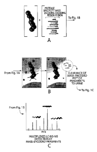
Figure 1 is a schematic depicting a method according to the invention for
multiplexed in vivo
enzyme profiling of mass-coded nanoparticle based pro-diagnostic reagents.
CA 2754072 2017-10-10
CA 02754072 2011-08-31
WO 2010/101628
PCT/US2010/000633
Figure 2 shows data depicting the process of tumor and wound targeting with
pro-
diagnostic reagents. Figure 2A is a schematic of the pro-diagnostic reagent,
with the circles
referring to the carrier, the star is a fluorescent molecule, and the zigzag
line refers to the
enzyme susceptible domain and the signature molecule (darker end region).
Figure 2B is an
electron micrograph of the pro-diagnostic reagent. Figure 2C is a graph
depicting the
circulation time of the pro-diagnostic reagent, by plotting detection of the
carrier in the blood
with respect to time after intravenous injection. Figure 2D is photographs of
mice having
either tumors or injuries (left and right panels, respectively) administered
the pro-diagnostic
reagent. Figure 2E is histopathological analysis of carrier homing to tumors
or regions of
injury.
Figure 3A is a schematic of the pro-diagnostic reagent, with the circles
referring to the
carrier, the star is a signature molecule, and the zigzag line refers to the
enzyme susceptible
domain. Figure 3B is a graph depicting fluorescence activation versus time.
Figure 3C
depicts data on 43 pro-diagnostic reagents (with enzyme susceptible domains
listed to the
right for detection of tumor and injury enzymes.
Figure 4 is a Table depicting the mass detection of ejected fragments in
vitro. The
results confirmed that the fluorescent results from the screen could also be
detected by
analyzing the mass of ejected fragments in vitro.
Figure 5 depicts the results of fluorescent detection of urinary reporter
activation by
tumors and injuries in vivo. Figure 5A is a schematic of the pro-diagnostic
reagent as shown
in Figure 3A, further depicting the portion of the molecule that undergoes
renal clearance and
the portion that undergoes RES clearance. Figure 5B is a set of photographs of
that were
intravenously administered the optimized pro-diagnostic reagent for injury
detection (top) or
tumor detection (bottom). Half the mice that were administered the optimized
pro-diagnostic
agents for injury-detection suffered bilateral hind limb injuries (left side
of the photograph)
while the control mice had no injuries (right side of the photograph). Half
the mice
administered the optimized pro-diagnostic agents for tumor-detection harbored
human
fibrosarcoma tumors (HT-1080) (left side of photograph), while the other mice
contained no
tumors (right side of photograph). Figure 5C is a set of graphs depicting
relative bladder
fluorescence for tumor (bottom panel) or injured (top panel) versus control
mice in order to
track the entrance of cleaved signature peptide into the urine after
injection.
Figure 6 shows LC/MS quantitation of signature molecules in urine. Figure 6A
is a
photograph of an experimental mouse, having bilateral injury and a control
uninjured mouse.
7
CA 02754072 2011-08-31
WO 2010/101628 PCT/US2010/000633
Figure 6B is a graph depicting the ratio of signature molecule (from thrombin
cleavable
proteolytic susceptible domain) to isotopically labeled product in injured
versus control mice.
Figure 7A shows a photograph of a typical implantable capsule in comparison to
a
penny and a ruler. Figure 7B is a graph showing measurements of thrombin-
cleaved peptide
efflux from implantable diagnostic capsules sealed with semi-permeable
membranes of
different pore sizes. Capsules made with membranes of pore size 10, 30, 50 or
80 nm were
loaded with nanoparticles functionalized with GGdFPipRSGGGC (SEQ ID NO: 8) and
exposed to solutions of thrombin or factor Xa (a cognate and a non-cognate
protease,
respectively). Thrombin-specific cleavage was monitored over time and is shown
in terms of
kinetics of reporter release.
Figure 8A shows representative proteolytic products appended with peptide caps
of
differing length and charge density. Figure 8B is a graph showing normalized
relative
intensities of the peptide reporters. The inset of Figure 8B shows a
magnification of the
normalized relative intensities of the peptide sequences Al-A6 as measured via
LC/MS.
Figure 8C shows a revised list of representative proteolytic products for
optimal LC/MC
detection.
Figure 9A is a schematic of the pro-diagnostic reagent, showing two identical
cocktails of 12 pro-diagnostic nanoparticles, each functional ized with a
different peptide,
with the circles referring to the carrier, the star is a signature molecule,
and the zigzag line
refers to the enzyme susceptible domain. Figure 9B is a graph depicting LC/MS
peak area
measurements of all twelve peptides after exposure of the first multiplex
cocktail to thrombin.
Figure 9C is a graph depicting LC/MS peak area measurements of all twelve
peptides after
exposure of the second multiplex cocktail to collagenase. Figure 9D is a graph
showing the
ratio of the LC/MS peak area for each peptide reporter after exposure to
thrombin over the
peak area measured after exposure to collagenase.
BRIEF DESCRIPTION OF THE SEQUENCES
SEQ ID NO: 1 is GGPQGIWGQC
SEQ ID NO: 2 is GGPLGVRGKC
SEQ ID NO: 3 is GGPLANvaDapARGC
SEQ ID NO: 4 is GGPVGLIGL
SEQ ID NO: 5 is GGPVPLSLVMC
SEQ ID NO: 6 is GGSGGPLGLRSWC
SEQ ID NO: 7 is GGGPWGIWGQGC
SEQ ID NO: 8 is GGdFPipRSGGGC
8
CA 02754072 2011-08-31
WO 2010/101628 PCT/US2010/000633
SEQ ID NO: 9 is GGLVPRGSGC
SEQ ID NO: 10 is GGPQG
SEQ ID NO: 11 is GGPLG
SEQ ID NO: 12 is GGPLA
SEQ ID NO: 13 is GGPVG
SEQ ID NO: 14 is GGPVPLS
SEQ ID NO: 15 is GGSGGPLG
SEQ ID NO: 16 is GGGPWG
SEQ ID NO: 17 is GGdFPipR
SEQ ID NO: 18 is GGLVP
SEQ ID NO: 191s GGVVVLS
SEQ ID NO: 20 is Fl-dR-dS-dR
SEQ ID NO: 21 is Fl-dR-G-dS-dR
SEQ ID NO: 22 is Fl-dR-dS-dR-G-G-P-Q-G-I-W-G-Q-C
SEQ ID NO: 23 is Fl-dR-G-dS-dR-G-G-P-L-G-V-R-G-K-C
SEQ ID NO: 24 is Fl-dR-G-dS-dR-G-G-P-L-A-Nva-Dpa-A-R-G-C
SEQ ID NO: 25 is Fl-dR-G-dS-dR-G-G-P-V-G-L-I-G-C
SEQ ID NO: 26 is Fl-dR-dS-dR-G-G-P-V-P-L-S-L-V-M-C
SEQ ID NO: 27 is Fl-dR-G-dS-dR-G-G-V-V-V-L-S-M-T-A-C
SEQ ID NO: 28 is Fl-dR-G-dS-dR-G-G-S-G-G-P-L-G-L-R-S-W-C
SEQ ID NO: 29 is Fl-dR-G-dS-dR-G-G-G-P-W-G-I-W-G-Q-G-C
SEQ ID NO: 30 is Fl-dR-G-G-dS-G-G-dF-Pip-R-S-G-G-G-C
SEQ ID NO: 31 is Fl-dR-dS-dR-G-G-L-V-P-R-G-S-G-C
SEQ ID NO: 32 is Fl-dR-G-G-dS-G-G-F-P-R-S-G-G-G-C
SEQ ID NO: 33 is Fl-dR-G-G-dS-G-G-G-dF-Pip-K-S-G-G-G-C
SEQ ID NO: 34 is Fl-dR-G-G-dS-G-G-G-dF-P-K-S-G-G-G-C
SEQ ID NO: 35 is dR-dS-dR
SEQ ID NO: 36 is dR-G-dS-dR
(Fl: Fluorescein; Nva: Norvaline; Dap = (N - p, - (2,4 - dinitrophenyl)) - L -
(1,13 -
diaminopropionic acid); Pip: pipecolic acid; d: D-isomer.)
DETAILED DESCRIPTION OF THE INVENTION
The status of physiological conditions of a subject can be assessed using the
methods
of the invention by identifying molecular properties also referred to as
"molecular
signatures". Such molecular signatures are useful for diagnosing diseases such
as cancer,
9
CA 02754072 2011-08-31
WO 2010/101628 PCT/US2010/000633
rheumatoid arthritis and arteriosclerosis, as well as for prognostic
indicators. The response of
most cancers to medical intervention is currently monitored by physical exams
and various
clinical imaging modalities. A few cancers such as prostate and ovarian cancer
are monitored
by use of single biomarkers in the blood. Such diagnostic techniques are
achieved, for
instance using fluorescence detection of molecular markers which are activated
in a particular
disease state.
The invention relates to a platform for functional characterization of disease
or
condition specific enzymatic repertoire as a method to monitor both disease
progression and
regression as well as response to therapeutics. The methods provide orders of
magnitude
.. more in vivo enzyme-substrate information than current fluorescent
detection technologies.
The platform provides a unique opportunity to functionally monitor cancer and
other disease
progression and response to therapy. It is particularly useful for prolonged
therapeutic
regimens, where the discovery of prognostic functional signatures would
greatly assist
intervention and where enzymatic signatures directly correlate to therapeutic
efficacy.
By administering a pro-diagnostic reagent, such as an exogenous detectable
substrate
library into animal models of disease it is possible to gain information into
substrate specific
enzymatic activities associated with diseases, such as cancer, cardiovascular
disease, arthritis,
and others. The technology allows for the potential simultaneous profiling of
hundreds of
enzyme-substrate activities in vivo using, for instance, -chaperoned, enzyme
sensitive
.. detectable compounds, an example of a compound referred to as pro-
diagnostic reagents.
The method leverages the distinct pharmacokinetics of modular structures and
small,
optionally hydrophilic, marker peptides (RES and renal clearance,
respectively). The pro-
diagnostic reagents have long circulation times and thus remain in circulation
or permeate
into tumors via porous angiogenic vascular networks, where upon local
molecules, such as
.. enzymes (MMPs, kallikreins, cathepsins, plasminogen activators, ADAMs) gain
access to the
enzyme susceptible regions of the pro-diagnostic reagents or substrates gain
access to the
enzymes of the pro-diagnostic reagents.
When the pro-diagnostic reagents, for example, the reagents including an
enzyme
susceptible domain are exposed to enzymes, for instance, proteases, the
reagent is cleaved,
such that a marker, referred to herein as a signature molecule, is released.
The marker is
renally-cleared and thus functions as a "messenger" of enzyme activity. For
instance, a
marker may include a self-quenched dye, such as Cy5.5 which is bound to a
larger molecule.
When the peptide containing the self-quenched dye is cleaved or modified by
specific
CA 02754072 2011-08-31
WO 2010/101628 PCT/US2010/000633
enzymes at the disease site the fluorophores are no longer self-quenched but
instead
developed fluorescent properties which can be detected at remote sites.
Alternatively, using
mass-encoded substrate libraries, the mass of enzyme substrates are designed
such that upon
cleavage, a distinct mass-specific messenger of cleavage will enter the urine
of a patient or
animal for detection using LC-MS technology. LC-MS urine analysis can generate
data that
is organized into a barcode of, for instance, cancer enzyme activity. In the
absence of
enzyme activity the pro-diagnostic reagents remain uncleaved and the whole
reagent
including the signature molecule is cleared through RES organs (liver, spleen,
and lymph
nodes) without producing urine markers. The use of mass to identify substrates
allows
unprecedented multiplexing capability with the potential to assay greater than
1,000
substrates.
When the pro-diagnostic reagents includes an enzyme, such as a protease, the
enzyme is exposed to endogenous substrates and the substrate is cleaved, such
that a marker,
referred to herein as a signature molecule, is released from the endogenous
substrate. The
marker is renally-cleared and thus functions as a "messenger" of enzyme
activity. For
instance, a marker may include a peptide, carbohydrate or nucleic acid
fragment which has
been cleaved from the substrate. Using the detection techniques, for instance,
LC-MS
technology, the mass of the signature can be detected. LC-MS urine analysis
can generate
data that is organized into a barcode of, for instance, cancer
enzyme/substrate activity. In the
absence of enzyme activity the signature molecule is not cleared through RES
organs (liver,
spleen, and lymph nodes) and does not produce urine markers.
Thus, the invention in some aspects involves administering to a subject a pro-
diagnostic reagent, identifying a biological sample from the subject in which
to detect the
signature molecule and optionally collecting the sample; and, subjecting the
biological
sample to an analysis method in order to detect the presence of one or more
signature
molecules. The presence of the signature molecule in the biological sample is
indicative of
an active enzyme or a substrate within the subject.
For example the invention in some aspects involves methods for administering
to a
subject a pro-diagnostic reagent, such that the pro-diagnostic reagent has a
modular structure
having a carrier domain linked to a signature producing domain , wherein the
signature
producing domain is capable of producing a signature molecule in the subject;
identifying a
biological sample for detection of the signature molecule, wherein the
biological sample is at
a site remote from the production of the signature molecule; and, subjecting
the biological
sample to an analysis method in order to detect the presence of the signature
molecule,
11
CA 02754072 2011-08-31
WO 2010/101628 PCT/US2010/000633
wherein the presence of the signature molecule in the biological sample is
indicative of a
biological predictor molecule within the subject.
The pro-diagnostic reagent comprises a modular structure having a carrier
domain
linked to a signature producing domain. A modular structure, as used herein,
refers to a
molecule having multiple domains.
The signature producing domain may be, for instance, an enzyme that can react
with
an endogenous substrate in a subject to produce a signature molecule or it may
be an enzyme
susceptible domain which is linked to a signature molecule. The carrier domain
may include
a single type of signature producing domain, such as, a single enzyme
susceptible domain and
or signature molecule or it may include multiple signature producing domains,
such as,
different enzyme susceptible domains and signature molecules. For instance
each carrier
may include 1 type of signature producing domain or it may include 2-1,000
different
signature producing domains or any integer therebetween. Alternatively each
carrier may
include greater than 1,000 signature producing domains. Multiple copies of the
pro-
diagnostic reagent are administered to the subject. Some mixtures of pro-
diagnostic reagents
may include signature producing domains that are enzymes, others may be
enzymatic
susceptible domains, and other may be mixtures of the two. Additionally a
plurality of
different pro-diagnostic reagents may be administered to the subject to
determine whether
multiple enzymes and/or substrates are present. In that instance, the
plurality of different pro-
diagnostic reagents includes a plurality of signature molecules, such that
each enzyme
susceptible domain is associated with a particular signature molecule or
molecules.
The carrier domain may serve as the core of the pro-diagnostic agent. A
purpose of
the carrier domain is to serve as a platform for the signature producing
domain. As such, the
carrier can be any material or size as long as it can serve as a carrier or
platform. Preferably
the material is non-immunogenic, i.e. does not provoke an immune response in
the body of
the subject to which it will be administered. Another purpose is that it may
function as a
targeting means to target the modular structure to a tissue, cell or molecule.
In some
embodiments the carrier domain is a particle. A particle, for example, a
nanoparticle, may,
for instance, result in passive targeting to tumors by circulation. Other
types of carriers,
include, for instance, compounds that cause active targeting to tissue, cells
or molecules.
Examples of carriers include, but are not limited to, microparticles,
nanoparticles, aptamers,
peptides (RGD, iRGD, LyP-1, CREICA, etc.) antibodies or antibody fragments
(e.g.
herceptin, cetuximab, panitumumab, etc.) and small molecules (e.g. erlotinib,
gefitinib,
sorafenib, etc.).
12
CA 02754072 2011-08-31
WO 2010/101628
PCT/US2010/000633
As used herein the term "particle" includes nanoparticles as well as
microparticles.
Nanoparticles are defined as particles of less than 1.0 p.m in diameter. A
preparation of
nanoparticles includes particles having an average particle size of less than
1.0 gm in
diameter. Microparticles are particles of greater than 1.0 gm in diameter but
less than 1 mm.
A preparation of microparticles includes particles having an average particle
size of greater
than 1.0 pim in diameter. The microparticles may therefore have a diameter of
at least 5, at
least 10, at least 25, at least 50, or at least 75 microns, including sizes in
ranges of 5-10
microns, 5-15 microns, 5-20 microns, 5-30 microns, 5-40 microns, or 5-50
microns. A
composition of particles may have heterogeneous size distributions ranging
from 10 nm to
mm sizes. In some embodiments the diameter is about 5 nm to about 500 nm. In
other
embodiments, the diameter is about 100 nm to about 200 nm. In other
embodiment, the
diameter is about 10 nm to about 100 nm.
The particles may be composed of a variety of materials including ceramic,
metallic,
natural polymer materials (including lipids, sugars, chitosan, hyaluronic acid
etc), synthetic
polymer materials (including poly-lactide-coglycolide, poly-glycerol sebacate,
etc), and
non-polymer materials, or combinations thereof.
The particles may be composed in whole or in part of polymers or non-polymer
materials. Non-polymer materials, for example, may be employed in the
preparation of the
particles. Exemplary materials include alumina, calcium carbonate, calcium
sulfate, calcium
phosphosilicate, sodium phosphate, calcium aluminate, calcium phosphate,
hydroxyapatite,
tricalcium phosphate, dicalcium phosphate, tricalcium phosphate, tetracalcium
phosphate,
amorphous calcium phosphate, octacalcium phosphate, and silicates. In certain
embodiments
the particles may comprise a calcium salt such as calcium carbonate, a
zirconium salt such as
zirconium dioxide, a zinc salt such as zinc oxide, a magnesium salt such as
magnesium
silicate, a silicon salt such as silicon dioxide or a titanium salt such as
titanium oxide or
titanium dioxide.
A number of biodegradable and non-biodegradable biocompatible polymers are
known in the field of polymeric biomaterials, controlled drug release and
tissue engineering
(see, for example, U.S. Pat. Nos. 6,123,727; 5,804,178; 5,770,417; 5,736,372;
5,716,404 to
Vacanti; U.S. Pat. Nos. 6,095,148; 5,837,752 to Shastri; U.S. Pat. No.
5,902,599 to Anseth;
U.S. Pat. Nos. 5,696,175; 5,514,378; 5,512,600 to Mikos; U.S. Pat. No.
5,399,665 to Barrera;
U.S. Pat. No. 5,019,379 to Domb; U.S. Pat. No. 5,010,167 to Ron; U.S. Pat. No.
4,946,929 to
d'Amore; and U.S. Pat. Nos. 4,806,621; 4,638,045 to Kohn; see also Langer,
Acc. Chem. Res.
13
CA 02754072 2016-08-18
64371-1124
33:94, 2000; Langer, J. Control Release 62:7, 1999; and Uhrich etal., Chem.
Rev. 99:3181,
1999).
Polymers include, but are not limited to: polyamides, polycarbonates,
polyallcylenes,
polyalkylene glycols, polyalkylene oxides, polyalkylene terepthalates,
polyvinyl alcohols,
polyvinyl ethers, polyvinyl esters, polyvinyl halides, polyglycolides,
polysiloxanes,
polyurethanes and copolymers thereof, alkyl cellulose, hydroxyalkyl
celluloses, cellulose
ethers, cellulose esters, nitro celluloses, polymers of acrylic and
methacrylic esters, methyl
cellulose, ethyl cellulose, hydroxypropyl cellulose, hydroxy-propyl methyl
cellulose,
hydroxybutyl methyl cellulose, cellulose acetate, cellulose propionate,
cellulose acetate
butyrate, cellulose acetate phthalate, carboxylethyl cellulose, cellulose
triacetate, cellulose
sulphate sodium salt, poly(methyl methacrylate), poly(ethylmethacrylate),
poly(butylmethacrylate), poly(isobutylmethaerylate), poly(hexlmethacrylate),
poly(isodecylmethacrylate), poly(lauryl methacrylate), poly(phenyl
methacrylate),
poly(methyl acrylate), poly(isopropyl acrylate), poly(isobutyl acrylate),
poly(octadecyl
acrylate), polyethylene, polypropylene poly(ethylene glycol), poly(ethylene
oxide),
poly(ethylene terephthalate), poly(vinyl alcohols), poly(vinyl acetate, poly
vinyl chloride and
polystyrene.
Examples of non-biodegradable polymers include ethylene vinyl acetate,
poly(meth)
acrylic acid, polyamides, copolymers and mixtures thereof.
Examples of biodegradable polymers include synthetic polymers such as polymers
of
lactic acid and glycolic acid, polyanhydrides, poly(ortho)esters,
polyurethanes, poly(butic
acid), poly(valeric acid), poly(caprolactone), poly(hydroxybutyrate),
poly(lactide-co-
glycolide) and poly(lactide-co-caprolactone), and natural polymers such as
algninate and
other polysaccharides including dextran and cellulose, collagen, chemical
derivatives thereof
(substitutions, additions of chemical groups, for example, alkyl, alkylene,
hydroxylations,
oxidations, and other modifications routinely made by those skilled in the
art), albumin and
other hydrophilic proteins, zein and other prolamines and hydrophobic
proteins, copolymers
and mixtures thereof. In general, these materials degrade either by enzymatic
hydrolysis or
exposure to water in vivo, by surface or bulk erosion. The foregoing materials
may be used
alone, as physical mixtures (blends), or as co-polymers. In some embodiments
the polymers
are polyesters, polyanhydrides, polystyrenes, polylactic acid, polyglycolic
acid, and
copolymers of lactic and glycoloic acid and blends thereof.
PVP is a non-ionogenic, hydrophilic polymer having a mean molecular weight
ranging from approximately 10,000 to 700,000 and the chemical formula (C61-
19N0)[n]. PVP
14
CA 02754072 2011-08-31
WO 2010/101628
PCT/US2010/000633
is also known as poly[1-(2-oxo-1 -pyrrolidinypethylene], PovidoneTM ,
PolyvidoneTM , RP
143Tm , KollidonTm , Peregal STTm , PeristonTm , PlasdoneTm , PlasmosanTm ,
ProtagentTm ,
SubtosanTm, and VinisiITM. PVP is non-toxic, highly hygroscopic and readily
dissolves in
water or organic solvents.
Polyethylene glycol (PEG), also known as poly(oxyethylene) glycol, is a
condensation polymer of ethylene oxide and water having the general chemical
formula
HO(CH2CH20)[n]H.
Polyvinyl alcohol (PVA) is a polymer prepared from polyvinyl acetates by
replacement of the acetate groups with hydroxyl groups and has the formula
(CH2CHOH)[n].
Most polyvinyl alcohols are soluble in water.
PEG, PVA and PVP are commercially available from chemical suppliers such as
the
Sigma Chemical Company (St. Louis, Mo.).
In certain embodiments the particles may comprise poly(lactic-co-glycolic
acid)
(PLGA).
The carrier may be composed of inorganic materials. Inorganic materials
include, for
instance, magnetic materials, conductive materials, and semiconductor
materials.
In addition to particles the carrier may be composed of any organic carrier,
including
biological and living carriers such as cells, viruses, bacteria, as well as
any non-living organic
carriers, or any composition enabling exposure of enzyme substrates to enzymes
in disease
(including extracellular, membrane-bound, and intracellular enzymes).
In some embodiments, the particles are porous. A porous particle can be a
particle
having one or more channels that extend from its outer surface into the core
of the particle.
In some embodiments, the channel may extend through the particle such that its
ends are both
located at the surface of the particle. These channels are typically formed
during synthesis of
the particle by inclusion followed by removal of a channel forming reagent in
the particle.
The size of the pores may depend upon the size of the particle. In certain
embodiments, the pores have a diameter of less than 15 microns, less than 10
microns, less
than 7.5 microns, less than 5 microns, less than 2.5 microns, less than 1
micron, less than 0.5
microns, or less than 0.1 microns. The degree of porosity in porous particles
may range from
greater than 0 to less than 100% of the particle volume. The degree of
porosity may be less
than 1%, less than 5%, less than 10%, less than 15%, less than 20%, less than
25%, less than
30%, less than 35%, less than 40%, less than 45%, or less than 50%. The degree
of porosity
can be determined in a number of ways. For example, the degree of porosity can
be
determined based on the synthesis protocol of the carriers (e.g., based on the
volume of the
CA 02754072 2011-08-31
WO 2010/101628 PCT/US2010/000633
aqueous solution or other channel-forming reagent) or by microscopic
inspection of the
carriers post-synthesis.
The plurality of particles may be homogeneous for one or more parameters or
characteristics. A plurality that is homogeneous for a given parameter, in
some instances,
means that particles within the plurality deviate from each other no more than
about +/- 10%,
preferably no more than about +/- 5%, and most preferably no more than about
+/- 1% of a
given quantitative measure of the parameter. As an example, the particles may
be
homogeneously porous. This means that the degree of porosity within the
particles of the
plurality differs by not more than +/- 10% of the average porosity. In other
instances, a
plurality that is homogeneous means that all the particles in the plurality
were treated or
processed in the same manner, including for example exposure to the same agent
regardless
of whether every particle ultimately has all the same properties. In still
other embodiments, a
plurality that is homogeneous means that at least 80%, preferably at least
90%, and more
preferably at least 95% of particles are identical for a given parameter.
The plurality of particles may be heterogeneous for one or more parameters or
characteristics. A plurality that is heterogeneous for a given parameter, in
some instances,
means that particles within the plurality deviate from the average by more
than about +/-
10%, including more than about +/- 20%. Heterogeneous particles may differ
with respect to
a number of parameters including their size or diameter, their shape, their
composition, their
surface charge, their degradation profile, whether and what type of agent is
comprised by the
particle, the location of such agent (e.g., on the surface or internally), the
number of agents
comprised by the particle, etc. The invention contemplates separate synthesis
of various
types of particles which are then combined in any one of a number of pre-
determined ratios
prior to contact with the sample. As an example, in one embodiment, the
particles may be
.. homogeneous with respect to shape (e.g., at least 95% are spherical in
shape) but may be
heterogeneous with respect to size, degradation profile and/or agent comprised
therein.
Particle size, shape and release kinetics can also be controlled by adjusting
the particle
formation conditions. For example, particle formation conditions can be
optimized to produce
smaller or larger particles, or the overall incubation time or incubation
temperature can be
increased, resulting in particles which have prolonged release kinetics.
The particles may also be coated with one or more stabilizing substances,
which may
be particularly useful for long term depoting with parenteral administration
or for oral
delivery by allowing passage of the particles through the stomach or gut
without dissolution.
For example, particles intended for oral delivery may be stabilized with a
coating of a
16
CA 02754072 2011-08-31
WO 2010/101628 PCT/US2010/000633
substance such as mucin, a secretion containing mucopolysaccharides produced
by the goblet
cells of the intestine, the submaxillary glands, and other mucous glandular
cells.
To enhance delivery the particles may be incorporated, for instance, into
liposomes,
virosomes, cationic lipids or other lipid based structures. The term "cationic
lipid" refers to
lipids which carry a net positive charge at physiological pH. Such lipids
include, but are not
limited to, DODAC, DOTMA, DDAB, DOTAP, DC-Chol and DMRIE. Additionally, a
number of commercial preparations of cationic lipids are available. These
include, for
example, LIPOFECTIN (commercially available cationic liposomes comprising
DOTMA
and DOPE, from GIBCO/BRL, Grand Island, N.Y., USA); LIPOFECTAMINE
(commercially available cationic liposomes comprising DOSPA and DOPE, from
GIBCO/BRL); and TRANSFECTAMO (commercially available cationic lipids
comprising
DOGS in ethanol from Promega Corp., Madison, Wis., USA). A variety of methods
are
available for preparing liposomes e.g., U.S. Pat. Nos. 4,186,183, 4,217,344,
4,235,871,
4,261,975, 4,485,054, 4,501,728, 4,774,085, 4,837,028, 4,946,787; and PCT
Publication
No. WO 91/17424. The particles may also be composed in whole or in part of
GRAS
components. i.e., ingredients are those that are Generally Regarded As Safe
(GRAS) by the
US FDA. GRAS components useful as particle material include non-degradeable
food
based particles such as cellulose.
The carrier domain can serve several functions. As discussed above, it may be
useful
for targeting the product to a specific region, such as a tissue. In that
instance it could include
a targeting agent such as a glycoprotein, an antibody, or a binding protein.
Further, the size of the carrier domain may be adjusted based on the
particular use of
the pro-diagnostic reagent. For instance, the carrier domain may be designed
to have a size
greater than 5 nm. Particles, for instance, of greater than 5 nm are not
capable of entering the
urine, but rather, are cleared through the reticuloendothelial system (RES;
liver, spleen, and
lymph nodes). By being excluded from the removal through the kidneys any
uncleaved pro-
diagnostic reagent will not be detected in the urine during the analysis step.
Additionally,
larger particles can be useful for maintaining the particle in the blood or in
a tumor site where
large particles are more easily shuttled through the vasculature. In some
embodiments the
carrier domain is 500 microns - 5nm, 250 microns- 5 nm, 100 microns ¨ 5nm, 10
microns -5
nm, 1 micron ¨ 5 nm, 100 nm-5 nm, 100nm ¨ 10 nm, 50nm ¨ lOnm or any integer
size range
therebetween. In other instances the carrier domain is smaller than 5 nm in
size. In such
instance the pro-diagnostic reagent will be cleared into the urine. However,
the presence of
17
CA 02754072 2011-08-31
WO 2010/101628
PCT/US2010/000633
free signature molecule can still be detected for instance using mass
spectrometry. In some
embodiments the carrier domain is 1-5nm, 2-5nm, 3-5nm, or 4-5nm.
Optionally the carrier domain may include a biological agent. In one
embodiment a
biological agent could be incorporated in the carrier domain or it may make up
the carrier
domain. For instance, it may form the scaffold or platform that the
proteolytic domain is
attached to. Thus the compositions of the invention can achieve two purposes
at the same
time, the diagnostic methods and delivery of a therapeutic agent. In some
embodiments the
biological agent may be a enzyme inhibitor. In that instance the biological
agent can inhibit
proteolytic activity at a local site and the signature molecule can be used to
test the activity of
that particular therapeutic at the site of action. HIV is an example of the
disease in which
active proteases can be monitored. In this embodiment the composition may
include a micro-
particle or other delivery device carrying a protease inhibitor. The protease
susceptible site
may be sensitive to the HIV proteases such that feedback can be provided
regarding the
activity of the particular protease inhibitor.
Biological agents include diagnostic, cosmetic, and therapeutic agents, such
as
releasable drugs. Thus, any biological agent can be incorporated within the
particles, which
can locally or systemically deliver or maintain the incorporated agents
following
administration or application to a subject. Any biocompatible or
pharmacologically
acceptable material can be incorporated into the particles or trapped in the
pores of the
particles using technology known to those skilled in the art. Biological
agents include but are
not limited to synthetic inorganic and organic compounds, proteins and
peptides,
polysaccharides and other sugars, lipids, and DNA and RNA nucleic acid
sequences having
therapeutic, prophylactic, cosmetic or diagnostic activities. Nucleic acid
sequences include
genes, plasmids, vectors, antisense molecules that bind to complementary DNA
to inhibit
transcription, siRNA, shRNA, and ribozymes.
In certain instances, the biological agent is an anti-microbial agent. An anti-
microbial
agent, as used herein, refers to a naturally-occurring or synthetic compound
which is capable
of killing or inhibiting infectious microorganisms. The type of anti-microbial
agent useful
according to the invention will depend upon the type of microorganism with
which the
subject is infected or at risk of becoming infected. Anti-microbial agents
include but are not
limited to anti-bacterial agents, anti-viral agents, anti-fungal agents and
anti-parasitic agents.
Phrases such as "anti-infective agent", "anti-bacterial agent", "anti-viral
agent", "anti-fungal
agent", "anti-parasitic agent" and "parasiticide" have well-established
meanings to those of
ordinary skill in the art and are defined in standard medical texts.
18
CA 02754072 2011-08-31
WO 2010/101628 PCT/US2010/000633
Growth factors may also be incorporated into the carrier. As used herein, the
term
growth factor refers to any agent that stimulates cellular proliferation
and/or differentiation.
Growth factors include but are not limited to fibroblast growth factor (FGF),
platelet-derived
growth factor (PDGF), insulin-like growth factors (IGF) I and II, TGF-13, TGF-
a, bone
morphogenetic protein (BMP) (e.g., BMP-2, BMP-3, BMP-4, BMP-6, or BMP-7),
hedgehog
proteins, growth differentiation factors, hematopoietic colony-stimulating
factors (CSF),
vascular endothelium growth factor (VEGF), osteoid-inducing factor (0IF),
angiogenins,
endothelins, hepatocyte growth factor, keratinocyte growth factor, ADMP-1,
interleukins (IL)
(e.g., IL-3 and IL-6), epithelial growth factors, dexamethasone, leptin,
sortilin,
transglutaminase, prostaglandin E, 1,25-dihydroxyvitamin D3, ascorbic acid,
pro-collagen,
glycerol phosphate, TAK-778, statins, growth hormone, steel factor (SF),
activin A (ACT),
retinoic acid (RA), epidermal growth factor (EGF), hematopoietic growth
factors, peptide
growth factors, erythropoietin, tumor necrosis factors (TNF), interferons
(IFN), heparin
binding growth factor (HBGF), nerve growth factor (NGF) and muscle morphogenic
factor
(MMP).
The biological agent may also be an anti-cancer therapy. Anti-cancer therapies
include for instance, radiotherapy, chemotherapy, adjuvant therapy, or any
combination of
the aforementioned.
The carrier domain may also be configured such that it can be detected in the
body
during the analysis. For instance, iron oxide can be incorporated into the
particles so that the
pro-diagnostic reagent can be tracked using MRI to provide non-invasive
imaging data.
The carrier is linked to the signature producing domain. A signature producing
domain, as used herein, is the portion of the modular structure that promotes
the enzymatic
reaction in the subject, causing the release of a signature molecule. The
signature producing
domain is either an active signature producing agent or an enzyme susceptible
domain linked
to a signature molecule. An active signature producing agent is a molecule
that causes the
formation of a signature molecule by modifying an endogenous molecule,
referred to herein
as a biological predictor molecule. For example the active signature producing
agent is an
enzyme that causes production of a signature molecule when it acts on an
endogenous
substrate. Enzymes include, for instance, proteases and glycosidases. The
biological
predictor molecule is the endogenous molecule acted upon by the active
signature producing
agent. Biological predictor molecules include for instance, substrates.
The enzyme susceptible site is dependent on enzymes that are active in a
specific
disease state. Alternatively, the enzyme specific site may be associated with
enzymes that are
19
CA 02754072 2011-08-31
WO 2010/101628 PCT/US2010/000633
ordinarily present but are absent in a particular disease state. For instance,
tumors are
associated with a specific set of enzymes. If the disease state being analyzed
is a tumor then
the product is designed with an enzyme susceptible site that matches that of
the enzyme
expressed by the tumor or other diseased tissue.
An enzyme, as used herein refers to any of numerous proteins produced in
living cells
that accelerate or catalyze the metabolic processes of an organism. Enzymes
act on
substrates. The substrate binds to the enzyme at a location called the active
site just before the
reaction catalyzed by the enzyme takes place. Enzymes include but are not
limited to
proteases, glycosidases, lipases, heparinases, phosphatases.
The enzyme susceptible site may be optimized to provide both high catalytic
activity
(or other enzymatic activity) for specified target enzymes but to also release
optimized
signature molecules for detection. For the specific detection modality of mass
spectrometry,
inclusion of tags for rapid affinity purification, specific charges in the
sequence to increase
ionization efficiency, mass defects to eliminate endogenous organic
background, and unique
mass signatures can improve detection. In addition to improving detection
sensitivity, by
programming the mass of these molecules, mass selection techniques can be
harnessed to
remove background during detection and focus on expected signature masses.
Patient outcome depends on the phenotype of individual diseases at the
molecular
level, and this is often reflected in expression of enzymes. The recent
explosion of
.. bioinformatics has facilitated exploration of complex patterns of gene
expression in human
tissues (Fodorõ S.A. Massively parallel genomics. Science 277, 393-395
(1997)).
Sophisticated computer algorithms have been recently developed capable of
molecular
diagnosis of tumors using the immense data sets generated by expression
profiling (Khan J,
Wei JS, Ringner M, Saal LH, Ladanyi M, Westermann F, et al. Classification and
diagnostic
prediction of cancers using gene expression profiling and artificial neural
networks. Nat Med
2001;7:673-679.). This information can be accessed in order to identify
enzymes and
substrates associated with specific diseases. Based on this information the
skilled artisan can
identify appropriate enzyme or substrates to incorporate into the pro-
diagnostic reagent.
Table 1 provides a non-limiting list of enzymes associated with (either
increased or decreased
with respect to normal) disease and in some instances, the specific substrate.
Table 2
provides a non-limiting list of substrates associated with disease or other
conditions.
Numerous other enzyme/substrate combinations associated with specific diseases
or
conditions are known to the skilled artisan and are useful according to the
invention.
CA 02754072 2011-08-31
WO 2010/101628 PCT/US2010/000633
Table 1
DISEASE ENZYME SUBSTRATE
Cancer MMP collagens, gelatin, various
ECM proteins
Cancer MMP-2 type IV collagen and gelatin
Cancer MMP-9 type IV and V collagens and
gelatin
Cancer kallilcreins kininogens, plasminogen
Cancer cathepsins broad spectrum of substrates
Cancer plasminogen activator, tPA Plasminogen
Cancer ADAM (A Diseintegrin And various extracellular
domains
Metalloprotease, also MDC, of transmembrane proteins
Adamalysin)
Pancreatic carcinoma MMP-7 various, e.g. collagen 18,
FasL,
HLE, DCN, IGFBP-3, MAG,
plasminogen, other MMPs
Pancreatic Cancer ADAM9, ADAM15 various extracellular
domains
of transmembrane proteins
Prostate adenocarcinoma Matriptase, a type II unspecific, cleaves after
Lys or
transmembrane serine protease Arg residues
Prostate cancer Kallilcrein 3 kininogens, plasminogen
Prostate cancer ADAM15 various extracellular
domains
of transmembrane proteins
Ovarian carcinoma Kallikrein 6 kininogens, plasminogen
Epithelial-derived tumors Matriptase, a type II
unspecific, cleaves after Lys or
(breast, prostate, ovarian, colon, transmembrane serine protease Arg
residues
oral)
Ovarian Cancer MMP-2, MMP-9, kallilcrein-10 type IV and V
collagens and
(hk-10) gelatin, kininogens,
plasminogen
Breast, gastric, prostate cancer cathepsins B, L and D
broad spectrum of substrates
Endometrial cancer cathepsin B unspecific cleavage of a
broad
spectrum of substrates without
clear sequence specificity
esophageal adenocarcinoma cathepsin B unspecific
cleavage of a broad
spectrum of substrates without
clear sequence specificity
Invasive cancers, metastases type II integral serine proteases
(dipeptidyl peptidase IV
(DPP4/CD26),seprase/fibroblast
activation protein alpha
(FAPalpha) and related type II
transmembrane prolyl serine
peptidases))
Invasive cancers, metastases Seprase various ECM
proteins
viral infections
All Retroviruses viral protease precursor GagPol fusion
HIV HIV protease (HIV PR, an precursor Gag and GagPol
aspartic protease) proteins
21
CA 02754072 2011-08-31
WO 2010/101628 PCT/US2010/000633
DISEASE ENZYME SUBSTRATE
Hepatitis C NS3 serine protease viral precursor polyprotein
Dengue Dengue protease auocleavage (NS2B/NS3),
NS3/NS4A and NS4B/NS5
cleavage
West Nile NS2B/NS3pro viral precursor polyprotein
bacterial infections
Legionella spp. zinc metalloprotease Me-Arg-Pro-Tyr
Meninogencephalitis histolytic cysteine protease
Streptococcus pyogenes (Group streptococcal pyrogenic exotoxin extracellular
matrix,
A Streptococcus) B (SpeB) immunoglobulins, complement
components
Chlostridium difficile Cwp84 fibronectin, laminin,
vitronectin and other ECM
proteins
Alzheimer's disease BACE-1,2 (Alzheimer secretase) p-amyloid precursor
protein
Stroke and recovery MMP, tPA
cardiovascular disease Angiotensin Converting Enzyme angiotensin I,
bradykinin
(ACE)
Atherosclerosis cathepsin K, L, S broad spectrum of substrates
arthritis MMP-1 triple-helical fibrillar
collagens
rheumatoid arthritis thrombin Osteopontin
osteoarthritis thrombin Osteopontin
osteoporosis/ostearthritis cathepsin K, S broad
spectrum of substrates
Arthritis, inflammatory joint Aggrecanase (ADAMTS4,
aggrecans (proteoglycans)
disease ADAMTS11)
thrombosis factor Xa (thrombokinase) Prothrombin
thrombosis ADAMTS13 von Willebrand factor (vWF)
thrombosis plasminogen activator, tPA Plasminogen
Stress-induced Renal pressure Prostasin epithelial Na
channel subunits
natriuresis
Table 2
DISEASE TARGET SUBSTRATE ENZYME
Inflammation Interleukin 1 beta MMP-2, MMP-3, MMP-9,
Trypsin, chymotrypsin, pepsin,
Lys-C, Glu-C, Asp-N, Arg-C
Pituitary gland IGFBP-3 MMP-1, MMP-3, MMP-9,
dysfunction, abnormal Trypsin, chymotrypsin, pepsin,
bone density, growth Lys-C, Glu-C, Asp-N, Arg-C
disorders
Cancer TGF-beta MMP-9, Trypsin, chymotrypsin,
pepsin, Lys-C, Glu-C, Asp-N,
22
CA 02754072 2011-08-31
WO 2010/101628
PCT/US2010/000633
DISEASE TARGET SUBSTRATE ENZYME
Arg-C
Cancer, autoimmune TNF MMP-7, Trypsin, chymotrypsin,
disease pepsin, Lys-C, Glu-C, Asp-N,
Arg-C
Cancer, autoimmune FASL MMP-7, Trypsin, chymotrypsin,
disease pepsin, Lys-C, Glu-C, Asp-N,
Arg-C
Wound healing, cardiac HB-EGF MMP-3, Trypsin, chymotrypsin,
disease pepsin, Lys-C, Glu-C, Asp-N,
Arg-C
Pfeiffer syndrome FGFR1 MMP-2, Trypsin, chymotrypsin,
pepsin, Lys-C, Glu-C, Asp-N,
Arg-C
Cancer Decorin MMP-2, MMP-3, MMP-7,
Trypsin, chymotrypsin, pepsin,
Lys-C, Glu-C, Asp-N, Arg-C
Cancer Tumor associated Endoglycosidases
carbohydrate antigens
Cancer Sialyl Lewis' 0-glycanase
Cancer Sialyl Lewis' 0-glycanase
Cancer/ Rheumatoid VEGF Trypsin, chymotrypsin, pepsin,
Arthritis, pulmonary Lys-C, Glu-C, Asp-N, Arg-C
hypertension
Cancer EGF Trypsin, chymotrypsin, pepsin,
Lys-C, Glu-C, Asp-N, Arg-C
Cancer IL2 Trypsin, chymotrypsin, pepsin,
= Lys-C, Glu-C, Asp-N, Arg-C
Cancer IL6 Trypsin, chymotrypsin, pepsin,
inflammation/angiogenesis Lys-C, Glu-C, Asp-N, Arg-C
Cancer IFN-y Trypsin, chymotrypsin, pepsin,
Lys-C, Glu-C, Asp-N, Arg-C
Cancer TNF-a Trypsin, chymotrypsin, pepsin,
inflammation/angiogenesis, Lys-C, Glu-C, Asp-N, Arg-C
Rheumatoid Arthritis
Cancer, Pulmonary TGF40 Trypsin, chymotrypsin, pepsin,
fibrosis, Asthma Lys-C, Glu-C, Asp-N, Arg-C
Cancer, Pulmonary PDGF Trypsin, chymotrypsin, pepsin,
hypertension Lys-C, Glu-C, Asp-N, Arg-C
Cancer, pulmonary Fibroblast growth factor Trypsin, chymotrypsin,
pepsin,
cystadenoma (FGF) Lys-C, Glu-C, Asp-N, Arg-C
Cancer Brain-derived neurotrophic Trypsin, chymotrypsin,
pepsin,
factor (BDNF) Lys-C, Glu-C, Asp-N, Arg-C
Cancer Interferon regulatory Trypsin, chymotrypsin,
pepsin,
factors (IRF-1, IRF-2) Lys-C, Glu-C, Asp-N, Arg-C
Inhibitor of tumor MIF Trypsin, chymotrypsin, pepsin,
suppressors Lys-C, Glu-C, Asp-N, Arg-C
Lymphomas/carcinomas, GM-CSF Trypsin, chymotrypsin, pepsin,
alveolar proteinosis Lys-C, Glu-C, Asp-N, Arg-C
Cancer invasion M-CSF Trypsin, chymotrypsin, pepsin,
23
CA 02754072 2016-08-18
64371-1124
DISEASE TARGET SUBSTRATE ENZYME
Lys-C, Glu-C, Asp-N, Arg-C
Chemical carcinogenesis, IL-12 Trypsin, chymotrypsin, pepsin,
multiple schlerosis, Lys-C, Glu-C, Asp-N, Arg-C
rheumatoid arthritis,
Crohn's disease
Natural Killer T cell IL-15 Trypsin, chymotrypsin, pepsin,
leukemias, inflammatory Lys-C, Glu-C, Asp-N, Arg-C
bowel disease, rheumatoid
arthritis
Cirrhosis Tissue inhibitor of MMPs Trypsin, chymotrypsin,
pepsin,
(TIMPs) Lys-C, Glu-C, Asp-N, Arg-C
Cirrhosis Collagen 1,111 MMP-1, MMP-8, Trypsin,
chymotrypsin, pepsin, Lys-C,
Glu-C, Asp-N, Arg-C
Cirrhosis Collagen IV, V MMP-2, Trypsin, chymotrypsin,
pepsin, Lys-C, Glu-C, Asp-N,
Arg-C
Several of the enzyme/Substrates described above are described in the
following
publications: Parks, W.C. and R.P. Mecham (Eds): Matrix metalloproteinases.
San Diego: Academic Press; 1998; Nagase, H. and J.F. Woessner, Jr.
(1999)3. Biol. Chem. 274:21491; Ito, A. et al. (1996) J.
Biol. Chem. 271:14657; Schonbecic, U. et al. (1998) J. Immunol. 161:3340;
Rajah, R. etal.
(1999) Am. J. Cell Mol. Biol. 20:199; Fowlkes, J.L. et al. (1994)
Endocrinology 135:2810;
Manes, S. et al. (1999) J. Biol. Chem. 274:6935; Mira, E. et al. (1999)
Endocrinology
140:1657; Yu, Q. and I. Stamenkovic (2000) Genes Dev. 14:163; Haro, H. etal.
(2000) J.
Clin. Invest. 105:143; Powell, C.P. et al. (1999) Curr. Biol. 9:1441; Suzuki,
M. et al. (1997) J.
Biol. Chem. 272:31730; Levi, E. et al. (1996) Proc. Natl. Acad. Sci. USA
93:7069; Imai, K.
et al. (1997) Biochem. J. 322:809; Smith, M.M. et al. (1995) J. Biol. Chem.
270:6440; and
Dranoff, G. (2004) Nat. Rev. Cancer 4: 11-22.
The signature producing domain may be attached directly to the carrier. For
instance
it may be coated directly on the surface of microparticles using known
techniques.
Alternatively if the carrier is a protein material it may be directly
connected through a peptide
bond. Additionally, the signature producing domain may be connected to the
carrier domain
through the use of a linker. As used herein "linked" or "linkage" means two
entities are
bound to one another by any physicochemical means. Any linkage known to those
of
ordinary skill in the art, covalent or non-covalent, is embraced. Thus, in
some embodiments
the carrier has a linker attached to an external surface, which can be used to
link the signature
producing domain. Another molecule can also be attached to the linker.
24
CA 02754072 2011-08-31
WO 2010/101628
PCT/US2010/000633
The signature producing domain is preferably a polymer made up of a plurality
of
chemical units. A "chemical unit" as used herein is a building block or
monomer which may
be linked directly or indirectly to other building blocks or monomers to form
a polymer. In
some embodiments the signature producing domain is a peptide that is
susceptible to
cleavage by an enzyme or causes cleavage of a substrate associated with a
disease or
condition. A number of examples of when the proteolytic cleavage site is a
peptide are
presented in the table above.
The enzyme susceptible domain may also be a polysaccharide. Some
polysaccharide
specific degrading enzymes are associated with tumors, angiogenesis and other
conditions.
A "polysaccharide" is a biopolymer comprised of linked saccharide or sugar
units. The
polysaccharides used as proteolytic susceptible domains may be isolated or
synthesized de
novo. For example, the polysaccharides may be isolated from natural sources
e.g. purified, as
by cleavage and gel separation or may be synthesized e.g., by chemical
synthesis and
incorporated into the pro-diagnostic reagent.
For instance, HSGAG degrading enzymes are enzymes that can be analyzed
according to the methods of the invention. HSGAG degrading enzymes include
heparinase-I,
heparinase- II, heparinase-III, D-glucuronidase and L-iduronidase. The
heparinases cleave at
the glycosidic linkage before a uronic acid. Heparinase I clips at a
glycosidic linkage before
a 2 -0 sulfated iduronic acid. Heparinase -III cleaves at a glycosidic linkage
before an
unsulfated glucuronic acid. Heparinase -II cleaves at both Hep-I and Hep-III
cleavable sites.
Glucuronidase and iduronidase, as their name suggests cleave at the glycosidic
linkage after a
glucuronic acid and iduronic acid respectively. Nitrous acid clips randomly at
glycosidic
linkages after a N-sulfated hexosamine and converts the six membered
hexosamine ring to a
5 membered anhydromannitol ring. Appropriate enzyme susceptible domains may be
designed based on the known substrates and cleavage sites of these enzymes.
The pro-diagnostic reagent may also include an implantable microdelivery
device that
houses the modular structure. An implantable microdelivery device is any type
of device,
that is sized for implantation into a body and can retain the modular
structure. For instance
the device may be an implantable capsule that contains the modular structure
housed there in.
The capsule may have a semi-permeable membrane, such that the modular
structure cannot
pass though the membrane, but which is permeable to endogenous molecules such
as
enzymes and substrates as well as signature molecules. Alternatively the
implantable
microdelivery device may be a chip having the modular structure attached
thereto. Examples
of implantable microdelivery devices include but are not limited to
implantable capsules,
CA 02754072 2011-08-31
WO 2010/101628 PCT/US2010/000633
chips, sustained-release formulations, multi-pulse drug delivery resorbable
polymeric
microchip device (Grayson et al. Nature Materials, VOL 2, Nov 2003, p. 767),
and controlled
release microchips (Santini et al Nature, vol 397, 1999, p.335). These devices
may be made
from many materials include many of the polymeric materials described herein.
Preferably
the implantable devices are biocompatible and non-toxic.
Modification of the enzyme susceptible domain by an enzyme in vivo, results in
the
production of a signature molecule. Alternatively, when the signature
producing domain is
an enzyme the enzyme cleaves an endogenous substrate producing a signature
molecule from
the endogenous substrate. The signature molecule is a detectable molecule. It
can be part of
the enzyme susceptible domain, e.g. the piece that is released or added upon
cleavage or it
can be a separate entity. The signature molecule may be, for instance, a
peptide, nucleic acid,
small molecule, fluorophore/quencher, carbohydrate, particle, radiolabel, MRI-
active
compound, inorganic material, organic material, with encoded characteristics
to facilitate
optimal detection.
The signature molecule may be detected by any known detection methods. A
variety
of methods may be used, depending on the nature of the signature
molecule/label. Labels on
signature molecules may be directly detected through optical or electron
density, radioactive
emissions, nonradiative energy transfers, or signature molecules may be
indirectly detected
with antibody conjugates, strepavidin-biotin conjugates, mass spectrometry,
liquid
.. chromatography-mass spectrometry, PCR analysis, DNA microarray, and
fluorescence
analysis.
The analysis step may be performed directly on the biological sample or the
signature
component may be purified to some degree first. For instance, a purification
step may
involve isolating the signature molecule from other components in the
biological sample.
.. Purification steps include methods such as affinity chromatography. As used
herein an
"isolated molecule" or "purified molecule" is a signature molecule that is
isolated to some
extent from its natural environment. The isolated or purified molecule need
not be 100%
pure or even substantially pure prior to analysis.
The methods for analysing signature molecules by identifying the presence of a
signature molecule may be used to provide a qualitative assessment of the
molecule (e.g.,
whether the signature molecule is present or absent) or a quantitative
assessment (e.g., the
amount of signature molecule present to indicate a comparative activity level
of the enzymes.
The quantitative value may be calculated by any means, such as, by determining
the percent
26
CA 02754072 2011-08-31
WO 2010/101628 PCT/US2010/000633
relative amount of each fraction present in the sample. Methods for making
these types of
calculations are known in the art.
A signature molecule can be determined using mass spectrometry. The mass
spectrometry data may be a valuable tool to ascertain information about the
signature
molecule in the biological sample. After a molecular weight of a signature
molecule is
identified, it may be compared to molecular weights of other known signature
molecules.
Molecular weight may be determined by several methods including mass
spectrometry. The use of mass spectrometry for determining the molecular
weight of
molecules is well known in the art. Liquid chromatography-mass spectrometry
(LC-MS, or
alternatively HPLC-MS) is an analytical chemistry technique that combines the
physical
separation capabilities of liquid chromatography (or HPLC) with the mass
analysis
capabilities of mass spectrometry. LC-MS is a powerful technique which has
very high
sensitivity and specificity. It can be used to detect signature molecules in a
complex mixture.
Other types of mass spectrometry known in the art, such as, matrix-assisted
laser desorption
ionization mass spectrometry (MALD1-MS), electron spray-MS, fast atom
bombardment
mass spectrometry (FAB-MS) and collision-activated dissociation mass
spectrometry (CAD)
can also be used to identify the molecular weight of the signature molecule.
Methods for performing mass spectrometry using nucleic acid samples have been
described. See e.g., US Patent No. 5,885,775. U.S. Patent Nos. 7,412,332 and
6,597,996
.. describe methods for detecting polysaccharide molecules using mass
spectrometry. As
shown in these patent applications, one technique for comparing molecular
weights is to
generate a mass line and compare the molecular weight of the unknown
polysaccharide to the
mass line to determine a subpopulation of polysaccharides which have the same
molecular
weight. A "mass line" is an information database, preferably in the form of a
graph or chart
which stores information for each possible type of polysaccharide having a
unique sequence
based on the molecular weight of the polysaccharide. Because mass spectrometry
data
indicates the mass of a fragment to 1Da accuracy, a length may be assigned
uniquely to a
fragment by looking up a mass on the mass line.
NMR spectroscopy is an analytical tool that allows for the determination of
molecular
structure. Utilizing the magnetic properties of some nuclei, the nuclear spins
of the nuclei
can be oriented randomly with an external magnetic field. Oriented nuclei that
are
subsequently irradiated at the correct frequency will absorb energy and
transition to a higher
energy state. Upon relaxation this energy is emitted and detected in various
NMR systems.
This irradiation of the nuclei occur in pulses. In basic one dimensional (1D)
NMR the
27
CA 02754072 2011-08-31
WO 2010/101628 PCT/US2010/000633
excitation is produced from a single pulse and emitted radiation is detected
as free induction
decay (FID). In two dimensional (2D) NMR spectroscopy the nuclei is irradiated
with two
pulses, and acquisition of the FID occurs at many time points with a delay
between the
pulses.
When the signature molecule is a nucleic acid, it can also be analyzed using
PCR and
microarrays. PCR methods are well-known in the art. For instance, U.S. Patent
No.
5,333,675, issued to Mullis et al. describes an apparatus and method for
performing
automated PCR. In general, performance of a PCR method results in
amplification of a
selected region of DNA by providing two DNA primers, each of which is
complementary to a
portion of one strand within the selected region of DNA. The primer is
hybridized to a
template strand of nucleic acid in the presence of deoxyribonucleotide
triphosphates (dATP,
dCTP, dGTP, and dTTP) and a chain extender enzyme, such as DNA polymerase. The
primers are hybridized with the separated strands, forming DNA molecules that
are single
stranded except for the region hybridized with the primer, where they are
double stranded.
The double stranded regions are extended by the action of the chain extender
enzyme (e.g.
DNA polymerase) to form an extended double stranded molecule between the
original two
primers. The double stranded DNA molecules are separated to produce single
strands which
can then be re-hybridized with the primers. The process is repeated for a
number of cycles to
generate a series of DNA strands having the same nucleotide sequence between
and including
the primers.
Chain extender enzymes are well known in the art and include, for example, E.
coil
DNA polymerase I, klenow fragment of E. coil DNA polymerase I, T4 DNA
polymerase, T7
DNA polymerase, recombinant modified T7 DNA polymerase, reverse transcriptase,
and
other enzymes. Heat stable enzymes are particularly preferred as they are
useful in
automated thermal cycle equipment. Heat stable polymerases include, for
example, DNA
polymerases isolated from bacillus stearothermophilus (Bio-Rad), thermus
thermophilous
(finzyme, ATCC number 27634), thermus species (ATCC number 31674), therms
aquaticus
strain TV11518 (ATCC number 25105), sulfolobus acidocaldarius, described by
Bukhrashuili et al., Biochem. Biophys. Acta., 1008:102-07 (1909), thermus
filiformus (ATCC
number 43280), Taq DNA polymerase, commercially available from Perkin-Elmer-
Cetus
(Norwalk, Connecticut), Promega (Madison, Wis.) and Stratagene (La Jolla,
Calif.), and
AmpliTaqTm DNA polymerase, a recombinant thermus equitus Taq DNA polymerase,
available from Perkin-Elmer-Cetus and described in U.S. Patent No. 4,889,818.
28
CA 02754072 2016-08-18
64371-1124
Preferably, the PCR-based methods performed according to the invention are
automated and performed using thermal cyclers. Many types of thermal cyclers
are well-
known in the art. For instance, M.J. Research (Watertown, MA) provides a
thermal cycler
having a pettier heat pump to provide precise uniform temperature control in
the thermal
cyclers; DeltaCycler thermal cyclers from Ericomp (San Diego, CA) also are
peltier-based
and include automatic ramping control, time/temperature extension programming
and a
choice of tube or microplate configurations. The RoboCyclerTm by Stratagene
(La Jolla, CA)
incorporates robotics to produce rapid temperature transitions during cycling
and well-to-well
uniformity between samples; and a particularly preferred cycler, is the Perkin-
Elmer Applied
Biosystems (Foster City, CA) ABI PrismTh 877 Integrated Thermal cycler, which
is operated
through a programmable interface that automates liquid handling and
thermocycling
processes for fluorescent DNA sequencing and PCR reactions.
The presence or absence of enzymes in the subject may also be determined using
hybridization techniques. Standard hybridization techniques of microarray
technology are
utilized to assess the presence of nucleic acids in the biological sample.
Microarray
technology, which is also known by other names including: DNA chip technology,
gene chip
technology, and solid-phase nucleic acid array technology, is well known to
those of ordinary
skill in the art and is based on, but not limited to, obtaining an array of
identified nucleic acid
probes on a fixed substrate, labeling target molecules with reporter molecules
(e.g.,
radioactive, chemiluminescent, or fluorescent tags such as fluorescein, Cye3-
dUTP, or Cye5-
dUTP), hybridizing target nucleic acids to the probes, and evaluating target-
probe
hybridization. A probe with a nucleic acid sequence that perfectly matches the
target
sequence will, in general, result in detection of a stronger reporter-molecule
signal than will
probes with less perfect matches. Many components and techniques utilized in
nucleic acid
microarray technology are presented in The Chipping Forecast, Nature Genetics,
Vol.21,
Jan 1999.
According to the present invention, microarray substrates may include but are
not
limited to glass, silica, aluminosilicates, borosilicates, metal oxides such
as alumina and
nickel oxide, various clays, nitrocellulose, or nylon. In all embodiments a
glass substrate is
preferred. An "array" as used herein is a set of molecules arranged in a
specific order with
respect to a surface. Preferably the array is composed of polynucleotides
attached to the
surface. Oligonucleotide arrays can be used to screen nucleic acid samples for
a target
nucleic acid, which can be labeled with a detectable marker. A fluorescent
signal resulting
from hybridization between a target nucleic acid and a substrate-bound
oligonucleotide
29
CA 02754072 2011-08-31
WO 2010/101628
PCT/US2010/000633
provides information relating to the identity of the target nucleic acid by
reference to the
location of the oligonucleotide in the array on the substrate. Such a
hybridization assay can
generate thousands of signals which exhibit different signal strengths. These
signals
correspond to particular oligonucleotides of the array. Different signal
strengths will arise
based on the amount of labeled target nucleic acid hybridized with an
oligonucleotide of the
array.
Conditions for optimal hybridization are known. The hybridization conditions
in
general are those used commonly in the art, such as those described in
Sambrook et al.,
"Molecular Cloning: A Laboratory Manual", (1989), 2nd Ed., Cold Spring Harbor,
NY;
Berger and Kimmel, "Guide to Molecular Cloning Techniques", Methods in
Enzymology,
(1987), Volume 152, Academic Press, Inc., San Diego, CA; and Young and Davis,
(1983),
PNAS (USA) 80:1194. In general, incubation temperatures for hybridization of
nucleic acids
range from about 20 C to 75 C. For probes 17 nucleotides residues and longer,
a preferred
temperature range for hybridization is from about 50 C to 54 C. The
hybridization
temperature for longer probes is preferably from about 55 C to 65 C and for
shorter probes is
less than 52 C. Rehybridization may be performed in a variety of time frames.
Preferably,
hybridization of SNP and RCGs performed for at least 30 minutes.
The signature molecule may be labeled. For example, a label may be added
directly
to a nucleic acid when the isolated signature molecule is subjected to PCR.
For instance, a
PCR reaction performed using labeled primers or labeled nucleotides will
produce a labeled
product. Labeled nucleotides (e.g., fluorescein-labeled CTP) are commercially
available.
Methods for attaching labels to nucleic acids are well known to those of
ordinary skill in the
art and, in addition to the PCR method, include, for example, nick translation
and end-
labeling.
Labels suitable for use in the methods of the present invention include any
type of
label detectable by standard means, including spectroscopic, photochemical,
biochemical,
electrical, optical, or chemical methods. Preferred types of labels include
fluorescent labels
such as fluorescein. A fluorescent label is a compound comprising at least one
fluorophore.
Commercially available fluorescent labels include, for example, fluorescein
phosphoramidides such as fluoreprime (Pharmacia, Piscataway, NJ), fluoredite
(Millipore,
Bedford, MA), FAM (ABI, Foster City, CA), rhodamine, polymethadine dye
derivative,
phosphores, Texas red, green fluorescent protein, CY3, and CY5.
Polynucleotides can be
labeled with one or more spectrally distinct fluorescent labels. "Spectrally
distinct"
fluorescent labels are labels which can be distinguished from one another
based on one or
CA 02754072 2011-08-31
WO 2010/101628 PCT/US2010/000633
more of their characteristic absorption spectra, emission spectra, fluorescent
lifetimes, or the
like. Spectrally distinct fluorescent labels have the advantage that they may
be used in
combination ("multiplexed"). Radionuclides such as 3H, 125/ 35^J, 14 C, or 32P
are also useful
,
labels according to the methods of the invention. A plurality of radioactively
distinguishable
radionuclides can be used. Such radionuclides can be distinguished, for
example, based on
the type of radiation (e.g. a, 13, or 6 radiation) emitted by the
radionuclides. The 32P signal
can be detected using a phosphoimager, which currently has a resolution of
approximately 50
microns. Other known techniques, such as chemiluminescence or colormetric
(enzymatic
color reaction), can also be used.
Quencher compositions in which a "donor" fluorophore is joined to an
"acceptor"
chromophore by a short bridge that is the binding site for the enzyme may also
be used. The
signal of the donor fluorophore is quenched by the acceptor chromophore
through a process
believed to involve resonance energy transfer (RET). Cleavage of the peptide
results in
separation of the chromophore and fluorophore, removal of the quench, and
generation of a
subsequent signal measured from the donor fluorophore.
Once the data is obtained, e.g. as a two-dimensional image, a computer can be
used to
transform the data into a displayed image which varies in color depending on
the intensity of
light emission at a particular location. Any type of commercial software which
can perform
this type of data analysis can be used. In general, the data analysis involves
the steps of
determining the intensity of the fluorescence emitted as a function of the
position on the
substrate, removing the outliers, and calculating the relative binding
affinity. One or more of
the presence, absence, and intensity of signal corresponding to a label is
used to assess the
presence or absence of an signature molecule. The presence and absence of one
or more
signature molecules can be used to determine the disease status of an
individual based on the
presence or absence of an enzyme.
The data may also be observed and analyzed manually. For instance, the
presence or
absence of a fluorescent label may be observed in order to provide the
diagnostic or
prognostic information from the data.
The disease or condition assessed acccording to the methods of the invention
is any
disease or condition that is associated with an enzyme. For instance, cancer,
cardiovascular
disease, arthritis, viral, bacterial, parasitic or fungal infection,
Alzheimer's disease
emphysema, thrombosis, hemophilia, stroke, organ disfunction, any inflammatory
condition,
vascular disease, parenchymal disease, or a pharmacologically-induced state
are all known to
be associated with enzymes. A pharmacologically induced state is a condition
in which
3'
CA 02754072 2011-08-31
WO 2010/101628
PCT/US2010/000633
enzyme inhibitors and other agents directly or indirectly affect enzyme
activities. Thus each
of the these can be assessed or monitored or studied according to methods of
the invention.
It is useful to be able to differentiate non-metastatic primary tumors from
metastatic
tumors, because metastasis is a major cause of treatment failure in cancer
patients. If
metastasis can be detected early, it can be treated aggressively in order to
slow the
progression of the disease. Metastasis is a complex process involving
detachment of cells
from a primary tumor, movement of the cells through the circulation, and
eventual
colonization of tumor cells at local or distant tissue sites. Additionally, it
is desirable to be
able to detect a predisposition for development of a particular cancer such
that monitoring
and early treatment may be initiated. For instance, an extensive cytogenetic
analysis of
hematologic malignancies such as lymphomas and leukemias have been described,
see e.g.,
Solomon et al., Science 254, 1153-1160, 1991. Early detection or monitoring
using the non-
invasive methods of the invention may be useful.
Solid tumors progress from tumorigenesis through a metastatic stage and into a
stage
at which several different active proteases can be involved. Some protease are
believed to
alter the tumor such that it can progress to the next stage, i.e., by
conferring proliferative
advantages, the ability to develop drug resistance or enhanced angiogenesis,
proteolysis, or
metastatic capacity.
Alzheimer's disease causes progressive dementia with consequent formation of
amyloid plaques, neurofibrillary tangles, gliosis and neuronal loss. The
disease occurs in both
genetic and sporadic forms whose clinical course and pathological features are
quite similar.
Three genes have been discovered to date which, when mutated, cause an
autosomal
dominant form of Alzheimer's disease. These encode the amyloid protein
precursor (APP)
and two related proteins, presenilin-1 (PSI) and presenilin-2 (PS2). Mutations
in any of the
three proteins have been observed to enhance proteolytic processing of APP via
an
intracellular pathway that produces amyloid beta peptide (Ar3 peptide), a 40-
42 amino acid
long peptide that is the primary component of amyloid plaque in Alzheimer's
disease.
Pathological processing of APP at the p- and y-secretase sites, which are
located N-terminal
and C-terminal to the a-secretase site, respectively, produces a very
different result than
processing at the a site. Sequential processing at the p- and y-secretase
sites releases the AD
peptide, a peptide possibly very important in Alzheimer's disease
pathogenesis. The 13
secretase enzyme, termed Aspartyl Protease 2 (Asp2) is thought to mediate this
processing.
The presence of Asp2 activity is important for the diagnosis and prognosis of
Alzheimer's
disease. This enzyme and it's substrate can also be used in the methods of the
invention to
32
CA 02754072 2011-08-31
WO 2010/101628
PCT/US2010/000633
monitor the ability of a therapeutic to function in slowing the progression of
Alzheimer's
disease.
As used herein, a subject is a human, non-human primate, cow, horse, pig,
sheep,
goat, dog, cat, or rodent. In all embodiments human subjects are preferred. In
aspects of the
invention pertaining to cancer diagnosis in general the subject preferably is
a human
suspected of having cancer, or a human having been previously diagnosed as
having cancer.
Methods for identifying subjects suspected of having cancer may include
physical
examination, subject's family medical history, subject's medical history,
biopsy, or a number
of imaging technologies such as ultrasonography, computed tomography, magnetic
resonance
imaging, magnetic resonance spectroscopy, or positron emission tomography.
As used herein, a biological sample is a tissue sample. The biological sample
may be
examined in the body, for instance, by detecting a label at the site of the
tissue, i.e. urine.
Alternatively the biological sample may be collected from the subject and
examined in vitro.
Biological samples include but are not limited to urine, blood, saliva, or
mucous secretion..
In preferred embodiments the tissue sample is obtained non-invasively, such as
the urine.
A "plurality" of elements, as used throughout the application refers to 2 or
more of the
elements.
The pro-diagnostic reagents of the invention are administered to the subject
in an
effective amount for detecting enzyme activity. An "effective amount", for
instance, is an
.. amount necessary or sufficient to cause release of a detectable level of
signature molecule in
the presence of an enzyme. In some instances when a therapeutic is
administered in the pro-
diagnostic reagent, the effective amount is that amount necessary to realize a
desired biologic
effect. An "effective amount for treating Alzheimer's disease", for instance,
could be that
amount necessary to (i) prevent further memory loss and/or (ii) arresting or
slowing memory
loss with respect to memory loss in the absence of the therapy. According to
some aspects of
the invention, an effective amount is that amount of a compound of the
invention alone or in
combination with another medicament, which when combined or co-administered or
administered alone, results in a therapeutic response to the disease, either
in the prevention or
the treatment of the disease. The biological effect may be the amelioration
and or absolute
elimination of symptoms resulting from the disease. In another embodiment, the
biological
effect is the complete abrogation of the disease, as evidenced for example, by
the absence of
a symptom of the disease.
The effective amount of a compound of the invention described herein may vary
depending upon the specific compound used, the mode of delivery of the
compound, and
33
CA 02754072 2016-08-18
64371-1124
whether it is used alone or in combination. The effective amount for any
particular
application can also vary depending on such factors as the disease being
assessed or treated,
the particular compound being administered, the size of the subject, or the
severity of the
disease or condition as well as the detection method. One of ordinary skill in
the art can
empirically determine the effective amount of a particular molecule of the
invention without
necessitating undue experimentation. Combined with the teachings provided
herein, by
choosing among the various active compounds and weighing factors such as
potency, relative
bioavailability, patient body weight, severity of adverse side-effects and
preferred mode of
administration, an effective regimen can be planned.
Pharmaceutical compositions of the present invention comprise an effective
amount
of one or more agents, dissolved or dispersed in a pharmaceutically acceptable
carrier. The
phrases "pharmaceutical or pharmacologically acceptable" refers to molecular
entities and
compositions that do not produce an adverse, allergic or other untoward
reaction when
administered to an animal, such as, for example, a human, as appropriate.
Moreover, for
animal (e.g., human) administration, it will be understood that preparations
should meet
sterility, pyrogenicity, general safety and purity standards as required by
FDA Office of
Biological Standards.
As used herein, "pharmaceutically acceptable carrier" includes any and all
solvents,
dispersion media, coatings, surfactants, antioxidants, preservatives (e.g.,
antibacterial agents,
antifimgal agents), isotonic agents, absorption delaying agents, salts,
preservatives, drugs,
drug stabilizers, gels, binders, excipients, disintegration agents,-
lubricants, sweetening agents,
flavoring agents, dyes, such like materials and combinations thereof, as would
be known to
one of ordinary skill in the art (see, for example, Remington's Pharmaceutical
Sciences
(1990)). Except insofar as any conventional carrier is incompatible with
.. the active ingredient, its use in the therapeutic or pharmaceutical
compositions is contemplated. The agent may comprise different types of
carriers depending
on whether it is to be administered in solid, liquid or aerosol form, and
whether it need to be
sterile for such routes of administration as injection.
Preferably the material is injected into the body but could also be
administered by
other routes. For instance, the compounds of the present invention can be
administered
intravenously, intradermally, intraarterially, intralesionally,
intratumorally, intracranially,
intraarticularly, intraprostaticaly, intrapleurally, intratracheally,
intranasally, intravitreally,
intravaginally, intrarectally, topically, intratumorally, intramuscularly,
intraperitoneally,
subcutaneously, subconjunctival, intravesicularlly, mucosally,
intrapericardially,
34
CA 02754072 2016-08-18
64371-1124
intraumbilically, intraocularally, orally, topically, locally, inhalation
(e.g., aerosol inhalation),
injection, infusion, continuous infusion, localized perfusion bathing target
cells directly, via a
catheter, via a lavage, in creams, in lipid compositions (e.g., liposomes), or
by other method
or any combination of the forgoing as would be known to one of ordinary skill
in the art (see,
for example, Remington's Pharmaceutical Sciences (1990)).
In certain embodiments, pharmaceutical compositions may comprise, for example,
at
least about 0.1% of an active compound. In other embodiments, the an active
compound may
comprise between about 2% to about 75% of the weight of the unit, or between
about 25% to
about 60%, for example, and any range derivable therein.
The agent may be formulated into a composition in a free base, neutral or salt
form.
Pharmaceutically acceptable salts, include the acid addition salts, e.g.,
those formed with the
free amino groups of a proteinaceous composition, or which are formed with
inorganic acids
such as for example, hydrochloric or phosphoric acids, or such organic acids
as acetic, oxalic,
tartaric or mandelic acid. Salts formed with the free carboxyl groups also can
be derived
from inorganic bases such as for example, sodium, potassium, ammonium, calcium
or ferric
hydroxides; or such organic bases as isopropylamine, trimethylamine, histidine
or procaine.
In embodiments where the composition is in a liquid form, a carrier can be a
solvent
or dispersion medium comprising but not limited to, water, ethanol, polyol
(e.g., glycerol,
propylene glycol, liquid polyethylene glycol, etc.), lipids (e.g.,
triglycerides, vegetable oils,
liposomes) and combinations thereof. The proper fluidity can be maintained,
for example, by
the use of a coating, such as lecithin; by the maintenance of the required
particle size by
dispersion in carriers such as, for example liquid polyol or lipids; by the
use of surfactants
such as, for example hydroxypropylcellulose; or combinations thereof such
methods. In
many cases, it will be preferable to include isotonic agents, such as, for
example, sugars,
sodium chloride or combinations thereof.
According to the methods of the invention, the compound may be administered in
a
pharmaceutical composition. In general, a pharmaceutical composition comprises
the
compound of the invention and a pharmaceutically-acceptable carrier.
Pharmaceutically-
acceptable carriers for peptides, monoclonal antibodies, and antibody
fragments are well-
known to those of ordinary skill in the art. As used herein, a
pharmaceutically-acceptable
carrier means a non-toxic material that does not interfere with the
effectiveness of the
biological activity of the active ingredients.
Pharmaceutically acceptable carriers include diluents, fillers, salts,
buffers, stabilizers,
solubilizers and other materials which are well-known in the art. Exemplary
CA 02754072 2011-08-31
WO 2010/101628
PCT/US2010/000633
pharmaceutically acceptable carriers for peptides in particular are described
in U.S. Patent
No. 5,211,657. Such preparations may routinely contain salt, buffering agents,
preservatives,
compatible carriers, and optionally other therapeutic agents. When used in
medicine, the
salts should be pharmaceutically acceptable, but non-pharmaceutically
acceptable salts may
conveniently be used to prepare pharmaceutically-acceptable salts thereof and
are not
excluded from the scope of the invention. Such pharmacologically and
pharmaceutically-
acceptable salts include, but are not limited to, those prepared from the
following acids:
hydrochloric, hydrobromic, sulfuric, nitric, phosphoric, maleic, acetic,
salicylic, citric,
formic, malonic, succinic, and the like. Also, pharmaceutically-acceptable
salts can be
prepared as alkaline metal or alkaline earth salts, such as sodium, potassium
or calcium salts.
The compounds of the invention may be formulated into preparations in solid,
semi-
solid, liquid or gaseous forms such as tablets, capsules, powders, granules,
ointments,
solutions, depositories, inhalants and injections, and usual ways for oral,
parenteral or
surgical administration. The invention also embraces pharmaceutical
compositions which are
formulated for local administration, such as by implants.
Compositions suitable for oral administration may be presented as discrete
units, such
as capsules, tablets, lozenges, each containing a predetermined amount of the
active agent.
Other compositions include suspensions in aqueous liquids or non-aqueous
liquids such as a
syrup, elixir or an emulsion.
The compounds of the invention may be administered directly to a tissue.
Direct
tissue administration may be achieved by direct injection. The compounds may
be
administered once, or alternatively they may be administered in a plurality of
administrations.
If administered multiple times, the compounds may be administered via
different routes. For
example, the first (or the first few) administrations may be made directly
into the affected
tissue while later administrations may be systemic.
For oral administration, the compounds can be formulated readily by combining
the
active compounds with pharmaceutically acceptable carriers well known in the
art. Such
carriers enable the compounds of the invention to be formulated as tablets,
pills, dragees,
capsules, liquids, gels, syrups, slurries, suspensions and the like, for oral
ingestion by a
subject to be treated. Pharmaceutical preparations for oral use can be
obtained as solid
excipient, optionally grinding a resulting mixture, and processing the mixture
of granules,
after adding suitable auxiliaries, if desired, to obtain tablets or dragee
cores. Suitable
excipients are, in particular, fillers such as sugars, including lactose,
sucrose, mannitol, or
sorbitol; cellulose preparations such as, for example, maize starch, wheat
starch, rice starch,
36
CA 02754072 2011-08-31
WO 2010/101628
PCT/US2010/000633
potato starch, gelatin, gum tragacanth, methyl cellulose, hydroxypropylmethyl-
cellulose,
sodium carboxymethylcellulose, and/or polyvinylpyrrolidone (PVP). If desired,
disintegrating agents may be added, such as the cross-linked polyvinyl
pyrrolidone, agar, or
alginic acid or a salt thereof such as sodium alginate. Optionally the oral
formulations may
also be formulated in saline or buffers for neutralizing internal acid
conditions or may be
administered without any carriers.
Dragee cores are provided with suitable coatings. For this purpose,
concentrated
sugar solutions may be used, which may optionally contain gum arabic, talc,
polyvinyl
pyrrolidone, carbopol gel, polyethylene glycol, and/or titanium dioxide,
lacquer solutions,
and suitable organic solvents or solvent mixtures. Dyestuffs or pigments may
be added to the
tablets or dragee coatings for identification or to characterize different
combinations of active
compound doses.
Pharmaceutical preparations which can be used orally include push-fit capsules
made
of gelatin, as well as soft, sealed capsules made of gelatin and a
plasticizer, such as glycerol
or sorbitol. The push-fit capsules can contain the active ingredients in
admixture with filler
such as lactose, binders such as starches, and/or lubricants such as talc or
magnesium stearate
and, optionally, stabilizers. In soft capsules, the active compounds may be
dissolved or
suspended in suitable liquids, such as fatty oils, liquid paraffin, or liquid
polyethylene
glycols. In addition, stabilizers may be added. Microspheres formulated for
oral
administration may also be used. Such microspheres have been well defined in
the art. All
formulations for oral administration should be in dosages suitable for such
administration.
For buccal administration, the compositions may take the form of tablets or
lozenges
formulated in conventional manner.
For administration by inhalation, the compounds for use according to the
present
invention may be conveniently delivered in the form of an aerosol spray
presentation from
pressurized packs or a nebulizer, with the use of a suitable propellant, e.g.,
dichlorodifluoromethane, trichlorofluoromethane, dichlorotetrafluoroethane,
carbon dioxide
or other suitable gas. In the case of a pressurized aerosol the dosage unit
may be determined
by providing a valve to deliver a metered amount. Capsules and cartridges of
e.g. gelatin for
use in an inhaler or insufflator may be formulated containing a powder mix of
the compound
and a suitable powder base such as lactose or starch. Techniques for preparing
aerosol
delivery systems are well known to those of skill in the art. Generally, such
systems should
utilize components which will not significantly impair the biological
properties of the active
agent (see, for example, Sciarra and Cutie, "Aerosols," in Remington's
Pharmaceutical
37
CA 02754072 2016-08-18
64371-1124
Sciences, 18th edition, 1990, pp 1694-1712). Those of skill in the
art can readily determine the various parameters and conditions for producing
aerosols
without resort to undue experimentation.
The compounds, when it is desirable to deliver them systemically, may be
formulated
for parenteral administration by injection, e.g., by bolus injection or
continuous infusion.
Formulations for injection may be presented in unit dosage form, e.g., in
ampoules or in
multi-dose containers, with an added preservative. The compositions may take
such forms as
suspensions, solutions or emulsions in oily or aqueous vehicles, and may
contain formulatory
agents such as suspending, stabilizing and/or dispersing agents.
Preparations for parenteral administration include sterile aqueous or non-
aqueous
solutions, suspensions, and emulsions. Examples of non-aqueous solvents are
propylene
glycol, polyethylene glycol, vegetable oils such as olive oil, and injectable
organic esters such
as ethyl oleate. Aqueous carriers include water, alcoholic/aqueous solutions,
emulsions or
suspensions, including saline and buffered media. Parenteral vehicles include
sodium
chloride solution, Ringer's dextrose, dextrose and sodium chloride, lactated
Ringer's, or fixed
oils. Intravenous vehicles include fluid and nutrient replenishers,
electrolyte replenishers
(such as those based on Ringer's dextrose), and the like. Preservatives and
other additives
may also be present such as, for example, antimicrobials, anti-oxidants,
chelating agents, and
inert gases and the like. Lower doses will result from other forms of
administration, such as
intravenous administration. In the event that a response in a subject is
insufficient at the
initial doses applied, higher doses (or effectively higher doses by a
different, more localized
delivery route) may be employed to the extent that patient tolerance permits.
Multiple doses
per day are contemplated to achieve appropriate systemic levels of compounds.
The data generated according to the invention may optionally be converted into
a bar
code or other human- or machine-readable form. For example, each line of a bar
code may
indicate the presence or absence of a specific enzyme or groups of specific
enzymes for a
particular subject. The bar code data can be compared with a database of
information on
other subjects or information on the disease to aid in the diagnosis,
prognosis or other
analysis of the test subject.
In one embodiment of the invention, the data generated herein is used to
select
clinical treatment paradigms for cancers. Treatment options, as described
herein, may
include but are not limited to: radiotherapy, chemotherapy, adjuvant therapy,
or any
combination of the aforementioned methods. Aspects of treatment that may vary
include, but
are not limited to: dosages, timing of administration, or duration or therapy;
and may or may
38
CA 02754072 2011-08-31
WO 2010/101628
PCT/US2010/000633
not be combined with other treatments, which may also vary in dosage, timing,
or duration.
Another treatment for cancer is surgery, which can be utilized either alone or
in combination
with any of the aforementioned treatment methods. One of ordinary skill in the
medical arts
may determine an appropriate treatment paradigm based on evaluation of data
generated by
the methods described herein.
In certain embodiments, software for calculating and processing the data as
described
herein can be provided on a computer connected by data link to a data
generating device,
such as a mass spectrometer, microarray reader or PCR machine. Any standard
data link can
be used, including serial or parallel cables, radio frequency or infrared
telemetry links, LAN
connections, WAN connections, etc. Alternatively, data can be transferred by
computer-
readable medium (e.g., magnetic or optical medium) and read by the software.
The data also
can be entered directly by the user via user interface, such as a keyboard,
monitor, mouse,
graphical user interface such as touch screen, etc. The computer may be
contained within the
data generating device, providing an integrated system for generating raw
data, calculating
ratios, and displaying such ratios. One or more computers also may be linked
to one or more
data generating devices and one or more display devices, such as in a local
area network or
wide area network.
In one embodiment of the invention, a visual display is used to display the
data for the
classification, diagnosis, prediction of prognosis and/or therapeutic
monitoring. The visual
display can be a graphical user interface, such as a monitor, or a printer.
The data can be processed individually or by a computer. For instance, a
computer-
implemented method for generating a data structure, tangibly embodied in a
computer-
readable medium, representing a quantitative value of a set of signature
molecules may be
performed according to the invention. The quantitative values may be compared
with a
reference database. Alternatively a qualitative pattern may be compared with a
reference
database.
A computer system that may implement the above as a computer program typically
may include a main unit connected to both an output device which displays
information to a
user and an input device which receives input from a user. The main unit
generally includes
a processor connected to a memory system via an interconnection mechanism. The
input
device and output device also may be connected to the processor and memory
system via the
interconnection mechanism.
One or more output devices may be connected to the computer system. Example
output devices include a cathode ray tube (CRT) display, liquid crystal
displays (LCD),
39
CA 02754072 2011-08-31
WO 2010/101628 PCT/US2010/000633
printers, communication devices such as a modem, and audio output. One or more
input
devices also may be connected to the computer system. Example input devices
include a
keyboard, keypad, track ball, mouse, pen and tablet, communication device, and
data input
devices such as sensors. The subject matter disclosed herein is not limited to
the particular
input or output devices used in combination with the computer system or to
those described
herein.
The computer system may be a general purpose computer system which is
programmable using a computer programming language, such as C+4-, Java, or
other
language, such as a scripting language or assembly language. The computer
system also may
include specially-programmed, special purpose hardware such as, for example,
an
Application-Specific Integrated Circuit (ASIC). In a general purpose computer
system, the
processor typically is a commercially-available processor, of which the series
x86, Celeron,
and Pentium processors, available from Intel, and similar devices from AMD and
Cyrix, the
680X0 series microprocessors available from Motorola, the PowerPC
microprocessor from
IBM and the Alpha-series processors from Digital Equipment Corporation, are
examples.
Many other processors are available. Such a microprocessor executes a program
called an
operating system, of which Windows NT, Linux, UNIX, DOS, VMS and 0S8 are
examples,
which controls the execution of other computer programs and provides
scheduling,
debugging, input/output control, accounting, compilation, storage assignment,
data
management and memory management, and communication control and related
services.
The processor and operating system define a computer platform for which
application
programs in high-level programming languages may be written.
A memory system typically includes a computer readable and writeable
nonvolatile
recording medium, of which a magnetic disk, a flash memory and tape are
examples. The
disk may be removable, such as a "floppy disk," or permanent, known as a hard
drive. A disk
has a number of tracks in which signals are stored, typically in binary form,
i.e., a form
interpreted as a sequence of one and zeros. Such signals may define an
application program
to be executed by the microprocessor, or information stored on the disk to be
processed by
the application program. Typically, in operation, the processor causes data to
be read from
the nonvolatile recording medium into an integrated circuit memory element,
which is
typically a volatile, random access memory such as a dynamic random access
memory
(DRAM) or static memory (SRAM). The integrated circuit memory element
typically allows
for faster access to the information by the processor than does the disk. The
processor
generally manipulates the data within the integrated circuit memory and then
copies the data
CA 02754072 2011-08-31
WO 2010/101628
PCT/US2010/000633
to the disk after processing is completed. A variety of mechanisms are known
for managing
data movement between the disk and the integrated circuit memory element, and
the subject
matter disclosed herein is not limited to such mechanisms. Further, the
subject matter
disclosed herein is not limited to a particular memory system.
The subject matter disclosed herein is not limited to a particular computer
platform,
particular processor, or particular high-level programming language.
Additionally, the
computer system may be a multiprocessor computer system or may include
multiple
computers connected over a computer network. It should be understood that each
module
may be separate modules of a computer program, or may be separate computer
programs.
Such modules may be operable on separate computers. Data may be stored in a
memory
system or transmitted between computer systems. The subject matter disclosed
herein is not
limited to any particular implementation using software or hardware or
firmware, or any
combination thereof. The various elements of the system, either individually
or in
combination, may be implemented as a computer program product tangibly
embodied in a
machine-readable storage device for execution by a computer processor. Various
steps of the
process may be performed by a computer processor executing a program tangibly
embodied
on a computer-readable medium to perform functions by operating on input and
generating
output. Computer programming languages suitable for implementing such a system
include
procedural programming languages, object-oriented programming languages, and
combinations of the two.
The invention further provides efficient methods of identifying
pharmacological
agents or lead compounds for agents active in vivo. Generally, the screening
methods involve
assaying for compounds that beneficially alter enzyme activity in vivo. Such
methods
according to the invention are adaptable to automated, high-throughput
screening of
compounds.
The methods may be used in any subject. For instance animal models of disease
may
be used to screen multiple putative therapeutic agents in order to assess the
activity level of
the putative therapeutic agents on particular enzymes associated with disease.
For instance, a
library of pro-diagnostic reagents having different putative therapeutic
agents associated
with the carrier can be administered to the animal model. If each therapeutic
agent is
associated with a unique signature molecule, then the activity of the putative
therapeutic
agent could be assessed by analyzing the level of signature molecule in the
urine as described
herein.
41
CA 02754072 2011-08-31
WO 2010/101628
PCT/US2010/000633
Additionally, the methods may be used for the advancement of personalized
medicine. For instance, a set of pro-diagnostic reagents having multiple
therapeutic agents,
each therapeutic agent associated with a discreet signature molecule could be
administered to
a subject having a disease to assess which therapeutic agent is most effective
in that
individual subject. Based on the data, an appropriate therapeutic strategy
could be designed.
An example of this may be seen in HIV. Protease inhibitors are used
therapeutically to
inhibit the activity of critical proteases associated with HIV survival and
activity. A set of
pro-diagnostic reagents could be generated having different enzyme inhibitors
as the carrier
or part of the carrier. Each enzyme inhibitor is associated with a particular
signature
molecule, such that the activity of the particular inhibitor on the enzyme can
be assessed by
monitoring the level of signature in the urine. For instance a particularly
active inhibitor
would cause a reduced level of signature molecule being transported to the
urine.
Typically, a known active therapeutic agent may serves as a negative control,
i.e., the
known therapeutic agent is incorporated into a pro-diagnostic reagent.
Putative therapeutic
agents, also referred to herein as candidate agents encompass numerous
chemical classes,
although typically they are organic compounds. Preferably, the candidate
pharmacological
agents are small organic compounds, i.e., those having a molecular weight of
more than 50
yet less than about 2500, preferably less than about 1000 and, more
preferably, less than
about 500. Candidate agents comprise functional chemical groups necessary for
structural
interactions with polypeptides and/or nucleic acids, and typically include at
least an amine,
carbonyl, hydroxyl, or carboxyl group, preferably at least two of the
functional chemical
groups and more preferably at least three of the functional chemical groups.
The candidate
agents can comprise cyclic carbon or heterocyclic structure and/or aromatic or
polyaromatic
structures substituted with one or more of the above-identified functional
groups. Candidate
agents also can be biomolecules such as peptides, saccharides, fatty acids,
sterols,
isoprenoids, purines, pyrimidines, derivatives or structural analogs of the
above, or
combinations thereof and the like. Where the agent is a nucleic acid, the
agent typically is a
DNA or RNA molecule, although modified nucleic acids as defined herein are
also
contemplated.
Candidate agents are obtained from a wide variety of sources including
libraries of
synthetic or natural compounds. For example, numerous means are available for
random and
directed synthesis of a wide variety of organic compounds and biomolecules,
including
expression of randomized oligonucleotides, synthetic organic combinatorial
libraries, phage
display libraries of random peptides, and the like. Alternatively, libraries
of natural
42
CA 02754072 2011-08-31
WO 2010/101628
PCT/US2010/000633
compounds in the form of bacterial, fungal, plant, and animal extracts are
available or readily
produced. Additionally, natural and synthetically produced libraries and
compounds can be
readily be modified through conventional chemical, physical, and biochemical
means.
Further, known pharmacological agents may be subjected to directed or random
chemical
modifications such as acylation, alkylation, esterification, amidification,
etc. to produce
structural analogs of the agents.
The invention also includes kits having a container housing a pro-diagnostic
reagent,
wherein the pro-diagnostic reagent comprises a carrier domain linked to a
signature
producing domain. The kit may also include a second container housing an
analytical
reagent.
The kits, also referred to as articles, include pharmaceutical or diagnostic
grade
compounds of the invention in one or more containers. The article may include
instructions
or labels promoting or describing the use of the compounds of the invention.
For instance,
the kit may include instructions for administering the pro-diagnostic reagent
to a subject and
for analyzing the signature molecule of the pro-diagnostic reagent in a
biological sample of
the subject.
As used herein, "promoted" includes all methods of doing business including
methods
of education, hospital and other clinical instruction, pharmaceutical industry
activity
including pharmaceutical sales, and any advertising or other promotional
activity including
written, oral and electronic communication of any form, associated with
compositions of the
invention.
"Instructions" can define a component of promotion, and typically involve
written
instructions on or associated with packaging of compositions of the invention.
Instructions
also can include any oral or electronic instructions provided in any manner.
Thus the agents described herein may, in some embodiments, be assembled into
pharmaceutical or diagnostic or research kits to facilitate their use in
therapeutic, diagnostic
or research applications. A kit may include one or more containers housing the
components
of the invention and instructions for use. Specifically, such kits may include
one or more
agents described herein, along with instructions describing the intended
therapeutic
application and the proper administration of these agents. In certain
embodiments agents in a
kit may be in a pharmaceutical formulation and dosage suitable for a
particular application
and for a method of administration of the agents.
The kit may be designed to facilitate use of the methods described herein by
physicians and can take many forms. Each of the compositions of the kit, where
applicable,
43
CA 02754072 2011-08-31
WO 2010/101628
PCT/US2010/000633
may be provided in liquid form (e.g., in solution), or in solid form, (e.g., a
dry powder). In
certain cases, some of the compositions may be constitutable or otherwise
processable (e.g.,
to an active form), for example, by the addition of a suitable solvent or
other species (for
example, water or a cell culture medium), which may or may not be provided
with the kit.
As used herein, "instructions" can define a component of instruction and/or
promotion, and
typically involve written instructions on or associated with packaging of the
invention.
Instructions also can include any oral or electronic instructions provided in
any manner such
that a user will clearly recognize that the instructions are to be associated
with the kit, for
example, audiovisual (e.g., videotape, DVD, etc.), Internet, and/or web-based
communications, etc. The written instructions may be in a form prescribed by a
governmental agency regulating the manufacture, use or sale of pharmaceuticals
or biological
products, which instructions can also reflects approval by the agency of
manufacture, use or
sale for human administration.
The kit may contain any one or more of the components described herein in one
or
more containers. As an example, in one embodiment, the kit may include
instructions for
mixing one or more components of the kit and/or isolating and mixing a sample
and applying
to a subject. The kit may include a container housing agents described herein.
The agents
may be prepared sterilely, packaged in syringe and shipped refrigerated.
Alternatively it may
be housed in a vial or other container for storage. A second container may
have other agents
prepared sterilely. Alternatively the kit may include the active agents
premixed and shipped
in a syringe, vial, tube, or other container.
The kit may have a variety of forms, such as a blister pouch, a shrink wrapped
pouch,
a vacuum sealable pouch, a sealable thermoformed tray, or a similar pouch or
tray form, with
the accessories loosely packed within the pouch, one or more tubes,
containers, a box or a
bag. The kit may be sterilized after the accessories are added, thereby
allowing the individual
accessories in the container to be otherwise unwrapped. The kits can be
sterilized using any
appropriate sterilization techniques, such as radiation sterilization, heat
sterilization, or other
sterilization methods known in the art. The kit may also include other
components,
depending on the specific application, for example, containers, cell media,
salts, buffers,
reagents, syringes, needles, a fabric, such as gauze, for applying or removing
a disinfecting
agent, disposable gloves, a support for the agents prior to administration
etc.
The compositions of the kit may be provided as any suitable form, for example,
as
liquid solutions or as dried powders. When the composition provided is a dry
powder, the
powder may be reconstituted by the addition of a suitable solvent, which may
also be
44
CA 02754072 2011-08-31
WO 2010/101628
PCT/US2010/000633
provided. In embodiments where liquid forms of the composition are sued, the
liquid form
may be concentrated or ready to use. The solvent will depend on the compound
and the
mode of use or administration. Suitable solvents for drug compositions are
well known and
are available in the literature. The solvent will depend on the compound and
the mode of use
or administration.
The kits, in one set of embodiments, may comprise a carrier means being
compartmentalized to receive in close confinement one or more container means
such as
vials, tubes, and the like, each of the container means comprising one of the
separate
elements to be used in the method. For example, one of the containers may
comprise a
positive control for an assay. Additionally, the kit may include containers
for other
components, for example, buffers useful in the assay.
The present invention also encompasses a finished packaged and labeled
pharmaceutical product. This article of manufacture includes the appropriate
unit dosage
form in an appropriate vessel or container such as a glass vial or other
container that is
hermetically sealed. In the case of dosage forms suitable for parenteral
administration the
active ingredient is sterile and suitable for administration as a particulate
free solution. In
other words, the invention encompasses both parenteral solutions and
lyophilized powders,
each being sterile, and the latter being suitable for reconstitution prior to
injection.
Alternatively, the unit dosage form may be a solid suitable for oral,
transdermal, topical or
.. mucosal delivery.
In a preferred embodiment, the unit dosage form is suitable for intravenous,
intramuscular or subcutaneous delivery. Thus, the invention encompasses
solutions,
preferably sterile, suitable for each delivery route.
In another preferred embodiment, compositions of the invention are stored in
.. containers with biocompatible detergents, including but not limited to,
lecithin, taurocholic
acid, and cholesterol; or with other proteins, including but not limited to,
gamma globulins
and serum albumins.
As with any pharmaceutical product, the packaging material and container are
designed to protect the stability of the product during storage and shipment.
Further, the
products of the invention include instructions for use or other informational
material that
advise the physician, technician or patient on how to appropriately prevent or
treat the disease
or disorder in question. In other words, the article of manufacture includes
instruction means
indicating or suggesting a dosing regimen including, but not limited to,
actual doses,
monitoring procedures.
CA 02754072 2011-08-31
WO 2010/101628
PCT/US2010/000633
More specifically, the invention provides an article of manufacture comprising
packaging material, such as a box, bottle, tube, vial, container, sprayer,
insufflator,
intravenous (i.v.) bag, envelope and the like; and at least one unit dosage
form of a
pharmaceutical agent contained within said packaging material. The invention
further
provides an article of manufacture comprising a needle or syringe, preferably
packaged in
sterile form, for injection of the formulation, and/or a packaged alcohol pad.
EXAMPLES
Example 1: Methods
Syntheses of nanoparticle pro-diagnostic reagents
40nm dextran-coated iron oxide nanoparticles (NP; 115,000 g/mole per iron
core)
were dissolved in lx phosphate buffered saline (PBS; 137 mM NaCl, 10 mM
Phosphate, 2.7
mM KCI, pH 7.4) at a concentration of 2 mg/mL. Vivotag-750 fluorophore was
labeled on
the NPs such that each NP has around 2 VT-750 fluorophores. The linker N-
succinimidyl
iodoacetate (SIA) was dissolved in DMSO at 20 mg/mL. The two solutions were
mixed to
obtain a 1-to-7 mass ratio between iron oxide NPs and SIA for 2 hr at room
temperature with
shaking. Size exclusion chromatography (column diameter x height = lem x 30cm;
media:
Sephadex G-50-coarse) was used to separate out the excess SIA and to exchange
NPs into
borate buffer (50mM sodium borate, 5mM EDTA, pH 8.3).
Each of the 43 fluorescein-labeled peptides ; MIT Biopolymers Laboratory) was
dissolved in DMSO at 25 mg/mL. Thiol polyethylene glycol (SH-PEG; MW = 20k)
was
dissolved in borate buffer at 1 mg/mL. Each of the 43 peptides, SH-PEGs, and
the activated
NPs were left to for > 12 hr at molar ratio of 1:20:95 = NP:SH-PEG:peptide,
making 43
different peptide-PEG-NPs. Additional borate buffer was added to the reactants
to bring the
DMSO to < 10% of the total reaction volume.
After the linkers on the NP surface had reacted with 20k SH-PEGs and
fluorophore-
peptides, the five final-product solutions were filtered on centrifugal filter
columns (Amicon,
Millipore; MW = 100k) at 4,200 rcfto remove the un-conjugated SH-PEGs and
peptides to <
0.1% of the original conjugated quantity. lx HEPES salt buffer (100mM 4-(2-
hydroxyethyl)-
1-piperazineethanesulfonic acid, 150mM NaC1, pH 7.5) was used to replace the
borate buffer
and DMSO during centrifugation until <0.1% volume of borate and < 0.02% volume
of the
DMSO were left in the samples.
The 43 newly made peptide-PEG-NPs (pP-NPs) were analyzed with a spectrometer
to
assess the number of peptides bound to each nanoparticle. Nanoparticle spectra
were
normalized to particles that had not been reacted with peptides to allow
quantification of the
46
CA 02754072 2011-08-31
WO 2010/101628 PCT/US2010/000633
attached fluorophore-peptide absorbance (fluorescein peak absorbance = 495 nm;
extinction
coefficient = 72,000 x 106 cm' M-1). NP concentration was assessed by
recording its
absorbance at 400 nm with an extinction coefficient of 2.07 x 106 cm-1 M4,
Comparison of
these concentrations allowed quantitation of the average peptide-to-NP ratios.
All samples
were normalized to 5 M based on peptide concentration and stored in 4 C.
In vitro screen of extracellular protease activations on nanoparticle pro-
diagnostic
reagents
The 43 normalized (based on 5 M peptide concentrations) pP-NPs were aliquoted
on a clear half-96-well plate. From the aliquoted "stock" plate, 20 1.J.L of
each sample was
taken out and put into a black half-area 96-well plate. MMP-2 was dissolved in
lx HEPES
salt buffer (with 5mM CaC12) at 64 nM, and the solution was heated at 37 C
for 15 minutes
to activate the enzyme. 20 L of the MMP-2 solution was put into each of all
43 wells
(which already contained the pP-NP samples) on the black half-96-well plate
such that each
well had 32nM MMP-2 and 2.5 M peptide concentrations. The plate was read
immediately
and every 1-min following for 90 min by using a microplate fluorimeter
(Molecular Devices
Corporation; Gemini EM; excitation: 485 nm, emission: 538 nm, cutoff: 530 nm)
to sense the
cleaved fluorophore signals. The protease activities for particular peptide
substrate were
measured by the fluorophore intensity levels over time. The basic physical
concept is that
when the peptide-fluorophore was attached to the NP iron core, the
fluorescence of the
fluorophore is quenched by the neighboring absorption iron cores. However,
when the
peptide were cleaved (now de-quench from iron core), an increase in
fluorescence can be
detected as it diffuses away from the particle core. Same steps were repeated
for MMP-7,
MMP-8, MMP-9, and MMP-14.
Other proteases (Thrombin, factor Xa, tissue factor, and Cathepsin B)
dissolved in lx
HEPES (but no CaCl2) were used to test the enzyme activities at a final
concentration of
32nM. 20 L Dulbecco's Modified Eagle Medium (DMEM, GibcoSRL, Rockville, MD)
with 10% fetal bovine serum was put into each well of the prepared black half-
96-well plate
as well. The DMEM with 10% serum served as the control for the experiment. The
initial
slope of time over fluorophore intensity level (Vo = milli-unit per minute)
was plotted for
each trials from each samples. There were total of 43 samples and 10 proteases
(MMP-2,
MMP-7, MMP-8, MMP-9, MMP-14, thrombin, factor Xa, tissue factor, Cathepsin B,
and
DMEM with 10% serum). The data were then collected and analyzed, and the
around 10
most effective samples were selected for future in vivo experiment.
47
CA 02754072 2011-08-31
WO 2010/101628 PCT/US2010/000633
HT-1080/MDA-MB-435 cancer cell culture and implantation
HT-1080 human fibrosarcoma cells (American Type Culture Collection; ATCC) were
cultured in tissue culture flasks (150 cm2) by using cell media solution
consist of DMEM
(89%), fetal bovine serum (10%; Invitrogen), and penicillin/streptomycin (1%).
The cells
were grown over several generation (n = 4-5) and were then concentrated to
around 2x10⁶
million cells/mL in a serum-free solution (66% DMEM, 33% matrix gel, and 1%
penicillin/streptomycin). 200 AL of cell solution were injected subcutaneously
into each of
the nude mice (Nu/Nu; strain code: 088; Charles River Laboratories, Inc.,
Wilmington, MA).
For each mouse, 100 p,L of the cell solution were injected into each of the
nude mouse on the
regions of upper right/left thigh (thus, 200 AL total per mouse) below the
skin but not into the
tissues. The cells would gradually settled and started growing into a sizable
tumor (diameter
= 1 cm) in around 14 days. MDA-MB-435 was processed in the exact same way as
HT-1080
with one exception: the injection solution contained 50% 2x10⁶ million
cells/mL and 50%
DMEM.
Circulation assessment of nanoparticle pro-diagnostic reagents in vivo
Thrombin-specific pP-NPs samples (2 n-mol based on peptide concentration) were
injected into each of four 1-month old adult female white mice (Swiss Webster;
strain code:
024; Charles River Laboratories, Inc., Wilmington, MA). The reagents (also
referred to
herein as NP-chaperones) were allowed to circulate in the bloodstream for 5
min (the
endpoint of which was defined as 0 hr). 15 AL of blood samples were taken
retro-orbitally
with heparinized capillary tubes at the following time points: 0 hr, 1 hr, 3
hr, 6 hr, 8 hr, and
12 hr. 10 AL of the blood sample was mixed gently with 40 AL lx PBS containing
10mM
EDTA to chelate blood calcium and prevent coagulation; the mixture was
vortexed and
centrifuged to pellet the red blood cells which were then discarded. 30 AL of
the supernatant
(blood plasma) was put into a well on a 24-by-36-microarray plate.
The intensity of the NP-linked Vivotag-750 fluorescence in the blood plasma
sample
was detected with the Odyssey imaging system (Westburg, Leusden, Netherlands).
All
fluorescence intensity levels were normalized to the 0 hr time point (defined
as 100%). Di-
exponential lines of best fit were drawn for all samples to calculate the half-
life points.
In vivo assessment of tumor and clotting-factor protease activity
48
CA 02754072 2011-08-31
WO 2010/101628 PCT/US2010/000633
Out of the nine selected peptide-PEG-NP (pP-NPs), the first seven peptide were
targeted for tumor monitoring while last two peptide were chosen for internal,
vascular injury
assessment. These nine pP-NPs were re-synthesized in larger quantity (at least
100 n-moles
based on peptide concentrations). Each of the nine pP-NPs (around 5 n-moles)
was reacted
with VT-750 flourophore (at five-fold molar excess to the peptide
concentrations on the NPs)
for 2 hr, and the excess un-reactive VT-750 were filtered out to <0.01% by
spinning down
on 100k filter column. The VT-750 was now covalently connected to the end of
the peptides
on pP-NPs, and we abbreviate these products as 750-pP-NPs.
The two thrombin-specific-cleavage 750-pP-NP chaperones were each taken out
based on 1-nmol VT-750 concentrations, combined together, and re-suspended to
200 uL of
lx PBS. After fed on non-flourophore diet for > 1 wk, 9 nude mice were each
injected
intravenously with the 200 uL of the 750-pP-NPs; immediately, 4 of the 9 mice
were slightly
injured on both thigh muscles while 1 of the 9 was injured on only one thigh
muscles. All
mice were imaged for the bio-distribution of NP chaperones in vivo (Odyssey
imaging
systems; Westburg, Leusden, Netherlands) by tracing VT-750 for 2-hr period
with 10-min
intervals. The bio-distributions of 750-pP-NPs over time in each mouse were
quantified
(ImageJ; NIH), and the organs of interest included bladder, kidney, liver, and
spleen. The
seven MMP-specific cleavage 750-pP-NP chaperones were each taken out based on
0.3-nmol
VT-750 concentrations, combined together, and re-suspended to 200 uL of lx
PBS. After fed
.. on non-flourophore diet for > 1 wk, 9 nude mice (3 with HT-1080 tumors, 3
with MDA-MB-
435 tumors, and 3 with no tumor) were each injected intravenously with the 200
uL of the
750-pP-NPs. The bio-distribution of 750-pP-NPs over time in each mouse were
traced and
quantified for 2-hr-period with 10-min intervals. If the NP chaperones have
been stored in
the fridge for more than 6hr, it is suggested to spin them down in 100k filter
column in a
volume 20-fold of the original samples to get rid of the free peptides and/or
VT-750.
The remaining pP-NPs from the nine selected samples were grouped into seven
MMP-specific pP-NPs (MMP:pP-NPs) and two thrombin-specific pP-NPs (thrombin:pP-
NPs); the pP-NPs in each group were present in the same amount based on
peptide
concentrations. 2 n-mol of MIVIP:pP-NPs (based on peptide concentrations) was
injected into
each of the 16 nude mice (4 with HT-1080 tumors, 4 with MDA-MB-435 tumors, 4
with no
tumor but injury, and 4 with no tumors and no injury); the urine of all 12
mice were collected
after 2 hr of injections. 2 n-mol of thrombin:pP-NPs were injected into 8
Swiss Webster
mice (immediately after injection, 4 were slightly injured on both thigh
muscles) and 4 nude
mice with MDA-MB-435 tumors; the urine of all 12 mice were collected after 1
hr of
49
CA 02754072 2011-08-31
WO 2010/101628
PCT/US2010/000633
injections. In addition, 2 n-mol of a free thrombin peptide was injected into
each of the 8
mice, and 4 were immediately injured afterward; the urine samples of all 8
mice were
collected after 1 hr of injections. In the waiting periods between the
injections of the NP
chaperones and the excretions of the urine, all mice were anesthetized with
isoflurane (2-
chloro-2-(difluoromethoxy)-1,1,1-trifluoro-ethane). After urine excretions for
each mouse,
each urine sample volume was diluted to 500 uL by adding ddH20. 100 AL of each
500uL
urine sample was put into a well on a black half-96-well plate, and a
microplate fluorimeter
(Molecular Devices Corporation; Gemini EM; excitation: 485 nm, emission: 538
nm, cutoff:
530 nm) was use to measure the relative fluorophore units (RFU) of each
sample.
Furthermore, 100 L of each 250uL urine sample was analyzed on HPLC-MS-MS for
specific peptide sequences injected into the mice.
Statistical analysis
Since that the sample sizes for both the nude mice and the white mice were
very small
and that a normal distribution could not be assumed, a Student two-tailed t-
test was
performed for all statistical tests in this study. The null hypothesis was
that the two groups
(e.g. tumor vs. non-tumor; injured vs. non-injured) do not differ in the
amount of peptides
excreted, and p-value <0.05 would reject the null hypothesis.
EXAMPLE 2: Preparation of pro-diagnostic reagents
43 pro-diagnostic reagents composed of peptide-PEG-nanoparticles (pP-NPs) were
prepared and analyzed with a spectrometer to assess the number of peptides
bound to each
nanoparticle. The reagents were designed to enable rapid high throughput
screening
methods. Figure 1 is a schematic depicting a method according to the invention
for
multiplexed in vivo enzyme profiling of mass-coded nanoparticle based pro-
diagnostic
reagents.
EXAMPLE 3: In vitro screen of extracellular protease activations on pro-
diagnostic
reagents
Initially, we established that the pro-diagnostic reagents could target tumors
or
injuries. We then conducted a screen of 43 pro-diagnostic reagents to
fluorescently find
optimal sequences for detection of tumor and injury proteases. The protease
activities for a
particular peptide substrate were measured by the fluorophore intensity levels
over time.
When the peptide-fluorophore (signature molecule) was attached to the
nanoparticle iron core
CA 02754072 2011-08-31
WO 2010/101628
PCT/US2010/000633
(carrier), the fluorescence of the fluorophore was quenched by the neighboring
absorption
iron cores. However, when the peptides were cleaved, the fluorophores were no
longer
quenched and, an increase in fluorescence was detected.
Figure 3A is a schematic of the pro-diagnostic reagent, with the circles
referring to the
.. carrier, the star is a signature molecule, and the zigzag line refers to
the enzyme susceptible
domain. Figure 3B is a graph depicting fluorescence activation versus time.
Figure 3C
depicts data on 43 pro-diagnostic reagents (with enzyme susceptible domains
listed to the
right for detection of tumor and injury enzymes.
Figure 4 is a Table depicting the mass detection of ejected fragments in
vitro. The
.. results confirmed that the fluorescent results from the screen could also
be detected by
analyzing the mass of ejected fragments in vitro.
EXAMPLE 4: Tumor and wound targeting with pro-diagnostic reagents
Tumor and wound targeting with pro-diagnostic reagents shows how the carriers
.. enhance the circulation time of the peptides (which would otherwise clear
within minutes)
and enable targeting to either tumors or injuries. Figure 2 shows data
depicting the process
of tumor and wound targeting with pro-diagnostic reagents. Figure 2A is a
schematic of the
pro-diagnostic reagent, with the circles referring to the carrier, the star is
a fluorescent
molecule, and the zigzag line refers to the enzyme susceptible domain and the
signature
molecule (darker end region). Figure 2B is an electron micrograph of the pro-
diagnostic
reagent. Figure 2C is a graph depicting the circulation time of the pro-
diagnostic reagent, by
plotting detection of the carrier in the blood with respect to time after
intravenous injection.
Figure 2D is photographs of mice having either tumors or injuries (left and
right panels,
respectively) administered the pro-diagnostic reagent. Figure 2E is
histopathological analysis
of carrier homing to tumors or regions of injury.
EXAMPLE 5: In vivo assessment of tumor and clotting-factor protease activity
The circulation time of pro-diagnostic reagents in vivo was assessed.
Optimized pro-
diagnostic reagents were injected intravenously into mice and the cleaved
peptide (signature
component) was fluorescently tracked into the urine after injection. The
intensity of the
fluorescence in the blood plasma sample was detected with the Odyssey imaging
system.
Figure 5 depicts the results of fluorescent detection of urinary reporter
activation by tumors
and injuries in vivo. Figure 5A is a schematic of the pro-diagnostic reagent
as shown in
Figure 3A, further depicting the portion of the molecule that undergoes renal
clearance and
51
CA 02754072 2011-08-31
WO 2010/101628
PCT/US2010/000633
the portion that undergoes RES clearance. Figure 5B is a set of photographs of
that were
intravenously administered the optimized pro-diagnostic reagent for injury
detection (top) or
tumor detection (bottom). Half the mice that were administered the optimized
pro-diagnostic
agents for injury-detection suffered bilateral hind limb injuries (left side
of the photograph)
while the control mice had no injuries (right side of the photograph). Half
the mice
administered the optimized pro-diagnostic agents for tumor-detection harbored
human
fibrosarcoma tumors (HT-1080) (left side of photograph), while the other mice
contained no
tumors (right side of photograph). Figure 5C is a set of graphs depicting
relative bladder
fluorescence for tumor (bottom panel) or injured (top panel) versus control
mice in order to
track the entrance of cleaved signature peptide into the urine after
injection. Bi-exponential
lines of best fit were drawn for all samples to calculate the half-life
points.
Figure 6 shows LC/MS quantitation of signature molecules in urine. Figure 6A
is a
photograph of an experimental mouse, having bilateral injury and a control
uninjured mouse.
Figure 6B is a graph depicting the ratio of signature molecule (from thrombin
cleavable
proteolytic susceptible domain) to isotopically labeled product in injured
versus control mice.
EXAMPLE 6: In vitro assessment of an implantable diagnostic capsule.
Figure 7 shows an exemplary embodiment of an implantable diagnostic capsule.
Figure 7A shows a photograph of a typical implantable capsule as described,
for example, in
Daniel, K.D., et al., Implantable diagnostic device for cancer monitoring.
Biosensors and
Bioelectronics 24 (2009) 3252-3257 which can be used according to the
invention for
loading nanoparticles. Nanoparticles can be loaded into the well-shaped
reservoir and sealed
by a semi-permeable polycarbonate membrane enabling free traffic of enzymes
and other
molecules but limiting the extracapsule diffusion of nanoparticles. Figure 7B
shows
measurements of thrombin-cleaved peptide efflux from implantable diagnostic
capsules
sealed with semi-permeable membranes of different pore sizes. Capsules made
with
membranes of pore size 10, 30, 50 or 80 nm were loaded with nanoparticles
functionalized
with GGdFPipRSGGGC (SEQ ID NO: 8) and exposed to solutions of thrombin or
factor Xa
(a cognate and a non-cognate protease, respectively). Thrombin-specific
cleavage was
monitored over time by measuring extracapsule fluorescence every 30 minutes,
normalizing
over factor Xa. The kinetics of reporter release is shown.
EXAMPLE 7: Optimization of peptide reporters for LC/MS detection
52
CA 02754072 2011-08-31
WO 2010/101628
PCT/US2010/000633
A series of peptides (Al-A14) of different sequence were investigated to
determine
optimal sequence length and charge density that would enable facile detection
via LC/MS.
Three representative proteolytic products, sequences GGVVVLS (SEQ ID NO: 19),
GGPVG
(SEQ ID NO: 13), and GGdFPipR (SEQ ID NO: 17) were selected and appended with
.. peptide caps of differing length and charge density (Figure 8A). The
sequences were then
detected via LC/MS. Figure 8B shows normalized relative intensities of the
peptide
reporters. The inset of Figure 8B shows a magnification of the normalized
relative intensities
of the peptide sequences Al-A6 as measured via LC/MS. In general, hydrophilic
sequences
appended with shorter caps were more readily detected. For example, peptides
A5 and A6,
containing the shortest caps of the group of peptides containing the
proteolytic product
GGVVVLS (SEQ ID NO: 19), were more readily detected than other peptides of
that group,
which contained longer, or less hydrophilic caps. A hydrophilic molecule is a
molecule that
can transiently hydrogen-bond with water and is, thus, soluble in water and,
in some
embodiments, in other polar solvents. Hydrophilicity of peptides can be
modulated according
to methods well known in the art. In some embodiments, removal of the
fluorescein tag will
increase the hydrophilicity of the peptides reported in this disclosure. In
some embodiments,
modification of the sequences to contain positively charged residues
(Histidine, Lysine, and
Arginine) and/or negatively charged residues (glutamic acid and aspartic acid)
will increase
the hydrophilicity. Similarly, peptides All and Al2, containing the shortest
caps of the group
of peptides containing the proteolytic product GGPVG (SEQ ID NO: 13), were
more readily
detected than other peptides of that group, which contained longer, or less
hydrophilic caps.
Of the tested cap sequences Fl-dR-dS-dR (SEQ ID NO: 20) and Fl-dR-G-dS-dR (SEQ
ID
NO: 21) were determined to be advantageous. Accordingly, for non-fluorescent
detection, the
cap sequences dR-dS-dR (SEQ ID NO: 35) and dR-G-dS-dR (SEQ ID NO: 36) were
determined to be advantageous. Using the results from the optimization
experiments as
guidelines, a revised list of pro-diagnostic peptides were designed for
optimal LC/MS
detection. The following pro-diagnostic peptides optimized for LC/MS detection
were
designed:
Peptide Sequence
A Fl-dR-dS-dR-G-G-P-Q-G-I-W-G-Q-C (SEQ ID NO: 22)
Fl-dR-G-dS-dR-G-G-P-L-G-V-R-G-K-C (SEQ ID
NO: 23)
Fl-dR-G-dS-dR-G-G-P-L-A-Nva-Dpa-A-R-G-C (SEQ ID NO: 24)
Fl-dR-G-dS-dR-G-G-P-V-G-L-I-G-C (SEQ ID
NO: 25)
Fl-dR-dS-dR-G-G-P-V-P-L-S-L-V-M-C (SEQ ID
NO: 26)
53
CA 02754072 2011-08-31
WO 2010/101628 PCT/US2010/000633
Fl-dR-G-dS-dR-G-G-V-V-V-L-S-M-T-A-C (SEQ ID
NO: 27)
Fl-dR-G-dS-dR-G-G-S-G-G-P-L-G-L-R-S-W-C (SEQ ID NO: 28)
Fl-dR-G-dS-dR-G-G-G-P-W-G-I-W-G-Q-G-C (SEQ ID
NO: 29)
Fl-dR-G-G-dS-G-G-dF-Pip-R-S-G-G-G-C (SEQ ID
NO: 30)
J Fl-dR-dS-dR-G-G-L-V-P-R-G-S-G-C (SEQ ID NO: 31)
Ia Fl-dR-G-G-dS-G-G-F-P-R-S-G-G-G-C (SEQ ID
NO: 32)
lb Fl-dR-G-G-dS-G-G-G-dF-Pip-K-S-G-G-G-C (SEQ ID
NO: 33)
lc Fl-dR-G-G-dS-G-G-G-dF-P-K-S-G-G-G-C (SEQ ID
NO: 34)
EXAMPLE 8: In vitro multiplexed analysis of protease activity by LC/MS
Two identical cocktails of 12 pro-diagnostic nanoparticles, each
functionalized with a
different peptide (Figure 9A), were exposed to thrombin or collagenase and the
proteolytically released reporters were analyzed by LC/MS. Six of the peptides
contained
target peptide sequences for thrombin, whereas the other six peptides
contained target peptide
sequences for collagenase. LC/MS peak area measurements of all twelve peptides
after
exposure of the first multiplex cocktail to thrombin and after exposure of the
second
multiplex cocktail to collagenase are shown in Figure 9B and Figure 9C,
respectively. In the
cocktail exposed to thrombin, peptide reporters cleaved from peptides
containing a thrombin
target site (left six bars) were predominantly detected over peptide reporters
cleaved from
peptides containing a collagenase target site (right six bars), as shown in
Figure 9B. In the
cocktail exposed to collagenase, peptide reporters cleaved from peptides
containing a
collagenase target site (right six bars) were predominantly detected over
peptide reporters
cleaved from peptides containing a thrombin target site (left six bars), as
shown in Figure 9C.
Figure 9 D shows the ratio of the LC/MS peak area for each peptide reporter
after exposure to
thrombin over the peak area measured after exposure to collagenase.
The foregoing written specification is considered to be sufficient to enable
one skilled
in the art to practice the invention. The present invention is not limited in
scope by the
examples provided, since the examples are intended as illustrations of various
aspect of the
invention and other functionally equivalent embodiments are within the scope
of the
invention. Various modifications of the invention in addition to those shown
and described
herein will become apparent to those skilled in the art from the foregoing
description and fall
within the scope of the appended claims. The advantages and objects of the
invention are not
necessarily encompassed by each embodiment of the invention.
54
CA 02754072 2016-08-18
64371-1124
SEQUENCE LISTING IN ELECTRONIC FORM
. In accordance with Section 111(1) of the Patent Rules, this
description contains a sequence listing in electronic form in ASCII
text format (file: 64371-1124 Seq 18-AUG-11 vl.txt).
A copy of the sequence listing in electronic form is available from
the Canadian Intellectual Property Office.
The sequences in the sequence listing in electronic form are
reproduced in the following table.
SEQUENCE TABLE
<110> Massachusetts Institute of Technology
Bhatia, Sangeeta
von Maltzahn, Geoffrey Albert
Kwong, Gabriel
<120> METHODS AND PRODUCTS FOR IN VIVO ENZYME PROFILING
<130> M0656.70181
<140> PCT/0S2010/000633
<141> 2010-03-02
<150> US 61/156660
<151> 2009-03-02
<160> 36
<170> PatentIn version 3.5
<210> 1
<211> 10
<212> PRT
<213> Artificial Sequence
<220>
<223> artificial poiypeptide
=
<400> 1
Gly Gly Pro Gin Gly Ile Trp Gly Gln Cys
1 5 10
CA 02754072 2011-10-14
<210> 2
<211> 10
<212> PRT
<213> Artificial Sequence
<220>
<223> artificial polypeptide
<400> 2
Gly Gly Pro Leu Gly Val Arg Gly Lys Cys
1 5 10
<210> 3
<211> 11
<212> PRT
<213> Artificial Sequence
<220>
<223> artificial polypoptido
<220>
<221> MISC
<222> (6)¨(6)
<223> Xaa is Norvaline (Nva)
<220>
<221> MISC
<222> (7)¨(7)
<223> Xaa is N - beta - (2,4 - dinitrophenyl) (Dap)
<400> 3
Gly Gly Pro Leta Ala Xaa Xaa Ala Arq Gly Cys
1 5 10
<210> 4
<211> 9
<212> PRT
<213> Artificial Sequence
<220>
<223> artifical polypeptide
<400> 4
Gly Gly Pro Val Gly Leu Ile Gly Leu
1 5
<210> 5
<211> 11
<212> PRT
<213> Artificial Sequence
<22C>
<223> artificial polypeptide
56
CA 02754072 2011-10-14
<400> 5
Gly Gly Pro Val Pro Leu Ser Leu Val Met Cys
1 5 10
<210> 6
<211> 13
<212> PRT
<213> Artificial Sequence
<220>
<223> artificial polypepfidc
<400> 6
Gly Gly Ser Gly Gly Pro Leu Sly Leu Arg Ser Trp Cys
1 5 10
<210> 7
<211> 12
<212> PRT
<213> Artificial Sequence
<220>
<223> artificial polypeptide
<400> 7
Gly Gly Gly Pro Trp Gly Ile Trp Gly Gin Gly Cys
1 5 10
<210> 8
<211> 10
<212> PRT
<213> Artificial Sequence
<220>
<223> artificial polypeptide
<220>
<221> MISC
<222> (3)..(3)
<223> D-isomer
<220>
<221> MISC
<222> (4)..(4)
<223> Xaa is pipecolic acid (Pip)
<400> B
Gly Gly Phe Xaa Arg Ser Gly Gly Gly Cys
1 5 10
<210> 9
<211> 10
<212> PRT
<213> Artificial Sequence
57
CA 02754072 2011-10-14
<220>
<223> artificial polypeptide
<400> 9
Gly Gly Leu Vol Pro Arg Gly Ser Gly Cys
1 5 10
<210> 10
<211> 5
<212> PRT
<213> Artificial Sequence
<220>
<223> artificial polypeptide
<400> 10
Gly Gly Pro Gin Gly
1 5
<210> 11
<211> 5
<212> PRT
<213> Artificial Sequence
<220>
<223> artificial polypeptide
<400> 11
Gly Gly Pro Leu Gly
1 5
<210> 12
<211> 5
<212> PRT
<213> Artificial Sequence
<220>
<223> artificial polypeptide
<400> 12
Gly Gly Pro Leu Ala
1 5
<210> 13
<211> 5
<212> PRT
<213> Artificial Sequence
<220>
<223> artificial polypcptidc
<400> 13
Gly Gly Pro Val Gly
1 5
58
CA 02754072 2011-10-14
<210> 14
<211> 7
<212> PRT
<213> Artificial Sequence
<220>
<223> artificial polypeptide
<400> 14
Gly Gly Pro Val Pro Leu Ser
1 5
<210> 15
<211> 0
<212> PRT
<213> Artificial Sequence
<220>
<223> artificial polypeptide
<400> 15
Gly Gly Ser Gly Gly Pro Leu Gly
1 5
<210> 16
<211> 6
<212> PRT
<213> Artificial Sequence
<220>
<223> artificial polypeptide
<400> 16
Gly Gly Gly Pro Trp Gly
1 5
<210> 17
<211> 5
<212> PRT
<213> Artificial Sequence
<220>
<223> artificial polypeptide
<220>
<221> MISC
<222> (3)..(3)
<223> n-isomer
<220>
<221> MiSC
<222> (4)..(4)
<223> Xaa is Pipecolic acid (Pip)
59
CA 02754072 2011-10-14
<400> 17
Gly Gly Phe Xaa Arg
1 5
<210> 18
<211> 5
<212> PRT
<213> Artificial Sequence
<220>
<223> artificial polypepLide
<400> 18
Gly Gly Leu Val Pro
1 5
<210> 19
<211> 7
<212> PRT
<213> Artificial Sequence
<220>
<223> artificial polypeptide
<400> 19
Gly Gly Val Val Val Leu Ser
1 5
<210> 20
<211> 3
<212> PRT
<213> Ar-fificial Sequence
<220>
<223> artificial polypeptide
<220>
<221> MTSC
<222> (1)..(1)
<223> N-,ierminal Fluorescein
<220>
<221> MISC
<222> (1)..(3)
<223> D-isomer
<400> 20
Arg Ser Arg
1
<210> 21
<211> 4
<212> PRT
<213> Artificial Sequence
CA 02754072 2011-10-14
<220>
<223> artificial polypeptide
<220>
<221> MISC
<222> (1)..(1)
<223> N-terminal Fluorescein
<220>
<221> MISC
<222> (1)..(1)
<223> D-isomer
<220>
<221> MISC
<222> (3)..(4)
<223> 3-isomer
<400> 21
Arg Gly Ser Arg
1
<210> 22
<211> 13
<212> PRT
<213> Artificial Sequence
<220>
<223> artificial polypeptide
<220>
<221> MISC
<222> (1)..(1)
<223> N-Lerminal Fluorescein
<220>
<221> MISC
<222> (1)..(3)
<223> D-isomer
<400> 22
Arg Ser Arg Gly Gly Pro Gln Gly Tie Tim Gly Gln Cys
1 5 10
<210> 23
<211> 14
<212> SRI
<213> Artificial Sequence
<220>
<223> artificial polypeptide
<220>
<221> MISC
<222> (1)..(1)
<223> N-terminal Fluorescein
61
CA 02754072 2011-10-14
<220>
<221> MISC
<222> (1)..(1)
<223> D-iscmer
<220>
<221> MISC
<222> (3)..(4)
<223> D-isomer
<40C> 23
Arg Gly Ser Arg Gly Gly Pro Leu Gly Val Arg Sly Lys Cys
1 5 10
<210> 24
<211> 15
<212> PR?
<213> Artificial Sequence
<220>
<223> artificial polypeptide
<220>
<221> MISC
<222> (1)¨(1)
<223> N-terminal Fluorescein
<220>
<221> MTSC
<222> (1)..(1)
<223> D-isomer
<220>
<221> MISC
<222> (3)..(4)
<223> D-isomer
<220>
<221> MISC
<222> (10)..(10)
<223> Xaa is Norvaline (Nva)
<220>
<221> MISC
<222> (11)..(11)
<223> Xaa is N - beta - (2,4 - dinitrophenyl) (Dap)
<400> 24
Arg Gly Ser Arg Gly Gly Pro Leo Ala Xaa Xaa Ala Arg Gly Cys
1 5 10 15
<210> 25
<211> 13
<212> PR?
<213> Artificial Sequence
62
CA 02754072 2011-10-14
<220>
<223> artificial polypeptide
<220>
<221> MISC
<222> (1)..(1)
<223> N-terminal Fluorescein
<220>
<221> MISC
<222> (1)..(1)
<223> D-isomer
<220>
<221> MISC
<222> (3)..(4)
<223> D-isomer
<400> 25
Arg Gly Set Arg Gly Gly Pro Val Gly Leu Ile Gly Cys
1 5 10
<210> 26
<211> 14
<212> PRT
<213> Artificial Sequence
<220>
<223> artificial polypeptide
<220>
<221> MISC
<222> (1)..(1)
<223> N-terminal Fluorescein
<220>
<221> MISC
<222> (1)..(3)
<223> D-isomer
<400> 26
Arg Ser Arg Gly Gly Pro Val Pro Leu Set Leu Val Met Cys
1 5 10
<210> 27
<211> 15
<212> PRT
<213> Artificial Sequence
<220>
<223> artificial polypeptide
<220>
<221> MISC
<222> (1)..(1)
<223> N-terminal Fluorescein
63
CA 02754072 2011-10-14
<220>
<221> MISC
<222> (1)..(1)
<223> ID-isomer
<220>
<221> MISC
<222> (3)..(4)
<223> D-isomer
<400> 27
Arg Gly Ser Arg Gly Gly Val Val Val Leu Ser Met Thr Ala Cys
1 5 10 15
<210> 28
<211> 17
<212> PRT
<213> Artificial Sequence
<220>
<223> artificial polypeptide
<220>
<221> MISC
<222> (1)..(1)
<223> N-Lerminal Fluorescein
<220>
<221> MISC
<222> (1)..(1)
<223> ID-isomer
<220>
<221> MISC
<222> (3)..(4)
<223> D-isomer
<400> 28
Arg Gly Ser Arg Gly Gly Ser Sly Gly Pro Leu Gly Leu Arg Ser Trp
1 5 10 15
Cys
<210> 29
<211> 16
<212> PRT
<213> Artificial Sequence
<220>
<223> artificial polypeptide
<220>
<221> MISC
<222> (1)¨(1)
<223> N-terminal Fluorescein
64
CA 02754072 2011-10-14
<220>
<221> MISC
<222> (1)..;1)
<223> D-isomer
<220>
<221> MISC
<222> (3)..(4)
<223> D-isomer
<400> 29
Arg Gly Ser Arg Gly Gly Gly Pro Trp Gly Ile Trp Gly Gin Gly Cys
1 5 10 15
<210> 30
<211> 14
<212> PRT
<213> Artificial Sequence
<220>
<223> artificial polypepicide
<220>
<221> MISC
<222> (1)..(1)
<223> N-terminal Fluorescein
<220>
<221> MISC
<222> (1)..(1)
<223> D-isomer
<220>
<221> MISC
<222> (4)..(4)
<223> D-isomer
<220>
<221> MISC
<222> (7)¨(7)
<223> 7-isomer
<220>
<221> misc_feature
<222> (8)..(8)
<223> Xaa is pipecolic acid (Pip)
<400> 30
Arg Gly Gly Ser Gly Gly Phe Xaa Arg Ser Gly Gly Gly Cys
1 5 10
<210> 31
<211> 13
<212> PRT
<213> Artificial Sequence
CA 02754072 2011-10-14
<220>
<223> artificial polypeptide
<220>
<221> MISC
<222> (1)..(1)
<223> N-terminal Fluorescein
<220>
<221> MISC
<222> (1)..(3)
<223> 0-isomer
<400> 31
Arg Ser Arg Gly Cly Leu Val Pro Arg Gly Ser Gly Cys
1 5 10
<210> 32
<211> 14
<212> POT
<213> Artificial Sequence
<220>
<223> artificial polypeptide
<220>
<221> MISC
<222> (1)..(1)
<223> N-terminal Fluorescein
<220>
<221> MISC
<222> (1)..(1)
<223> 0-isomer
<220>
<221> MTSC
<222> (4)..(4)
<223> C-isomer
<400> 32
Arg Gly Gly Ser Gly Gly Phe Pro Arg Ser Gly Gly Gly Cys
1 5 10
<210> 33
<211> 15
<212> PRT
<213> Artificial Sequence
<220>
<223> artificial polypeptide
<220>
<221> MISC
<222> (1)..(1)
<223> N-terminal Fluorescein
66
CA 02754072 2011-10-14
<220>
<221> MISC
<222> (1)..(1)
<223> D-isomer
<220>
<221> MISC
<222> (4)..(4)
<223> D-isomer
<22C>
<221> MISC
<222> (2)..(8)
<223> D-isomer
<220>
<221> misc_feature
<222> (9)..(9)
<223> Xaa is pipecolic acid (Pip)
<400> 33
Arg Gly Gly Ser Gly Gly Gly Phe Xaa Lys Ser Gly Gly Gly Cys
1 5 10 15
<210> 34
<211> 15
<212> PRI
<213> Artificial Sequence
<220>
<223> artificial polypepIlide
<220>
<221> MISC
<222> (1)..(1)
<223> N-terminal Fluorescein
<220>
<221> MISC
<222> (1)..(1)
<223> D-isomer
<220>
<221> MISC
<222> (4)..(4)
<223> 9-isomer
<220>
<221> MISC
<222> (8)..(8)
<223> D-isomer
<400> 34
Arg Gly Ply Ser Gly Gly Gly Phe Pro Lys Ser Gly Gly Gly Cys
1 5 10 15
67
CA 02754072 2011-10-14
<210> 35
<211> 3
<212> PRT
<213> Artificial Sequence
<220>
<223> artificial polypeptide
<220>
<221> MISC
<222> (1)..(3)
<223> D-isomer
<400> 35
Arg Ser Arg
1
<210> 36
<211> 4
<212> PRT
<213> Artificial Sequence
<220>
<223> artificial polypeptide
<220>
<221> MISC
<222> (1)..(1)
<223> D-isomer
<220>
<221> MISC
<222> (3)..(4)
<223> D-isomer
<400> 36
Arg Gly Ser Arg
1
68