Note: Descriptions are shown in the official language in which they were submitted.
CA 02754124 2011-08-31
WO 2010/102117
PCT/US2010/026225
MRI COMPATIBLE MEDICAL DEVICE TEMPERATURE
MONITORING SYSTEM AND METHOD
FIELD OF THE INVENTION
[0001] The invention relates to medical devices used in diagnostic and
therapeutic procedures and in particular to a system and method for monitoring
temperature of a medical device in a magnetic resonance imaging environment.
BACKGROUND OF THE INVENTION
[0002] MRI has achieved prominence as a diagnostic imaging modality,
and
increasingly as an interventional imaging modality. The primary benefits of
MRI
over other imaging modalities, such as X-ray, include superior soft tissue
imaging
and avoiding patient exposure to ionizing radiation produced by X-rays. MRI's
superior soft tissue imaging capabilities have offered great clinical benefit
with
respect to diagnostic imaging. Similarly, interventional procedures, which
have
traditionally used X-ray imaging for guidance, stand to benefit greatly from
MRI's
soft tissue imaging capabilities. In addition, the significant patient
exposure to
ionizing radiation associated with traditional X-ray guided interventional
procedures is eliminated with MRI guidance.
[0003] MRI uses three fields to image patient anatomy: a large static
magnetic field, a time-varying magnetic gradient field, and a radiofrequency
(RF)
electromagnetic field. The static magnetic field and time-varying magnetic
gradient field work in concert to establish both proton alignment with the
static
magnetic field and also spatially dependent proton spin frequencies (resonant
frequencies) within the patient. The RF field, applied at the resonance
frequencies, disturbs the initial alignment, such that when the protons relax
back
to their initial alignment, the RF emitted from the relaxation event may be
detected and processed to create an image.
[00041 Each of the three fields associated with MRI present safety
risks to
patients when a medical device is in close proximity to or in contact either
CA 02754124 2011-08-31
WO 2010/102117
PCT/US2010/026225
externally or internally with patient tissue. One important safety risk is the
heating that can result from an interaction between the RF field of the MR1
scanner and the medical device (RF-induced heating), especially medical
devices
which have elongated conductive structures with tissue contacting electrodes,
such
as electrode wires in pacemaker and implantable cardioverter defibrillator
(1CD)
leads, guidewires, and catheters. Thus, as more patients are fitted with
implantable medical devices, and as use of MIll diagnostic imaging continues
to
be prevalent and grow, the need for safe devices in the MM environment
increases.
[0005] A variety of MM techniques are being developed as an alternative to
X-ray imaging for guiding interventional procedures. For example, as a medical
device is advanced through the patient's body during an interventional
procedure,
its progress may be tracked so that the device can be delivered properly to a
target
site. Once delivered to the target site, the device and patient tissue can be
monitored to improve therapy delivery. Thus, tracking the position of medical
devices is useful in interventional procedures. Exemplary interventional
procedures include, for example, cardiac electrophysiology procedures
including
diagnostic procedures for diagnosing arrhythmias and ablation procedures such
as
atrial fibrillation ablation, ventricular tachycardia ablation, atrial flutter
ablation,
Wolfe Parkinson White Syndrome ablation, AV node ablation, SVT ablations and
the like. Tracking the position of medical devices using MM is also useful in
oncological procedures such as breast, liver and prostate tumor ablations; and
urological procedures such as uterine fibroid and enlarged prostate ablations.
[0006] In many of the foregoing cases, elongated or large
surface area
metallic structures may be present in interventional devices that are used
during a
procedure to deliver therapy or provide a diagnosis, implanted devices that
are
placed within the body to provide therapy or deliver a diagnosis, or the tools
used
to deploy or deliver the interventional or implanted device to the patient.
Examples of interventional devices having metallic structures may include
plaque
excision devices, embolic traps, electrophysiology catheters, biopsy
needles/tools,
and stem cell delivery catheters. Examples of implanted devices having
metallic
_
- 2 -
CA 02754124 2011-08-31
WO 2010/102117
PCT/US2010/026225
structures may include cochlear implants, pacemakers, implantable cardioverter
defibrillators, Insulin pumps, nerve stimulators, lead wires, prosthetic heart
valves,
hemostatic clips, and non-ferromagnetic stapedial implants. Finally, examples
of
deployment or delivery tools having metallic structures may include catheters,
sheaths, introducers, guidewires, transseptal devices, and trochars.
[0007] As appreciated by those skilled in the art, these metallic
structures
may undergo heating during an MRI scanning process. This heating may be
caused by numerous factors, including but not limited to eddy currents from MM
gradient switching, RF induced heating due to electromagnetic interactions
between the metallic structure and the MM transmit coil, and large current
densities at metal/tissue interfaces (where heating may occur in both the
metallic
structure as well as the connected tissue). In all of these cases, it may be
important to monitor the device temperature at a single or multiple points
such
that a safe level of device heating may be maintained.
[0008] In some of the foregoing cases, the interventional procedure may
also
include delivery of ablative therapy in the ft:um of either heat, such as by
radiofrequency delivery, laser delivery, microwave delivery, or highly focused
ultrasound delivery, or freezing, such as by delivery of a cryogenic fluid.
When
the interventional procedure includes the delivery of ablative energy, it may
be
especially important to monitor the temperature of the therapy delivery point
such
that the therapy can be appropriately titrated. Thus, temperature monitoring
is an
important step for interventional procedures performed under MM guidance.
[0009] Numerous methods and devices for measuring temperature are known
and used in the medical device field. One exemplary device for measuring
temperature is a theimocouple. Generally speaking, a thermocouple may be any
conductor that generates a voltage when subjected to a theimal gradient.
Thermocouples typically use two dissimilar metals to create a circuit in which
the
two legs generate different voltages that may be measured to determine a
temperature value. Thermopile devices operate in a similar manner and are
constructed by connecting a plurality of theimocouples in series or parallel.
- 3 -
CA 02754124 2011-08-31
WO 2010/102117
PCT/US2010/026225
Another exemplary device for measuring temperature is a resistance thermometer
or resistance temperature detector (RTD). This type of device operates by
exploiting the predictable change in electrical resistance of materials with
changing temperature, and is typically made of platinum. Yet another exemplary
device for measuring temperature is a thermistor. Thennistors utilize a type
of
resistor that exhibits a varying resistance according to its temperature. Both
positive and negative coefficient devices exist (PTC and NTC). As opposed to
RTDs which are formed from pure metals, thermistors are generally formed from
a ceramic or polymer.
[0010] One exemplary method of measuring temperature is known as
radiation thermometry. Every object emits radiant energy, and the intensity of
this
radiation per unit area is a function of its temperature. In radiation
thermometry,
infrared thermometers are used to measure intensity of radiation. Radiation
thermometry is also commonly referred to as optical pyrometry, radiometric
temperature measurement, infrared thermometry, optical fiber thermometry, two
color radiation thermometry, and infrared thermometry. Another exemplary
method of measuring temperature is based upon the semiconductor absorption
theory, and may be referred to as the method of "spectral analysis." Spectral
analysis uses gallium arsenide (GaAs) tipped fibers, and operates on the
absorption/transmission properties of gallium arsenide crystal semiconductors.
As
the crystal temperature increases, its transmission spectrum shifts to a
higher
wavelength. The relationship between temperature and the wavelength at which
the absorption shift takes place is predictable. The temperature value may be
obtained by analyzing the absorption spectrum. Yet another method of measuring
temperature is known as fluoroptic thermometry. When thenno-sensitive
phosphor is stimulated with red light it emits light over a broad spectrum in
the
near infrared region. The time required for the fluorescence to decay is
dependent
upon the sensor's temperature. The measured decay time may be converted to
temperature using a calibrated conversion table.
[0011] The foregoing known devices and methods for measuring temperature
have numerous disadvantages and limitations. Thermocouples are inaccurate,
- 4 -
CA 02754124 2011-08-31
WO 2010/102117
PCT/US2010/026225
susceptible to MRI-induced heating due to their metallic nature, and require
conductive leads that can create a non-MRI safe condition. Resistance
thermometers or RTDs require conductive leads that can create a non-MRI safe
condition and are mechanically fragile. Thermistors also require conductive
leads
that can create a non-MM safe condition and are mechanically fragile. With
regard to radiation thenuometry, radiation amplitude at body temperatures is
small
and requires large area detectors. Further, it is difficult to provide
sufficient
lensing at the tip of the catheter. Spectral analysis is expensive,
potentially toxic
in the body due to the use of gallium arsenide, and the fibers are difficult
to
manufacture. Fluoroptic theunometry is also an expensive and inaccurate
process
that requires calibration before each use. Further, it is difficult to
localize the
temperature measurement point, and process testing cannot be exposed to
ambient
light.
[0012] Current technologies for measuring temperature in an MRI
environment are inadequate. Therefore, what is needed is a real-time
temperature
measurement system that is MM safe, accurate, biocompatible, and cost
effective.
BRIEF SUMMARY OF THE INVENTION
[0013] The present invention solves the foregoing needs by providing a
novel
MM compatible temperature measurement system and method for a medical
device. In one exemplary embodiment, a temperature monitoring system is
provided that includes an optical transmit/receive unit, an elongate optical
fiber
having a proximal end, a distal end, and an inner core extending between the
proximal end and the distal end, and one or more fiber Bragg grating elements
founed in the inner core of the optical fiber. The optical fiber is operably
coupled
to the transmit/receive unit at the proximal end. At least a portion of the
optical
fiber is also operably coupled to a medical device and is structured to
measure
temperature at one or more temperature sensing locations on the medical
device.
[0014] In accordance with another aspect of the present invention, a
method
of estimating temperature is provided that generally includes the steps of
selecting
- 5 -
CA 02754124 2013-12-16
74105-46
a plurality of known calibration temperature values, determining a bulk
wavelength for each
of the calibration temperature values, formulating a calibration data set that
includes the
plurality of known temperature values and the corresponding plurality of bulk
wavelengths,
and using the calibration data set to determine an estimated current
temperature value based
upon a current bulk wavelength, wherein the current temperature value is
estimated based
upon one or more data points in the calibration data set.
[0014a] According to one aspect of the present invention, there is
provided a method of
estimating temperature comprising: selecting a plurality of known calibration
temperature
values; determining a bulk wavelength for each of the calibration temperature
values;
formulating a calibration data set of data points that includes the plurality
of known
temperature values and the corresponding plurality of bulk wavelengths; and
using the
calibration data set to determine an estimated current temperature value based
upon a current
bulk wavelength, wherein the current temperature value is estimated based upon
one or more
of the data points in the calibration data set.
BRIEF DESCRIPTION OF THE DRAWINGS
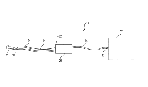
[0015] FIG. 1 is a diagram illustrating one exemplary temperature
monitoring system
in accordance with the present invention.
[0016] FIG. 2 is a diagram illustrating a modified design for the
temperature
monitoring system of FIG. 1.
[0017] FIG. 3 is a block diagram illustrating the components of an
exemplary optical
transmit/receive circuitry unit.
[0018] FIG. 4 is a block diagram illustrating the basic operation of
the temperature
monitoring system of FIG. 1.
[0019] FIG. 5 is a block diagram illustrating the basic operation of
an alternative
temperature monitoring system in accordance with the present invention.
-6-
CA 02754124 2013-12-16
74105-46
[0020] FIG. 6 is an exemplary embodiment of an implantable device
having the
temperature monitoring system of FIG. 1 embedded therein.
[0021] FIG. 7 is an exemplary embodiment of an ablation catheter
having the
temperature monitoring system of FIG. 5 embedded therein.
[0022] FIG. 8 is an exemplary embodiment of a biopsy needle device having
the
temperature monitoring system of FIG. 1 embedded therein.
-6a-
CA 02754124 2011-08-31
WO 2010/102117
PCT/US2010/026225
[0023] FIG. 9 is an exemplary embodiment of a stem cell delivery device
having the temperature monitoring system of FIG. 1 embedded therein.
[0024] FIG. 10 is an exemplary embodiment of an interventional device
delivery system having the temperature monitoring system of FIG. 1 embedded
therein.
[0025] FIG. 11 is an exemplary embodiment of another alternative
temperature monitoring system in accordance with the present invention.
[0026] FIG. 12 is a flow diagram illustrating exemplary steps in a
process for
determining bulk wavelength in accordance with one embodiment of the present
invention.
[0027] FIG. 13 is a graphical illustration showing an exemplary data set
consisting of transmit wavelengths and associated received light magnitudes.
[0028] FIG. 14 is a graphical illustration depicting an exemplary bulk
range
on the data set of FIG. 13.
[0029] FIG. 15 is a flow diagram illustrating exemplary steps in a
temperature calibration process in accordance with one embodiment of the
present
invention.
[0030] FIG. 16 is a graphical illustration depicting a calibration data
set
collected during a temperature calibration process.
[0031] FIG. 17 is an exemplary calibration data set in table fonn.
[0032] FIG. 18 is a flow diagram illustrating exemplary steps in a
temperature measuring process in accordance with one embodiment of the present
invention.
[0033] FIG. 19 is a graphical illustration depicting a step for
determining an
estimated temperature value using interpolation.
- 7 -
CA 02754124 2011-08-31
WO 2010/102117
PCT/US2010/026225
[0034] FIG. 20 is a graphical illustration depicting a step for
determining an
estimated temperature value using extrapolation.
DETAILED DESCRIPTION OF THE INVENTION
[0035] FIG. 1 is a diagram illustrating one exemplary temperature
monitoring
system 10 in accordance with the present invention. As illustrated in FIG. 1,
the
temperature monitoring system 10 generally includes an optical
transmit/receive
circuitry unit 12 and a fiber 14 operably coupled on a proximal end 16 to the
optical transmit/receive circuitry 12. The fiber 14 may preferably be fowled
from
glass or plastic, and further includes a fiber Bragg grating (FBG) element 18
adjacent a distal end 20. In the exemplary embodiment of FIG. 1, the
temperature
monitoring system 10 is shown as being used with a catheter 22. The catheter
22
includes a main body 24 structured to receive at least a portion of the fiber
14 and
a catheter handle 26 structured to be grasped and held by a surgeon or support
device. As will be appreciated by those skilled in the art, the catheter 22 is
represented generically herein and may be structured for use in numerous types
of
medical procedures to deliver therapy or provide a diagnosis.
[0036] The fiber 14 of the temperature monitoring system 10 may be
structured such that it is completely removable from the catheter 22 and may
be
reused in a different catheter or another type of medical device.
Alternatively, as
illustrated in FIG. 2, the fiber may comprise a first portion 14' that is
fixedly
coupled to the optical transmit/receive circuitry 12 and a second portion 14"
that
is fixedly coupled to the catheter 22. As will be appreciated by those skilled
in the
art, the first and second portions 14' and 14" may be fowled as separate fiber
segments. In this alternative fiber design, the catheter handle 26 may include
a
connector 28 that allows the first portion 14' of the fiber to be optically
coupled to
the second portion 14" to transmit light waves toward the FBG element 18.
Optionally, the optical transmit/receive circuitry 12 may include a connector
29
that allows the first portion 14' to be removably coupled thereto. As will be
appreciated by those skilled in the art, connectors 28 and 29 may comprise any
- 8 -
CA 02754124 2011-08-31
WO 2010/102117
PCT/US2010/026225
suitable connection means without departing from the intended scope of the
present invention.
[0037] The FBG element 18 positioned or embedded within the main body
24
of the catheter 22 allows a user such as a surgeon to monitor temperature
during a
medical procedure. As will be appreciated by those skilled in the art, one or
more
FBGs may be used to monitor temperature during therapy delivery. Alternatively
or additionally, one or more FBGs may be used to monitor medical device
heating
during scanning such that safe levels of heating may be maintained.
[0038] Generally speaking, an FBG is one type of distributed Bragg
reflector
that is constructed in a segment of optical fiber and is structured to reflect
predetermined wavelengths of light and to transmit all others therethrough.
This
selective reflection is accomplished by adding a periodic variation to the
refractive
index of the optical fiber core, thereby creating a wavelength specific
dielectric
mirror. Thus, FBGs act as "filters" to block or reflect certain wavelengths.
[0039] FBGs are typically formed in an optical fiber by either "writing" or
"inscribing" the periodic (or aperiodic) variation of refractive index into
the core
of the optical fiber using an ultraviolet source. The methods used to create
the
variations include "interference" and "masking." The interference method,
which
may be useful for uniform gratings, utilizes an ultraviolet laser that is
split into
two separate beams that interfere with one another to create a periodic
intensity
distribution along the interference pattern. The magnitude of the refractive
index
is dependent upon the intensity of the laser light used. The masking method,
which is well-suited for the manufacture of chirped FBGs, utilizes a photomask
placed between an ultraviolet light source directed at the fiber and creates a
grating structure based upon the intensity of the light that impinges upon the
fiber.
In another common method, an ultraviolet laser beam may be operated to "write"
the grating into the fiber point-by-point.
[0040] As appreciated by those skilled in the art, FBGs operate on a
principle
known as "Fresnel reflection," wherein light traveling between media of
different
- 9 -
CA 02754124 2011-08-31
WO 2010/102117
PCT/US2010/026225
refractive indices may be both reflected and refracted at the interface. The
grating
of the FBG element includes a varying sinusoidal refractive index over the
length
of the element. The wavelength reflected by the grating, which is known as the
Bragg wavelength, may be approximated as follows:
Bragg wavelength = 24A,
where 11 represents the average refractive index in the grating of the
fiber and A represents the grating period.
[0041] The refractive index and the grating period are determined by the
structure of the FBG element. Generally speaking, there are six known and
common structures for FBGs, including chirped, superstructure, Gaussian
apodized, discrete phase shift, uniform positive-only index change, and raised-
cosine apodized.
[0042] Because the Bragg wavelength is sensitive to temperature, FBGs
may
be used as sensing elements in optical fiber sensors. In a FBG element, the
measurand causes a shift in the Bragg wavelength. The relative shift in the
Bragg
wavelength due to an applied strain (e) and a change in temperature (AT), may
be
approximated as follows:
Relative shift in Bragg wavelength = Cse + CTAT,
wherein Cs is the coefficient of strain and CT is the coefficient of
temperature.
[0043] Based upon the foregoing relationship, FBGs may be used to
directly
sense the temperature and determine changes in temperature. Various other
methods of estimating temperature with FBGs are also possible. A more
detailed,
exemplary method for estimating temperature using FBGs will be described in
further detail to follow.
[0044] The fiber 14 of the temperature monitoring system 10 may be
either a
single-mode or multi-mode fiber optic cable. As appreciated by those skilled
in
the art, single-mode fiber optical cables are structured for carrying only a
single
- 10-
CA 02754124 2011-08-31
WO 2010/102117
PCT/US2010/026225
ray or mode of light, which may contain a variety of different wavelengths.
Single-mode cables have a small light carrying core, and are well-suited for
long
distance transmissions. Conversely, multi-mode fiber optic cables have a
relatively larger light carrying core, and are well-suited for short distance
transmissions.
[0045] Although only one FBG element 18 is illustrated in FIG. 1,
temperature monitoring systems having any number of FBG elements are within
the intended scope of the present invention. In one exemplary embodiment each
FBG element may have an axial length (along the axis of the fiber) between
about
2 mm and about 6 mm. However, the lengths of the FBGs may be greater than 6
mm or less than 2 mm depending upon the requirements and intended operation of
the system. For example, in one alternative embodiment the fiber may include a
plurality of FBGs each having a length less than 2 mm in order to optimize
spatial
selectivity.
[0046] The optical transmit/receive circuitry 12 is illustrated as being
external
to the catheter 14 of FIG. 1 merely for purposes of example and not
limitation. In
alternative embodiments, the optical transmit/receive circuitry 12 may instead
be
positioned within or embedded into the medical device in which the temperature
is
being monitored.
[0047] FIG. 3 is a block diagram illustrating the components of an
exemplary
optical transmit/receive circuitry 12. As illustrated in FIG. 3, the optical
transmit/receive circuitry 12 includes a light source 30, a tunable filter 32,
a scan
generator 34, a processor 36, and a detector 38. The light source 30 may be a
narrowband or broadband light source (i.e. white light). The tunable filter 32
and
the detector 38 are operable to detect wavelength of the received light (i.e.
tunable
wavelength filter).
[0048] In operation, the scan generator 34 may tune the light source 30
by
sweeping it across a predeteimined range so that the wavelength of light being
transmitted down the fiber 14 is known at all times. When the wavelength
emitted
-11-
CA 02754124 2011-08-31
WO 2010/102117
PCT/US2010/026225
by the light source 30 matches the specified Bragg wavelength of the FBG
element 18, light is reflected back along the fiber 14 towards the detector
38. The
scan generator 34 is operable to transmit a timing signal to the processor 36.
This
timing signal allows the processor to create a "spectrum" based upon the
"intensity" versus "time" information it has received. The processor may be
operable to identify various characteristics of the spectrum such as peak
positions,
which may then be used to estimate temperature.
[0049] FIG. 4 is a block diagram illustrating the basic operation of
the
temperature monitoring system 10 in accordance with the present invention. As
shown in FIG. 4, light waves 40 (either narrowband or broadband) are
transmitted
from the optical transmit/receive circuitry 12 towards the FBG element 18. The
FBG element 18 reflects a predetermined narrow or broad range of wavelengths
of
light 42 incident on the grating while passing all other wavelengths of light
44.
The reflected wavelengths 42 are redirected back towards the optical
transmit/receive circuitry 12 where they are detected by the tunable filter 32
and
the detector 38 as previously described above with regard to the system block
diagram of FIG. 3.
[0050] FIG. 5 is a block diagram illustrating the basic operation of an
alternative temperature monitoring system 10A in accordance with the present
invention. The temperature monitoring system 10A is similar to the temperature
monitoring system 10 previously described, but further includes a second FBG
element 45 positioned along the fiber 14. Because wavelengths other than the
Bragg wavelength are passed with little or no attenuation, multiple FBGs may
be
used on a single fiber. As shown in FIG. 5, light waves 40 (either narrowband
or
broadband) are transmitted from the optical transmit/receive circuitry 12
towards
the FBG element 18. The FBG element 18 reflects a predetermined narrow or
broad range of wavelengths of light 42 incident on the grating while passing
all
other wavelengths of light 44. The reflected wavelengths 42 are redirected
back
towards the optical transmit/receive circuitry 12 where they are detected by
the
tunable filter 32 and the detector 38 as previously described above with
regard to
the system block diagram of FIG. 3. The wavelengths of light 44 that are
allowed
- 12-
CA 02754124 2011-08-31
WO 2010/102117
PCT/US2010/026225
to pass through the FBG element 18 are directed towards the second FBG element
45, where a second predetelinined narrow or broad range of wavelengths of
light
46 are reflected back towards the optical/transmit receive circuitry 12 where
they
are also detected by the tunable filter 32 and the detector 38. All other
wavelengths of light 48 are passed through the second FBG element 45 towards
the distal end 20 of the fiber 14. As will be appreciated by those skilled in
the art,
the FBG element 18 and the second FBG element 45 must have their own
wavelength segments to ensure that various signals do not overlap and the
temperature monitoring system operates properly.
[0051] FIG. 6 is an exemplary embodiment of an implantable device 50 such
as a defibrillator having the temperature monitoring system 10 of FIG. 1
embedded therein. As illustrated in FIG. 6, the implantable device 50 may be
inserted under the skin of a patient P adjacent the heart 51, and may
generally
include a main housing 52 along with one or more elongate electrodes 53
insertable through a vein 54 and sized to extend into the right atrium 55 and
the
right ventricle 56. As will be appreciated by those skilled in the art, the
size and
structure of the implantable device 50 may vary without departing from the
intended scope of the present invention.
[0052] As further illustrated in FIG. 6, both the fiber 14 and the
optical
transmit/receive circuitry 12 are positioned or embedded within the housing 52
such that the temperature monitoring system 10 is completely contained within
the
implantable device 50. In operation, the temperature monitoring system 10 is
operable to sense temperature adjacent to the implantation position of the
housing
52. Although a single FBG element that produces a corresponding single
temperature sensing location is shown, those skilled in the art will
appreciate that
any number of FBG elements may be used to achieve any desired number of
temperature sensing locations within the housing 52 or along the axial length
of
the electrodes 53 without departing from the intended scope of the present
invention.
- 13 -
CA 02754124 2011-08-31
WO 2010/102117
PCT/US2010/026225
[0053] FIG. 7 is an exemplary embodiment of an ablation catheter 60
having
the temperature monitoring system 10A of FIG. 5 embedded therein. As
illustrated in FIG. 7, the ablation catheter 60 includes a generally tubular
main
body 62, an ablation tip 64, and a lumen 66 extending along the axial length
of the
main body 62 towards the ablation tip 64. As will be appreciated by those
skilled
in the art, the ablation catheter 60 may be structured to deliver any suitable
ablative therapy to the ablation tip 64 through the lumen 66 including, but
not
limited to, radiofrequency energy, laser energy, microwave energy, highly
focused
ultrasound energy, cryogenic fluid and the like.
[0054] As further illustrated in FIG. 7, the embedded fiber 14 of the
temperature monitoring system 10A may be operable to sense temperature at a
first sensing location 68A adjacent to the FBG element 18 and at a second
sensing
location 68B adjacent to the FBG element 45. Although the ablation catheter 60
is shown as including two temperature sensing locations 68A and 68B, any
number of temperature sensing locations may be created by simply varying the
number of FBG elements in the fiber. Additionally, the axial positions of the
temperature sensing locations may be altered by modifying the spacing between
the FBG elements.
[0055] FIG. 8 is an exemplary embodiment of a biopsy needle device 70
having the temperature monitoring system 10 of FIG. 1 embedded therein. As
illustrated in FIG. 8, the biopsy needle device 70 includes a generally
tubular main
body 72, an open distal tip 74, and a lumen 76 extending along the axial
length of
the main body 72. As will be appreciated by those skilled in the art, the size
and
structure of the biopsy needle device 70 may vary without departing from the
intended scope of the present invention.
[0056] As further illustrated in FIG. 8, the embedded fiber 14 of the
temperature monitoring system 10 may be operable to sense temperature at a
single sensing location 78 adjacent to the FBG element 18. However, as will be
appreciated by those skilled in the art, any number of FBG elements may be
used
to achieve any desired number of temperature sensing locations along the axial
- 14-
CA 02754124 2011-08-31
WO 2010/102117
PCT/US2010/026225
length of the biopsy needle device 70. Additionally, although the FBG element
18
of the fiber 14 is positioned such that it produces a temperature sensing
location
78 adjacent to the distal end of the main body 72, the temperature sensing
location
may be modified by placing the FBG element at another axial location.
[0057] FIG. 9 is an exemplary embodiment of a stem cell delivery device 80
having the temperature monitoring system 10 of FIG. 1 embedded therein. As
illustrated in FIG. 8, the stem cell delivery device 80 includes a catheter 82
and a
stem cell delivery needle 84. The catheter 82 includes a generally tubular
main
body 85 with an open distal end 86 structured to allow the stem cell delivery
needle 84 to pass therethrough. The stem cell delivery needle 84 includes an
elongate main body 88 have a lumen 90 therein. The fiber 14 with the FBG
element 18 is embedded within the lumen 90 of the main body 88 of the stem
cell
delivery needle 84. The main body 88 may include an aperture at a distal end
that
is structured and sized for passing cell structures therethrough. As will be
appreciated by those skilled in the art, the size and structure of the stem
cell
delivery device 80 may vary without departing from the intended scope of the
present invention.
[0058] As further illustrated in FIG. 9, the embedded fiber 14 of the
temperature monitoring system 10 may be operable to sense temperature at a
single sensing location 92 adjacent to the FBG element 18. As will be
appreciated
by those skilled in the art, a surgeon may move the stem cell delivery needle
84
relative to the open distal end 86 of the catheter 84 in order to position the
temperature sensing location at the desired point (or as close as possible to
the
desired point) where the surgeon wants to obtain a temperature reading.
Similar
to the medical devices previously described above, any number of FBG elements
may be used to achieve any desired number of temperature sensing locations
along the axial length of the stem cell delivery device 80. Additionally,
although
the FBG element 18 of the fiber 14 is positioned such that it produces a
temperature sensing location 92 adjacent to the distal end of the main body 88
of
the stem cell delivery needle 84, the temperature sensing location may be
modified by placing the FBG element at another axial location.
- 15 -
CA 02754124 2011-08-31
WO 2010/102117
PCT/US2010/026225
[0059] FIG. 10 is an exemplary embodiment of an interventional device
delivery system 100 having the temperature monitoring system 10 of FIG. 1
embedded therein. As illustrated in FIG. 10, the delivery system 100 includes
a
generally tubular catheter body 102, an expansion means 104 such as a balloon
adjacent a distal end, and a lumen 106 extending along the axial length of the
catheter body 102. In one exemplary embodiment, the lumen 106 may be
structured for passage of an inflation means such as air or saline for
inflation and
deflation of the expansion means 104. The delivery system 100 may be
structured
for delivery of any suitable interventional device such as an expandable stent
or
the like.
[0060] As further illustrated in FIG. 10, the embedded fiber 14 of the
temperature monitoring system 10 may be operable to sense temperature at a
single sensing location 108 adjacent to the FBG element 18. However, as will
be
appreciated by those skilled in the art, any number of FBG elements may be
used
to achieve any desired number of temperature sensing locations along the axial
length of the interventional device delivery system 100. Additionally,
although
the FBG element 18 of the fiber 14 is positioned such that it produces a
temperature sensing location 108 adjacent to the distal end of the catheter
body
102, the temperature sensing location may be modified by placing the FBG
element at another axial location.
[0061] FIG. 11 is an exemplary embodiment of another alternative
temperature monitoring system 10B in accordance with the present invention. As
illustrated in FIG. 11, the temperature monitoring system 10B is similar to
the
temperature monitoring system 10A, but further includes a third FBG element
110
and a fourth FBG element 112. As will be appreciated by those skilled in the
art
based on the foregoing discussion, having multiple FBG elements positioned
along an axial length of the fiber 14 between the proximal end 16 and the
distal
end 20 allows for multiple point temperature measurements along a pathway in a
medical device. This type of "pathway" temperature monitoring may be useful in
any medical device where it may be important to monitor temperature at more
than one location, including but not limited to the devices previously
described.
- 16 -
CA 02754124 2011-08-31
WO 2010/102117
PCT/US2010/026225
As illustrated in FIG. 11, the spacing S between the various FBG elements may
be
equal or alternatively may vary by any desired amount. Thus, the temperature
monitoring device 10B may be customized for particular applications and uses.
[0062] Although the various embodiments of medical devices were
described
above as including a single fiber element, temperature monitoring systems
utilizing multiple fiber elements each having one or more FBG elements therein
are also possible. Thus, a single medical device such as an ablation catheter
may
be structured with two or more fibers positioned or embedded therein. This
type
of design may be used for measuring the temperature of one or more therapy
delivery points or one or more locations for safety monitoring during therapy
delivery or delivery of a medical device using MRI guidance. Further, although
the fiber and FBG elements of the temperature monitoring systems have been
generally described as embedded or removably positioned within the medical
devices, they may alternatively be fixedly or removably coupled to an outer
surface of the device without departing from the intended scope of the present
invention.
[0063] As will be appreciated by those skilled in the art, the optical
fiber may
be positioned or embedded within a device, positioned on an outer surface of a
device, or any combination thereof without departing from the intended scope
of
the present invention. For example, in one exemplary embodiment the fiber may
be partially exposed to the exterior of the device. In another exemplary
embodiment the device may include a fiber with at least one portion completely
positioned/embedded within the device and at least one additional portion
positioned on the exterior of the device. Thus, numerous alternative designs
are
contemplated and within the intended scope of the present invention.
[0064] As a Bragg diffraction grating system does not posses the
technical
shortcomings of other temperature measuring techniques inside an MRI system,
another alternative embodiment of the present invention may include external
in
=
vitro or in vivo temperature measurement of a medical device. In this
embodiment, a fiber optic cable having one or more FBG elements is placed
- 17 -
CA 02754124 2011-08-31
WO 2010/102117
PCT/US2010/026225
external to the medical device. As will be appreciated by those skilled in the
art,
this embodiment may be useful in determining the safety of a medical device in
MRI with regard to joule heating at tissue/electrode interfaces, dielectric
heating
along the length of a metallic structure, gradient induced heating and the
like.
[0065] Although the temperature monitoring system of the present invention
has been described with reference to a discrete number of medical devices,
those
skilled in the art will appreciate that the temperature monitoring system may
be
incorporated into any medical device that is used in an MRI environment. Thus,
the embodiments set forth herein have been described merely for purposes of
example and not limitation.
[0066] Now that several exemplary embodiments of the temperature
monitoring system have been described with reference to various medical
devices,
one exemplary method of operating the temperature monitoring systems to
deteimine temperature measurements will be described in detail. The exemplary
method of the present invention may generally be separated into three
processes,
including detelmining bulk wavelength 200, calibrating temperature 300, and
measuring temperature 400. Each of these processes will now be described with
reference to FIGS. 12-19.
[0067] FIG. 12 is a flow diagram illustrating exemplary steps in the
process
of determining bulk wavelength 200 in accordance with one embodiment of the
present invention. Beginning with step 202, three wavelength values are
predefined. These wavelength values include the minimum wavelength, Amin, the
maximum wavelength, Amax, and the wavelength step, Astep. Then, at step 204
the transmit wavelength, Atx, is set to the minimum wavelength, Amin.
[0068] Starting at the minimum wavelength, the optical transmit/receive
unit
transmits narrowband (or broadband) light into the proximal end of a fiber
containing one or more FBG elements at step 206. The light reflected off of
the
one or more FBG elements is received and measured by a photo detector in the
-18-
CA 02754124 2011-08-31
WO 2010/102117
PCT/US2010/026225
optical transmit/receive unit at step 208, and the magnitude and transmit
wavelength, ktx, are recorded into memory at step 210.
[0069] A processor then deteiiiiines whether the transmit wavelength,
ktx, is
greater than or equal to the maximum wavelength, ?max, at step 212. If the
transmit wavelength, ktx, is determined to be less than the maximum
wavelength,
?max, the transmit wavelength, ktx, is incremented by the wavelength step,
kstep,
at step 214 and the process 200 enters a loop 216 where steps 206-212 are
repeated for transmit wavelengths from ?min to Amax at incremental steps of
kstep. Once the processor deterniines that the transmit wavelength, ktx, is
greater
than or equal to the maximum wavelength, kmax, at step 212, this portion of
the
process is complete and a data set now exists consisting of transmit
wavelengths
and associated received light magnitudes. An exemplary data set is represented
by the graph in FIG. 13.
[0070] Although one exemplary method of forming the data set
represented
by the graph in FIG. 13 has been described in detail, those skilled in the art
will
appreciate that any suitable method may be used without departing from the
intended scope of the present invention. One alternative method is to transmit
a
broad spectrum of light down the fiber and measure return light intensity
variations as the path difference in an interferometer is varied. Another
alternative method is to transmit a broad spectrum of light down the fiber and
utilize a second fiber Bragg grating element with a known pass/reject ratio
through which the returned light is passed, wherein the intensities of the
light
transmitted through the second fiber Bragg grating element and the light
rejected
by the second fiber Bragg grating element may be compared to determine the
wavelength of the returning light. Yet another alternative method is to
transmit a
broad spectrum of light down the fiber and split the returned light into
several
beams which may be fed into many narrowband detectors, each narrowband
detector being designed to detect light at a specific wavelength. Yet another
alternative method is to transmit multiple narrowband light signals down the
fiber,
each signal being centered at a different wavelength and each uniquely
modulated
or coded such that the returned signal can be demodulated or decoded to
- 19 -
CA 02754124 2011-08-31
WO 2010/102117
PCT/US2010/026225
determine the corresponding intensity vs. wavelength characteristics. As will
be
appreciated by those skilled in the art, the foregoing alternative methods are
presented merely for purposes of example and not limitation.
[0071] For purposes of discussion and not limitation, the bulk
wavelength
may be defined as a single wavelength value that represents the center
wavelength
of the received light. To find the bulk wavelength, the wavelength at which
the
magnitude is maximum is first identified at step 218. This step is depicted
graphically in FIG. 14. Next, in step 220, the range of contiguous wavelengths
(which includes the wavelength at which the magnitude is maximum) for which
the corresponding magnitude is greater than the maximum magnitude minus some
threshold is determined. This range may be referred to as the "bulk range." In
one exemplary embodiment the preferred threshold may be about 3dB, although
any suitable threshold may be used as will be appreciated by those skilled in
the
art. The bulk wavelength is then calculated in step 222 as the center of mass
of
the magnitudes within the defined bulk range. This step is also depicted
graphically in FIG. 14. As illustrated in the exemplary graph of FIG. 14, the
bulk
range does not include the small magnitude peak to the left of the main peak.
Once the bulk wavelength is calculated at step 222, the process 200 may
terminate
at step 224.
[0072] As will be appreciated by those skilled in the art, bulk wavelength
may be calculated using numerous alternative methods without departing from
the
intended scope of the present invention. For example, bulk wavelength may be
determined using peak detection (i.e. finding the absolute peak magnitude
value),
filtered peak detection (i.e. filtering the wavelength magnitudes followed by
finding the absolute peak magnitude value), filtered center of mass (i.e.
filtering
the wavelength magnitudes followed by finding the center of mass of the
magnitudes), or the like. Thus, the bulk wavelength process 200 is one of many
processes that may be used, and was discussed herein for purposes of example
and
not limitation.
- 20 -
CA 02754124 2011-08-31
WO 2010/102117
PCT/US2010/026225
[0073] Turning next to FIG. 15, a flow diagram is presented
illustrating
exemplary steps in the process of calibrating temperature 300 in accordance
with
one embodiment of the present invention. Beginning with step 302, a plurality
of
known calibration temperature values are selected that will be used to perform
the
calibration procedure. Then, in step 304, a bulk wavelength, kbulk, is
determined
for each of the selected calibration temperature values. The result of the
bulk
wavelength determination step is depicted graphically in FIG. 16. These bulk
wavelengths may be determined using the bulk wavelength process 200
previously described, or any other known and suitable process for determining
bulk wavelength. Once a bulk wavelength is determined for each of the selected
calibration temperature values in step 304, a calibration data set is
formulated and
stored in memory in step 306. In one exemplary embodiment as depicted in the
table set forth in FIG. 17, the calibration data set may be stored as a
plurality of
calibration wavelengths, kcal, and a corresponding plurality of calibration
temperatures, Teal. Once the calibration data set is stored in memory, the
calibration process may terminate at step 308. The stored calibration data set
may
then be used in the temperature monitoring process 400 to determine the
temperature at one or more temperature sensing locations.
[0074] Turning next to FIG. 18, a flow diagram is presented
illustrating
exemplary steps in the process of measuring temperature 400 at one or more
temperature sensing locations in accordance with one embodiment of the present
invention. Beginning with step 402, the current bulk wavelength, ?bulk, is
determined using any suitable bulk wavelength determination process, such as
the
bulk wavelength process 200 previously discussed. Next, the calibration data
set
is accessed from memory in step 404. Using the calibration data set, an
interpolation is performed between the appropriate Teal and kcal points at
step
406 to estimate the current temperature based on the current bulk wavelength.
This interpolation process is depicted graphically in FIG. 19. The
interpolation
step may use linear interpolation or any suitable higher order interpolation,
such
as polynomial interpolation. If the current bulk wavelength, kbulk, falls
outside of
the range of Tcal and kcal points in the calibration data set, step 406 may
- 21 -
CA 02754124 2011-08-31
WO 2010/102117
PCT/US2010/026225
alternatively utilize extrapolation to estimate the current temperature. This
extrapolation process is depicted graphically in FIG. 20. As will be
appreciated
by those skilled in the art, the extrapolation step may use linear
extrapolation or
any suitable higher order extrapolation, such as polynomial extrapolation.
Once
the current temperature is determined in step 406, the temperature monitoring
process may tenuinate at step 408. As will be appreciated by those skilled in
the
art, the temperature monitoring process 400 may be repeated at any desired
time
interval in order to continuously or periodically monitor, with or without
temporal
interpolation or extrapolation, temperature of a device.
[0075] Although several exemplary steps were described with reference to
the bulk wave deteimination, temperature calibration, and temperature
measurement processes, those skilled in the art will appreciate that the order
and
number of steps may be modified without departing from the intended scope of
the present invention. Thus, the exemplary steps were provided merely for
purposes of example and not limitation.
[0076] As will further be appreciated by those skilled in the art, the
processes
previously described may be embodied as a system, method or computer program
product. Accordingly, the present invention may take the form of an entirely
hardware embodiment, an entirely software embodiment (including firmware,
resident software, micro-code, etc.) or an embodiment combining software and
hardware aspects that may all generally be referred to as a "circuit,"
"module" or
"system." Furthermore, the present invention may take the form of a computer
program product embodied in any tangible medium of expression having
computer usable program code embodied in the medium.
[0077] The processes comprising the method of the present invention have
been described with reference to flow diagrams illustrating exemplary steps.
It
will be understood that each block of the flowchart diagrams, and combinations
of
blocks in the flowchart diagrams, can be implemented by computer program
instructions. These computer program instructions may be provided to a
processor of a general purpose computer, special purpose computer, or other
- 22 -
CA 02754124 2013-12-16
74105-46
programmable data processing apparatus to produce a machine, such that the
instructions, which execute via the processor of the computer or other
programmable data processing apparatus, create means for implementing the
= functions/acts specified in the flowchart diagram block or blocks.
[0078] These computer program instructions may also be stored in a
computer-readable medium that can direct a computer or other programmable data
processing apparatus to function in a particular manner, such that the
instructions
stored in the computer-readable medium produce an article of manufacture
including instruction means which implement the function/act specified in the
flowchart block or blocks.
[0079] The computer program instructions may also be loaded onto
computer or other programmable data processing apparatus to cause a series of
operational steps to be performed on the computer or other programmable
apparatus to produce a computer implemented process such that the instructions
which execute on the computer or other programmable apparatus provide
processes for implementing the functions/acts specified in the flowchart
diagram
block or blocks.
[0080] Although the present invention has been described with
reftience to
preferred embodiments, workers skilled in the art will recognize that changes
may
be made in form and detail without departing from the scope of the
invention.
-23-