Note: Descriptions are shown in the official language in which they were submitted.
CA 02754576 2016-08-15
1
EYE SAMPLING DEVICE
BACKGROUND
The invention relates to a device with which conjunctival
impressions may be taken.
The conjunctival impression technique was developed at the
end of the 1970s and is now an accepted technique allowing
conjunctival epithelial cells to be collected from the
conjunctival mucosa which is an immunologically reactive and
complex tissue, in a quasi-painless and non-invasive way, for
purposes of analyses and diagnose. The most superficial cells
of the conjunctival epithelium, i.e. the cells undergoing
desquamation, are collected by this technique. Conjunctival
epithelium renews itself from the basal layers, and the most
superficial cells which have reached their final
differentiation are regularly discharged into the lachrymal
fluid. It is this surface layer comprising the terminally
differentiated conjunctival cells, which may be sampled by
the conjunctival impression for subsequent analysis.
There are three types of cell populations which may be found
on conjunctival impressions:
- epithelial covering cells which apart from their
participation in coating the conjunctival mucosa, are
involved in many biological reactions, particularly in
inflammatory and apoptotic processes;
- goblet cells (mucus-secreting cells) which release
soluble mucins in the tear film, and which play major
defense and regulatory roles at in the ocular surface ;
and,
- inflammatory cells including dendritic cells known for
their property of immunocompetence, Langerhans cells and
also lymphocyte populations localized in the epithelium.
With conjunctival impressions, various cell populations,
their size, number, density and pathological modifications
CA 02754576 2016-08-15
-
,
2
may be analyzed and important information may be provided on
the status of the eye surface. They also provide information
on the conjunctival attack, notably in the field of the dry
eye, iatrogenic pathologies induced by collyria, pathologies
of the limbus and chronic inflammation of the eye surface.
In order to take a conjunctival impression, the most current
present method consists in using membrane filters commonly
used in biochemical industry, the most often in cellulose
ester (a mixture of cellulose ester and cellulose nitrate) or
in polyethersulfone, for example, and which are applied on
the conjunctiva, generally without any local anesthesia. The
sampling is totally bloodless, painless and non-invasive
contrary to other more aggressive systems such as brushing or
biopsy. During the sampling, the filter is generally applied
on the upper bulbar conjunctiva of the eye, protected by the
eyelid and removed after a contact of 3-5 seconds. The
desquamating conjunctival cells then adhere to the membrane
surface and form a thin and homogenous cell layer. The
filters are then treated in order to recover the cells to be
analyzed.
Within the scope of scientific evaluations or when the
examinations will be repeated, the impression should always
be taken at the same location because there are disparities
in the distribution of the sampled cells on the bulbar
conjunctiva. This is neither obvious nor easy with the
present sampling methods which remain relatively manual even
today, in spite of various attempts to improve the procedure
(different materials, porosities, holding devices.. .etc.)
They are relatively cumbersome and time-consuming for a
clinician or a pathologist within the scope of routine daily
use.
CA 02754576 2016-08-15
3
SUMMARY
An object of the invention is to provide a device for taking
an eye impression which is simple to implement while
supplying reliable and reproducible sampled impressions.
For this purpose, provision is made according to the
invention for, an eye sampling device comprising supporting
means comprising a sampling surface, the supporting means
being elastically deformable between a rest position and a
sampling position and conformed so that, upon being used for
sampling, the sampling surface applied in the sampling
position in a coinciding way onto a sampling area
(2;402;502), is detached therefrom according to a peeling
movement when passing from the sampling position to a rest
position.
Thus, by the use of elastically deformable supporting means
bearing the sampling surface and conformed in order to allow
the latter to be detached from the eye sampling area
according to a peeling movement, eye impressions which are of
optimum quality, may be obtained reliably and reproducibly.
Further, this allows a sampling device to be produced, which
is simple to use.
There is provided, an eye sampling device for taking an eye
impression comprising: a body with an end intended to come
into contact with an eye during sampling, wherein said
contact by said end is limited to a sampling area located on
the conjunctiva of the eye being sampled, a piston including
an upper end and an opposite end, said piston is slidably
mounted in the body between a rest position and a sampling
position, a spring configured to exert a return force on the
piston to force it to the rest position, a supporting element
at the opposite end of the piston to support a detachable
sampling surface, said sampling surface including a sampling
support for taking said eye impressions, said sampling
support formed with a material forming a support for taking
CA 02754576 2016-08-15
3a
said eye impressions and configured to be pressed against an
eye when the upper end of the piston is pressed towards the
end of the body and the piston is in the sampling position,
said sampling surface also including attachment edges to
cooperate with the supporting element and attach the sampling
surface to the sampling element, said attachment edges of the
sampling surface configured to detach from the supporting
element when the piston moves further than the rest position
after a sampling has been completed, wherein said sampling
support is further configured to deform in use so as to
become coincident in all points between said sampling support
and said sampling area and to collect an eye impression upon
becoming coincident with said sampling area.
There is provided, a method for taking an eye impression with
a device including a body with an end, a piston comprising an
upper end and an opposite end, said piston slidably mounted
in the body between a rest position and a sampling position,
said device comprising a spring configured to exert a return
force on the piston to force it to the rest position, said
piston comprising a supporting element at the opposite end of
the piston to support a detachable sampling surface, said
method comprising: placing the end of the body onto an eye;
applying pressure to the upper end of the piston to move the
piston from a rest position to a sampling position, wherein
the sampling surface is pressed against the eye and deforms
so as to become coincident with a sampling area of the eye,
said sampling area located on the conjunctiva of the eye, and
collects an eye impression upon becoming coincident in all
points between said sampling surface and said sampling area;
releasing the pressure on the upper end of the piston to move
the sampling surface away from the eye according to a peeling
movement.
Advantageously, but optionally, the device according to the
invention has at least one of the following features:
CA 02754576 2016-08-15
3b
- the sampling surface, in the rest position, includes at
least one high point and at least one low point, both
distinct so that during the peeling movement, a
detachment line gradually passes from the high point to
the low point;
- the supporting means are conformed so that, upon being
used for sampling, the sampling surface is applied onto
the sampling area, according to a movement opposite to
the peeling movement when passing from a rest position
to a sampling position;
- the supporting means include a block of elastomeric
material or of foam;
- the supporting means include a flexible blade;
- the supporting means include a series of spring blades;
- the sampling surface is substantially planar;
- the sampling surface has a section of generally cross-
sectional convex shape;
CA 02754576 2011-09-06
WO 2010/100258 PCT/EP2010/052832
4
- the sampling surface includes a sampling support and
attachment means on the supporting means;
- the attachment means comprise an edge including an
extending protrusion which juts out, intended to
cooperate with a notch of supporting means of
substantially complementary shape;
- the device further includes a body and a piston forming
component intended to be slidably moved in the body
against elastic return means, the supporting means being
attached on one end of the piston forming component;
- the body includes means for separating the sampling
surface from the supporting means; and,
- the separation means include shoulders (33,34) intended
to bear upon the attachment means (13,14) in order to
disengage them.
Other features and advantages of the invention will emerge
from the description hereinafter of a preferred embodiment
and also of variants. In the appended drawings:
- Figs. la and lb are schematic views of the sampling
kinematics achieved by a device according to the
invention;
- Figs. 2a and 2b are schematic views of alternative
embodiments of the deformable portion of the device
according to the invention;
- Figs. 3a-3c are schematic views of alternative
embodiments of the sampling surface of the device
according to the invention;
- Fig. 4 is a half-sectional view of an embodiment of
the device according to the invention;
- Fig. 5 is a simplified cross-sectional view of the
kinematics for recovering the eye impression taken by
the device of Fig. 4;
- Fig. 6 is a perspective schematic view illustrating
the shape of the impression taken by the device of
Fig. 4;
CA 02754576 2011-09-06
WO 2010/100258 PCT/EP2010/052832
- Figs. 7a and 7b are three-dimensional schematic views
of alternative shapes of impressions taken by a
device according to the invention; and,
- Fig. 8 is an anatomical view of an eye intended to
5 receive the device according to the invention.
As a liminary remark, the sampling area 2 of an eye
impression is located on the conjunctiva, an area located at
the periphery of the cornea, which extends from the limbus
which delimits the junction between the cornea (nominal
diameter of the cornea: about 12 mm for an adult) and the
sclera, and extends outwardly to an area externally delimited
by a circle with a diameter of about 22 mm and centered on
the cornea. For example, as illustrated in Fig. 6, the
sampling area 2 (and therefore the sampling surface 11) is
ring-shaped with an inner diameter of about 12 mm and an
outer diameter of about 17 mm. For a child, whose eye has not
reached the adult size, these dimensions have to be adapted
in proportion.
The area 2 for taking an eye impression is moreover located
on the eyeball which may be assimilated to a sphere with a
diameter of 24 mm +/- 1 mm, except at the cornea. The eye's
anatomy is recalled and illustrated in Fig. 8.
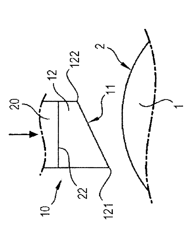
With reference to Figs. la and lb, we shall describe the
sampling kinematics of a device 10 for taking an eye
impression according to the invention.
This kinematics is important in order to ensure reliability
and reproducibility as well as quality of the eye impressions
taken by means of the device 10 according to the invention in
order to maximize the quality of the sampled cells while
maintaining their integrity.
As a matter of principle, the device 10 according to the
invention includes a piston forming component 30 having an
end 22 on which supporting means 12 are attached. These
supporting means 12 are elastically deformable between a rest
CA 02754576 2011-09-06
WO 2010/100258 PCT/EP2010/052832
6
position illustrated in Fig. la and a sampling position
illustrated in Fig. lb. On the other hand, these supporting
means include a sampling face or surface 11 which is intended
to face and to be pressed against a sampling area 2 of an eye
1. This sampling surface is made in one or more suitable
materials forming a support for taking eye impressions and
moreover known to one skilled in the art. Finally, the
sampling surface, when the supporting means 12 are in the
rest position, has at least one low point 121 (or low contact
line) and at least one high point 122 (or high contact line),
along a longitudinal axis of the device depending on the
direction in which a sampling movement is performed.
This sampling movement is obtained by presenting the sampling
surface 11 in a non-parallel or non-coinciding way towards
the sampling area 2 of the eye surface. Thus, the sampling
surface 11, defined by a planar geometry here, is presented
so as to face the sampling area 2 of the eye surface
according to a given orientation, so that, at the time of
contact between both surfaces, there is no coincidence of
these surfaces. Indeed, contact between the sampling surface
11 and the sampling area 2 of the eye 1 is initiated by the
contact of the low point (or low line) 121 on said sampling
area and proceed by moving this contact point (or line) along
the sampling surface 11, by sweeping through the latter,
right up to the high point (or high line) 122 which lastly
comes into contact with the sampling area 2, and this under
the effect of a movement along the longitudinal axis of the
device 10 according to the invention as illustrated by the
arrow of Fig. la. As the contact of the sampling surface 11
gradually advances on the sampling area 2 of the eye surface,
the sampling surface 11 gradually deforms so as to become
coincident with the sampling area 2 of the eye surface. This
deformation also affects the supporting means 12 which deform
elastically, until the sampling surface 11 is stuck on,
i.e. intimately contacting, the sampling area 2 of the eye 1
as this is illustrated in Fig. lb. There is then coincidence
CA 02754576 2016-08-15
7
in all points between the sampling surface 11 and the
sampling area 2.
The sampling support, once it is placed on the eye 1, is then
detached from the eye 1 in the least traumatic way, by
exerting a peeling movement. Indeed, with a stress mode of
the peeling type, concentrated stresses may exist at the
location of the detachment which, here, is a point on the
schematic view of Figs. la and lb (or a line passing through
this point) and which, during detachment, passes from the
high point (or high line) 122 to the low point (or low line)
121, while the sampling surface 11 under the effect of the
supporting means 12 which gradually resume their initial
resting shape under the effect of the elastic forces, initial
shape which does not coincide with the sampling area 2 of the
eye 1.
The supporting means 12 are made in a block of elastically
deformable material such as a foam or a suitable elastomer
(elastic polymer): polyurethane, polyethylene, silicone foam,
a thermoplastic elastomer such as Styrene-ethylene/butylene-
styrene (SEBS), Styrene-ethylene/propylene-styrene (SEPS)
ethylene-propylene copolymer/polypropylene (EPDM-
PP),
polyether block amides (PEBAX), polyurethane or further a
vulcanizable elastomers such as silicone rubber, latex,
polybutadiene, a fluorinated elastomer, polychloroprene,
polyisoprene...etc.
Two alternative embodiments of the supporting means are
illustrated in Figs. 2a and 2b. In Fig. 2a, the supporting
means are produced as a flexible blade 112 mounted on the
component 20 forming a piston via a supporting rod 112a. In
Fig. 2b, the supporting means are produced as a plate
supported by spring blades 212, for example made in plastic,
in metal or any other material having similar elastic
properties.
CA 02754576 2011-09-06
WO 2010/100258 PCT/EP2010/052832
8
Likewise, with reference to Figs. 3a-3c, the sampling surface
alternatively has different convex sectional geometries:
- as a quarter of a circle or an ellipse (Fig. 3a)
- as a half-circle or half-ellipse (Fig. 3b)
- as a substantially triangular or conical tip
(Fig. 3c)
which respectively corresponds to volume portions of spheres
or ellipsoids.
For these different geometries, the sampling surface
111,211,311 always has a low point and at least one high
point in order to be able to allow the sampling movement
according to the kinematics described earlier, with reference
to Figs. la and lb. Thus, regardless of the geometry, the
contact between the sampling surface 11,111,211,311 and the
sampling area 2 of the eye surface is gradually made and as a
dynamic contact point or/and then line gradually sweeping
through the sampling area right up to intimate contact
between both surfaces, the sampling surface being deformed as
well as the supporting means.
Upon removing or detaching the sampling surface, the
progressive movement of this same dynamic contact line is the
inverse of the movement performed during the contacting.
Thus, the detachment movement is analogous to a peeling
movement with which the cells of the sampling area 2 may be
gradually detached from the eye surface, which have then
adhered onto the sampling support of the sampling surface.
This adhesion on the sampling support is achieved according
to different well-known principles: by hydrophilicity, by
standard surface tension, by adhesion of the cell mucus, or
anionic electrostatic charges of the cells.
With reference to Fig. 4, we shall describe an embodiment of
a device 10 for taking eye impressions according to the
invention which applies the principles and characteristics
described earlier. The device 10 here is ring-shaped with a
CA 02754576 2016-08-15
9
longitudinal axis X-X. It includes a piston 20. The piston 20
includes an upper end 23 comprising an interface for applying
said piston 20. It includes an opposite end 22 substantially
perpendicular to the longitudinal axis X-X on which the
supporting means 12 are positioned and attached, here as a
sectional shape of a elastomeric trapezoidal block (or of a
block formed with a truncated cylinder, an upper end of which
is perpendicular to the axis, in contact on the end 22 of the
piston 20 and an opposite lower face tilted with respect to
said axis). These supporting means 12 as seen earlier, are
elastically deformable between a rest position and a sampling
position. An end of the supporting means 12, tilted with
respect to the longitudinal axis, bears the sampling surface
11.
This sampling surface 11 includes the sampling support
intended to collect an eye impression upon sampling on a
sampling area 2 of the eye 1. The sampling support includes
at the periphery, around the sampling surface 11, a flexible
edge 13, 14 (added by insert-molding, adhesive bonding,
welding...etc.), this edge having a protrusion 13a, 14a
directed radially towards the supporting means 12, which will
fit onto a notch 15, 16 located on the perimeter of the
supporting means 12. Once it is in place, the edge 13, 14
extends and protrudes from the supporting means 12.
In the case illustrated in Fig. 4, as the device 10 according
to the invention is ring-shaped, the sampling support is
itself also ring-shaped and includes a first edge 13 on a
radially inner periphery and a second edge 14 on a radially
outer periphery. Also, the supporting means 12 are ring-
shaped and include a first notch 15 on a radially inner face
into which the protrusion 13a of the first edge 13 will fit
and a second notch 16 on a radially outer face into which the
protrusion 14a of the second edge 14 will fit.
The piston 20 is slidably mounted in a body 30 of the device
10 according to the invention and this against an elastic
CA 02754576 2016-08-15
return means 27, here as a compression spring. The body 30
includes a first radially inner wall including an end 32
intended to come into contact with the eye 1 during sampling.
Additionally, the radially inner wall of the body 30 includes
5 a radially outer shoulder 34, optionally with a frusto-
conical shape, the role of which will be described
subsequently with reference to Fig. 5.
Given that the conjunctiva is a very mobile tissue relatively
to the underlying sclera S, that the eye 1 is capable of
10 reflex eye movements upon sampling (with the risk of damaging
the sample by a shearing effect between the sampling support
placed on the conjunctiva and the conjunctiva), that the
conjunctiva is attached at the limbus L and since there is a
change in the radius of curvature between the cornea C and
the sclera S, the end 32 of the device 10 according to the
invention includes a semi-circular supporting area intended
to come into contact, by fitting its contour, with the limbus
L of the eye 1, thereby ensuring proper positioning on the
one hand and properly maintaining the relative position
between the eye and the device according to the invention
during the sampling, on the other hand.
The body 30 further includes a second radially outer wall
comprising an end 31 intended to come into contact with the
eye 1 during sampling. Additionally, the radially outer wall
of the body 30 includes a radially inner shoulder 33, the
role of which will be described subsequently with reference
to Fig. 5.
Given that the edges of the eyelids are often contaminated by
bacterial strains different from those present at the eye
surface, there is a risk of contamination of the taken sample
specifically dedicated to the analysis of this surface.
Consequently, before putting the sampling support in contact
with the sampling area of the eye, the surface including the
sampling area of the edge of the eyelids has to be isolated.
This is the role of the end 31 which is conformed in order to
CA 02754576 2016-08-15
11
face the eyelid, by being accommodated below the eyelid,
while maintaining the latter open and preventing any crossed
contamination between the sample and the eyelid. For this,
the end 31 has a foot-shaped section, the sole of which has a
section of a concave shape mating that of the eye surface
upon which the end 31 is intended to bear during sampling,
the eyelid will then come into contact and be retained by the
top of said foot.
On the other hand, it should be noted that the eye surface is
covered with a lachrymal film consisting of tears and of a
thin layer of lipids secreted by the Meibomian glands located
at the lid margin of the lower and upper eyelids. This thin
lipidic and hydrophobic layer will limit or prevent adhesion
of the cells on the highly hydrophilic surface of the
sampling support. Consequently, before having the sampling
support come into contact with the sampling area of the eye,
the tear film has to be broken. This is the additional role
of the ends 31 and 32 of the body 30. For this, these ends 31
and 32 are made in or covered with a semi-rigid or
elastomeric (so as not to injure the eye surface and to
achieve intimate contact with the latter) material and which
is both absorbent and lipophilic so as to absorb the lipid
portion of the tear film at the sampling area of the sampling
surface thereby delimited by these ends 31 and 32.
With reference to Fig. 4, we shall describe operation of the
device 10 for taking an eye impression according to the
invention which has just been described.
The device 10 according to the invention is placed on the eye
1 so that the end 32 of the body 30 will contact the limbus L
and that the end 31 of the body 31 is inserted under the
eyelids. Next, pressure is exerted on the end 23 of piston 20
in order to accomplish the sampling. This pressure is exerted
along the longitudinal axis X-X and oriented towards the eye
1 (see the arrow of Fig. 4). The sampling support 11 is then
pressed on the eye according to the sampling kinematics which
CA 02754576 2016-08-15
12
was described earlier with reference to Figs. la and lb.
After a few seconds, the pressure on the end 23 of the piston
20 is released, the so compressed springs 27 exert a return
force on the piston 20 forcing it to slide in the body 30
along the longitudinal axis X-X in the direction of the arrow
as illustrated in Fig. 6. The sampling support 11 is driven
by the piston and is delicately detached according to the
peeling movement described earlier. There is no suction cup
effect between the sampling support 11 and sampling area 2 of
the eye. The device 10 is then removed from the eye.
Now the sampling support 11 remains to be recovered in order
to be able to carry out the intended analyses. Recovery of
the sampling support 11 is performed after having placed at
least the portion including the sampling support 11 of the
device according to the invention inside a collecting means,
such as for example a collecting tube.
The piston 20 is then pulled backwards according to the arrow
of Fig. 6 and the edges 14 and 13 of the sampling support 11
will bear upon the shoulders 33 and 34 of the body 30. As the
movement of the piston 20 being pulled backwards continues,
the edges 14 and 13 retained by the shoulders 33 and 34, each
protrusion of the edges 14 and 13 are then disengaged from
the notches 16 and 15 of the supporting means 12,
respectively. The sampling support 11 is detached and falls
into the collecting means.
Alternatively, the detachment of the sampling support 11 is
carried out according to a gradual movement defined by the
initial position of the shoulders 33 and 34 along the travel
covered by the piston 20 when it is pulled backwards in the
body 30: the edge 14 will first bear upon the shoulder 33 and
its protrusion disengages from the notch 16, and then the
edge 14 will first bear upon the shoulder 34 and its
protrusion disengages from the notch 15, thereby releasing
the sampling support 11. For this, the shoulders 33 and 34
are not located at a same height along the axis of the device
CA 02754576 2011-09-06
WO 2010/100258 PCT/EP2010/052832
13
and the edges 14 and 13 themselves are not at the same
height, which is the case in the example of Fig. 5.
Alternatively, the shoulders 33 and 34 are located at a same
height along the axis of the device and the edges 14 and 13
themselves are not at the same height. In still another
alternative, the shoulders 33 and 34 are not located at a
same height along the axis of the device and the edges 14 and
13 themselves are at the same height.
The sampling support 11 falls into the collecting means
without any other handling from behalf of the user and with
very limited contact with open air. This thereby reduces the
risks of accidental contamination (desirable for analyzing
the germs present at the eye surface and detecting
infestations of microorganisms, for example) and of damage
before utilization.
Indeed, cytology for example requires the cells to be very
well attached onto the sampling support 11 so as to be marked
with fluorescent markers and allow observation by
conventional microscopy (with transparization of the support)
or confocal microscopy.
Cytometry as for it, requires the cells to adhere as much as
possible on the support, while allowing to be easily detached
when desirable into a collecting solution with order to
proceed to cell counting and sorting.
Finally, sample analysis by means of polymerase chain
reaction (PCR), assumes destruction of the sampling support
into the products used for cell lysis allowing DNA
extraction.
By using the device according to the invention, eye
impressions may be optimized to the various kinds of analysis
techniques by using different supports adapted to the
analysis procedure.
The sampling support 11 is here an insert-molded membrane
filter type with a flexible plastic edge (which has the
advantage of being a versatile solution, allowing the use of
CA 02754576 2011-09-06
WO 2010/100258 PCT/EP2010/052832
14
several types of membranes). Alternatively, it is possible to
mould a shaped one-piece part (membrane + edge) in a plastic
material ad hoc, and if necessary including a treated
surface, optimized for the sampling (roughness, plasma
treatment in order to increase hydrophilicity,...etc.) in
order to produce the sampling support.
Considering the foregoing, the device 10 according to the
invention therefore provides control of the two following
parameters when placing and removing the sampling support,
for making the samplings more reliable:
- Movement for placing the support
- Pressure (controlled and reproducible)
Further, with the device 10 according to the invention, as
described earlier, the problem of the tear film may be
solved, and the sampling surface may be defined and localized
always at the same location and the eye may be immobilized,
as well as recovery may be optimum without damaging the taken
eye impression.
As alternative embodiments, the device according to the
invention, which has just been described according to one
embodiment, where it has an axisymmetrical ring shape, may
appear in any geometrical (sector, round, oval, ovoid
...etc.) shape able to be inscribed between the diameter at
the limbus and the diameter of about 22 mm.
A first alternative embodiment illustrated in Fig. 7a
delimits a sampling area 402 (and so the sampling support
411) formed by a half-disc with a diameter of about 13 mm,
the centre of which is tangential at the limbus and placed on
the upper area of the conjunctiva. The disc is inscribed in
the ring-shaped sampling surface with an inner diameter of
about 12 mm and an outer diameter of about 18.5 mm in this
case.
A second alternative embodiment illustrated in Fig. 7b
delimits a sampling area 502 (and so the sampling support
CA 02754576 2011-09-06
WO 2010/100258 PCT/EP2010/052832
511) as a sector with an inner diameter of about 12 mm at the
limbus and an outer diameter of about 17 mm.
In both of these alternative embodiments, as well as in
others where the device according to the invention does not
5 have an axisymmetrical ring shape, the shoulders 33 and 34
form a single and same shoulder inside the body of the
device, as well as the notches 13 and 14 which are only a
single notch at the periphery of the supporting means 12.
The device according to the invention described earlier
10 therefore operates by having the conjunctiva come into
contact with a material allowing controlled adhesion of the
cells. Most particularly, it ensures that:
(i) the parameters which may influence the quality of
the taken sample (amount of non-lysed cells) are
15 stabilized such as pressure, application time and
sampling kinematics.
Additionally with it, it is possible to:
(ii) accurately delimit a determined eye surface located
on the pars plana;
(iii) immobilize the eye during the sampling time;
(iv) isolate the sample from the tear film and from the
lid margins; and,
(v) prevent contamination of the sample (no contact
with the eyelids, the physician and the
environment).
And this while ensuring that it is as atraumatic as possible.
The material allowing the sampling and forming the sampling
support 11 (foam, woven or non-woven fabric filter, hydrogel,
polymer whether treated or not.. .etc.) is compatible with the
present methods of analysis, i.e:
- Conventional microscopy (with transparization of the
support) or confocal microscopy
(without
transparization) for cytology;
CA 02754576 2011-09-06
WO 2010/100258 PCT/EP2010/052832
16
- Flow cytometry (detachment of the cells from the
support);
- PCR assays (either detachment of the cells from the
support, or complete dissolution of the filter in the
lysis solution of the cells).
Other exemplary uses of the taken sample may relate to
recovery of DNA (within the scope of forensic medicine for
example) or to the analysis of germs present at the eye
surface (viruses and bacteria).
It is also possible to search for atmospheric pollutants or
even radioactive particles in the sample and it would thus be
simpler to measure contamination than by means of a blood
sample. When the eye is open, it is continually in contact
with its environment and exposed to airborne substances or to
substances present in the atmosphere, and in some way, by
collecting these substances, it is acting as a sensor.
Collection of substances collected by the eye with this
method may have advantages as compared with other techniques
used such as skin biopsies or blood samples. More generally,
it is/will be possible to collect and also analyze the
composition of tears and to detect the content of chemical
mediators, hormones (insulin,...), DNA, blood proteins, ions,
enzymes, in normal and/or pathological situations.
In view of the teaching contained herein, one skilled in the
art can identify various other configurations to the
invention without departing from the scope thereof.