Note: Descriptions are shown in the official language in which they were submitted.
CA 02755056 2017-01-04
77501-51
Pattern Noise Correction for Pseudo Projections
FIELD OF THE INVENTION
The present invention relates generally to analysis of medical imaging data,
and, more particularly, to pattern noise correction in a biological cell
imager.
BACKGROUND OF THE INVENTION
3D tomographic reconstructions require projection images as input. A
projection image assumes that an object of interest is translucent to a source
of
exposure such as a light source transmitted through the object of interest.
The
projection image, then, comprises an integration of the absorption by the
object
along a ray from the source to the plane of projection. Light in the visible
spectrum is
used as a source of exposure in optical projection tomography.
In the case of producing projections from biological cells, the cells are
typically
stained with hematoxyln, an absorptive stain that attaches to proteins found
cell
chromosomes. Cell nuclei are approximately 15 microns in diameter, and in
order to
promote reconstructions of sub-cellular features it is necessary to maintain
sub-
micron resolution. For sub-micron resolution, the wavelength of the
illuminating
source is in the same spatial range as the biological objects of interest.
This can
result in undesirable refraction effects. As a result a standard projection
image
cannot be formed. To avoid these undesirable effects, as noted above, the
camera
aperture is kept open while the plane of focus is swept through the cell. This
approach to imaging results in equal sampling of the entire cellular volume,
resulting
in a pseudo-projection image. A good example of an optical tomography system
has
been published as United States Patent Application Publication 2004-0076319,
on
April 22, 2004, corresponding to pending US Patent Application No. 10/716,744,
filed
November 18, 2003, to Fauver, et al. and entitled "Method and Apparatus of
Shadowgram Formation for Optical Tomography." US Patent Application No.
10/716,744.
Pattern Noise
Pattern noise represents a kind of distortion that is fixed and present to the
same degree for all pseudo-projection images acquired in any optical
tomography
CA 02755056 2011-09-09
WO 2010/104976 PCT/US2010/026862
system. The source of this distortion is any component in the optical path
from
illumination to the image formation that causes light to deviate from its
ideal path in a
way that is consistent from projection to projection. Pattern noise does not
arise
from the cell or any components in the cell-CT that are in movement during
collection
of the pseudo-projection images.
Referring, for example, to FIG. 2, a typical pseudo-projection image
exhibiting
some causes of pattern noise is shown. These include dust and illumination
variation. Also shown in FIG. 2 are two cells Cl, C2 embedded in an optical
gel. In a
system employing a CCD camera for acquiring pseudo projections or the like
sources of pattern noise include:
1. Non-constant illumination,
2. Dust on a CCD camera,
3. Non-uniformity in the CCD camera response, and
4. Distortions in illumination arising from dirt/debris on the reflecting
surfaces
encountered in the optical path.
Referring now to FIG. 2A, there shown is a selected portion 40 of the pseudo-
projection image that has been enhanced as section 40A to better visually
illustrate
some subtle effects of pattern noise. Section 40A exhibits more subtle
distortion that
results from dirt and debris on the reflecting surfaces in the optical path.
This
distortion is exemplified by taking a segment of the pseudo projection and
expanding
it to fill the entire space gray scale dynamic range. Note the mottling
distortion in the
background 44.
Distortions Arising from Pattern Noise
Using an optical tomography system as described in Fauver, pseudo-
projection images are formed as an object, such as a cell, is rotated. The
formed
pseudo-projection images are back-projected and intersected to form a 3D image
of
the cell. The pattern noise in the pseudo projections is also intersected and
results
in a noise that is additive to the reconstruction of the object of interest.
While noise
in each pseudo projection may be rather small, in the resulting reconstruction
this
noise may be quite large as the patterning may reinforce in a constructive way
across multiple pseudo projections.
Referring now to FIG. 3, a reconstructed slide that has been enhanced to
show the effect of the pattern noise on a reconstructed image is shown. The
swirling
pattern 30 in the background is one obvious manifestation of pattern noise.
2
CA 02755056 2017-01-04
,
77501-51
Unfortunately, previously known techniques for spatial filtering do not
adequately correct images because they do not effectively address the causes
of
pattern noise. Spatial filtering does not adequately correct for low frequency
illumination variations. Further, spatial filtering does not adequately remove
impulse
distortions, arising from dust. Further still, the spatial frequency of
pattern noise in the
form of mottling is in the same range as other features whose 3D
reconstruction is
desired. Consequently a different approach to pattern noise removal is needed.
The present invention described herein provides, for the first time, a new and
novel system and method for removing the detrimental effects of pattern noise
in
medical imagers.
SUMMARY
This summary is provided to introduce a selection of concepts in a simplified
form that are further described below in the Detailed Description. This
summary is not
intended to identify key features of the claimed subject matter, nor is it
intended to be
used as an aid in determining the scope of the claimed subject matter.
A system and method for correcting pattern noise projection images includes
acquiring a set of projection images with an optical tomography system
including a
processor, where each of the set of projection images is acquired at a
different angle
of view. A threshold is applied to each projection image produce a set of
threshold
images. Each threshold image may optionally be dilated to produce a set of
dilated
images. The set of threshold images (or dilated images) are summed to form an
ensemble image. Each of the threshold images (or dilated images) is processed
to
produce a set of binary images. The set of binary images are summed to form an
ensemble mask. The ensemble image is divided by the ensemble mask to yield a
background pattern noise image. Each projection image is multiplied by a
scaling
factor and divided by the background pattern noise to produce a quotient image
that
is filtered to produce a noise corrected projection image.
According to one aspect of the present invention, there is provided a system
for correcting pattern noise projection images comprising: means for acquiring
a set
of projection images, where each of the set of projection images is acquired
at a
different angle of view; means for thresholding each projection to produce a
set of
3
CA 02755056 2017-01-04
77501-51
threshold images, where the thresholding means is coupled to receive the set
of
projection images; means for summing the set of threshold images to form an
ensemble image, where the summing means is coupled to receive the set of
threshold images; means for processing each of the set of threshold images to
produce a set of binary images, where the binary processing means is coupled
to
receive the set of threshold images; means for summing the set of binary
images to
form an ensemble mask, where the summing means is coupled to receive the
ensemble mask; means for dividing the ensemble image by the ensemble mask to
yield a background pattern noise image, where the dividing means is coupled to
receive the ensemble image and the ensemble mask; means for multiplying each
projection image by a scaling factor and dividing by the background pattern
noise to
produce a quotient image, where the multiplying means is coupled to receive
each
projection image and the background pattern noise; and means, coupled to
receive
the quotient image, for filtering the quotient image to produce a noise
corrected
projection image.
According to another aspect of the present invention, there is provided a
method for correcting pattern noise projection images, the method comprising
the
steps for: acquiring a set of projection images with an optical tomography
system
including a processor, where each of the set of projection images is acquired
at a
different angle of view; thresholding each of the set of projection images by
operating
the processor to produce a set of threshold images; summing the set of
threshold
images by operating the processor to form an ensemble image; processing each
of
the set of threshold images by operating the processor to produce a set of
binary
images; summing the set of binary images by operating the processor to form an
ensemble mask; dividing the ensemble image by the ensemble mask by operating
the processor to yield a background pattern noise image; multiplying each
projection
image by a scaling factor and dividing by the background pattern noise by
operating
the processor to produce a quotient image; and filtering the quotient image by
operating the processor to produce a noise corrected projection image.
3a
CA 02755056 2017-01-04
77501-51
BRIEF DESCRIPTION OF THE DRAWINGS
While the novel features of the invention are set forth with particularity in
the
appended claims, the invention, both as to organization and content, will be
better
understood and appreciated, along with other objects and features thereof,
from the
following detailed description taken in conjunction with the drawings, in
which:
3b
CA 02755056 2011-09-09
WO 2010/104976 PCT/US2010/026862
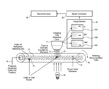
FIG. 1 is a highly schematic view of an optical projection tomography system
including a pattern noise correction processor.
FIG. 2 shows a typical pseudo-projection image with pattern noise.
FIG. 2A shows a selected portion of the pseudo-projection image of FIG. 2
that has been enhanced to better visually illustrate some subtle effects of
pattern
noise.
FIG. 3 shows a processed slice from 3D reconstruction showing the effect of
pattern noise.
FIG. 4A shows a masked pseudo projection of the cells shown in FIG. 2 and
FIG. 4B shows a mask image for the cells.
FIG. 5A shows a masked pseudo projection of the cells shown in FIG. 2 with
capillary advanced by 45 and FIG. 5B shows a mask image for the cells.
FIG. 6 shows a masked pseudo projection of the cells shown in FIG. 2 with
capillary reversed by 45 and FIG. 6B shows a mask image for the cells.
FIG. 7 shows an image resulting from summation of all masked pseudo
projections.
FIG. 8 shows an image resulting from summation of all mask images.
FIG. 9 shows a noise image with grayscale expanded to fill image dynamic
range.
FIG. 10 shows a noise correction schematic.
FIG. 11 illustrates the image of FIG. 2 after application of noise correction.
FIG. 12A and FIG. 12B show a comparison of image slices from a 3D
reconstruction of pseudo projections without noise correction and with noise
correction respectively.
FIG. 13 shows a graphical representation of threshold selection criteria.
DESCRIPTION OF THE PREFERRED EMBODIMENT
The following disclosure describes several embodiments and systems for
imaging an object of interest. Several features of methods and systems in
accordance with example embodiments of the invention are set forth and
described
in the figures. It will be appreciated that methods and systems in accordance
with
other example embodiments of the invention can include additional procedures
or
features different than those shown in figures.
Example embodiments are described herein with respect to biological cells.
However, it will be understood that these examples are for the purpose of
illustrating
4
CA 02755056 2011-09-09
WO 2010/104976 PCT/US2010/026862
the principles of the invention, and that the invention is not so limited.
Additionally,
methods and systems in accordance with several example embodiments of the
invention may not include all of the features shown in these figures.
Throughout the
figures, like reference numbers refer to similar or identical components or
procedures.
Unless the context requires otherwise, throughout the specification and claims
which follow, the word "comprise" and variations thereof, such as, "comprises"
and
"comprising" are to be construed in an open, inclusive sense that is as
"including, but
not limited to."
Reference throughout this specification to "one example" or "an example
embodiment," "one embodiment," "an embodiment" or various combinations of
these
terms means that a particular feature, structure or characteristic described
in
connection with the embodiment is included in at least one embodiment of the
present disclosure. Thus, the appearances of the phrases "in one embodiment"
or "in
an embodiment" in various places throughout this specification are not
necessarily all
referring to the same embodiment. Furthermore, the particular features,
structures,
or characteristics may be combined in any suitable manner in one or more
embodiments.
Generally as used herein the following terms have the following meanings
when used within the context of optical microscopy processes:
"Capillary tube" has its generally accepted meaning and is intended to include
transparent microcapillary tubes and equivalent items with an inside diameter
generally of 500 microns or less.
"Depth of field" is the length along the optical axis within which the focal
plane
may be shifted before an unacceptable image blur for a specified feature is
produced.
"Object" means an individual cell, item, thing, particle or other microscopic
entity.
"Pseudo projection" includes a single image representing a sampled volume
of extent larger than the native depth of field of a given set of optics. One
concept of a pseudoprojection is taught in Fauver '744.
"Specimen" means a complete product obtained from a single test or
procedure from an individual patient (e.g., sputum submitted for analysis, a
biopsy, or a nasal swab). A specimen may be composed of one or more
CA 02755056 2011-09-09
WO 2010/104976 PCT/US2010/026862
objects. The result of the specimen diagnosis becomes part of the case
diagnosis.
"Sample" means a finished cellular preparation that is ready for analysis,
including all or part of an aliquot or specimen.
As used in this specification, the terms "processor" and "computer processor"
encompass a personal computer, a microcontroller, a microprocessor, a field
programmable object array (FPOA), a digital signal processor (DSP), an
application-
specific integrated circuit (ASIC), a field programmable gate array (FPGA), a
programmable logic array (PLA), or any other digital processing engine, device
or
equivalent including related memory devices, transmission devices, pointing
devices,
input/output devices, displays and equivalents.
Referring now to FIG. 1 a highly schematic view of an optical projection
tomography system including a pattern noise correction processor is shown.
Cells
15 are suspended in an index of refraction matching gel 12 contained in a
capillary
tube 18. Pressure 10 is applied to the gel 12 to move the cells into the
optical path of
a high-magnification microscope including an objective lens 5. The objective
lens 5 is
scanned or vibrated by, for example, a (not shown) piezo-electric element. The
capillary tube 18 is positioned to be scanned by the vibrating objective lens
5. An
illumination source 20 operates to illuminate objects, such as biological
cells passing
through the field of view of the objective lens 5. An image sensor 25 is
located to
acquire images transmitted from the objective lens 5. A plurality of pseudo-
projection
images, here exemplified by pseudo-projection images 22A, 22B and 22C are
acquired by the image sensor 25 at varying angles of view as presented by the
rotating capillary tube 18. An image processor with noise correction 35 is
coupled to
receive the pseudo-projection images. Corrected pseudo-projection images are
then
passed to a reconstruction processor 36 for producing 3-D images.
VisionGate, Inc. of Gig Harbor Washington, assignee of this application, is
developing an optical tomography system incorporating pattern noise correction
under the trademark "Cell-CTTm." The Cell-CTTm optical tomography system
employs
scores, designed to detect lung cancer in its pre-invasive and treatable
stage. In one
example embodiment the operation is as follows.
1. A specimen for examination is processed to remove non-diagnostic elements
and is fixed and stained.
6
CA 02755056 2011-09-09
WO 2010/104976 PCT/US2010/026862
2. The specimen is then suspended in a gel medium. The cells in gel mixture
are then inserted into a glass micro-capillary tube 18 of approximately 50
inner diameter 16.
3. Pressure is applied to the gel to move the cells into the optical path 14
of a
high-magnification microscope.
4. Once the cells are in place the tube is rotated to permit capture of 500
high
resolution images of the desired object taken over 360 degrees of tube
rotation. These images are simulations of projection images created by
integrating the light from the objective lens as the objective scans the
nucleus.
The simulated projection or pseudo-projection images thus represent the
entire nuclear content in a single image, taken from a single perspective.
5. Pseudo-projection images are processed to correct for residual noise and
motion artifact.
6. The corrected pseudo projections are processed using filtered back
projection
to yield a 3-D tomographic representation of the cell. An example section of
such a 3-D rendering is shown in FIG. 3 for an Adenocarcinoma cell grown in
culture.
7. Based on the tomographic reconstruction, features are computed that are
used to detect cells with the characteristics of cancer and its precursors.
These features are used in a classifier whose output designates the likelihood
that object under investigation is a cancer cell. Classifier outputs are based
on a scoring system developed by VisionGate, Inc. called LuCEDTM scores.
Among other things, good quality reconstruction and classification depends on
good quality corrected pseudo projections input to the reconstruction
algorithm in
step 6. This document discloses a method to correct for pattern noise present
in
pseudo projections at the time of data capture.
Pattern Noise Correction
As noted above, pattern noise results from additive distortion. A pseudo
projection may be modeled as an ideal pseudo projection plus pattern noise. If
the
pattern noise is found then the ideal, noise free, pseudo projection can be
found by
subtracting the pattern noise from the noisy pseudo projection. Hence a
challenge
for doing a subtractive correction is to find the pattern noise image. The
creation of a
pattern noise image is enabled by recognizing and using the fact that pseudo-
projection images are comprised of two image parts. A first image part is
stable and
7
CA 02755056 2011-09-09
WO 2010/104976 PCT/US2010/026862
common to the entire set of pseudo projections and a second image part which
is
dynamic and changeable from one projection to the next. The dynamic part is
the
part that is associated with a sample such as a cell and other material that
is
suspended in the gel. In an optical tomography system design, the cell changes
its
position as the capillary tube is rotated. Because the cell and other material
are dark
relative to the background the gel-suspended part of the image may be
thresholded
out, leaving a partial representation of the stable part of the image.
An image after application of a threshold is shown for the pseudo projection
of
FIG. 2 in FIG. 4A. Note that FIG. 4B contains a mask image that is a binary
version
of the grayscale version of FIG. 4A where all non-zero pixels are set to one.
FIG. 5A
and FIG. 5B and FIG. 6A and FIG. 6B show similar images for rotations plus and
minus 45 degrees respectively from the position represented in FIG. 4A and
FIG. 4B.
The axes are in pixel counts.
Referring now jointly to FIG. 4A, FIG. 5A and FIG. 6A, note that each image
contains a different part of the background, or pattern noise containing part
of the
image. In this observation the key to the formation of the background image is
found. The thresholded images for the entire set of masked pseudo-projections
may
be summed together to form an ensemble grey scale image as shown in FIG. 7 for
an entire set of 500 pseudo-projections. It will be understood that, while in
some
examples a set of 500 pseudo-projections was used, the invention is not so
limited
and more or less pseudo-projections may be included in a set. The amount and
rate
of rotation may also be varied for different applications or results.
Referring now jointly to FIG. 4B, FIG. 5B and FIG. 6B the mask images there
shown may be summed together to form an ensemble mask. Summed images for an
entire set of 500 pseudo-projections are shown in FIG. 8.
Referring now jointly and respectively to FIG. 7 and FIG. 8 it can be seen
that
at no spot in the images is there a point where some information concerning
the
background is not available. By design, the background generally indicated as
70
and 70A in the respective figures is not substantially modulated through
rotation of
the tube. Cellular material is evidenced by modulated patterns, for example,
72 and
72A in the respective figures. Therefore, it is a good assumption that the
background
as computed through by averaging all 500 pseudo-projections may be
approximated
by the background in any one pseudo-projection. As a result, the pattern noise
8
CA 02755056 2011-09-09
WO 2010/104976 PCT/US2010/026862
image may be found by dividing the ensemble grey scale image by the ensemble
mask.
The result is shown in FIG. 9 where the noise image has been processed to
expand the grey scale range to fill the entire dynamic range for the image.
Note that
FIG. 9 shows that the noise image represents all the relevant distortions for
which a
correction is desired including
a. Illumination variation,
b. Dust, and
c. Mottling.
Correction of any one pseudo-projection is then a matter of division.
Referring now to FIG. 10 a noise correction schematic is shown. A typical 3D
reconstruction for a biological cell requires acquisition of 500 pseudo-
projection
images, PP0_PP499, each acquired as the capillary tube rotates through 500
incremental rotation angles, where PPo is acquired at angle 0 and P P499 is
acquired
at about 3600. In operation loop 100 is repeated through 500 incremental
angles
according to the command i=0:499. Each pseudo projection, PP,, is processed
through a threshold operation 104 to produce a threshold image. Optionally,
the
threshold image may then be dilated 106 to produce a dilated image. However,
dilation is not an essential step for pattern noise correction and may be
bypassed or
left out. The dilated image or threshold image, as the case may be, is sent to
a
summer 110 which accumulates images with removed objects, and the summation
of all images forms an ensemble image 114. The dilated image or threshold
image,
as the case may be, is also processed into a binary image at 108 to form a
mask that
is summed at mask summer 112 ultimately producing an ensemble mask 116.
Threshold procedures are described further below with reference to FIG. 13.
The
operations of thresholding, dilating and mask creation may be implemented in a
computer as a software program, dedicated processor, computer processor,
electronic circuits or the like including processors and related devices
listed above.
Referring now to FIG. 13, a graphical representation of a histogram marked
with threshold selection criteria is shown. Correct functioning of the noise
correction
algorithm depends upon correct selection of the threshold used to remove
objects
from pseudo-projections. In one example, threshold selection is accomplished
through a two-part process and performed separately for each pseudo-
projection.
The two-part process of threshold selection is based on two principles. First
a
9
CA 02755056 2011-09-09
WO 2010/104976 PCT/US2010/026862
histogram 101 is generated that combines two influences from the image, the
background and that of an object, such as a cell. The
histogram 101 is
characterized by a mode ("Mode") and a maximum ("Max"). The mode represents
the most frequently occurring value, which here is the average value of the
background. A cell in the image influences the histogram to its dark side.
Hence the
variance in the background may be estimated by finding the difference between
the
maximum and the mode. An initial estimate for the threshold for separating
cell from
background in the image may therefore be made according to the formula:
Thresh=0.9(2*Mode ¨ Max) as indicated by broken line 102. The estimated
threshold is then applied to the image and the total area below the threshold
is
found.
The second principle governing threshold calculation is derived from the fact
that a profile of any of the various objects changes little from pseudo-
projection to
projection. This is because the capillary tube rotates in small increments
from one
pseudo-projection to the next. This fact is used to further refine the
threshold as it is
iteratively adjusted until the total area of pixels beneath the threshold is
within 10%
of the area for the previous threshold.
Referring again to FIG. 10, once the summations are available the ensemble
image 114 is divided by the ensemble mask to yield the background pattern
noise
118. Each PP, is multiplied by a scaling factor (here, for example, 360000)
and the
product is divided by the background pattern noise 118. The quotient image is
filtered by a low pass filter 122 that passes low-frequency signals but
attenuates
signals with frequencies higher than the cutoff frequency, where the cutoff
frequency
is selected to filter out high frequency artifacts as may be caused, for
example, by
camera noise. The cutoff frequency is selected so as to preserve the highest
spatial
frequencies for which response in the reconstruction is desired. A filtered
image is
produced at 124 as a noise corrected pseudo projection.
Referring now to FIG. 11, the result of correction for the pseudo-projection
of
FIG. 2 is shown. A comparison of FIG. 11 with FIG. 2 shows that illumination
variation has been corrected, dust removed and mottling substantially reduced.
Referring now to FIG. 12A and FIG. 12B, a comparison of image slices from a
3D reconstruction volume of pseudo projections without noise correction and
with
noise correction respectively is shown. The first image in FIG. 12A resulted
from
reconstruction with no noise correction. The second image in FIG. 12B has been
CA 02755056 2011-09-09
WO 2010/104976 PCT/US2010/026862
processed with noise correction. Note the much cleaner presentation of
cellular
detail for the noise corrected reconstruction.
In an optical tomography system or similar system, noise correction according
to the methods and systems described herein may be effectively performed when
there is sufficient movement of the cell so that the background may be imaged
in at
least a small number of pseudo-projections. When this is not the case the
noise
correction may not be effective. Further, correct execution of the technique
depends
on the ability to remove the cells from the background so that the grey matter
in an
image resulting from summation of all masked pseudo projections, as shown, for
example, in FIG. 7, represents only the background. This occurs when the
algorithm
that determines the threshold correctly identifies the threshold to segment
cells.
When thresholds are incorrectly identified, an image resulting from summation
of all
masked pseudo projections can include cellular residues which leads to an
incorrect
normalization. In such a circumstance the resulting pattern noise image,
unlike that
shown in FIG. 9, exhibits high variance. When variance of the noise image
exceeds
a predetermined level, noise correction cannot be effectively performed.
While specific embodiments of the invention have been illustrated and
described herein, it is realized that numerous modifications and changes will
occur to
those skilled in the art. It is therefore to be understood that the appended
claims are
intended to cover all such modifications and changes as fall within the true
spirit and
scope of the invention.
What is claimed is:
11