Note: Descriptions are shown in the official language in which they were submitted.
CA 02755071 2011-10-17
ENDOSCOPE CLEANER
BACKGROUND
Technical Field
[0001] The present disclosure relates to a cleaning apparatus configured to
remove debris
from the lens of a minimally invasive viewing instrument.
Background of Related Art
[0002] Minimally invasive surgery has become increasingly popular in recent
years.
Minimally invasive surgery eliminates the need to cut a large incision in a
patient, thereby reducing
discomfort, recovery time, and many of the deleterious side effects associated
with traditional open
surgery. Minimally invasive viewing instruments, e.g., laparoscopes and
endoscopes, are optic
instruments to facilitate the viewing of internal tissues and/or organs.
[0003] Laparoscopic surgery involves the placement of a laparoscope in a small
incision in
the abdominal wall of a patient to view the surgical site. Endoscopic surgery
involves the
placement of an endoscope in a naturally occurring orifice, e.g., mouth, nose,
anus, urethra, and
vagina to view the surgical site. Other minimally invasive surgical procedures
include video
assisted thoracic surgery and cardiovascular surgery conducted through small
incisions between the
ribs. These procedures also utilize scopes to view the surgical site.
[0004] A typical minimally invasive viewing instrument, e.g., a laparoscope or
an
endoscope, includes a housing, an elongated lens shaft extending from one end
of the housing, and
a lens that is provided in the distal end of the lens shaft. A camera
viewfinder extends from the
other end of the housing. A camera is connected to the housing and transmits
images of the
surgical field viewed through the lens to a monitor on which the images are
displayed. During a
surgical procedure, the distal end portion of the lens shaft is extended into
the patient, while the
proximal end portion of the lens shaft, the housing and the camera viewfinder
remain outside the
patient. In this manner, the laparoscope/endoscope is positioned and adjusted
to view particular
anatomical structures in the surgical field on the monitor.
i
CA 02755071 2011-10-17
[0005] During insertion of an endoscope or a laparoscope into the body and
during the
surgical procedure, debris, e.g., organic matter and moisture, may be
deposited on the lens of the
endoscope. The buildup of debris and condensation on the lens impairs
visualization of the surgical
site, and often necessitates cleaning of the lens.
SUMMARY
[0006] The present disclosure is generally related to an instrument for
cleaning the lens of a
medical viewing instrument, such as an endoscope, during a minimally invasive
surgical procedure.
In one aspect of the present disclosure, an instrument for cleaning the lens
of a surgical scope is
provided comprising an elongated sheath with proximal and distal end portions
and having an
interior and exterior and a fluid conduit for transporting fluid. The interior
is dimensioned and
configured to slidingly receive a scope therein. The fluid conduit has a fluid
discharge opening to
deliver fluid to the lens of the scope and is coupled to the sheath exterior.
[0007] Preferably, the instrument further includes a roller mechanism coupled
to the distal
portion of the sheath exterior and includes at least one movable wiping arm.
The roller mechanism
can in some embodiments be configured such that when the roller mechanism is
in a cleaning
position, the fluid conduit is in the dispensing state. The roller mechanism
can be further
configured such that when the roller mechanism is in a non-cleaning position,
the fluid conduit is in
the non-dispensing state.
[0008] The instrument may further comprise a wiping arm operatively connected
to the
sheath wherein insertion of the scope actuates the wiping arm. In some
embodiments, advancement
of the scope with respect to the sheath automatically discharges fluid through
the discharge opening
of the fluid conduit.
[0009] In some embodiments, the roller mechanism is in the non-cleaning
position when
the scope retracts inside the elongated sheath interior.
[0010] An attachment clip can be provided to attach the fluid conduit to the
elongated
sheath exterior.
[0011] The roller mechanism can include first and second wiping arms extending
from a
ring like member, and the wiping arms can move transversely over the scope
lens.
2
CA 02755071 2011-10-17
[0012] In some embodiments, the instrument includes a pump configured to
switch
between first and second positions, the first position being to deliver fluid
through the fluid
discharge opening and the second position being to close the discharge
opening.
[0013] The present disclosure provides in another aspect an instrument for
cleaning a lens
of a surgical scope comprising an elongated sheath with proximal and distal
end portions and an
interior and exterior and a pair of wiping arms supported by the sheath. The
arms are movable from
a first position adjacent one another to a second position spaced from each
other. The arms are
movable from the first to the second position upon contact by the lens of the
scope inserted through
the interior of the sheath.
[0014] In some embodiments, the wiping arms extend from a ring-like member
positioned
at a distal end portion of the sheath. The sheath preferably includes a fluid
conduit for delivering a
cleaning fluid to a lens of a scope inserted through the sheath.
[0015] In some embodiments, a fluid discharge nozzle communicates with the
fluid
conduit.
[0016] In some embodiments, movement of the wiping arms opens a valve for
delivering
cleaning fluid to a lens.
BRIEF DESCRIPTION OF THE DRAWINGS
[0017] The above and other aspects, features, and advantages of the present
disclosure will
become more apparent in light of the following detailed description when taken
in conjunction with
the accompanying drawings in which:
[0018] Figure IA is a schematic view of a scope lens cleaner according to an
embodiment
of the present disclosure;
[0019] Figure 1 B is close up view of the area of detail of Figure 1 A;
[0020] Figures 2A-2B are enlarged perspective views of the scope lens cleaner
of Figure
1A shown in different positions;
[0021] Figure 2C is a view similar to Figure 2B showing an alternate
embodiment for
opening the nozzle;
[0022] Figure 3 is an enlarged side view of the scope lens cleaner of Figure
IA;
3
CA 02755071 2011-10-17
[0023] Figure 4 is a cross-sectional view taken along section lines A-A in
Figure 1A;
[0024] Figure 5 is a cross-sectional view of a fluid discharge nozzle element
of the scope
lens cleaner according to an embodiment of the present disclosure;
[0025] Figure 6 is a cross-sectional view of an alternate embodiment of the
fluid discharge
nozzle of the present disclosure;
[0026] Figure 7 is a schematic view of a scope lens cleaner according to
another
embodiment of the present disclosure; and
[0027] Figures 8A-8B are enlarged perspective views of the scope lens cleaner
of Figure 7.
DETAILED DESCRIPTION
[0028] An endoscope typically includes an endoscope housing or body which can
be rigid
or flexible, depending on its surgical application. A camera viewfinder, e.g.
an eyepiece, is located
at a proximal (imaging) end of the scope housing. A lens is provided at the
distal end of the scope
body.
[0029] In typical use of the endoscope, the viewfinder is adapted to sight
images of a
surgical field in the patient, e.g. an abdominal cavity, thoracic cavity,
etc., as the position of the
scope is adjusted to view a particular anatomical structure or structures in
the surgical field. The
camera is adapted to receive images of the surgical field sighted through the
lens and transmit the
images to an external monitor that is connected to the camera and on which the
images of the
surgical field are displayed. That is, a visual display device is operatively
connected to the eyepiece
to convert the optical signal into a video signal to produce a video image on
the monitor (or for
storage on select media). Accordingly, the monitor enables a surgical team to
view the anatomical
structure or structures in the surgical field inside the patient as the
surgical procedure is carried out
using minimally invasive or endoscopic surgical instruments. Throughout the
surgical procedure,
condensation, smoke particles, biological tissue or matter has a tendency to
contact and build up on
the lens of the scope. This tends to obscure the images of the surgical field
as they are displayed on
the monitor.
4
CA 02755071 2011-10-17
[0030] The instrument of the present disclosure enables cleaning of the scope
lens during
the surgical procedure to maintain a clear image without having to remove the
scope from the
patient's body.
[0031] Particular embodiments of the present disclosure will be described
herein with
reference to the accompanying drawings. In the figures and in the description
that follows, in which
like reference numerals identify similar or identical elements, the term
"proximal" will refer to the
end of the apparatus that is closer to the operator during use, while the term
"distal" will refer to the
end that is further from the operator during use.
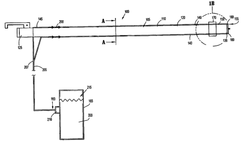
[0032] Referring to Figure 1A of the drawings, an illustrative embodiment of
an instrument
lens cleaner according to the present disclosure is generally indicated by
reference numeral 100.
The instrument 100 includes a generally elongated, cylindrical or tubular
sheath 105 having a
sheath wall 110. Wall 110 may be a substantially rigid or semi-rigid plastic
material. The sheath
wall can also be flexible to accommodate a flexible scope. The tubular sheath
105 has a proximal
end 125 and a distal end 130. A cross section of the sheath wall 110 at
section A-A is shown in
Figure 4. Referring to Figure 4, the sheath wall 110 typically has a generally
annular cross-
sectional configuration and defines a sheath interior 115 and a sheath
exterior 120 which defines the
outer surface of the sheath wall 110.
[0033] The tubular sheath 105 is dimensioned and configured to slidingly
receive a
conventional scope therein. The scope can be inserted into an already placed
sheath or alternatively
positioned within the sheath and together inserted into the body. The scope
can be fully removed
from the sheath if desired. The sheath can accommodate various types of
scopes, including but not
limited to laparoscopes, thoracoscopes, etc. For example, during video
assisted thorascopic
surgery, a thoracic port is inserted through the ribs to provide access to the
thoracic cavity for access
to lung or other tissue. A separate access is provided through the ribs to
insert a scope to visualize
the thoracic cavity during the surgical procedure. The sheath of the present
disclosure can be
utilized with the thoracoscope to maintain a clean lens to provide consistent
visibility and imaging
during the surgical procedure. The sheath can also be utilized with a flexible
scope if composed of
a sufficiently flexible material.
CA 02755071 2011-10-17
[0034] Referring back to Figure IA, a fluid conduit 140, provided on each side
of the
tubular sheath 105, has a generally annular cross-sectional configuration and
is connected along the
sheath exterior 120. The fluid conduits 140 each have a proximal end portion
145 and a distal end
portion 150 having an opening 155. The fluid conduits 140 extend generally
parallel to the
longitudinal axis of the tubular sheath 105. Each fluid conduit 140 has a
proximal inlet end that is
in fluid communication with a fluid reservoir 165 and a distal outlet end at
the opening 155. The
outlet 155 can in some embodiments include a fluid discharge nozzle 180. The
diameter or width
of the fluid conduits 140 may in some embodiments be substantially equal to or
slightly larger than
the thickness of the sheath wall 110. As shown, preferably two fluid conduits
140 are provided,
preferably identical and illustratively spaced about 180 degrees apart,
although other spacings are
also contemplated. A different number of fluid conduits could also be
provided. If a flexible
sheath is utilized for a flexible scope, the fluid conduits would also be
composed of sufficiently
flexible material.
[0035] Referring to Figure 1 B, the fluid conduit 140 is connected to the
sheath exterior 120
by attachment clip 170. The attachment clip 170 is normally biased in the
locking configuration
with arms 171 in a closer position. The clip 170 is coupled to the fluid
conduit 140 by bending the
clip arms 171 outwardly as shown by the phantom lines and sliding the clip 170
over the fluid
conduits 140. When the arms 171 are released, they return to their original
state, applying a holding
force against the fluid conduits 140.
[0036] The fluid discharge nozzle 180 of each fluid conduit 140 communicates
with the
outlet end of the fluid conduit 140 and protrudes radially inwardly therein
toward a longitudinal
axis of the sheath in a direction toward the lens of a scope inserted through
the sheath 105. As
illustrated in Figure 5, in one embodiment, the fluid discharge nozzle 180 has
a nozzle wall 180a
that is continuous with a wall 185 of the fluid conduit 140 and defines a
nozzle interior 180b. The
nozzle interior 180b of the fluid discharge nozzle 180 communicates with the
fluid conduit 140.
The wall 180a functions as a flow diverter to direct the fluid toward a nozzle
plate 190. The nozzle
plate 190 includes a plurality of spaced apart exit openings to deliver the
cleaning fluid from
reservoir 165 in a spray fashion. In an alternate embodiment illustrated in
Figure 6, a single nozzle
opening 195 is formed in the nozzle plate, preferably extending through the
central portion of the
6
CA 02755071 2011-10-17
nozzle plate 190, to deliver the cleaning fluid in a single stream fashion.
Other configurations for
delivering the fluid are also contemplated.
[0037] Referring back to Figure IA, an elongated, flexible fluid connecting
conduit 205,
fitted with a conduit connector 210 at inlet end 160, is disposed in fluid
communication with the
inlet end of the fluid conduits 140. The conduit 205 in some embodiments is
integral with the fluid
conduits 140 and is therefore an extension thereof. In other embodiments, the
conduit 205 is a
separate tube connected to the fluid conduits 140. The fluid connecting
conduit 205 is adapted for
connection to a discharge outlet (not illustrated) of a fluid pump and supply
apparatus 215, such as
through the conduit connector 210. The fluid pump and supply apparatus 215 may
be conventional
and include the fluid reservoir 165 that is adapted to contain a cleaning
fluid 200 such as saline
solution. As shown, conduit 205 splits at region 207 so the cleaning fluid 200
is transported in the
direction of the arrows through both fluid conduits 140 for discharge through
associated nozzles
180.
[0038] A roller mechanism 230 (Figure 2A) is connected to the distal end 130
of the
elongated sheath exterior 120. As illustrated in Figures 2A-2B and 3, the
roller mechanism 230 is
connected adjacent the fluid discharge nozzles 180. The roller mechanism 230
includes two wiping
arms 235, 238 of a material for wiping and cleaning the lens 265 of an
endoscope 250 inserted
through and extending distally from the tubular sheath 105.
[0039] The roller mechanism 230 in the illustrated embodiment is formed into a
substantially ring shape as shown in Figure 2B. Two arms 231, 233 extend
inwardly from the ring-
like member to form the wiping arms 235, 238, respectively. The roller
mechanism can be formed
of a wire or tubular member with a normal position of that shown in Figure 2B.
For example, it can
be composed of a naturally sprung material or a shape memory material with a
memorized position
of Figure 2B so it automatically returns to this position after the scope
retraction described below.
When the scope is advanced as described below, it forces the wiping arms 235,
258 to separate in a
transverse sweeping motion over the lens to the position of Figure 2A. When
the scope is retracted,
the wiping arms 235, 238 return to their normal (initial) position of Figure
2B. The roller
mechanism 230 includes two collars 255 which frictionally engage a respective
recess 260 in the
fluid conduit 140 to retain the roller mechanism 130.
7
CA 02755071 2011-10-17
[0040] The region adjacent the arms 231, 233 can be considered to function as
levers 240.
The levers 240 move between a non-cleaning position where the arms are
adjacent (Figure 2B) to a
cleaning position where the arms separate to move across the lens of the scope
(Figure 2A). That
is, in the cleaning position, the levers 240 swing away from the distal end
130 of the elongated
sheath 105 and the wiping arms 235, 238 of roller 235 wipe across the
endoscope lens 265 as
illustrated in Figure 2A. In other words, in the dispensing (cleaning)
position, the swinging of the
levers 240 in an arc enable arms 235, 238 to remove fluid 200 and/or debris
from the surface of the
endoscope lens. Note in the non-cleaning position, the levers 240 are
positioned at an angle with
respect to the longitudinal axis of the elongated sheath 105 as illustrated in
Figure 2B. The levers
240 are preferably normally in a non-cleaning position.
[0041] In one embodiment, an actuator 245 is in communication with each fluid
conduit
140. Only one of the actuators is shown in the drawings. The actuators 245
enable the automatic
delivery of cleaning fluid upon insertion of the scope 265 through the sheath
105. More
specifically, the actuator is operatively connected to a valve (not shown).
The valve provides for a
cutoff of fluid to the discharge nozzle 180. When the scope 265 is in the
advanced position of
Figure 2A to view the surgical site during surgery, the actuator 245 is in the
pivoted position. To
clean the scope of the lens, the scope 265 is retracted proximally within the
sheath, thereby
allowing actuator 245 to move to its position of Figure 3. This opens the
valve to allow fluid to be
dispensed through the nozzles 180 as the actuator moves the valve. As the
scope is re-advanced, it
pivots actuator 245 to close the valve to cut off fluid flow. Consequently, in
this embodiment,
scope retraction enables the actuator 245 to open the valve to open the
conduits 140 for fluid flow
through the discharge nozzle and subsequent distal movement of the scope
returns the actuator 245
to its original position and actuates the wiping arms 235, 238 to wipe and
clean the surface of the
lens 265 of scope 250. It is also contemplated that rather than the sequential
operation of the valve
and wiping arms, scope advancement could simultaneously open the nozzles for
fluid flow and
actuate the wiping arms 235, 238. Consequently, when the roller mechanism is
in a cleaning
position, the fluid conduit is in the dispensing state and when the roller
mechanism is in a non-
cleaning position, the fluid conduit is in the non-dispensing state.
8
CA 02755071 2011-10-17
[0042] As can be appreciated, the scope lens can therefore be cleaned in situ,
i.e. without
requiring withdrawal of the scope from the patient's body, as it can be
cleaned by slight retraction
and re-advancement multiple times during a surgical procedure.
[0043] Note in an alternate embodiment, the actuator can include a conduit
engaging
structure to pinch the fluid conduit(s) 140 to close it off to fluid flow. In
this embodiment,
movement of the scope would contact the actuator to pivot it so the conduit
engaging structure
would be released from pinching engagement with the fluid conduit 140 to
enable the flow of
cleaning fluid through the conduit 140 and through the nozzle. Thus, the
actuator would switch the
fluid discharge nozzle 180 between dispensing and non-dispensing states in
response to scope
movement within the elongated sheath 105.
[0044] In another embodiment illustrated in Figure 2C, the movement of the
scope from the
retracted position to the advanced position of Figure 2B would move hinged
nozzle covers to
automatically open a valve. That is, hinges 181 are hingedly connected to the
discharge nozzles
and cover the discharge nozzles in their normal position. When the scope is
advanced, the scope
contacts arm 183 of each hinge to pivot the cover portion 184 away from the
nozzle to provide an
opening for fluid flow. In an alternate embodiment, the hinges are connected
to the lever arms
240', so that when distal movement of the scope moves the lever arms, the
cover of the hinges is
moved away from the nozzle to open the nozzle for fluid flow. Otherwise, the
wiper is the same as
in Figure 2B and includes roller mechanism 230' with arms 231', 233' and
wiping arms 235, 238'.
[0045] In operation, as the endoscope 250 is moved distally relative to the
elongated sheath
105, the endoscope 250 makes contact with the actuator 245 and as a result,
the fluid discharge
nozzle 180 automatically sprays an amount of fluid 200 onto the endoscope lens
265. The duration
of the spray creates a mist onto the endoscope lens 265. As the endoscope
continues to be
advanced distally it contacts arms 235, 238 at roller 230 to pivot the arms
235, 238 to move
transversely over the lens 265 to wipe the fluid 200 and/or debris from the
surface of the lens 265.
The endoscope continues to extend distally after the arms 235, 238 wipe the
fluid 200 and/or debris
and is out of the spray zone. The endoscope lens 265 captures images of the
surgical field without
having arms 235, 238 being in the camera field of view as the scope housing
(body) maintains the
arms 235, 238 in a spaced position (see Figure 2A). If at any time during the
surgical procedure the
9
CA 02755071 2011-10-17
lens needs to be cleaned, the endoscope 250 can be withdrawn into the sheath
105, and then re-
advanced to pivot the wiping arms 235, 238 to clean the lens 265 and to open
the nozzle 180 in the
embodiments having an actuator activated by scope movement.
[00461 Note in some embodiments, the fluid pump and supply apparatus 215 are
selectively
operated to pump a cleaning fluid 200, such as saline solution, through the
fluid connecting conduit
205 and the fluid discharge nozzle 180. The cleaning fluid 200 can be
discharged from the fluid
discharge nozzle 180 through the nozzle openings in a spray pattern, or
alternatively, through a
single nozzle opening in a single stream pattern (or alternatively in other
patterns), against and
across the surface of the lens 265. Thus, when desired to clean the lens, the
scope is retracted and
the user actuates a pump or other device to advance the fluid through the
conduit and nozzle. This
is shown for example in the embodiment of Figure. 7. In other embodiments, as
described above,
rather than the user selectively pumping cleaning fluid, the endoscope
movement would
automatically deliver cleaning fluid to the lens.
[0047) In the embodiment of Figure 7 the instrument lens cleaner 300 is
similar to the
instrument lens cleaner 100 of Figure 1A, with the exception of the actuating
system to spray the
fluid 200'. The instrument lens cleaner 300 includes a bulb pump 305 to
inject, e.g. to spray, the
fluid 200' to clean the endoscope lens 265. In operation, a surgeon, when
desired, manually
squeezes the bulb 305 to discharge the cleaning fluid 200' under pressure from
the fluid reservoir
165 and into the fluid connecting conduit 205'. Although one conduit 340 is
shown in fluid
communication with connecting conduit 205', two fluid conduits (or additional
conduits) as in the
embodiment of Figure 1 are also contemplated. The conduit(s) 340 can include a
pressure-release
valve therein that is normally closed but opens when the bulb is squeezed to
inject the fluid at high
pressure. Thus, at sufficient pressure, the valve opens to enable fluid
discharge nozzle 380 to
release high-pressure but low volume fluid 200' such as saline. As the surgeon
releases the bulb
305 to return to its initial position, the valve self-closes and the bulb 305
is refilled for the next shot
of fluid. Note other types of liquid pumps could alternatively be provided.
Note the scope can be
withdrawn during use to place the lens adjacent the discharge nozzle for
application of cleaning
fluid. Advancement would then actuate the wiping arms in the manner described
above.
CA 02755071 2011-10-17
[0048] Figures 8A-8B are side, partially schematic views of the instrument
lens cleaner 300
of Figure 7. Figure 8A illustrates the fluid discharge nozzle 380 prior to
spraying cleaning fluid
200 on the scope lens 265. In response to the surgeon squeezing the bulb 305,
the fluid discharge
nozzle 380 sprays a mist (or jet or stream) of cleaning fluid on the scope
lens 265 (Figure 8B) to
remove debris. It should be understood that other mechanisms beside the bulb
305 can be used to
inject cleaning fluid 200', such as a foot pedal (not shown) or other hand-
controlled device such as
a syringe type mechanism or a trigger to control the discharge of cleaning
fluid 200'. Automated or
semi-automated delivery mechanisms can be provided. Also, as noted above, two
fluid conduits
340 could be provided, with connecting conduit 205' split as in conduit 205 of
FIG. IA to deliver
fluid to both conduits 340.
[0049] The lens cleaner 300 of Figures 7-8 includes a roller mechanism
identical to that of
Figure 2A or identical to that of Figure 2B. Such roller mechanism is not
shown in Figures 7-8 for
clarity.
[0050] In each of the embodiments herein, one or more fluid conduits with
respective fluid
openings or nozzles can be provided. The fluid can be delivered as a mist,
spray, jet, stream, etc.
[0051] While several embodiments of the disclosure have been shown in the
drawings
and/or discussed herein, it is not intended that the disclosure be limited
thereto, as it is intended that
the disclosure be as broad in scope as the art will allow and that the
specification be read likewise.
Therefore, the above description should not be construed as limiting, but
merely as exemplifications
of particular embodiments. Those skilled in the art will envision other
modifications within the
scope and spirit of the claims appended hereto.
11