Note: Descriptions are shown in the official language in which they were submitted.
CA 02755555 2011-09-15
WO 2010/105628 PCT/DK2010/050063
1
System and method for effective planning, visualization, and
optimization of dental restorations
Field of the invention
The present invention relates to a system and a method for planning dental
restorative work. The invention furthermore relates to a system and a method
for interactive CAD design and realistic 3D presentation and visualization of
dental restorations and subsequent physical realization by means of CAM.
Background of the invention
In dental practice, diagnostic wax-ups are created to visualize and plan
restorative treatment, e.g., veneers or crowns on the anterior/front teeth.
Diagnostic wax-ups are traditionally created in wax on gypsum casts by the
dental laboratory for the dentist who uses it for treatment planning as well
as
for visualization and discussion of the restorative result with the patient.
The
dialog between dentist and patient is an important tool for improved patient
satisfaction and often enables more expensive treatments. To transfer the
design from the diagnostic wax-up to the patient's teeth, the dental
technician
typically looks at the original diagnostic wax-up and manually tries to
replicate this design for the real restorations, incorporating potential
comments from the dentist and the patient. This manual replication process
is both costly, possibly inaccurate, and time consuming.
Because of the manual labor involved, diagnostic wax-ups are generally
expensive, often several hundred US dollars. Creating a wax-up model is
also time-consuming, such that the patient generally has to return for another
appointment to evaluate it. Because diagnostic wax-ups are models of teeth
only, they also fail to convey the full aesthetic impact of a restorative
CA 02755555 2011-09-15
WO 2010/105628 PCT/DK2010/050063
2
treatment. The visual impression of a patient's smile is also determined by
the gingiva and the entire face [1]. Furthermore, a free standing wax-up
model cannot convey the lighting to which teeth are subject to inside the
mouth. In the field of orthodontics, treatment planning has more commonly
involved 3D models of both the teeth and the face, or even the head. Data
sources include 2D color pictures of the face and CT scans of the head [2, 3].
WO 2006/065955 discloses methods and systems for orthodontic treatment
including a method for generating a photo-realistic image of a predicted
result
of a dental treatment on a patient, the method comprising: acquiring one or
more images of the patient's pre-treatment face and teeth; generating a 3D
digital model of the patient's pre-treatment face and teeth from the images of
the patient's pre-treatment face and teeth; acquiring a 3D digital model of
the
patient's pre-treatment tooth arch; acquiring a 3D digital model of the
patient's predicted tooth arch resulting from the treatment; generating a 3D
digital model of the patient's predicted face and teeth from the 3D digital
models of the patient's pre- treatment face and teeth, pre-treatment tooth
arch, and predicted tooth arch; and rendering a photo-realistic image from
the 3D digital model of the patient's predicted face and teeth.
WO 2004/098378 relates to orthodontic treatment and discloses a system for
use in diagnosis and planning treatment of a human patient, comprising: a
general purpose computer system having a processor and a user interface; a
memory accessible to said general purpose computer system storing a) a
first set of digital data representing patient craniofacial image information
obtained from a first imaging device, and b) a second set of digital data
representing patient craniofacial image information obtained from a second
image device different from said first image device, said first and second
sets
of data representing at least in part common craniofacial anatomical
structures of said patient, at least one of said first and second sets of
digital
data including data representing the external visual appearance or surface
CA 02755555 2011-09-15
WO 2010/105628 PCT/DK2010/050063
3
configuration of the face of the patient; and a set of computer instructions
stored on a machine readable storage medium accessible to said general
purpose computer system, wherein said set of instructions comprises
instructions for causing said general computer system to: 1) automatically,
and/or with the aid of operator interaction, superimpose said first set of
digital
data and said second set of digital data so as to provide a composite,
combined digital representation of said craniofacial anatomical structures in
a
common coordinate system; 2) displaying said composite, combined digital
representation of said craniofacial anatomical structures to a user of said
system.
Thus, in the field of orthodontics, treatment planning involving 3D models of
both the teeth and the face are known.
US 2008/153061 discloses a method for planning and performing dental
treatments, comprising: an acquisition phase of a set of data relating to the
position, to the conformation and to the dimension of at least one site inside
the oral cavity of a patient who has to undergo a dental treatment and
relating to the conformation of at least one portion of the face of said
patient;
a design phase of a virtual prototype of at least one dental prosthesis that
can be fitted at said site during said treatment starting from said set of
data
and by means of a software program implemented on a computer; a
determination phase, by means of said software program and starting from
said set of data and from said virtual prototype of the dental prosthesis, of
at
least one virtual model suitable for visually reproducing said portion of the
face following the fitting of said dental prosthesis; a preparation phase of
said
site by means of a dental instrument, with the assistance of said software
and starting from said virtual prototype of the dental prosthesis and from
said
virtual model, before the installation and the manufacture of said dental
prosthesis.
CA 02755555 2011-09-15
WO 2010/105628 PCT/DK2010/050063
4
US 2008/153061 does not describe how to combine the various sources of
geometry information, especially of the teeth, which are represented in both
the scan of the face and that of the oral cavity. Neither does US 2008/153061
describe how to transfer the results of the design phase to the actual post-
preparation dental geometry. Furthermore, US 2008/153061 assumes the
reading phase of the virtual impression to be performed by the same dental
instrument that executed the preparation of the oral site.
WO 2009/091438 discloses a method for designing a custom dental device,
comprising the steps of: obtaining a set of time-based 3-dimensional images
of the oral anatomy of a person during jaw motion; obtaining 3-dimensional
data of a dental object of the person; registering the 3-dimensional data of
the dental object to at least one of the time-based 3-dimensional images;
using the time-based 3-dimensional images and registered 3-dimensional
data to design a dental device.
WO 2009/091438 assumes that the 3-dimensional images be acquired at a
rate of 50 per second; however, no such scanner exists at present nor is it
disclosed. Furthermore, it appears unrealistic that the supposedly required
accuracy of tracking dental objects, where the accuracy is about 20 m, can
be achieved with any of the technologies referenced, nor is any new
appropriate technology disclosed.
In all, it remains a problem to provide improved systems and methods for
planning and visualizing dental restorations on teeth of a patient.
CA 02755555 2011-09-15
WO 2010/105628 PCT/DK2010/050063
Summary of the invention
Dental restorations, both indirect restorations and e.g. partial dentures and
implant-retained structures are more and more often designed using CAD
5 software and a digital model of the patient's teeth, a digital model
usually
obtained by means of a 3D scanner. After design in the CAD software, the
restoration can be produced by CAM software. Present dental CAD software,
however, does not support interaction with the patient, if anything because
the present CAD modeling process generally starts from prepared teeth ¨ too
late for the patient to influence the treatment to a significant degree. Thus,
a
main object of the invention is to provide digital design of dental
restorations
at an earlier stage of the design process.
This is achieved by a method for planning dental restoration on at least a
part of the pre-prepared teeth of a patient, wherein said method comprises
the steps of:
- providing at least one 3D digital model of at least a part of the pre-
prepared
teeth;
- designing at least one dental restoration CAD model based on the 3D
digital
model of at least a part of the pre-prepared teeth;
- providing at least one 3D digital model of at least a part of the
prepared
teeth, where the prepared teeth are provided by preparing the pre-prepared
teeth by dental restorative work, preferably at least partly based on the
dental
restoration CAD model; and
- aligning the 3D models of the pre-prepared and the prepared teeth.
Thus it is an advantage that the method provides alignment or merging of
multiple 3D data sources and exploitation of the results of pre-treatment
analysis and planning.
CA 02755555 2011-09-15
WO 2010/105628 PCT/DK2010/050063
6
It is an advantage of the method that the dentist can design and show a
dental restoration CAD model, which is a virtual model, to the patient, before
the dentist starts preparing the patient's teeth, such that the patient can
see
what the dental restoration will look like. Since the dental restoration CAD
model is based on the 3D digital model of patient's pre-prepared teeth, the
dental restoration will give a true image of how the dental restoration will
really look. Thus the patient has a chance to say if he wishes the dental
restoration to have a different shape, size etc. before the dentist starts
preparing the patient's tooth/teeth.
Then after the dentist has prepared the patient's teeth to receive or fit to
the
agreed dental restoration, a 3D digital model of the prepared teeth is
provided. There is now a 3D model of the pre-prepared teeth and a 3D model
of the prepared teeth, and these two models are then aligned. When aligning
the two models it is possible to obtain a dental restoration with a perfect
fit
because both the pre-prepared teeth and the prepared teeth are used in the
design such that the original teeth and the prepared teeth are taken into
account.
The pre-prepared teeth can be the patient's teeth before any treatment has
been applied. However, the pre-prepared teeth may also be the patient's
teeth prior to the preparation work that is often required prior to a dental
restoration. Therefore the pre-prepared teeth may have received some
(typically minor) treatment, such as cleaning, polishing, minor grinding
and/or
the like, but the pre-prepared teeth have not been prepared for a dental
restoration. A preparation for a dental restoration typically requires
grinding,
drilling, removal, endodontic treatment and/or the like, of relevant
tooth/teeth.
All in all: by the present invention a possible dental restoration can be
provided by means of CAD with basis in a 3D model of the pre-prepared
teeth.
CA 02755555 2011-09-15
WO 2010/105628 PCT/DK2010/050063
7
Thus embodiments of the invention relates to planning, visualizing, optimizing
and/or executing dental restorative work by means of CAD.
Prior to dental restorative work relevant tooth/teeth are prepared. Thus, the
3D model of the pre-prepared teeth may also be prepared. In a further
embodiment of the invention a dental preparation CAD model is designed,
preferably at least partly based on the model of the pre-prepared teeth.
Thus, the present invention provides procedures to effectively transfer pre-
preparation design work to the actual preparation procedure, and even to the
post-preparation design phase. This is illustrated in fig. 1. And furthermore,
duplicate design work for pre-prepared and prepared teeth is avoided.
A related objective is to avoid the manual production of diagnostics wax-ups
in relation to planning, evaluation and execution of dental restorations.
The prior art documents related to orthodontics do not disclose designing
dental restorations, since orthodontics is related to moving teeth by means of
appliances, such as dental braces, headgear etc., and therefore in
orthodontics no dental restorations are designed.
Models, such as virtual 3D models, mentioned in relation to orthodontics are
models of the configurations or arrangements of teeth in the different steps
in
an orthodontic treatment and planning, because the teeth will be moved
stepwise over longer time by means of the appliances.
The prior art document US 2008/0153061 does for example not disclose the
steps of aligning the 3D models of the pre-prepared and the prepared teeth.
In some embodiments the method further comprises transferring the design
of the dental restoration CAD model to the model of the prepared teeth.
When transferring the design of the dental restoration CAD model to the
CA 02755555 2011-09-15
WO 2010/105628 PCT/DK2010/050063
8
model of the prepared teeth, the design can be adjusted to fit the model
automatically and/or manually.
A further object of the invention is to visualize proposed restorations,
possibly
along with the patient's face. This is achieved by providing a facial 3D
digital
model of the patient, preferably with at least a part of the teeth being
visible
and/or exposed, preferably provided by means of scanning at least a part of
the face of the patient, preferably optical scanning.
A further embodiment of the invention comprises the step of at least partly
aligning the 3D model of the pre-prepared teeth and/or the dental restoration
CAD model with the visible teeth in the facial 3D model.
In a further embodiment of the invention the dental restoration CAD model is
at least partly designed based on the facial 3D model.
A further embodiment of the invention comprises the step of providing a
preparation guide for the dentist prior to preparing the teeth, said
preparation
guide preferably at least partly based on the dental preparation CAD model.
In a further embodiment of the invention said preparation guide provides
assistance in relation to lengthening of crown(s), location and/or type of the
margin, and/or the like, and wherein the generation of said preparation guide
is at least partly based on the dental restoration CAD model and/or the 3D
model of the pre-prepared teeth and/or the dental preparation CAD model
and/or segmentation of said models.
In a further embodiment of the invention said preparation guide comprises
instructions for execution of a machine generated preparation and/or
preparation model.
CA 02755555 2011-09-15
WO 2010/105628 PCT/DK2010/050063
9
In a further embodiment of the invention said preparation guide comprises a
dental model of the preparation, such as a gypsum model and/or a wax-up
model, such as a marked-up dental model.
A further embodiment of the invention comprises the step of transferring the
design of the dental restoration CAD model comprises aligning the dental
preparation CAD model with the 3D model of the prepared teeth.
In a further embodiment of the invention aligning is at least partly based on
detecting and/or demarcating and/or aligning margin lines of the models.
In a further embodiment of the invention transferring the design of the dental
restoration CAD model comprises morphing part of the dental restoration
CAD model to the 3D model of the prepared teeth.
In a further embodiment of the invention morphing is applied near the margin
line of the dental restoration CAD model and/or the 3D model of the prepared
teeth.
In a further embodiment the impact of morphing is highest near the margin
line of the dental restoration CAD model and/or the 3D model of the prepared
teeth, with decreasing impact of the morphing when increasing the distance
to the margin line.
A further embodiment of the invention, the step of transferring the design of
the dental restoration CAD model comprises creating an inner surface of the
dental restoration CAD model as an offset to the 3D model of the prepared
teeth, said offset preferably in the occlusal / incisal direction from the
margin
line of the 3D model of the prepared teeth.
CA 02755555 2011-09-15
WO 2010/105628 PCT/DK2010/050063
In a further embodiment of the invention said offset is provided
automatically.
In a further embodiment of the invention a significant part of the outer
surface
of the dental restoration CAD model is maintained when transferred to the 3D
5 model of the prepared teeth, the contour of the inner surface of the
dental
restoration CAD model is substantially similar to the outer surface of the 3D
model of the prepared teeth and the margin line area of the dental restoration
CAD model and the 3D model of the prepared teeth are morphed together.
10 Yet a further embodiment of the invention comprises the step of
transferring
the design of the dental restoration CAD model comprises morphing the
dental preparation CAD model with the 3D model of the prepared teeth,
thereby providing a transformation of the dental preparation CAD model to
the 3D model of the prepared teeth, and subsequently applying this
transformation to the dental restoration CAD model.
A further embodiment of the invention comprises the step of modifying the
design of the dental restoration CAD model subsequent to the step of
transferring said dental restoration CAD model to the 3D model of the
prepared teeth.
Yet another embodiment of the invention relates to a method for planning,
visualizing, and/or optimizing dental restorative work on at least a part of
the
teeth of a patient, said method comprising the steps of:
- providing a 3D digital model of at least a part of the face of the patient,
preferably with at least a part of the patient's teeth being visible and/or
exposed, preferably provided by means of optically scanning at least a part of
the face of the patient,
- obtaining at least one 3D digital model of at least a part of the prepared
teeth, where the prepared teeth are prepared by dental restorative work,
CA 02755555 2011-09-15
WO 2010/105628 PCT/DK2010/050063
11
- aligning the 3D model of the prepared teeth with the visible teeth in the
3D
facial model, and
- designing at least one dental restoration CAD model based on the 3D
model of the prepared teeth and at least partly based on the 3D facial model.
In a further embodiment of the invention the 3D model of the pre-prepared
and/or the 3D model of the prepared teeth are provided by means of
scanning, such as scanning intra orally, scanning an impression of the teeth
and/or the antagonist, scanning a cast of the teeth and/or the antagonist,
and/or the like scanning methods.
Yet a further embodiment of the invention comprises the step of calculating
margin lines of the 3D models.
In a further embodiment of the invention the 3D facial model face, the 3D
model of the pre-prepared teeth and/or 3D model of the prepared teeth
and/or the dental restoration CAD model and/or the dental preparation CAD
model comprises information of geometry and/or texture (color).
In a further embodiment of the invention color is detected by means of at
least one color sensitive sensor and/or by means of stacking of color
channels.
In a further embodiment of the invention the 3D facial model is provided by
means of aligning and/or combining multiple sub-scans of the face,
preferably sub-scans provided from different angles.
In a further embodiment of the invention at least part of the sub-scans are at
least partially overlapping.
CA 02755555 2011-09-15
WO 2010/105628 PCT/DK2010/050063
12
In a further embodiment of the invention at least a part of the sub-textures
of
at least a part of the sub-scans are color adjusted and/or color interpolated,
such as by texture weaving, to provide the texture of the 3D facial model.
In a further embodiment of the invention at least part of the hair of the
patient
is powdered with a reflective powder.
In a further embodiment of the invention silhouettes from multiple sub-scans
are extruded and subsequently intersected to provide a visual hull
approximation.
Yet a further embodiment of the invention comprises the step of cutting
and/or removing at least a part of the teeth from the 3D facial model.
In a further embodiment of the invention design of the dental restoration CAD
model is at least partly based on biometric information for optimizing the
aesthetic impression of the dental restoration, biometric information such as
degree of maxillary anterior tooth display (Morley ratio), upper lip drape and
gingival display.
In a further embodiment of the invention wherein the facial midline is
substantially aligned with the arch midline, and/or the incisal plane and the
interpupillary line are provided substantially parallel.
In some embodiments of the invention the face scanner is used to measure
features of the face of the patient, such as the facial midline, the arch
midline,
the incisal plane, and/or the interpupillary line.
Yet a further embodiment of the invention comprises the step of providing a
least one X-ray image of at least a part of the head, the jaw, the pre-
prepared
CA 02755555 2011-09-15
WO 2010/105628 PCT/DK2010/050063
13
and/or the prepared teeth.
In a further embodiment of the invention multiple X-ray images obtained from
different angles are combined to provide a 3D X-ray model.
In a further embodiment of the invention the 3D X-ray model is aligned with
and/or visualized along one or more of the 3D models and/or the CAD
models.
In a further embodiment of the invention automatic and/or semi-automatic
assistance is provided in the design of the dental restoration CAD model
and/or the dental preparation CAD model, assistance such as automatic
suggestions, evaluation of basic rules and requirements and/or the like,
requirements such as medical and/or biologic requirements.
In a further embodiment of the invention a library of standard restorations
and/or standard preparations is provided when designing the dental
restoration CAD model and/or the dental preparation CAD model, a library
such as a library of CAD models.
Yet a further embodiment of the invention comprises the step of estimating
the strength of a planned dental restoration, such as estimating by means of
finite-element simulation.
A further embodiment of the invention comprises the step of visualizing the
dental restoration CAD model, for example for the patient, dentist and/or
dental technician.
In a further embodiment of the invention the dental restoration CAD model is
visualized side-by-side, along and/or on top of the model of the pre-prepared
teeth.
CA 02755555 2011-09-15
WO 2010/105628 PCT/DK2010/050063
14
A further embodiment of the invention comprises the step of visualizing the
dental restoration CAD model aligned in the facial model.
A further embodiment of the invention comprises the step of predicting and/or
visualizing the facial soft-tissue-change occurring as a result of the dental
restorative work.
In a further embodiment of the invention visualization is provided in 3D, such
as visualization of 3D models and CAD models.
In a further embodiment of the invention visualization is provided by means of
at least one computer screen and/or by means of manufacturing of at least
one diagnostic wax-up. Thus, the 3D models and/or the CAD models can be
presented on a computer screen, however the models may also be physically
realized e.g. by 3D printing in gypsum or wax.
In a further embodiment of the invention visualization is provided over a
computer network, such as the internet.
Yet a further embodiment of the invention comprises the step of predicting
and/or visualizing the facial soft-tissue-change occurring as a result of the
dental restorative work.
Yet a further embodiment of the invention comprises the step of at least
partially segmenting teeth and tissue, such as gingival, in the 3D model of
the
pre-prepared teeth and/or in the 3D model of the prepared teeth and/or in the
3D facial model.
In a further embodiment of the invention segmentation is at least partly
provided by means of a computer implemented algorithm, such as a shortest-
CA 02755555 2011-09-15
WO 2010/105628 PCT/DK2010/050063
path algorithm applied on a 3D matrix representing curvature of the tooth
surface.
In a further embodiment of the invention segmentation is at least partly based
5 on color information in the 3D model(s).
A further aspect of the invention relates to a method for planning,
visualizing,
and/or optimizing dental restoration on at least a part of the pre-prepared
teeth of a patient, where said method comprises the steps of:
10 - providing at least one 3D digital model of at least a part of the pre-
prepared
teeth;
- designing at least one dental restoration CAD model based on the 3D digital
model of at least a part of the pre-prepared teeth;
where the method further comprises the step of:
15 - simulating and estimating dynamic occlusal interferences, and
wherein said interferences are deduced at least partly from a plurality of
scans that record said patient's jaw articulation by tracking at least one
reference object fixed to the patient's teeth
Yet a further embodiment of the invention comprises the step of calculating
the articulation of the jaw and thereby simulating and/or estimating dynamic
occlusal interferences.
In some embodiments of the invention the face scanner is used to measure
3D movements of the jaws and face of the patient in real time.
In some embodiments of the invention the face scanner is used to measure
the position of the upper jaw and/or lower jaw with respect to the skull. Thus
the face scanner may then replace a face-bow, which is traditionally used for
this measurement.
CA 02755555 2011-09-15
WO 2010/105628 PCT/DK2010/050063
16
Thus the face scanner can be used to measure planes of the face, such as
centric determination or the midline, it can be used to measure jaw
movement, and/or it can be used to measure the attachment and/or
movement of the jaws relative to the rest of the skull.
Thus the measured jaw motions, which are the physically true motions or
movements, are used to simulate the movement in a dynamic virtual
articulator, such that dental restorations can be designed, where the dental
restorations have improved functionality and aesthetics. Thus the face
scanner can perform the relevant measurements for providing a dental
restoration, and thereby replacing the use of e.g. face-bows etc..
In a further embodiment of the invention calculation and/or estimation of the
articulation of the jaw and/or the dynamic occlusal interferences is at least
partly based on a plurality of face scans and at least one 3D model of the
pre-prepared and/or prepared teeth, a 3D model that comprises the
antagonist. For optimal accuracy and precision, it is advantageous to fix one
or more reference spheres or objects to the teeth.
Yet a further embodiment of the invention comprises the step of interactively
modifying and/or optimizing the design of the dental restoration CAD model,
preferably based on input from a dentist and/or the patient and/or from
considerations relating to aesthetic appearance, biometrics, medial and/or
biological rules and/or requirements, estimation of strength, soft-tissue
change, occlusal interferences, color issues, cost of restoration and/or the
like.
Design and/or design modifications of the dental restoration CAD model can
be provided by a dentist and/or dental technician in cooperation with the
patient. However, with digital models the involved patients do not have to be
at the same location because the models can be distributed, presented
and/or visualized via a computer network. Thus, in a further embodiment of
CA 02755555 2011-09-15
WO 2010/105628 PCT/DK2010/050063
17
the invention wherein interactive modification and optimization of the dental
restoration CAD model is provided across a computer network, such as
patient, dentist and/or dental technician being located at different
geographic
locations. E.g. the patient may be at home while the dentist is presenting the
dental restoration CAD model, such as via a web page. Or the dentist and
the patient may be at a dental clinic, together evaluating a dental
restoration
model for the patient provided by a dental technician at a dental lab in
another location.
A further embodiment of the invention comprises the step of evaluating
and/or validating a preparation guide and/or a set of prepared teeth,
preferably at least partly based on a 3D model of said prepared teeth.
In a further embodiment of the invention evaluation and/or validation
comprises estimating and/or evaluating a proposed dental restoration, choice
of materials, choice of restorative method, and/or the like.
In a further embodiment of the invention a dental restoration can be one or
more inlays, onlays, veneers, crowns, bridges or combinations thereof and/or
a dental restoration can be a removable partial denture framework and/or an
implant-retained structure.
In another embodiment of the invention planning, visualizing, optimizing
and/or executing dental restorative work is combined with planning,
visualizing, optimizing and/or executing of plastic surgery applied to the
head
and/or face.
In a further embodiment the method further comprising planning,
visualization, and/or optimization of at least one "snap on", wherein a "snap-
on" CAD model is created by subtracting the 3D model of the pre-prepared
teeth from the dental restoration CAD model.
CA 02755555 2011-09-15
WO 2010/105628 PCT/DK2010/050063
18
Yet a further embodiment of the invention comprises the step of
manufacturing of a dental restoration for the prepared teeth based on the
dental restoration CAD model, preferably by means of CAM.
A further embodiment of the invention comprises the step of manufacturing of
a diagnostic wax-up based on the dental restoration CAD model, preferably
by means of CAM.
A further embodiment of the invention comprises the step of manufacturing of
a preparation guide for the prepared teeth based on the dental preparation
CAD model, preferably by means of CAM.
A further embodiment of the invention comprises the step of manufacturing of
a diagnostic wax-up based on the dental preparation CAD model and/or the
preparation guide, preferably by means of CAM.
In a further embodiment of the invention CAM instructions for manufacturing
of the dental restoration are provided and/or distributed by means of a
computer network, such as transferred to a processing centre via the
internet.
In a further embodiment of the invention any listed step at least partly is
provided by means of CAD or can be provided by means of CAD.
In a further embodiment the method further comprises designing a temporary
crown, where the temporary crown is derived from the CAD design.
A further embodiment of the invention relates to design and/or manufacture
of snap-ons.
CA 02755555 2011-09-15
WO 2010/105628 PCT/DK2010/050063
19
The entire process of deciding upon ¨ preferably interactively with the
patient
¨ and then designing a dental restoration is now fully digitally supported.
The invention furthermore relates to a system comprising means for carrying
out any of the listed methods.
The invention furthermore relates to a computer program product having a
computer readable medium, said computer program product comprising
means for carrying out any of the listed methods.
A preferred embodiment of this invention allows for interactive design of
restorative treatment, thus increasing the chance for complete patient
satisfaction. In terms of interactivity this invention is based on 3D models,
contrary to for example US 6,786,726 that only relates to 2D digital images.
One embodiment of the invention provides a method and a system to plan
and execute dental restorative treatment mainly relying on 3D data and
without the need for a physical diagnostic wax-up. Preferably, also color 3D
scans of the patient's head are obtained and used within the planning
process, making it even more comprehensive and realistic. Methods
described this application can be interactive between the patient and the
dentist, thus ascertaining the patient's accept of the proposed treatment. As
another advantage, the 3D data obtained in the pre-treatment phase can be
exploited when the restoration is actually designed for manufacture by CAM.
In one embodiment, the invention concerns a system and method for
planning dental restorative treatment and designing a dental restoration
based on a 3D digital model of the patient's teeth in the pre-preparation
state,
where this planning and design is implemented in software only. Thereby, the
system and method has the advantages of a diagnostic wax-up without its
disadvantages of high costs and tedious and time-consuming manufacture.
CA 02755555 2011-09-15
WO 2010/105628 PCT/DK2010/050063
The dentist can even design the restoration interactively with the patient.
Once a design has been decided on, the dentist will generally prepare the
teeth accordingly, and generate another 3D model of the prepared teeth. The
final design will be based on the prepared state, but can exploit the pre-
5 preparation design.
Optionally in said embodiment, the invention includes a system and a method
to obtain a colored 3D model of the patient's head. This latter model is
usually obtained with another type of scanner, and it need not have the same
10 high level of detail as the 3D model of the teeth. To visualize the
effects of
treatment, the teeth in the head model are replaced by the CAD-designed
teeth (i.e., the teeth as they would appear post-treatment), using some kind
of alignment technique and information from the 3D model of the teeth prior
to CAD design. The result is a composite 3D model of head and teeth that
15 can visualize the effect of potential restorative work even better than
a 3D
model of teeth alone.
In another embodiment of the invention, the colored 3D model of the patient's
head is required, whereas the digital model of teeth in their pre-preparation
state is not. The design of the restoration after a model of the prepared
teeth
20 is obtained can take advantage of the information in the face model in
the
same way as the previous embodiment.
The present invention relates to different aspects including the method
described above and in the following, and corresponding methods, systems,
devices, uses, and/or product means, each yielding one or more of the
benefits and advantages described in connection with the first mentioned
aspect, and each having one or more embodiments corresponding to the
embodiments described in connection with the first mentioned aspect and/or
disclosed in the appended claims.
CA 02755555 2011-09-15
WO 2010/105628 PCT/DK2010/050063
21
In particular, disclosed is a system for planning, visualizing, and/or
optimizing
dental restoration on at least a part of the pre-prepared teeth of a patient,
wherein said system comprises:
- means for providing at least one 3D digital model of at least a part of
the
pre-prepared teeth;
- means for designing at least one dental restoration CAD model based on
the 3D digital model of at least a part of the pre-prepared teeth;
- means for providing at least one 3D digital model of at least a part of
the
prepared teeth, where the prepared teeth are provided by preparing the pre-
prepared teeth by dental restorative work, at least partly based on the dental
restoration CAD model; and
- means for aligning the 3D models of the pre-prepared and the prepared
teeth.
Definitions
A 3D model (aka a 3D digital model) can be either point clouds, surface
(faceted / meshed), or volumetric. Faceted/meshed models are preferred
over point clouds, but faceted/meshed models can be generated from point
clouds, for example by triangulation. Volumetric models can be obtained with
a scanner applying penetrating radiation, such as CT scanners.
A restoration CAD model is a virtual computer model of a restoration.
Similarly: a preparation CAD model is a virtual computer model of a
preparation. CAD models are created in a software program and can be
based on one or more 3D models of the patient teeth. Thus, whereas a 3D
model is typically a digital representation of a physical object, a CAD model
is a virtual digital model, however possibly at least partly comprising a
digital
representation of at least a part of a physical object.
CA 02755555 2011-09-15
WO 2010/105628 PCT/DK2010/050063
22
A restoration is a classical fixed restoration such as inlays/onlays, veneers,
crowns, bridges, implant-retained structures etc, but by analogy also
removable restorations such as dentures. A restoration requires dental
restorative work.
A preparation guide is a recommended procedure to execute a dental
preparation. It may be in the form of documents, audiovisual material, or
physical artifacts such as example dental models. It may contain information
concerning which equipment to use and how to use it. Thus a preparation
guide is typically directed at a dentist, a dental technician, a dental lab
and/or
the like. A preparation guide may comprise (software) instructions that can be
executed by a machine used for the preparation.
A patient is the person for whom a restoration is designed. There may be
medical indications for dental treatment of this patient, but also cosmetic
considerations can be a relevant motivation for having a dental restoration
designed.
Description of drawings
The above and/or additional objects, features and advantages of the present
invention, will be further elucidated by the following illustrative and non-
limiting detailed description of embodiments of the present invention, with
reference to the appended drawings, wherein:
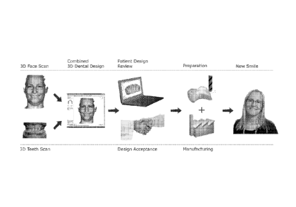
Figure 1: Motivation for this invention, outline of flowchart with graphical
illustrations for clarity.
Figure 2: Detailed flow chart for variant V1 of the method described in this
invention.
CA 02755555 2011-09-15
WO 2010/105628 PCT/DK2010/050063
23
Figure 3: Detailed flow chart for variant V2 of the method described in this
invention.
Figure 4: Sagital section of a schematic tooth, visualizing various steps of
the
method described in this invention.
Figure 5: Zoomed sagital section of a schematic tooth, illustrating step the
transfer of the pre-preparation design to the prepared teeth.
Figure 6: Graphical representation of some steps in this invention.
Figure 7: Example screen snapshots of CAD software showing face model
with part of the smile cut out and model of restoration (affecting teeth 6-11)
and tissue (segmented) aligned to that of the face. For the sake of being able
to distinguish face scan and restoration model in this Figure, the color of
the
restoration was intentionally not attempted matched that of the teeth in the
face scan (this is visible even in the black-and-white pictures). (a):
anterior
view, (b): lateral view.
Detailed description
In the following description, reference is made to the accompanying figures,
which show by way of illustration how the invention may be practiced.
In one embodiment of the invention (in the following termed "V1" and
illustrated in the flow chart in fig. 2) a pre-treatment (pre-prepared) 3D
model
of the patient's teeth is used, preferably obtained with a 3D scanner.
Optionally, another 3D model of the patient's face (possibly obtained with
another type of scanner) is exploited for optimal alignment and/or aesthetic
look of the restoration.
In another embodiment of the invention (in the following termed "V2" and
illustrated in the flow chart in fig. 3), the 3D facial model is required,
while the
pre-treatment 3D model is optional.
CA 02755555 2011-09-15
WO 2010/105628 PCT/DK2010/050063
24
Both V1 and V2 may comprise similar steps, however in a different
combination and with slight differences. Optional steps and models in V1 and
V2 are indicated by dashed borders in the flowchart elements. Some steps
are optional only in V1 or V2.
Some steps may be implemented in software, while others may represent
manual work and/or application of machinery. The software is preferentially a
single program, for optimal ease of use.
Some steps are also illustrated graphically in figs. 4 ¨ 7.
Step 1: Obtain 3D digital model of pre-treatment teeth and gingiva
(required in V1, not applicable in V2):
There are several commercial systems available for obtaining 3D digital
models 100 of teeth (e.g., Cadent iTero, 3M ESPE Lava, 3Shape D640).
Among these are intra-oral scanners and scanners for dental impressions or
casts thereof (e.g., 3Shape D640). Scanners can be for example be optical
scanners (laser, structured light). Guidelines in the relevant scanner
manufacturer's operations manual should be followed for obtaining the 3D
model 100. This model 100 will in the following also be denoted as the pre-
preparation model. Its contour in figure 4 is C100.
Potentially, scanners with penetrating radiation such as (cone beam) CT
scanners (Imaging Science International's i-CAT, Kodak/lmtec's Iluma) can
be used to obtain model 100. They have the advantage of providing
volumetric models showing also decay inside the teeth, while disadvantages
include concerns about radiation dose or high price of treatment. A teeth
scan is shown in figure 6, step 1.
Step 2: Segment pre-prepared teeth from gingiva (optional in V1, not
applicable in V2):
CA 02755555 2011-09-15
WO 2010/105628 PCT/DK2010/050063
Optical scanners generally obtain a 3D digital model of an object's surface.
While this model describes geometry, it does not differentiate between any
materials or sub-objects that make up the surface. Specifically for dental
applications, the 3D model does not differentiate between teeth and gingival,
5 some of which inadvertently will be included in a teeth scan. For
visualization
and CAD design of dental restorations, it can therefore be advantageous to
segment the combined 3D model into teeth and tissue, respectively.
Segmentation can be applied by means of an algorithm implemented in
software, yielding model 101. A segmented teeth model is shown in figure 6.
In one embodiment of the invention, the segmentation algorithm uses vectors
perpendicular to each tooth, or a single vector, perpendicular to the whole
model, and one point in the middle of each tooth or two points on the distal
and mesial sides of the tooth. A preferred version of the separation algorithm
is based on using a 3D shortest path algorithm, preferably capable of
handling negative weights, for example the Bellman-Ford algorithm. The
algorithm is preferably applied on a 3D matrix with elements representing
curvature of the surface of the tooth model 100.
In another embodiment, the scanner used to generate model 100 can
capture color as well. Segmentation can then be based on color information.
If step 2 is skipped, models 100 and 101 are identical.
Step 3: Obtain 3D digital model(s) of face/head (optional in V1, required in
V2):
There are several systems available for obtaining 3D digital models of the
head, particularly the face (e.g., Konica Minolta Vivid, Breuckmann
faceScan). Head/face and dental scanners are generally different
instruments, because the required resolution for head/face scans is generally
CA 02755555 2011-09-15
WO 2010/105628 PCT/DK2010/050063
26
lower, while the volume of interest is larger. Most optical head/face scanners
employ structured light.
In a preferred embodiment of this invention, the head/face scanner can
detect not just surface geometry, but also color. Color (also termed texture)
information is important in visualization. Color can be detected directly by
choosing a color-sensitive sensor in the scanner's camera(s). Another
approach is to use a sensor sensitive to total light intensity only, but take
several images where the illumination is a single base color in each, and then
reconstruct the color by combining those images. This process is also called
stacking of color channels, and typical base colors are red, green, and blue.
It is preferable to scan the head/face with the patient exposing his or her
teeth. This constellation can be exploited in step 6. Generally, the patient
will
want to smile, because the aesthetic appearance of a dental restoration is
often viewed most critical for a smile.
A 3D model of the head may require several scans from different angles.
Multiple such scans have to be aligned to a combined model. Many
algorithms exist for this purpose, for example Iterative Closest Point. They
all
require some overlap of at least pairs of sub-scans. As lighting in every sub-
scan generally will differ, the sub-textures need to be color-adjusted for the
combined texture. For example, texture weaving can be employed to smooth
color differences between different sub-scans [4].
Due to limited reflectivity, the hair portion of the head is generally
difficult to
capture with optical scanners. This limitation can be overcome by powdering
the hair with a reflective powder. Another method to reconstruct the hair
portion in 3D is to extrude the silhouettes in multiple head images (taken
from
different angles) and then to intersect them to form the visual hull
approximation.
CA 02755555 2011-09-15
WO 2010/105628 PCT/DK2010/050063
27
Subsequent to step 3, the flow charts splits into two branches. These
branches are not alternatives, but can both be executed. They start with
steps 4a and 4b, respectively.
Step 4a: Derive jaw motion (optional in V1, optional in V2):
Especially for crown design, it can be advantageous to account for dynamic
occlusal interferences. With a plurality of face scan models 200, it is
possible
to deduct the articulation of the jaw and thus simulate dynamic occlusal
interferences given a 3D model of the teeth 101 that includes the antagonist.
To deduct the articulation from 3D facial models, it may be advantageous to
fix one or more reference sphere(s) to the patient's mandibular teeth,
preferably between the lower lip and the mandibular incisors, and to track
that sphere's motion. The procedure is described for a single sphere and 2D
images in [9], but can be expected to be more precise with 3D data, and to
correctly detect rotational movements if more than one sphere is used.
Additional sphere(s) or object(s) can also be fixed on the patient's maxillary
teeth. With 3D data, any concurrent movement of the head can be separated
from movement of the jaw during chewing. WO 2009/091439 discloses a
procedure where 3D movements are deducted by tracking dental objects.
This is however much less accurate than using reference sphere(s), because
spheres, unlike dental objects, have a perfect geometric surface from which it
is possible to determine a center position with high precision and accuracy.
Accordingly, many metrological standards employ reference spheres, e.g.
ISO 10360-3.
Step 4b: Remove teeth (optional in V1, optional in V2):
If any face/head model 200 shows the teeth (and possibly the gingival
scaffold), it can be advantageous to cut them out, and to later (e.g. in step
6)
display the teeth/gingiva model 101 (V1) or 401 (V2) in their place. The
latter
model will often have a higher degree of detail, as a high level of detail is
CA 02755555 2011-09-15
WO 2010/105628 PCT/DK2010/050063
28
required for modeling the dental restoration in step 6. The cutting could also
apply to only some of the teeth, for example if the model 101 or 401, resp.,
only has some teeth, or even a single tooth. Said cutting is performed in
software, where it can be performed interactively or at least partially
automated.
Interactive cutting can for example be performed in 3D software by placing a
3D line on the model, cutting out all points and/or facets inside the line.
Possibly, facets can be sub-divided along the cutting line, such that the
cutting line is respected precisely. One way of entering the cutting line in
the
3D software is to click on some reference points, and use a spline to connect
them. The spline should follow the surface of model 200.
Automated cutting can be performed by detecting teeth (and possibly
gingiva) by software algorithms. For example, teeth can be detected as such
by their color and/or their shape.
If model 101 or 401, resp., includes a complete set of teeth, the inner
commissure is the preferred section in model 200 to cut out, as delineated by
the innermost confluences of the vermillion of the lips at the corners of the
mouth [1].
Step 5: Align teeth model to face scan (optional in V1, not applicable in
V2):
In this step, the teeth/gingiva model 101 is aligned with some head/face
model 200, or ¨ if step 4 was performed - the cut head face model 201. In
other words, the position of model 101 becomes that of the corresponding
portion of model 200/201, and both can be displayed simultaneously in a
meaningful way. Alignment is thus a rigid transformation of at least one
model, either into the local coordinate system of the other, or into some
other
common coordinate system.
CA 02755555 2011-09-15
WO 2010/105628 PCT/DK2010/050063
29
The alignment is preferably performed in software, interactively and/or
automatically. Interactive alignment can be performed in the graphical user
interface provided by the software by dragging a model (translation), or
dragging some control points for rotations. Another way to transform a model
is to enter or adjust the transformation matrix directly.
The criterion for alignment can be a subjective visual fit or be defined
mathematically. A common such criterion is the sum of squared distances
between the two models. Distances are usually measured in the direction of
the surface normals. Other criteria could be based on the distances between
certain features, such as the incisal planes, or the midline(s) between
incisors.
Automatic alignment can be performed using the same algorithms as in step
3. Possibly, the user will have to place control points for corresponding
points
of the models to be aligned, those serving as a first guess for the automated
fine alignment. Automated alignment is an optimization of the mathematically
defined fit criterion. In case the head/face model 200 does not expose the
teeth on the surface, alignment with the teeth/gingiva model 101, alignment
can still be possible if the head/face model is a cephalogram (x-rays of the
head) [5].
Step 6: Design restoration in CAD system (required in V1, required in V2):
This step is largely identical in both variants (i.e. V1 and V2), but starts
from
the pre-prepared teeth in variant V1, while in variant V2, it starts from the
prepared teeth. The earlier position in the work flow in variant V1 allows
some additional possibilities in this variant.
Common features in step 6 in both variants V1 and V2:
Dental restorations that can be designed in a CAD system include inlays,
onlays, veneers, crowns, bridges, combinations thereof, and others. By
CA 02755555 2011-09-15
WO 2010/105628 PCT/DK2010/050063
analogy, the term "restoration" also covers removable partial denture
frameworks and implant-retained structures. Several dental CAD software
packages that allow such design are available, for example 3Shape
DentalDesigner. Model 300 is that of the restoration only. In this step 6, it
is
5 only a digital model. Its contour in figure 4 is C300. The restoration
implies
requirements for the preparation. As model 300 is digital, the preparation is
also virtual in this step 6. For a given restoration model 300, there can be
many possible virtual preparations C102, however some may be more
advisable than others (see step 7). An example contour of a virtual
10 preparation in figure 4 is C102. Mainly, C102 is offset from C300 by the
cement space. Note that the thickness of the cement space in figure 4 is
exaggerated for graphical clarity only.
The software used in this step 6 should preferably assist the dentist/dental
15 technician in designing the restoration, for example by making automatic
suggestions and/or evaluating basic rules and requirement.
Basic rules and requirements, preferably implemented in the software, may
include the minimum thickness for the restoration (generally dependent on
20 material) and biologic width. Other rules could ascertain the mandatory
continuous circumferential height of a preparation for a crown. The strength
of a restoration could be determined numerically, for example by measuring
the thickness or preferably a finite-element simulation. Yet another rule
could
be to not to penetrate the antagonist and proximal teeth.
In the common case of the head model 101 not being a volumetric one, it can
be advantageous to integrate x-ray images in this step 6, because the extent
of decay visible in these will constrain the choice of restoration. If
multiple x-
ray images are taken from different angles, it will be possible to create an
approximate 3D model from the silhouettes in all images, analogously to how
the hair can be reconstructed in 3D in step 3. The resolution in 3D of this
CA 02755555 2011-09-15
WO 2010/105628 PCT/DK2010/050063
31
model will however generally be poor, because only few X-rays can be taken.
Because of this poor quality, said integration of x-ray images in step 6 may
not be a proper alignment to the other models, but at least a concurrent
visualization in the software. Possibly, the software can detect the image
planes of the x-rays in the 3D model of the teeth (101 in variant V1, 401 in
variant V2) by a best fit between their sections, and then automatically set
the view port in the 3D visualization of the latter models to match the image
planes of the x-rays.
A major advantage of this invention is that it enables a dialog between the
patient and the dentist regarding the treatment, optionally involving the lab
also. For example, the dentist can visualize the proposed restoration on a
computer screen. Preferably, the CAD software that the dental
technician/dentist uses for the virtual design of the restoration itself
provides
such visualization and can be used interactively to update the design in
dialog with the patient. The technician/dentist could propose visual
appearance and aesthetic as well as explain functional advantages and
disadvantages of potential restorations, along with cost. A physical
diagnostic
wax-up could also be manufactured by CAM, still more cheaply and quickly
than traditional diagnostic wax-ups.
It is advantageous to be able to render the available 3D models photo-
realistically. Graphics functionality on PCs, like OpenGL, aids towards this
goal. Proper, or even adjustable, coloring of gingiva and teeth, respectively,
or regions thereof, in teeth / gingival models (101 in variant V1, 401 in
variant
V2) is likewise advantageous. Even if said models were obtained with a
color-enabled scanner, the lighting used to capture it is generally different
from that applied when capturing model 200, leading to a visual mismatch in
the display of all models aligned (step 5). Special computer graphical
techniques, like ray tracing, can improve the visual appearance, along with
the modeling of more than one light source.
CA 02755555 2011-09-15
WO 2010/105628 PCT/DK2010/050063
32
When a face/head model 201 is available, biometric information can be
exploited for optimizing the aesthetic impression of the dental restoration
[6].
For example, it often appears ideal to align the facial midline with the arch
midline, or to achieve parallelism between the incisal plane and the
interpupillary line. Metrics for of smile anatomy include the degree of
maxillary anterior tooth display (Morley ratio), upper lip drape, and gingival
display [1].
If the dental restoration deviates significantly from the existing conditions,
it
may have effect on the soft tissue near the mouth. 3D facial soft-tissue-
change prediction after simulated orthognathic surgical planning has been
presented in the literature [e.g., 7], and an analogous procedure could be
applied in the context of this invention. The outcome of any (optional) soft-
tissue change simulation could be visualized as model 202.
If step 4a has been performed and a trajectory of the mandibular teeth has
been determined, dynamic occlusal interferences can be tested in the
present invention, allowing the dentist/dental technician to modify model 300
in order to avoid such interferences. This procedure may be at least partially
automated removing any parts of model 300 that collide with the antagonist
given said trajectory.
When the CAD design is finished, a physical diagnostic wax-up model of the
digital model 300, or parts thereof, can be manufactured by CAM. Such
manufacturing requires essentially no manual labor and is much less
expensive than traditional manual production. The physical wax-up gives the
dentist and/or patient another opportunity of evaluating the proposed
treatment before it is executed. This may be a relevant procedure especially
when the restoration design is performed in a dental lab at another location
or the dentist is very traditional. If a physical diagnostic wax-up is
created, the
lab technician may be required to grind on the pre-preparation model before
CA 02755555 2011-09-15
WO 2010/105628 PCT/DK2010/050063
33
scanning. In case no physical model exists one can be manufactured by
CAM.
In another embodiment of the invention the CAD design can used to create
"snap on"s, which can mounted directly on the patient teeth visualizing the
treatment result. The "snap on"s are directly created by subtracting the pre-
prepared teeth from the design. I.e. the 3D model of the pre-prepared teeth is
subtracted from the CAD model of the designed "snap-on" The resulting
subtracted design provides the a model of the snap-on's that subsequently
can be manufactured by CAM whereupon the snap-on's are ready-to-use.
Communication networks provide other means of establishing interactivity
with patient and/or dentist in a situation where the restoration design is
performed in another location. For example, the patient and/or dentist could
follow the design process via a life internet connection to the designer's
computer.
Variant V1 only: In one embodiment of this invention, the dentist or dental
technician demarcates the desired margin for the restorative design on the
teeth model 101 in the software. In another embodiment, the dentist chooses
a desired surface of the restoration, e.g., from a crown library (potentially
but
not necessarily the same as in the corresponding parts in model 101), and
the software calculates a margin line. Any combination of said embodiments
is also possible, particularly for bridges. Possible automatic suggestions in
the software include margin placement, particularly apical placement
dependent on tooth number. The dentist may also be offered a selection
among a library of standard restorations, which then can be modified.
In this step 6, but also with relevance for the preparation (step 7 below),
also
temporary crowns can be designed. The temporary crown will be directly
derived from the full CAD design in step 6, but with additional cement space
CA 02755555 2011-09-15
WO 2010/105628 PCT/DK2010/050063
34
e.g. 0.2 mm between the virtual preparation and the inside of the temporary
crown. The increased cement space is created to accommodate for
inaccuracies in the actual preparation performed by the dentist.
Step 7: Generate preparation guide (optional in V1, not applicable in V2):
In a preferred embodiment of the invention the software assists the dentist
with the preparative work. In many cases, general preparation guides are
provided by manufacturers of dental material and equipment. To ease the
dentist's work and to improve the restorative strength and overall quality,
the
invention may provide the preparation guides automatically for the particular
design obtained at the end of step 6.
Possibly, the software can assist with planning crown lengthening. In this
context, step 2 can be beneficial, preventing the margin from being placed
too sub-gingivally. Also the type of margin (bevel, shoulder) could be
suggested by the software.
Besides proposing details of the preparation, the software that generates a
preparation guide can possibly also validate a preparation that the dentist
and/or dental technician have devised by other means. For example, the
software can evaluate restorative strength and/or choice of materials, and/or
even the choice of restorative treatment method.
The preparation guide can take many forms including instruction text,
multiple 2D screen shoots, 3D animations, computer visualization, videos
and/or instructions for machined/robot preparation. A preparation guide may
also include a physical model of the desired, positive, preparation, or a
physical negative representation which can be tested in the mouth of the
patient. For example in the case where model 100 is a scanned cast model,
the dental technician could prepare this cast. Because the virtual preparation
CA 02755555 2011-09-15
WO 2010/105628 PCT/DK2010/050063
is also available in digital form (the dental preparation CAD model, contour
C102 in fig 4), it could also be manufactured by CAM.
Step 8: Prepare teeth (required in V1, required in V2):
5 Based on the agreed restorative treatment and with or without any guide
from
step 7, the dentist prepares the patient's teeth. The preparation is typically
performed by the dentist grinding down the teeth such that the restorative
work can be glued on. In variant V1, the preparation will be for the
restoration
designed in step 6, whereas in variant V2, no prior design determines the
10 preparation work.
Snap-ons (a commercial product by Snap-on Smile) require no invasive
preparation.
15 Step 9: Obtain 3D digital model of prepared teeth and gingiva (required
in V1, required in V2):
In terms of procedure, this scanning step is identical to step 1, however in
this step, the prepared teeth are scanned. The contour of the actual
preparation in figure 4 is C400. For the sake of simplicity in the figure, it
is
20 identical with that of the virtual preparation C102, but this need not
be the
case.
Step 10: Segment prepared teeth from gingiva (optional in V1, optional in
V2):
25 The segmentation of teeth and gingival in the prepared model can be
executed analogously to step 2, but applied to the prepared model instead of
the pre-prepared model. If this step is skipped, models 400 and 401 are
identical.
30 Step 11: Align (required in V1, required in V2):
CA 02755555 2011-09-15
WO 2010/105628 PCT/DK2010/050063
36
Logically and procedurally, this step is similar in variants V1 and V2;
however
this step relates to different models in either variant.
Variant V1: Alignment of restoration designed for the pre-prepared teeth
(step 6, model 101), prepared teeth (model 401) can be performed by the
same software algorithms as described in step 3. Again, it is important to
have some overlap in the models. Such areas will generally exist unless the
preparation affects all teeth. The model of the restoration 300 is already in
the same local coordinate system as model 101, based on which it was
designed. Therefore, model 300 is also aligned with model 401 without any
further processing. If the head/face scan (model 201) is available, it can be
aligned to models 101/300 and 401 such that all three models match. Figure
7 shows a typical result of this step 11 for such constellation.
Variant V2: Alignment of prepared teeth (model 401) and the head/face scan
(model 201) can be performed by the same software algorithms as described
in step 3.
Step 12: Transfer CAD design from pre-prepared to prepared teeth
(required in V1, not applicable in V2):
Due to the manual preparation the actual preparation C402 (fig 5) will in
general differ, at least slightly, from the virtual preparation C102 created
in
the design step 6. Thus, the restoration design needs to be modified
accordingly, but preferably the transfer should maintain as much of the
design created in step 6 as possible. This procedure is preferably
implemented in software.
The automation provided by this step 12 is what lacks in the manual and
subjective process that is the current state of technology. Typically today,
to
transfer the design, the dental technician looks at the original diagnostic
wax-
up and manually tries to replicate this design for the real restorations,
CA 02755555 2011-09-15
WO 2010/105628 PCT/DK2010/050063
37
incorporating potential comments from the dentist and the patient. This
manual replication process is both costly, inaccurate, and time consuming.
A preferred algorithm for this step 12 starts by demarcating the margin line
in
both the virtual preparation (600 in fig 5) and the actual preparation (601 in
fig 5). While the margins are points in the 2D cross section that is fig 5, in
reality they are curves in 3D, and can for example be represented by (B-
)splines. Dental CAD software like 3Shape's DentalDesigner can
automatically detect margin lines and place said splines, but user interaction
should also be allowed. The transformation between 600 and 601 is denoted
T
A free form deformation (FFD) model can used to generalize T to surfaces.
This process is often also called "morphing". The morphing operation affects
the near-margin portion of model 300, with decreasing impact for surface
portions with decreasing distances from the margin. The relevant parameters
of the algorithm can be adjusted by the user. A similar procedure for "crown
matching" has been proposed in [8], however outside the scope of dental
restorations. Colloquially speaking, morphing is like stretching a rubber
balloon by pulling or pushing its "lips" (the thick ring through which the air
is
blown in, which corresponds to the margin line).
Note that in the example shown in figure 5, the prepared margin is located
gingivally from the virtual one, so therefore the exterior surface of model
300
needs to be extended to arrive at model 301. The opposite case is however
also possible. If the preparation ends up removing less material than
assumed when creating model 300, the exterior surface of model 301 can be
smaller than that of model 300. In other words, morphing can both be a
contraction and a stretching operation. For a given tooth, morphing can even
be a combination of contraction and stretching along various sections of the
CA 02755555 2011-09-15
WO 2010/105628 PCT/DK2010/050063
38
margin, namely when there are deviations between the virtual and actual
preparation in both the gingival and the occusal / incisal direction.
Away from the margin towards the interior portions of the
preparation/restoration, morphing need not be applied. Instead, the inner
surface of the restoration can be computed in the normal fashion, i.e., the
surface is created by an offset of the preparation above the margin line
controlled by several parameters.
Away from the margin along the exterior of the restoration and beyond the
radius of influence of the morphing operation (section 600 to 602 in fig 5),
the
surfaces of pre-prepared design and final design are identical, i.e. contours
C300 and C301 overlay each other.
By combining the identical, the morphed, and the preparation-generated
surface the final CAD design 301 (contour C301) is completed. When
creating the final CAD design, material and manufacturing process
requirements should be included, e.g. the actual design can be split into two
files for pressing. If step 7 was skipped and thus model 102 is not truly
available (it is the same as model 101), the virtual margin line can also be
taking from the model of the restoration 300.
Further modifications to the design of the restoration can be made with the
same procedures as mentioned under step 6. If color was adjusted in step 6,
it may be advantageous to transfer the color information to the design and
later manufacture of the restoration.
Only in the unlikely event of the actual preparation matching the virtual one,
and no other modifications being desirable, will models 300 and 301 be
identical.
CA 02755555 2011-09-15
WO 2010/105628 PCT/DK2010/050063
39
Step 13: Produce (optional in V1, optional in V2):
Once model 301 has been finalized, it can be produced using CAM
(Computer Aided Manufacturing). Both rapid prototyping (RP) machines and
milling machines can be used for the actual production. A CAM software (e.g.
3Shape CAMbridge) prepares the data (including model 301) for production.
For RP machines this preparation typically involves 3D rotation, placement
(nesting), supports, slicing, ID-tags, etc. For milling machines the
preparation
typically involves 3D rotation, placement (nesting), sprues (connector pins),
drops, engraving, milling path generation and post processing, etc. Some
dental CAD/CAM solutions include the same internal steps of preparation for
production and are thus technically suitable for the method described in this
invention, but are currently not open to 3D models generated by other
manufacturers' equipment (e.g., Sirona CEREC).
The production process can either manufacture the restoration immediately
(e.g., from blocks of zirconia), or indirectly. In the indirect process, for
example wax is milled or printed and then cast using traditional "lost wax"
techniques. Many manufacturers offer RP (SLA, SLS, SLM, DLP, FDM,
Polyjet, etc.) and/or milling machines suitable for such work, e.g., Roland,
3DSystems, EnvisionTec, Solidscape, DWS, EOS, ProMetal, and others.
Manufacturing may in many cases be performed at another location than the
preceding steps. Digital models and designs can for example be transferred
to a processing center via the internet.
Although some embodiments have been described and shown in detail, the
invention is not restricted to them, but may also be embodied in other ways
within the scope of the subject matter defined in the following claims. In
particular, it is to be understood that other embodiments may be utilised and
structural and functional modifications may be made without departing from
the scope of the present invention.
CA 02755555 2011-09-15
WO 2010/105628 PCT/DK2010/050063
In device claims enumerating several means, several of these means can be
embodied by one and the same item of hardware. The mere fact that certain
measures are recited in mutually different dependent claims or described in
5 different embodiments does not indicate that a combination of these
measures cannot be used to advantage.
It should be emphasized that the term "comprises/comprising" when used in
this specification is taken to specify the presence of stated features,
integers,
10 steps or components but does not preclude the presence or addition of
one
or more other features, integers, steps, components or groups thereof.
The features of the method described above and in the following may be
implemented in software and carried out on a data processing system or
15 other processing means caused by the execution of computer-executable
instructions. The instructions may be program code means loaded in a
memory, such as a RAM, from a storage medium or from another computer
via a computer network. Alternatively, the described features may be
implemented by hardwired circuitry instead of software or in combination with
20 software.
References
25 [1] Ackerman MB, Ackerman JL. Smile analysis and design in the digital
era.
J Clin Orthod. 2002; 36, 221-36.
[2] Xia J, Wang D, Samman N, Wai R, Yeung K, Tideman H. Computer-
assisted three-dimensional surgical planning and simulation: 3D color facial
model generation. Int J Oral & Maxillofacial Surgery 2002, 29 (1), 2-10.
CA 02755555 2011-09-15
WO 2010/105628 PCT/DK2010/050063
41
[3] Rangel FA, Maal TJ, Berge SJ, van Vlijmen OJ, Plooij JM, Schutyser F,
Kuijpers-Jagtman AM. Integration of digital dental casts in 3-dimensional
facial photographs. Am J Orthod Dentofacial Orthop. 2008, 134 (6), 820-6.
[4] Callieri M, Cignoni P, Scopigno R. Reconstructing textured meshes from
multiple range + rgb maps.VMV 2002, Erlangen, Nov 20-22, 2002.
[5] Zhao B, Ong SH, Foong KWC. Multimodal Registration of Dental and
Facial Images. Proc (444) Signal and Image Processing. Hawaii, Aug 23-25,
2004.
[6] Ahmad I: Anterior dental aestethic: Facial perspective. Brit Dental J
2005,
199 (1), 15-21.
[7] Xia J, Samman N, Yeung RW, Wang D, Shen SG, lp HH, Tideman H.
Computer-assisted three-dimensional surgical planning and simulation. 3D
soft tissue planning and prediction. Int J Oral Maxillofac Surg. 2000 29 (4),
250-8.
[8] Hassana H, El-Baza A, Faraga AA, Farmanb AG, Tasmanb S, Millera
WM. A volumetric 3D model of the human jaw, CARS 2005: Computer
Assisted Radiology and Surgery 2005. 1244-
1249.
doi :10.1016/j.ics.2005.03.345
[9] Pinheiro AP, Andrade AO, Pereira AA, Bellmo D. A computational method
for recording and analysis of mandibular movements. J Appl Oral Sci.
2008;16 (5): 321-7