Note: Descriptions are shown in the official language in which they were submitted.
CA 02755731 2011-09-16
WO 2010/112815 PCT/GB2010/000564
1
MEDICAL APPARATUS
FIELD OF THE INVENTION
The present invention relates to medical apparatus for monitoring one or more
physiological conditions of a patient and a method for monitoring one or more
physiological
conditions of a patient.
BACKGROUND OF THE INVENTION
In medical devices, such as patient monitors, physiological measurements may
be
performed in which a cable is attached directly to a patent at one end by
electrode wires to
perform physiological tests, such as an electrocardiogram (ECG). A common
design used to
perform ECG measurements, consists of a number of electrode patches connected
to the
patient's skin in which voltage variations are recorded over a period of time,
and the resulting
signals are processed, stored and interpreted. The electrical signals sensed
by the electrodes
are commonly amplified and filtered in order to generate useful data. Although
there are
systems in the prior art for monitoring the physiological condition of a
patient many of these
prior art systems require a patient to wear a type of body monitor which then
sends signals to
a computer device such as a pda or a laptop. Example prior art systems are
described in US
2006/009697, US 2008/058614, US 5,417,222 W02002/30277, W098/30145,
US2007/0213622, US5377687, EP0059172, US2008/0058614, W02005/018447,
W099/16351, EP1 127538 and W02000/51677.
Certain prior art devices provide a computer print out or alternatively
connect to third
party computers. This limits the uses of such devices. Furthermore, a number
of patents
describe physiological monitors including portable ECG monitors such as US
5,701,894
which describes an ambulatory physiological recorder that includes multiple
selective plug-
and-play signal input conditioners, a microprocessor system and operating and
analysing
software, and a removable memory module for data storage. In US 6,454,708
there is
described a system for monitoring health parameters and capturing data from a
subject. The
system includes a cordless sensor band with sensors for measuring full
waveform ECG, full
waveform respiration, skin temperature, and motion, and a connector which
accepts a
memory card or a smart card for storage of measured data.
Prior art devices are limited because:
1. Although cordless and wireless sensor bands and other prior art devices can
give a
full waveform ECG they do not always give a full 12 lead diagnostic quality
ECG recording as
required by a qualified medic.
2. Cordless and wireless sensor bands have inherent problems regarding battery
and
power usage e.g. potentially, at the critical moment, the battery runs out.
CONFIRMATION COPY
CA 02755731 2011-09-16
WO 2010/112815 PCT/GB2010/000564
2
3. Cordless and wireless sensor bands have inherent problems regarding
bandwidth
e.g. potentially, at the critical moment, there is data loss.
4. Prior art devices are not fully integrated to or with a computer or a
computer
network e.g. patient data management becomes problematic with data loss and/or
loss of
resolution and/or integrity, and/or minimal or poor computing functionality.
5. Furthermore, these devices are not fully integrated with or into a computer
or a
computer network and in those cases. where data is transferred onto a memory
card, this can
be problematic for real time remote viewing by a health care professional
(HCP).
Existing ECG devices used currently by the health care system do not lend
themselves to portability and are kept in the surgery or health centre. These
are also usually
trolley mounted devices which merely produce a printed report which has to be
scanned in or
faxed to a specialist, resulting in low quality reports. This type of paper
system also generates
a patient confidentially problem for a user, where the patient details are
exposed.
There is therefore a need in the art to provide a medical apparatus which: 1)
is fully
integrated to or within a computer system or a computer network 2) optionally,
complies with
regulatory safety standards for medical equipment 3) optionally, provides
clinical grade
resolution recordings, 4) optionally, provides the bandwidth facility to
monitor a patient in real
time 5) optionally, provides the facility to monitor a patient remotely, 6)
optionally, has minimal
computational impact on a computer it is integrated with.
Furthermore, prior art medical data gathering devices e.g. electrocardiogram
(ECG)
measurement devices do not provide the functionality of a computer with a
standard
operating system and standard programs such as Microsoft Word , Microsoft
Outlook . This
functionality is often missed by health care professionals in their day to day
activities,
especially now that patient data records are being centralized or where these
are being
reviewed remotely.
It is an object of at least one aspect of the present invention to obviate or
mitigate at
least one or more of the aforementioned problems. It is a further object of at
least one
example embodiment of the present invention to provide improved medical
apparatus which
is capable of measuring data relating to a physiological condition of a
patient and directly
transmitting collected information to a computer. It is a further object of at
least one aspect of
the invention to provide improved medical apparatus.
SUMMARY OF THE INVENTION
In a first aspect of the invention there is provided apparatus for measuring
medical
data comprising: at least one medical data gathering module, at least one
microprocessor, in
CA 02755731 2011-09-16
WO 2010/112815 PCT/GB2010/000564
3
which the apparatus further comprises a medical data handling module separate
from the at
least one microprocessor for buffering the transfer of medical data between
the medical data
gathering module and the at least one microprocessor.
Optionally, the medical data handling module is arranged to gather and store
data in
at least one predetermined group of data and the at least one microprocessor
is arranged to
retrieve data from the medical data handling module in one or more multiples
of the at least
one predetermined group of data. Optionally, the predetermined group of data
is N bits of
data and/or, optionally, T1 seconds worth of data. Optionally, the medical
data handling
module is arranged to gather N bits of data every T1 seconds from the least
one medical data
gathering module and store the sampled bits of data in the predetermined group
of data.
Optionally, 1/T1 is the rate that the medical data handling module samples
data from the at
least one medical data gathering module. Optionally, one or more multiples of
the
predetermined groups of data comprises one or more multiples of T1 seconds
worth of data,
and/or one or more multiples of N bits of data. Optionally, N equals 24 to 54,
24, 48, 54, 56 or
any multiple thereof. Optionally, gathered medical data is stored and
retrieved by the at least
one microprocessor intermittently from the at least one medical data handling
module.
Optionally, the microprocessor is a personal computer microprocessor or a
laptop
microprocessor and/or the microprocessor comprises Microsoft Windows or Apple
Mac or
Linux operating systems and/or, optionally, the apparatus comprises a computer
comprising
the at least one microprocessor. Optionally, the at least one medical data
gathering module,
the at least one microprocessor and the medical data handling module are
located within a
unitary housing or, optionally, within a unitary personal computer housing or,
optionally, within
a unitary laptop computer housing, Optionally, either 1) the at least one
medical data
gathering module, at least one microprocessor and the medical data handling
module are co-
located within a first housing, or, optionally, 2) the at least one medical
data gathering module
and data handling module are co-located in a first housing and the least one
microprocessor
is located within a second housing and communication means are provided to
enable the
medical data handling module and at least one microprocessor to communicate
via wire
and/or wirelessly, or, optionally, 3) the at least one medical data gathering
module is located
within a first housing and the at least one medical data handling module and
at least one
microprocessor are co-located within a second housing and communication means
are
provided to enable at least one medical data gathering module and at least one
medical data
handling module to communicate via wire and/or wirelessly. Optionally, where a
wireless
connection is provided this may be any one or more wireless connections
standards such as
infrared, WIFI, Bluetooth, 3G, satellite capability and so on.
CA 02755731 2011-09-16
WO 2010/112815 PCT/GB2010/000564
4
Optionally, the housing, or first housing where first and second housings are
provided,
comprises at least one socket for connecting at least one medical data
gathering module(s) to
at least one medical data gathering connecting cable. Optionally, 1) a first
housing for the at
least one medical data gathering module is provided and a second housing for
the
microprocessor, is provided and the first and second housing are adapted so
that the first
housing can sit beneath the second housing or vice versa; and/or, optionally,
2) an outer
periphery of the first housing substantially matches an outer periphery of the
second housing
or vice versa; and, in either case, the medical data handling module is
located within the first
housing or the second housing. Optionally, the apparatus further comprises a
cable
identifying circuit for identifying the at least one medical data gathering
cable connected
thereto and/or, when the at least one medical data gathering module comprises
an
electrocardiogram data gathering module, for identifying whether a three lead
or 10 lead
electrocardiogram connecting cable is connected and/or, optionally, whether no
ECG cable is
connected.
Optionally, the medical data handling module comprises either 1) a further
microprocessor or, optionally, 2) a further microprocessor and memory or,
optionally, 3)
system programmable on a chip or, optionally, 4) a field programmable gate
array (FPGA).
Optionally, the medical data handling module comprises at least one first in
first out (FIFO)
memory buffer. Optionally, the medical data handling module comprises a field
programmable gate array and the field programmable gate array comprises a
first in first out
(FIFO) buffer. Optionally, the medical data handling module comprises either,
1) a
programmable read only memory, or, optionally, 2) an erasable programmable
read-only
memory (EPROM), or, optionally, 3) an electronically erasable programmable
read only
memory (EEPROM), for delivering instructions to the medical data handling
module.
Optionally an EEPROM is used such as a Serial Flash Memory (SFM) to hold the
FPGA
program.
Optionally, the at least one data gathering module comprises one or more of an
electrocardiogram data gathering module, an invasive or non-invasive blood
pressure (BP)
monitoring data gathering module; spirometry (lung function) data gathering
module; pulse
oximetry data gathering module; temperature data gathering module; both
invasive and non-
invasive blood pressure (BP) data gathering module; audiometry testing data
gathering
module; retinal testing data gathering module; ultrasound data gathering
module;
dermatology screening (image capture) data gathering module; imaging, tissue
and/or wound
care data gathering module; video endoscopy data gathering module; video
conferencing
data gathering module e.g. for live remote consultations; video and/or image
CA 02755731 2011-09-16
WO 2010/112815 PCT/GB2010/000564
capture data gathering module e.g. for remote consultations that may or may
not be live;
audio for detection of heart and/or lung sounds data gathering module;
scaliometer (height)
data gathering module; foetal heart Doppler ultrasound and/or audio
acquisition and/or
analysis data gathering module; and weighing scales data gathering module.
Optionally, an
5 electrocardiogram (ECG) data gathering module and/or a blood pressure (BP)
data gathering
module are provided. Optionally, simultaneous video conferencing functionality
is provided
e.g. for live consultations. This may be combined with live video/image
medical data capture
functionality for forwarding medical video/images during a video conference.
As will be appreciated by those skilled in the art, optionally, in addition to
any one or
more of the above mentioned data gathering modules being provided for
interface with a
microprocessor via a medical data handling module, one or more of the above
medical data
gathering modules may also be provided for connection directly to the at least
one
microprocessor, for example, via an internal data bus, such as an internal USB
data bus
which may have plug and play functionality. Typically, the apparatus is
arranged so that data
from at least one medical data gathering module is buffered by the medical
data handling
module. Optionally this may be one or both of an ECG data gathering module and
a BP data
gathering module. Optionally in addition, one or more further medical data
gathering modules
is provided connected to an internal data bus of the apparatus so as to
deliver data to the at
least one microprocessor directly (such as spirometer head 46 in figure 9). It
will be
appreciated by those skilled in the art that additional medical data gathering
functionality can.
be added using medical data gathering modules connected to the internal data
bus of a
computer using for example a USB plug and play port, and by modification
and/or upgrading
and/or expanding the medical data processing software in the microprocessor.
Whilst it is preferred in one aspect of the invention for at least one medical
data
gathering module and at least one medical data handling module for buffering
data therefrom
to at least one microprocessor to be provided, in another separate aspect of
the invention, the
medical apparatus may comprise at least one microprocessor, and at least one
internal data
bus, and at least one medical data gathering module adapted for connection to
the internal
data bus, for example via a USB or other plug and play connector.
Optionally, in one example embodiment of either aspect, the apparatus
comprises at
least one medical data gathering module, at least one medical data handling
module for
buffering data therefrom to at least one microprocessor, at least one internal
data bus, and at
least further one medical data gathering module adapted for connection to the
internal data
bus, for example, via a USB or other plug and play connector.
CA 02755731 2011-09-16
WO 2010/112815 PCT/GB2010/000564
6
Optionally, the medical data handling module is connected to at least one
microprocessor of the personal computer or laptop computer, optionally, using
an internal
serial bus or an internal universal serial bus (USB). Optionally, the
apparatus further
comprises a revision controlled motherboard for hosting the at least one
microprocessor.
Optionally, a medical grade power supply is provided, optionally within a
housing where one
is provided, optionally, to power the at least one medical data gathering
module, optionally,
the at least one microprocessor and, optionally, the data handling module.
Optionally, the apparatus further comprises a medical data gathering cable
lead off
circuit for identifying when a medical data gathering cable has become
disconnected from a
patient, and/or, optionally, the apparatus further comprises a medical data
gathering cable.
Optionally, the microprocessor comprises a medical data request retrieval
module to
request data retrieval from the medical data handling module. Optionally, the
at least one
medical data gathering module gathers data quasi-continuously, and/or,
optionally, at a
regular intervals and/or, optionally, the at least one microprocessor
retrieves data
intermittently and/or, optionally, or the at least one microprocessor
retrieves data
intermittently, optionally, either at regular or irregular intervals.
Optionally, the medical data
handling module samples data from the at least one medical data gathering
module across N
channels once every T1 seconds. Optionally, T1 is in the range 0.25ms
(millisecond) to 5ms,
or 0.5ms to 2.5ms, or T1 is 0.5ms, 1 ms, 2ms or 5ms. Optionally, the least one
microprocessor attempts to retrieve data in multiples of N from the data
handling module
once every T2 seconds, and/or, optionally, the at least one microprocessor may
comprise.
medical data request retrieval module to retrieve data in multiples of N from
the data handling
module once every T2 seconds. Optionally, when the at least one microprocessor
misses
retrieving data either, optionally, 1) the at least one microprocessor
attempts to retrieve data
in multiples of N from the data handling module at another time and/or at the
end of the next
T2 second interval, and/or, optionally, 2) the at least one microprocessor
comprises medical
data retrieval module to retrieve data in multiples of N from the data
handling module at
another time and/or, optionally, at the end of the next T2 second interval.
Optionally, a
medical data gathering rate of sampling once every Ti seconds is the same as a
medical
data retrieval request rate of once every T2 seconds i.e. T1 equals T2.
Optionally, a medical
data retrieval transfer rate is QxN bits per T2 seconds where Q is the number
of sampling
rounds of data to be retrieved and/or the medical data retrieval rate is up to
the data transfer
rate of an internal communications bus of the medical apparatus. Optionally,
data in excess
of a multiple of N and/or, optionally, in excess of a multiple of T1 seconds
worth of data, is left
in the medical data handling module until the next retrieval round.
CA 02755731 2011-09-16
WO 2010/112815 PCT/GB2010/000564
7
Optionally, analysis and calculation means is provided for use by the
microprocessor
to conduct analysis and calculation on the medical data at a resolution of T1
seconds, the
resolution of the medical data gathering rate. Optionally, drawing and display
means, and
optionally a display, are provided for use by the microprocessor to draw every
Mth data value
to a display.
In a further aspect of the invention there is provided a method for measuring
medical
data, comprising: providing at least one medical data gathering module;
providing at least one
microprocessor; providing a medical data handling module separate from the at
least one
microprocessor; buffering medical data transfer between the medical data
gathering module
and the microprocessor the medical data handling module. Optionally, the
method comprises
gathering and storing data in predetermined groups of data in the medical data
handling
module and retrieving data from the medical data handling module in one or
more multiples of
the predetermined groups of data. Optionally, the method comprises sampling N
data bits
every T1 seconds, optionally, storing N data bits in memory every T1 seconds,
optionally,
reading N or, optionally, a multiple of N data bits every T2 seconds,
optionally, emptying the
memory of multiples of N data, optionally, leaving any fragments of multiples
of data until
following data collection round. Optionally, Ti equals T2, and/or, optionally,
data retrieval
transfer rate is QxN bits per T2 seconds where Q the number of sampling rounds
of data to
be gathered.
According to a further aspect of the invention there is provided apparatus
comprising
a cable and a computer, said cable comprising: at least one lead capable of
connecting the
cable to a computer; and at least one further lead comprising medical
components capable of
detecting a physiological condition of a patient and the computer comprising
cable
connections components so that the cable is capable of detecting and
transmitting
information relating to the physiological condition of the patient to the
computer. In this
aspect, the invention therefore relates to a cable capable of providing a
direct connection
between medical components (e.g. ECG electrodes) capable of measuring and/or
detecting a
physiological condition of a patient and then transmitting collected
information to the
computer.
In particular embodiments, the medical components may therefore comprise a
physiological measurement electronic circuit which may then transmit the
detected and/or
measured information. There may be at least one or a plurality of leads
capable of connecting
the at least one or a plurality of cables to the computer. At the end of at
least one of the leads
there may be an end socket which may comprise a series of pins which are
capable of
attaching the cable directly to a computer. The pins may insert into a
suitable receiving socket
CA 02755731 2011-09-16
WO 2010/112815 PCT/GB2010/000564
8
in the computer. Typically, the receiving socket may be within a wall of a
housing of the
computer. There may be one or a plurality of leads capable of connecting the
cable to
medical components. The medical components such as electrodes may be used to
detect at
least one or a plurality of physiological conditions of the patient by
measuring, for example,
vital signals from a patient. The electrodes may be attached to a patient's
head, limbs and/or
the chest area.
Optionally, the invention may comprise: an identifying mechanism to identify
the cable
and the medical components to the computer. Optionally, the invention may
comprise a
bespoke connector e.g. to prevent connection of foreign cables.
Optionally, in particular embodiments, the medical data gathering module may
monitor
and detect any one or more or any combination of the following physiological
conditions:
electrocardiogram (ECG) signals; invasive or non-invasive blood pressure;
spirometry (lung
function); and pulse oximetry; temperature; both invasive and non-invasive
blood pressure
monitoring; audiometry testing; retinal testing; ultrasound screening;
dermatology screening
(image capture); imaging, tissue viability and wound care screening; video
endoscopy; video
for remote consultations; video conferencing; audio for detection of heart
and/or lung sounds;-
scaliometer (height); foetal heart Doppler ultrasound audio acquisition and
analysis and
weighing scales.
Electrical components of the cable and/or computer in one example embodiment
may
contain improved circuitry that may be capable of measuring and/or detecting
ECG signals. In
certain embodiments, the improved circuitry provides a method of data transfer
to reduce or
substantially avoid data loss within the bandwidth required by a standard PC.
Alternatively or in addition the improved circuitry may provide a safe method
of
connecting a patient directly to a computer system and network, for example by
means of
defibrillation protection circuits in the cable and/or within the computer
e.g. optical isolator
circuit(s) on the medical data gathering circuit board. For example, the
improved circuitry may
comprise a first stage where an input to the ECG consists of a defibrillator
and/or mains
potential electric protection. This protection may be contained within the
cable and/or may
also be within the first stage of an ECG input.
Optionally, a physiological signal sensed from the skin of a patient by one or
all
electrodes of the cable may form an input to a buffer amplifier. Outputs of
the buffer amplifier
may be connected to a resistor array network in, for example, a star delta
formation. A centre
of the resistor network may form a common signal which may be used for common
mode
signal rejection and may form an input to a separate amplifier which is
connected and drives
the screen of the cable. The aim of this is to reduce noise which is common to
all electrodes
CA 02755731 2011-09-16
WO 2010/112815 PCT/GB2010/000564
9
and reduce capacitively generated noise in the cable. Optionally, each
electrode or
considered lead position may have its own buffer amplifier and the output of
the buffer
amplifier may be routed to the input of a different amplifier. The different
amplifier circuit may
provide a signal gain of approximately 140 and may provide a high degree of
slew rate
limiting. Quad amplifiers may be used where the first stage of the quad
amplifier serves as a
fixed gain. The second stage may serve as a slew rate limiter. The output of
third and fourth
stages may be common together and may be used as comparators to detect if the
electrode
has become removed from the patient's skin, resulting in amplifier saturation.
The output of
the second stage of the quad amplifier and the common output of the third and
fourth stages
may form independent inputs to a plurality of analogue to digital converters.
In one example embodiment, the invention may therefore be computationally
efficient
by providing a plurality of analogue to digital converters in the improved
circuitry. Optionally in
addition, in a further effort to reduce computational cycles, the controlling
signals to the
analogue to digital converters may be common and the analogue to digital
converters may be
set to run in synchrony. For example, at least two, and in particular three
analogue to digital
converters may be utilised thus reducing the computational cycles to address
the analogue to
digital converters three fold. The analogue to digital converters may be
arranged to sample
the signals at a rate of 1 kHz to 20kHz, or more, say once every 0.5ms
(millisecond), or once
every 1 ms. Each output from the plurality of analogue to digital converter
may be interfaced
to high speed electromagnetic isolators with a high degree of isolation from
the input to the
output to ensure patient safety.
Optionally, an integrated circuit, such as a fully programmable gate array
(FPGA),
optionally having a first in first out (FIFO) memory module may be employed to
sample from
the isolators (and hence from the analogue to digital converters) optionally,
at a pre-
programmed rate of, for example, between about 1 - 10kHz, say at l kHz or once
every
millisecond. Optionally, this may be half the rate of the sampling by the
analogue to digital
converters. This reduces computational cycles and in particular the
computational burden on
the computer because this part of the circuit is independent from the host
computer. The
computer may then sample the first in first out buffer at a sample retrieval
request rate of
between, for example, about 1 - 20 Hz to empty the buffer and store the data
into the
computer memory. The actual rate of transfer of sampled data may be within the
sample
rates achievable by data bus architecture embedded within central processing
units (typically
much faster than 10 kHz). The data may then be ready for analysis by suitable
software and
displayed on a display of the computer or transmitted. For example, the data
may first be
compressed and then encrypted to preserve patient confidentially before being
stored into
CA 02755731 2011-09-16
WO 2010/112815 PCT/GB2010/000564
memory of the computer and/or transmitted to a remote location via a network
connection.
Once received at the remote location, the data may be de-encrypted and
decompressed in
order to be displayed on another computer.
This electrocardiogram circuitry and signal analyser may be housed in a
computer or
5 within a laptop computer such as on a shelf which may, for example fit into
a standard
computer ROM bay drive. Any personal computer with available bays or suitable
laptop
computer may be used for the present invention but in one example embodiment,
optionally,
it is preferred if the computer is fitted with a revision controlled mother-
board. Optionally, it is
also preferred that the computer is fitted with a medical grade power supply.
The computer
10 used in the present invention may be any suitable computer. The computer
may comprise a
recess within which, for example, a ROM enclosure may be inserted. A circuit
board may also
be attached to, for example, a shelf in the computer. A front face plate may
also be attached
to the front of the computer. The front face plate may comprise an aperture
through which the
computer may be connected to medical apparatus. The computer may comprise an
electronic
circuit which is designed to specifically use less computational cycles and
therefore be
computationally efficient in sampling physiological signals. For example, the
electrocardiogram may comply with EN ISO 60601-2-25 relating to ECG equipment
and
ENISO 60601-2-27 relating to ECG monitoring equipment.
According to a further aspect of the present invention there is provided a
method of
detecting and transmitting information relating to the physiological condition
of the patient to
the computer using apparatus comprising cable and a computer according to one
aspect of
the invention.
According to a further aspect of the present invention there is provided
apparatus for
detecting and/or monitoring a physiological condition of a patient, said
apparatus comprising:
a computer; and a cable comprising at least one lead capable of connecting the
cable to a
computer and at least one further lead comprising medical apparatus capable of
detecting a
physiological condition of a patient.
The computer used may be a desktop computer or alternatively may be a laptop
computer. The computer may be specially adapted for the present invention. For
example,
the computer may comprise an amplifier such as an ECG amplifier. Optionally,
the computer
may comprise an increased depth to accommodate an ECG interface and/or a blood
pressure interface. Optionally, the computer may also be portable such as a
laptop computer.
Within the computer there may be monitoring apparatus for monitoring a
patient's vital signs.
The apparatus according to the present invention may allow a healthcare worker
to make a
decision on the vital signs health status of a patient without the need for
any other external
CA 02755731 2011-09-16
WO 2010/112815 PCT/GB2010/000564
11
patient monitoring equipment required. The electronic circuitry of the data
handling module
overcomes the technical difficulties relating to computer processing power and
real time
analysis when using, for example, the relatively low clock speed of the
operating system by
providing a separate medical data handling facility. In more detail:-
1. Diagnostic quality patient monitoring requires a sample rate of the signal
that is
reliable with no data loss i.e. (1 to 10 kHz) x 24 signals to sample.
2. Although computers are fast (currently around 3GHz), the operating system
clock
actually runs quite slowly (around 20 Hz).
3. The present inventors had to find a way of monitoring the patient with
diagnostic
quality under the constraints of the operating system.
4. Optionally, the circuitry samples all 24 of the analogue signals at 1 kHz
(e.g. all 24
channels at every 1 ms) or at 2 KHz (e.g. 24 channels once every 0.5ms).
5. Optionally, A/D's operate in synchrony to sample the data to save
computation
cycles
6. Optionally, the data is held in a medical data handling module, optionally
comprising for example a FIFO buffer, optionally, in 0.25ms, 0.5ms, lms, 2ms,
5ms, 10ms,
20ms, 25ms, 30ms, 50ms chunks. Optionally, the FIFO buffer may be 4kbits or
32kbits. For
example, if 54 bits of data are held in 1ms groups of data than the 4kbit FIFO
can hold
54bitsx74ms = 3996 bits of data i.e. 74 groups of data of size 54 bits. A
32kbit FIFO would be
able to hold 592ms of data held in 54 bit groups of data (54bits x 592ms =
31968bits) i.e. 592.
bits of data of size 54 bits.
7. Optionally, the software then samples the data and is transferred to the PC
CPU at
a leisurely 20Hz.
Thus the present inventors have found a way to transfer the data with minimal
data
loss.
In a particular embodiment of the present invention, a user may affix ECG
electrodes
to a patient's chest in the normal way, but plug the electrode cables directly
into the computer
via a built in ECG socket. The ECG socket may be wired to circuitry which
amplifies and
signal conditions the ECG waveform. The signal may be digitised before being
electrically
isolated. A digital signal emitted from the apparatus may be routed to an
internal bus of the
computer, directly to the CPU for analysis. The computer may perform cardiac
analysis and
may display waveforms and results on a screen. The screen may be a standard
LCD screen.
The user may also wrap a blood pressure cuff around a patient's arm and plug a
pneumatic
hose directly into a blood pressure port on the side of a computer. The
computer may then
perform blood pressure analysis and display waveforms on a display screen. The
circuitry
CA 02755731 2011-09-16
WO 2010/112815 PCT/GB2010/000564
12
required to perform the detection and analysis of a patient's vital signs may
be built into and
incorporated into a computer such as a laptop computer. Typically, the
circuitry which
interfaces with the computer for either or both, for example, ECG and blood
pressure
analysis, may utilise an improved method of data transfer which uses very low
computer
processing power. The computer may also comprise means such as memory and
associated
software which may save all results for future pending analysis or comparison
of future
recordings. The computer may also be configured to send encrypted vital signs
data through
an available network connection to a specialist unit (accident and emergency
or coronary
care unit) which may help make an informed decision on the health status of a
patient. Data
sent across a network may also be automatically encrypted by the device to
prevent patient
data information being accessed by an unauthorised person. The laptop computer
may also
be suitable for transmitting data for "live" remote monitoring by a special
unit. A web cam may
also be installed into the computer to enable a remote specialist to see the
patient via video
or captured pictures which will help with remote diagnosis, for example, skin
cancer, wound,
fracture advice and the like. Alternatively or in addition, the apparatus may
comprise video
conferencing capability e.g. for real time video conferencing with a health
care professional.
This may be in addition to the functionality of video or image recording of a
physiological
condition. Computer software in the computer may also be used to access local
patient
information databases to update the patient record with the current results
and any findings or
further information or referral advice.
The apparatus according to the present invention may therefore be used to make
more acute or emergency decisions. For example, currently if a person has
suffered a
myocardial infarction (i.e. a heart attack), there is a time period in which a
patient needs to
have specialist treatment. This time period is often critical especially if a
patient is away from
specialist help. The apparatus according to the present invention may be used
to help local
healthcare personnel to administer specialist treatment under the remote
specialist advice.
The apparatus according to the prevent invention may therefore help to prevent
unnecessary
journeys to hospital. Currently remote or rural patients have to travel
considerable distances
to gain specialist advice or treatments, when actually, some patients do not
need to make
such a journey. The apparatus according to the present invention may therefore
help a local
healthcare professional make an informed decision on transferring- a patient
to a territory
hospital or specialist unit.
The apparatus therefore provides advantages over existing methods relating to
patient safety (where devices may be plugged into an unknown computer which
may not be
CA 02755731 2011-09-16
WO 2010/112815 PCT/GB2010/000564
13
approved for safe use within the patient environment) and convenience (because
all of the
required functions are within 1 unit).
A method of assessing the ECG of a subject under test may comprise any one of
or
combination of the following steps: performing an ECG test at recording
apparatus located at
a first physical location; propagating from the first physical location via a
network connection,
to a receiver apparatus located at a second physical location, spaced from the
first location;
conducting at a receiver computer the assessment of the ECG and generating a
test result;
and propagating from the receiver computer to the recording apparatus a
clinical assessment
statement; and presenting the result in a readable manner to a user at the
recording
apparatus. The method may also comprise the step of propagating from the
recording
apparatus at the first physical location via a network connection which also
includes the step
of propagating from the recording apparatus via a wireless network connection.
The ECG
signal may be encrypted for the process of transmission, and encryption
reversal at the
receiver apparatus.
The patient recording may be capable of displaying the clinical data in
predetermined
format, displaying the electrocardiograph data in a graphical format, using
software custom
design for this purpose. The patient report may include addition fields for
use by the user,
wherein the user is capable of adding to the patient report within the
addition fields. The
additional fields may be capable of receiving text information from a user who
is a cardiologist
and wherein the text information includes analysis and diagnosis data.
Optionally, it is preferred that the apparatus may be able to filter noise.
Optionally,
noise filtering can be done in software and/or in the electronics. Optionally
raw data is
delivered to the software in the microprocessor with minimal or no filtering.
Optionally, filtering
is done in the software using a finite impulse response (FIR) filter or using
an infinite impulse
response (IIR) filter. Optionally, the apparatus may signal condition the
signal by buffering the
shape of the signal to the A/D converter(s) on the way to the FPGA.
Optionally, there is also
provided a means of the signal sensed from each electrode being routed to a
buffer amplifier
and each electrode has its own buffer amplifier. Typically, the apparatus may
utilize a plurality
of analogue to digital converters arranged in such a way as to enable
simultaneous or
substantially simultaneous monitoring of each electrode to reduce computation
operations
and therefore be computationally efficient.
Optionally, the invention seeks to ensure that a multi channel ECG may be
simultaneously monitored, for example by using at least two analogue to
digital converters,
whilst using the minimum of computing effort through use of minimal computing
operations
and may safely be used to perform medical measurements.
CA 02755731 2011-09-16
WO 2010/112815 PCT/GB2010/000564
14
BRIEF DESCRIPTION OF THE DRAWINGS
Example embodiments of the invention will now be described by way of example
only
with reference to the following figures in which like reference numerals refer
to like referenced
features.
Figure 1 shows a schematic perspective view of a personal computer and its
housing
and an exploded perspective view of an ECG interface card assembly according
to an
example embodiment of the invention.
Figure 2 shows an exploded perspective view of an ECG interface card assembly
according to an example embodiment of the invention.
Figure 3 shows an exploded perspective view of a blood pressure (BP) interface
card
assembly according to an example embodiment of the invention.
Figure 4 shows four perspective views of a laptop computer and its housing
according
to an example embodiment of the invention.
Figure 5 shows a schematic functional block diagram of apparatus according to
an
example embodiment of the invention.
Figure 6 shows a schematic block diagram of functional and software modules of
apparatus according to an example embodiment of the invention.
Figure 7 shows schematic diagram of ECG and BP interface cards, data handling
module interface card and computer microprocessor motherboard according to an
example
embodiment of the invention.
Figure 8 shows schematic block diagram of various components for ECG and BP
measurements, and interconnections via data handling module interface card and
internal
computer address bus according to an example embodiment of the invention.
Figure 9 shows a schematic diagram of a further medical data gathering module
in the
form of spirometer head and a universal serial bus (USB) connector according
to an example
embodiment of the invention.
Figure 10 shows, schematic perspective view of an example ECG cable according
to
an example embodiment of the invention.
Figure 11 shows a plan view of an example 10 lead ECG cable according to an
example embodiment of the invention.
Figure 12 shows a plan view of an example 3 lead ECG cable according to an
example embodiment of the invention.
Figure 13 shows a schematic block diagram of an example data handling module
and
connections to 2 medical data gathering modules according to an example
embodiment of
the invention.
CA 02755731 2011-09-16
WO 2010/112815 PCT/GB2010/000564
Figure 14 shows a further schematic block diagram of an example data handling
module according to an example embodiment of the invention.
Figure 15 shows a schematic block diagram of a field programmable gate array
(FPGA) and associated electronically programmable read-only memory (EPROM) and
clock
5 oscillator according to an example embodiment of the invention.
Figure 16 shows a schematic circuit diagram of a circuit for use in an ECG
medical
data gathering module according to an example embodiment of the invention.
Figure 17 shows a schematic block diagram of a circuit for use in a BP medical
data
gathering module according to an example embodiment of the invention.
10 Figure 18 shows a schematic circuit diagram of an example circuit for use
in a BP
medical data gathering module according to an example embodiment of the
invention.
Figure 19 shows a schematic circuit diagram of an alternative circuit for use
in a BP
medical data gathering module according to an example embodiment of the
invention.
Figure 20 shows a schematic block diagram of a flow diagram for a BP medical
data
15 gathering module according to an example embodiment of the invention.
Figure 21A shows a schematic representation of a medical data gathering
module,
data handling module and computer including microprocessor and the timing of
data transfer
therebetween according to an example embodiment of the invention.
Figure 21 B shows a schematic representation of steps taken by the
microprocessor in
displaying data at a relatively low resolution, and in analysing data at a
relatively high
resolution according to an example embodiment of the invention.
Figure 22 shows a schematic representation of medical data gathering modules
(medical data gathering interfaces such as ECG and BP interfaces), data
handling module in
the form of an FPGA, a communications interface in the form of a USB, and
software running
on a PC microprocessor according to an example embodiment of the invention.
Figure 23 shows a representation of a user interface screen for patient
details and
reports according to an example embodiment of the invention.
Figure 24 shows a representation of a user interface screen for use with a
blood
pressure data gathering module according to an example embodiment of the
invention.
Figures 25 and 26 show representations of user interface screens for display
of ECG
data and calculations in connection therewith, according to an example
embodiment of the
invention.
Figure 27 shows a representation of the user interface screen for use with a
spirometer according to an example embodiment of the invention.
CA 02755731 2011-09-16
WO 2010/112815 PCT/GB2010/000564
16
Figure 28 shows a table of N data bits relating to ECG data, skin temperature
data
and BP data according to an example embodiment of the invention.
Figure 29 shows an exploded view of part of a laptop computer according to an
example embodiment of the invention.
DETAILED DESCRIPTION OF THE DRAWINGS
Turning now to the figures, figure 1 shows apparatus 1, according to one
example
embodiment of the invention. Apparatus I comprises a personal computer (PC) 2
having
housing 4 and a medical data gathering module 6. Here, medical data gathering
module is in
the form of an electrocardiogram (ECG) interface assembly 6 mounted in a PC
ROM bay 10
so that a front wall 7 of ECG interface assembly 6 forms part of housing 4 of
the PC 2 (see
figure 2). In figure 2, an ECG socket 12 is mounted in a front wall or front
face plate 7 for
providing direct connection to a patient via an ECG patient cable (not shown).
An ECG
interface card 14 is provided within ECG interface assembly 6 to mount ECG
power, control
and data gathering components thereon.
In figure 3, an additional or alternative medical data gathering module is
provided in
the form of a blood pressure (BP) interface assembly 16. In this example, BP
interface
assembly 16 is for non-invasive blood pressure measurement and has a lid 8. A
BP interface
card 18 is provided within BP interface assembly 16 to mount BP power, control
and data
gathering components thereon as well as a pump 21. Alternatively, pump 21 is
located
adjacent BP interface card 18. The pump 21 leads to a cuff connector 20 via a
pump
connector 20'. BP interface assembly 16 is mounted in a PC ROM bay 10 (see
figure 1), so
that a front wall 7 (see figure 2) of BP interface assembly 16 forms part of
housing 4 of the
PC 2. The BP cuff connector 20 is mounted in the front wall 7 of BP interface
assembly 16 for
providing direct connection to a patient via a BP (air) cable and cuff (not
shown).
Figure 4 shows perspective views of a laptop computer 22 adapted to form
apparatus
according to one example embodiment of the invention. The laptop computer 22
has a
housing 24 and, in this example embodiment, a housing extension 26. As in the
case of a
desktop computer 2 seen in figure 1, one or more data gathering modules, for
example, in the
form of ECG interface band/or BP Interface assembly 16 are built into the
laptop computer
and are not visible here being hidden by housing 24. However, an ECG socket 12
and a BP
cuff connector 20 for providing direct connection to a patient are visible in
housing 24. In this
example, these are located in optional housing extension 26. The housing
extension 26 has
been formed in line with and contiguous with the rear wall of housing 24 of
laptop computer
22, so as to provide a clean finished line to rear wall of housing 24.
CA 02755731 2011-09-16
WO 2010/112815 PCT/GB2010/000564
17
Briefly turning now to figure 29, an exploded view of part of a laptop
computer base
22' is shown. Optionally, laptop computer base 22' has a display (not shown).
Laptop
computer 22' has a housing assembly 24 comprising a housing base 24' and a
first part of a
housing lid 24". A further cover (not shown), optionally, in the form of a
keyboard, may be
fixedly mounted on housing lid 24" to form a lid of unitary housing 24. Covers
25 below base
24' may also form a part of unitary housing 24, for example when fixedly
mounted on housing
base 24'. Housing lid part 24" is typically formed from a metal plate for
strength and
optionally has an extension 26' having a front plate 7' with an aperture 12'
and another
aperture for mounting a cuff connector 20. A pump 250 for connecting to cuff
connector 20 is
shown. ECG socket 12 is shown, and in normal use, is fixedly mounted in
aperture 12'.
Laptop 22' comprises a revision controlled mother board 42 to which ECG
interface
card 14, BP interface card 18, FPGA board 150 and CPU assembly 43' are
connected and/or
mounted thereon. Laptop 22' also comprises a hard drive 300, rechargeable
battery 160' and
DVD drive 301. A front wall 7 of housing extension 26 in housing base 24' is
arranged so that
aperture 12" is aligned with aperture 12' for mounting ECG socket 12 therein.
Likewise an
aperture in front wall 7 is provided for aligning with cuff connector 20.
Thus, in figures 1, 4 and 29, the PC 2 and laptop computer 22/22' also
typically
comprise at least one core microprocessor and standard operating system such
as Microsoft
Windows , Apple Mac or Linux operating system. Thus, apparatus according to
the
invention provides all the normal functionality of a personal computer or
laptop computer, as
well as one or more medical data gathering modules. In these embodiments, this
is provided
within a single housing (housing 4 in figure 1 and housing 24 in figure 4). By
providing both
data gathering and computing functionality within a single unitary integrated
housing, a
number of advantages are provided. Firstly, whilst taking measurements with a
patient, a
HCP has access to all the usual computing functionality for carrying out other
parallel
activities (e.g. Microsoft Word , Microsoft Outlook for email etc). Secondly,
the data
collected by the one or more data gathering modules is immediately available
for use by the
usual suite of programs available on computers, e.g. word processing,
spreadsheets, email
etc). Thirdly, there is no need to provide a separate computing facility
separate from the
medical apparatus, since this is provided along with the computer. By
utilizing the features of
the invention and in particular the features of claim 1 of the invention, the
medical device side
of the apparatus can operate with minimal affect on the operation of the
computer and vice
versa, i.e. the computer can operate with minimal effect on the operation and
indeed data
collection of the medical device. Fourthly, because the computer either
desktop or Laptop is
adapted, in effect now a piece of medical apparatus and easily identifiable as
such (for
CA 02755731 2011-09-16
WO 2010/112815 PCT/GB2010/000564
18
example in one embodiment by the presence of one or more sockets or connectors
(12, 20)
and/or housing extension 26 in the housing 4, 24), it is a much less
attractive proposition for
theft, an important criterion when expensive computing devices, especially
portable ones, are
used in a public health care setting.
In an alternative embodiment (not shown), a medical data gathering module such
as
ECG interface assembly 6 and/or BP interface assembly 16 can be mounted in a
separate
second housing, distinct and separate to a first housing 4 or 24. In such
circumstances, wired
or wireless communication components may be built into each housing to enable
communication between the first and second housings, and the components
therein. In one
version of this alternative embodiment, medical data handling means for
sampling data from
medical data gathering modules (e.g. from modules 6 and 16) is built into the
first (computer)
housing 4, 24. In another version of this alternative embodiments, medical
data handling
means for sampling data from the medical data gathering modules e.g. from 6,
16, is built into
the second housing (not shown) along with the medical data gathering modules.
In either
case, where two housings are provided, it may be advantageous e.g. from a
space saving
perspective, if one housing is adapted so the other can be located above it,
for example, by
having a flat upper surface, or other suitable mounting means. Alternatively
or in addition, the
size and shape of an outer periphery of the one housing may be substantially
similar or
identical to that of the other housing. For example, the size and shape of the
second housing
may be substantially similar or identical to that of the first housing so that
the first PC/laptop
housing 4, 24 can be placed thereon and adopt a similar overall outline. This
arrangement is
particularly suitable for a laptop computer embodiment such as that shown in
figure 4, so that
first laptop housing 24 may be placed upon the second housing containing the
medical data
gathering modules. Alternatively, and particularly suitable for a desk top PC
housing 4 (see
figure 1), the upper surface of the PC housing may be adapted so that the
second housing
can sit thereon. For example, it may be that the size and/or shape of an upper
portion of the
first (PC) housing may be substantially similar to or identical to that of the
second housing
containing the medical data gathering modules so that the second housing can
be placed
thereupon (not shown).
Figure 5 shows schematic functional block diagram of apparatus according to
one
embodiment of the invention. Figure 5 shows patient contacting hardware 30
comprising an
ECG patient lead 32, a blood pressure cuff 34 and a spirometer mouthpiece 36.
Figure 5 also
shows internal hardware and software 40 comprising an ECG interface card 14, a
BP
interface card 18, an internal data bus 38, a personal computer platform 42
comprising a
microprocessor (such as a PC motherboard) and a separate data handling module
48.
CA 02755731 2011-09-16
WO 2010/112815 PCT/GB2010/000564
19
Additional internal hardware in the form of a spirometer 46 is shown connected
directly to the
PC platform 42. BP cuff 34 connects to PC platform 42 via, firstly, BP
Interface card 18 and,
secondly, via data handling module 48. Similarly, ECG patient lead 32 connects
to PC
platform 42, firstly, via ECG interface 14 and, secondly, via data handling
module 48.
Figure 6 shows a schematic block diagram of the hardware and software
functional
modules of apparatus according to one embodiment of the invention. Here the
apparatus
comprises an analysis system 50 having hardware modules 52 and software
modules 54.
ECG interface card 14 and BP interface card 18 are connected to an ECG
acquisition module
56 and BP measurement module 62 via ECG and BP output command lines 68,
control lines
70, ECG and BP data feedback lines 72 and ECG and BP data input lines 74 via
medical
data handling module 48. In practice output command lines 68 and input data
lines 74 are
part of internal data bus 38 (see figure 5) Thus lines 68 and 70 transmit
control signals and
lines 72 and 74 feedback data sampled as described elsewhere herein.
Similarly, spirometer
46 is controlled by and feedbacks to spirometry measurement modules 64 via
output
command line 78 and data feedback line 80. Patient data is fed into a patient
data software
module 66 (and optional patient data file) by bidirectional patient data input
line 76. The
patient data may be encrypted and/or compressed within a special format file.
ECG
acquisition module 56 connects to an ECG data module 58 which in turn connects
to an ECG
measurement and calculation module 60 (here a dynamic link library module).
Optionally,
ECG feedback line 74 also feedbacks results to ECG measurement and calculation
module
60 from medical data handling module 48 (optionally in the form of an FPGA
module).
Figure 7 shows an ECG interface assembly 6, ECG interface card 14 and ECG
socket
12 mounted in front wall 7. Figure 7 also shows BP interface assembly 16, BP
Interface card
18, pump 21 and cuff connector 20 mounted in front wall 7. An interface data
bus 38, here a
40 way IDE (integrated drive electronics) connector links ECG interface card
14 and BP
Interface card 18 to medical data handling module 48, for example, here a
peripheral
interface board or a field programmable gate array (FPGA) board. A medical
data handling
microprocessor 49 is mounted on medical data handling module 48. Medical data
handling
microprocessor 49 may be a system-programmable-on-a-chip (SPOC) in the form of
a field
programmable gate array (e.g. an FPGA manufactured by Altera) as will be
described in
more detail later. Alternatively, an ASIC such as a Rapid Chip from LSI may be
used as a
medical data handling module. The PC motherboard 42 having computer
microprocessor 43
and PCI standard card socket 45 is also shown. It should be noted that medical
data handling
microprocessor 49 is separate and distinct from computer microprocessor 43. In
this example
embodiment, medical data handling module 48 is also provided on a PCI standard
card
CA 02755731 2011-09-16
WO 2010/112815 PCT/GB2010/000564
having a PCI standard connections for slotting into PCI standard card socket
45 on PC
motherboard 42. Typically, this is used in the desktop PC version. In the
laptop computer
version, the data handling module 48 typically communicates with the mother
board 42 via a
USB wire connection located internal to the laptop housing 24 (see figures 4
and 29), or via
5 wireless connections where a first and second housing are provided. Other
variations on
these arrangements can be envisaged by those skilled in the art. It will be
also understood by
those skilled in the art that whereas both ECG and BP data gathering modules
are described,
one or both or alternate medical data gathering modules may be used.
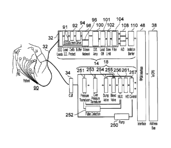
Figure 8 shows ECG and BP measurement components in more detail. A patient 90
is
10 connected to ECG interface card 14 via an ECG patient lead 32. ECG
interface card 14
comprises a first protection circuit, for example, in the form of
defibrillation protection circuit
92, lead identifier circuit 186, a buffer circuit 94, ECG screen drive
amplifier 96, a Wilson
network circuit 98, a differential amplifier circuit 100, a lead off circuit
101, a slew rate limiter
circuit 102, a filter circuit 104, at least one and here three analogue to
digital converters 108
15 and a second protection circuit, for example, in the form of an isolation
barrier 110. ECG
interface card 14 connects to data handling module 48 in the form of a bespoke
interface card
that connects via internal data bus 38 to computer microprocessor 43 (not
shown). An ECG
lead screen drive signal 91 for screening the ECG leads 32 is derived from the
Wilson
network 96. A medical data handling module 48, here in the form of an FPGA
module, and an
20 internal data bus 38 are also shown.
BP interface card 18 comprises pressure transducer 251, pulse detection
circuit 252,
over pressure transducer 253, dump valve 254, bleed valve 255, multiplexer
256, analogue to
digital (A/D) converter 261, pump control circuit 257 and pump 250. A cuff 34
connects the
BP interface to a patient.
Figure 9 shows a spirometer head 46 connected to a USB connector 112 for
connecting to a USB port in the housing 4, 24 of PC 2 or laptop 22. Thus as
shown in figures
8 and 9, two medical data gathering modules 14, 18 are connected via medical
data handling
module 48 to internal communications bus 38. A separate medical data gathering
module,
here a spirometer head 46, is connected via USB connector 112 to internal
communications
bus 38 and directly to microprocessor 43 (seen in figure 7). One or more
additional medical
data handling modules, such as any medical data gathering module described
herein, may
thus be added and connected to the microprocessor e.g. using USB connectors to
connect to
internal data bus of the microprocessor. It is therefore possible to add
additional functionality
with only additional software changes required, no or minimal hardware changes
required.
Thus in one example embodiment, the present invention provides seamless
addition of
CA 02755731 2011-09-16
WO 2010/112815 PCT/GB2010/000564
21
separate functionality of any one or more medical data gathering modules by
plugging directly
into internal communications bus 38, e.g. via USB ports or other plug and play
facility by
appropriate modification and/or upgrade of functionality of internal software
of the apparatus.
Figures 10 and 11 show respectively, perspective and plan views of a ten lead
ECG
patient cable 32. Ten lead ECG cable 32 has an optional central connection
point in the form
of an ECG lead hub 120, ECG connecting lead 122, and ECG cable plug 124. ECG
lead plug
124 has numerous pins 125 connected to patient lead connections N, R, C1, C2,
C3, C4, C5,
C6, L and F (or RA, LA, LL, C1, C2, C3, C4, C5, C6, RL and S depending upon
terminology
used) via ECG lead hub 120. Lead connections, N, R, C1, C2, C3, C4, C5, C6, L
and F
connect to the body of a patient in a standard pattern well documented and
understood by
those skilled in the art. In brief, six leads namely C1, C2, C3, C4, C5 and C6
are placed
around the chest using connection leads and electrode patches for detecting
heart rhythm
signals with respect to a lead placed on a limb. This gives six waveforms.
Three original lead
positions (as in a 3-ECG cable) gives three more signals, then with respect to
a second (other
limb lead) this gives three new augmented waveforms. In total 12 waveforms
result from a ten
lead ECG measurement.
Figure 12 shows a three lead ECG cable 32 having an ECG lead hub 120, ECG
connecting lead 122, and ECG cable plug 124. Defibrillation protection in the
form of ECG
protection circuit 128 up to 5 kV may be provided within ECG lead hub 120 in
the three lead
and 10 lead ECG cables of figures 11 and 12.
Figure 13 shows a schematic block diagram of a medical data handling module 48
in
the form of an FPGA board, in this example an FPGA printed circuit board (PCB)
150. Also
shown are ECG interface card 14 and BP interface card 18. A system-
programmable-on-a-
chip (SPOC) in the form of a field programmable gate array integrated circuit
152 receives
instructions in the form of software from an electronically programmable read-
only memory
154 (either EEPROM or EPROM may be used although it is preferred that the
program is
fixed within the medical apparatus during manufacturing at its factory
settings). Typically the
EEPROM 154 is programmed during manufacturing. Alternative integrated circuits
such as
microprocessors, ASICS etc may be used in alternative medical data handling
modules. The
advantage in using an FPGA is that it contains both a central processing unit
and memory,
and it can be programmed on the fly by an associated separate device such as
EEPROM 154
that can be addressed either during manufacturing or by factory based post
manufacturing
adjustment. EEPROM such as serial flash memory (SFM) may be used.
The FPGA board 150 has an internal interface 158 with a power enable section
156
for enabling power from voltage regulators 162 via power enable line 168 to
ECG interface
CA 02755731 2011-09-16
WO 2010/112815 PCT/GB2010/000564
22
card 14 and BP interface card 18. Internal interface 158 may be a PCI
interface e.g. for use in
a desktop computer, or an internal USB interface e.g. for use with a laptop
computer. Three
voltage regulators 162 are provided which deliver 3.3 V, +5 V and -5 V from a
battery 160 via
battery power in leads 164. A voltage monitor 166 monitors voltage from
battery 160 and from
each of the three voltage regulators 162 via voltage monitor power input lines
170. Voltage
monitor 166 delivers power to FPGA 152 via FPGA power input line 172. ECG
interface card
14 and BP Interface card 18 receive regulated voltages via power input lines
174. FPGA 152
delivers control and clock signals via ECG control and clock line 178, data
output buffer 180
and data output line 182 to ECG interface card 14. A clock portion 182' of
data output line
182 is delivered to three analogue to digital converters 108. Optionally,
these then run
synchronously saving components and computing time. Thus, in more preferred
embodiments of the invention, at least two and, optionally, three analogue to
digital
converters are provided. Optionally, a three lead/ten lead (and/or lead off)
identifier circuit 186
is also provided for identifying whether a three lead or ten lead ECG patient
cable is
connected to ECG interface card 14 (and/or if no cable is connected). Three
ECG data output
lines 188 deliver data to ECG data input buffer 190. An ECG data input line
192 then
connects to FPGA 152.
A/D converters 108 may sample data at 1 kHz (every 1 ms (millisecond)) or 2
kHz
(every 0.5ms) or at any other suitable sampling rate. FPGA 152 then samples
data typically
at 1 kHz or 2 kHz or at a suitable sampling rate to provide sufficient data
resolution, for
example, for any subsequent measurement and calculations that may be required.
Thus, in
some embodiments, the FPGA 152 may take every other measurement delivered by
the A/D
converters, in other embodiments it may take every measurement delivered by.
the A/D
converters. In yet further embodiments, the FPGA data collections rate is
variable, and/or
selected to match the data resolution required by the subsequent medical data
measurement
and calculation module, which will of course depend upon the nature of the
medical data and
the measurement or calculation required. For ECG measurements in one
embodiment of the
invention, the A/D converters collect data every 0.5ms (at 2 kHz) and the FPGA
samples the
A/D converters every 1 ms (at 1 kHz)
A BP control line 196 delivers control Instructions to BP control circuit 198
and onward
via BP interface control line 202 to BP interface card 18. BP patient cable
35, typically for
connecting to a BP cuff 34 (see figure 8), is connected to BP interface card
18. Data is sent
via BP interface data output line 204 to BP data input buffer 200 and onward
to FPGA 152 via
BP data input line 194. FPGA connects to FPGA internal data interface 158 via
a bidirectional
data line 157. FPGA internal data interface 158 connects to internal data bus
e.g. USB or PCI
CA 02755731 2011-09-16
WO 2010/112815 PCT/GB2010/000564
23
(not shown) via line 206. EPROM 154 provides instructions in the form of
software to FPGA
152. A clock oscillator 176 provides a clock signal (for example 20 MHz) to
FPGA 152.
Figure 14 shows a schematic block diagram of an example data handling module
48
very similar to that shown in figure 13. In this example embodiment, data
handling module 48
comprises an FPGA interface board 150 for example, in the form of printed
circuit board. Also
shown are an ECG control module 180 and ECG control lines 178. A battery
and/or power
supply 160, optionally a medical grade battery and/or a medical grade power
supply, is
connected via voltage regulators 162, voltage power monitoring lines 170 and
FPGA power
input/monitoring line 172 to FPGA 152. FPGA 152 provides power enable 156 to
voltage
regulators 162 via voltage regulator power enable lines 168 and via ECG power
input lines
174. In this embodiment, USB data is sent from USB interface 158 via line 206.
FPGA
program is provided to FPGA 152 from EPROM 154. A clock is provided by
oscillator 176.
ECG data and BP data is delivered via ECG data input line 188, BP data input
line 204, ECG
data buffer 190, BP data buffer 200, and data lines 192 and 194 to FPGA
microprocessor
152.
Figure 15 is a more detailed schematic view of FPGA microprocessor 152, here
in the
form of a system on a programmable chip comprising first in first out (FIFO)
memory 214 and
CPU 216. Power monitoring logic may also be provided (not shown). EPROM 154 is
pre-
programmed with instructions for CPU 216. Oscillator 176 provides a clock
signal to CPU
216. Control lines 178 and 96 are controlled by CPU 216. Data input lines 192,
194 deliver
data to FIFO memory 214 upon request by CPU 216.
Figure 16 shows an example embodiment of an ECG interface circuit comprising
patient lead input pins 125', three lead/10 lead identifier circuit 220,
defibrillation protection
circuits 222, buffer circuit 224, Wilson network circuit 226, ten lead off and
amplification
circuits 230 optionally including a slew rate limiting circuit (not shown),
three amplification
circuits 234, three analogue to digital converters 108, power isolation
circuit 239 and high-
speed optical isolation barrier 240. Thus, optionally, each line from pins
125' has its own
amplification circuit, either 230 or 234. Furthermore, two forms of voltage
protection are
provided in the defibrillation protection circuit 222 and in the high speed
optical isolation
barrier 240. Alternatively or in addition, voltage protection may also be
provided in patient
lead 32 (see figure 8).
When identifier circuit 220 shows 2.5V, no patient cable is present. When
identifier
circuit 220 shows 1.6V, a three lead patient cable is connected. When
identifier circuit 220
shows OV, 10 lead patient cable is connected.
CA 02755731 2011-09-16
WO 2010/112815 PCT/GB2010/000564
24
Figure 17 shows a higher level block diagram of a BP interface card 18 and
connections to a data handling module in the form of FPGA interface card 150.
BP Interface
card 18 is connected to a pump 250 mounted thereon (as shown) or adjacent
thereto (not
shown). BP control lines 286 deliver binary data to hex to decimal converter
266 from FPGA
150. Decimal control lines 288 are connected to line driver amplification 272
and then to
diode steering array 268. The diode steering array 268 controls the pump stop
circuit 274 and
the pump go circuit 276, which in turn control pump 250. Pulse extraction
circuit 278,
pressure transducer 280 and over pressure transducer 282 collect signals from
cuff 34 and
deliver these to three inputs 290 of multiplexer circuit 284. Multiplexer
circuit 284 has an
output 292 that delivers data to medical data handling module 48 (e.g. FPGA
150).
Figures 18 and 19 are similar to figure 17 but show more detail, in particular
these
figures show pump 250 for delivering air to a BP cuff 34, a pressure
transducer 251, a pulse
extraction circuit 252, an over pressure transducer 253 for detecting over
pressure, a dump
valve 254, a bleed valve 255, a multiplexing circuit 256, a pump control
system 257 (including
a diode steering array 268 - not shown)and an overpressure circuit 258 for
detecting and
dealing with over pressure, a pump safety circuit 259, an overall timer
circuit 260, an
analogue to digital converter 261. In figure 19, Item 266 is binary (or hex)
to decimal
converter for converting hex data from the FPGA to a decimal control signal
controlling the.
pump and associated control and measurement circuits. Amplifying line drivers
267 amplify
control signals as required. Optionally, each BP input line has its own
amplifying line driver
267. A diode steering array 268 controls the pump circuits. Pump control 257
(in figure 18)
comprises a pump stop 274, a pump go 276, a pump timer and controller 273, a
BP control
198, binary to decimal converter 266, line driver 267, sounder 270 and diode
steering array
268 (as seen in figurel9)
Turning now to figure 20, components and data flow within a BP interface card
18 are
shown. Control lines 286 deliver control signals to BP control module 198.
Binary to decimal
converter 266 converts these to decimal data on decimal BP control lines 288
for
amplification by line driver(s) 267. Diode steering array 268 uses these
control signals to
control dump valve 254, bleed valve 255, sounder 270, pump stop circuit 274,
pump go circuit
276, over pressure supply relay and timer circuit 282 and overall control and
timer circuits
273. Over pressure transducer 253 delivers signals to over pressure relay via
comparators
271. A pulse extraction circuit 252 derives signals from the pressure
transducer 280
associated with cuff 34 (see figure 8) and both deliver inputs to multiplexer
284. An analogue
to digital converter 261 delivers signals to medical data handling module (not
shown) from
multiplexer 284.
CA 02755731 2011-09-16
WO 2010/112815 PCT/GB2010/000564
Figure 21A shows a schematic representation of medical data gathering modules
14,
18, medical data handling module 48, a computer platform 42 and computer
microprocessor
43. At any given point in time, microprocessor 43 may be scheduled or called
away to carry
out tasks other than retrieving data from the medical data handling module 48.
These other
5 tasks are represented by dotted line arrows. Microprocessor 43 has an
internal clock, which
can operate at up to 3 GHz. However, most operating systems such as Microsoft
Windows ,
Apple Macintosh operating systems and Linux operating systems have an
internal clock
much slower around 20 Hz or 55ms. By utilising a software clock, such as a
system clock
loop set to go round enough times to reach 1 ms and counting the loop that
number of times,
10 the system/software can be asked to retrieve data from the medical data
handling module
once every half millisecond or once every millisecond etc. However, as already
discussed,
the microprocessor 43 has many other tasks to perform such as checking ports
etc and
cannot reliably be counted upon to retrieve data as scheduled on time, e.g.
every millisecond.
The medical data handling module 48 therefore assists the microprocessor 43.
Firstly,
15 the medical data handling module 48 samples N bits of data every Ti
seconds. In this
example T1 equals, 0.25 ms to 10 ms, or 0.5 ms to 5 ms, or lms, or 2ms.
Examples of the
medical data sampled is outlined in figure 28, which consists here of 54 bits
of data from ECG
and BP modules and skin temperature measurements. These are split by the A/Ds
108 (see
figures 8, 13 and 16) into two parts, namely most significant bit (Msbit) and
least significant bit
20 (Lsbit) giving 2x24=48 data bits, plus six BP control and data bits and
voltage monitoring data
bits are added giving a total of 54 bits. Furthermore, two more bits of USB
data for
handshaking is added by the FPGA (or by the FPGA user interface) giving a
total of 56 bits.
Every T1 seconds, in this example every 1 ms, N bits of data are sampled from
incoming data
lines to the medical data handling module, here a FPGA, and stored in the
First In First Out
25 (FIFO) FPGA memory 214. The designation N1, N2, N3 indicates the data were
collected in
the first, second and third time periods respectively. The predetermined
group, here a
predetermined number, of data bits collected each time was the same, namely,
N. In this
example N equals 54. Other quantities of N can be envisaged depending upon the
nature and
type of medical data being collected. A predetermined group of data may be T1
seconds
worth of data.
The microprocessor 43 is programmed to retrieve data from medical data
handling
module 48, in this case from FPGA 150, every fixed time period T2. Thus, T2 is
the nominal
data retrieval request rate. Optionally, and indeed typically, T2 is equal in
length to the time
period T1 for sampling data. Thus, it may be arranged that T1 equals T2
equals, in this
preferred example, 1ms. In this example, then if microprocessor 43 is able to
request data
CA 02755731 2011-09-16
WO 2010/112815 PCT/GB2010/000564
26
from memory 43 every T2 seconds then the FPGA memory 214 is emptied and all
data is
transferred to microprocessor 43 for analysis and calculation, drawing and
display. However,
if, as is shown in figure 21A, microprocessor is busy for 3 ms and only gets
round to retrieving
data at T2actual of 3.25 ms (M times x T1 plus a bit), then it retrieves data
from the data
handling modules in multiples of N (or in multiples of T1 time period's of
data) and leaves any
remaining data in excess of a multiple of N (or in excess of a T1 time
period's worth of data)
to be collected in the next data retrieval round.
Interestingly enough, while the data retrieval request rate is T2, typically
equal to the
data sampling rate of T1, a data retrieval transfer rate is limited only by
the capacity of the
internal serial bus, typically by the internal USB or PCI bus. Thus, if three
lots of multiples of
N data bits are transferred and N=54, then the data retrieval transfer rates
may be 3 x (54+2)
per millisecond, whereas, if one lot of N data bits is transferred the data
retrieval transfer
rates may be 1 x (54+2) per millisecond.
Thus, in the above example embodiments, regardless of the microprocessor's
ability
to clock at 1 ms, or indeed to act upon clocking information every 1 ms to
enact a data
retrieval round, data sampled at a resolution of T1 seconds (here 1 ms) is
stored ready for
collection at a later round. This ability to separate the action of the
medical data sampling
rates (by the medical data handling module 48) and the medical data retrieval
request rates
(by the microprocessor 43 of computer 2, 22), allows the integration of these
components in a
single medical apparatus with minimal or no loss of functionality or data
resolution, Thus, in
this example embodiment, medical data gathering modules, such as ECG and/or BP
data
gathering modules, can be integrated with a computer motherboard having one or
more
standard microprocessor and running standard operating system such as
Microsoft
Windows , Apple Mac and Linux operating systems that have limited operating
system
clock speeds, with minimal risk of losing data resolution, enabling gathering
and recording of
diagnostic quality grade data.
Turning now to figure 21B, data resolution is preserved at the sampling rate
required
for complex calculations such as heart rate determination or heart rate
variability
measurements e.g. for diabetic autonomic Neuropathy test (step 300).
Typically, these
calculations can be commenced once a suitable amount of data is collected
e.g., 3 seconds
to 40 seconds, or more typically after 5 or 30 seconds of data (step 305).
Appropriately high
data resolution is necessary for completing accurate calculations of this sort
(step 306).
When displaying data, however, such a high level of resolution is not required
and, in
one example embodiment, drawing modules select every Mth measurement to
display and
draw this to the screen every K seconds. For example, the Mth measurement may
be any of
CA 02755731 2011-09-16
WO 2010/112815 PCT/GB2010/000564
27
the 5th to 15th measurements and may typically be every 10th measurement.
Also, for
example, K may be from 5ms to 15ms and is typically every 10ms. This can be
seen more
clearly in figure 22.
In general now in figure 22, in step 310, PC software enables the FPGA 152.
The PC
software reads the buffer size in step 314 and then asks the question does the
buffer have
more than a given multiple of data, e.g., greater than N bits and/or greater
than a given time
period's worth of data (e.g. greater than time period T1, here 1ms), in step
316. If the answer
is yes, the microprocessor 43 (not shown) reads the data from the buffer in
multiples of N
(and/or in multiples of T1 seconds worth of data) in step 318.
In one example, the drawing and display portion of the software draws every
10t" data
points, i.e. the data points from every 10 ms sampling to the screen after 500
ms of data has
been collected, in step 320. The drawing step may be carried out every number
of seconds,
say every K seconds; K may be equal to 2ms, 5ms, 10ms or 20ms. In step 322,
the software
performs complex calculations on data signals, e.g. a heart rate etc after
five seconds, on the
complete data set sampled by the FPGA 150.
Example data and user interface screens are shown in figure is 23, 24, 25, 26
and 27.
Figure 26 shows results such as durations, intervals, etc calculated on the
complete high-
resolution data sets at a resolution equivalent to the data sampling rate of
the medical data
handling module from the medical data gathering modules such as the ECG data
gathering
module. Figure 28 shows a table of N data bits where N=54, and ECG, BP and
skin
temperature are measured. Other examples of medical data gathering modules
which could
be utilised in this invention individually or in any combination of two or
more are
electrocardiogram (ECG) signals; blood pressure; invasive or non-invasive
blood pressure
monitoring; spirometry (lung function); pulse oximetry; temperature; both
invasive and non-
invasive blood pressure; audiometry testing; retinal testing; dermatology
screening (image
capture); video endoscopy; video for remote consultations; audio for detection
'Of heart
sounds; scaliometer (height); and weighing scales. Thus, one or more of any of
these medical
data gathering modules may be used in one or more embodiments of the
invention.
Whilst specific embodiments of the present invention have been described
above, it
will be appreciated by those skilled in the art that departures from the
described embodiments
may still fall within the scope of the present invention.