Note: Descriptions are shown in the official language in which they were submitted.
81679978
PSMA-TARGETING COMPOUNDS AND USES THEREOF
CROSS-REFERENCE TO RELATED APPLICATION
[0001] This application claims priority to U.S. Provisional Application
Nos. 61/161,484
filed March 19, 2009, 61/161,485, filed March 19, 2009, 61/248,067, filed
October 2, 2009, and
61/248,934, filed October 6, 2009. This invention was made using U. S.
Government support
under NIH grant NIH U24 CA92871.
BACKGROUND
Field of the Invention
[0002] The present invention relates to prostate specific membrane antigen
(PSMA)
binding compounds, chemical precursors of PSMA binding compounds and imaging
methods of
using the compounds.
Background
[0003] Prostate cancer (PCa) is the most commonly diagnosed malignancy and
the second
leading cause of cancer-related death in men in the United States (Cancer
Facts & Figures;
American Cancer Society: Atlanta, GA, 2009). In 2009, it is estimated that
192,000 men will he
diagnosed with prostate cancer and 27,000 men will die of the disease. Only
one half of tumors
due to PCa are clinically localized at diagnosis and one half of those
represent extracapsular
spread. Localization of that spread as well as determination of the total body
burden of PCa have
important implications for therapy, particularly as new combination and focal
therapies become
available.
[0004] The prostate-specific membrane antigen (PSMA), while expressed in
prostate
tumor epithelium, has a curious property in that it is expressed in the
neovasculature of many solid
tumors but not in that of prostate cancer (Chang et al., Cancer Res., vol. 59,
pp. 3192-3198, 1999;
Chang et al., Clin. Cancer Res., vol. 5, pp. 2674-2681, 1999; Gong et al.,
Cancer Metastasis Rev.,
vol. 18, pp. 483-490, 1999; Chang et al., Mol. Urol., vol. 3, pp. 313-320,
1999; Baccala et al.,
Urology, vol. 70, pp. 385-390, 2007; Chang et al., Urology, vol. 57, pp. 801-
805,
1
CA 2755965 2017-11-06
CA 02755965 201 09-19
WO 2010/108125 PCT/US2010/028020
2001Milowsky et al., J. Clin. Oncol., vol. 25, pp. 540-547, 2007). Because of
that property, an
In-labeled monoclonal antibody to an extracellular epitope of PSMA, In-J591,
was capable
of identifying renal, bladder, lung, breast, colorectal and pancreatic tumors
in a Phase I clinical
imaging study (Milowsky et al., J. Clin. Oncol., vol. 25, pp. 540-547, 2007).
That study
validated 1 1 lIn-J591 as a vascular targeting agent in human subjects. Since
then other reports
have further studied PSMA expression in certain tumor types. Baccala et al.
noted that clear cell
renal cell carcinoma expresses significantly more PSMA in its neovasculature
than does the
papillary variety (Baccala et al., Urology, vol. 70, pp. 385-390, 2007).
Furtheiniore,
angiomyolipoma, a benign renal lesion, did not express PSMA. As an enzyme with
an
extracellular active site, PSMA represents an excellent target for imaging and
therapy directed
toward solid tumor neovasculature in addition to prostate cancer itself. PSMA-
based agents can
report on the presence of this marker, which is increasingly recognized as an
important
prognostic determinate in PCa (Murphy et al., Urology, vol. 51, pp. 89-97,
1998). It is also the
target for a variety of new PCa therapies (Galsky et al., J Clin Oncol, vol.
26, pp. 2147-2154,
2008).
[0005] ProstaScintTM is an '111n-labeled monoclonal antibody against PSMA
that is
clinically available for imaging PCa. Radioimmunotherapy based on
ProstaScintTM and
radiolabeled variations of this antibody are fraught with similar difficulties
to the use of
radiolabeled antibodies for imaging, including prolonged circulation times,
poor target to
nontarget tissue contrast, unpredictable biological effects and the occasional
need for pre-
targeting strategies, limiting the utility of these agents (Lange, P. H.,
Urology, vol. 57, pp. 402-
406, 2001; Haseman et al., Cancer Biother Radiopharm, vol. 15, pp. 131-140,
2000; Rosenthal
et al., Tech Urol, vol. 7, pp. 27-37, 2001). Furthermore, antibodies may have
less access to
tumor than low molecular weight agents, which can be manipulated
pharmacologically.
[0006] The development of low molecular weight radiotherapeutic agents is
much
different from developing radiopharmaceuticals for imaging in that longer
tumor residence times
can often be important for the former.
[0007] Complete detection and eradication of primary tumor and metastatic
foci are
required to effect a cure in patients with cancer; however, current
preoperative assessment often
misses small metastatic deposits. More sensitive imaging techniques than
computed
tomography, magnetic resonance imaging and even positron emission tomography
(PET), which
can be used easily in the operating suite, are required. An old technique,
recently revisited
2
CA 2755965 2017-03-16
81679978
because of improved optics and fluorescent dye chemistry, is intraoperative
photodiagnosis (PDD)
(Toda, Keio J. Med.. vol. 57, pp. 155-161, 2008). Fluorescein dyes have been
used
intraoperatively to identify brain tumors and verify the clarity of tumor
margins since 1948
(Toda, Keio J. Med., vol. 57, pp. 155-161, 2008). A recent report describes
its utility in
identifying brain metastases (Okuda et al., Minim. Invasive Neurosurg.. vol.
50, pp. 382-384,
2007). A long history of the use of 5-aminolevulinic acid (5-ALA) for brain
tumor resection is
also evident, and its use has been associated with improvement in progression-
free survival
(Stummer et al., Lancet Oncol., vol. 7, pp. 392-401, 2006). PDD can be
performed easily during
surgery due to the lack of a need for complex imaging equipment. All that is
needed is a light-
emitting diode to excite the fluorophore, which can be administered
systemically or "painted" on
the tissue directly. More recent incarnations of PDD have used quantum dots
(Arndt-Jovin et al.,
IEEE Trans Nanobioscience, vol. 8. pp. 65-71. 2009), and more advanced dyes,
such as
indocyanine green (ICG) (Gotoh et al., J. Surg. Oncol., vol. 100, pp. 75-79,
2009), which emit in
the near-infrared (NIR) region of the spectrum, enabling reasonable tissue
penetration of emitted
(and detected) light. Applications have included nontareeted approaches, such
as preoperative
evaluation of the vascular integrity of surgical flaps or identification of
nodules of hepatocellular
carcinoma (Matsui et al., Plast. Reconstr. Sure., vol. 123, pp. 125e-127e,
2009). Targeted
approaches are also emerging, such as use of a fluorophore-conjugated anti-CEA
antibody to
identify colon or pancreatic cancer (Kaushal et al., J. Gastrointest. Surg.,
vol. 12, pp. 1938-1950,
2008), or the use of NIR activatable probes that emit light only when cleaved
by a tumor-
associated protease (Sheth et al.. Gynecol. Oncol.. vol. 112, pp. 616-622,
2009).
100081 Recently, the application of68Ga-labeled peptides has attracted
considerable
interest for cancer imaging because of the physical characteristics of Ga-68
(Reubi et al., J Nucl
Med, vol. 49, pp. 1735-1738, 2008). Ga-68 is available from an in-house
68Ge/68Ga generator
(68Ge, t112 = 270.8 day), which renders it independent of an onsite cyclotron.
Therefore, 68Ga-based
PET agents possess significant commercial potential and serve as a convenient
alternative to
cyclotron-based isotopes for positron emission tomography (PET), such as 18F
or '241. "Ga has a
high positron-emitting fraction (89% of its total decay). The maximum positron
energy of68Ga
(max. energy = 1.92 MeV, mean = 0.89 MeV) is higher than that of I8F (max =
0.63 MeV,
mean = 0.25 MeV). However, a study of spatial resolution using Monte Carlo
analysis revealed
that under the assumption of 3 mm spatial resolution for most PET detectors,
the full-width-at-
half-maximum (FWHM) of18F and 68Ga are indistinguishable in soft tissue (3.01
mm vs. 3.09
3
CA 02755965 201 09-19
WO 2010/108125 PCT/US2010/028020
mm) (Sanchez-Crespo et al., Eur J Nucl Med Mol Imaging, vol. 31, pp. 44-51,
2004). That
finding implies that with the standard spatial resolution of 5 to 7 mm for
current clinical
scanners, image quality using 68Ga-based radiotracers will likely be
indistinguishable from that
of '8F-based agents, stimulating interest in the development of 68Ga-labeled
compounds for
medical imaging (Sanchez-Crespo et al., Eur J Nucl Med Mol Imaging, vol. 31,
pp. 44-51, 2004;
Khan et al., Eur J Surg Oncol, vol. 35, pp. 561-567, 2009; Fani et al.,
Contrast Media Mol
Imaging, vol. 3, pp. 67-77, 2008). With a physical half-life of 68 min, 68Ga
is also matched
nicely to the pharmacokinetics of many peptides used for imaging. Few 68Ga-
labeled,
mechanism-based radiotracers for prostate cancer have been reported
previously, and none for
PSMA. Furthermore, 68Ga is introduced to biomolecules through macrocyclic
chelators, which
allows possible kit formulation and wide availability of the corresponding
imaging agents.
SUMMARY OF THE INVENTION
[0009] The present invention satisfies the long standing and unmet need
for new
imaging and therapeutic compounds for targeting prostate cancer and cancer
angiogenesis. The
present invention, in particular, provides therapeutic compounds and imaging
agents which
differ from the prior art in modifications which were not previously known or
suggested.
Furthermore, the invention provides imaging agents that offer better contrast
between target
tissues and non-target tissues. The invention also provides compounds with
greater cellular
retention and low molecular weight.
[0010] Embodiments of the invention include compounds having the
structure
- R1
R _
R3 -
G L _________________ R
- ____________________ 0 _______________________ 2r W Tz {(CH2)m¨Y¨IV
P
R2 r -
- (cH2)a o
Q02C---1--'NANCO2Q
H H
[0011] wherein the subunits associated with elements p, q, r, and s may
be in any
order. Z is tetrazole or CO2Q; each Q is independently selected from hydrogen
or a protecting
group, a is 1, 2, 3, or 4, and R is each independently H or CI-C.4 alkyl.
[0012] Variable r is 0 or 1. Tz is a triazole group selected from the
group consisting of
4
CA 02755965 201 09-19
WO 2010/108125 PCT/US2010/028020
N=N N=N
L2 L1 _______ c \N L2
and
R5
- )
is 1-(C1-12)d- or -1-X2 2 -A-(CH2bI
where LI , L is or
R5
--kCH2)b __ 1
^ , X1 is -NRC(0)-, -NRC(0)NR-, -NRC(S)NR-, or -NRC(0)0-; X2 is
-C(0)NR-, -NRC(0)NR-, -NRC(S)NR-, or -0C(0)NR-; R5 is H, CO2H, or CO2R6, where
R6 is
a C1-C6 alkyl, C2-C12 aryl, or C4-C16 alkylaryl; b is 1, 2, 3, or 4; and d is
1, 2, 3, or 4.
[0013] Variable q is 0 or 1. W is -NRC(0)-, -NRC(0)NR-, NRC(S)NR-, -
NRC(0)0-,
-0C(0)NR-, -0C(0)-,-C(0)NR-, or -C(0)0-; R2 and R3 are independently H, CO2H,
or CO2R4,
where R4 is a C1-C6 alkyl, C2-C12 aryl, or C4-C16 alkylaryl, wherein if one of
R2 and R3 is CO2H
or CO2R2, then the other is H; n is 1, 2, 3, 4, 5 or 6.
[0014] Variable s is 0 or 1. Y is -C(0)-, -NRC(0)-, -NRC(S)-, -0C(0); and
m is 1, 2,
3, 4, 5, or 6.
[0015] Variable p is 0, 1, 2, or 3, and when p is 2 or 3, each RI may be
the same or
different. R' is H, C1-C6 alkyl, C2-012 aryl, or C4-C16 alkylaryl.
[0016] G is a moiety selected from the group consisting of
-õ
ChV
- FG N
1-N3 i ,
0
A __ V'
\NH R HO NH2
-i-
A-V N
,and
where Ch is a metal chelating moiety, optionally including a chelated metal;
FG is a fluorescent
dye moiety which emits in the visible or near infrared spectrum; one of A and
A is Ch and the
other is FG; V and V' are independently -C(0)- , -NRC(0)- , -NRC(S)-, or -
0C(0)-; and g is
1, 2, 3, 4, 5, or 6. The following conditions also apply:
CA 02755965 201 09-19
WO 2010/108125 PCT/US2010/028020
\NH R
Ch N2 IN -(CH2)9 -ty C-555'
A¨V
1) when G is R ,or 0 and r
is 0, then q and s are
both 1;
2) when G is R and r is 0, then q and s are both 0 or both 1;
0
HO NH2
'111-
3) when G is N 'N then p is 0 and R2 is H,
and the structure optionally
includes a chelated metal ion.
Ch N
4) when G is R and r is 0, then if p is 0, then one of R2 and R3 is
CO2R2,
and the other is H; and
5) when g is 1-N3 or ------, then r is 0.
[0017] Embodiments include
compounds having the structure
R1
,
R_
R3
Ch N 'Nr--(CH2)n¨L
I 0 R2 W¨(CH2),¨Y¨N,
R - P (CH2)a 0
Q02C)--,NN.,^..0O2Q
H H
wherein Z is tetrazole or CO2Q; each Q is independently selected from hydrogen
or a protecting
group, a is 1, 2, 3, or 4, and R is each independently H or C i-C4. alkyl. Ch
is a metal chelating
moiety optionally including a chelated metal. W is -NRC(0)-, -NRC(0)NR-,
NRC(S)NR-,
-NRC(0)0-, -0C(0)NR-, -0C(0)-, ¨C(0)NR-, or -C(0)0-. Y is ¨C(0)-, -NRC(0)-, -
NRC(S)-,
-0C(0). V is ¨C(0)- , ¨NRC(0)- , ¨NRC(S)- , or ¨0C(0)-. In exemplary
embodiments m is 1,
2, 3, 4, 5, or 6; n is 1,2,3,4,5 or 6; and p is 0, 1, 2, or 3, and when p is 2
or 3, each RI may be
the same or different. RI is H, C1-C6 alkyl, C2-C12 aryl, or C4-C16 alkylaryl.
R2 and R3 are
independently H, CO211, or CO2R4, where R4 is a C1-C6 alkyl, C2-Ci2 aryl, or
C4-C16 alkylaryl,
6
CA 02755965 201 09-19
WO 2010/108125 PCT/US2010/028020
wherein when one of R2 and R3 is CO2H or CO2R2, the other is H, and when p is
0, one of R2
and R3 is CO2R4, and the other is H.
[0018] Some embodiments further include a chelated metal. In some
embodiments,
the chelated metal is Tc, In, Ga, Y, Lu, Re, Cu, Ac, Bi, Pb, Sm, Sc, Co, Ho,
Gd, Eu, Tb, or Dy.
In some embodiments, the chelated metal an isotope, for example. In some
embodiments, the
isotope is Tc-94m, Tc-99m, In-111, Ga-67, Ga-68, Y-86, Y-90, Lu-177, Re-186,
Re-188, Cu-64,
Cu-67, Co-55, Co-57, Sc-47, Ac-225, Bi-213, Bi-212, Pb-212, Sm-153, Ho-166, or
Dy-166.
Embodiments include compounds having the structure
0
HO NH2
R3
N, 7
1\1(CH2)n-L ______________________
W (CH2)õ-Y-IV
-s (CF12)a 0
Q02CL,NANCO2Q
H H
optionally including a chelated metal ion. Z is tetrazole or CO2Q; each Q is
independently
selected from hydrogen or a protecting group, and a is 1, 2, 3, or 4. R is
each independently H
or C1-C4. alkyl. W is -NRC(0)-, -NRC(0)NR-, NRC(S)NR-, -NRC(0)0-, -0C(0)NR-,
-0C(0)-, -C(0)NR-, or -C(0)0-. Y is -C(0)-, -NRC(0)-, -NRC(S)-, -0C(0)-;
[0019] In exemplary embodiments m is 1, 2, 3, 4, 5, or 6; n is 1, 2, 3,
4, 5 or 6; q is 0 or
1; and s is 0 or I. R3 is H, CO2H, or CO2R4, where R4 is a C1-C6 alkyl, C2-C12
aryl, or C4-C16
alkylaryl. Some embodiments further include a chelated metal ion. In some
embodiments, the
metal ion is Tc, Re, Cu, or Ga. In some embodiments, the metal ion is Tc-99m,
Re-186, Re-188,
Cu-64, or Ga-68. In some embodiments, the metal ion is Tc-99m.
[0020] Embodiments include compounds having the structure
R1
hF R3
I
- 0 R2(CH2),--W--(CH2)m---Y-N
- s (CH2)a 0
Q02C N N CO2u
H H
where p, q, and s are in the order drawn, and q and s are either both 0 or
both 1. Z is tetrazole or
CO2Q; each Q is independently selected from hydrogen or a protecting group,
and a is 1, 2, 3, or
7
CA 02755965 201 09-19
WO 2010/108125 PCT/US2010/028020
4. FG is a fluorescent dye moiety which emits in the visible or near infrared
spectrum. R is
each independently H or CI-C.4 alkyl. V is -C(0)- or -NRC(0)- or -NRC(S)-. W
is -NRC(0)-,
-NRC(0)NR-, NRC(S)NR-, -NRC(0)0-, -0C(0)NR-, -0C(0)-, -C(0)NR-, or -C(0)0-. Y
is
-C(0)-, -NRC(0)-, -NRC(S)-, -0C(0). In exemplary embodiments m is 1, 2, 3, 4,
5, or 6; n is 1,
2, 3, 4, 5 or 6; p is 0, 1, 2, or 3, and when p is 2 or 3, each RI may be the
same or different. RI is
H, C1-C6 alkyl, C2-C12 aryl, or C4-C16 alkylaryl. R2 and R3 are independently
H, CO2H, or
CO2R2, where R2 is a CI-C6 alkyl, C2-C12 aryl, or C4-C16 alkylaryl, wherein
when one of R2 and
R3 is CO2H or CO2R2, the other is H. In some embodiments, the fluorescent dye
moiety emits in
the near infrared spectrum.
[00211 Embodiments include compounds having the structure
A'-V
R \NI R 0 R-
R3
A-V
W-(CH2),-Y-N
'--(CF12)4 0
"P
Q02C N N CO2u
H H
wherein Z is tetrazole or CO2Q; each Q is independently selected from hydrogen
or a protecting
group, and a is 1, 2, 3, or 4. One of A and A is Ch and the other is FG, where
FG is a
fluorescent dye moiety which emits in the visible or near infrared spectrum
and Ch is metal
chelating moiety optionally including a chelated metal. R is each
independently H or C1-C4
alkyl. V or V' are independently -C(0)- , -NRC(0)- , or -NRC(S)-. W is -NRC(0)-
,
-NRC(0)NR-, NRC(S)NR-, -NRC(0)0-, -0C(0)NR-, -0C(0)-, -C(0)NR-, or -C(0)0-. Y
is
-C(0)-, -NRC(0)-, -NRC(S)-, -0C(0). In exemplary embodiments m is 1, 2, 3, 4,
5, or 6; n is 1,
2, 3, 4, 5 or 6; and g is 1, 2, 3, 4, 5, or 6; p is 0, 1,2, or 3, and when p
is 2 or 3, each R1 may be
the same or different. RI is H, C1-C6 alkyl, C2-C12 aryl, or C4-C16 alkylaryl.
R2 and R3 are
independently H, CO2H, or CO2R4, where R4 is a Ci-C6 alkyl, C2-C12 aryl, or C4-
C16 alkylaryl,
wherein when one of R2 and R3 is CO2H or CO2R2, the other is H. In some
embodiments, the
fluorescent dye moiety emits in the near infrared spectrum. Some embodiments
further include
a chelated metal.
8
CA 02755965 201 09-19
WO 2010/108125 PCT/US2010/028020
[00221 Embodiments include compounds having the structure
- R1 R-
R3 -
G1 N
R -
_ 0 W __ Tz [(CH2)rn - Y -N
R2
- r s (cH2). o
QO2CN N CO2Q
H H
wherein subunits associated with p, q, r, and s may be in any order. Z is
tetrazole or CO2Q; each
Q is independently selected from hydrogen or a protecting group, and a is 1,
2, 3, or 4. R is each
independently H or C1-C4 alkyl. In this exemplary embodiment r is 1. Tz is a
triazole group
having the structure
N=N N=N
_________________ Li Ni .,õL2 ____________ c
Or
R5
where 1,1 is 1-(CH2)dl- or 1-X2 ; L2 is -1-(CH2)b-i
or
R5
4-(CH2)b-L,1
^ X1 is -NRC(0)-, -NRC(0)NR-, NRC(S)NR-, or -NRC(0)0-; X2 is -
C(0)NR-, -NRC(0)NR-, NRC(S)NR-, or -0C(0)NR-; R5 is H, CO2H, or CO2R6, where
R6 is a
C1-C6 alkyl, C2-C12 aryl, or C4-C16 alkylaryl; b is 1, 2, 3, or 4; and d is 1,
2, 3, or 4. In
exemplary embodiments q is 0 or 1, W is -NRC(0)-, -NRC(0)NR-, NRC(S)NR-, -
NRC(0)0-, -
OC(0)NR-, -0C(0)-, -C(0)NR-, or -C(0)0-; n is 1, 2, 3, 4, 5 or 6; and R2 and
R3 are
independently H, CO2H, or CO2R4, where R4 is a C1-C6 alkyl, C2-C12 aryl, or C4-
C16 alkylaryl,
wherein if one of R2 and R3 is CO2H or CO2R2, then the other is H. In
exemplary embodiments
s is 0 or 1; Y is -C(0)-, -NRC(0)-, -NRC(S)-, -0C(0); and m is 1, 2, 3, 4, 5,
or 6. In exemplary
embodiments p is 0, 1, 2, or 3, and when p is 2 or 3, each R1 may be the same
or different; and
R1 is H, C1-C6 alkyl, C2-C12 aryl, or C4-C16 alkylaryl. Gi is a moiety
selected from the group
consisting of
\NH R
Ch,V.,Nµ -(CH2)g N1/,
R , andA -V 0
9
CA 2755965 2017-03-16
81679978
where Ch is a metal chelating moiety, optionally including a chelated metal;
FG is a
fluorescent dye moiety which emits in the visible or near infrared spectrum;
one of A and A'
is Ch and the other is FG; V and V' are each independently -C(0)- , -NRC(0)- ,
-NRC(S)-, or
-0C(0)-; and g is 1, 2, 3,4, 5, or 6. In some embodiments, the fluorescent dye
moiety emits in
the near infrared spectrum. Some embodiments include a chelated metal.
[0023] Embodiments of the invention include methods of imaging one or more
cells,
organs or tissues by exposing the cell to or administering to a organism an
effective amount of
a compound discussed above, where the compound includes a fluorescent dye
moiety, or a
metal isotope suitable for imaging.
[0024] Embodiments of the invention include methods of treating a tumor
comprising
administering a therapeutically effective amount of a compound discussed
above, where the
compound includes a therapeutically effective radioisotope.
[0025] Embodiments of the invention include methods for sorting cells by
exposing
the cells to a compound discussed above, where the compound includes a
fluorescent dye
moiety, followed by separating cells which bind the compound from cells which
do not bind
the compound.
[0026] Embodiments of the invention include methods of intraoperative
tumor
mapping comprising administering an effective amount of a compound discussed
above to a
subject, where the compound includes a fluorescent dye moiety.
[0026a] The invention as claimed relates to:
- a compound having the structure
-
p
- 0 Tz (CH2) ¨Y ¨N
P
R2
- q (OH2)9 0 rZ.
002C. N N CO20
H H
wherein the subunits associated with elements p, q, r, and s may be in any
order:
Z is tetrazole or COQ; each Q is independently selected from hydrogen or a
protecting group,
a is 1, 2, 3, or 4;
CA 2755965 2017-03-16
81679978
R is each independently H or Ci-C4 alkyl;
risOorl;
Tz is a triazole group selected from the group consisting of
N=N N=N
1-2¨Fand 4¨L1¨(01-12-1¨
R5
Li is 2d or
R5
cHob_l_ ¨HCH2)h¨Iss,
L2 is x4...
X1 is -NRC(0)-, -NRC(0)NR-, -NRC(S)NR-, or -NRC(0)0-;
X2 is -C(0)NR-, -NRC(0)NR-, -NRC(S)NR-, or -0C(0)NR-;
R5 is H, CO2H, or CO2R6, where R6 is a C1-C6 alkyl, C6-C12 aryl, or C4-C16
alkylaryl;
b is 1, 2, 3, or 4;
d is 1, 2, 3, or 4;
q is 0 or 1;
W is -NRC(0)-, -NRC(0)NR-. NRC(S)NR-, -NRC(0)0-, -0C(0)NR-, -0C(0)-, -C(0)NR-,
or -C(0)0-
R2 and R3 are independently H. CO2H, or CO2R4, where R4 is a C1-C6 alkyl, C6-
C12 aryl, or
C4-Ci 6 alkylaryl, wherein if one of R2 and R3 is CO2H or CO2R4, then the
other is II;
n is l , 2, 3, 4, 5 or 6;
s is 0 or 1;
10a
81679978
Y is -C(0)-, -NRC(0)-, -NRC(S)-, -0C(0)-;
m is 1, 2, 3, 4, 5, or 6;
p is 0, 1, 2, or 3, and when p is 2 or 3, each R1 may be the same or
different;
R1 is H, C1-C6 alkyl, C6-C12 aryl, or C4-C16 alkylaryl;
Ch N
G is
wherein V is ¨C(0)-, -NRC(0)-, -NRC(S)-, or ¨0C(0)-;
where Ch is a metal chelating moiety, optionally including a chelated metal;
with the condition that:
when r is 0, then q and s are both 1; and
when r is 0, then if p is 0, then one of R2 and R3 is CO2R4, and the other is
H;
- use of an effective amount of a compound as described herein for imaging
one or more cells, organs or tissues, where the compound comprises a
fluorescent dye moiety,
or a metal isotope suitable for imaging;
- use of a therapeutically effective amount of a compound as described
herein
for the treatment of a tumor, where the compound comprises a therapeutically
effective
radioisotope:
- a method for sorting cells by exposing the cells to a compound as
described
herein, where the compound comprises a fluorescent dye moiety, followed by
separating cells
which bind the compound from cells which do not bind the compound;
- use of an effective amount of a compound as described herein for
intraoperative tumor mapping, where the compound comprises a fluorescent dye
moiety;
10b
CA 2755965 2018-06-27
81679978
- a kit comprising a compound as described herein and instructions for use of
the compound for imaging one or more cells, organs or tissues, where the
compound
comprises a fluorescent dye moiety, or a metal isotope suitable for imaging;
- a kit comprising a compound as described herein and instructions for use of
a
therapeutically effective amount of the compound for the treatment of a tumor,
where the
compound comprises a therapeutically effective radioisotope;
- a kit comprising a compound as described herein and instructions for use
of
the compound for sorting cells, where the compound comprises a fluorescent dye
moiety; and
- a kit comprising a compound as described herein and instructions for use
of
the compound for intraoperative tumor mapping, where the compound comprises a
fluorescent dye moiety.
BRIEF DESCRIPTION OF THE DRAWINGS
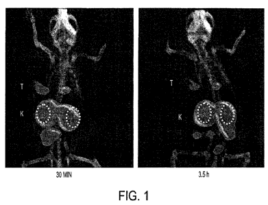
[0027] Figure 1 shows SPECT-CT images of a PSMA+ LNCaP tumor-bearing mouse
injected intravenously with exemplary compound [99mTc]SRV32.
[0028] Figure 2. GE eXplore VISTA pseudodynamic PET image (co-registered
with
the corresponding CT image) of a PSMA+ LNCaP tumor-bearing mouse injected
intravenously with 0.2 mCi (7.4 MBq) of exemplary compound [68GalSRV27.
[0029] Figure 3. GE eXplore VISTA PET image (co-registered with the
corresponding CT image) of a PSMA+ PIP and PSMA- flu tumor-bearing mouse
injected
intravenously with 0.2 mCi (7.4 MBq) of exemplary compound [68Ga]SRV100.
[0030] Figure 4 shows a synthetic scheme for exemplary compound SRV100 and
1111In]SRV100.
10c
CA 2755965 2018-06-27
CA 02755965 201 -0,9-19
WO 2010/108125 PCT/US2010/028020
[0031] Figure 5 shows SPECT-CT images of a PSMA+ PC-3 PIP tumor-bearing
mouse injected intravenously with exemplary compound [111In]SRV27.
[0032] Figure 6 shows SPECT-CT images of a PSMA+ PC-3 PIP tumor-bearing
mouse injected intravenously with exemplary compound [111In]SRV100.
[0033] Figure 7 shows SPECT-CT images of a PSMA+ PC-3 PIP tumor-bearing
mouse injected intravenously with exemplary dual modality compound
[111Ir]SRV73.
[0034] Figure 8 shows the absorbance and emission spectra, and quantum
yield of
exemplary compound YC-27.
[0035] Figure 9 shows the fluorescence decay of exemplary compound YC-27.
[0036] Figure 10 shows an IC50 curve of compound YC-27 using a
fluorescence-based
NAALADase assay
[0037] Figure 11 shows in vivo imaging of a NOD/SCID mouse (mouse #1),
bearing
PC3-PIP (forward left flank) and PC3-flu (forward right flank) tumors. Mouse
#1 received 10
nmol of YC-27 and dorsal (animal prone) and ventral (animal supine) views were
obtained.
Dorsal and ventral views at 40 mm p.i. (A, B, respectively); 18.5 h (C, D); 23
h (E, F); 42.5 h
(G, H); 68 h (I, J). Dorsal view of pre-injection image (K). Dorsal and
ventral views 70.5 h p.i.
(L, M). Images after midline laparotomy (N) and individually harvested organs
(0) on a Petri
dish at 70.5 h p.i.. Images were scaled to the same maximum (arbitrary units).
[0038] Figure 12 shows in vivo imaging of a NOD/SCID mouse (mouse #2)
(left
panel), bearing PC3-PIP (forward left flank) and PC3-flu (forward right flank)
tumors. Mouse
#2 received 1 nmol of YC-27 and dorsal (animal prone) and ventral (animal
supine) views were
obtained. Dorsal and ventral views of the pre-injection image (A, B,
respectively); 10 min p.i.
(C, D); 20.5 h (E, F); 24 h (G, H). Images after midline laparotomy (I) and
individually
harvested organs (J) on a Petri dish at 24 h p.i.. Right Panels: Mouse #3 in
same orientation as
mouse #2. Mouse #3 received 1 nmol of YC-27 co-injected with 1 mol of DCIBzL,
which
served as a blocking agent to test binding specificity. Images were scaled to
the same maximum
(arbitrary units).
[0039] Figure 13 shows SPECT-CT images of a PSMA+ LNCaP tumor-bearing
mouse injected intravenously with exemplary compound [99mTc]SRVI34B.
[0040] Figure 14 shows SPECT-CT images of a PSMA+ PC3-PIP tumor-bearing
mouse injected intravenously with exemplary compound [991"Te]SRVI34B.
11
CA 02755965 201 09-19
WO 2010/108125 PCT/US2010/028020
[0041] Figure 15 shows SPECT-CT images of a PSMA+ PC3-PIP (forward left
flank)
and PSMA- PC3-flu (forward right flank) tumor-bearing mouse injected
intravenously with
exemplary compound 199mTc1SRVI34A.
[0042] Figure 16 shows SPECT-CT images of a PSMA+ PC3-PIP (forward left
flank)
and PSMA- PC3-flu (forward right flank) tumor-bearing mouse injected
intravenously with
exemplary compound [99mTc[SRVI34B.
[0043] Figure 17 shows PC3-PIP and PC3-flu cells treated with fluorescent
compound
YC-VIII-36 (green, top left) and DAPI (blue), and PC3-PIP and PC3-flu cells
treated with both
YC-VIII-36 and PSMA inhibitor, PMPA.
100441 Figure 18 shows PC3-PIP cells treated with DAPI (blue) and varying
concentrations of YC-VIII-36 (green).
[0045] Figure 19 shows time dependent internalization of YC-VIII-36 into
PC3-PIP
cells treated with YC-VIII-36 (green) and DAPI (blue).
[0046] Figure 20 shows titration and detection of varying amounts of YC-
VIII-36
injected subcutaneously into a nude mouse. (IVIS spectrum with 10 second
exposure followed
by spectral unmixing)
[0047] Figure 21 shows fluorescence images of a PSMA+ PC3-PIP and PSMA-
PC3-
flu tumor-bearing mouse injected intravenously with exemplary compound YC-VIII-
36.
[0048] Figure 22 shows fluorescence images of a PSMA+ PC3-PIP and PSMA-
PC3-
flu tumor-bearing mouse injected intravenously with exemplary compound YC-VIII-
36 180
minutes after injection (top) and biodistribution of exemplary compound YC-
VIII-36 180
minutes after injection (bottom).
[0049] Figure 23 shows FACS analysis showing the percent subpopulation of
PSMA
positive cells in PC3-flu, PC3-PIP, and LNCaP cells.
[0050] Figure 24 shows cell sorting results for PC3-PIP cells treated
with exemplary
compound YC-VIII-36, including initial percentage (top center), and after 3
passages of sorting
(bottom).
[0051] Figure 25 shows the number of spiked PIP-pos cells into 10 million
of PC3-flu
detectable by 100 nM compound YC-VIII-36 in flow cytometry (BD LSR-II). Gate
P1 is total
number of single cells counted; gate P2 at higher intensity is the number of
Pip-pos cells
detected and gate P3 at lower intensity.
12
CA 2755965 2017-03-16
81679978
DETAILED DESCRIPTION OF EXEMPLARY EMBODIMENTS
[0052] Some embodiments of the current invention are discussed in detail
below. In
describing embodiments, specific terminology is employed for the sake of
clarity. However,
the invention is not intended to be limited to the specific terminology so
selected. A person
skilled in the relevant art will recognize that other equivalent components
can be employed
and other methods developed without departing from the broad concepts of the
current
invention.
[0053] Where a range of values is provided in the present application, it
is understood
that each intervening value, to the tenth of the unit of the lower limit
unless the context clearly
dictates otherwise, between the upper and lower limit of that range and any
other stated or
intervening value in that stated range, is encompassed within the invention.
The end values of
any range are included in the range.
Definitions
[0054] The following terms below generally have the meaning that would be
readily
understood by persons skilled in the art. The definitions are provided herein
for clarity. Where
a definition excludes an art-recognized meaning, the ten-n should be taken to
have the
meaning set forth below. Where the art-recognized meaning and the meaning
below differ but
are not exclusive, the intended meaning is clear by the context in which it is
used.
[0055] As used herein, "agent" is a non-peptide, small molecule compound.
[0056] By "cell substrate" is meant the cellular or acellular material
(e.g., extracellular
matrix, polypeptides, peptides, or other molecular components) that is in
contact with the cell.
[0057] By "control" is meant a standard or reference condition.
[0058] By "disease" is meant any condition or disorder that damages or
interferes with
the normal function of a cell, tissue, organ or subject.
[0059] By "effective amount" is meant a quantity sufficient to produce a
measurable
difference, when compared with a control. For example, an amount sufficient to
produce a
measurable image, when the compound is used for imaging, or an amount
sufficient to
ameliorate the symptoms of a disease, when the compound is used for therapy.
The effective
amount of an active therapeutic agent for the treatment of a disease or injury
varies depending
13
CA 02755965 201 09-19
WO 2010/108125 PCT/US2010/028020
upon the manner of administration, the age, body weight, and general health of
the subject.
Ultimately, the attending clinician will decide the appropriate amount and
dosage regimen.
[0060] By "modifies" is meant alters. An agent that modifies a cell,
substrate, or
cellular environment produces a biochemical alteration in a component (e.g.,
polypeptide,
nucleotide, or molecular component) of the cell, substrate, or cellular
environment.
[0061] As used herein, the terms "prevent," "preventing," "prevention,"
"prophylactic
treatment" and the like refer to reducing the probability of developing a
disorder or condition in
a subject, who does not have, but is at risk of or susceptible to developing a
disorder or
condition.
[0062] By "subject" is meant a mammal, including, but not limited to, a
human or non-
human mammal, such as a bovine, equine, canine, ovine, or feline.
[0063] By "therapeutic delivery device" is meant any device that provides
for the
release of a therapeutic agent. Exemplary therapeutic delivery devices include
tablets and pills,
described below, as well as syringes, osmotic pumps, indwelling catheters,
delayed-release and
sustained-release biomaterials.
[0064] As used herein, the terms "treat," treating,'' "treatment,"
"therapeutic" and the
like refer to reducing or ameliorating a disorder and/or symptoms associated
therewith. It will be
appreciated that, although not precluded, treating a disorder or condition
does not require that
the disorder, condition or symptoms associated therewith be completely
eliminated.
[0065] The compounds herein described may have one or more asymmetric
centers or
planes. Compounds of the present invention containing an asymmetrically
substituted atom may
be isolated in optically active or racemic forms. It is well known in the art
how to prepare
optically active forms, such as by resolution of racemic forms (racemates), by
asymmetric
synthesis, or by synthesis from optically active starting materials.
Resolution of the racemates
can be accomplished, for example, by conventional methods such as
crystallization in the
presence of a resolving agent, or chromatography, using, for example a chiral
HPLC column.
Many geometric isomers of olefins, C=N double bonds, and the like can also be
present in the
compounds described herein, and all such stable isomers are contemplated in
the present
invention. Cis and trans geometric isomers of the compounds of the present
invention are
described and may be isolated as a mixture of isomers or as separated isomeric
forms. All chiral
(enantiomeric and diastereomeric), and racemic forms, as well as all geometric
isomeric forms
14
CA 02755965 201 09-19
WO 2010/108125 PCT/US2010/028020
of a structure are intended, unless the specific stereochemistry or isomeric
form is specifically
indicated.
[0066] The compounds herein described may have one or more charged atoms.
For
example, the compounds may be zwitterionic, but may be neutral overall. Other
embodiments
may have one or more charged groups, depending on the pH and other factors. In
these
embodiments, the compound may be associated with a suitable counter-ion. It is
well known in
the art how to prepare salts or exchange counter-ions. Generally, such salts
can be prepared by
reacting free acid forms of these compounds with a stoichiometric amount of
the appropriate
base (such as Na, Ca, Mg, or K hydroxide, carbonate, bicarbonate, or the
like), or by reacting
free base forms of these compounds with a stoichiometric amount of the
appropriate acid. Such
reactions are typically carried out in water or in an organic solvent, or in a
mixture of the two.
Counter-ions may be changed, for example, by ion-exchange techniques such as
ion-exchange
chromatography. All zwitterions, salts and counter-ions are intended, unless
the counter-ion or
salt is specifically indicated. In certain embodiments, the salt or counter-
ion may be
pharmaceutically acceptable, for administration to a subject. Pharmaceutically
acceptable salts
are discussed later.
[0067] As used herein, a "protecting group" is a chemical substituent
which can be
selectively removed by readily available reagents which do not attack the
regenerated functional
group or other functional groups in the molecule. Suitable protecting groups
are known in the
art and continue to be developed. Suitable protecting groups may be found, for
example in
Wutz et al. ("Greene's Protective Groups in Organic Synthesis, Fourth
Edition," Wiley-
Interscience, 2007). Protecting groups for protection of the carboxyl group,
as described by
Wutz et al. (pages 533-643), are used in certain embodiments. In some
embodiments, the
protecting group is removable by treatment with acid. Specific examples of
protecting groups
include but are not limited to, benzyl, p-methoxybenzyl (PMB), tertiary butyl
(43u),
methoxymethyl (MOM), methoxyethoxymethyl (MEM), methylthiomethyl (MTM),
tetrahydropyranyl (THP), tetrahydrofuranyl (THF), benzyloxymethyl (BOM),
trimethylsilyl
(TMS), triethylsilyl (TES), t-butyldimethylsilyl (TBDMS), and triphenylmethyl
(trityl, Tr).
Persons skilled in the art will recognize appropriate situations in which
protecting groups are
required and will be able to select an appropriate protecting group for use in
a particular
circumstance.
CA 02755965 201 -0,9-19
WO 2010/108125 PCT/US2010/028020
100681 As used herein, "alkyl" is intended to include branched, straight-
chain, and
cyclic saturated aliphatic hydrocarbon groups. Examples of alkyl include, but
are not limited to,
methyl, ethyl, n-propyl, iso-propyl, n-butyl, sec-butyl, tert-butyl, n-pentyl,
and sec-pentyl. In
certain embodiments, alkyl groups are Ci-C6 alkyl groups or Ci-C4 alkyl
groups. Particular alkyl
groups are methyl, ethyl, propyl, butyl, and 3-pentyl. The term "C1-C6 alkyl"
as used herein
means straight-chain, branched, or cyclic C1-C6 hydrocarbons which are
completely saturated
and hybrids thereof such as (cycloalkyl)alkyl. Examples of C1-C6 alkyl
substituents include
methyl (Me), ethyl (Et), propyl (including n-propyl (n-Pr, "Pr), iso-propyl (i-
Pr, 'Pr), and
cyclopropyl (c-Pr, el3r)), butyl (including n-butyl (n-Bu, "Bu), iso-butyl (i-
Bu, 'Bu), sec-butyl (s-
Bu, sBu), tert-butyl (t-Bu, 13u), or cyclobutyl (c-Bu, 1311)), and so forth.
"Cycloalkyl" is
intended to include saturated ring groups, such as cyclopropyl, cyclobutyl,
cyclopentyl, or
cyclohexyl. Cycloalkyl groups typically will have 3 to about 8 ring members.
In the term
"(cycloalkyl)alkyl'", cycloalkyl, and alkyl are as defined above, and the
point of attachment is on
the alkyl group. This term encompasses, but is not limited to,
cyclopropylmethyl,
cyclopentylmethyl, and cyclohexylmethyl. The alkyl group may be substituted or
unsubstituted.
Substituents are not counted towards the total number of atoms in the alkyl
group, so long as the
total atoms in the substituent(s) are not larger than the alkyl group.
100691 As used herein, the term "aryl" includes aromatic groups that
contain 1 to 3
separate or fused rings and from 2 to about 12 carbon atoms, and up to 3
heteroatoms as ring
members. Examples of heteroatoms include nitrogen, oxygen or sulfur atoms. The
aryl group
may have 0, 1, 2 or 3 heteroatoms as ring members. Examples of aryl groups
include but are not
limited to phenyl, biphenyl and naphthyl, including 1-napthyl and 2-naphthyl.
Examples of aryl
groups having heteroatoms include quinolinyl, isoquinolinyl, quinazolinyl,
pyridyl, pyrazinyl,
pyrimidyl, furanyl, pyrrolyl, thienyl, oxadiazolyl, thiadiazolyl, thiazolyl,
triazinyl, oxazolyl,
isoxazolyl, imidazolyl, indolyl, benzofuranyl, and benzothiazolyl, among
others. The aryl group
may be substituted or unsubstituted. Substituents are not counted towards the
total number of
atoms in the aryl group, so long as the total atoms in the substituent(s) are
not larger than the
aryl group.
100701 As used herein, the term "alkylaryl" includes alkyl groups, as
defined above,
substituted by aryl groups, as defined above. The aryl group may be connected
at any point on
the alkyl group. The term C4-C16 alkylaryl includes alkylaryl groups having a
total of 4 to 16
carbon atoms, counting the carbon atoms on the alkyl group and aryl group
together. Examples
16
CA 02755965 201 09-19
WO 2010/108125 PCT/US2010/028020
of alkylaryl groups include but are not limited to benzyl (phenylmethyl),
phenylethyl, and
naphthylmethyl. The alkylaryl group may be substituted or unsubstituted.
Substituents are not
counted towards the total number of atoms in the alkylaryl group, so long as
the total atoms in
the substituent(s) are not larger than the alkylaryl group.
[0071] The term "substituted," as used herein, means that any one or more
hydrogens
on the designated atom or group is replaced with a substituent, provided that
the designated
atom's normal valence is not exceeded, and that the substitution results in a
stable compound.
When a substituent is oxo (keto, i.e., =0), then 2 hydrogens on an atom are
replaced. The
present invention is intended to include all isotopes (including
radioisotopes) of atoms occurring
in the present compounds. When the compounds are substituted, they may be so
substituted at
one or more available positions, typically 1, 2, 3 or 4 positions, by one or
more suitable groups
such as those disclosed herein. Suitable groups that may be present on a
"substituted" group
include e.g., halogen; cyano; hydroxyl; nitro; azido; amino; alkanoyl (such as
a Ci-Co alkanoyl
group such as acyl or the like); carboxamido; alkyl groups (including
cycloalkyl groups, having
1 to about 8 carbon atoms, for example 1, 2, 3, 4, 5, or 6 carbon atoms);
alkenyl and alkynyl
groups (including groups having one or more unsaturated linkages and from 2 to
about 8, such
as 2, 3, 4, 5 or 6, carbon atoms); alkoxy groups having one or more oxygen
linkages and from 1
to about 8, for example 1, 2, 3, 4, 5 or 6 carbon atoms; aryloxy such as
phenoxy; alkylthio
groups including those having one or more thioether linkages and from 1 to
about 8 carbon
atoms, for example 1, 2, 3, 4, 5 or 6 carbon atoms; alkylsulfinyl groups
including those having
one or more sulfinyl linkages and from 1 to about 8 carbon atoms, such as 1,
2, 3, 4, 5, or 6
carbon atoms; alkylsulfonyl groups including those having one or more sulfonyl
linkages and
from 1 to about 8 carbon atoms, such as 1, 2, 3, 4, 5, or 6 carbon atoms;
aminoalkyl groups
including groups having one or more N atoms and from 1 to about 8, for example
1, 2, 3, 4, 5 or
6, carbon atoms; carbocyclic aryl having 4, 5, 6 or more carbons and one or
more rings, (e.g.,
phenyl, biphenyl, naphthyl, or the like, each ring either substituted or
unsubstituted aromatic);
arylalkyl having 1 to 3 separate or fused rings and from 6 to about 18 ring
carbon atoms, (e.g.
benzyl); arylalkoxy having 1 to 3 separate or fused rings and from 6 to about
18 ring carbon
atoms (e.g. 0-benzyl); or a saturated, unsaturated, or aromatic heterocyclic
group having 1 to 3
separate or fused rings with 3 to about 8 members per ring and one or more N,
0 or S atoms,
(e.g. coumarinyl, quinolinyl, isoquinolinyl, quinazolinyl, pyridyl, pyrazinyl,
pyrimidyl, furanyl,
pymalyl, thienyl, thiazolyl, triazinyl, oxazolyl, isoxazolyl, imidazolyl,
indolyl, benzofuranyl,
17
CA 02755965 201 09-19
WO 2010/108125 PCT/US2010/028020
benzothiazolyl, tetrahydrofuranyl, tetrahydropyranyl, piperidinyl,
morpholinyl, piperazinyl, and
pyrrolidinyl). Such heterocyclic groups may be further substituted, e.g. with
hydroxy, alkyl,
alkoxy, halogen and amino.
[0072] As used herein, where an internal substituent is flanked by bonds
(for example
-NRC(0)-) the order of the atoms is fixed, the orientation of the group may
not be reversed, and
is inserted into a structure in the orientation presented. In other words
¨NRC(0)- is not the
same as ¨C(0)NR-. As used herein the term C(0) (for example -NRC(0)-) is used
to indicate a
carbonyl (C=0) group, where the oxygen is bonded to the carbon by a double
bond.
[0073] A substituent bearing a broken bond, such as the example shown
below, means
that the substituent is directly bonded to the molecule at the indicated
position. No additional
methylene (CH2) groups are implied.
0
HO NH2
N 'N
N
[0074] Substituents bearing two broken bonds, such as the example shown
below,
means that the orientation of the atoms is as-indicated, left to right and
should be inserted into a
molecule in the orientation shown. No additional methylene (CH2) groups are
implied unless
specifically indicated.
N=N
Embodiments
[0075] As described herein, all embodiments or subcombinations may be
used in
combination with all other embodiments or subcombinations, unless mutually
exclusive.
[0076] In some of the following embodiments, Z is CO2Q. In some of the
following
embodiments, Q is H. In some of the following embodiments, m is 4, 5, or 6. In
some of the
following embodiments, m is 6. In some of the following embodiments, n is 2,
3, or 4. In some
of the following embodiments, n is 3. In some of the following embodiments, a
is 3 or 4. In
some of the following embodiments, a is 4. In some of the following
embodiments, Y is
-C(0)-. In some of the following embodiments, W is ¨NHC(0)-.
18
CA 02755965 201 09-19
WO 2010/108125 PCT/US2010/028020
[0077] Embodiments of the invention include compounds having the
structure
-
N -L3 _____________ R
0 w Tz (C H2), - Y
P
R2
- -
(CH2)a 0
Q02C N N,CO2Q
H H
wherein the subunits associated with elements p, q, r, and s may be in any
order. Z is tetrazole
or CO2Q; each Q is independently selected from hydrogen or a protecting group,
a is 1, 2, 3, or
4, and R is each independently H or C1-C4 alkyl.
[0078] Variable r is 0 or 1. Tz is a triazole group selected from the
group consisting of
N=N N=N
4-Ll-r\iõ;') _____ L2 _______ and L1 (k.,;N L2
R5
2 ________________________________________
j--(CH2)d
where LI is (CH2)d- or 1-X2 , L is ----(C1.12)b or
R5
X
(CH2)b-Li
- , XI is -NRC(0)-, -NRC(0)NR-, -NRC(S)NR-, or -NRC(0)0-; X2 is
-C(0)NR-, -NRC(0)NR-, -NRC(S)NR-, or -0C(0)NR-; R5 is H, CO2H, or CO2R6, where
R6 is
a C1-C6 alkyl, C2-C12 aryl, or C4-C16 alkylaryl; b is 1, 2, 3, or 4; and d is
1, 2, 3, or 4.
[0079] Variable q is 0 or 1. W is -NRC(0)-, -NRC(0)NR-, NRC(S)NR-, -
NRC(0)0-,
-0C(0)NR-, -0C(0)-, -C(0)NR-, or -C(0)0-; R2 and R3 are independently H, CO2H,
or
CO2R4, where R4 is a Ci-C6 alkyl, C2-C12 aryl, or C4-C16 alkylaryl, wherein if
one of R2 and R3
is CO2H or CO2R2, then the other is H; n is 1, 2, 3, 4, 5 or 6.
[0080] Variable s is 0 or 1. Y is -C(0)-, -NRC(0)-, -NRC(S)-, -0C(0)-;
and m is 1, 2,
3, 4, 5, or 6.
[0081] Variable p is 0, 1, 2, or 3, and when p is 2 or 3, each RI may be
the same or
different. RI is H, C1-C6 alkyl, C2-C12 aryl, or C4-C alkylaryl.
[0082] G is a moiety selected from the group consisting of
FG--\k'N\
-1-N3 i R
19
CA 02755965 201 -0,9-19
WO 2010/108125 PCT/US2010/028020
0
HO NH2
NH R
A -V --Ly Ngs_
0 , and
where Ch is a metal chelating moiety, optionally including a chelated metal;
FG is a fluorescent
dye moiety which emits in the visible or near infrared spectrum; one of A and
N is Ch and the
other is FG; V and V are independently ¨C(0)- , ¨NRC(0)- , ¨NRC(S)-, or ¨0C(0)-
;and g is 1,
2, 3, 4, 5, or 6. The following conditions also apply:
A ¨N./\
NH R
\N¨(CH2)g¨LyN/_
Ch N
A- V
1) when G is R , or 0 and r is 0, then q and s are both 1;
FG N
2) when G is R and r is 0, then q and s are both 0 or both 1;
0
HO)Ccr\NH2
3) when G is N:WNI- then p is 0 and R2 is H, and the structure optionally
includes a chelated metal ion;
Ch N;2õ-L,
4) when G is R and r is 0, then if p is 0, then one of R2 and R3 is CO2R2,
and the
other is H; and
5) when g is I-N3 or __ , then r is 0.
[0083] In some embodiments, Z is CO2Q. In some embodiments, Q is H. In
some
embodiments, m is 4, 5, or 6. In some embodiments, m is 6. In some
embodiments, n is 2, 3, or
4. In some embodiments, n is 3. In some embodiments, a is 4. In some
embodiments, subunits
associated with elements p, q and s are in the order drawn and r may be in any
location,
including between one of p, q, or s. In some embodiments r is 0.
CA 02755965 201 -0,9-19
WO 2010/108125 PCT/US2010/028020
[0084] Embodiments include compounds having the structure
R1 R-
R3
N
Ch NI 02(CH2)n-INVV-(CH2),-Y-N
R - - P (CF12)a 0
II
N N
H H
wherein Z is tetrazole or CO2Q; each Q is independently selected from hydrogen
or a protecting
group, a is 1, 2, 3, or 4, and R is each independently H or C1-C4 alkyl. Ch is
a metal chelating
moiety optionally including a chelated metal. W is -NRC(0)-, -NRC(0)NR-,
NRC(S)NR-,
-NRC(0)0-, -0C(0)NR-, -0C(0)-, -C(0)NR-, or -C(0)0-. Y is -C(0)-, -NRC(0)-, -
NRC(S)-,
-0C(0). V is -C(0)- , -NRC(0)- , -NRC(S)- , or -0C(0)-. In exemplary
embodiment m is 1,
2, 3, 4, 5, or 6; n is 1, 2, 3,4, 5 or 6; and p is 0, 1, 2, or 3, and when p
is 2 or 3, each RI may be
the same or different. RI is H, C1-C6 alkyl, C2-C12 aryl, or C4-C16 alkylaryl.
R2 and R3 are
independently H, CO2H, or CO2R4, where R4 is a C1-C6 alkyl, C2-C12 aryl, or C4-
C16 alkylaryl,
wherein when one of R2 and R3 is CO2H or CO2R2, the other is H, and when p is
0, one of R2
and R3 is CO2R4, and the other is H.
[0085] In some embodiments, the compound has the structure shown below.
R1 R_
R3
N
Ch N -r-(CF12)n
0 R2 (CH2)a 0
Q02C N
H H
[0086] In some embodiments, the compound has the structure shown below.
Ri
- R-
I R3 C)11 0 R
N
Ch N `1--(CH2)n-LN __________________ (CH2),--)L- N --(CH2)a
0
0 R2
PR
N CO2()
H H
[0087] In some embodiments, p is 1, 2 or 3. When p is 2 or 3, each RI may
be the
same or different. When two RI groups are different, the two may be in any
order. In some
embodiments, p is 2. In some embodiments, p is 2, and both RI are the same. In
some
embodiments, RI is C2-C12 aryl. In some embodiments RI is phenyl. In some
embodiments, R3
is CO2H and R2 is H. In some embodiments, R2 is CO2H and R3 is H. In some
embodiments, R2
and R3 are both H.
21
CA 02755965 201 -0,9-19
WO 2010/108125 PCT/US2010/028020
[0088] In some embodiments, p is 0. In some embodiments where p is 0, R2
is CO2R4,
and R3 is H. In some embodiments where p is 0, R3 is CO2R4, and R2 is H. In
some
embodiments R4 is C2-C12 aryl, or C4-C16 alkylaryl. In some embodiments R4 is
benzyl.
[0089] Ch is a metal chelating moiety optionally including a chelated
metal. A metal
chelating moiety is a chemical moiety that non-covalently binds a metal atom,
usually with a
plurality of non-covalent interactions. Ch includes any additional atoms or
linkers necessary to
attach the metal chelating moiety to the rest of the compound. For instance
linking groups
having alkyl, aryl, combination of alkyl and aryl, or alkyl and aryl groups
having heteroatoms
may be present in the chelating moiety. Numerous metal chelating moieties are
known in the
art. Any acceptable chelator can be used with the present invention as long as
compatible and
capable of chelating a desired metal. Examples of metal chelating moieties
(Ch) include, but are
not limited to 1,4,7,10-tetra a7acyclododecane-1,4,7,10-tetraacetic acid
(DOTA) and Diethylene-
triaminepentaacetic acid (DTPA). In some embodiments, Ch has a structure shown
below.
(CO2H
0
r, CO2H ___N7----IN,._co2H
tv.
rAril---
CO2H __----f¨N.,CO2H
csss...¨NN___ \ CO2H ---- N
--,,(
( CO2H ( CO2H ( CO2H CO2H
CO2H CO2H CO2H
3 3 5
CO2H
CO2H HO 0 HO \O
NG HO 0 HO \O
HO2C---.''N)
"'NJ ois (N
) ( ______________ ,--N HO" NI \ N 71 N e.
HO N.õ.õ.0O2H")\ N N , K CO2H
CO2H 0 \ ______ / ---- CO2H
H0,10 HO\O CO2H /10
( /------, 0 / \ Zn/------1
N NI ...--N I ,--iq 0 NH HN
0 i ,....- N
N HO N
HOA, 'N NI> //C) ----NJ '',S HN0
HO2C \---
0O2H
HO' = ( + r 1-",
c- CO2H CO2H
22
CA 02755965 201'-09-19
WO 2010/108125
PCT/US2010/028020
0 0 1
HO--Fic,--ki \:. / \ )
HO2C-4--F is NH N \
N_
OH N--.'"----
O NH
H N __ /
'N 1µ1
HO2C -/ 1 \-CO2H
HO CO2H
,
I I
1\1Th r1\1
CO2H CO21-I
/ \) HO2C¨/N
9N\--1-
,ss
H2N___CNNr\NN- 1__NH = i- NH
Niii 1\1 K.,1\1 CNN ---
CO2H CO2H I HO2C---/ L.,.)
I
C'r\r- N N¨' - CO,H (--0--
/¨N N--
, oc 2H
HO2C
/¨
= / N HO2C
J
HO2C Ho2c----
µ \
,
[0090] Examples of specific compounds include the compounds shown below.
CO2H
/ ii \ ,ç-CO2H
rN N,i
HO2C, L., ) 0 o,,0
,:= 0
'---N NN).1,,,
\ _______ / NN NH
0yOH
H H 0 L-
0 )
H,oyNA NL11,, H
o"H n " 0
CO2H
/ \ 7¨CO2H
0 rN Nõ)
HO2C L, ) 0 Oõ0 0
N--N /NNA,õ
\ _____________ NN NH
H H 0 OH
0
0
H Ir'NA N .=. H
0 H H H H 0
23
CA 02755965 201'-09-19
WO 2010/108125 PCT/US2010/028020
HO
0 \--0
0 --- 0
\ / \
o. /A NH)-L ,--.,./\,_,------.
N N NH
HO r N N .õ1 H H L.. CO2H
HO
0
L N N ) Ph/ )
\ N. 0
// __ / \ __ / =
--:-,
0 HO/. HO2CN.11-.
N ,z, CO2H
H H u H -
rl
Ph HO
\
0 0 \ -'0 0
0\\ H j-N N NH
N
N II H N.
HO __ r N N 0/ H 0 CO2H
)
0
HO \ EN N Ph
"--
// / \ __ / = H 02C' NA N CO2H
H H H n
0 /k.,
HO
Ph HO
0 \ 0
HO2C,,,K ilj-LN N NH
0 N-Thf
O\ H H H I\ CO2H
OPh/ 0
) \ /----N / IL-N".
)
HO N N..,1 H \ 0
-It. ,--,COH
HO2C--'N N ,-,-
HO N N) OH H H H rl 2
/1 __ /\ __ /\
0 0
(002H
N
41
----N
( 0
S 0
CO2H
NH
NAN N
1---- oy0H
H H H 0
0 )
. O.
H' ANr-2'ir H
H H H H
0 0
24
CA 02755965 201'-09-19
WO 2010/108125
PCT/US2010/028020
CO2H
N /r---0O2H
C N
4.
N
( S 0
CO2H
NH
N)CNN
I¨, (:)/OH
H H H 0
0 )
,O, ,A.. A
H Ti AN NAH H
0 n ri o
CO2H
.,..--N //---CO2H
N
---'N
( S 0
H
NH
CO2H
N)--,N H
H H L-,
0 HO(D 0 CO2H
\ )
0
. -
H020 A.-1-. hiA hir, co2H
0 H 0
H H H
N, N N N ,-----õ,..õ.õ---,õ-N
D
H NH
0 K
(CO2H
S HO:) CO2H
r,-- N \ )
L N 0
N CO2H HO2C - N
N :\
( H H H
HCO2H
CO2H
0 H H 0
H
H N CO2H
N ...,..,--...õ..õ...---..õ...õ N
,,,NH N
(CO2H N11 NH
0 H 0 HOcl L'. S
\ )
0
r-N
L N
HO2C ,t N N = CO2H
N \ )¨ CO2 H H H H H
(
CO2H
CA 02755965 201 09-19
WO 2010/108125 PCT/US2010/028020
0 0
./(
NH
0 NH HN
0 CO2H
HN 0 Ph H
0
.4N Phph Ph NThr 0 H02c NA N CO2
H
H H H
0 \ 0 HO
Ph
0
NH
HN
[\ CO2H
0
Ph
0
0 /
H C)11
HO2C = N H
NH H H H H 2
0
0
''.1\17/1 /S,i7ph Ph HO
1/ Ph Ph
0
[0091] In some embodiments, the compound further includes a chelated
metal. In
some embodiments, the chelated metal is Tc, In, Ga, Y, Lu, Re, Cu, Ac, Bi, Pb,
Sm, Sc, Co, Ho,
Gd, Eu, Tb, or Dy. In some embodiments, the chelated metal is Tc, Ga, In, Cu,
Y, Ac, Lu, Re,
or Bi. In some embodiments the metal is an isotope, for example a radioactive
isotope. In some
embodiments, the isotope is Tc-99m, In-111, Ga-67, Ga-68, Y-86, Y-90, Lu-177,
Re-186, Re-
188, Cu-64, Cu-67, Co-55, Co-57, Sc-47, Ac-225, Bi-213, Bi-212, Pb-212, Sm-
153, Ho-166, or
Dy-166. In some embodiments, the isotope is Tc-99m, In-111, Ga-67, Ga-68, Y-
90, Lu-177,
Re-186, Re-188, Cu-67, Ac-225, Bi-213, or Bi-212.
[0092] Embodiments include compounds having the structure
0
HO NH2 _
-
(CH2)n-l`R3
N vv (CH2),-Y-N
----5-(CH2)a 0Z
O02C-NANCO20
H H
optionally including a chelated metal ion. Z is tetrazole or CO2Q; each Q is
independently
selected from hydrogen or a protecting group, and a is 1, 2, 3, or 4. R is
each independently H
or Ci-C4 alkyl. W is -NRC(0)-, -NRC(0)NR-, NRC(S)NR-, -NRC(0)0-, -0C(0)NR-,
26
CA 02755965 201 09-19
WO 2010/108125 PCT/US2010/028020
-0C(0)-, -C(0)NR-, or -C(0)0-. Y is -C(0)-, -NRC(0)-, -NRC(S)-, -0C(0)-. In
some
embodiments, subunits associated with q and s may be in the order shown or the
reverse thereof.
[0093] In exemplary embodiment m is 1, 2, 3, 4, 5, or 6; n is 1, 2, 3, 4,
5 or 6; q is 0 or
1; and s is 0 or 1. R3 is H, CO2H, or CO2R4, where R4 is a C1-C6 alkyl, C2-C12
aryl, or C4-C16
alkylaryl. Some embodiments further include a chelated metal ion. In some
embodiments, the
metal ion is Tc, Re, Ga, or Cu. In some embodiments, the metal ion is Tc-99m,
Re-186, Re-188,
Cu-64, or Ga-68. In some embodiments, the metal ion is Tc-99m, Re-186 or Re-
188.
[0094] In some embodiments, the compound has the structure
0
HO NH2
R3
N N
(CH2)n
-N
(CH2)a
Q02C- NA N''CO2Q
H H
optionally including a chelated metal ion. Z is tetrazole or CO2Q; each Q is
independently
selected from hydrogen or a protecting group and a is 1, 2, 3, or 4. R is each
independently H or
Ci-C4 alkyl. W is -NRC(0)-, -NRC(0)NR-, NRC(S)NR-, -NRC(0)0-, -0C(0)NR-, -
0C(0)-,
-C(0)NR-, or -C(0)0-. Y is -C(0)-, -NRC(0)-, -NRC(S)-, -0C(0)-. In exemplary
embodiments m is 1, 2, 3, 4, 5, or 6; and n is 1, 2, 3, 4, 5 or 6. R3 is H,
CO2H, or CO2R4, where
R4 is a C1-C6 alkyl, C2-C12 aryl, or C4-C16 alkylaryl.
[0095] In some embodiments, the compound has the structure
0
HO'VNI-12
' N-(CH2),-Y -N
N (CH2)a 0
N A N''CO2Q
H H
where a is 1, 2, 3, or 4. Y is -C(0)-, -NRC(0)-, -NRC(S)-, -0C(0)-, and In
exemplary
embodiment m is 1, 2, 3, 4, 5, or 6.
27
CA 02755965 201 09-19
WO 2010/108125 PCT/US2010/028020
[0096] In some embodiments, the compound has the structure
0
HO NH2
R3
NN'(CH2),
(CH2)a 0 Z
N HNCO2C)
H
where a is 1, 2, 3, or 4. W is -NRC(0)-, -NRC(0)NR-, NRC(S)NR-, -NRC(0)0-, -
0C(0)NR-,
-0C(0)-,¨C(0)NR-, or -C(0)0-, and n is 1, 2, 3, 4, 5 or 6. R3 is H, CO2H, or
CO2R4, where R4
is a C1-C6 alkyl, C2-C12 aryl, or C4-C16 alkylaryl.
[0097] In some embodiments, the compound has the structure
0
HO N H2
N 'N-----(CF12)a
N
O02C N N
H H
where a is 1, 2, 3, or 4.
[0098] In some embodiments, Y is is ¨C(0)-.
[0099] In some embodiments, W is ¨NHC(0)-.
[00100] In some embodiments, m is 4, 5, or 6. In some embodiments, m is 6.
[00101] In some embodiments, n is 2, 3, or 4. In some embodiments, n is 3.
[00102] In some embodiments, R3 is CO2H. In some embodiments, R3 is H. In
some
embodiments, R3 is CO2R4.
[00103] Examples of compounds include those having the structure shown
below
28
CA 02755965 201 09-19
WO 2010/108125 PCT/US2010/028020
HO2C
H2
0
0 OH
HO--%
0
H H
0 0
[00104] In some embodiments, the compound further includes a chelated
metal ion. In
some embodiments, the metal ion is Tc, Re, Cu, or Ga. In some embodiments, the
metal ion is
Tc-99m, Re-186, Re-188, Cu-64, or Ga-68. In some embodiments, the metal ion is
Tc-99m.
[00105] The metal ion chelates to the triazole amino acid portion of the
molecule to
form a structure shown below using Tc as an example.
0
0 H2N N , N
Tc
/IN
OC CO C
[00106] Embodiments include compounds having the structure
R3
FG
N7'y N Th ______________ (CH2),---N
I _ 0 R2 W-(CH2)m-Y-N
- s (CF12)a 0 VZ
002C) N Nr-'CO20
H H
where p, q, and s are in the order drawn, and q and s are either both 0 or
both 1. Z is tetrazole or
CO2Q; each Q is independently selected from hydrogen or a protecting group,
and a is 1, 2, 3, or
4. FG is a fluorescent dye moiety which emits in the visible or near infrared
spectrum. R is
each independently H or C1-C4 alkyl. V is -C(0)- or -NRC(0)- or -NRC(S)-. W is
-NRC(0)-,
-NRC(0)NR-, NRC(S)NR-, -NRC(0)0-, -0C(0)NR-, -0C(0)-, -C(0)NR-, or -C(0)0-. Y
is
-C(0)-, -NRC(0)-, -NRC(S)-, -0C(0). In exemplary embodiments m is 1, 2, 3, 4,
5, or 6; n is 1,
2, 3, 4, 5 or 6; p is 0, 1, 2, or 3, and when p is 2 or 3, each R1 may be the
same or different. RI is
H, C1-C6 alkyl, C2-C12 aryl, or C4-C16 alkylaryl. R2 and R3 are independently
H, CO2H, or
29
CA 02755965 201 09-19
WO 2010/108125 PCT/US2010/028020
CO2R2, where R2 is a Ci-C6 alkyl, C2-C12 aryl, or C4-C16 alkylaryl, wherein
when one of R2 and
R3 is CO2H or CO2R2, the other is H. In some embodiments, the fluorescent dye
moiety emits in
the near infrared spectrum.
1001071 Some embodiments have the structure shown below.
-,R1
R3
FGV
N N (CH2)n
--LW ¨(CH2)m ¨ Y
(CH2)a 0
Q02C) N N"--''CO2Q
H H
wherein Z is tetrazole or CO2Q; each Q is independently selected from hydrogen
or a protecting
group, and a is 1, 2, 3, or 4. FG is a fluorescent dye moiety which emits in
the visible or near
infrared spectrum. R is each independently H or C1-C4 alkyl. V is ¨C(0)- or -
NRC(0)- or
-NRC(S)-. W is -NRC(0)-, -NRC(0)NR-, NRC(S)NR-, -NRC(0)0-, -0C(0)NR-, -0C(0)-,
-C(0)NR-, or -C(0)0-. Y is ¨C(0)-, -NRC(0)-, -NRC(S)-, -0C(0)-. In exemplary
embodiments m is 1,2, 3,4, 5, or 6; n is 1, 2, 3, 4, 5 or 6; p is 0, 1, 2, or
3, and when p is 2 or 3,
each RI may be the same or different. RI is H, C1-C6 alkyl, C2-C12 aryl, or C4-
C16 alkylaryl. R2
and R3 are independently H, CO2H, or CO2R2, where R2 is a C1-C6 alkyl, C2-C12
aryl, or C4-C16
alkylaryl, wherein when one of R2 and R3 is CO2H or CO2R2, the other is H. In
some
embodiments, the fluorescent dye moiety emits in the near infrared spectrum.
[00108] In some embodiments, the compound has the structure shown below.
R1 R_
R3
õV, N
0
0 R2
(CH2)a
P
Q02CNNCO2 Q
H H
1001091 In some embodiments, the compound has the structure shown below.
R_
N ,J1,
FG NI if N __
(CH26,
0 R2 N(CH2)a 0
P R A
QO2C N N CO2Q
H H
CA 02755965 201 -0,9-19
WO 2010/108125 PCT/US2010/028020
[00110] In some embodiments, the compound has the structure shown below.
FG N
T¨(CH2)n¨L. N.--(CH2)rrr)L" N
0
R2 (CH2)a
N NCO2Q
H H
[00111] In some embodiments, the compound has the structure shown below.
FG N/
(CF12)a 0
N
H H
[00112] In some embodiments, p is 1, 2 or 3. In some embodiments, p is 2.
In some
embodiments RI is C2-C12 aryl. In some embodiments, RI is phenyl.
[00113] In some embodiments, p is 0.
[00114] In some embodiments, R3 is CO2H and R2 is H. In some embodiments,
R2 is
CO2H and R3 is H. In some embodiments, R2 is CO2R4, and R3 is H. In some
embodiments, R3
is CO2R4, and R2 is H. In some embodiments R4 is C2-C12 aryl, or C4-C16
alkylaryl. In some R4
is benzyl. In some embodiments, R2 is H, and R3 is H.
[00115] In some embodiments V is ¨C(0)- or ¨NRC(S)-.
[00116] FG is a fluorescent dye moiety that emits light in the visible or
near infrared
spectrum. In some embodiments, FG is a fluorescent dye moiety which emits in
the near
infrared spectrum. FG includes any additional atoms or linkers necessary to
attach the
fluorescent dye moiety to the rest of the compound. For instance linking
groups having alkyl,
aryl, combination of alkyl and aryl, or alkyl and aryl groups having
heteroatoms may be present
in the chelating moiety, so long as the linker does not interfere with the
fluorescence of the dye.
In some embodiments, the fluorescent dye moiety includes a
poly(ethyleneglycol) linker.
Numerous fluorescent dye moieties are known in the art, and will be readily
apparent to one of
ordinary skill. Many fluorescent dyes are commercially available with
activated groups used to
react with protein sidechains or other compounds.
[00117] Examples of fluorescent compounds which may form all or part of
the structure
of FG include carbocyanine, indocarbocyanine, oxacarbocyanine,
thiacarbocyanine,
merocyanine, polymethine, coumarine, rhodamine, xanthene, fluorescein, and
boron-
dipyrromethane (BODIPY) compounds, to name a few.
31
CA 2755965 2017-03-16
81679978
[00118] Examples of fluorescent dye moieties include those described in
WO 20089/109832.
[00119] Specific dyes which emit in the near infrared spectrum include
commercially
available compounds Cy5, Cy5.5, and Cy7, available from GE Healthcare; VivoTag-
680,
VivoTag-S680, and VivoTag-S750, available from VisEn Medical; AlexaFluor660,
AlexaFluor680, AlexaFluor700, AlexaFluor750, and AlexaFluor790, available from
Invitrogen; Dy677, Dy676, Dy682, Dy752, and Dy780, available from Dyonics;
DyLight547,
and Dylight647, available from Pierce; HiLyte Fluor 647, HiLyte Fluor 680, and
HiLyte Fluor 750. available from AnaSpec: IRDye 800CW, IRDye 800RS, and
IRDye 700DX, available from Li-Cor; and ADS780WS, ADS830WS, and ADS832WS,
available from American Dye Source.
[00120] In some embodiments. FG is a structure shown below.
0
00
0
1411
0
\ 11,
N =-=
N
iiO3S
Wi 0 __________________________
0
N "-=
83$
32
CA 02755965 201'-09-19
WO 2010/108125 PCT/US2010/028020
HO3S
N
HO 0 0
HO3S HO3S 0
SO3-
HO3S
HO3S so3H
0 0
N
reNC)0
03/
HO
HO
¨
HN 0
\ -N 0
o
0 ,
HOOC
0
N N H 0
0
,N
HN ¨Nµ+ 0
OH
N(CH2CH3)2
N+-N
0
(H3CH2C)2HN
0/ NO
33
CA 02755965 201 -0,9-19
WO 2010/108125
PCT/US2010/028020
[00121] Exemplary compounds include
those shown below.
0
OP N
H ¨ NCi
0
NH
0 OH
c
.-------\-----... HO 0 /
SO3 H
- 0 0
H 03s 40
/ 0
N s'= '-= '1C-1
=,õ,".,..õ....--õ,...,N H
NH
03S OH
- /
0
HO -)
N
,,...i.,,,OH
HH PH II
0 0
HO3S lei
H03s s03H
0 0
N -.. .., -...., ,..k
,..",0.... ...., "
N
H Of`j
NH
cy,OH
03g/
0 )
H0.1),..
H H H H il
0 0
Ho3s
\----\---\ H H 0
0 COOH 0 oy,OH
0 0
Ho3s -
HO3S SO3 HO 51, OH
o H N ['iqThr
o
34
CA 02755965 201'-09-19
WO 2010/108125 PCT/US2010/028020
HO3S rai
. / qr 0
= 0
0
N
NH
0 OH
COOFP
03/
0
H0.1.-..N--icNOH
H H H H
0 0
0
H
0
N
H
N
NH
I. 00H
COOH
0
/
0
HO ,OH
0 H Hi A If
0
\-----\---s03-
HN 0
, _
---"N+ ¨
0 H H
\ F -13k--N N NN/NyNT,N
\ F
NH H
0 coo R 0 OH
H01) N N. ..-Z. OH
u-
OH H"O
0
/ H
/
H
N
i N -..
0 H
OH
HN
..__N
¨
',..
H01), ijk cr()H
N N 6
H H H "
0 0
0
0
H,., ,,NH H
N
N ----..--
NH
0 ,OH
H0> S
COOH
HO
/
\
0
0 HO
H 11j
IIH H n
0 0
0
CA 02755965 201'-09-19
WO 2010/108125
PCT/US2010/028020
F
HO 0 0
0
./
F NH
0/0H
,-'
0
HO A . OH
0 0
N N."--''ir
H H H H
S
1 ' N+ni )'L
I ¨ NH
H
.--- OH
0./ _
/
---N 0
/ HO
H H H I-1 0
0 0
HOOC / \ H H 0
\zNzNy,N
NH
0 COOH (:)õ,,OH
0
.)
0
HO .-5)C OH
0 H HN HN :(F1 0
OH
S
1 ' N.f1\1)NH
1 H H 0
/ \ZN=Nz-N,,N
_
NH
COOFP
----- N
/ 0 /
HO _UN
H H H H II
0 0
N(CH2CH3)2
0 1
1
H H H 0
,N N N
(H3CH2C)2HN ,S . \/NZY 0 NH
0' µ0 0 0/10H
COOH
)
HO ,
,N NiOH
nH H n
0 0
36
CA 02755965 201 09-19
WO 2010/108125 PCT/US2010/028020
[00122] Embodiments include compounds having the structure
A'-V'
1
\y- _L.-' R3
-R1R-
R3 \1-(CH2)g---L--
A-V
-P (CF12)4 0
Q02C-'N AN---'"CO2Q
H H
wherein Z is tetrazole or CO2Q; each Q is independently selected from hydrogen
or a protecting
group, a is 1, 2, 3, or 4. One of A and A' is Ch and the other is FG, where FG
is a fluorescent
dye moiety which emits in the visible or near infrared spectrum and Ch is
metal chelating
moiety optionally including a chelated metal. R is each independently H or C1-
C4 alkyl. V or V'
are independently -C(0)- , -NRC(0)- , or -NRC(S)-. W is -NRC(0)-, -NRC(0)NR-,
NRC(S)NR-, -NRC(0)0-, -0C(0)NR-, -0C(0)-, -C(0)NR-, or -C(0)0-. Y is -C(0)-, -
NRC(0)-, -NRC(S)-, -0C(0)-. In exemplary embodiments m is 1, 2, 3, 4, 5, or 6;
n is 1, 2, 3, 4,
or 6; and g is 1, 2, 3, 4, 5, or 6; p is 0, 1,2, or 3, and when p is 2 or 3,
each RI may be the same
or different. RI is H, C1-C6 alkyl, C2-C12 aryl, or C4-C16 alkylaryl. R2 and
R3 are independently
H, CO2H, or CO2R4, where R4 is a C1-C6 alkyl, C2-C12 aryl, or C4-C16
alkylaryl, wherein when
one of R' and R" is CO2H or CO2R2, the other is H. In some embodiments, the
fluorescent dye
moiety emits in the near infrared spectrum. Some embodiments further include a
chelated metal.
[00123] In some embodiments, the compound has the structure shown below.
A'-V' R1
\N R R3
I
A-V
1\1-(CH2)g--(y.N- 1-(c1--12)n-LN.---(CH26-Y-N-
(CH2)a 0
o - o2
Q02C)"---..N N
H H
[00124] In some embodiments, the compound has the structure shown below.
A'-V'
N R R 3 C 0
)11 R
N
µN-(cHog-LiiN o
0 R2
A-V
0 -
QO2C NCO2Q
H H
37
CA 02755965 201 -0,9-19
WO 2010/108125 PCT/US2010/028020
[00125] In some embodiments, the compound has the structure shown below.
A' ____________ V'
RR
N 0 R
1
R3 01 N
N )1 _____________________________________________ (CH2),õJ.L_NI
A-V (CF12)a 0
0 R2
Q02C--)\
H H
[00126] In some embodiments, the compound has the structure shown below.
A' ¨ V'
\N -R R
R3
N¨(CH2)g¨Ly
A - V
0 W-(CH2),¨Y¨N,
R2 (CI-12)4 0
Q02C N N
H H
[00127] In some embodiments, p is 1, 2 or 3. In some embodiments, p is 2.
In some
embodiments RI is C2-C12 aryl. In some embodiments, RI is phenyl.
[00128] In some embodiments, p is 0.
[00129] In some embodiments, R3 is CO2H and R2 is H. In some embodiments,
R2 is
CO2H and R3 is H. In some embodiments, R2 is CO2R4, and R3 is H. In some
embodiments, R3
is CO2R4, and R2 is H. In some embodiments R4 is C2-C12 aryl, or C4-C16
alkylaryl. In some R4
is benzyl. In some embodiments, R2 is H, and R3 is H.
[00130] In some embodiments V and V are individually ¨C(0)- or ¨NRC(S)-.
This
means that one of V and V' may be ¨C(0)-, while the other is ¨NRC(S)-, or that
both V and V'
are either ¨C(0)- or ¨NRC(S)-. V and V' will be determined, in part, on the
type of FG and Ch
used.
[00131] FG is a fluorescent dye moiety which emits light in the visible or
near infrared
spectrum. Exemplary fluorescent dye moieties are described previously.
[00132] Ch is a metal chelating group. Suitable metal chelating groups are
described
previously. In some embodiments, the compound further includes a chelated
metal. The list of
Exemplary metals and isotopes thereof are described previously.
38
CA 02755965 201 --19
WO 2010/108125
PCT/US2010/028020
[00133] Some embodiments have the structure
HN¨V¨FG
HN 0
Ch N NH
0
HO-N CO2H
0
HO2C H hi hi 1.=_,
co2H
where FG and Ch are described previously, and each V is individually ¨C(0)- or
¨NRC(S)-.
[00134] Specific examples of compounds include the structures shown below.
(H3cH2q2N 0 N(cH2cH3)2
-03S
NH-S=0
8
0
0 HN H 0
/ NH
ON\/N
0 0
HO-N CO 2H
( NAN
0
0A0 H020002H
H H H H
[00135] Embodiments include compounds having the structure
- R1 R
R3
Gi
_ 0 _______________ (CH2)n¨LW Tz I (CH2),¨Y¨N
P
R`
r -
(cH2), o
N N
H H
wherein the subunits associated with elements p, q, r, and s may be in any
order. Z is tetrazole
or CO2Q; each Q is independently selected from hydrogen or a protecting group,
and a is 1, 2, 3,
39
CA 02755965 201 -0,9-19
WO 2010/108125 PCT/US2010/028020
or 4. R is each independently H or Ci-C4 alkyl. In an exemplary embodiment r
is 1. Tz is a
triazole group having the structure
N=N N=N
_________________ Li _____ L2 ___ or L1 __ IsNy'N-L2
R5
(CH2)d-1-
2 -
where LI is -HCF12)a---1- or -1-X2 ; L is -1-(CH2)b- or
R5
A-(CH2)b-iNvi
^ XI is -NRC(0)-, -NRC(0)NR-, NRC(S)NR-, or -NRC(0)0-; X2 is
-C(0)NR-, -NRC(0)NR-, NRC(S)NR-, or -0C(0)NR-; R5 is H, CO2H, or CO2R6, where
R6 is a
C1-C6 alkyl, C2-C12 aryl, or C4-C16 alkylaryl; b is 1, 2, 3, or 4; and d is 1,
2, 3, or 4. In
exemplary embodiments q is 0 or 1, W is -NRC(0)-, -NRC(0)NR-, NRC(S)NR-, -
NRC(0)0-,
-0C(0)NR-, -0C(0)-, -C(0)NR-, or -C(0)0-; n is 1, 2, 3, 4, 5 or 6; and R2 and
R3 are
independently H, CO2H, or CO2R4, where R4 is a C1-C6 alkyl, C2-C12 aryl, or C4-
C16 alkylaryl,
wherein if one of R2 and R3 is CO214 or CO2R2, then the other is H. In
exemplary embodiments
s is 0 or 1; Y is -C(0)-, -NRC(0)-, -NRC(S)-, -0C(0); and m is 1, 2, 3, 4, 5,
or 6. In exemplary
embodiments p is 0, 1, 2, or 3, and when p is 2 or 3, each RI may be the same
or different; and
RI is H, C1-C6 alkyl, C2-C12 aryl, or C4-C16 alkylaryl. GI is a moiety
selected from the group
consisting of
NNH R
Ch N FG
ty-c-. N sN-(CH2)g-Ly N;cvs,.
R , andA-V 0 ,
where Ch is a metal chelating moiety, optionally including a chelated metal;
FG is a fluorescent
dye moiety which emits in the visible or near infrared spectrum; one of A and
A' is Ch and the
other is FG; V and V are each independently -C(0)- , -NRC(0)- , -NRC(S)-, or -
0C(0)-;and g
is 1, 2, 3, 4, 5, or 6. In some embodiments, the fluorescent dye moiety emits
in the near infrared
spectrum. Some embodiments include a chelated metal. In some embodiments the
subunits
associated with elements p, q, and s are in the order shown. In some
embodiments, p is 0.
CA 02755965 201 09-19
WO 2010/108125 PCT/US2010/028020
[00136] Some embodiments have the structure shown below.
R3
G1
VV¨Tz¨(CF12),¨Y¨N
R2
(CH2)a 0
Q02CN1NCO2Q
H H
[00137] Some embodiments have the structure shown below.
G1 _________________ Tz¨(CH2)m¨Y¨N
(CH2)a 0
Q02C)-.'NAN"--''CO2Q
H H
[00138] Some embodiments have the structure shown below.
R3
G1
W __________________________________ Tz
R2
(CH2)a 0
H H
[00139] In some embodiments Y is ¨C(0)-. In some embodiments, W is ¨NRC(0)-
.
[00140] In some embodiments, p is 1, 2 or 3. In some embodiments, p is 2.
In some
embodiments RI is C2-C12 aryl. In some embodiments, RI is phenyl.
[00141] In some embodiments, p is 0.
[00142] In some embodiments, R3 is CO2H and R2 is H. In some embodiments,
R2 is
CO2H and R3 is H. In some embodiments, R2 is CO2R4, and R3 is H. In some
embodiments, R3
is CO2R4, and R2 is H. In some embodiments R4 is C2-C12 aryl, or C4-C16
alkylaryl. In some R4
is benzyl. In some embodiments, R2 is H, and R3 is H.
[00143] In some embodiments, V and V' are individually ¨C(0)- or ¨NRC(S)-.
[00144] FG is a fluorescent dye moiety which emits light in the visible or
near infrared
spectrum. Exemplary fluorescent dye moieties are described previously and may
be used in
these embodiments.
[00145] Ch is a metal chelating group. Suitable metal chelating groups are
described
previously. In some embodiments, the compound further includes a chelated
metal. The list of
exemplary metals and isotopes thereof are described previously and may be used
in these
embodiments.
41
CA 02755965 201 09-19
WO 2010/108125 PCT/US2010/028020
[00146] Examples of compounds include the structures shown below
0
HO
0
0 Ns,N.N NH
HO G1 0 CO2H
0
HO2C--'N N CO2H
H H H ¨
HO
HN / N CO2H
0 \ 0 )
-
H02C 11 1)11=i CO2H
0
HO
NH
0 CO2H
0
N CO H
H H H 2
[00147] Embodiments of the invention also include intermediates used to
make triazole
compounds according to the various embodiments of the invention. Embodiments
include
compounds having the structure
42
CA 02755965 201 09-19
WO 2010/108125 PCT/US2010/028020
- R-
R3
H2),,¨ Y ¨ N
P
R2
q (CF12)a 0 ''NZ
002C N N
H H
wherein the subunits associated with elements p, q, r, and s may be in any
order. Z is tetrazole
or CO2Q; each Q is independently selected from hydrogen or a protecting group,
a is 1, 2, 3, or
4, and R is each independently H or Ci-C4 alkyl. Variable q is 0 or 1. W is -
NRC(0)-, -
NRC(0)NR-, NRC(S)NR-, -NRC(0)0-, -0C(0)NR-, -0C(0)-, ¨C(0)NR-, or -C(0)0-; R2
and
R3 are independently H, CO2H, or CO2R4, where R4 is a C1-C6 alkyl, C2-C12
aryl, or C4-C16
alkylaryl, wherein if one of R2 and R3 is CO2H or CO2R2, then the other is H;
n is 1, 2, 3, 4, 5 or
6.
[00148] Variable s is 0 or 1. Y is ¨C(0)-, -NRC(0)-, -NRC(S)-, -0C(0); and
m is 1, 2,
3,4, 5, or 6.
[00149] Variable p is 0, 1, 2, or 3, and when p is 2 or 3, each RI may be
the same or
different. RI is H, C1-C6 alkyl, C2-Ci2 aryl, or C4-C16 alkylaryl. G2 is 1-N3
or
[00150] In some embodiments, the compound has the structure shown below.
¨N
R2
(CH2)a 0
Q02C N N
H H
[00151] In some embodiments, the compound has the structure shown below.
G2 ¨(CH2),õ ¨ Y ¨N
(CH2)a 0 Z
Q02C---C NA N"--0O2Q
H H
[00152] In some embodiments, the compound has the structure shown below.
G2
(CH2)a 0
Q02C''N N
H H
43
CA 02755965 201 -0,9-19
WO 2010/108125 PCT/US2010/028020
[00153] Examples include the compounds shown below.
N3 0
CO2H CO2H
N3
0 0
HO2C1'N)I''1\1-CO9H HO2CINAN2 'CO H
H H H 171 - H H H
0
CO 2H
N3
0 0
0 OH
HO2C-TµNAN1'-" CO,H
H H H.-. -
[00154] Other
embodiments include pharmaceutically acceptable salts of the
compounds described in any of the previous embodiments. As used herein,
"pharmaceutically
acceptable salts" refer to derivatives of the disclosed compounds wherein the
parent compound
is modified by making non-toxic acid or base salts thereof. Examples of
pharmaceutically
acceptable salts include, but are not limited to, mineral or organic acid
salts of basic residues
such as amines; alkali or organic salts of acidic residues such as carboxylic
acids; and the like.
The pharmaceutically acceptable salts include the conventional non-toxic salts
or the quaternary
ammonium salts of the parent compound formed, for example, from non-toxic
inorganic or
organic acids. For example, conventional non-toxic acid salts include those
derived from
inorganic acids such as hydrochloric, hydrobromic, sulfuric, sulfamic,
phosphoric, nitric and the
like; and the salts prepared from organic acids such as acetic, propionic,
succinic, glycolic,
stearic, lactic, malic, tartaric, citric, ascorbic, pamoic, malefic,
hydroxymaleic, phenylacetic,
glutamic, benzoic, salicylic, mesylic, sulfanilic, 2-acetoxybenzoic, fumaric,
toluenesulfonic,
methanesulfonic, ethane disulfonic, oxalic, isethionic, HOOC-(CH2)õ-COOH where
n is 0-4, and
the like. The pharmaceutically acceptable salts of the present invention can
be synthesized from
a parent compound that contains a basic or acidic moiety by conventional
chemical methods.
Generally, such salts can be prepared by reacting free acid forms of these
compounds with a
stoichiometric amount of the appropriate base (such as Na, Ca, Mg, or K
hydroxide, carbonate,
bicarbonate, or the like), or by reacting free base forms of these compounds
with a
stoichiometric amount of the appropriate acid. Such reactions are typically
carried out in water
44
CA 02755965 201 -0,9-19
WO 2010/108125 PCT/US2010/028020
or in an organic solvent, or in a mixture of the two. Generally, non-aqueous
media like ether,
ethyl acetate, ethanol, isopropanol, or acetonitrile are used, where
practicable. Lists of additional
suitable salts may be found, e.g., in Remington 's Pharmaceutical Sciences,
17th ed., Mack
Publishing Company, Easton, PA, p. 1418 (1985).
Preparation
[00155] The compounds described in the above embodiments may be made using
procedures
known in the art. In general, the materials used will be determined by the
desired structure, and
the type of linkage used.
[00156] Often, the compounds are prepared by sequentially adding components to
a
preformed urea, such as the lysine-urea-glutamate compounds described in
Banerjee et al. (J.
Med. Chem. vol. 51, pp. 4504-4517, 2008). Other urea-based compounds may also
be used as
building blocks.
[00157] Compounds are assembled by reactions between different components, to
form
linkages such as ureas (-NRC(0)NR-), thioureas (-NRC(S)NR-), amides (-C(0)NR-
or ¨
NRC(0)-), or esters (-C(0)0- or ¨0C(0)-). Urea linkages may be readily
prepared by
reaction between an amine and an isocyanate, or between an amine and an
activated
carbonamide (-NRC(0)-). Thioureas may be readily prepared from reaction of an
amine with an
isothiocyanate. Amides (-C(0)NR- or ¨NRC(0)-) may be readily prepared by
reactions
between amines and activated carboxylic acids or esters, such as an acyl
halide or N-
hydroxysuccinimide ester. Carboxylic acids may also be activated in situ, for
example, with a
coupling reagent, such as a carbodiimide, or carbonyldiimidazole (CDI). Esters
may be formed
by reaction between alcohols and activated carboxylic acids. Triazoles are
readily prepared by
reaction between an azide and an alkyne, optionally in the presence of a
copper (Cu) catalyst.
[00158] Protecting groups may be used, if necessary, to protect reactive
groups while the
compounds are being assembled. Suitable protecting groups, and their removal,
will be readily
available to one of ordinary skill in the art.
[00159] In this way, the compounds may be easily prepared from individual
building blocks,
such as amines, carboxylic acids, and amino acids.
[00160] Often, a Ch or FB group is placed on the compound by adding a metal
chelating
group or fluorescent dye to the compound toward the end of a synthesis, for
example by reacting
a reactive amine on the compound with an activated metal chelating group or
fluorescent dye. A
CA 02755965 201 09-19
WO 2010/108125 PCT/US2010/028020
wide variety of metal chelating groups and fluorescent dyes are known in the
art, with activated
functional groups for reacting with amines. The type of metal chelating group
will be
determined, in part by the desired metal. Selecting a metal chelating group
for a particular metal
atom will be apparent to one of ordinary skill in the art. The fluorescent dye
used with be
determined, in part, by the desired wavelength of fluorescence, and may be
readily selected by
one of ordinary skill in the art.
[00161] Exemplary procedures for specific compounds are described in the
Examples below.
Other compounds within the scope of the claims can be prepared using readily
apparent
modifications of these procedures.
Uses
[00162] Compounds described above, including various radiolabeled compounds,
may be
used for diagnostic, imaging, or therapeutic purposes. In general, the
suitability of a particular
radioisotope for a particular purpose (i.e. imaging or therapeutic) is well
understood in the art.
Other exemplary embodiments are compounds used as precursors for radiolabeled
compounds,
in which a metal or radioactive isotope of a metal may be added to the
compound. Some
compounds according to the invention are intermediates for forming other
compounds of the
invention.
Imaging
[00163] Embodimens include methods of imaging one or more cells, organs or
tissues
comprising exposing cells to or administering to a subject an effective amount
of a compound
with an isotopic label suitable for imaging. In some embodiments, the one or
more organs or
tissues include prostate tissue, kidney tissue, brain tissue, vascular tissue
or tumor tissue. The
cells, organs or tissues may be imaged while within an organism, either by
whole body imaging
or intraoperative imaging, or may be excised from the organism for imaging.
[00164] In another embodiment, the imaging method is suitable for imaging
studies of
PSMA inhibitors, for example, by studying competitive binding of non-
radiolabeled inhibitors.
In still another embodiment, the imaging method is suitable for imaging of
cancer, tumor or
neoplasm. In a further embodiment, the cancer is selected from eye or ocular
cancer, rectal
cancer, colon cancer, cervical cancer, prostate cancer, breast cancer and
bladder cancer, oral
cancer, benign and malignant tumors, stomach cancer, liver cancer, pancreatic
cancer, lung
46
CA 02755965 201 -0,9-19
WO 2010/108125 PCT/US2010/028020
cancer, corpus uteri, ovary cancer, prostate cancer, testicular cancer, renal
cancer, brain cancer
(e.g., gliomas), throat cancer, skin melanoma, acute lymphocytic leukemia,
acute myelogenous
leukemia, Ewing's Sarcoma, Kaposi's Sarcoma, basal cell carinoma and squamous
cell
carcinoma, small cell lung cancer, choriocarcinoma, rhabdomyosarcoma,
angiosarcoma,
hemangioendothelioma, Wilms Tumor, neuroblastoma, mouth/pharynx cancer,
esophageal
cancer, larynx cancer, lymphoma, neurofibromatosis, tuberous sclerosis,
hemangiomas, and
lymphangiogenesis.
[00165] The imaging methods of the invention are suitable for imaging any
physiological process or feature in which PSMA is involved. Typically, imaging
methods are
suitable for identification of areas of tissues or targets which express high
concentrations of
PSMA. Typical applications include imaging glutamateric neurotransmission,
presynaptic
glutamatergic neurotransmission, malignant tumors or cancers that express
PSMA, prostate
cancer (including metastasized prostate cancer), and angiogenesis. Essentially
all solid tumors
express PSMA in the neovasculture. Therefore, methods of the present invention
can be used to
image nearly all solid tumors including lung, renal cell, glioblastoma,
pancreas, bladder,
sarcoma, melanoma, breast, colon, germ cell, pheochromocytoma, esophageal and
stomach.
Also, certain benign lesions and tissues including endometrium, schwannoma and
Barrett's
esophagus can be imaged according to the present invention.
[00166] The methods of imaging angiogenesis are suitable for use in
imaging a variety
of diseases and disorders in which angiogenesis takes place. Illustrative, non-
limiting, examples
include tumors, collagen vascular disease, cancer, stroke, vascular
malformations, and
retinopathy. Methods of imaging angiogenesis are also suitable for use in
diagnosis and
observation of normal tissue development.
[00167] PSMA is frequently expressed in endothelial cells of capillary
vessels in
peritumoral and endotumoral areas of various malignancies such that compounds
of the
invention and methods of imaging using same are suitable for imaging such
malignancies.
[00168] In certain embodiments, the radiolabeled compound is stable in
vivo.
[00169] In certain embodiments, the radiolabeled compound is detectable by
positron
emission tomography (PET) or single photon emission computed tomography
(SPECT).
[00170] In some embodiments, the subject is a human, rat, mouse, cat, dog,
horse,
sheep, cow, monkey, avian, or amphibian. In another embodiment, the cell is in
vivo or in vitro.
Typical subjects to which compounds of the invention may be administered will
be mammals,
47
CA 02755965 201 09-19
WO 2010/108125 PCT/US2010/028020
particularly primates, especially humans. For veterinary applications, a wide
variety of subjects
will be suitable, e. g. livestock such as cattle, sheep, goats, cows, swine
and the like; poultry
such as chickens, ducks, geese, turkeys, and the like; and domesticated
animals particularly pets
such as dogs and cats. For diagnostic or research applications, a wide variety
of mammals will
be suitable subjects including rodents (e.g. mice, rats, hamsters), rabbits,
primates, and swine
such as inbred pigs and the like. Additionally, for in vitro applications,
such as in vitro
diagnostic and research applications, body fluids and cell samples of the
above subjects will be
suitable for use such as mammalian, particularly primate such as human, blood,
urine or tissue
samples, or blood urine or tissue samples of the animals mentioned for
veterinary applications.
In other in vitro applications, the cells or tissues are present in culture or
in suspension.
[00171] Imaging agents of the invention may be used in accordance with the
methods of
the invention by one of skill in the art. Images can be generated by virtue of
differences in the
spatial distribution of the imaging agents which accumulate at a site when
contacted with
PSMA. The spatial distribution may be measured using any means suitable for
the particular
label, for example, a gamma camera, a PET apparatus, a SPECT apparatus, a
fluorescence
camera and the like. The extent of accumulation of the imaging agent may be
quantified using
known methods for quantifying radioactive emissions. A particularly useful
imaging approach
employs more than one imaging agent, or a bimodal agent having a fluorescent
dye moiety and a
metal chelating group, such as those described above, to perform simultaneous
studies.
[00172] In general, a detectably effective amount of the imaging agent is
administered
to a subject. As used herein, "a detectably effective amount" of the imaging
agent is defined as
an amount sufficient to yield an acceptable image using equipment which is
available for clinical
use. A detectably effective amount of the imaging agent may be administered in
more than one
injection. The detectably effective amount of the imaging agent can vary
according to factors
such as the degree of susceptibility of the individual, the age, sex, and
weight of the individual,
idiosyncratic responses of the individual, and the dosimetry. Detectably
effective amounts of
the imaging agent can also vary according to instrument and film-related
factors. Optimization
of such factors is well within the level of skill in the art. The amount of
imaging agent used for
diagnostic purposes and the duration of the imaging study will depend upon the
radionuclide
used to label the agent, the body mass of the patient, the nature and severity
of the condition
being treated, the nature of therapeutic treatments which the patient has
undergone, and on the
idiosyncratic responses of the patient. Ultimately, the attending physician
will decide the
48
CA 02755965 201 09-19
WO 2010/108125 PCT/US2010/028020
amount of imaging agent to administer to each individual patient and the
duration of the imaging
study.
[00173] In some embodiments, the compounds are excreted from tissues of
the body
quickly to prevent prolonged exposure to the radiation of the radiolabeled
compound
administered to the patient. Generally, the compounds are excreted from
tissues of the body
slowly enough to allow sufficient time for imaging or other use. Typicaly
compounds of the
invention are eliminated from the body in less than about 24 hours. More
typically, compounds
of the invention are eliminated from the body in less than about 16 hours, 12
hours, 8 hours, 6
hours, 4 hours, 2 hours, 90 minutes, or 60 minutes. Exemplary compounds are
eliminated in
between about 60 minutes and about 120 minutes.
[00174] In some embodiments of the invention, the compounds are designed
to increase
uptake in PSMA positive cells (i.e. tumor cells). For example, highly
hydrophilic compounds
may be excreted quickly. Compounds with increased hydrophobicity, such as
compounds
having hydrophobic linkers, may have longer circulation times, thereby
providing more
prolonged supply of tracer to bind to cells. According to embodiments of
compounds according
to the invention, hydrophobicity can be increased when, for example, p is 1 or
more, or when R2
or R3 is CO2R4
Therapeutic Uses
[00175] Embodiments of the invention include methods of treating a tumor
comprising
administering a therapeutically effective amount of a compound discussed
above, where the
compound includes a therapeutically effective radioisotope. The development of
low
molecular weight radiotherapeutic agents is much different from developing
radiopharmaceuticals for imaging in that longer tumor residence times may be
important for the
former.
[00176] In some embodiments, the tumor cells may express PSMA, such as
prostate
tumor cells or metastasized prostate tumor cells. In other embodiments, a
tumor may be treated
by targeting adjacent or nearby cells which express PSMA. For example,
vascular cells
undergoing angiogenesis associated with a tumor may be targeted. Essentially
all solid tumors
express PSMA in the neovasculture. Therefore, methods of the present invention
can be used to
treat nearly all solid tumors including lung, renal cell, glioblastoma,
pancreas, bladder, sarcoma,
melanoma, breast, colon, germ cell, pheochromocytoma, esophageal and stomach.
Also, certain
49
CA 02755965 201 -0,9-19
WO 2010/108125 PCT/US2010/028020
benign lesions and tissues including endometrium, schwannoma and Barrett's
esophagus can be
treated according to the present invention. Examples of therapeutically
effective radioisotopes
include 90Y, 177/,u5 186R¨ e5 '"Re, 67c/15 225Ac5 213B/5 212B/, 67Ga5 111/14
153sm, 212pb 131/ and 211A.t.
Cell Sorting
[00177] Embodiments include methods for sorting cells by exposing the
cells to a
compound discussed above, where the compound includes a fluorescent dye
moiety, followed
by separating cells which bind the compound from cells which do not bind the
compound.
[00178] Fluorescent compounds described above bind to PSMA on cells that
express
PSMA on the cell surface. In some cases, fluorescent compound is internalized.
Cells binding
the fluorescent compound appear fluorescent, and may be imaged using
fluorescence
microscopy. Fluorescence-activated cell sorting (FACS) or flow cytometry may
be used to
separate PSMA positive cells from PSMA negative cells.
Intraoperative Tumor Mapping
[00179] Embodiments of the invention include methods of intraoperative
tumor
mapping or intraoperative photodiagnosis (PDD) by administering an effective
amount of a
compound discussed above to a subject, where the compound includes a
fluorescent dye moiety.
According to such embodiments, an effective amount of a compound is an amount
sufficient to
produce a detectable level of fluorescence when used for intraoperative tumor
mapping or PDD.
The compounds bind to, and may be internalized into, cells, particularly tumor
cells, that express
PSMA. The fluorescent compounds thereby define the boundaries of the tumor,
allowing for
accurate surgical removal. The compounds that includes a fluorescent dye
moiety may also be
used to visualize circulating tumor cells that express PSMA.
Pharmaceutical Compositions and Kits
[00180] The compounds discussed herein can be formulated into various
compositions,
for use in diagnostic, imaging or therapeutic treatment methods. The
compositions (e.g.
pharmaceutical compositions) can be assembled as a kit. Generally, a
pharmaceutical
composition comprises an effective amount (e.g., a pharmaceutically effective
amount, or
detectably effective amount) of a compound described above.
CA 02755965 201 09-19
WO 2010/108125 PCT/US2010/028020
[00181] A composition of the invention can be formulated as a
pharmaceutical
composition, which comprises a compound of the invention and pharmaceutically
acceptable
carrier. By a "pharmaceutically acceptable carrier" is meant a material that
is not biologically or
otherwise undesirable, i.e., the material may be administered to a subject
without causing any
undesirable biological effects or interacting in a deleterious manner with any
of the other
components of the pharmaceutical composition in which it is contained. The
carrier would
naturally be selected to minimize any degradation of the active ingredient and
to minimize any
adverse side effects in the subject, as would be well known to one of skill in
the art. For a
discussion of pharmaceutically acceptable carriers and other components of
pharmaceutical
compositions, see, e.g., Remington's Pharmaceutical Sciences, 18th ed., Mack
Publishing
Company, 1990. Some suitable pharmaceutical carriers will be evident to a
skilled worker and
include, e.g., water (including sterile and/or deionized water), suitable
buffers (such as PBS),
physiological saline, cell culture medium (such as DMEM), artificial cerebral
spinal fluid, or the
like.
[00182] A pharmaceutical composition or kit of the invention can contain
other
pharmaceuticals, in addition to the compound. The other agent(s) can be
administered at any
suitable time during the treatment of the patient, either concurrently or
sequentially.
[00183] One skilled in the art will appreciate that the particular
formulation will depend,
in part, upon the particular agent that is employed, and the chosen route of
administration.
Accordingly, there is a wide variety of suitable formulations of compositions
of the present
invention.
[00184] One skilled in the art will appreciate that a suitable or
appropriate formulation
can be selected, adapted or developed based upon the particular application at
hand. Dosages
for compositions of the invention can be in unit dosage form. The term "unit
dosage form" as
used herein refers to physically discrete units suitable as unitary dosages
for animal (e.g. human)
subjects, each unit containing a predetermined quantity of an agent of the
invention, alone or in
combination with other therapeutic agents, calculated in an amount sufficient
to produce the
desired effect in association with a pharmaceutically acceptable diluent,
carrier, or vehicle.
[00185] One skilled in the art can easily determine the appropriate dose,
schedule, and
method of administration for the exact formulation of the composition being
used, in order to
achieve the desired effective amount or effective concentration of the agent
in the individual
patient.
51
CA 02755965 201 -0,9-19
WO 2010/108125 PCT/US2010/028020
[00186] The dose of a composition of the invention, administered to an
animal,
particularly a human, in the context of the present invention should be
sufficient to produce at
least a detectable amount of a diagnostic or therapeutic response in the
individual over a
reasonable time frame. The dose used to achieve a desired effect will be
determined by a variety
of factors, including the potency of the particular agent being administered,
the
pharmacodynamics associated with the agent in the host, the severity of the
disease state of
infected individuals, other medications being administered to the subject,
etc. The size of the
dose also will be determined by the existence of any adverse side effects that
may accompany
the particular agent, or composition thereof, employed. It is generally
desirable, whenever
possible, to keep adverse side effects to a minimum. The dose of the
biologically active material
will vary; suitable amounts for each particular agent will be evident to a
skilled worker.
[00187] Other embodiments provide kits including a compound according to
the
invention. In certain embodiments, the kit provides packaged pharmaceutical
compositions
having a pharmaceutically acceptable carrier and a compound of the invention.
In some
embodiments the packaged pharmaceutical composition will include the reaction
precursors
necessary to generate the compound of the invention upon combination with a
radionuclide.
Other packaged pharmaceutical compositions provided by the present invention
further comprise
indicia comprising at least one of: instructions for preparing compounds
according to the
invention from supplied precursors, instructions for using the composition to
image cells or
tissues expressing PSMA, or instructions for using the composition to image
glutamatergic
neurotransmission in a patient suffering from a stress-related disorder, or
instructions for using
the composition to image prostate cancer.
[00188] In certain embodiments, a kit according to the invention contains
from about 1
mCi to about 30 mCi of the radionuclide-labeled imaging agent described above,
in combination
with a pharmaceutically acceptable carrier. The imaging agent and carrier may
be provided in
solution or in lyophilized form. When the imaging agent and carrier of the kit
are in lyophilized
form, the kit may optionally contain a sterile and physiologically acceptable
reconstitution
medium such as water, saline, buffered saline, and the like. The kit may
provide a compound of
the invention in solution or in lyophilized form, and these components of the
kit of the invention
may optionally contain stabilizers such as NaCl, silicate, phosphate buffers,
ascorbic acid,
gentisic acid, and the like. Additional stabilization of kit components may be
provided in this
embodiment, for example, by providing the reducing agent in an oxidation-
resistant form.
52
CA 2755965 2017-03-16
=
81679978
Determination and optimization of such stabilizers and stabilization methods
are well within
the level of skill in the art.
[00189] A "pharmaceutically acceptable carrier" refers to a biocompatible
solution,
having due regard to sterility, p[Eta], isotonicity, stability, and the like
and can include any
and all solvents, diluents (including sterile saline, Sodium Chloride
Injection, Ringer's
Injection, Dextrose Injection. Dextrose and Sodium Chloride Injection,
Lactated Ringer's
Injection and other aqueous buffer solutions), dispersion media, coatings,
antibacterial and
antifungal agents, isotonic agents, and the like. The pharmaceutically
acceptable carrier may
also contain stabilizers, preservatives. antioxidants, or other additives,
which are well known
to one of skill in the art, or other vehicle as known in the art.
[00190] The invention and the manner and process of making and using it,
are
described in such full, clear, concise and exact terms as to enable any person
skilled in the art
to which it pertains, to make and use the same.
[00191] While the invention has been described and illustrated with
reference to certain
particular embodiments thereof, those skilled in the art will appreciate that
various
adaptations, changes, modifications, substitutions, deletions, or additions of
procedures and
protocols may be made without departing from the spirit and scope of the
invention. It is
intended, therefore, that the invention be defined by the scope of the claims
that follow and
that such claims be interpreted as broadly as is reasonable.
[00192] It is to be understood that the foregoing describes exemplary
embodiments of
the present invention and that modifications may be made therein without
departing from the
spirit or scope of the present invention as set forth in the appended claims.
EXAMPLES
[00193] The present invention is further illustrated by the following
examples which
should not be construed as limiting in any way. The practice of the present
invention will
employ, unless otherwise indicated, conventional techniques, which are within
the skill of the
art. Such techniques are explained fully in the literature.
53
CA02755965201--ovg
WO 2010/108125 PCT/US2010/028020
EXAMPLE 1
[00194] 2-13-[5-(7-{5-14-(2-Amino-2-carboxy-ethyl)-11,2,31triazol-1-y1]-1-
carboxly-
pentylcarbamoy1)-heptanoylamino)-1-carboxy-penty11-ureidol-pentanedioic acid,
compound SRV32. The compound SRV32 was prepared in three steps following the
scheme
shown below.
0 o
,c,./OPMB
0 /
0 0
PMBO-NA Nn. A .-=,.,,,OPMB
H H
0 1 0
.\H2
a HO 0
H 0
N3 -,, N 0,./0PMB
N.I
H00 N 0 /
PMBOy^N, ANn ,--,,,õOPMB
H H
by/ NHBoc ¨
A 0
H025_ .,...,,,,L,,, SRV25
NHBoc '''-, CO2H
---- \ H 0
N. N ,7^..._ ,N oõ,. OPMB
N' -....- -....- -..-
---I
H000 N N 0 /
PMBO¨c NA N ri ---,___OPMB
H H
0 0
SRV29
HO2C
\
NH2
N - NJi
il'I,
HO'N 0 /
0
SRV32 HO..._v",NAN.OH
II H H
0 0
a. TEA, DMF, 16h, it; b.Cu(0Ac)2,Sodium ascorbate, water/t-BuOH(1:1), it, 18
h;
b. Cu(0Ac)2, Na-ascorbate, H20, it, 12 h;
c. TFA/CH2C12(1:1), 16h, it.
[00195] The compound 1 was prepared from literature method (Banerjee et
al., J Med
Chem, vol. 51, pp. 4504-4517, 2008). To a solution of compound 1 (100mg, 0.107
mmol in 5 ml
54
CA 02755965 201 09-19
WO 2010/108125 PCT/US2010/028020
DMF) was added H-Lys(c-azide)-0H(20 mg, 0.107mmol) (Boc-Lys(Azide)-OH was
purchased
from Anaspec. The removal of Boc group was done by treating the commercial
compound with
1:1 TFA:CH2C12 at room temperature for 4 hr, and the solution was stirred for
16h at rt. The
solvent was removed under vacuum. The solid residue thus obtained was
dissolved in 10 ml
ethyl acetate and extracted with 3x 10 mL water. Organic layer was dried under
vacuum to get a
colorless solid as the protected azido urea compound SRV25. ESIMS: 991[M+1]+.
To the
Compound SRV25(60 mg, 0.06 mmol in 1 ml t-BuOH), was added N(a)-Boc-L-
propargylglycine(Anaspec) (14 mg, 0.012 mmol in 1 ml t-BuOH), followed by
Cu(OAc)2.H20
(2 mg, 0.012 mmol in 1 ml water) and sodium ascorbate (4.75 mg, 0.024 mmol in
1 ml water)
and the mixture stirred at room temperature for 12 h. The product was
extracted into CH2C12 and
washed twice with aqueous NaCl. The aqueous phases were re-extracted with
CH2C2. The
organic phases were combined, dried over Na2SO4 and evaporated.. The product,
compound
SRV29, was purified by a silica gel pipette column eluted with solution of
90/10 CH2C12/
Me0H. ESIMS: Calcd for C601-182N8018 1203.57, found 1204[M+1]+.
[00196] The compound SRV29 was dissolved in 2 ml 1/1 CHC13/TFA and stirred
overnight. The solution was removed under vacuum to get a colorless solid. The
solid was
washed 3 times with 5 ml CH2C12 to remove impurities. The crude solid,
compound SRV32 was
further purified by HPLC using a 85/15 water/ acetonitrile (0.1% TFA in each)
flow rate 4 ml,
Rt=10.2 min. ESMS: 742.77[M+1]+, 1H NMR (D20) 8: 7.46(M, 114), 5.2(m, 2H)
4.35(m, 1H),
4.26(m, 111), 4.18(m, 1H), 3.80-3.70(m, 1H), 3.18(t, J= 6 Hz, 2H), 2.69(m,
2H), 2.51(t, J = 7.6
Hz, 2H), 2.40-2.18(m, 25H).
[00197] Radiolabeling with Tc-99m was performed by the same procedure
described
previously (Banerjee et al., J Med Chem, vol. 51, pp. 4504-4517, 2008).
Biodistribution and Imaging
[00198] A single SCID mouse implanted with both a PC-3 PIP (PSMA+) and a
PC-3 flu
(PSMA-) xenograft was injected intravenously with compound 99mTc-SRV32 in
saline. At 0.5
hr, 1 hr, 2 h, and 5 h p.i. the mouse was anesthetized with isoflurane and
maintained under 1%
isoflurane in oxygen. The mouse was positioned on the X-SPECT (Gamma Medica,
Northridge,
CA) gantry and was scanned using two low energy, high-resolution pinhole
collimators (Gamma
Medica) rotating through 360 in 6 increments for 45 seconds per increment.
All gamma
images were reconstructed using Lunagem software (Gamma Medica, Northridge,
CA).
CA 02755965 201 09-19
WO 2010/108125 PCT/US2010/028020
Immediately following SPECT acquisition, the mice were then scanned by CT (X-
SPECT) over
a 4.6 cm field-of-view using a 600 A, 50 kV beam. The SPECT and CT data were
then
coregistered using the supplier's software (Gamma Medica, Northridge, CA) and
displayed
using AMIDE (http://amide.sourceforge.net/). Data were reconstructed using the
Ordered
Subsets-Expectation Maximization (OS-EM) algorithm.
[00199] Tissue biodistribution was measured. Results are summarized in the
following
table. 99mTc-SRV32 exhibited high uptake (-7% ID/g at 30 minutes), and good
clearance from
non-target tissues.
Tissue 30 min 60 min 120 min 300 min
blood 1.38 0.4 0.63 + 0.1 0.61 0.3 0.19 0.1
liver 14.26 1.0 9.81 2.0 5.65 0.5 3.06 0.6
stomach 0.77 0.1 0.42 0.09 0.29 0.1 0.18 0.1
spleen 26.10 9.0 17.31 6.6 5.80 1.9 1.26 0.5
kidney 139.53 17.2 144.65 15.1 151.23 37.1 80.00 8.4
muscle 0.56 0.1 0.40 + 0.2 0.16 0.1 0.51 0.6
small
intestine 1.94 1.1 0.74 + 0.3 0.39 0.2 0.26 + 0.2
large
intestine 0.61 0.1 0.36 0.1 0.53 0.4 2.96 1.3
bladder 1.07 0.1 3.09 2.6 5.39 7.1 2.74 2.1
PC-3 PIP 6.67 + 1.6 5.32 1.2 3.77 0.8 2.19 0.5
PC-3 flu 0.75 0.2 0.45 0.3 0.35 0.2 0.43 0.4
[00200] A single SCID mouse implanted with a PSMA+ LnCaP xenograft was
injected
intravenously with compound 99mTc-SRV32 in saline. At 0.5 hr, and 3.5 hr p.i.
the mouse was
anesthetized with isoflurane and maintained under 1% isoflurane in oxygen. The
mouse was
positioned on the X-SPECT (Gamma Medica, Northridge, CA) gantry and was
scanned using
two low energy, high-resolution pinhole collimators (Gamma Medica) rotating
through 360 in
6 increments for 45 seconds per increment. All gamma images were
reconstructed using
Lunagem software (Gamma Medica, Northridge, CA). Immediately following SPECT
acquisition, the mice were scanned by CT (X-SPECT) over a 4.6 cm field-of-view
using a 600
A, 50 kV beam. The SPECT and CT data were then coregistered using the
supplier's software
(Gamma Medica, Northridge, CA) and displayed using AMIDE
(http://amide.sourceforge.net/).
Data were reconstructed using the Ordered Subsets-Expectation Maximization (OS-
EM)
algorithm. Images are shown in Figure 1.
56
CA 02755965 201 09-19
WO 2010/108125 PCT/US2010/028020
COMPARATIVE EXAMPLE 1
[00201] Under the same conditions, tumor uptake for compound 99mTc-L1,
shown
below, was determined. Results are summarized in the following table. The data
show that
while 99mTC-L1 shows good retention, compound 99mTc-SRV32 has greater
retention in vivo
both for target tumor and nontarget tissues, and lower GI uptake than the
previous 99mTcL1
compound at initial time points.
oc
Ni H0
0C-9/9mTc-N
OC/ (3-""I'N HO---% 0
HO,C
N N
H H
0 0 99I"Tc-L1
Tissue 30 min 60 min 120 min 300 min
PC-3 PIP 7.9 4 3.9 0.6 2.0 0.8 0.8 0.5
PC-3 flu 0.3 0.2 0.2 0.1 0.05 0.02 0.01 0.01
EXAMPLE 2¨ 68Ga Compounds
General
[00202] Solvents and chemicals obtained from commercial sources were of
analytical
grade or better and used without further purification. All experiments were
performed in
duplicate or triplicate to ensure reproducibility. Analytical thin-layer
chromatography (TLC)
was performed using Aldrich aluminum-backed 0.2 mm silica gel Z19, 329-1
plates and
visualized by ultraviolet light (254 nm), 12 and 1% ninhydrin in Et0H. Flash
chromatography
was performed using silica gel purchased from Bodman (Aston PA), MP SiliTech
32-63 D 60A.
11-1 NMR spectra were recorded on either a Varian Mercury 400 MHz or on a
Bruker
ultrashieldTM 400 MHz spectrometer. Chemical shifts (8) are reported in ppm
dovvnfield by
reference to proton resonances resulting from incomplete deuteration of the
NMR solvent. Low
resolution EST mass spectra were obtained on a Bruker Daltonics Esquire 3000
Plus
spectrometer. Higher-resolution FAB mass spectra were obtained on a JOEL JMS-
AX505HA
mass spectrometer in the mass spectrometer facility at the University of Notre
Dame. Optical
rotation was measured on a Jasco P-1010 polarimeter. Infrared spectra were
obtained on a
57
CA 02755965 201 -0,9-19
WO 2010/108125 PCT/US2010/028020
Bruker Tensor 27 spectrometer. High-performance liquid chromatography (HPLC)
purification
of new compounds was performed using a Phenomenex C18 Luna 10-x 250 mm2 column
on a
Waters 600E Delta LC system with a Waters 486 tunable absorbance UVNis
detector, both
controlled by Empower software.
[00203] For purification of radiolabeled [68Ga]SRV100, a Varian Microsorb-
Mv C18
250 x 4.6mm2 column was used. HPLC was performed using the following isocratic
conditions:
For Method 1, the mobile phase was 80% solvent A (0.1% TFA in water) and 20%
solvent B
(0.1% TFA in CH3CN), flow rate 4 mL/min; for Method 2, the mobile phase was
80% solvent A
and 20% solvent B, flow rate 1 mL/min. Method 1 was used for purification of
compounds
SRV27, [69/71Ga]SRV27, SRV100, [69171Ga] SRV100 and [68Ga]SRV27.
[00204] For purification of [68Ga]SRV100 Method 2 was used. For
radiosynthetic
purification, HPLC was performed on a Varian Prostar System (Palo Alto, CA),
equipped with a
model 490 UV absorbance detector and a Bioscan NaI scintillation detector
connected to a
Bioscan Flow-count system controlled by Empower software.
Radiochemistry
[00205] 68Ga labeling protocol for compound SRV27 was done following a
literature
procedure (Zhernosekov et al., J Nucl Med, vol. 48, pp. 1741-1748, 2007). A
detailed
description is given below.
1. 13.2mCi of 68Ga in 7 mL of 0.1N HC1 were obtained from more than 1-year-
old 740-
MBq generator. The solution was transferred on a cation-exchange cartridge,
Phenomenex
Strata-X-C tubes (33i.tm strong cation exchange resin, part no. 8B-S029-TAK,
30 mg/1m1).
2. The column was eluted with 5 ml of a solution of 20/80 0.10N hydrochloric
acid/
acetone. The eluant remaining on the cation-exchanger was removed by passage
of nitrogen.
These two processes aimed to remove most of the remaining chemical and
radiochemical
impurities from the resin, whereas 68Ga(III) should quantitatively remain on
the column.
3. The column was filled with 150 IAL of a 2.4/97.6 0.05N HC1/acetone
solution. About 2
mm standing appeared to be best for complete desorption of the 68Ga(III) from
the resin into
the liquid phase. An additional 250 ILtL of this mixture were applied, and the
purified
68Ga(III) was obtained in 400 L of this eluent overall.
4. The fraction (400 1_, eluent) was used directly for the labeling of DOTA-
urea
compound. The processed activity was added to 500 pL pure H20 in a standard
glass reagent
58
CA 02755965 201 09-19
WO 2010/108125 PCT/US2010/028020
vial containing 100 1 (92 nmol, 1 mg/mL solution) of ligand. No buffer
solution was added.
The reaction vial was heated at 95 C for 10 min. The complexation was
monitored by
injecting aliquots of 100 1 (210 p,Ci) of the solution in HPLC. Product
obtained =160 Ci.
Radiochemical Yield=(160/210)x100=76.19% (without decay correction). Solvent
system
80/20 water/acetonitrile (0.1% TFA in each), Rt (retention time) =25 min for
the compound
and R=19 min for the free ligand. Product obtained = 5.92 MBq. For
[68Ga]SRV27,
radiochemical yield: 76.2% (without decay correction). HPLC was performed by
Method 1
as described in the General experimental section. Rt = 25 min for the desired
product and Rt
= 19 min for the free ligand. For [68Ga]SRV100, radiochemical yield: 70%. HPLC
was
performed by Method 2 as mentioned in General experimental section. Rt = 22.5
min for the
desired product and Rt = 16 mm for the free ligand.
Cell Lines and Tumor Models
[00206] PC-3 PIP (PSMA+) and PC-3 flu (PSMA-) cell lines were obtained
from Dr.
Warren Heston (Cleveland Clinic) and were maintained as previously described
(Mease et al.,
Clin Cancer Res, vol. 14, pp. 3036-3043, 2008). LNCaP cells were obtained from
American
Type Culture Collection (ATCC, Manassas, VA) and were maintained as per ATCC
guidelines.
All cells were grown to 80-90% confluence before trypsinization and
formulation in Hank's
Balanced Salt Solution (HBSS, Sigma, St. Louis, MO) for implantation into
mice.
[00207] Animal studies were undertaken in compliance with institutional
guidelines
related to the conduct of animal experiments. For biodistribution studies of
[68GalSRV27, and
[68Ga]SRV100 and imaging studies of [68Ga]SRV100, male SCID mice (NCI) were
implanted
subcutaneously with 1 - 5 x 10⁶ PSMA+ PC-3 PIP and PSMA- PC-3 flu cells behind
either
shoulder. For imaging studies of [68Ga]SRV27, male SCID mice (NCI) were
implanted
subcutaneously with 5 x 10⁶ LNCaP cells behind the right shoulder. Mice were
imaged or used
in biodistribution studies when the tumor xenografts reached 3 - 5 mm in
diameter.
Synthesis of SRV27
[00208] 2-1345-(7-11-Benzyloxycarbony1-542-(4,7,10-tris-carboxymethyl-
1,4,7,10tetraazacyclododec-1-y1)-acetylaminq-pentylcarbamoy1}-heptanoylamino)-
1-
carboxy-pentyll-ureido}-pentanedioic acid (SRV27). Compound SRV27 was prepared
in
three steps according the following scheme.
59
CA 02755965 201 09-19
WO 2010/108125
PCT/US2010/028020
0 0
NH
00PMB
0 0 (-1,,
0
PM13-0- jr'" '(:).PMB
01-IH HH0
1
NH2
BocHN--0
a 0
49
n
0 0
NH
BocHNWN)1 0 OPMB H2NWN NH
lo OH
0 b Zo 0
0
PMB' HH N 'PMB FlaAN =(aH
o Hno 011H HI-
10
2 3
CO2H
Cs T-CO2H
(N
HO2C ) 0 01/0
NH
____________________ NWN ID OH
0
a) NEt3, DMF, it, 16h, b)TFA/CH2Cl2, it, 16 h, c)DOTA-NHS, DMF,TEA, it, 16h.
H H N N - H
oH HHo
SRV27-
1002091
Compound 1 was prepared according to a literature method (Banerjee et al., J
Med Chem, vol. 51, pp. 4504-4517, 2008). To a solution of compound 1 (100 mg,
0.11 mmol in
mL DMF) was added H-Lys(Boc)-0Bz (36 mg, 0.11 mmol) (Hamachi et al., Chem.
Eur. J.,
vol. 5, pp. 1503-1511, 1999). The solution was stirred for 16 h at ambient
temperature. The
solvent was removed under vacuum. The solid residue thus obtained was
dissolved in 10 mL
ethyl acetate and extracted with 3 x 10 mL water. The organic layer was dried
under vacuum to
provide a colorless solid ESIMS: 1154 [M+1]+. This crude compound was
dissolved in 3 mL
CHC13 followed by addition of 3 mL TFA at 0 C. The solution was allowed to
stir overnight at
ambient temperature. The volume of the solution was reduced under vacuum and
the solid
residue was washed with 3 x 5 mL CH2C12 to remove impurities. The colorless
solid residue, 3,
was dried under vacuum to give 80 mg of compound 3. Compound 3 was purified
further by
using a 2 g Sep Pak C18 cartridge with a solution of 85/15 water/acetonitrile
(0.1% TFA in each).
1H NMR (D20, 6): 7.5 (bm, 5H), 4.27 (m, 1H), 4.12 (m, 1H), 3.99 (m, 1H), 3.04
(m, 4H), 2.38
(m, 2H), 2.3-1.0 (m, 27H). ESIMS: 694 [M+1]+. To a solution of DOTA-mono-NHS
(54 mg,
0.11 mmol in 5 mL DMF) was added 3 (80mg, 0.08 mmol) and TEA (60 pL, 0.43
mmol) and
the solution was allowed to stir for 16 h at ambient temperature. Solvent was
removed under
vacuum and the crude solid, SRV27, was purified by HPLC Method 1, retention
time 19 mm.
Yield: ¨ 40%. ESMS: 1080[M+1] , HRESI+- MS: Calcd. for C49H77N9018, 1080.5487
[M+H],
CA 02755965 201 -0,9-19
WO 2010/108125 PCT/US2010/028020
found: 1080.5459. 1H NMR (D20) 6: 7.88 (m, 4H), 4.26-4.1 (m, 5H), 3.45-3.18
(m, 16H), 2.52-
2.43 (m, 16H), 2.40-2.18 (m, 25H). 13C (CD3CO2D) d: 177.5, 177.6, 175.3,
172.3, 160.6, 160.2,
159.8,159.5, 135.5, 128.5, 128.4,119.9, 117, 114.0, 111.3, 67.3, 55.5, 53.1,
51.0, 49.9, 30.7,
28.0, 26.4, 25.1.
1002101 2-(3-[5-(7-11-Benzyloxycarbony1-542-(4,7,10-tris-carboxymethyl-
1,4,7,10tetraaza-cyclododec-1-y1)-acetylaminoPpentylcarbamoy1}-heptanoylamino)-
1-
carboxy-pentylFureido}-pentanedioic acid Gallium (III), SRV31 ([69/71Gal-
SRV27). To a
solution of GaNO3 (10 mg, 39umo1) in deionized water was added compound
SRV27(4.2mg,
39umol) in 1 mL deionized water and the resulting solution was heated in
boiling water for 10
mm. The solvent was evaporated to dryness and the crude residue was purified
by HPLC using a
80/20 water/acetonitrile (0.1% TFA in each), flow rate 8 ml/min. Retention
time for the product
was at 12 mm. Yield:-35% ESMS: 1146[M+1]+,11-INMR (D20) 8: 7.88 (m, 411), 4.26-
4.1 (m,
5H), 3.45 (m, 8H) 3.18 (m, 811), 2.69 (m, 811), 2.51 (m, 8H), 2.40-2.18 (m,
2511).
SRV100
[00211] 2-13-(1-Carboxy-5-17-15-carboxy-5-(3-phenyl-2-{3-pheny1-2-12-
(4,7,10-tris-
carboxymethy1-1,4,7,10-tetraaza-cyclododec-1-y1)-acetylamino]-propionylamino}-
propionylamino)-pentylcarbamoyll-heptanoylaminol-penty1)-ureido]-pentanedioic
acid,
(SRV100). Compound SRV100 was prepared according to the scheme shown in Figure
4.
Fmoc-Lys(Boc)-Wang resin (100 mg, 0.43 mM) was allowed to swell with CH2C12 (3
mL)
followed by DMF (3 mL). A solution of 20 % piperidine in DMF (3 x 3 mL) was
added to the
resin that was then shaken gently on a mechanical shaker for 30 min at ambient
temperature.
The resin was washed with DMF (3 x 3 mL) and CH2C12 (3 x 3 mL). Formation of
free amine
was assessed by the Kaiser test (Kaiser et al., Anal Biochem, vol. 34, pp. 595-
598, 1970). After
swelling the resin in DMF, a solution of Fmoc-Phe-OH (3eq), HBTU (3eq), HOBt
(3eq), and
DIPEA (4.0eq) in DMF was added and gently shaken for 2 h. The resin was then
washed with
DMF (3 x 3 mL) and CH2C12 (3 x 3 mL). The coupling efficiency was assessed by
the Kaiser
Test. That aforementioned sequence was repeated for two more coupling steps
with Fmoc-Phe-
OH and DOTA-(t-butyl ester)3-CO2H. The resulting compound was cleaved from the
resin using
TFA: CH2C12 (1:1) and concentrated under vacuum to produce the free amine. The
concentrated
product was purified by using a C18 SepPak Vac 2g column. The product was
eluted with a
solution 70/30 water/acetonitrile (0.1% TFA in each). ESIMS:827 [M+1]+.
Lyophilized amine
61
CA 02755965 201 09-19
WO 2010/108125 PCT/US2010/028020
(10 mg, 12 mol in 2 mL DMF) was added to 1 (prepared separately) (20 mg, 21.4
mol in 1
mL DMF) followed by TEA (214 1=01, 30 L) and then stirred at 25 C for 16 h.
After solvent
removal, solid residue was treated with 3 mL TFA: CH2C12 to remove the PMB
group. The
residue was washed 2 x 5 mL CH2C12 to remove impurities. The colorless solid
residue thus
obtained was purified by a C18 SepPak Vac 2g column using an eluent of 70/30
water/acetonitrile (0.1% TFA in each) to produce SRV100 (SR-V-100). The
product was further
purified using preparative RP-HPLC by Method 1, retention time 17 min. Yield:
¨ 30%. ESMS
m/Z: 1284[M+H]+, HRESI+-MS: Calcd. for C68F190N11020, 1284.6365 [M+H], found:
1284.6358. 1HNMR (CD3CO2D) 8: 7.35-7.20(m, 10H), 4.86 (bm, 2H), 4.57-4.46
(4H), 4.4-2.8
(m, 14 H), 2.51 (t, 2h), 2.4-1.2 (m, 28H). 13C (CD3CO2D) 8: 176.5, 177,
177.06, 177.6, 173.6,
173.24, 161.3, 160.92, 160.53, 160.14, 159.77, 137.95, 137.06, 130.5, 129.5,
127.9, 127.71õ
120.8, 118.0, 115.1, 112.3, 56.1, 55.5, 53.5, 53.3, 40.1, 38.8, 36.832.6,
31.8, 30.7, 29.42, 27.9,
26.53.
[00212] 2-[3-(1-Carboxy-5-(7-15-carboxy-5-(3-phenyl-2-{3-phenyl-2-12-
(4,7,10-tris-
carboxymethy1-1,4,7,10tetraaza-cyclododec-1-y1)-acetylaminol-propionylamino)-
propionylamino)-pentylcarbamoyll-heptanoylamino}-penty1)-ureidol-pentanedioic
acid
Gallium (III), [69/7 IGa]SRV100. This compound was prepared according to the
same general
procedure as described for [69/71Ga]SRV27. Compound [69/71Ga]SRV100 was
purified by
Method 1, retention time 22 min. Yield: ¨ 30%. ESMS m/Z: 1351[M+H], HRESI+-MS:
Calcd.
For C68H86GaNi iNa020, 1372.5204 [M+Na], found: 1372.5199.
Compound Characterization ¨ Lipophilicity
[00213] Partition coefficients, log04 (pH = 7.4) values were determined
according to a
literature procedure (Antunes et al., Bioconjug Chem, vol. 18, pp. 84-92,
2007). Briefly, a
solution of either [68Ga]SRV27 or [68Ga]SRV100 was added to a presaturated
solution of 1-
octanol (5 mL) mixed with phosphate buffered saline (PBS) (5 mL) in a 15 mL
centrifuge tube.
After vigorously shaking the mixture, it was centrifuged at 3,000 rpm for 5
min. Aliquots (100
L) were removed from the two phases and the radioactivity was measured in a y-
counter, 1282
Compugamma CS (LKB, Wallac, Turku, Finland).
[00214] On analysis of the reaction mixture by HPLC, the retention time of
the
radiolabeled compound was slightly longer than the corresponding free ligand.
The specific
radioactivity of purified [68Ga]SRV27 and [68Ga]SRV100 was between 3.0 and 6.0
MBq/nmol.
62
CA 02755965 201 09-19
WO 2010/108125 PCT/US2010/028020
The log Poctanol/water values for [68Ga]SRV27 and [68Ga]SRV100 were
approximately ¨3.9 as
determined by the shake-flask method (Antunes et al., Bioconjug Chem, vol. 18,
pp. 84-92,
2007). However, using an HPLC method, we found that the HPLC retention times
for SRV100
(28 min) and [69171Ga]SRV100 (32 min) were longer than for SRV27 (19 mm) and
[69/71Ga]SRV27 (24 min). It is evident that SRV100 and the corresponding
gallium compound
were more lipophilic than SRV27 and its gallium¨labeled analog, which is
reasonable in light of
the presence of two phenylalanine residues in the long linker of SRV100, while
SRV27 has only
one lysine residue protected as the benzyl ester.
Cell Binding Assay
1002151 Ki values for SRV27, [69' 71Ga]SRV27, SRV100 and [69' 71GalSRV100
were
determined using a competitive N-acetyl aspartyl glutamate (NAAG) fluorescence
cell binding
assay adapted from the literature (Kozikowski et al., J Med Chem, vol. 47, pp.
1729-1738,
2004). All compounds were found to be strong inhibitors of PSMA. Compounds
SRV27 and
[69'7IGa]SRV27 had inhibitory capacities of 2.9 nM and 29 nM, respectively.
For SRV100 and
[69' 71Ga]SRV100, values were 1.23 nM and 0.44 nM, respectively.
Ex vivo Biodistribution
[00216] PSMA+ PC-3 PIP and PSMA- PC-3 flu xenograft-bearing SCID mice were
injected via the tail vein with 30 iLiCi (1.1 MBq) of [68Ga]SRV27 or
[68Ga]SRV100. In case each
four mice were sacrificed by cervical dislocation at 30, 60, 120, 180 min p.i.
For [68Ga]SRV27
and at 5, 60, 120, 180 min p.i. for [68Ga]SRV100. The heart, lungs, liver,
stomach, pancreas,
spleen, fat, kidney, muscle, small and large intestines, urinary bladder, and
PC-3 PIP and flu
tumors were quickly removed. A 0.1 mL sample of blood was also collected. Each
organ was
weighed, and the tissue radioactivity was measured with an automated gamma
counter (1282
Compugamma CS, Pharmacia/LKB Nuclear, Inc., Gaithersburg, MD). The %ID/g was
calculated by comparison with samples of a standard dilution of the initial
dose. All
measurements were corrected for decay.
[00217] Compound [68Ga]SRV27 was assessed for its pharmacokinetics ex vivo
in
severe-combined immunodeficient (SCID) mice bearing both PSMA+ PC3-PIP and
PSMA-
PC3- flu xenografts (Chang et al., Cancer Res, vol. 59, pp. 3192-3198, 1999).
Table 1 shows the
percent injected dose per gram (%ID/g) of radiotracer in selected organs for
[68GalSRV27.
63
CA 02755965 201 09-19
WO 2010/108125 PCT/US2010/028020
Table 1. Ex vivo tissue biodistribution of [68Ga]SRV27
Tissue 30 min 60 min 120 min 180 min
blood 2.20 0.90 1.93 0.70 0.80 0.30 0.62 + 0.34
heart 0.70 + 0.13 0.50 0.08 0.21 0.08 0.20 0.02
liver 0.84 0.24 0.83 0.10 0.42 +Ø07 0.50 0.03
stomach 0.73 0.13 0.75 0.32 0.24 0.07 0.24 0.05
spleen 4.90 1.10 3.35 1.20 0.43 0.19 0.32 0.13
kidney 97.19 16.07 64.68 4.10 5.35 2.12 2.13 0.11
muscle 0.46 0.16 0.25 0.07 0.08 + 0.04 0.05 0.01
small intestine 0.79 0.12 0.70 0.34 0.26 + 0.11 0.34 0.20
large intestine 0.77 0.14 0.95 0.53 0.34 0.10 0.46 0.10
bladder 8.96 5.30 25.29 + 8.63 2.70 4.02 5.39 2.98
PC-3 PIP 3.78 + 0.90 3.32 0.33 1.31 0.06 1.10 0.19
PC-3 flu 0.82 0.20 0.67 0.08 0.41 0.09 0.39 0.02
PIP:flu 4.61 4.93 3.24 2.77
PIP:muscle 8.30 13.13 17.40 20.37
flu:muscle 1.80 2.67 5.37 7.34
[00218] Compound [68Ga]SRV27 showed clear PSMA-dependent binding in PSMA+
PC3 PIP xenografts, reaching a maximum uptake of 3.78 0.90 (SEM) %ID/g at 30
mm post-
injection (p.i.). The blood, spleen and kidney displayed highest uptake at 30
mm. By 60 mm, the
urinary bladder showed highest uptake, however, this uptake represents
excretion at all time
points. The high values noted in kidney are partially due to high expression
of PSMA within
proximal renal tubules (Silver et al., Clin Cancer Res, vol. 3, pp. 81-85,
197; Slusher et al., J
Comp Neurol, vol. 315, pp. 271-229, 1992). Rapid clearance from the kidneys
was
demonstrated, decreasing from 97.19 16.07 %ID/g at 30 min to 2.31
0.11%ID/g at 3 h. The
radioactivity in the PSMA+ PIP tumor cleared more slowly, from its
aforementioned value at 30
min to 1.08 0.19 %ID/g at 3 h.
[00219] Compound [68Ga]SRV100 was also investigated for its
pharmacokinetic
characteristics in tumor bearing mice at 5 min, 1 h, 2 h and 3 h p.i. Table 2
shows the %ID/g of
radiotracer in selected organs for [68Ga]SRV100.
64
CA 02755965 201 -0,9-19
WO 2010/108125 PCT/US2010/028020
Table 2. Ex vivo tissue biodistribution of [68Ga]SRV100
Tissue 5 min 60 min 120 min 180 min
blood 6.28 0.08 0 .41 0.05 0.15 0.07 0.13 0.01
heart 2.01 0.24 0.19 0.07 0.05 0.03 0.03 0.01
lung 4.59 0.68 0.74 0.54 0.20 0.05 0.14 0.03
liver 1.57 0.16 0.24 0.09 0.19 0.03 0.14 0.02
stomach 2.38 0.35 0.38 0.16 0.18 + 0.02 0.04 + 0.02
pancreas 1.52 0.19 0.25 0.14 0.08 0.03 0.04 + 0.02
spleen 5.17 2.22 2.43 1.07 0.78 0.15 0.34 0.09
fat 1.03 0.02 0.40 0.04 0.08 0.02 0.02 0.01
kidney 64.75 12.00 26.57 10.93 12.25 1.79 10.04 1.22
muscle 1.58 0.33 0.12 0.08 0.03 0.02 0.00 0.01
small intestine 2.04 0.25 0.23 0.05 0.09 0.04 0.06 0.03
large intestine 2.02 0.49 0.50 0.70 0.12 0.03 0.12 0.03
bladder 5.97 1.50 7.65 3.34 1.41 1.17 0.75 0.54
PC-3 PIP 6.61 0.55 2.80 1.32 3.29 0.77 1.80 0.16
PC-3 flu 2.63 0.51 0.16 0.08 0.18 0.03 0.12 0.03
PIP:flu 2.50 17.30 18.28 15.20
Pip:muscle 4.17 23.27 122.13 436.29
flu:muscle 1.67 1.34 6.68 28.70
[00220] As for [68Ga]SRV27, [68Ga]SRV100 showed PSMA-dependent tumor
uptake.
After a peak, flow-related, uptake at 5 mm p.i. of 6.61 + 0.55%, [68Ga]SRV100
demonstrated a
2 h tumor uptake value of 3.29 0.77%, which dropped to 1.80 0.16% at 3 h.
Uptake in blood
was high at 5 mm and rapidly washed out within 1 h. Non-target organs such as
kidney, spleen
and lung showed high uptake at 5 min and rapidly washed out with time. With
the exception of
the kidneys and spleen, clearance from blood and normal organs was faster for
[68Ga]SRV100
than for [68Ga]SRV27. Again, high kidney uptake is associated with high
expression of PSMA
within proximal renal tubules (Silver et al., Clin Cancer Res, vol. 3, pp. 81-
85, 197; Slusher et
al., J Comp Neurol, vol. 315, pp. 271-229, 1992). Similar to [68Ga]SRV27,
[68Ga]SRV100
demonstrated faster clearance of radioactivity from kidney than from the PSMA+
tumor.
However, the rate of clearance from kidney for [68Ga]SRV100 was much slower
than for
[68Ga]SRV27, i.e., 65 12% at 5 min p.i. and 10.04 1.22% at 3 h.
Small Animal PET Imaging
[00221] A single SCID mouse implanted with a PSMA+ LNCaP xenograft was
injected
intravenously with 0.2 mCi (7.4 MBq) of [68Ga]SRV27 in 2001AL 0.9% NaCl. At
0.5 h p.i., the
mouse was anesthetized with 3% isoflurane in oxygen for induction and
maintained under 1.5%
isoflurane in oxygen at a flow rate of 0.8 L/min. The mouse was positioned in
the prone position
CA 02755965 201 09-19
WO 2010/108125 PCT/US2010/028020
on the gantry of a GE eXplore VISTA small animal PET scanner (GE Healthcare,
Milwaukee,
WI). Image acquisition was performed using the following protocol: The images
were acquired
as a pseudodynamic scan, i.e., a sequence of successive whole-body images were
acquired in
three bed positions for a total of 120 mm. The dwell time at each position was
5 mm, such that a
given bed position (or mouse organ) was revisited every 15 mm. An energy
window of 250 -
700 keV was used. Images were reconstructed using the FORE/2D-OSEM method (two
iterations, 16 subsets) and included correction for radioactive decay, scanner
dead time, and
scattered radiation. After PET imaging, the mobile mouse holder was placed on
the gantry of an
X-SPECT (Gamma Medica Ideas, Northridge, CA) small animal imaging device to
acquire the
corresponding CT. Animals were scanned over a 4.6 cm field-of-view using a 600
A, 50 kV
beam. The PET and CT data were then co-registered using Amira 5.2.0 software
(Visage
Imaging Inc., Carlsbad, CA).
[00222] Imaging studies of [68Ga]SRV100 and blocking studies of
[68Ga]SRV27 were
carried out on PSMA+ PC-3 PIP and PSMA- PC-3 flu xenograft-bearing SCID mice
or PSMA+
PC-3 PIP (25.9 MBq in 100 pi, NaC1) xenograft-bearing SCID mice. At 30 mm, 1 h
and 2 h p.i.
the mice were anesthetized and whole-body images were obtained using the PET
scanner as
mentioned above, in two bed positions, 15 mm at each position for a total of
30 min using the
same energy window. Images were reconstructed and co-registered with the
corresponding CT
images using the same methods as described above.
[00223] Figures 2 and 3 demonstrate the high target selectivity of
[68Ga]SRV27 and
[68Ga]SRV100 by delineating the PSMA+ tumors. Although a PSMA- control tumor
was not
included in Figure 2, a separate blocking study was performed for [68Ga]SRV27,
in which an
animal pre-treated with 50 mg/kg of the known PSMA-binding ligand, 2-
(phosphonomethyl)pentanedioic acid (2-PMPA) (Jackson et al., J Med Chem, vol.
39, pp. 619-
622, 1996), did not demonstrate PSMA+ tumor uptake, attesting to the binding
specificity of this
compound. The more quantitative, ex vivo studies of [68Ga]SRV27 and
[68Ga]SRV100 further
supported high PSMA target specificity, demonstrating target-to-nontarget
(PIP/flu) ratios of
approximately 5 and 18 at 1 h and 2 h p.i., respectively. One hour and 2 h
PSMA+ tumor uptake
values for these compounds, 3.32 0.33% and 3.29 0.77%, respectively, for
[68Ga]SRV27 and
[68Ga]SRV100, are comparable to other radiometallated PSMA inhibitors
(Banerjee et al., J Med
Chem, vol. 51, pp. 4504-4517, 2008). As shown in Figures 2 and 3 those values
are sufficient
for clear tumor imaging. Notably, PIP tumors contain about one order of
magnitude lower
66
CA 02755965 201 -0,9-19
WO 2010/108125 PCT/US2010/028020
PSMA than LNCaP tumors (data not shown), which are often employed to assess
for binding
specificity of PSMA-targeting agents. PIP/flu is the preferred comparison as
both are derived
from PC-3 cells, providing a more controlled study.
[00224] Intense radiotracer uptake was seen only in the kidneys and tumor
for both
[68¨a,
jSRV27 (Figure 2) and [68Ga]SRV100 (Figure 3). As noted above for the ex vivo
study,
the intense renal uptake was partially due to specific binding of the
radiotracer to proximal renal
tubules (Silver et al., Clin Cancer Res, vol. 3, pp. 81-85, 197; Slusher et
al., J Comp Neurol, vol.
315, pp. 271-229, 1992) as well as to excretion of this hydrophilic compound.
Apart from the
kidneys, only the PSMA+ tumor demonstrated significant radiotracer uptake.
Discussion
[00225] Because of its demonstrated clinical utility and the appearance of
dual modality
(PET/computed tomography (CT)) systems, clinical PET imaging has been
accelerating
worldwide and may soon become the dominant technique in nuclear medicine. PET
isotopes
tend to be short-lived and enable synthesis of "physiologic" radiotracers,
namely, those that
incorporate 150, 13N or "C, enabling precise conformity to the tracer
principle. Being essentially
isosteric to H, 18F enables nearly tracer-level studies, with important
caveats, particularly for
[18F]fluorodeoxyglucose (FDG), which is by far the most commonly used
radiopharmaceutical
for PET. But, in part because FDG does not accumulate well within many tumor
types,
including prostate cancer, there has been a re-emergence in the development of
radiometallated
peptides, often employing 99mTc, that target G-protein coupled receptors.
Gallium-68 provides a
link between PET and single photon emission computed tomography (SPECT) since
metal
chelating methodology needed for 99mTc can also be applied to 68Ga. A further
analogy is the
convenience of use of a 68Ge/68u,-,a generator (PET), as with 99M0/99mTc
(SPECT), to provide
readily available isotope, with no need for an in-house cyclotron. Although
18F-labeled, low
molecular weight PSMA inhibitors have shown promise in preclinical imaging
studies (Mease et
al., Clin Cancer Res, vol. 14, pp. 3036-3043, 2008; Lapi et al., J Nuel Med,
vol. 50, pp. 2042-
2048, 2009), the availability of generator-produced 68Ga and the extension to
PET from our
published 99mTc-labeled series of PSMA-binding radiometallated imaging agents
(Banerjee et
al., J Med Chem, vol. 51, pp. 4504-4517, 2008) provided the rationale for this
study.
67
CA 02755965 201'-09-19
WO 2010/108125 PCT/US2010/028020
EXAMPLE 3
[00226] Compound SRV27 and SRV100 were prepared as described in Example 2.
In-
111 labeling was generally performed by treatment of SRV27 or SRV100 or SRV73
with
111InC13, in 200 mM aqueous Na0Ac ¨60 C for 30 minutes. Specifically, for
SRV27, 60 1 of
SRV27 (2 mg/mL, sodium acetate) was combined with 100 1 sodium acetate and
3mCi 111InC13
in a 1.5 ml appendorf tube and left at for 60 C for 30 min. The radiolabled
product was diluted
with 8001A1 water and purified by HPLC. Radiolabeling yield is 1.7 mCi (-57%)
and
radiochemical purity was > 99.9%
SRV73
[00227] Compound SRV73 was prepared by the method outlined in the scheme
below.
Compound SRV73 is a bimodal compound having a fluorescent dye moiety and a
metal
chelating moiety
00
O
NH 0 OPMB
0
0
PM13-alr'N'ILN aPMB
01-1 H 0
1
0 0-tBu
V 0
BocHN'WN NH
0 OPMB
0
0
p mi3,0 N NZ.O.pms
OH 01-IH
HI:10
H2NNftlI
0
NH
0 OH
0
0
HONA,N 0.H
PhIN NI:10
0
DOTA Rhodamine-red
HN H 0
a. (i)H-Lys-(Boc)-0-tBu, DMF,TEA; NH
b. (i)TFA/CH2a2i 0 0 0
OH
c. (i)Fmoc-Lys(Boc)-0Su, TEA, DMF; (ii) HO 0
CH2C12/TFA; (iii) DOTA-NHS, TEA, DMF;
(iv) 20% piperidine/DMF; (v) Rhodamine- 0
)1, OH
red-X,TEA, DMF; HO
SRV73, Ki(17 nM) ii1=1.
68
CA 02755965 201 -0,9-19
WO 2010/108125 PCT/US2010/028020
Small animal PET imaging
[00228] SPECT imaging experiments for [111IniSRV27,1111In]SRV100 and
[111In]SRV73 were performed using the same general procedure described for
[99mTc]SRV32
described in Example 1.
[00229] SPECT-CT imaging experiment of compound [111In]SRV27 (Figure 5)
illustrated clear PSMA-dependent binding in PSMA+ PC3 PIP xenografts within 1
h post
injection. The high values noted in kidney are partially due to high
expression of PSMA within
proximal renal tubules (Silver et al., Clin Cancer Res, vol. 3, pp. 81-85,
197; Slusher et al., J
Comp Neurol, vol. 315, pp. 271-229, 1992). Rapid clearance from the kidneys
was observed
while the activity retained in PSMA+ tumor even after four days post
injection.
[00230] SPECT-CT imaging experiment of compound [111In]SRV100 (Figure 6)
demonstrated similar clear PSMA-dependent binding in PSMA+ PC3 PIP xenografts
within 2 h
post injection. The high values noted in kidney are partially due to high
expression of PSMA
within proximal renal tubules (Silver et al., Clin Cancer Res, vol. 3, pp. 81-
85, 197; Slusher et
al., J Comp Neurol, vol. 315, pp. 271-229, 1992). Rapid clearance from the
kidneys was
observed while the activity retained in PSMA+ tumor even after four days post
injection. The
longer tumor activity retention for [111In]SRV27 and [111In]SRV100 might be
useful Y-90/Lu-
177 based radiotherapeutic application.
[00231] Figure 7 demonstrates clear tumor uptake for [111In]SRV73 at 7 h
post
injection. This is significant since after attaching a bulky fluorescent dye,
rhodamine, the
compound retains its PSMA binding activity. This is an example of dual
modality application
for this class of compounds.
EXAMPLE 4
SRVI34
[00232] 2-{3-11-Carboxy-5-(7-15-carboxy-5-13-pheny1-2-(3-pheny1-2-{2-[2-(2-
trity1su1fany1-acetylamino)-acety1amino1-acety1amino)-propiony1amino)-
propionylaminol-
pentylearbamoy1}-heptanoylamino)-pentylFureido}-pentanedioic acid (SRVI34).
SRVI34
was prepared according to the scheme below. Lys(Boc)-Wang resin (100 mg, 0.43
mM) was
allowed to swell with CH2C12 (3 mL) followed by DMF (3 mL). A solution of 20%
piperidine in
DMF (3 x 3 mL) was added to the resin that was then shaken gently on a
mechanical shaker for
30 mm at ambient temperature. The resin was washed with DMF (3 x 3 mL) and
CH2C12 (3 x 3
69
CA 02755965 201 -0,9-19
WO 2010/108125 PCT/US2010/028020
mL). Formation of free amine was assessed by the Kaiser test. After swelling
the resin in DMF,
a solution of Fmoc-Phe-OH (3 eq), HBTU (3eq), HOBt (3 eq), and DIPEA (4.0 eq)
in DMF was
added and gently shaken for 2 h. The resin was then washed with DMF (3 x 3 mL)
and C112C12
(3 x 3 mL). The coupling efficiency was assessed by the Kaiser Test. That
aforementioned
sequence was repeated for four more coupling steps with Fmoc-Phe-OH, Fmoc-Gly-
OH, Fmoc-
Gly-OH and S-trityl mercaptoacetic acid. Finally the product was cleaved from
the resin using
TFA:CH2C12 (1:1) and concentrated under vacuum to produce the free amine
(SRVI32). The
concentrated product was purified by using a C18 SepPak Vac 2g column. The
product was
eluted with a solution 70/30 water/acetonitrile (0.1% TFA in each). ESIMS:
[M+11+.
Lyophilized SRVI32 (10 mg, 12 pmol in 2 mL DMF) was added to the urea
(compound 1
described in Example 2) (20 mg, 21.4 umol in 1 mL DMF) followed by TEA (214
ginol, 30 pt)
and then stirred at 25 C for 16 h. The residue was washed 2 x 5 mL CH2C12 to
remove
impurities. The colorless solid residue thus obtained was purified by a C18
SepPak Vac 2g
column using an eluent of 70/30 water/acetonitrile (0.1% TFA in each). The
product was further
purified using preparative RP-HPLC by Method 1, retention time 17 mm. Yield: ¨
30%. ESMS
m/Z: 1328 [M+11]+, 1H NMR (D20/CD3CN (1:1) 6: 7.98 (m, 5H), 7.90-7.76 (m,
18H), 7.66 (m,
2H), 5.11 (m, 1H), 4.82-4.72 (m, 3H), 4.28 (m, 2H), 4.16 (m, 2H), 3.68 (m,
5H), 3.49-3.32 (m,
21-I), 3.00 (m, 211), 2.69 (m, 411), 2.64-1.74 (m, 2611).
CA 02755965 201 -0,9-19
WO 2010/108125 PCT/US2010/028020
BocHN H2N
Ph
0 ---- 0 0
N J- jil
NHFmoc a ----.'
0 0 H
Ph
0 OH Ph SRV132 0
Resin b y-0rNH
0 1 0 CO2H
0 Z
a . HO2C H N NI Fi
CO2H
NH
HN
1`... 0 ) CO 2H
Ph \ 0
0 N Ho2C CO2H
H NThr H'''')INN n H H n
H H
0 \ 0
SR-V1-34
NH Sõ?\_Ph Ph HO
0
0
1/ /Ph"Ph NH
HN
(-.. CO2H
0
lc
Ph \ 0)
0/ 1_1 0 (S)A(S)
/ H HO2CIN N"->CO2
0-,,,N 0 NThrliN"-AN H H H H
Tc 0 \ 0
INI/ \S Ph HO
0
a. (i) 20% piperidine/DMF; (ii) Fmoc-Phe-OH, HOBT, HBTU, DIEA; (iii) 20%
piperidine/DMF; (iv) Fmoc-Phe-OH, HOBT, HBTU, DIEA; (v) 20% piperidine/DMF;
(vi)Fmoc-Gly-OH, HOBT, HBTU, DIEA; (vii) 20% piperidine/DMF; (viii)Fmoc-Gly-
OH,
HOBT, HBTU, DIEA; (ix) 20% piperidine/DMF; (x) (S-Ph3)-CH2CO2H, HOBT, HBTU,
DIEA; (xi) TFA/CH2C14; b. TEA, DMF; c. Tc04" , SnC12, sodium ascorbate, Na-
tartarate,
NH40Ac, pH 7.5.
[00233] Radiolabeling with Tc-99m: Radiolabeling was performed following
a
literature procedure (Wang et al., Nature Protocols, vol. 1, pp. 1477-1480,
2006). Briefly, 1 mg
(75.3 umol) of compound SRVI34 was dissolved in 1 ml of 0.5 M ammonium acetate
buffer at
pH 8. Disodium tartarate dihydrate was dissolved in the labelling buffer of
0.5 M ammonium
acetate (pH 8) to a concentration of 50 mg/ml. Ascorbic acid-HC1 solution was
prepared by
dissolving ascorbic acid in 10 mM HC1 to a concentration of 1.0 mg/ml. A
solution of SRVI34
(80 i.t1) was combined to a solution of 45 I 0.25 M ammonium acetate, 15 pi
tartarate buffer,
followed by 5 I of the freshly prepared 4 mg/ml SnC12. 2H20 solution in the
ascorbate-HC1
71
,
CA 02755965 201 -0,9-19
WO 2010/108125 PCT/US2010/028020
solution. The final pH will be about 8-8.5. After vortexing, was added 20 mCi
of 99mTc-
pertechnetate in 200 I saline and was heated the solution at 90-100 C for 20
min. Reaction
mixture was cooled, diluted 8501.11 of water and purified by HPLC using a
Phenomenex C18 Luna
x 250 mm2 column on a Waters 600E Delta LC system with a Waters 486 tunable
absorbance
UVNis detector, both controlled by Empower software. HPLC solvent system, flow
rate= 4
ml/min, a gradient, 0-5 mm, 80/20 water/acetonitrile (0.1% TFA in each
solvent), 5-25 min
40/60 water/acetonitrile (0.1% TFA in each solvent) and 25-35 mm 80/20 (0.1%
TFA in each
solvent) was used. Two radiolabeled products were found, called as
[99mTc]SRVI34A (5.52
mCi) (retention time 17.5 mm) and [99mTc]SRVI34B (6 mCi) (retention time 18.9
min).
SRVI34A and SRVI32B are diastereomers, syn and anti-isomers with respect to
the Tc=0
group. Each product was neutralized with 50 I of 1 M sodium bicarbonate and
evaporated to
dryness under vacuum. The obtained solid residues was dissolved in 200 saline
and used for
imaging and biodistribution studies.
Ex vivo Biodistribution
100234] PSMA+ PC-3 PIP and PSMA- PC-3 flu xenograft-bearing SCID mice were
injected via the tail vein with 30 Ci [99"Ic]SRVI34B. Four mice were
sacrificed by cervical
dislocation at 30, 60, 120, and 300 mm p.i. The heart, lungs, liver, stomach,
pancreas, spleen,
fat, kidney, muscle, small and large intestines, urinary bladder, and PC-3 PIP
and flu tumors
were quickly removed. A 0.1 mL sample of blood was also collected. Each organ
was weighed,
and the tissue radioactivity was measured with an automated gamma counter
(1282
Compugamma CS, Pharmacia/LKB Nuclear, Inc., Gaithersburg, MD). The %ID/g was
calculated by comparison with samples of a standard dilution of the initial
dose. All
measurements were corrected for decay.
[00235] Compound [99mTc[SRVI3413 was assessed for its pharmacokinetics ex
vivo in
severe-combined immunodeficient (SCID) mice bearing both PSMA+ PC3-PIP and
PSMA-
PC3- flu xenografts (Chang et al., Cancer Res, vol. 59, pp. 3192-3198, 1999).
Table 3 shows the
percent injected dose per gram (%ID/g) of radiotracer in selected organs for
[99'M SRVI34B.
72
CA 02755965 201 09-19
WO 2010/108125 PCT/US2010/028020
Table 3: Biodistribution data for [99mTc]SRVI34B (n = 4)
30 min 60 min 120 min 300 min
Blood 1.13+1.06 0.69+0.08 0.27+0.09 0.23+0.00
heart 1.11+0.06 0.70+0.16 0.61+0.07 0.46+0.05
lung 4.08+0.31 4.84+1.26 4.02+0.73 2.79+0.74
liver 1.55+0.23 0.92+0.37 0.50+0.085 0.24+0.09
stomach 0.79+0.23 0.77+0.17 0.54+12 0.27+0.08
pancreas 1.72+0.74 1.42+0.45 1.02+0.29 0.94+0.46
spleen 56.44+16.49 64.24+13.29 58.27+18.26 24.49+3.63
fat 2.18037+0.50 2.13+0.58 1.82+0.37 0.99+0.03
kidney 62.45+1.63 96.38+22.74 104.84+19.03 116.14+2.71
muscle 1.20+0.12 0.74+0.04 1.29+1.31 0.45+0.31
small intestine 1.03+0.40 1.43+0.66 0.79+0.33
0.23+0.12
large intestine 0.61+0.03 0.63+0.38 0.35+0.12
1.30+0.08
bladder 1.28+0.25 2.07+0.96 0.87+0.33 0.51+0.00
PC-3 PIP 6.11+0.94 7.99+2.26 6.96+1.13 4.81+0.66
PC-3 flu 0.98+0.38 0.76+0.51 0.50+0.28 0.22+0.11
PIP:flu 6.28 10.56 14.05 22.18
Small Animal SPECT-CT Imaging
[00236] Imaging experiments for [99mTc]SRVI34A and [99mTc]SRVI34B were
done
following the same procedures as was done for [99mTe]SRV32 (Example 1).
[00237] Figures 13, 14, 15, and 16 demonstrate the high target selectivity
of
[99mTc]SRVI34B by delineating the PSMA+ tumors. The compound [99mTe]SRVI34B
exhibited
high uptake in PSMA+ tumor and no uptake in PSMA- tumor. The tumor uptake
remains high
4.88% ID/g even at 5 hr post inject (p.i.). However this compound showed very
high kidney
uptake 116% ID/g even at 5 hr p.i. In addition this compound showed high
spleen uptake 24.5
%ID/g at 5 hr p.i.
EXAMPLE 5
General
[00238] All reagents and solvents were purchased from either Sigma-Aldrich
(Milwaukee, WI) or Fisher Scientific (Pittsburgh, PA). 2-13-[547-(2,5-Dioxo-
pyrrolidin-l-
yloxycarbony1)-heptanoylamino]-1-(4-methoxy-benzyloxycarbony1)-pentyll-ureidol-
pentanedioic acid bis-(4-methoxy-benzyl) ester (1) was prepared according to
(Banerjee et al., J.
Med. Chem., vol. 51, pp. 4504-4517, 2008). H-Lys(Boc)-0Bu=FIC1 was purchased
from Chem-
73
,
CA 02755965 201 09-19
WO 2010/108125 PCT/US2010/028020
Impex International (Wood Dale, IL). The N-hydroxysuccinimide (NHS) ester of
IRDye
800CW was purchased from LI-COR Biosciences (Lincoln, NE). 1H NMR spectra were
obtained on a Bruker Avance 400 mHz Spectrometer. ESI mass spectra were
obtained on a
Bruker Esquire 3000 plus system. Purification by high-performance liquid
chromatography
(HPLC) was performed on a Varian Prostar System (Varian Medical Systems, Palo
Alto, CA).
YC-27
[00239] Compound YC-27 was prepared according the scheme shown below.
0
N-0
NH
NH
0 0 OPMB
0 COOH 0 c),OH
a, b
0 0
HO wicN OH
PMBO N)cN OPMB
H H H
0 0 0H H H "
PMB = p-methoxybenzyl
1 YC-VIII-24
HO3S
\-1-1
0
N
NH
H.,s
0 COOH 0 OH
0
so,-
HO3S
HO 1 OH
o H
0
YC-27
(a) H-Lys(Boc)-0Bu=HC1, Et3N, CH2C12, (b) TFA:CH2C12 = 1:1; (c) IRDye800CW-
NHS,
DIPEA, DMSO
[00240] Trifluoroacetate salt of 2-(3-{5-[7-(5-amino-1-carboxy-
pentylcarbamoy1)-
heptanoylamino]-1-carboxy-penty1}-ureido)-pentanedioic acid (YC-VIII-24). To a
solution
of 1 (0.065 g, 0.020 mmol) in CH2C12(2 mL) was added triethylamine (0.040 mL,
0.285 mmol),
followed by H-Lys(Boc)-0Bu=HC1 (0.028 g, 0.083 mmol). After stirring for 2 h
at room
temperature, the solvent was evaporated on a rotary evaporator. A solution of
TFA/CH2C12 1:1
(2 mL) was then added to the residue and stirred for 1 h at room temperature.
The crude
material was purified by HPLC (column, Econosphere C18, 10ft, 250 x 10 mm;
retention time,
15 min; mobile phase, A = 0.1% TFA in H20, B = 0.1% TFA in CH3CN; gradient, 0
min = 5%
B, 25 min = 25% B; flow rate, 4 mL/min) to afford 0.032 g (66%) of YC-VIII-24.
1HNMR
74
CA 02755965 201 -0,9-19
WO 2010/108125 PCT/US2010/028020
(400 MHz, D20) 84.24-4.28 (m, 1H), 4.17-4.20 (m, 1H), 4.08-4.12 (m, 1H), 3.08-
3.12 (m, 2H),
2.88-2.92 (m, 2H), 2.41-2.44 (m, 2H), 2.19-2.21 (m, 2H), 2.05-2.16 (m, 3H),
1.57-1.93 (m, 7H),
1.21-1.50 (m, 10H), 1.21 (m, 4H). ESI-Mass calcd for C26H46N501 [M]604.3,
found 604Ø
[00241] YC-27. To a solution of YC-VIII-24 (0.3 mg, 0.43 mop in DMSO (0.1
mL)
was added /V,N-diisopropylethylamine (0.002 mL, 11.4 p,mol), followed by the
NHS ester of
IRDye 800CW (0.3 mg, 0.26 ilmol). After stirring for YC-VIII-24 for 2 h at
room temperature,
the reaction mixture was purified by HPLC (column, Econosphere C18 5p,, 150 x
4.6 mm;
retention time, 22 min, mobile phase, A = 0.1% TFA in H20, B = 0.1% TFA in
CH3CN;
gradient, 0 min = 0% B, 5 min = 0% B, 45 min = 100% B; flow rate, 1 mL/min) to
afford 0.3 mg
(72%) of YC-27. ESI-Mass calcd for C72H97N7025S4 [M] 1587.5, found 794.3
[M+H]2+,
1587.6 [M].
Synthesis of precursor YC-VI-54
NH3+
0 TBTU--"N"--CI OPMB BocHN
NH
0 0
OPMB
OPMB OH
IIHH DIEA, DMF
HH0 0
PM BO opmg
Lys-Urea-Glu N N
H H H H
0 0
YC-VI-53
+1-13N----N--' 0õ.---õf0 1 TFA/arasole
NH
l),o OH
Ho N
..3N 0H
N =
H H H H
0 0
YC-VI-54
[00242] To a solution of Lys-Urea-Glu (0.103 g, 0.121 mmol, Banerjee et al
J. Med.
Chem., vol. 51, pp. 4507-4517, 2008) in DMF (2 mL) was added Boc-NH-PEG-COOH
(0.060
g, 0.135 mmol) and TBTU (0.040g, 0.125 mmol), followed by N,N'-
diisopropylethylamine
(0.042 mL, 0.241 mmol). After stirring overnight at room temperature, the
solvent was
evaporated on a rotary evaporator. The crude material was purified by a silica
column using
methanol/methylene chloride (5:95) to afford 0.101 g (0.109 mmol, 90%) of YC-
VI-53, which
was dissolved in a solution of 3% anisole in TFA (1 mL). The mixture was
reacted at room
CA 02755965 201 09-19
WO 2010/108125 PCT/US2010/028020
temperature for 10 min, then concentrated on a rotary evaporator. The crude
material was
purified by HPLC (Econosphere C18 10u, 250 x 10 mm, H20/CH3CN/TFA (92/8/0.1),
4
mL/min, Compound YC-VI-54 eluting at 11 min) to afford 0.035 g (57%) of
compound YC-VI-
54. 114 NMR (400 MHz, D20) 84.17-4.21 (m, 111), 4.10-4.13 (m, 111), 4.00 (s,
2H), 3.67-3.71
(m, 6H), 3.14-3.20 (m, 4H), 2.43-2.46 (m, 2H), 2.08-2.13 (m, 1H), 1.87-1.93
(m, 1H), 1.76-1.79
(m, 1H), 1.63-1.67 (m, 1H), 1.45-1.50 (m, 2H), 1.33-1.40 (m, 2H). ESI-Mass
calcd for
C181133N4010 [M]465.2, found 465.2.
YC-VIII-11
Ho3s dim
4111 0
0
N 0
NH
OH
-03S 0),/
0
HONkS -OH H
0 MHD
[00243] To a solution of compound YC-VI-54 (0.3 mg, 53 mol) in DMSO (0.05
mL)
was added N,N-diisopropylethylamine (0.002 mL, 11.4 timol), followed by NHS
ester of IRDye
800RS (0.2 mg, 0.21 mop. After 2 hour at room temperature, the reaction
mixture was purified
by HPLC (column, Econosphere C18 511, 150 x 4.6 mm; retention time, 28 min;
mobile phase,
A = 0.1% TFA in 1120, B = 0.1% TFA in CH3CN; gradient, 0 mins =0% B, 5 mins =
0% B, 45
mins = 100% B; flow rate, 1 mL/min) to afford 0.2 mg (75%) of compound YC-VIII-
11. ESI-
Mass calcd for C641184N6018S2 [Mr 1288.5, found 1288.9.
YC-VIII-12
ho,s Ho,s
so
cxy
NH
OH
HO3S
NAN OH
11 H H H
0 0
76
CA 02755965 201 09-19
WO 2010/108125 PCT/US2010/028020
[00244] To a solution of compound YC-VI-54 (0.3 mg, 53 mol) in DMSO (0.05
mL)
was added /V,N-diisopropylethylamine (0.002 mL, 11.4 pmol), followed by NHS
ester of
IRDye800CW (0.2 mg, 0.17 mop. After 2 hour at room temperature, the reaction
mixture was
purified by HPLC (column, Econosphere C18 5p,, 150 x 4.6 mm; retention time,
22 min; mobile
phase, A = 0.1% TFA in H20, B = 0.1% TFA in CH3CN; gradient, 0 mins =0% B, 5
mins =0%
B, 45 mins = 100% B; flow rate, 1 mL/min) to afford 0.2 mg (80%) of compound
YC-VIII-12.
ESI-Mass calcd for C64H84N6024S4 [M]+ 1448.4, found 1448.7.
YC-VIII-28
0
+113N
NH
COOH ox0H IRDye800RS-NHS
___________________________________________ 3
D1EA, DMSO
HO N j.õ
H H H H
0 0
2
HO3S
e 0
-1'0 0
N
NH
\ZNNV),'N
COON 0
NH
OH
-03S
0
N OH
N =
H H H
0 0
[00245] To a solution of YC-VIII-24 (prepared as described previously for
YC-27) (0.3
mg, 0.42 mop in DMSO (0.1 mL) was added /V,N-diisopropylethylamine (0.002 mL,
11.5
pmol), followed by NHS ester of IRDye 800RS (0.3 mg, 0.31 p,mol). After 2 hour
at room
temperature, the reaction mixture was purified by HPLC (column, Econosphere
C18 511, 150 x
4.6 mm; retention time, 27 min; mobile phase, A = 0.1% TFA in H20, B = 0.1%
TFA in
CH3CN; gradient, 0 mins = 0% B, 5 mins = 0% B, 45 mins = 100% B; flow rate, 1
mL/min) to
afford 0.3 mg (67%) of compound YC-VIII-28. ESI-Mass calcd for C72H97N7019S2
[M]
1427.6, found 714.4 [M+F112+, 1427.8 [M]t
77
CA 02755965 201 m.
WO 2010/108125 PCT/US2010/028020
YC-VIII-30
HN 0
0
F
NH
0 COOH 0 OH
,2r/
HO
H
0 " o
[00246] To a
solution of YC-VIII-24 (0.5 mg, 0.70 mop in DMSO (0.1 mL) was
added N,N-diisopropylethylamine (0.005 mL, 28.7 mop, followed by NHS ester of
BODIPY
650/665-X (0.3 mg, 0.47 rnol). After 2 hour at room temperature, the reaction
mixture was
purified by HPLC (column, Econosphere C18 5 , 150 x 4.6 mm; retention time, 28
min; mobile
phase, A = 0.1% TFA in H20, B = 0.1% TFA in CH3CN; gradient, 0 mins = 0% B, 5
mins = 0%
B, 45 mins = 100% B; flow rate, 1 mL/min) to afford 0.4 mg (75%) of compound
YC-VIII-30.
ESI-Mass calcd for C55H73BF2N9014 [M+Hr 1132.5, found 1132Ø
YC-VIII-31
H 0 N H
0 OH
),/
HO
H H H
0 0
[00247] To a solution of YC-VI-54 (0.5 mg, 0.70 mol) in DMSO (0.1 mL) was
added
N,N-diisopropylethylamine (0.005 mL, 28.7 mop, followed by NHS ester of BODIPY
650/665-X (0.3 mg, 0.47 prnol). After 2 hour at room temperature, the reaction
mixture was
purified by HPLC (column, Econosphere C18 5 ,, 150 x 4.6 mm; retention time,
29 min; mobile
phase, A = 0.1% TFA in H20, B = 0.1% TFA in CH3CN; gradient, 0 mins =0% B, 5
mins = 0%
B, 45 mins = 100% B; flow rate, 1 mL/min) to afford 0.4 mg (86%) of compound
YC-VIII-31.
ESI-Mass calcd for C47H59BF21\18013 [Mr 992.4, found 992.9.
78
CA 02755965 201 -0,9-19
WO 2010/108125 PCT/US2010/028020
YC-VIII-41
F
HO 0 0
0
./
(NH3 F NH
0 0...),,OH
.1 HO N,A,N _E+-1,1i3OH )-... 0 OH
Marina Blue-NHS
TEA, DM:
HO.OH
0 11 H I-1 0 OHH HR 0
Lys-Urea-Glu
1002481 To a solution of Lys-Urea-Glu (4.0 mg, 9.6 mop in DMF (0.5 mL)
was added
triethylamine (0.01 mL, 71.7 p.mol), followed by Marina Blue-NHS ester (1.8
mg, 4.9 mop.
After 2 hour at room temperature, the reaction mixture was purified by HPLC
(column,
Econosphere C18 104, 250 x 10 mm; retention time, 14 min; mobile phase,
H20/CH3CN/TFA =
85/15/0.1; flow rate, 4 mL/min) to afford 2.5 mg (89%) of compound YC-VIII-41.
1H NMR
(400 MHz, D20) 6 7.40 (d, J = 11.6 Hz, 1H), 4.23-4.31 (m, 111), 4.15-4.19 (m,
1H), 3.64 (s, 2H),
3.19-3.23 (m, 2H), 2.49-2.53 (m, 2H), 2.39 (s, 3H), 2.06-2.17 (m, 1H), 1.95-
1.99 (m, 111), 1.83-
1.90 (m, 1H), 1.72-1.80 (m, 1H), 1.52-1.55 (m, 2H), 1.40-1.45 (m, 211). ESI-
Mass calcd for
C24H28F2N301 1 [M+H] 572.2, found 571.8.
YC-VIII-52
S
I H NH
./ 0 OH
_
--"N 0
/ HO N --lc N i OH
H H H H
0 0
[00249] To a solution of Lys-Urea-Glu (4.0 mg, 9.61.tmo1) in DMSO (0.5 mL)
was
added /V,N-diisopropylethylamine (0.020 mL, 114.8 mop, followed by 4-[244-
dimethylamino-pheny1)-viny1]-1-(3-isothiocyanato-propy1)-pyridium (3 mg, 7.4
mol). After 2
hour at room temperature, the reaction mixture was purified by HPLC (column,
Econosphere
C18 101.1, 250 x 10 mm; retention time, 13 min; mobile phase, A = 0.1% TFA in
H20, B = 0.1%
79
CA 02755965 201 -0,9-19
WO 2010/108125 PCT/US2010/028020
TFA in CH3CN; gradient, 0 mins = 10% B, 20 mins = 60% B; flow rate, 4 mL/min)
to afford 1.3
mg (24%) of compound YC-VIII-52. ESI-Mass calcd for C311-143N607S [M] 643.3,
found
642.9.
YC-VIII-74
I H H 0
N
NH
COON o
HO NA OH
H H HHII
[00250] To a solution of YC-VIII-24 (3.0 mg, 4.2 pmol) in DMSO (0.5 mL)
was added
/V,N-diisopropylethylamine (0.020 mL, 114.8 mop, followed by 442-(4-
dimethylamino-
pheny1)-viny1]-1-(3-isothiocyanato-propy1)-pyridium (2 mg, 4.9 mol). After 2
hour at room
temperature, the reaction mixture was purified by HPLC (column, Econo sphere
C18 5p, 150 x
4.6 mm; retention time, 15 mm; mobile phase, A = 0.1% TFA in H20, B = 0.1% TFA
in
CH3CN; gradient, 0 mins = 0% B, 5 mins = 0% B, 45 mins = 100% B; flow rate, 1
mL/min) to
afford 2 mg (47%) of compound YC-VIII-74. ESI-Mass calcd for C45H67N8011S [Mr
927.5,
found 927Ø
YC-VIII-63
0 HOOC
0
I N
NH
0 0 HOOC 0 OH
0
HO hi NOH
0 H H
OH 0
[00251] To a solution of YC-VIII-24 (5.0 mg, 7.0 mop in DMF (1 mL) was
added
triethylamine (0.020 mL, 143.5 mol), followed by NHS ester of 5-(and-6)-
carboxynaphthofluorescein (4.0 mg, 7.0 mop. After 1 hour at room temperature,
the reaction
CA 02755965 201 -0,9-19
WO 2010/108125 PCT/US2010/028020
mixture was purified by HPLC (column, Econosphere C18 1011, 250 x 10 mm;
retention time,
minor product at 17 min, major product at 20 min); mobile phase, H20/CH3CN/TFA
=
70/30/0.1; flow rate, 4 mL/min) to afford 0.3 mg of minor and 2.2 mg of major
product (two
isomers of YC-VIII-63). ESI-Mass calcd for C551-159N5017 [M]+ 1061.4, found
1061.6 (for both
minor and major product).
YC-IX-92
Hoss
N Ns, sNs
e /XI NH
OH
0
HO H NOH
H H H 8
[00252] To a solution of Lys-Urea-Glu (0.2 mg, 0.48 mop in DMSO (0.05 mL)
was
added N,N-diisopropylethylamine (0.002 mL, 11.5 mop, followed by NHS ester of
IRDye
800RS (0.2 mg, 0.21 ilmol). After 2 hour at room temperature, the reaction
mixture was
purified by HPLC (column, Econosphere C18 51.4 150 x 4.6 mm; retention time,
23 mm; mobile
phase, A = 0.1% TFA in H20, B = 0.1% TFA in CH3CN; gradient, 0 mins =0% B, 5
mins = 0%
B, 45 mins = 100% B; flow rate, 1 mL/min) to afford 0.2 mg (84%) of compound
YC-IX-92.
ESI-Mass calcd for C58H73N5015 S2 [1\4] 1143.5, found 572.5 [M+H] 2+, 1144.0
[M]+.
Characterization - Fluorescence
[00253] Fluorescence spectra were recorded using a Varian Cary Eclipse
fluorescence
spectrophotometer (Varian Medical Systems) with excitation from a Xenon arc
lamp. YC-27
was dissolved in water. All of the fluorescence measurements were performed in
aqueous
solution under ambient conditions. The fluorescence quantum yield of YC-27 was
measured
using an aqueous solution of ICG = 0.016 (Sevick-Muraca etal., Photochem.
Photobiol., vol.
66, pp. 55-64, 1997), excitation wavelength at 775 nm) as the standard (Figure
8). The
fluorescence intensity data were collected in the spectral region 780 ¨ 900 nm
over which
quantum yield was integrated. Time-resolved intensity decays were recorded
using a PicoQuant
Fluotime 100 time-correlated single-photon counting (TCSPC) fluorescence
lifetime
spectrometer (PicoQuant, Berlin, DE). The excitation was obtained using a
pulsed laser diode
81
CA 02755965 201 09-19
WO 2010/108125 PCT/US2010/028020
(PicoQuant PDL800-B) with a 20 MHz repetition rate. The fluorescence intensity
decay of YC-
27 was analyzed in terms of the single-exponential decay using the PicoQuant
Fluofit 4.1
software with deconvolution of the instrument response function and nonlinear
least squares
fitting. The goodness-of-fit was determined by the X2 value.
[00254] The electronic spectrum of YC-27 exhibited an absorbance maximum
at 774
nm with an extinction coefficient of 158,900 M-1. Upon excitation, YC-27
provided intense
fluorescence with an emission maximum at 792 nm and a fluorescence lifetime of
443 psec in
aqueous solution (Figure 9). Using an excitation wavelength of 775 nm, YC-27
demonstrated
a fluorescence quantum yield of 0.053 in aqueous solution relative to ICG,
which demonstrated
a quantum yield of 0.016 (Figure 8) (Sevick-Muraca et al., Photochem.
Photobiol., vol. 66, pp.
55-64, 1997), attesting to the efficiency of this IRDye 800CW-based compound.
That is
significant because ICG has been used previously for intraoperative tumor
mapping (K. Gotoh,
T. Yamada, 0. Ishikawa, H. Takahashi, H. Eguchi, M. Yano, H. Ohigashi, Y.
Tomita, Y.
Miyamoto, and S. Imaoka, A novel image-guided surgery of hepatocellular
carcinoma by
indocyanine green fluorescence imaging navigation. J. Surg. Oncol., 2009).
In vitro NAALADase Activity
[00255] PSMA inhibitory activity of YC-27 was determined using a
fluorescence-
based assay according to a previously reported procedure (Chen et al., J. Med.
Chem., vol. 51,
pp. 7933-7943, 2008). Briefly, lysates of LNCaP cell extracts (25 pL) were
incubated with the
inhibitor (12.5 L) in the presence of 4 1.tM N-acetylaspartylglutamate (NAAG)
(12.5 L) for
120 min. The amount of glutamate released by NAAG hydrolysis was measured by
incubation
with a working solution (50 1.LL) of the Amplex Red Glutamic Acid Kit
(Molecular Probes Inc.,
Eugene, OR) for 60 min. Fluorescence was measured with a VICTOR3V multilabel
plate reader
(Perkin Elmer Inc., Waltham, MA) with excitation at 530 nm and emission at 560
nm.
Inhibition curves were determined using semi-log plots, and IC50 values were
determined at the
concentration at which enzyme activity was inhibited by 50%. Assays were
performed in
triplicate. Enzyme inhibitory constants (IC; values) were generated using the
Cheng-Prusoff
conversion [19]. Data analysis was performed using GraphPad Prism version 4.00
for Windows
(GraphPad Software, San Diego, California).
[00256] This assay is free from the interference of IRDye 800CW because
the
excitation/emission maxima of IRDye 800CW are remote from those of resorufin
(?ex = 563 nm,
82
CA 02755965 201 09-19
WO 2010/108125 PCT/US2010/028020
kern = 587 nm), which provides the fluorescent readout in the assay. The K,
value of YC-27 was
0.37 nM with 95% confidence intervals from 0.18 nM to 0.79 nM. Under the same
experimental conditions, the K, value of the known PSMA inhibitor ZJ-43 (Zhou
et al., Nat.
Rev. Drug Discov., vol. 4, pp. 1015-1026, 2005) was 2.1 nM, indicating the
high inhibitory
capacity of YC-27 . The inhibition curve of YC-27 , which is expressed with
respect to the
amount of glutamate released from hydrolysis of NAAG, is shown in Figure 10.
Biodistribution and Imaging
[00257] Cell Culture and Animal Models. Both PSMA-expressing (PSMA+ PC3-
PIP) and non-expressing (PSMA- PC3-flu) prostate cancer cell lines (Chang et
al, Cancer Res.,
vol. 59, pp. 3192-3198, 1999) were grown in RPMI 1640 medium (Invitrogen,
Carlsbad, CA)
containing 10% fetal bovine serum (FBS) (Invitrogen) and 1% Pen-Strep
(Biofluids, Camarillo,
CA). All cell cultures were maintained in 5% carbon dioxide (CO2), at 37.0 C
in a humidified
incubator. Animal studies were undertaken in compliance with the regulations
of the Johns
Hopkins Animal Care and Use Committee. Six- to eight-week-old male, non-obese
diabetic
(NOD)/severe-combined immunodeficient (SCID) mice (Charles River Laboratories,
Wilmington, MA) were implanted subcutaneously (s.c.) with PC3-PIP and PC3-flu
cells (2 x 10⁶
in 100 1, of Matrigel) at the forward left and right flanks, respectively.
Mice were imaged or
used in ex vivo biodistribution assays when the xenografts reached 5 to 7 mm
in diameter.
[00258] In vivo Imaging and Ex vivo Biodistribution. Mouse #1 was injected
with 10
nmol and mouse #2 with 1 nmol of VC-27 in 200 L of PBS intravenously (i.v.)
via the lateral
tail vein. Mouse #3 was injected with 1 nmol of YC-27 and also co-injected
with 1 lamol of the
known PSMA inhibitor 2-{3-[1-carboxy-5-(4-iodo-benzoylamino)-pentyl]-ureidol-
pentanedioic
acid (DCIBzL) (Chen et al., J. Med. Chem., vol. 51, pp. 7933-7943, 2008;
Barinka et al., J. Med.
Chem. vol. 51, pp. 7737-7743, 2008) in 2004, of PBS i.v. to assess for PSMA
binding
specificity. Images were acquired at an array of post-injection (p.i.) time
points starting at 10
mm p.i. using a dedicated small animal optical imaging instrument, the Pearl
Imager (LI-COR
Biosciences). The Pearl Imager uses diffusive lasers optimized for IRDye
800CW. The
instrument employs a CCD camera with a field-of-view of 11.2 cm x 8.4 cm at
the surface of the
imaging bed. The scan time was less than 30 sec to complete white light, 700
nm channel and
800 nm channel image acquisition. Images are displayed using a pseudocolor
output with
83
CA 02755965 201 09-19
WO 2010/108125 PCT/US2010/028020
corresponding scale. All images were acquired at the same parameter settings
and are scaled to
the same maximum values. Imaging bed temperature was adjusted to 37 C. Animals
received
inhalational anesthesia (isoflurane) through a nose cone attached to the
imaging bed. Animals
were sacrificed by cervical dislocation for ex vivo imaging studies at the end
of acquisition of the
in vivo images. Ex vivo images were acquired first by midline surgical
laparotomy and then
again by harvesting liver, spleen, stomach, small intestine, kidneys, urinary
bladder, PC3-PIP
and PC3-flu tumors and displaying them individually on plastic Petri dishes.
Estimates of signal
output were provided by drawing three circular regions of interest within each
tumor and
determining the average signal (arbitrary units)/area using the manufacturer's
software.
[00259] Figure 11 (mouse #1) depicts the pharmacokinetic behavior of YC-27
in vivo.
In this experiment 10 nmol of VC-27 was administered intravenously and the
animal was
imaged repeatedly over a three day period. Although difficult to quantify as
these are planar
images, one can see clearly increased uptake in the PSMA+ PC3-PIP tumor
relative to the
control (PSMA-negative) PC3-flu tumor at 18.5 h p.i. through 70.5 h p.i.
(Figure 11C through
11M). Using quantitative real time polymerase chain reaction (qRT-PCR) we
measured the
relative amounts of PSMA mRNA expression in extracts of the tumors in mice #1-
3, and
confirmed that PC3-PIP tumors (left flank) expressed PSMA mRNA at levels
several million
times higher than PC3-flu tumors (right flank) (data not shown). Panels 11L
and 11M show
emission from the intact, living, unshaven animal, while panels 11N and 110
are postmortem
studies with organs exposed. Note that in 11L one can barely discern the
kidneys, a known
target site for PSMA (Tasch et al., Crit. Rev. Immunol., vol. 21, pp. 249-261,
2001; Pomper et
al., Mol. Imaging, vol. 1, pp. 96-101, 2002; Kinoshita et al., World J. Surg.,
vol. 30, pp. 628-
636, 2006), while the kidneys are clearly visible in 110 when exposed. A
portion of that renal
light emission may be due to clearance of this relatively hydrophilic
compound. The estimated
target-to-nontarget ratio (PC-3 PIP vs. PC-3 flu light output) was 10 when
comparing the tumors
from panel M (70.5 h p.i.).
[00260] The experiment in Figure 12 was performed with 10-fold less YC-27
administered than in the previous experiment. Despite reducing the
concentration of YC-27 ,
PSMA+ PC3-PIP tumor could be seen clearly at one day p.i. (Figure 12, mouse
#2, Left
Panels). DCIBzL, a known, high-affinity PSMA inhibitor, was co-administered
with YC-27 as
a test of binding specificity (Figure 12, mouse #3, Right Panels). Nearly all
of the light
emission from target tumor, as well as kidneys, was blocked, demonstrating the
specificity of
84
CA 02755965 201 09-19
WO 2010/108125 PCT/US2010/028020
this compound for PSMA in vivo. The estimated target-to-nontarget ratio (PC-3
PIP vs. PC-3
flu light output) was 26 when comparing the tumors from panel F (20.5 h p.i.).
By
administering 1 nmol to this - 25 g mouse, we have realized the high
sensitivity of in vivo
optical imaging, rivaling that of the radiopharmaceutical-based techniques.
For example, 1 nmol
converts to 1.6 pg injected. Uwe synthesized a similar compound labeled with
18F or other
radionuclide at 1,000 mCi/ mol (37 GBq/pmol), and administered a standard dose
of 200 Ci
(7.4 MBq) to a mouse, we would be injecting 0.3 g.
[00261] Interestingly, in mouse #1, which received 10 nmol of YC-27 , we
observed a
small degree of non-specific uptake at the 23 h time point, manifested as
uptake within PSMA-
negative PC3-flu tumors. That finding could be due to enhanced permeability
and retention of
YC-27. No non-specific uptake/retention was observed at a similar, 20.5 h,
time point in mouse
#2, which received a 10-fold lower dose. That finding suggests the need for
further optimization
of dose and timing for in vivo applications.
Discussion
[00262] A wide variety of low molecular weight PSMA-based imaging agents
have
been synthesized, including those using the urea scaffold (Banerjee et al., J.
Med. Chem., vol.
51, pp. 4504-4517, 2008; Chen et al., J. Med. Chem., vol. 51, pp. 7933-7943,
2008; Zhou et al.,
Nat. Rev. Drug Discov., vol. 4, pp. 1015-1026, 2005; Pomper et al., Mol.
Imaging, vol. 1, pp.
96-101, 2002; Foss et al., Clin. Cancer Res., vol. 11, pp. 4022-4028,2005;
Humblet et al., Mol.
Imaging, vol. 4, 448-462, 2005; Misra et al., J. Nucl. Med., vol. 48, pp. 1379-
1389, 2007; Mease
et al., Clin. Cancer Res., vol. 14, pp. 3036-3043, 2008; Liu et al., Prostate,
vol. 68, pp. 955-964,
2008; Humblet et al., J. Med. Chem., vol. 52, pp. 544-550, 2009; Kularatne et
al., Mol. Pharm.,
vol. 6, pp. 790-800, 2009; Hillier et al., Cancer Res., vol. 69, pp. 6932-
6940, 2009). Those
compounds have primarily been radiopharmaceuticals, but optical agents exist.
In two separate
studies Humblet et al. reported the synthesis of mono- and polyvalent NIR
fluorescent
phosphonate derivatives for imaging PSMA, but little accumulation in PSMA-
expressing tumors
was evident in the former study (Humblet et al., Mol. Imaging, vol. 4, pp. 448-
462, 2005) while
no in vivo results were reported in the latter (Humblet et al., J. Med. Chem.,
vol. 52, pp. 544-
550, 2009). Liu et al have also synthesized fluorescent phosphonate
derivatives and have
demonstrated their PSMA-binding specificity and intracellular localization in
vitro (Liu et al.,
Prostate, vol. 68, pp. 955-964, 2008). Recently Kularatne et al. have
synthesized fluorescent
CA 02755965 201 09-19
WO 2010/108125 PCT/US2010/028020
(fluorescein and rhodamine) urea derivatives that demonstrate PSMA migration
to endosomes
(Kularatne et al., Mol. Phann., vol. 6, pp. 790-800, 2009). We arrived at VC-
27 based on
structure-activity relationships developed for PSMA-binding ureas, which were
focused on
improving pharmacokinetics for use in vivo by optimization of the linker-
chelate complex
(Banerjee et al., J. Med. Chem., vol. 51, pp. 4504-4517, 2008). Calculated
hydrophobicity
values (Ghose et al., J. Phys. Chem. A, vol. 102, pp. 3762-3772, 1998) suggest
that YC-27
should be considerably more hydrophobic (ALogD = 5.96) than
radiopharmaceuticals such as
[125I]DCIBzL (ALogD = 1.19), perhaps accounting for its long tumor retention,
which is
desirable for an optical imaging agent intended for intraoperative use. We
confirmed greater
hydrophobicity of YC-27 relative to DCIBzL through reverse-phase HPLC (data
not shown)
EXAMPLE 6
Synthesis of YC-VIII-36
H H 0
0
HO COOH 0 NH
HO
0)/OH
0 0
HO NA OH
o H IA1 T
0
[00263] To a solution of YC-VIII-24 (prepared as described in Example 5)
(1.5 mg,
0.21 !mop in DMF (1 mL) was added triethylamine (0.005 mL, 35.91Amol),
followed by
fluorescein isothiocyanate isomer 1 (1 mg, 2.57 mop. After 2 hours at room
temperature, the
reaction mixture was purified by HPLC (column, Econosphere C18 5, 150 x 4.6
mm; retention
time, 15 mm; mobile phase, H20/CH3CN/TFA = 75/25/0.1; flow rate, 1 mL/min) to
afford 1.5
mg (72%) of compound YC-VIII-36. ESI-Mass calcd for C47H57N6016S [M+H] 993.4,
found
992.8.
Cell Labeling
[00264] PSMA positive PIPcells, and PSMA negative FLU cells were treated
with
compound YC-VIII-36 (40nM) and 4',6-diamidino-2-phenylindole (DAPI, blue) .
Figure 17
shows fluorescence of cells expressing PSMA (green fluorescence, top left).
PIP and FLU cells
86
CA 02755965 201 09-19
WO 2010/108125
PCT/US2010/028020
were treated with both YC-VIII-36 and PSMA inhibitor PMPA (5 tM), showing
inhibition of
cellular fluorescence by PMPA (Figure 17, bottom).
[00265] Figure 18 shows PC3-PIP cells treated with DAPI (blue) and varying
concentrations of YC-VIII-36 (green).
[00266] Figure 19 shows time dependent internalization of YC-VIII-36 into
PC3-PIP
cells treated with YC-VIII-36 (green) and DAPI (blue). The time dependent
internalization
study was done as described (Liu et al., Prostate vol. 68, pp. 955-964, 2008)
with appropriate
modifications. Briefly, PC3-PIP cells were seeded as above. The cells were
first pre-chilled by
incubating with ice cold complete growth media and then incubated with ice
cold complete
growth media containing 500 nM of compound YC-VIII-36 at 40 C for 1 hr. After
lhr of
incubation the excess compound was removed by washing the wells twice with ice-
cold
complete growth media and then the wells were replenished with pre-warmed
complete growth
media. The chamber slides containing cells were incubated for 10 mm, 30 min,
60 min and 180
min at 37 C in a humidified incubator.
In vivo Imaging
[00267] Figure 20 shows titration and detection of varying amounts of YC-
VIII-36
injected subcutaneously into a nude mouse. (IVIS spectrum with 10 second
exposure followed
by spectral unmixing)
[00268] Figure 21 and 22 (top) shows fluorescence images of a PSMA+ PC3-
PIP and
PSMA- PC3-flu tumor-bearing mouse injected intravenously with exemplary
compound VC-
VIII-36. Compound YC-VIII-36 (150 pg) was injected into the tail vein of a
nude mouse. The
excitation frequency was 465 nm with a 5 s exposure. Fluorescence emission was
measured at
500, 520, 540, and 580 nm, followed by spectral unmixing.
[00269] Figure 22 (bottom) shows the biodistribution of compound YC-VII-36
(150
jig) 180 minutes after injection.
FACS and Cell Sorting
[00270] Flow cytometric analysis (FCA): Confluent flasks of PC3-PIP, PC3-
flu and
LNCap cells were trypsinized, washed with complete growth media (to neutralize
tryp sin) and
counted. Approximately 5 million of each cell type in suspension was incubated
with 1mM of
compound YC-VIII-36 for 30 min with occasional shaking at 37 C in the
humidified incubator
87
CA 02755965 201 09-19
WO 2010/108125 PCT/US2010/028020
with 5% CO2. After incubation, the cells were washed twice with ice cold KRB
buffer and fixed
with 2% paraformaldehyde (ice cold). The samples were stored on ice and
protected from light
until the FCA was done. FCA was performed using a FACS Calibur flow cytometer
(Becton
Dickinson, San Jose, CA). For data acquisition, singlets were gated as the
prominent cluster of
cells identified from a plot of side scatter (S SC) width versus forward
scatter (FSC) width to
ensure that cell aggregates were excluded from analysis. 50,000 total events
were counted to
estimate the positively stained cells from a plot of F1-1 (X-axis) versus F1-2
(Y-axis). All data
were analyzed using CellQuest version 3.3 software.
[00271] Flow sorting: PC3-PIP cells were labeled with 1mM of compound YC-
VIII-36
for 30 mm at 37 C in the humidified incubator with 5% CO2. Cells were washed
twice with ice
cold KRB buffer and stored on ice. Flow sorting was performed using FACS Aria
system
(Becton Dickinson, San Jose, CA) within 10-15 minutes after completion of last
wash. Both the
stained (positive) and also the unstained (negative) subpopulations were
collected in sterile tubes
containing 3 ml of complete growth media. Following sorting, cells were
centrifuged,
resuspended in warm complete growth media, transferred to tissue culture
flasks and incubated
at 37 C in the humidified incubator with 5% CO2 for culture. The sorted
subpopulations, "PIP-
positive (PIP-pos)" and "PIP-negative (PIP-neg)" cells, were re-analyzed by
FCA (as above) at
passage 3 for further confirmation of their heterogeneity.
[00272] Determination of saturation dose in flow cytometry: Approximately
5 million
cells each of PIP-pos (sorted) and PC3-flu were labeled as above with varying
doses of
compound #. The cells were washed twice with ice cold KRB buffer and fixed
with 2%
paraformaldehyde (ice cold). The samples were stored on ice and protected from
light till the
FCA was done. Singlets were gated as above in a plot of SSC vs. FSC to exclude
the aggregates.
Standard gating was used on X-axis (F1-1) for analysis of stained cells in all
the doses.
[00273] PC3-flu, PC3-PIP, and LNCaP cells were treated with compound YC-
VIII-36,
and analyzed using fluorescence activated cell sorting (FACS) to determine the
percentage of
cells expressing PSMA on the cell surface. Figure 23 shows FACS analysis
showing the
percent subpopulation of PSMA positive cells in PC3-flu, PC3-PIP, and LNCaP
cells. As
expected PC3-flu (PSMA-) cells (left) show a very small percentage, while PC3-
PIP (PSMA+,
center) and LNCaP (PSMA+ right) show greater percentages.
[00274] PC3-PIP (PSMA+) cells were sorted using FACS following treatment
with
compound YC-VIII-36. Figure 24 shows cell sorting of PC3-PIP cells, including
initial
88
CA 02755965 201 09-19
WO 2010/108125 PCT/US2010/028020
percentage (top center), and after 3 passages of sorting (bottom). Region R2
indicates positive
PSMA surface expression, as indicated by binding compound YC-VIII-36. The
results show an
increase in the percentage of PSMA expressing cells following three rounds of
cell sorting.
1002751 Determination of detection limit (Figure 25): PIP-pos cells were
mixed with 10
million of PC3-flu cells in triplicates in different ratios- 1 in 106, 105,
104, 103 and 102
respectively. All the tubes containing cell suspensions in complete growth
media including
controls [10 million PC3-flu cells with 0% PIP-pos cells and 10 million PIP-
pos cells (100%)]
were incubated with 100 nM of compound # YC-VIII-36 at 37 C in the humidified
incubator
with 5% CO2 as above, with occasional stirring. The cells were washed, fixed
with 2%
paraformaldehyde as above and analyzed with LSRII (Becton Dickinson, San Jose,
CA) for the
determination of detection limit. Singlets were gated as above in a plot of
SSC vs. FSC to
exclude the aggregates. 1 million total events were counted to estimate the
positively stained
cells from plot of F1-1 (X-axis) versus F1-2 (Y-axis). Two gates, P2 at higher
intensity (103 and
above) and P3 at lower intensity (102-103) on X-axis (F1-1) was applied for
analysis of positive
cells as described in figure 4. All the data were analyzed using DIVA 6.1.3
software.
89