Note: Descriptions are shown in the official language in which they were submitted.
1-0&20
WO 2010/111514 PCT/US2010/028698
1
RAPID ANTEMORTEM DETECTION OF INFECTIOUS AGENTS
CROSS REFERENCE TO RELATED APPLICATION
[001] This application claims the benefit of priority of U.S. Provisional
Patent
Application 61/211,265, filed March 25, 2009, incorporated by reference herein
in its entirety.
This application claims the benefit of priority of U.S. Provisional Patent
Application 61/211,264,
filed March 25, 2009, incorporated by reference herein in its entirety.
STATEMENT OF FEDERAL RIGHTS
[002] These inventions were made with government support under Contract No. DE-
AC52-06 NA 25396, awarded by the U.S. Department of Energy. The government has
certain
rights in the inventions. These inventions further were made with support from
grant number
DAMD17-03-1-0368, awarded by the Army Medical Research and Materiel Command,
and grant
number HL063837, awarded by the National Heart Lung Blood Institute.
FIELD OF THE INVENTIONS
[003] The present inventions relate to methods of rapid, antemortem detection
of trace
amounts of biological and chemical products, exemplary among those are the
conformationally
altered form of cellular prion protein in biological samples.
BACKGROUND OF THE INVENTION
[004] The transmissible spongiform encephalopathies (TSEs), or prion diseases,
are
infectious neurodegenerative diseases of mammals that include bovine
spongiform
encephalopathy ("mad cow" disease), chronic wasting disease of deer and elk,
scrapie in sheep,
and Creutzfeldt-Jakob disease (CJD) in humans. TSEs may be passed from host to
host by
ingestion of infected tissues or blood transfusions. Clinical symptoms of TSEs
include loss of
movement coordination and dementia in humans. They have incubation periods of
months to
years, but after the appearance of clinical signs, they are rapidly
progressive, untreatable and
invariably fatal. Attempts at TSE risk reduction have led to significant
changes in the production
and trade of agricultural goods, medicines, cosmetics, blood and tissue
donations, and
biotechnology products.
[005] TSEs are associated with the conversion of host-encoded, cellular prion
protein
(PrPs) into a conformationally altered form (PrPs ). Post-mortem
neuropathological examination
of brain tissue from an animal or human has remained the `gold standard' of
TSE diagnosis and
typically reveals astrocytosis and spongiform changes, sometimes accompanied
by the formation
of PrPs -containing amyloid deposits. It is very specific but less sensitive
than other techniques
1-0&20
WO 2010/111514 PCT/US2010/028698
2
(Wells and Wilesmith, 1995; Gavier-Widen et al., 2005). Although the
sensitivity of microscopic
observation can be increased by immunohistochemical techniques that use
antibodies specific to
PrP to detect accumulation of PrPs in amyloid deposits (van Keulen et al.,
1995; 1996), these
methods are ill-suited to rapid, routine analysis. An additional concern is
that laboratory
diagnosis of TSEs is complicated by the uneven distribution of TSE associated
molecules in body
tissues, with highest concentrations consistently found in nervous system
tissues and very low
concentrations in easily accessible body fluids such as blood or urine.
[006] PrPs has distinct physiochemical and biochemical properties such as
aggregation,
insolubility, protease digestion resistance, and a 3-sheet-rich secondary
structure. One such
altered property of PrPs , namely, partial resistance to protease digestion,
forms the basis of the
majority of diagnostic biochemical tests. To differentiate between PrPc and
PrPs , the sample is
typically pretreated with proteinase K (PK). Since PrPs is partially
digestion resistant and PrPc
is easily digested by PK, pretreatment results in elimination or reduction of
interference from
PrPc, and in a sample that is rich in PrPs as compared to PrPc. However, it
has been suggested
by others that the majority of PrPs in the brains of patients who died from
CJD is a PK-sensitive
version of PrPs (sPrPS ), making the use of PK treatment in an antemortem
assay, where PrPs
concentrations are very low, impractical. The development of diagnostic assays
that do not
require proteolytic treatment of samples would eliminate the issues associated
with proteolytic
digestion and reduced assay sensitivity.
[007] Current PrPs detection methods are time-consuming and employ post-
mortem
analysis after suspicious animals manifest one or more symptoms of the
disease. Current
diagnostic methods are based mainly on detection of physicochemical
differences between PrPC
and PrPs which, to date, are the only reliable markers for TSEs. For example,
the most widely
used diagnostic tests exploit the relative protease resistance of PrPs in
brain samples to
discriminate between PrPc and PrPs , in combination with antibody-based
detection of the PK-
resistant portion of PrPs . It has as yet not been possible to detect prion
diseases by using
conventional methods such as polymerase chain reaction, serology or cell
culture assays. An
agent-specific nucleic acid has not yet been identified, and the infected host
does not elicit an
antibody response.
[008] Antibody-antigen binding events of PrPs to three antibodies discussed
herein
(8E9, 11F12, and 5D6) have been characterized in an electronically published
October 31, 2008
publication by Chang, et al., PrP Antibody Binding-Induced Epitope Modulation
Evokes
Immunocooperativity, 205 J. Neuroimmunol., 94, 94-100, the contents of which
are hereby
1-0&20
WO 2010/111514 PCT/US2010/028698
3
incorporated herein in its entirety. These antibodies interact with different
epitopes on PrPs
Monoclonal antibody (Mab) 8E9 binds in the region of amino acids 155-200 of
PrPs . Mab
11F12 binds in the region of amino acids 93-122 of PrPs . Mab 5D6 binds to an
undefined
conformational epitope of PrPs . A conformational epitope does not bind to a
specific continuous
sequence of amino acids. Rather it binds to a region of the protein's
structure that can include
amino acid residues from several, disconnected areas of the amino acid primary
structure.
[009] Capture enzyme-linked immunosorbent assays (ELISAs) were performed using
these three antibodies. Only using Mab 11F12 as the capture reagent and using
the biotinylated
monoclonal antibody 5D6 as the detector was successful in binding to and
identifying PrPs
Only this combination of antibodies in this order provided the same results in
263K-infected
hamsters, scrapie sheep or CWD-affected deer. Detection was further enhanced
using heat and or
sodium dodecylsulfate (SDS) denaturation. It is believed that this increased
detection is due to
antibody induced epitope unmasking in PrPs . In essence, bindng of one
antibody (Mab 11F12)
to PrPs unmasks an epitope in some way to allow a second antibody (Mab 5D6)
to bind better. It
is not known whether this occurs through PrP conformational alterations,
refolding of PrPc into
PrPs and/or changes in the PK-resistant or sPrPs forms to make them more
accessible to
additional antibody binding.
[010] Surround optical fiber immunoassay (SOFIA) was also disclosed in an
electronically published February 27, 2009 publication by Chang et. al.,
Surround Optical Fiber
Immunoassay (SOFIA): An Ultra-Sensitive Assay for Prion Protein Detection, 159
Journal of
Virological Methods, 15, 15-22. SOFIA combines the specificity inherent in
Mabs for antigen
capture with the sensitivity of surround optical detection technology. To
detect extremely low
signal levels, a low noise, photo-voltaic diode was used as the detector for
the system. SOFIA
utilizes a laser illuminating a micro-capillary holding the sample. Then, the
light collected from
the sample is directed to transfer optics from optical fibers. Next, the light
is optically filtered for
detection, which is performed as a current measurement and amplified against
noise by a digital
signal processing lock-in amplifier. The results are displayed on a computer
and stored on
computer software designed for data acquisition.
[011] Rhodamine Red was detectable by SOFIA to a concentration of 0.1
attograms
(ag). Thus, SOFIA shown there had a detection limit of approximately 10 ag of
PrPs from non-
PK treated hamster brain, and extrapolating, about 1 femtogram of PrPs from
sheep and deer
brain material. However, assuming equal antibody reactivity, western blotting
indicated that
there is at least 10-100 fold more PrPs in hamster brains than in sheep and
deer brain material on
1-0&20
WO 2010/111514 PCT/US2010/028698
4
a gram equivalent basis suggesting that detection of the protein in the latter
two species could be
in the range of 10-100 ag or better.
[012] The laboratory technique of protein misfolding cyclic amplification
(PMCA) has
been reported to support the specific reproducible conversion of PrPC to PrPs
resulting in the
amplification of minute quantities of PrPs . Although the CWD infectious agent
has been
detected in saliva, blood, urine and feces, the direct immunodetection of PrPs
from this material
has been unsuccessful (Haley et al., 2009a, b). Furthermore, the successful
detection of the CWD
infectious agent for some of this material required bioassays of the serial
PMCA (sPMCA)
products (Mathiason et al., 2006, 2009; Haley et al., 2009a, b; Tamguney et
al., 2009). To
facilitate preclinical detection of TSEs in peripheral tissues, notably blood,
the target PrPs in a
sample can be amplified by means of PMCA (Saborio et al., 2001). PMCA has been
reported to
increase the sensitivity of the detection of PrPs from brains of
experimentally scrapie-infected
rodents (Saborio et al., 2001; Deleault et al., 2003; Bieschke et al., 2004),
cattle and sheep
naturally infected with bovine spongiform encephalopathy and scrapie,
respectively (Soto et al.,
2005), and more recently from humans with Creutzfeldt-Jakob disease (Jones et
al., 2007) and
deer with chronic wasting disease (Kurt et al., 2007). Furthermore, PMCA has
been reported to
detect PrPs in sheep and hamster blood, both at terminal stages of disease
and in pre-
symptomatic animals (Castilla et al., 2005a, b; Saa et al., 2006; Murayama et
al., 2007; Thorne
and Terry, 2008) and in urine and cerebrospinal fluid (Atarashi et al., 2007,
2008; Murayama et
al., 2007) making this technology a useful diagnostic tool. However, to date
PMCA is hindered
by the need for many rounds of cycling in order to visualize the final product
by immunoblotting.
In fact, performing many rounds of PMCA can lead to false-positive results. By
192 cycles,
control blood samples showed the spontaneous conversion of PrPc to PrPs , thus
making this
technique somewhat inadequate for diagnostic purposes. PMCA has great
potential, but is
hampered by various fundamental and technical difficulties including the
length of time
necessary to achieve optimal sensitivity (approximately 3 weeks).
[013] The current dogma is that PrPs directly correlates with infectivity and
their
accumulation in the brain causes neuropathology and clinical disease. It is
also assumed that the
rate and pattern of PrPS accumulation, and, therefore, the rate of formation
of neuropathology,
determines the incubation periods of the disease (Prusiner et al., 1990;
Carlson et al., 1994).
However, it has also been shown that in the CNS and contrary to expectation,
overall
accumulation of PrPs and infectivity to a high level can be present in
asymptomatic mice (Bueler
et al., 1994). Additional studies on naturally and experimentally infected
sheep (Madec et al.,
1-0&20
WO 2010/111514 PCT/US2010/028698
2004; Bulgin et al., 2006) have also demonstrated inconsistencies between the
levels of PrPs
IHC staining topology, extent of histological lesions and clinical disease.
[014] To improve food safety it would be highly beneficial to screen all the
animals for
prion disease using antemortem, pre-clinical testing, i.e., testing prior to
presentation of
symptoms. However, PrPs levels are very low in pre-symptomatic hosts. In
addition, PrPs s are
generally unevenly distributed in body tissues, with highest concentrations
consistently found in
nervous system tissues and very low concentrations in easily accessible body
fluids such as blood
or urine. Therefore, any such test would be required to detect extremely small
amounts of PrP
and would have to differentiate PrPC and PrPs
[015] The ability to secure early diagnosis is vital for therapeutic
interventions to be of
real value. With respect to animals destined for the human food chain and
blood and tissue
donors, prion agents must be detectable well before the appearance of any
clinical symptoms.
Thus, there is a continuing need for more sensitive methods of prion
detection.
SUMMARY OF THE INVENTION
[016] The conformationally altered form of PrPc is PrPs . Some groups believe
that
PrPs is the infectious agent (prion agent) in TSEs, while other groups do
not. PrPs could be a
neuropathological product of the disease process, a component of the
infectious agent, the
infectious agent itself or something else altogether. Regardless of what its
actual function in the
disease state is, what is clear is that PrPs is specifically associated with
the disease process and
detection of it indicates infection with the agent that causes prion diseases.
[017] The present inventions provide, among other things, methods to diagnose
prion
diseases by detection of PrPs in a biological sample. This biological sample
can be brain tissue,
nerve tissue, blood, urine, lymphatic fluid, cerebrospinal fluid, or a
combination thereof.
Absence of PrPs indicates no infection with the infectious agent up to the
detection limits of the
methods. Detection of a presence of PrPs indicates infection with the
infectious agent associated
with prion disease. Infection with the prion agent may be detected in both
presymptomatic and
symptomatic stages of disease progression.
[018] These and other improvements have been achieved with SOFIA, a laser-
based
immunoassay which has been developed for the detection of PrPs (Chang et al.,
2009). SOFIA's
sensitivity and specificity (Chang et al., 2009) eliminates the need for PK
digestion to distinguish
between the normal and abnormal PrP isoforms. Further, the detection of PrPs
in blood plasma
has now been addressed by limited PMCA followed by SOFIA. Because of the
sensitivity of
1-0&20
WO 2010/111514 PCT/US2010/028698
6
SOFIA, PMCA cycles can be reduced, thus decreasing the chances of spontaneous
PrPs
formation and the detection of falsely positive samples.
[019] The present inventions meet the aforementioned needs of increased
sensitivity in
the detection of prion diseases in both presymptomatic and symptomatic TSE
infected animals,
including humans, by providing methods of analysis using highly sensitive
instrumentation,
which requires less sample preparation than previously described methods, in
combination with
recently developed Mabs against PrP. The methods of the present inventions
provide sensitivity
levels sufficient to detect PrPs in brain tissue. When coupled with limited
sPMCA, the methods
of the present inventions provide sensitivity levels sufficient to detect
PrPSC in blood plasma,
tissue and other fluids collected antemortem. The time between sample
collection and analysis
can be less than 24 hrs for brain material The methods combine the specificity
of the Mabs for
antigen capture and concentration with the sensitivity of a surround optical
fiber detection
technology. In contrast to previously described methods for detection of PrPs
in brain
homogenates, these techniques, when used to study brain homogenates, does not
utilize seeded
polymerization, amplification, or enzymatic digestion (for example, by
proteinase K, or "PK").
This is important in that previous reports have indicated the existence of
PrPs isoforms with
varied PK sensitivity, which decreases reliability of the assay. The
sensitivity of this assay makes
it suitable as a platform for rapid prion detection assay in biological
fluids. In addition to prion
diseases, the method may provide a means for rapid, high-throughput testing
for a wide spectrum
of infections and disorders.
[020] While it was found that about 40 cycles of sPMCA combined with
immunoprecipitation was inadequate for PrPs detection in plasma by ELISA or
western blotting,
the PrPs has also been found to be readily measured by SOFIA methods. In
accordance with this
invention the limited number of cycles necessary for the present assay
platform virtually
eliminates the possibility of obtaining PMCA-related false positive results
such as those
previously reported (Thorne and Terry, 2008).
[021] The following represent non-limiting embodiments of the present
invention.
According to a first embodiment, methods for detection of the presence or
absence of PrPs in a
biological sample suspected of having them are disclosed comprising the steps
of concentrating
PrPs as may be present in the sample by substantially separating the PrPs
from sample matrix;
labeling concentrated PrPs with at least one molecular label to produce
labeled PrPs ; and
detecting the labeled PrPs on analytical instrumentation.
1-0&20
WO 2010/111514 PCT/US2010/028698
7
[022] According to a second embodiment of the present invention, methods for
detection
of the presence or absence of PrPs in a biological sample suspected of having
them are disclosed
comprising the steps of concentrating PrPs as may be present in the sample by
substantially
separating the PrPs from sample matrix; labeling concentrated PrPs with at
least one molecular
label to produce labeled PrPs ; and detecting the labeled PrPs on analytical
instrumentation. In
this embodiment, the PrPs are undigested.
[023] According to a third embodiment of the present invention, methods for
detection
of the presence or absence of PrPs in a biological sample suspected of having
them are disclosed
comprising the steps of concentrating PrPs as may be present in the sample by
substantially
separating the PrPs from sample matrix; labeling concentrated PrPs with at
least one molecular
label to produce labeled PrPs ; and detecting the labeled PrPs on analytical
instrumentation. The
duration of time between concentrating the PrPs and analyzing the labeled
PrPs is preferably
about 48 hours or less.
[024] According to a further embodiment of the present invention, methods for
detection
of the presence or absence of PrPs in a biological sample suspected of having
them are disclosed
comprising the steps of amplifying PrPs in the sample by sPMCA; concentrating
PrPs as may be
present in the sample by substantially separating the PrPs from sample
matrix; labeling
concentrated PrPs with at least one molecular label to produce labeled PrPs ;
and detecting the
labeled PrPs on analytical instrumentation.
[025] According to a further embodiment of the present invention, methods for
detection
of the presence or absence of PrPs in a biological sample suspected of having
them are disclosed
comprising the steps of amplifying PrPs in the sample by sPMCA; concentrating
PrPs as may be
present in the sample by substantially separating the PrPs from sample
matrix; labeling
concentrated PrPs with at least one molecular label to produce labeled PrPs ;
and detecting the
labeled PrPs on analytical instrumentation. In this embodiment, the
biological sample is brain
tissue, nerve tissue, blood, urine, lymphatic fluid, cerebrospinal fluid or a
combination thereof.
BRIEF DESCRIPTION OF THE DRAWINGS
[026] Figure 1 is a schematic representation showing one embodiment of
instrumentation suitable for analysis of PrPs according to some method of the
present invention.
[027] Figure 2 is a schematic representation showing a side view of one
embodiment of
an end port assembly of instrumentation suitable for analysis of PrPs
according to some methods
of the present invention.
1-0&20
WO 2010/111514 PCT/US2010/028698
8
[028] Figure 3 is a schematic representation of one embodiment of a sample
container of
instrumentation suitable for analysis of PrPs according to methods of the
present invention.
[029] Figure 4 is a schematic representation of the sample container of FIGURE
3, as
viewed from one side.
[030] Figure 5 is a schematic representation of the sample container of FIGURE
3, as
viewed from the top.
[031] Figure 6 depicts a western blot analysis of untreated and PK treated
total brain
lysates from 263K-infected hamsters (H), scrapie-infected sheep (S) and CWD-
infected deer (D)
using Mabs 08-1/5D6 (A), 08-1/11F12 (B), and 08-1/8E9 (C).
[032] Figure 7 depicts antibody binding measured colorimetrically at OD405.
Capture
ELISA assay using Mabs 11F12 as the capture reagent and biotinylated 5D6 as
the detection
reagent. Brain tissue homogenates from normal and infected hamsters, sheep and
deer. The
assay was performed on non-PK and PK-treated brain lysates.
[033] Figure 8 depicts a western blot analysis of non-PK treated brain
homogenates
following capture ELISA. The capture ELISA was carried out on normal sheep
(NS), scrapie-
infected sheep (SS), normal deer (ND), CWD-infected deer (CWD), normal hamster
(NH) and
263-K-infected hamsters (263K) under the same conditions as described in
Figure 7 using a non-
biotinylated detection reagent. Immunostaining was carried out using Mab 8E9.
[034] Figure 9 depicts a comparison of reversing the capture and detection
reagents in
the capture ELISA using brain lysates from uninfected and infected hamsters,
sheep and deer.
Studies using 5D6 as the capture reagent and 11F12 as the biotinylated
detection reagent
(5D6/Biotin 11F12) are compared to using 11F12 as the capture reagent and 5D6
as the
biotinylated detection reagent (11F12/Biotin 5D6).
[035] Figure 10 depicts data obtained on the instrument of Figure 1, showing
dilutions
of Rhodamine Red (^) and relative signal intensities from rPrP (recombinant
PrP) from mouse
(*), hamster (+), sheep (Y) and deer(*).
[036] Figure 11 depicts PrP detection by the instrument of Figure 1 in PK-
treated and
untreated normal (open bar) and infected (solid bar) brain homogenates from
infected hamsters,
sheep and deer. The x-axis numbers represent the degree of 10-fold serial
dilutions of the
original samples. For example, -10 for hamster indicates that the sample has
been diluted by a
factor of 1 x 10⁻¹⁰
[037] Figure 12 depicts a western blot analysis of PrP Following Mab 8E9
immunoprecipitation.
1-0&20
WO 2010/111514 PCT/US2010/028698
9
[038] Figure 13 depicts the results of a capture ELISA analysis of Mab 8E9
immunoprecipitation of PrP.
[039] Figure 14 depicts a western blot of PrPs following sPMCA.
[040] Figure 15 depicts immunohistochemistry of scrapie sheep third eyelid
lymphoid
tissue. PrPs immunohistostaining (red) can be seen inside follicles.
[041] Figure 16 depicts PrPs detection in sheep scrapie blood samples using
SOFIA
with and without sPMCA.
[042] Figure 17 depicts PrPs detection in CWD blood samples using SOFIA with
and
without sPMCA.
DETAILED DESCRIPTION OF THE INVENTION
[043] "PrPs " will be understood to mean the conformationally altered form of
PrPC.
PrPs is specifically associated with the disease process and detection of it
indicates infection
with the agent that causes prion diseases. (TSE's) will be understood to
include, but are not
limited to, the human diseases Creutzfeldt-Jakob disease (CJD), Gerstmann-
Straussler-Scheinker
syndrome (GSS), fatal familial insomnia (FFI), and kuru, as well as the animal
forms of the
disease: bovine spongiform encephalopathy (BSE, commonly known as mad cow
disease),
chronic wasting disease (CWD) (in elk and deer), and scrapie (in sheep). It is
to be understood
that "proteinaceous" means that the prion may comprise proteins as well as
other biochemical
entities, and thus is not intended to imply that the prion is comprised solely
of protein.
[044] "Substantially separating," as used in the context of concentrating the
PrPs , is
understood to mean that any sample matrix or non-PrPs material that remains
in the sample is
insufficient to be detected, or to interfere with detection, by the method
described herein.
[045] "Labeled PrPs " will be understood to mean PrPs to which a fluorescent
label has
been covalently or non-covalently attached. Preferably, one fluorescent label
is attached to a
single PrPs molecule.
[046] "Capable of detecting" means that an instrument produces a signal that
is
significantly higher than the background noise signal of the instrument when a
sample containing
no labeled PrPs is analyzed. Although the particular sample may contain
greater than attomole
quantities, it is understood were the sample to be diluted to approximately
0.1 attomole per
milliter of sample of labeled PrPs that, upon analysis, the instrument would
produce a
reproducible and statistically significant signal.
[047] "Attomole quantities," means from 0.1 attomole to 1 femtomole.
1-0&20
WO 2010/111514 PCT/US2010/028698
[048] "Antemortem" is understood to mean prior to death of the organism from
which
the sample is collected.
[049] "Preclinically" or "presymptomatically" is understood to mean that the
sample is
collected from an organism that does not exhibit symptoms of a prion disease.
[050] "Seeded polymerization" is understood to mean inducing conversion of
PrPc to
PrPs that has higher beta-pleated sheet content and that is protease
resistant.
[051] "Enzymatic digestion" is understood to mean breakdown of proteins by
proteases,
intentionally introduced into the sample, which induce selective cleavage
between specific amino
acids. "Enzymatic digestion" is understood not to include autodigestion or
digestion due to
enzymes naturally present in the sample. "Undigested," as used herein, is
understood to mean
that PrPs s are at no time during the sample preparation or analysis subjected
to enzymatic
digestion.
[052] The methods of the present invention comprise the step of obtaining a
sample that
may or may not contain the abnormal isoform of PrP (PrPs ), for example, from
an animal or
human of which it is desired to determine whether infection has occurred. If
the sample is from
an infected organism, the sample comprises PrPs and a sample matrix,
understood to include
non- PrPs components such as cells, cellular components, biomolecules, non-
PrPs proteins, etc.
The sample may be collected from and comprise nervous tissue, blood, urine,
lymphatic fluid,
cerebrospinal fluid, other bodily fluids, and combinations thereof.
[053] Once collected, the PrPs are at least semi-purified, or concentrated,
by separating
the PrPs of interest from the sample matrix. The concentration may occur by a
variety of means
that would be known to one of skill in the art, including but not limited to
the use of molecular
antibodies, immunoprecipitation, magnetic beads, antibody capture on a plastic
surface, methods
utilizing sodium phosphotungstate, methanol, and combinations thereof. In one
embodiment, the
concentration occurs by using monoclonal antibodies. In SOFIA, several PrP-
specific Mabs,
which have recently been described to have a synergistic effect when used
together in a capture
ELISA were used. (Chang et al., PrP Antibody Binding-induced Epitope
Modulation Evokes
Immunocooperativity, J. of Immunology, v.205, issue 1-2, pp. 94-100 (2008)).
[054] The concentration further may occur by means of the technique described
in Kim
et al., 2005, incorporated herein by reference, which is an
immunoprecipitation-based capture
assay using a dye-labeled anti-PrP Mab along with a second biotinylated anti-
PrP Mab and
streptavidin-conjugated magnetic beads. Variations of this technique included
dye-labeled anti-
PrP Mabs with a second PrP Mab conjugated directly to magnetic beads.
1-0&20
WO 2010/111514 PCT/US2010/028698
11
[055] The concentrated sample may comprise at least 0.1 attomole of PrPs ,
alternatively
at least 200 attomole, alternatively from about 0.1 attomole to about 1.0
nanomole, alternatively
from about 0.1 attomole to about 1 femtomole, and alternatively from about 0.4
to about 1.0
attomole of PrPs
[056] The PrPs in the concentrated sample may be labeled with one or more
fluorescent
molecules to produce labeled PrPs . The labeling may occur by a variety of
methods known to
one of skill in the art, including but not limited to fluorescent labeling,
phosphorescent labeling,
radioisotope labeling, biotinylation, and other means of labeling that would
be understood by one
of skill in the art. In one embodiment, the labeling is fluorescent labeling,
and the fluorescent
label is Rhodamine Red.
[057] In an alternative embodiment, the PrPs may be detected by means other
than
fluorescence, including but not limited to phosphorescence, absorption of
infrared, visible and
ultraviolet wavelengths, and by other spectroscopic means that would be
understood by one of
skill in the art.
[058] In one embodiment, the concentrated sample is then analyzed on a
suitable
analytical instrument which is capable of sensitive and rapid detection of the
PrPs . In one
embodiment, the instrument is capable of detection of attomole quantities of
labeled PrPs In
one embodiment, the time comprising the steps of concentrating the PrPs ,
labeling the PrPs and
detection is 48 hours or less, alternatively 24 hours or less, and
alternatively is 12 hours or less,
and alternatively is 3 hours or less.
[059] In one embodiment, instruments such as those described in U.S. Patent
Application No. 11/634,546, filed on December 7, 2005, and incorporated herein
by reference in
its entirety may be employed for the purposes of this invention. An
alternative embodiment of
the system 100 is depicted in Figure 1. In this embodiment, four linear arrays
101 extend from a
sample holder 102, which houses an elongated, transparent sample container
306, to an end port
103. The distal end of the endport 104 is inserted into an end port assembly
200. The linear
arrays comprise a plurality of optical fibers having a first end and a second
end, the plurality of
optical fibers optionally surrounded by a protective and/or insulating sheath.
The number of
fibers may vary, and in one embodiment is from about 10 to about 100,
alternatively is from
about 25 to about 75, and alternatively is about 50. The number of linear
arrays may vary, and is
at least two. The maximum number of linear arrays is dependent upon the size
of the sample
holder in that the sample holder must be large enough to afford sufficient
space for the first ends
of the optical fibers to surround and be in close proximity (e.g., from about
1 mm to about 1 cm)
1-0&20
WO 2010/111514 PCT/US2010/028698
12
to a sample container. In one embodiment, the number of linear arrays is from
2 to 10,
alternatively is from about 4 to 6, and alternatively is 4. In one embodiment,
the linear arrays are
disposed in a planar array, wherein the adjacent linear arrays are oriented
equidistantly from one
another and surrounding the sample holder. When the number of linear arrays is
four, the
adjacent linear arrays are oriented at 90 degree angles with respect to each
other. The length of
the linear array may vary widely and is dependent upon the number and nature
of the optical
fibers. The length must be sufficient to allow bundling of the optical fibers
from each linear
array without compromising the integrity of the optical fibers. In principle,
there is no upper
limit on the length of the optical fibers, which would allow for a sample to
be located remotely
from the diagnostic equipment used to analyze the sample.
[060] The first ends of the optical fibers may be disposed in a substantially
linear
manner along the length of the container comprising the sample. The second
ends of the optical
fibers are bundled together to form a single end port. In other words, a given
length of the second
ends of the fibers from each linear array are intermingled to form a single
bundle. Preferably, the
second ends of the fibers from each linear array are randomly interspersed
within the bundle. The
plurality of optical fibers receives the signal emitted from the analyte of
interest and transmits the
signal from the first ends of the fibers to the end port comprising the second
ends of the fibers.
The fibers have a high numerical aperture (NA), which corresponds to sine 0/2,
where 0 is the
angle of accepted incident light (optical acceptance angle). In this
embodiment, the NA may
range from about 0.20 to about 0.25 and the optical acceptance angle of from
about 20 degrees to
about 45 degrees. The optical acceptance angle is chosen such that
substantially all of the emitted
signal may be intercepted by the plurality of fibers. This ensures optimum
collection efficiency
of the signal from dilute analytes, such as PrPs
[061] In one embodiment, the optical fibers comprise fused silica. The fibers
may have
a diameter of from about 50 micrometers to about 400 micrometers.
[062] The bundling of the optical fibers from each linear array offers several
advantages.
Rather than separate detectors for each linear array being required, a single
detector may be used.
For a system comprising four linear arrays, this results in a detection area
having one-quarter the
size of four individual detectors. The background noise thus is dramatically
decreased, which in
turn increases the signal to noise ratio and thus lowers the limit of
detection. In one embodiment,
the size of the detector is from about 0.5 mm x 0.5 mm to about 1 mm x 1 mm.
The limit of
detection of the system of this embodiment is at least 0.1 attomole of
analyte, alternatively is at
least 200 attomole, alternatively is from about 0.1 attomole to about 1.0
micromole, alternatively
1-0&20
WO 2010/111514 PCT/US2010/028698
13
is from about 0.1 attomole to about 1 nanomole, and alternatively is from
about 0.4 to about 1.0
attomole of analyte. Alternatively, in this embodiment, the limit of detection
of the system is at
least 0.1 attogram of analyte, and alternatively is at least 10 attogram of
analyte.
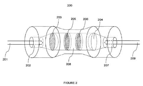
[063] Figure 2 depicts one embodiment of an endport assembly of this
embodiment.
The distal end of the single endport 104 comprising the bundled optical fibers
is inserted into the
entrance 202 of endport assembly 200. The signal is transmitted by the optical
fibers through the
endport assembly 200 to the exit 207, and is then transmitted to outgoing
optical fiber 208 which
in turn is in contact with a detector. Outgoing optical fiber 208 may have a
diameter of from
about 300 microns to about 500 microns, and preferably is about 400 microns.
Therefore, the end
port assembly optically couples the single end port to the detector. The
endport assembly may
comprise a first lens 203, which serves to collimate the incident signal. The
endport assembly
further may comprise a second lens 204, which serves to focus the outgoing
signal to a NA
suitable for outgoing optical fiber 208. The endport assembly further may
comprise at least one
notch filter 205 and at least one bandpass filter 206.
[064] Non-limiting examples of suitable detectors include photo-diode
detectors, photo-
multipliers, charge-coupled devices, a photon-counting apparatus, optical
spectrometers, and any
combination thereof.
[065] Figure 3 depicts one embodiment of a suitable sample holder 102 of this
embodiment. Spacers 303 are positioned such as to provide a space for an
elongated, transparent
container 306 to pass through the sample holder 300. In one embodiment, the
sample holder 300
is a capillary, and may be made of glass, quartz, or any other suitable
material that would be
known to one of skill in the art. By way of example only, the capillary may
hold 100 microliters
of fluid. Spacers 303 further are positioned to provide a slot 304, or space,
for the first ends of
the optical fibers to surround and be in close proximity to the transparent
container. Spacers 302
are held in place by top end plate 305 and bottom end plate 302, both of which
are attached to the
spacers 303 by a means for fastening 301, such as a screw.
[066] The emitted signal that is captured is converted to an electrical signal
by photo-
detector and transmitted to an analyzer (not shown), which receives the
electrical signal and
analyzes the sample for the presence of the analyte. Examples of analyzers
would be well-
understood by those of skill in the art. The analyzer may include a lock-in
amplifier, which
enables phase sensitive detection of the electrical signal, or any other means
known in the art for
analyzing electric signals generated by the different types of photo-detectors
described herein.
1-0&20
WO 2010/111514 PCT/US2010/028698
14
[067] The apparatus developed for these assays may be optimized for the
collection of
the light from the reporter molecule. The dyes currently used in fluorescence
based assays have
quantum efficiencies near or above 90%. In one embodiment, the dye is
Rhodamine Red X
(Invitrogen Corp., Carlsbad CA). In addition, the transconductance pre-
amplifier and the lock-in
detector settings are optimized to facilitate low signal/low noise detection.
First, an appropriate
modulation frequency is chosen for the optical chopper, which should be
incommensurate with
the line-frequency or other electrical sources of noise in the environment. In
addition, line
filtering by a lock-in amplifier should be employed. In one embodiment, the
modulation
frequency is 753 Hz, and the lock-in amplifier is set to filter at 60 Hz and
120 Hz. The sensitivity
for the transconductance pre-amplifier was chosen based on expected signal
level, and to
maximize the pre-amplifier's input impedance, and in one embodiment is set to
1 nA/V. In one
embodiment, the bandpass filter is centered on the chopper frequency, which is
e.g. 753 Hz.
EXAMPLES
1. Collection of Tissue Samples
[068] The procurement and propagation of the hamster-adapted 263K scrapie
strain was
as described Chang, B. et al., "PrP Antibody Binding-Induced Epitope
Modulation Evokes
Immunocooperativity," J. Neuroimmunol. v.205, issue 1-2, pp. 94-100 (2008)).
Brains from
sheep infected with scrapie and white-tailed deer infected with CWD were
harvested at the time
of clinical disease and frozen at -800 C. Brains from uninfected animals were
similarly harvested
and frozen. The coding region of the full-length deer, hamster, mouse and
sheep PrP was cloned
into a pET-23 vector to produce a tag-free protein (rPrP) as described in D.R.
Brown et al.,
"Normal prion protein has an activity like that of superoxide dismutase,"
Biochem J. vol. 344 pp.
1-5 (1999). Expression and purification was substantially identical to
procedures C.E. Jones et
al., "Preferential Cue coordination by His96 and His"' induces a-sheet
formation in the
unstructured amyloidogenic region of the prion protein," J. Biol. Chem. 279,
pp. 32018-32027
(2004).
[069] Experimental oral infections used a 20% scrapie sheep brain homogenate
(derived
from a composite of 7 scrapie brains from clinically and immunohistochemically
positive
animals) prepared in phosphate-buffered saline (PBS). All uninfected animals
were housed in a
separate scrapie-free facility. Clinical signs of sheep scrapie included: fine
head tremors
progressing to body trembling, wool loss from rubbing, nibbling at
extremities, hypersensitivity
and gait abnormalities.
1-0&20
WO 2010/111514 PCT/US2010/028698
[070] Genotyping of the sheep was performed commercially (Gene Check, Inc.,
Greeley,
CO).
[071] For IHC, formalin fixed third eyelid tissues were washed for 15 min in
water and
soak in 99% formic acid for 1 hr. After a 3 hr water wash, the tissues were
paraffinized in a
Microm STP 120, and cut at 4 microns for mounting. The slides were allowed to
dry for at least
24 hours, deparaffinized and then immunostained using the Ventana (Ventana
Medical Systems
Inc., Oro Valley, AZ) proprietary reagents (prion enhancing solution and anti-
PrP antibody) and
Benchmark LT automated system.
[072] For blood collection (IACUC approved), the animals were restrained and a
needle
was inserted into the jugular vein. Immediately following blood collection
(using sodium citrate
as the anticoagulant), one half of the blood was chilled and shipped
immediately. The remaining
half of the collected whole blood sample was centrifuged at low speed for 15
min at 4 C.
Plasma was removed, frozen and shipped on dry ice.
[073] White-tailed deer care and sampling protocols were approved by the
Colorado
Division of Wildlife's (CDOW) IACUC. Neonatal white-tailed deer fawns acquired
from several
free-ranging sources were bottle-raised using canned evaporated bovine milk
and established
protocols (Wild and Miller 1991; Wild et al. 1994). Deer were confined to
biosecure paddocks
throughout the study, except during times of sample collections. Food, water
and supplements
were provided ad libitum in all paddocks. At about 6 months of age, white-
tailed deer fawns
were orally inoculated with about 0.5 g of conspecific, pooled, infectious
brain material placed at
the base of the tongue; previous analyses showed that this inoculum pool was
infectious and
contained about 6 g PrPcwl) per g of brain tissue (Raymond et al. 2000; Wolfe
et al. 2007). All
deer were evaluated by a veterinarian experienced in recognizing clinical
signs of CWD, and
subjectively scored for behavioral changes, loss of body condition, ataxia,
and salivation or
polydipsia. The five deer for this study were heterozygous for glycine and
serine at codon 96 of
the native prion protein gene, had PrPCWD accumulation in tonsil biopsies by
253 or 343 days post
infection (dpi) (Wolfe et al. 2007), and were confirmed to be prion infected
at postmortem
examination 891 to 1774 dpi.
[074] Blood samples were collected from the five inoculated white-tailed deer
at 891
dpi. At the time of sampling, one animal (BC04) was in end-stage clinical
chronic wasting
disease, two (N204 and W1004) were showing some loss of body condition, and
the other two
(1304, K304) were clinically normal. For blood sampling, deer were sedated
with xylazine, skin
overlying the jugular vein was aseptically prepared, and about blood was
collected via jugular
1-0&20
WO 2010/111514 PCT/US2010/028698
16
venipuncture into a plastic bag treated with sodium citrate. Bags of blood
were cooled and
shipped overnight for processing.
2. Generation of Monoclonal Antibodies
[075] PK-treated PrPs , which consists of the core protein containing amino
acids (aa)
90-231 (PrP90_231), was isolated from the brains of 263K infected hamsters
using a procedure
originally reported by Hilmert and Diringer (1984) and modified by Rubenstein
et al. (1994).
This material was solubilized using guanidine hydrochloride extraction and
methanol precipited
as previously described (Kang et al., 2003) and used as the immunogen. PrP-/-
mice were
immunized and their immune responses monitored by ELISA as previously
described (Kascsak et
al., 1987). One of the immunized mice was used to produce hybridomas. The
mouse received a
final immunization of antigen by the intravenous route in phosphate-buffered
saline ("PBS") 4
days before fusion. Spleen cells were fused to an SP2/0 myeloma cell line
expressing reduced
levels of cell surface PrPc (Kim et al., 2003). The hybridomas were screened
by ELISA as
previously described (Kascsak et al., 1987) and the resulting cells were
cloned three times by
limiting dilution. Large scale Mab production was carried out using disposable
bioreactor flasks
(Integra Biosciences, Switzerland) and antibody was purified from media using
protein G
immunoaffinity chromatography (Pierce, Rockford, IL). Protein was determined
by the micro
BCA protein assay (Pierce) and isotyping was performed using the mouse Mab
isotyping kit
(Pierce). Each of the Mabs was biotinylated using the EZ-link biotinylation
kit (Pierce).
Sc
[076] Numerous Mabs were generated using the solubilized PrP as immunogen and
the low PrP expressing SP2/0 myeloma cell line. Three of these Mabs, 08-1/5D6
(5D6), 08-
1/11F12 (11F12) and 08-1/8E9 (8E9) were selected for this study and have been
isotyped as
IgGI, IgG2b and IgG2b respectively. Individually, all three Mabs react with
both the normal and
disease associated PrP isoforms.
[077] Western blotting of total brain lysates (Figure 6) demonstrated that all
three Mabs
Sc
were reactive against PrP from non-protease treated brain samples and PK-
treated PrP from
263K-infected hamsters, scrapie-infected sheep and CWD-infected deer (Figure
6). Similar
sc
results were observed using untreated and PK-treated partially purified PrP
preparations (data
not shown). These Mabs were also immunoreactive against the normal and
abnormal PrP
Sc
isoforms and PK-treated PrP isolated from mouse brains infected with the ME7,
139A and 22L
c
mouse-adapted scrapie strains and CJD-infected human brain as well as PrP
derived from
uninfected brain material from all the species tested including cattle (data
not shown).
1-0&20
WO 2010/111514 PCT/US2010/028698
17
[078] By indirect ELISA, the three Mabs were immunoreactive to PK-treated
PrPsc
purified from 263K-infected hamster brains. The degree of reactivity was
dependent on the
extent of the denaturation treatment. Either heat or SDS treatment alone
increased
immunoreactivity but a combination of the two treatments resulted in the
highest levels of
antibody binding and immunoreactivity (Table 1) approximating an additive
effect of the two
treatments and suggesting that epitope exposure is a multi-mechanistic
process. Interestingly,
although 5D6 binds to a conformational epitope, reactivity of this Mab is not
lost, but rather
enhanced upon PrP denaturation. It has previously been reported (Tayebi et
al., 2004) that heat
sc
denaturation is not sufficient to disrupt the polymeric structure of PrP .
Furthermore, the Mabs
c
were equally immunoreactive by ELISA to both PrP from uninfected brains and
total PrP
(normal and abnormal PrP isoforms) in non-denatured brain homogenate.
Immunoreactivity was
equally enhanced approximately 2-fold following denaturation with SDS and
heat. Following
Sc
PK treatment and denaturation, the immunoreactivity of PrP was increased an
additional 3-fold
due to the presence of less exogenous brain protein binding as a result of the
proteolytic digestion
(data not shown).
[079] To increase specificity and sensitivity for PrP detection, a capture
ELISA assay
was used incorporating a biotinylated detection antibody. As expected, for
each of the Mabs
biotinylated, 5-6 biotins were bound to each antibody molecule. Further, the
biotinylation of the
Mabs did not interfere with or reduce their immunoreactivity as assessed by
indirect ELISA using
Sc
partially purified PK-treated PrP (data not shown). Therefore, any differences
in the binding
and reactivity of the detection antibodies are not the result of the physical
biotinylation process.
sc
Using PK-treated PrP that had been denatured with SDS and heat, several Mab
combinations
were examined and each antibody was assessed both as the capture reagent and
as the detection
reagent (Table 2). Only one of the antibody combinations, Mab 11F12 as the
capture reagent and
Sc
biotinylated 5D6 as the detector, was successful in binding to and identifying
PrP . The results
sc
were the same regardless of whether the PrP was derived from 263K-infected
hamsters, scrapie
sheep or CWD-affected deer. The capture ELISA assay utilizing the 11F12-5D6
Mab
combination was next assessed for its ability to detect PrP in total and PK-
treated brain
homogenates from uninfected and infected hamsters, sheep and deer (Figure 7).
Similar to the
Sc
results described above for the indirect ELISA assay on purified hamster brain
PrP , the
detection of PrP in the capture ELISA assay was also dependent on epitope
availability and
1-0&20
WO 2010/111514 PCT/US2010/028698
18
determined by the initial treatment of brain lysate. Untreated brain lysate
from infected animals
showed a slight (1.5-fold) increase in signal intensity compared to uninfected
brain material
whereas either detergent or heat denaturation alone resulted in a 4 to7-fold
increase. Not
sc
surprisingly, the highest levels (greater than 10-fold) of PrP detection were
achieved when a
combination of SDS and heat treatment were used. Furthermore, increasing the
concentration of
sc
SDS above 1% reduced PrP detectability most likely due to an inhibition and/or
reversal of
antibody-antigen binding. This harsh denaturation treatment, as will be seen
below, was not
sufficient to completely destroy PrP conformation. It has previously been
reported that scrapie
Sc
infectivity, and presumably some degree of PrP conformation, could be
maintained in purified
Sc
PrP preparations following treatment with SDS, heat and SDS-PAGE (Brown et
al., 1990;
Rubenstein et al., 1994).
c
[080] PrP could be detected in non-PK treated normal brain homogenates by
capture
ELISA from all three species. In all cases, the signal intensity (-0.25-0.3)
was no greater than
twice above background (-0.12-0.15). This material was eluted from the wells
and examined by
c
western blotting. In contrast to the results described above where PrP was
detected directly
from non-PK-treated brain homogenates, western blotting of eluted samples
resulted only in the
c
detection of IgG light and heavy chains. PrP was not detectable due to the low
levels of bound
material. Following PK digestion, ELISA values were reduced to background
levels indicating
c sc
the elimination of PrP . PrP could readily be detected by the capture ELISA
assay in PK-
treated brain homogenates from 263K-infected hamsters, sheep scrapie and CWD.
Interestingly,
c
capture ELISA assays performed on non-PK treated brain homogenates, which
contain both PrP
sc c
and PrP , showed signal intensities higher than what could be attributed to
the PrP (determined
sc
from the non-PK normal tissue) and PrP (determined from the PK-treated
infected tissue)
aggregate (Figure 7). It is possible that the increased signal intensity is
due to the presence and
sc
binding of sPrP . An alternative explanation is that the binding of the
protein, presumably full-
length PrP , to the capture Mab induces a spatial change in the antigen which
results in the
sc
epitope for the second Mab becoming more accessible. This process is referred
to as positive
immunocooperativity.
[081] With the given set of Mabs used in this study, the degree of positive
Sc
immunocooperativity, as shown in Figure 7, was species dependent. PrP from CWD-
infected
1-0&20
WO 2010/111514 PCT/US2010/028698
19
deer showed the greatest levels with a 58% increase in 5D6 binding beyond that
calculated solely
from the combination of PrPc and PrPsC, while sheep scrapie PrPsC showed a 46%
increase. PrPsC
from 263Kinfected hamsters exhibited the least, but still significant, with
40%. The values in
Figure 7 are based on triplicate readings for six individual samples for each
species and
expressed as the mean + standard deviation.
Sc
[082] An antibody-induced spatial rearrangement and/or conformational change
in PrP
can be demonstrated by showing that the 11F12-5D6 captured material has
altered the epitope for
another PrP-specific Mab. The capture assay was performed on non-PK-treated,
SDS and heat
sc
denatured PrP . This was followed by incubation with biotinylated Mab 8E9,
streptavidin-
alkaline phosphatase and substrate. The lack of a signal above background
indicated that the
epitope for Mab 8E9 was either no longer available or accessible. However,
elution of the 11F12-
5D6 captured material from the microtiter wells followed by Western blotting
and
Sc
immunostaining with Mab 8E9 demonstrated robust PrP staining indicating that
the Mab 8E9
epitope was once again available (Figure 8). Presumably treatment with SDS-
PAGE sample
sc
buffer, along with electrophoresis in the presence of SDS, alters the 11F12-
5D6 binding to PrP
sc
and reverses the antibody-induced PrP changes to once again enable 8E9
binding.
C Sc
[083] Although Mab 8E9 was able to bind to PrP and PrP directly on Western
blots
and indirect ELISA assays, replacing 5D6 with 8E9 in the capture ELISA assay
resulted in no
detectable PrP indicating the absence of biotinylated 8E9 binding to the
antigen. Furthermore,
sc
the PrP specificity of the 11F 12-5D6 antibody pair was not only due to the
presence of these
specific Mabs but also to the sequence of the binding events. Reversing the
antibodies by
utilizing 5D6 as the capture reagent and biotinylated 11F12 as the detection
reagent (5D6/ Biotin
Sc
11F12) resulted in minimal PrP binding from non-PK treated brain lysates when
compared to
the 11F12-biotinylated 5D6 combination (11F12/Biotin 5D6) (Figure 9). A signal
to noise (S/N)
ratio was obtained by comparing the PrP signal obtained with the capture assay
using infected
brain lysates with the variance in the background signal obtained from
uninfected material from
hamster, sheep and deer brain tissue (S/N = (S-SO)/(36SO); where S=signal,
SO=mean
background signal, 60 = standard deviation of the background signal). A S/N
ratio of less than
1 indicates that a binding of the Mab is sufficiently weak that the signal
measured contains a
significant amount of noise. On the other hand, a S/N of 1 or greater
indicates that the noise in
the measurement is not significant indicating that most of the power in the
measurement results
1-0&20
WO 2010/111514 PCT/US2010/028698
from specific Mab binding. The confidence level increases exponentially as the
S/N ratio
increases. For the 5D6/Biotin 11F12 pair, the S/N ratios were approximately
0.6, 0.1 and 0.3 for
hamster, sheep and deer, respectively, indicating that the Mab binding was
nonspecific.
However, with the 11F12/Biotin 5D6 combination the S/N ratios were
approximately 19
(hamster), 28 (sheep) and 42 (deer). These ratios are indicative of the highly
significant nature of
the specific Mab binding. The values in Figure 9 are based on triplicate
readings for six
individual samples for each species and the ELISA results calculated as the
mean + standard
deviation. The increased antibody binding from infected samples (based on the
OD405) are
compared to the uninfected controls. Plotted on a logarithmic scale is the
signal to noise ratio
(S/N) as calculated from the signal power of the infected samples to the power
in the control
samples (noise).
3. Immunoassays
[084] For the preparation of 10% brain homogenates, brain tissues were
homogenized in
10 vol. of ice-cold lysis buffer (10 mM Tris-HC1, 150 mM NaCl, 1% IgepalTM CA-
630 (Nonidet
P-40), 0.5% deoxycholate, 5 mM EDTA, pH 8.0) in the presence of 1 mM
phenylmethylsulfonyl
fluoride (PMSF) (if the homogenate was to be treated with proteinase K (PK),
PMSF was omitted
from the lysis buffer). After centrifugation at 1,000 x g for 10 min, the
supernatants were
aliquoted and stored at -80 C.
[085] The protocol and reagents for the capture assays are described in Chang,
B. et al.,
"PrP Antibody Binding-Induced Epitope Modulation Evokes Immunocooperativity,"
J.
Neuroimmunol. v.205, issue 1-2, pp. 94-100 (2008), the contents of which are
hereby
incorporated herein in its entirety. Hybridoma cell lines producing the murine
monoclonal
antibodies used herein have been deposited as indicated, infra. For the
capture ELISA assay, 96-
well plates were coated with affinity-purified 11F12 capture monoclonal
antibody (Mab) (5
g/ml) at room temperature for 2-3 hrs. The coated wells were blocked with 3%
bovine serum
albumin (Sigma) in PBS overnight at 4 C. The wells were washed three times
with PBST. The
antigen was either non-PK- or PK- (100 g/ml PK at 50 C for 30 min) treated
brain lysates to
which was added a final concentration of 1% PMSF. All samples were treated
with 1% SDS
(final concentration), heated at 100 C for 10 min. and centrifuged at 16,000
x g for 5 min. The
supernatants were serially diluted 10-fold and 100 pl was added to each well.
The plates were
incubated at 370 C for 1 hr. The wells were washed three times with PBST and
100 l of the
biotinylated 5D6 detector Mab (5 g/ml) was added. After 60 min the wells were
washed with
1-0&20
WO 2010/111514 PCT/US2010/028698
21
PBST and 100 pl streptavidin conjugated to alkaline phosphatase (1:5,000) was
added for 60 min
at 370 C. PNPP (4-Nitrophenyl phosphate disodium salt hexahydrate) (Sigma)
substrate solution
was added to each well (100 l) and after 60 min, product was measured with an
ELISA reader
(Bio-Tek, Vermont, NY) at OD405.
[086] For laser analysis, incubation with the biotinylated Mab 5D6 was
followed by the
addition of streptavidin conjugated to Rhodamine Red (1:1000). Following a 60
min incubation
at 37 C, the wells were washed with PBST and treated with 100 pl IN NaOH for
10 min at
100 C and then shaken at room temperature for 20 min. The material was placed
into a 100 ul
MicrocapTM (Drummond Scientific, Broomall, PA) microcapillary tube which was
then inserted
into a specifically designed tube sample holder for laser excitation and
emission detection.
Dilutions are calculated relative to the original starting brain tissue. Each
value (data point)
represents the mean standard deviation (SD) from multiple assays as
described in the figure
legends.
4. Western Blotting
[087] Ten percent brain homogenates were prepared in lysis buffer as described
above.
The samples were centrifuged at low speed (2000 x g for 10 min). Ten
microliters of the
supernatants were mixed with a final of lx sample buffer, heated at 100 C for
4 min and
subjected to SDS-polyacrylamide gel electrophoresis (SDS-PAGE) using 12%
acrylamide gels,
transferred to nitrocellulose membranes and immunostained using either
streptavidin-conjugated
to alkaline phosphatase with NBT and BCIP as the substrate (Kascsak et al.,
1986) or horseradish
peroxidase-conjugated goat anti-mouse IgG (PierceTM) with super signal west
femto maximum
sensitivity substrate (Pierce) as previously described (LaFauci et al., 2006).
For samples that
were PK digested prior to SDS-PAGE, 40 ul of the supernatants from the low
speed
centrifugation were incubated with 100 g/ml PK (final concentration) for 30
min at 50 C
followed by the addition of 1% PMSF, 1X SDS-PAGE sample buffer and heating at
100 C for 5
min.
5. Immunoprecipitation
[088] MagnaBind protein G beads (Pierce) were washed 3 times with PBS,
resuspended
in 50 pl of PBS and 200 pl of 10% brain homogenate was added with 50 pg of Mab
8E9 (10
mg/ml) in a total volume of 1.2 ml PBS. After mixing at room temperature for 1
hr, the beads
were magnetically separated, washed 3 times with PBS containing 0.2% Tween 20
(PBST) and
then resuspended in 600 p1 PBS. After heating at 100 C for 10 min and
microcentrifugation at
16,000 x g for 3 min, the supernatants were used for capture ELISA.
1-0&20
WO 2010/111514 PCT/US2010/028698
22
[089] For blood samples, magnaBind protein G beads were resuspended in 100 l
PBS,
followed by the addition of 100 pg Mab 8E9 in a final volume of 5 ml PBS and
mixed at room
temperature for 1 hr. The beads were washed with PBST, resuspended in 5 ml PBS
containing
500 pl plasma and incubated for an additional 1 hr. As described above for
brain, the beads were
isolated, washed in PBST, heated and the microcentrifuged supernatant analyzed
by capture
ELISA.
6. Protein misfolding cyclic amplification (PMCA)
[090] As a source of PrPc for sPMCA of both brain and blood, 10% (wt/vol)
brain
homogenates from normal hamsters, sheep and deer [prepared in PBS containing
150 mM NaCl,
1.0% Triton X-100, 4 mM EDTA, and complete protease inhibitor cocktail
(Calbiochem)] were
centrifuged (1,500 x g, 30 sec) and the supernatants quick frozen. A 500 1
aliquot of serial 10-
fold dilutions (10-8 to 10-11) of brain homogenates from 263K-infected
hamster, sheep scrapie
and CWD deer were mixed with 100 1 of the 10% normal brain supernatant from
the
corresponding species and incubated (1 hr at 37 C) with shaking. Each sample
was then
sonicated (28W power output) followed by the addition of another 100 l of 10%
normal brain
homogenate and incubation (1 hr at 37 C with shaking). This was defined as
one cycle of
amplification. After each round of 5 cycles, PMCA was continued by
transferring 500 l of the
PMCA reaction mix from the original reaction tube to new tube and adding 100
pl 10% normal
brain homogenate. PMCA on 500 it aliquots of undiluted scrapie sheep or CWD
deer plasma
was carried out similarly as described for brain. Following PMCA, samples were
centrifuged at
2,000 x g for 10 min. For brain samples, 200 l of supernatant was digested
with proteinase K
(PK) (100 g/ml, 500 C, 30 min), followed by the addition of 1% protease
inhibitor cocktail and
1% SDS. Samples were heated at 100 C for 10 min and 10 pl aliquots were
analyzed by western
blotting (Chang et al. 2009). For blood, following PMCA 500 l of amplified
blood samples
were either untreated or PK-treated followed by Mab 8E9 immunoprecipitation
prior to analysis
by western blotting or SOFIA. For all PMCA samples, dilutions were expressed
relative to the
original undiluted brain or blood samples. Blood samples not subjected to PMCA
were
immunoprecipitated and processed similarly as above without the sonication and
37 C
incubation cycling.
7. SOFIA
[091] Ninety-six well High Binding plates (Costar, NY) were coated with
capture Mab
11F12 (5 pg/wel1 in 100 1) at room temperature for 3 hrs and blocked with
casein in TBS
overnight at 4 C. Magnetic protein G beads and Mab 8E9 were mixed for 60 min,
washed 3
1-0&20
WO 2010/111514 PCT/US2010/028698
23
times with PBST and the pellets resuspended in 800 l PBS. Blood samples were
centrifuged
(800 x g, 5 min) and 800 pl of the supernatants were combined with SDS (1%
final cone), heated
at 100 C for 10 min, mixed with the G beads-Mab 8E9 for 60 min and wash 3
times with PBST.
The final washed pellets were resuspended in 800 l PBS and heated for 10 min.
After
centrifugation at 16,000 x g for 5 min, 100 l of supernatant was added to
each well. After
incubation at room temperature (1 hr) and washing with PBST, 100 l of
biotinylated Mab 5D6
(2 pg/well) was added for 1 hr. followed by washing and incubation with 100 l
streptavidin-
Rhodamine Red-X conjugate (Invitrogen) for 1 hr. After 4 PBST washes, 100 1
of IN NaOH
was added and the plates were heated (100 C for 10 min), mixed for 30 min at
room temperature
and neutralized with equimolar amounts of HCI. Analysis was performed on 90 tl
aliquots.
8. Instrumentation
[092] The setup is designed around a commonly used disposable 100 microliter
micro-
capillary (Drummond Scientific Co., Broomall, PA) as a sample holder. The
sample is excited by
focusing temporally modulated light from a solid state, frequency-doubled
Nd:YAG laser (Beam
of Light Tech. TM, Clackamas, OR) along the axis of the capillary, with
typical power of 30 mW
continuous wave at a wavelength of 532 nm, which matches well with the
absorption peak of
Rhodamine. A fiber optic assembly was designed comprised of four linear arrays
which span
approximately a third of the length of the capillary and are positioned at 90
degrees with respect
to each other around the perimeter of the capillary. Because of the large
numerical aperture (0.22,
or an acceptance angle of -23 deg.) of the fibers, this orientation of the
fibers results in complete
coverage of the sample's field of view. The light collected by the four linear
arrays is ganged (i.e.,
bundled, or combined) and focused into transfer optics in which a holographic
notch filter (Kaiser
Optical Systems Inc. Ann Arbor, MI), and band pass filters (Omega Optical,
Inc. Brattleboro,
VT) are mounted. These are used to eliminate the scattered light from the
excitation source, and
band-limit the detection of the fluorescence of the reporter dye,
respectively. The light is then
focused back into a single, multi-mode, 400 micron optical fiber (ThorlabsTM,
Inc. Newton, NJ)
and coupled to a single low noise photo-voltaic diode detector (United
Detector Technology,
Hawthorne, CA) which is mounted on a BNC connector directly on the pre-
amplifier of the
detection electronics. Detection of the signal employs a phase sensitive, or
"lock-in", detection
scheme. The excitation source is modulated with an optical chopper (Thorlabs
Inc.) which serves
to generate the reference frequency for the detection system. The diode
detector is mounted on
the input of the transconductance pre-amplifier (Stanford Research Systems,
Inc. Sunnyvale, CA)
to reduce the total line impedance and eliminate difficulties in impedance
matching of the signal
1-0&20
WO 2010/111514 PCT/US2010/028698
24
at these low levels. The signal is then detected with a lock-in amplifier
(Stanford Research
Systems) and data acquisition is performed through a LabViewTM (National
Instruments Inc.,
Austin, TX). The program consists of an electronic strip chart which poles the
lock-in amplifier
for its reading in voltage periodically displays the time history of the
measurements to the
operator, and stores the values with a time stamp in an ASCII file. The time
constant of the lock-
in amplifier should be chosen to provide a bandwidth of a few tenths of a
Hertz. For these
measurements a time constant of 3 seconds was chosen. The lock-in requires
several time
constants in duration to obtain a stable reading (3 to 30 seconds in this
case). The values for the
measurements were taken after the signal had stabilized (20 to 30 sec.) after
loading a new
sample. The modulation of the excitation source, and reference frequency for
the lock-in
detector, were 753 Hz which was chosen to minimize environmental noise. In
addition to this
filtering of the signal at line-frequency and two times line frequency was
done with the lock-in
amplifier and the pre-amplifier signal was band-pass filtered at the
modulation frequency. For the
samples the pre-amplifier sensitivity of 1 nA/V was chosen, giving an input
impedance of 1 M
Ohm. In making the measurements a set of startup procedures was maintained
which included: a
warm up of 15 minutes for all electronics (the laser, lock-in amplifier, pre-
amplifier), a visual
check of dark signal levels to assure that system is properly electrically
grounded, a measurement
of laser power to check for stability and output level, a visual check of
laser alignment. Control
measurement of baseline signal is checked using a capillary with distilled,
deionized water.
[093] The sensitivity limits of the instrument were tested by measuring the
fluorescence
signal emission of Rhodamine Red at decreasing concentrations. Rhodamine Red
was detectable
to a concentration of 0.01 attograms (ag) [20 attomoles (am)] (Figure 10).
Determination of
specificity and sensitivity was carried out by performing assays using full-
length recombinant PrP
(rPrP) from deer, hamster, mouse and sheep. Regardless of the species tested,
the limits of
detectability were > 10 ag rPrP. Turning to Figure 10, data was obtained on
the instrument of
Figure 1, wherein dilutions of Rhodamine Red (^) in water were added to 100 1
micro-capillary
tubes, and surround optical fiber fluorescent signal emission was recorded.
The relative signal
intensities were calculated based on the fluorescence signal emission of water
alone. In the case
of the rPrP from mouse (*), hamster(*), sheep (V) and deer (9), the rPrP was
diluted in 1% PrP-
'- brain homogenate and subjected to SOFIA. The relative fluorescent signal
intensities were
calculated based on similar assays performed with rPrP diluent (1% PrP-~-
brain homogenate)
alone. Triplicate assays at a preamplifier setting of 1 nA/V were performed
for each rPrP
1-0&20
WO 2010/111514 PCT/US2010/028698
concentration and the data was plotted as the mean of the signal intensities
(% increase compared
to control) + SD.
[094] Brain homogenates from normal and infected hamsters, deer and sheep were
examined for their use in the method of the present invention. Western
blotting of 10% brain
homogenates confirmed the presence of PrPs in the starting material. Typical
PrP banding
patterns were evident in the 10% brain homogenates prior to PK treatment with
the characteristic
band shifting to lower molecular sizes of PrPs following PK digestion along
with the elimination
of PrPc from the normal hamster brain material as confirmation of complete
proteolytic
digestion. Serial dilutions of detergent extracted brain homogenates from
clinical animals have
demonstrated that the limits of PrPs detection by Western blotting is
approximately 10-3 - 10-4
while detection of PrPs by capture ELISA was sensitive following an
additional 10' - 10' fold
dilution (data not shown). In comparison, using the same Mabs and brain
homogenates, the
sensitivity of the assay reported in this manuscript exceeded that for Western
blotting and capture
ELISA by at least 5 orders of magnitude. Using the method of the present
invention, the signal to
baseline ratios (S/B) were used to evaluate PrP detestability in brain
homogenates. It was
determined that an S/B ratio of greater than 1.1 indicated the presence of
PrP. Serial dilutions of
PK-treated and untreated brain homogenates from normal and infected brain
tissue of hamsters,
sheep and deer were assayed by the method of the present invention (Figure
11). Values were
expressed as a ratio of signal from the samples' Rhodamine Red fluorescence
emission (S) vs.
background baseline signal derived from fluorescent emission of the diluent
(1% PrP-'- brain
homogenate or homogenizing buffer) alone (B). The data represents the mean
SD from three
independent experiments, each performed in triplicate at a preamplifier
setting of 1 nA/V, for
each brain homogenate dilution. As expected, following PK treatment all
samples from normal
brain tissues had S/B ratios of less than 1.1 regardless of the concentration
tested indicating the
absence of PrPc. As demonstrated by total signal output or S/B ratios above
1.1, protease
resistant PrPs , from serial 10-fold dilutions of PK-treated infected hamster
brain homogenates,
was detectable to a dilution of 10-" and from sheep and deer to 10-10. In
addition, maximum
PrPs detection from the PK-treated brain homogenates ranged from dilutions of
10-7 - 10-8 for
hamsters as well as sheep and deer.
[095] In the case of 10-fold serially diluted non-PK treated normal brain
homogenates,
PrPC was detectable by SOFIA to a dilution of 10-11 for hamsters and 10-10 for
deer and sheep
(with peak detection at 10-6 -10-7 dilutions) after which the S/B ratios all
fell below 1.1. The S/B
ratios from of non-PK treated brain tissue of 263K infected hamsters, scrapie-
infected sheep and
1-0&20
WO 2010/111514 PCT/US2010/028698
26
CWD-infected deer continued to indicate the presence of PrP. Serially diluted
brain homogenates
from infected tissues all showed S/B values greater than 1.1 to a dilution of
10-" for sheep and
deer (with peak detection at 10-') and 10-13 for hamsters (peak detection at
10-8). These results
indicate that PrP from non-protease treated, infected brain tissue can be
diluted beyond the levels
of PrPc detectability while still maintaining the capability to detect total
PrPs . These results
further suggest that there is at least 1 log more total PrPSC than PrPc in an
infected brain at
clinical disease. In support of this, it has previously been reported that
PrPs accumulates in the
brain during scrapie infection and attains concentrations 10 times greater
than that of PrPC.
Using previously published data on 263K-infected hamsters (R. Atarashi et al.
"Ultrasensitive
detection of scrapie prion protein using seeded conversion of recombinant
prion protein," Nature
Meth. vol. 4 (2007) pp. 645-650), SOFIA has a detection limit of approximately
10 ag of PrPs
from non-PK treated hamster brain. Extrapolation directly from the hamster
data suggests that 1
femtogram of PrPs can be detected from sheep and deer brain material.
However, assuming
equal antibody reactivity, Western blotting of diluted samples indicated that
there is at least 10 -
100 fold more PrPs in hamster brains than in sheep and deer brain material on
a gram equivalent
basis (data not shown) suggesting that detection of the protein in the latter
two species could be in
the range of 10 - 100 ag or better.
9. Detection of PrPSC in Blood
[096] Immunoprecipitation with Mab 8E9 serves as a bridge linking PMCA and
SOFIA,
so, the utility of this Mab for PrP immunoprecipitations was examined.
Mixtures of various
ratios of 10% brain homogenates from uninfected and 263K-infected hamsters
(uninfected:infected (%) - 100:0, 90:10, 70:30, 50:50) were immunoprecipitated
and analyzed by
western blotting (Figure 12). Ten percent normal brain homogenates (NBH) and
263K-infected
hamster brain homogenates (263K BH) were combined in various proprtions (lanes
1, 5 - NBH
only; lanes 2, 6-90 L NBH and 10 pL 263K BH; lanes 3, 7-70 L NBH + 30 L263
BH; lanes 4,
8 - 50 L NBH + 50 L 263K BH) and immunoprecipitated with Mab 8E9. The
immunoprecipitated samples were either untreated (lanes 1-4) or PK-treated
(lanes 5-8) prior to
western blotting and immunostaining with Mab 11F 12. In the absence of PK
digestion, all
samples, regardless of the brain homogenate ratios, showed similar
immunostaining intensities
(lanes 1-4). Western blots of PK-treated immunoprecipitants (lanes 5-8)
demonstrated similar
immunostaining when directly compared to samples containing only PK-treated
263K-infected
brain homogenates (data not shown). These results indicate that Mab 8E9
immunoprecipitated
both PrPc and PrPs and the presence of PrPc did not inhibit or reduce maximal
PrPs
1-0&20
WO 2010/111514 PCT/US2010/028698
27
immunoprecipitation. To quantitatively and qualitatively evaluate the PrP
isolated, capture
ELISA was performed on Mab 8E9 immunoprecipitants from the combinations of PK-
untreated
normal and 263K-infected hamster brain homogenates (Figure 13). The capture
ELISA utilized
the same Mab pair (11F12 as the capture Mab and 5D6 as the detector Mab) as
that used for
SOFIA. Mab 8E9 immunoprecipitation of the normal brain:infected brain
combinations followed
by capture ELISA resulted in increasing ELISA signal intensities as the levels
of infected brain
material increased in the starting mixtures. Since the brain material was not
proteolytically
digested, each mixture contained either PrPc alone or a mixture of both PrPc
and PrPs , as
confirmed by western blotting (Figure 12). However, analysis of the
immunoprecipitants by the
capture ELISA indicates that the increasing signal intensities are dependent
on the presence of
PrPs and not PrPc. This points to the utility of these specific Mabs and the
methodology for the
detection of PrPs Although the immunoprecipitation-capture ELISA format could
readily detect
brain-derived PrPs from 263K-infected hamsters, scrapie sheep and CWD deer,
PrPs could not
be detected in blood from clinical animals.
[097] The ability to detect PrPs in blood from 263K-infected hamsters and
sheep
scrapie samples following serial PMCA (sPMCA) has previously been reported.
However, the
large number of PMCA cycles necessary for PrPs detection makes the technique
impractical for
use as a diagnostic assay. The issue of PrPs detection in blood has been
approached by
incorporating sPMCA, followed by immunoprecipitation of the amplified target,
and detection
with the sensitive SOFIA assay (Chang et al., 2009). sPMCA was evaluated and
validated using
hamster brain (Figure 14)). Dilutions of hamster brain homogenates were
subjected to 7, 14 and
40 cycles of PMCA (lanes 7-10) while identical samples were processed
similarly without
sonication (lanes 3-6). Ten L aliquots of each sample was analyzed by western
blotting with
Mab 11F12. Control samples were 10.2 dilutions of 263K-infected hamster brain
homogenates
without (lane 1) and with (lane 2) PK digestion prior to western blotting 10-8-
10-"dilutions
(relative to the original brain tissue) of 10% 263K-infected hamster brain
homogenate were
subjected to sPMCA using normal hamster brain homogenate as a PrPc source.
PrPs was
undetectable at all the dilutions following 7 cycles of sPMCA but could be
detected in the 10-8
diluted sample by the completion of 14 cycles (Figure 14, lane 10). After 40
cycles of sPMCA
(sPMCA40), PK-resistant PrPs was detectable at all dilutions of 263K-infected
brain
homogenates tested (Figure 14 lanes 7-10). Similarly diluted hamster brain
homogenates that
were processed in parallel with the PMCA sonication steps omitted, did not
show any PrPs
amplification as demonstrated by the absence of PK-resistant PrPs
immunostaining (Fig. 3, lanes
1-0&20
WO 2010/111514 PCT/US2010/028698
28
3-6). Comparing detection limits of PrPs in the absence of PMCA (10-6
relative to the original
brain tissue) with detection after sPMCA40, and taking into account the sample
size analyzed, the
results estimate a 104 fold amplification as a result of PMCA.
[098] Similar PMCA experiments with diluted sheep brain or CWD brain
homogenates
from clinical animals (along with the uninfected brain homogenates of the
corresponding species
as the source of PrPc) demonstrated the initial detection of amplified PrPs
by western blotting at
28 cycles of PMCA at the 10-8 dilution of infected brain. The increased number
of cycles needed
for the initial detection of PrPs from sheep and deer brain compared to
hamster brain was due to
the lesser amount of starting PrPs found in the original brain tissue. As
expected, by the end of
sPMCA40, the amplified PrPs from the scrapie sheep and CWD deer brain
homogenates was
demonstrated by increased immunostaining intensity and detection limits of PK-
resistant PrPs
Although the sheep and deer brain tissue contained less PrPs compared to
infected hamster brain,
the levels of amplification were still approximately 4 logs relative to the
initial PrPs levels (not
shown).
[099] Plasma from scrapie sheep and CWD deer were subjected to sPMCA40. The
sheep
samples consisted of three groups (Table3, groups 1-3) of scrapie sheep,
which, at the time of
blood collection, were differentiated based on the presence or absence of
clinical signs and PrPs
immunohistochemical (IHC) staining of third eyelid lymphoid follicles (Figure
15). All animals
in group 3 that did not display clinical symptoms at the blood collection time
points eventually
progressed to clinical disease. The group of uninfected sheep (Table 3, group
4) were housed and
maintained in an isolated, scrapie-free area. CWD samples consisted of several
experimentally
infected (oral route) preclinical and clinical white-tailed deer (Table 4).
All of the sheep and
CWD samples were individually subjected to sPMCA40 and analyzed by western
blotting
following PK digestion. Following sPMCA40 of plasma, western blotting of PK-
treated PMCA
products either prior to or after PrPs concentration by Mab 8E9
immunoprecipitation, did not
reveal any PrPs . The addition of polyadenylic acid [poly(A)], which has been
reported to
facilitate rapid detection of low levels of PrPs from sheep blood (Thorne and
Terry, 2008), did
not improve amplification efficiency to the point of PrPs detection from
sheep scrapie or CWD
deer plasma following sPMCA40. The lack of PrPs detection following sPMCA40
from sheep
blood was independent of the sheep genotypes used (data not shown). That is,
the pairing of
sheep genotypes between the source of PrPs and the normal sheep brain PrPc
did not sufficiently
increase the amplified product for detection by immunoblotting. Since western
blotting was not
informative, it is unclear whether PrPs was initially present in the blood
from CWD and any of
1-0&20
WO 2010/111514 PCT/US2010/028698
29
the animals comprising the three groups of sheep scrapie or whether PMCA was
successful but
western blotting was not sensitive enough to detect the amplified PrPs after
only 40 cycles. It
has been reported that PMCA of sheep blood could lead to false positive
results due to the
apparent spontaneous generation of PrPs (Thorne and Terry, 2008). Therefore,
rather than
continue increasing the number of PMCA cycles, surround optical fiber
immunoassay (SOFIA)
(Chang et al., 2009) was used for PrPSc detection of untreated and PK-treated
Mab 8E9
immunoprecipitated sPMCA40 products. Our studies demonstrated that in the
absence of
sPMCA40, the readings obtained by SOFIA from scrapie sheep and uninfected
sheep plasma
samples were similar and approached baseline levels (Figure 16). Prior to
sPMCA40, the SOFIA
signal intensities (sample/background) for the individual samples ranged from
0.5 - 0.9 (group 1),
0.7 - 1.2 (group 2), 0.8 - 1.3 (group 3) and 0.6 - 1.1 (group 4). Since
previous studies on the
dynamic range of SOFIA (Chang et al., 2009) demonstrated that in PK-untreated
clinical sheep
brain PrPs was detectable in femtomole range (Chang et al., 2009), it is
likely that the levels of
PrPs in scrapie sheep plasma samples are below the detectable range. In an
attempt to amplify
the levels of PrPs to within the dynamic range of SOFIA, sPMCA40 was
performed followed by
Mab 8E9 immunoprecipitation. Non PK-treated PrPs could be detected by SOFIA
on the
immunoprecipitated sPMCA40 products (Figure 16). Following sPMCA40, the range
of signal
intensities (0.7 - 1.2) for the individual samples of the control group (group
4) did not
significantly differ from those samples prior to PMCA. However, the range of
SOFIA signal
intensities for all three groups of scrapie sheep were similar to each other
(group 1: 4.3 - 4.8,
group 2: 4.4 - 5.1), group 3: 4.8 - 5.3), regardless of their clinical
manifestations, and
significantly greater than both the pre-PMCA values as well as the uninfected
samples (group 4).
The value of this approach is realized when one considers that confirmation of
disease was
dependent on the sheep being scrapie infected but was independent of the
presence of clinical
signs and the neuropathology as all three groups of sheep tested positive for
the presence of PrPs
(Figure 16). PrPs amplification was also independent of genotype
compatibility since there was
no difference in the amplification when normal brain homogenates from either
ARQ/ARQ or
ARQ/VRQ sheep were used with any of the infected sheep plasma samples.
Furthermore, the
need for PK digestion to distinguish PrPc from PrPs was unnecessary since the
results of SOFIA
were the same regardless of whether the sPMCA40 products were untreated
(Figure 16) or PK-
treated (not shown) prior to immunoprecipitation and immunoassay analysis. The
data in Figure
16 was generated by dividing plasma samples into 3 groups according to the
appearance of
clinical signs and immunohistochemistry (IHC) associated with sheep scrapie.
Each plasma
1-0&20
WO 2010/111514 PCT/US2010/028698
sample was subjected to PMCA40 (s) or incubated without PMCA (o), Each sample
was either
untreated or PK digested followed by Mab $E9 immunoprecipitation and analysis
of PrPSc by
SOFIA. Plasma samples from each of the 3 groups was assayed in triplicate and
the data for all
the samples in each group combined and expressed as mean standard deviation_
[0100[ Similar studies were carried out with plasma obtained from several
preclinical and
clinical cases of deer CWD (Figure 17). Similar to the sheep plasma samples
described above,
the signals obtained by SOFIA on the CWD samples in the absence of sPMCA4o did
not differ
from the uninfected controls, which themselves approached background. In
addition, PK-
resistant PrPS'could not be detected by either capture ELISA or western
blotting following
sPMCA40. However following immunoprecipitation of the sPMCA4o products, PrPs
was
detectable by SOFIA from all preclinical. and clinical CWD blood (Figure 17).
Furthermore,
similar to the scrapie sheep samples, the SOFIA values were dependent on the
samples
originating from infected animals but confirmation of disease by SOFIA was
independent of the
clinical status of the diseased animal. The data in Figure 17 was generated by
subjecting each of
the plasma samples from the five CWD cases to sPMCA40 (a) or maintained in the
absence of
PMCA (^). All samples were either undigested or PIS treated followed by Mab
SE9
irn.rnunoprecipitation and SOFIA. Results are shown for the PK-untreated
samples and the values
represent the mean of triplicate assays SD. In the case of the 4 uninfected
deer plasma samples,
each of the 4 samples was analyzed in triplicate and the combined results of
the 4 samples are
expressed as the mean SD. In all embodiments of the present invention, all
percentages are by
weight of the total composition, unless specifically stated otherwise.
11011 All ratios are weight ratios, unless specifically stated otherwise. All
ranges are
inclusive and combinable. The number of significant digits conveys neither a
limitation on the
indicated amounts nor on the accuracy of the measurements. All numerical
amounts are
understood to be modified by the word "about" unless otherwise specifically
indicated.
[1021 All documents cited in the Detailed Description of the Invention are, in
relevant
part, incorporated herein by reference; the citation of any document is not to
be construed as an
admission that it is prior art with respect to the present invention. To the
extent that any meaning
or definition of a term in this document conflicts with any meaning or
definition of the same term
in a document incorporated by reference, the meaning or definition assigned to
that term in this
document shall govern.
[1031 Whereas particular embodiments of the present invention have been
illustrated and
described, it would be obvious to those skilled in the art that various other
changes and
1-0&20
WO 2010/111514 PCT/US2010/028698
31
modifications can be made without departing from the spirit and scope of the
invention. It is
therefore intended to cover in the appended claims all such changes and
modifications that are
within the scope of this invention.
REFERENCE LIST
Atarashi, R. et al. "Simplified ultrasensitive prion detection by recombinant
PrP conversion with
shaking," Nat. Methods 5, 211-212 (2008).
Bieschke, J. et al., "Autocatalytic self-propagation of misfolded prion
protein," Proc. Natl. Acad.
Sci. U. S. A. 101, 12207-12211 (2004).
Brown, P. et al. "Conservation of infectivity in purified fibrillary extracts
of scrapie-infected
hamster brain after sequential enzymatic digestion or polyacrylamide gel
electrophoresis," Proc.
Natl. Acad. Sci. U.S.A. 87, 7240-7244 (1990)
Brown, DR. et al., "Normal prion protein has an activity like that of
superoxide dismutase,"
Biochem. J. 344, 1-5 (1999). Erratum in: Biochem. J. 345, 767 (2000).
Bueler, H. et al., "High prion and PrPSc levels but delayed onset of disease
in scrapie-inoculated
mice heterozygous for a disrupted PrP gene," Mol. Medicine 1, 19-30 (1994).
Bulgin, M. S. et al., "Association between incubation time and genotype in
sheep experimentally
inoculated with scrapie-positive brain homogenate," Am. J. Vet. Res. 67, 49 8-
504 (2006)
Carlson, G. A. et al., "Prion isolate specified allotypic interactions between
the cellular and
scrapie prion proteins in congenic and transgenic mice," Proc. Natl. Acad.
Sci. U. S.A. 91, 5690-
5694 (1994).
Castilla, J. et al. "In vitro generation of infectious scrapie prions," Cell
121, 195-206 (2005a).
Castilla, J. et al. "Detection of prions in blood," Nat. Medicine. 11, 982-985
(2005b).
Chang, B. et al. "Surround optical fiber immunoassay (SOFIA): more than an
ultra-sensitive
assay for PrP detection," J. Virol. Methods 159, 15-22 (2009).
Chang, B. et al. "PrP antibody binding-induced epitope modulation evokes
immunocooperativity," J. Neuroimmunol. 205, 94-100 (2008).
Deleault, N. R. et al., "Protease-resistant prion protein amplification
reconstituted with partially
purified substrates and synthetic polyanions," J. Biol. Chem. 280, 26873-26879
(2003).
Gavier-Widen, D. et al., "Diagnosis of transmissible spongiform
encephalopathies in animals: a
review," J. Vet. Diagn. Invest. 17, 509-527 (2005).
Haley, N. J. et al. "Detection of sub-clinical CWD infection in conventional
test-negative deer
long after oral exposure to urine and feces from CWD+ deer," PLoS One 4, e7990
(2009).
1-0&20
WO 2010/111514 PCT/US2010/028698
32
Haley, N. J. et al., "Detection of CWD prions in urine and saliva of deer by
transgenic mouse
bioassay," PLoS One 4, e4848 (2009).
Hilmert, H. and Diringer, H. "A rapid and efficient method to enrich SAF-
protein from scrapie
brains of hamster," Biosci. Rep. 2, 165-70 (1984).
Houston, F. et al. "Transmission of BSE by blood transfusion in sheep," Lancet
356, 999-1000
(2000).
Jones, C.E. et al. "Preferential Cu2+ coordination by His96 and His 111
induces beta-sheet
formation in the sunstructured amyloidogenic region of prion protein," J.
Biol. Chem. 279,
32018-27 (2004).
Jones, M. et al. "In vitro amplification and detection of variant Creutzfeldt-
Jakob disease
PrP(Sc)," J. Pathol. 213, 21-26 (2007).
Kang, S.C. et al. :Guanidine hydrocholide extraction and detection of prion
proteins in mouse and
hamster prion diseases by ELISA," J. Pathol. 199, 534-41 (2003).
Kascsak, R.J. et al. "Immunological comparison of scrapie-associated fibrils
isolated from
animals infected with four different scrapie strains". J. Virol. 59, 676-683
(1986).
Kascsak, R.J. et al. "Mouse polyclonal and monoclonal antibody to scrapie-
associated fibril
proteins," J Virol. 61, 3688-3693 (1987).
Kim, J.I. et al. "Comparison of PrP transcription and translation in two
murine myeloma cell
lines," J. Neuroimmunol. 140:137-142 (2003).
Kocisko, D. A. et al. õCell-free formation of protease-resistant prion
protein" Nature 370, 471-
474 (1994).
Kurt, T. D. et al. "Efficient in vitro amplification of chronic wasting
disease PrPres," J. Virol. 81,
9605-9608 (2007).
LaFauci, G. et al. "Passage of chronic wasting disease prion into transgenic
mice expressing
Rocky Mountain elk (Cervus elaphus nelson) PrPC,: J. Gen. Virol. 87, 3773-3780
(2006).
Llewelyn, C. A. et al. "Possible transmission of variant CJD disease by blood
transfusion,"
Lancet 363, 417-421 (2004).
Madec, J-Yk. et al. "Abnormal prion protein in genetically resistant sheep
from a scrapie -
infected flock," J Gen. Virol. 85, 3483-3486 (2004).
Mathiason, C. K. et al., "Infectious prions in the saliva and blood of deer
with chronic wasting
disease," Science 314, 133-136 (2006).
Mathiason, C. K. et al., "Infectious prions in pre-clinical deer and
transmission of chronic
wasting disease solely by environmental exposure," PLoS One 4, e5916 (2009).
1-0&20
WO 2010/111514 PCT/US2010/028698
33
Murayama, Y. et al., õUrinary excretion and blood level of prions in scrapie-
infected hamsters,"
J. Gen. Virol. 88, 2890-2898 (2007).
Peden, A. H. et al. "Preclinical vCJD after blood transfusion in a PRNP codon
129 heterozygous
patient" Lancet 364, 527-529 (2004).
Prusiner, S. B. et al., "Transgenetic studies implicate interactions between
homologous PrP
isoforms in scrapie prion replication," Cell 63, 673-686 (1990).
Raymond, G. J. et al. "Evidence of a molecular barrier limiting susceptibility
of humans, cattle,
and sheep to chronic wasting disease" EVBO 19, 4425-4430 (2000).
Rubenstein R. et al. "Concentration and distribution of infectivity and PrPSc
following partial
denaturation of a mouse-adapted and a hamster-adapted scrapie strain." Arch.
Virol. 139, 301-
311(1994).
Saa, P. et al., "Presymptomatic detection of prions in blood," Science 313, 92-
94 (2006).
Saborio, G. P. et al., `Sensitive detection of pathological prion protein by
cyclic amplification of
protein misfolding," Nature 411, 810-813 (2001).
Soto, C. et al., "Pre-symptomatic detection of prions by cyclic amplification
of protein
misfolding," FEES Lett. 579, 63 8-642 (2005).
Tamguney, G. et al., "Asymptomatic deer excrete infectious prions in faeces,"
Nature 461, 529-
532 (2009).
Tayebi. M.et al. "Disease-associated prion protein elicits immunoglobulin M
responses in vivo,"
Mol. Med. 10, 104-111 (2004).
Thorne, L. & Terry, L. A., "In vitro amplification of PrPSc derived from the
brain and blood of
sheep infected with scrapie," J. Gen. Virol. 89, 3177-3184 (2008).
van Keulen, L. J., et al. "Immunohistochemical detection of prion protein in
lymphoid tissues of
sheep with natural scrapie, "J. Clin. Microbiol. 34, 1228-1231 (1996).
van Keulen, L. J., et al.. "Immunohistochemical detection and localization of
prion protein in
brain tissue of sheep with natural scrapie," Vet. Pathol. 32, 299-308 (1995).
Wells, G. A. and Wilesmith, J. W., "The neuropathology and epidemiology of
bovine spongiform
encephalopathy, "Brain Pathol. 5, 91-103 (1995).
Wild, M. A. and Miller, M. W., "Bottle-raising wild ruminants in captivity,"
Colorado Division
of Wildlife, Outdoor Facts 114 (1991).
Wild, M. A. et al, "Comparison of growth rate and milk intake of bottle-raised
and dam-raised
bighorn sheep, pronghorn antelope and elk neonates" J Wildlife Management 58,
340-347
(1994).
1-0&20
WO 2010/111514 PCT/US2010/028698
34
Wolfe, L. L. et al. "PrPCWD in rectal lymphoid tissue of deer (Odocoileus
spp.)," J. Gen. Virol.
88, 2078-2082 (2007).
1-0&20
WO 2010/111514 PCT/US2010/028698
ouip.-trisioti of D i ti r :i r~::t~f on Pry Ih m-limoreactivi:f d_,
T3'k#i:nn,nit L~Lilt~: SastrrES:i
0-6 11F1y. 8E9
LTntre t a.] 1 c$.01. j33 ..110
I ,,` ,M .223 O.O1t1 1. GS` 4..12ry4 1. '! ,.., ..33,1
!PD's=Plea: 2 - 2 32 2 p I- 2 21 0
`Pt. -sE. wa-:, n fled E. -M the k,.im of c1in td i h..3 fe.x: is 1ec e v it
G: I:,a tex a :jxtec +K ~,2n p e main an A Pa try Fl a. wet t.ed in ?
Id lvle>hod .
R "::ai is to is t IV,
q;s nesau ci by indirec EU A at OD:),
_~~,]
s'D . was 7W.de'd t a fir n] ceut a icou of 1%.
t
k r.
Sanmples iv e5:4 . eater I W C tar Efflk
TABLE 1
Page I
1-0&20
WO 2010/111514 PCT/US2010/028698
36
a1 ass o M rh Pain f r Capture :ELi A Assay` .
:;3tue 3:I;rlr IYet~:r3i :9. Zlae rin.~F:tdr ly
4'
111212 E#c 5 S
fit `: p.a. ate. .=A h a- sty b11 Fra4c Ytis; SD3 -nd h--a; ncr .h~-capfl r
Ma'. re.e U tnd a
TABLE 2
Page II
1-0&20
WO 2010/111514 PCT/US2010/028698
37
Description and Timing of Sheep Scrapie Disease Progression and Blood
Collection.*
Group 1: Clinical +, IHC
Blood Clinical
ID # Genotype lnfectiont lH }) Collection (dp ;V Signs d i . Death (dpi)
3040 AR h: RR E Neg. 2019 1936 2290
4102 A193" PC E Neg. 1565 1145 i885.
4110 , R i. R i E leg. 1565 1413 1855
663$ ARC'''s'RR E Neg. 783 604 11
Group 2: Clinical +, IHC +
15027 ARQa'~`VyRQ N 8040 1303 .883 1.320
5033 ARONR $21 1304 884 131
5066 ARQ RO N 74 1295 875 1.574
6004 ARQNRQ N 779 931 925 1249
6026 ARQ RQ N -402 926 926 1085
Group 3: Clinical 1, NC +
6006 ARC.'%,RQ N 732 934 934 1080
6014 AR,0'%, RO N 778 936 >^ 936 1256.
6024 Air ^NRO E 631 783 -783 1:213
6050 ARQ1VRQ E 259 76.3 >-783 1243
60160 ARCI1`RQ E 581 783 783 945
6066 ARQA RQ N 393 917 917 1076
Group 4: Uninfected
3058 ARQ/ARQ
5202 ARQiVRQ
5215 :AR ,,VR
5240 ARQfVRQ
5255 ARQivRQ
72:94 ARQ/VRQ
7212 ARQ'VRR
Animals are grouped based on clinical signs and third eyelid iynm hoid
follicle irnrnunoNstochernistry
dHCr at the time of hood collection,
tE - experimental (oral infection, N - natural infection ass,.r, red to occur
at birth Dexter st al., 2009). da'y C u,. t infection
''All an i nals ,,were clinically negative at the time of l l w8,c : 1 do but
all displayed ciNcal symptoms
at time of death.
TABLE 3
Page I I I
1-0&20
WO 2010/111514 PCT/US2010/028698
38
Description and Timing of Blood Collection, Disease Progression and Clinical
Presentation for White-Tailed Deer CWD
Blood Collection Death
lD # IHCA (dpi) animal status dpi cause
EC:D4 253 891 clinical CWD 891 clinical CWD
1N'1004 253 891 subtle changes 981 hemorrhagic disease (preci#n cal)
N204 25:3 891 subtle changes 1012 hemorrhagic disease (precl nical))
1304 343 891 preclinical 1182 clinical CWD
K304 253 891 preclinicai 1774 clinical CWD
PrPs im: unohistocher istry of tonsil biopsy.
Table 4
Page IV