Note: Descriptions are shown in the official language in which they were submitted.
CA 02756081 2011 09 21
WO 2010/096589 PCT/US2010/024628
PARTIALLY IMPLANTABLE MEDICAL DEVICES, FLUID CARTRIDGES FOR
USE WITH SAME, AND ASSOCIATED APPARATUS AND METHODS
BACKGROUND
1. Field
The present inventions relate generally to implantable medical devices.
2. Description of the Related Art
Fully implantable infusion devices, which are carried entirely within the
patient's body and include a reservoir, a fluid transfer device and a battery,
have been used to provide patients with a medication or other substance
(collectively "infusible substance"). The reservoir is used to store the
infusible
substance and, in some instances, fully implantable infusion devices are
provided with a fill port that allows the reservoir to be transcutaneously
filled
(and/or re-filled) through a hypodermic needle. The reservoir is coupled to
the
fluid transfer device, which is in turn connected to an outlet port. A
catheter,
which has an outlet at the target body region, may be connected to the outlet
port. As such, infusible substance from the reservoir may be transferred from
the reservoir to the target body region by way of the fluid transfer device
and
catheter.
The present inventors have determined that, while generally useful,
there are a number of issues associated with conventional fully implantable
infusion devices. For example, the present inventors have determined that
conventional fully implantable infusion devices are relatively large. In
particular, the batteries tend to be relatively large because they must last
many years and the reservoirs tend to be relatively large in order to minimize
refills, which may necessitate a visit to a physician for a percutaneous
needle-
based refilling procedure. Another issue identified by the present inventors
relates to control. Conventional fully implantable infusion devices are
controlled by way of an external remote control which can be lost or
misplaced. Another issue identified by the present inventors is maintenance.
Should, for example, the catheter be damaged or blocked, surgery is required
to remove and replace the catheter.
1
CA 02756081 2011 09 21
WO 2010/096589 PCT/US2010/024628
SUMMARY
An apparatus in accordance with one of the present inventions includes a
percutaneous port and an implantable operative portion.
An apparatus in accordance with one of the present inventions includes a
housing member defining an opening, a fluid transfer device and a housing
cover carried by the fluid transfer device and secured to the opening.
An apparatus in accordance with one of the present inventions includes a
percutaneous port configured to receive a cartridge and an implantable
operative portion, and is configured to sense movement of the cartridge
relative to the percutaneous port.
A method in accordance with one of the present inventions includes the
step of sensing movement of a cartridge relative to a percutaneous port.
A cartridge in accordance with one of the present inventions includes a
housing, a needle and a plurality of sensible members.
An apparatus in accordance with one of the present inventions includes a
cartridge with at least one sensible member and a partially implantable
medical device adapted to sense the at least one sensible member.
A fluid and power cartridge in accordance with one of the present
inventions includes a housing, a needle, a power source carried by the
housing, and power contacts.
An apparatus in accordance with one of the present inventions includes a
fluid and power cartridge and a partially implantable medical device including
a percutaneous port with an interior configured to receive the fluid and power
cartridge.
An apparatus in accordance with one of the present inventions includes a
percutaneous port configured to receive a cartridge, an implantable operative
portion including a fluid transfer device with an inlet and an outlet, and a
delivery/manifold tube operably connected to the inlet and the outlet.
An apparatus in accordance with one of the present inventions includes a
manifold portion, with first and second fluid lumens and a lumen-free portion
that prevents direct flow from the first fluid lumen to the second fluid
lumen,
and a delivery portion including a delivery lumen that is operably connected
to
the second fluid lumen.
2
CA 02756081 2016-09-15
77742-63
A method in accordance with one of the present inventions includes the
steps of delivering a first substance to a location within a patient's body
with a
partially implantable medical device and delivering a second substance to the
patient
with a device other than a partially implantable medical device.
A method in accordance with one of the present inventions includes the
steps of providing a patient with a first medication stored in a cartridge
that is
configured to be received by a partially implanted medical device and
providing the
patient with a second medication in an inhalable form.
A method in accordance with one of the present inventions includes the
steps of supplying a patient with an insulin cartridge that stores liquid
insulin and is
configured to be received by a partially implantable medical device and
supplying the
patient with insulin in an inhalable form.
Some embodiments disclosed herein relate to an apparatus for use with
a dermis and a fluid cartridge, the apparatus comprising: a percutaneous port
including a tubular wall, with an open and unobstructed first longitudinal
end, a
second longitudinal end, and an exterior, an end wall at the second
longitudinal end,
a resilient needle-puncturable septum at the end wall, and a porous region on
the
exterior of the tubular wall configured to promote soft tissue ingrowth, the
porous
region being located on the tubular wall such that a fluid cartridge located
within the
percutaneous cartridge port and at least partially within dermis that is
ingrown into the
porous region can be removed from the dermis by way of the open and
unobstructed
first longitudinal end and independently of the apparatus; and an implantable
operative portion, including a pump, operably connected to the percutaneous
port.
BRIEF DESCRIPTION OF THE DRAWINGS
Detailed description of exemplary embodiments will be made with
reference to the accompanying drawings.
3
CA 02756081 2016-09-15
77742-63
Figure 1 is a block diagram in accordance with one embodiment of a
present invention.
Figure 2 is a perspective view of a medical device in accordance with
one embodiment of a present invention.
Figure 3 is another perspective view of the medical device illustrated in
Figure 2.
Figure 4 is an elevation view showing the medical device illustrated in
Figures 2 and 3 implanted in a patient with the cartridge in place. Figure 4A
is an
enlarged elevation view showing the medical device illustrated in Figures 2
and 3
implanted in a patient with the cartridge removed.
Figure 5 is a side, partial section view of the medical device illustrated
in Figures 2 and 3 implanted in a patient with the cartridge in place.
Figure 6 is an exploded perspective view of a portion of the medical
device illustrated in Figures 2 and 3.
Figure 7 is a perspective view of a portion of the medical device
illustrated in Figures 2 and 3.
Figure 7A is a perspective view of a rechargeable battery.
3a
CA 02756081 2011 09 21
WO 2010/096589 PCT/US2010/024628
Figure 7B is a block diagram in accordance with one embodiment of a
present invention.
Figure 70 is a perspective view of a portion of a charger in accordance
with one embodiment of a present invention.
Figure 8 is a perspective view of a portion of a percutaneous port in
accordance with one embodiment of a present invention.
Figure 9 is a perspective view of a battery case in accordance with one
embodiment of a present invention.
Figure 10 is a perspective view of a portion of the medical device
illustrated in Figures 2 and 3.
Figure 11 is a perspective view of a septum in accordance with one
embodiment of a present invention.
Figure 12 is a section view taken along line 12-12 in Figure 11.
Figure 13 is an elevation view of a cartridge in accordance with one
embodiment of a present invention.
Figure 14 is a section view taken along line 14-14 in Figure 13.
Figure 15 is an exploded perspective view the cartridge illustrated in
Figure 13.
Figure 16 is an enlarged view of a portion of Figure 14.
Figure 17 is an enlarged view of a portion of Figure 14.
Figure 18 is a perspective view of a fluid transfer device in accordance
with one embodiment of a present invention.
Figure 19 is a partial section view taken along line 19-19 in Figure 18.
Figure 20 is an exploded perspective view of a portion of the medical
device illustrated in Figures 2 and 3.
Figure 21 is a perspective view of a portion of the medical device
illustrated in Figures 2 and 3.
Figure 22 is a perspective view of a portion of the medical device
illustrated in Figures 2 and 3.
Figure 23 is a perspective view of a portion of the medical device
illustrated in Figures 2 and 3.
Figure 24 is a section view of a portion of the medical device illustrated in
Figures 2 and 3.
4
CA 02756081 2011 09 21
WO 2010/096589 PCT/US2010/024628
Figure 25 is a perspective view of a portion of the medical device
illustrated in Figures 2 and 3.
Figure 26 is a perspective view of a portion of the medical device
illustrated in Figures 2 and 3.
Figure 27 is a block diagram in accordance with one embodiment of a
present invention.
Figure 28 is a section view of a portion of the medical device illustrated in
Figures 2 and 3.
Figure 29 is a perspective view of a delivery/manifold tube in accordance
with one embodiment of a present invention.
Figures 30-35 are plan views showing a plurality of sensible members
moving relative to a pair of sensors.
Figure 36 is a flow chart in accordance with one embodiment of a
present invention.
Figure 36A is a flow chart in accordance with one embodiment of a
present invention.
Figure 37 is a side view of a medical device in accordance with one
embodiment of a present invention.
Figure 38 is a partial section view of a portion of the medical device
illustrated in Figure 37.
Figure 39 is a side of a medical device in accordance with one
embodiment of a present invention.
Figure 40 is plan view of a portion of the medical device illustrated in
Figure 39.
Figure 41 is plan view of a portion of the medical device illustrated in
Figure 39.
Figure 42 is section view taken along line 42-42 in Figure 39.
Figure 43 is a side of a medical device in accordance with one
embodiment of a present invention.
Figure 44 is a perspective view of a medical device in accordance with
one embodiment of a present invention.
Figure 45 is a plan view of a portion of the medical device illustrated in
Figure 44.
5
CA 02756081 2011 09 21
WO 2010/096589 PCT/US2010/024628
Figure 46 is a perspective view of a portion of the medical device
illustrated in Figure 44.
Figure 47 is a perspective view of a portion of the medical device
illustrated in Figure 44.
Figure 48 is a perspective view of a portion of the medical device
illustrated in Figure 44.
Figure 49 is a perspective view of a cartridge in accordance with one
embodiment of a present invention.
Figure 50 is another perspective view of the cartridge illustrated in Figure
49.
Figure 51 is a plan view of the cartridge illustrated in Figure 49.
Figure 52 is a perspective view of the cartridge illustrated in Figure 49
with the battery and battery cover removed.
Figure 53 is a plan view of the cartridge illustrated in Figure 49 with the
battery cover removed.
DETAILED DESCRIPTION OF EXEMPLARY EMBODIMENTS
The following is a detailed description of the best presently known modes
of carrying out the inventions. This description is not to be taken in a
limiting
sense, but is made merely for the purpose of illustrating the general
principles of
the inventions.
The detailed description of the preferred embodiments is organized as
follows:
I. Introduction and Overview
II. Exemplary Percutaneous Port
III. Exemplary Replaceable Cartridge
IV. Exemplary Implantable Operative Portion
V. Exemplary Delivery/Manifold Tube
VI. Exemplary Control Methodologies
VII. Exemplary Internal Port
VIII. Additional Exemplary Implementations
IX. Exemplary Treatment Methodologies
6
CA 02756081 2011 09 21
WO 2010/096589 PCT/US2010/024628
The section titles and overall organization of the present detailed
description are
for the purpose of convenience only and are not intended to limit the present
inventions.
I. Introduction and Overview
The present inventions are generally directed to partially implantable
medical devices, i.e. medical devices that are configured such that, after
implantation, a portion of each device will extend completely through the
epidermis. The present medical devices may be used for therapeutic and/or
diagnostic purposes such as, for example, delivering a drug to a patient. As
illustrated for example in Figure 1, a medical device 10 in accordance with at
least some of the inventions disclosed herein includes a percutaneous port
12, a replaceable cartridge 14 that is configured to be received by the
percutaneous port, and an implantable operative portion 16.
The percutaneous port 12 extends through the epidermis from a
location within the body (e.g. a location within the abdomen) and,
accordingly,
allows a patient or physician to access various portions of the medical device
10 from outside the body. Access is attained without further incision into the
patient or the use of other devices and methods to facilitate percutaneous
access. By way of comparison, to refill many fully implantable infusion
devices
with a drug or other infusible substance, a physician must push a needle
through the patient's skin and into the abdomen in order to access the refill
port on the infusion device. An implanted battery would ultimately be depleted
and the device would likely be surgically replaced unless the battery was
rechargeable. Also, a surgical procedure may be required to replace a
blocked delivery catheter on a fully implantable infusion device.
The present percutaneous port 12, on the other hand, allows the
patient or physician to easily remove and/or replace the replaceable cartridge
14 from the outside of the patient's body. In at least some implementations,
the percutaneous port 12 also allows the patient or physician to remove and
replace certain aspects of the percutaneous port itself, remove and replace
certain aspects of the implantable operative portion 16 through the
percutaneous port, remove and replace some other device that may be used
in combination with the medical device 10 (e.g. a battery or other power
supply) and/or recharge a rechargeable battery. There are a variety of
7
CA 02756081 2011 09 21
WO 2010/096589 PCT/US2010/024628
advantages associated with such percutaneous access. For example, in some
conventional fully implantable infusion devices, the battery must last many
years and, accordingly, is relatively large (e.g. as much as about one-fourth
of
the total device volume). The ability to replace and/or recharge the
battery(s)
by way of the present percutaneous port 12 facilitates the use of smaller
batteries, which results in a smaller medical device. Moreover, in at least
some implementations, the tube that delivers fluid to the target body region
may be removed and replaced by way of the percutaneous port 12, thereby
eliminating the need for a surgical procedure should the tube become
blocked.
The percutaneous port 12 may be configured so as to encourage
tissue ingrowth into a portion thereof. Such tissue ingrowth creates an
infection resistant barrier around the percutaneous port 12. The percutaneous
port 12 may be carried by the implantable operative portion 16. The
percutaneous port 12 may, alternatively, be operatively connected to the
implantable operative portion 16 by a suitable structure such as, in the
exemplary context of an implantable infusion device, a fluid tube.
In the exemplary embodiment, the replaceable cartridge 14 supplies
the implantable operative portion 16 with something that is transferred to the
patient and/or is otherwise consumed by the implantable operative portion. In
the exemplary context of a partially implantable infusion device, the
replaceable cartridge 14 may function as the medical device reservoir and be
used to provide the drug or other infusible substance that is supplied to the
patient by the implantable operative portion 16. There are a variety of
advantages associated with a percutaneous port and cartridge-based
reservoir. For example, the reservoir may occupy as much as two-thirds of the
total volume of a conventional fully implantable infusion device. The large
reservoir is dictated by the difficulties associated with the refill of a
fully
implantable infusion device, e.g. it may require a visit to a physician for a
percutaneous needle-based refilling procedure, and the desirability of
limiting
the frequency of such procedures. In the exemplary context of high
concentration insulin delivery, the conventional reservoir is configured to
carry
a three to six month supply. The volume of the replaceable cartridge 14 may,
on the other hand, be considerably less. In the exemplary context of high
8
CA 02756081 2011 09 21
WO 2010/096589 PCT/US2010/024628
concentration insulin delivery, a cartridge could be configured to store a
seven
day supply, which results in about an approximately 90% volumetric reduction
in the overall medical device as compared to a device that stores a 3 month
supply.
The replaceable cartridge 14 may, in some implementations, also be
configured to supply power to the implantable operative portion 16 by way of
the percutaneous port 12, thereby obviating the issues associated with a more
permanent battery.
The replaceable cartridge 14 may also be used to perform a variety of
other functions, such as providing a direct user interface to the implantable
operative portion 16, either alone or in combination with other structures.
For
example, the user or physician may control certain aspects of the implantable
operative portion 16 (e.g. delivery rate) by rotating the cartridge 14
relative to
the percutaneous port 12. There are a variety of advantages associated with
such a user interface. For example, conventional fully implantable infusion
devices are generally controlled by way of telemetric communication from an
external remote control. The additional expense associated with this
communication method notwithstanding, patients are unable to interface with
their fully implanted infusion devices should they find themselves without
their
remote controls.
The implantable operative portion 16 performs the therapeutic and/or
diagnostic functions associated with the medical device 10 and, as used
herein, an "implantable" operative portion is an operative portion that is
sized,
shaped and otherwise constructed (e.g. sealed) such that it can be entirely
carried within the patient's body. In the exemplary context of a partially
implantable infusion device, the implantable operative portion may include,
among other things, a fluid transfer device (e.g. a pump and valve
arrangement) and control apparatus.
One example of a medical device which incorporates many of the
present inventions is the medical device 20 illustrated in Figures 2 and 3.
The
illustrated example includes a percutaneous port 100, a replaceable cartridge
200, and an implantable operative portion 300. A replaceable
delivery/manifold tube 400 may be provided in some implementations. The
9
CA 02756081 2011 09 21
WO 2010/096589 PCT/US2010/024628
particulars of the exemplary medical device 20 are discussed in Sections II-VI
below.
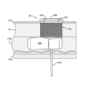
Turning to Figures 4, 4A and 5, the medical device 20 may, for
example, be implanted into the abdomen of a patient such that the
replaceable cartridge 200 will be adjacent to exterior surface of the skin and
available for removal and/or other manipulation. One suitable location is the
front side of the abdomen. For cosmetic purposes, the exterior color of the
cartridge 200 (or at least the visible surface thereof) may be chosen to match
the patient's skin color. Alternatively, the visible surface of the cartridge
may
be configured to resemble jewelry (such as that sometimes carried by body
piercings), may resemble a tattoo, or may resemble some other decorative
instrumentality. The replaceable cartridge 200 may be removed (Figure 4A)
from the percutaneous port 100, replaced, or otherwise manipulated, without
disturbing the implantable operative portion 300. The percutaneous port 100
may also, for example, be used to obtain physical or electronic access to
certain aspects of the port, and/or to replace the delivery/manifold tube 400,
when there is no cartridge 200 in the port.
In the exemplary context of insulin delivery, the implantable operative
portion 300 may be located subcutaneously within fat and/or muscle F/M, but
outside the peritoneal cavity, and the delivery portion of delivery/manifold
tube
400 may extend through the peritoneal wall PW and into the peritoneal cavity,
as illustrated in Figure 5. There are variety of advantages associated with
delivering insulin to the peritoneal cavity. For example, it is less likely
that
there will be tissue build-up at the outlet end of a delivery tube that is
located
in the peritoneum, as compared to the outlet end of a delivery tube that is
located subcutaneously. Delivery of insulin into the peritoneal cavity, as
opposed to subcutaneous delivery, results in better delivery kinetics,
eliminates the depot effect, is more natural (a healthy pancreas delivers
insulin to the peritoneal cavity), and the insulin peaks almost twice as fast
as
for subcutaneous injections of insulin.
It should also be noted that, depending on the therapy, the present
partially implantable medical devices may be used for subcutaneous delivery,
venous delivery, intranodal delivery, and delivery to any organ.
CA 02756081 2011 09 21
WO 2010/096589 PCT/US2010/024628
II. Exemplary Percutaneous Port
Referring first to Figures 2, 3 and 5, the exemplary percutaneous port
100 includes a tubular wall 102 with a rounded rim 104 and a layer of porous
material 106. The rounded rim 104, which may be located adjacent to the
epidermal surface when the medical device 20 is implanted into the patient,
strengthens the tubular wall 102 and eliminates what might otherwise be a
sharp edge that is uncomfortable to the touch. The layer of porous material
106, which may at a minimum be located just below the patients epidermis ED
and in contact with the dermis D (Figure 5), is configured to encourage tissue
ingrowth that creates an infection resistant barrier around the tubular wall
102
after implantation. The layer of porous material 106 extends around the entire
circumference of the tubular wall 102 (as shown) and may extend from one
longitudinal end of the tubular wall to the other, or over only a portion of
the
tubular wall below the rim 104, or over a portion of the tubular wall adjacent
to
the implantable operative portion 300, or over a portion of the tubular wall
therebetween. In certain exemplary implementations, the layer of porous
material 106 may be a mesh of intersecting fibers of any suitable
biocompatible material, such as a biocompatible metal (e.g., titanium,
nitinol,
stainless steel, gold, or platinum) or a biocompatible polymeric material
(e.g.,
polyolefins, Teflon, nylon, Dacron, or silicone). The mesh may be formed by
cross-winding the fibers in multiple layers to define a porosity conducive to
promoting tissue ingrowth (e.g., pore sizes within a range of 50 to 200
microns and having a porosity of 60 to 95%). The infection resistant barrier
may be enhanced by incorporating antimicrobial and/or anti-inflammatory
constituents into or beyond the layer of porous material 106. Additional
details
concerning such porous material layers may be found in U.S. Patent Pub.
Nos. 2004/0204686, 2007/0112334 and 2007/0149949, each of which is
incorporated herein by reference.
The exemplary percutaneous port 100 is circular in cross-section in
order to accommodate the cylindrical cartridge 200. It should be noted,
however, that the present percutaneous port may have cross-sectional
shapes other than circular in order to, for example, accommodate cartridges
that are oval, square, rectangular, or otherwise.
11
CA 02756081 2011 09 21
WO 2010/096589 PCT/US2010/024628
Turning to Figures 6-9, the exemplary percutaneous port 100 also
includes an end wall 108. The tubular wall 102 and the end wall 108 together
define an interior cartridge receiving region 110. The end wall 108 includes a
plurality of apertures and indentations that are associated with various
structures and functions that are related to the percutaneous port 100. For
example, the end wall 108 includes an aperture 112 that allows one or more
batteries 114 to be inserted into, and removed from, a battery case 116, which
has a positive battery contact 118. The battery case 116 is discussed in
greater detail below with reference to Figures 9 and 10. The end wall 108 also
includes a pair of apertures 120 and 122 for control sensors 124 and 126. The
control sensors 124 and 126, which are discussed in greater detail below with
reference to Figure 9 and in Section VI, are used to sense rotation of the
cartridge 200 relative to the port 100. In the illustrated implementation, the
control sensors are at the surface or extend slightly above the surface of the
end wall 108. An aperture 128 is provided for the replaceable
delivery/manifold tube 400, which is discussed in greater detail in Section V
below, while an aperture 130 is provided for a septum 132 that is located over
the delivery/manifold tube. The septum 132, which is discussed in greater
detail below with reference to Figures 11 and 12, is the structure through
which fluid from the cartridge 200 is delivered to the implantable operative
portion 300 (by way of the delivery/manifold tube 400). To that end, the
exemplary cartridge 200 includes a delivery needle 204 (Figures 13-15) and
the septum 132 is configured to allow passage of the needle. The septum 132
also functions as a seal, both when the needle 204 is extending therethrough
and after the needle has been removed, to prevent contaminants within the
interior cartridge receiving region 110 of the percutaneous port from entering
the delivery/manifold tube 400. The seal also prevents infusible substance
within the delivery/manifold tube 400 leaking into the interior cartridge
receiving region 110 of the percutaneous port 100.
It is anticipated that the batteries 114 may need replacement, that the
septum 132 may fail in response to the repeated needle puncturing
associated with cartridge replacement, and/or that the delivery/manifold tube
400 may become blocked or damaged. As such, the batteries 114, septum
132 and delivery/manifold tube 400 are removable and replaceable and may
12
CA 02756081 2011 09 21
WO 2010/096589 PCT/US2010/024628
be removed and replaced by way of the percutaneous port 100. To that end,
the wall that defines the aperture 130 in the illustrated embodiment is also
configured to mate with a releasable retainer 134 that holds the batteries
114,
the septum 132 and the delivery/manifold tube 400 in place. The exemplary
retainer 134 includes a flat retainer disk 136 and a post 138. The end wall
108
includes an indentation 140 that is substantially the same diameter and
thickness as the flat retainer disk 136 and, accordingly, the end wall and
flat
retainer disk will be essentially flush when the retainer 134 is in the locked
position illustrated in Figure 7. In the illustrated embodiment, the exterior
surface of the post 138 includes threads 142, while the wall that defines the
aperture 130 includes threads 144 which are configured to mate with the post
threads. The retainer 134 may be secured to the end wall 108 by inserting the
post 138 into the aperture 130, and then rotating the retainer until the flat
retainer disk 136 engages indentation 140. The flat retainer disk 136, which
also engages the adjacent battery 114 when the retainer 134 is in the locked
position (Figure 7), functions as the negative battery contact. The electrical
paths from the positive and negative battery contacts are discussed in Section
IV below.
A lumen 146 extends through the retainer 134 in the exemplary
implementation. The purpose of the lumen 146 is two-fold. The lumen 146
provides a passageway, which leads to the septum 132 and to the
delivery/manifold tube 400, for the cartridge needle 204 (Figure 28). The
lumen 146 is also configured to receive a tool (not shown) that may be used
to rotate the retainer 134. In the illustrated embodiment, the lumen 146 is
hexagonally-shaped and, accordingly, is configured to receive a tool such as
an Allen wrench that is correspondingly hexagonally-shaped. Other suitable
lumen/tool configurations include, but are not limited to square (or
"Robertson"), triple square and star shapes.
It should also be noted here that the inner surface of the exemplary
tubular wall 102 may be provided with an indentation 148 that is configured to
mate with a sealing ring 216 (Figure 16) on the cartridge 200, as is discussed
in Section III below. Additionally, and referring to Figure 8, the side of the
end
wall 108 opposite the interior cartridge receiving region 110 includes a ring
150 onto which the battery case 116 (Figures 9 and 10) is mounted, a base
13
CA 02756081 2011 09 21
WO 2010/096589 PCT/US2010/024628
member 152 in which the apertures 128 and 130 (Figures 6) are formed, a
ring 154 on which the delivery/manifold tube receiver 366 (Figures 23 and 24)
is mounted, one or more anchors 156, and a pin 158. The ring 154, anchors
156 and pin 158 are discussed in greater detail below in Section IV below. An
adhesive may be used to secure the battery case 116 to the ring 150.
As illustrated in Figure 9, the exemplary battery case 116 includes a
cylindrical wall 160, an end wall 162 that carries the battery positive
contact
118, and a flange 164 that carries the control sensors 124 and 126. The
cylindrical wall 160 has indentations 166 and 168 to accommodate the ring
150 and base member 152 and allow the battery case 116 to be mounted on
the percutaneous port end wall 108 in the manner illustrated in Figure 10.
Suitable materials for the battery case 116 include, but are not limited to,
polyethylene, polycarbonate, PEEK, Teflon, epoxy and others. The present
medical devices are not limited to any particular type of control sensor. The
type of control sensor will depend, at least in part, upon the type of
sensible
members carried by the cartridge 200. In the illustrated embodiment, the
circumferentially spaced control sensors 124 and 126 respectively consist of
pairs of electrical contacts ("contact pairs") 170a/170b and 172a/172b, and
the electrical contacts within each contact pair are substantially
circumferentially aligned (note Figure 7).
Turning to Figures 11 and 12, the exemplary replaceable septum 132
includes seal member 133 and an annular low friction retainer engagement
member 135. The seal member 133 has a relatively wide portion 174, a
relatively narrow portion 176 and a hollow region 178. The relatively wide
portion 174 is configured to fit within the aperture 130 and rest on the base
member 152 (note Figure 6), creating a seal. The relatively narrow portion
176 is configured to fit within the aperture 128 and rest on the
delivery/manifold tube 400. The retainer engagement member 135, which is
engaged by the lock post 138 (Figure 6) and is carried by the relatively wide
portion 174, defines an aperture 180 through which the cartridge needle 204
may pass, and includes a curved surface 182. The low friction lock
engagement member 135 allows the lock 134 to be rotated without rotating, or
rotationally deforming, the replaceable septum 132, while compressing the rim
of the septum assembly at 133 to effect the seal. Suitable materials for the
14
CA 02756081 2011 09 21
WO 2010/096589 PCT/US2010/024628
seal member 133 include, but are not limited to, resilient materials such as
silicone rubber and polyurethane, while the low friction member 135 may be
formed from materials, such as Teflon, a polished metal (e.g. titanium or
stainless steel), or a film (e.g. Teflon, nylon or polycarbonate) that is
adhered
to the seal member 133, which have a lower coefficient of friction than the
seal member. In other implementations, the retainer engagement member
may simply be in the form of a non-stick coating, such as a coating of a
Teflon
or a low friction polymer, on the seal member 133. It should also be noted
that
the septum is not limited to the illustrated shape with a narrow portion and a
wide portion and could, for example, simply be disk-shaped.
There are a variety of advantages associated with the present
percutaneous port 100. By way of example, but not limitation, the
percutaneous port 100 may be used to receive a replaceable cartridge (e.g.
cartridge 200) that is used to store the drug or other infusible substance
that is
supplied to the patient by the implantable operative portion 300. As noted
above, providing the infusible substance in this manner is more convenient
and greatly reduces the overall size of the medical device 20 as compared to
fully implantable infusion devices. The percutaneous port 100 may also be
used for maintenance. To that end, and as noted above, the percutaneous
port allows the batteries 114, the septum 132 and the delivery/manifold tube
400 to be removed and replaced. In addition to eliminating the need for
surgical procedures to replace the delivery tube, the ability to replace the
batteries 114 facilitates the use of a smaller power source than is required
for
a fully implantable infusion device that must remain implanted for many years.
Other advantages, which are associated with the sensing features of the
percutaneous port 100, are discussed in Section VI below.
III. Exemplary Replaceable Cartridge
As illustrated in Figures 13-15, the exemplary replaceable cartridge
200 includes a housing 202, which stores the infusible substance, and a
needle 204. Although the present cartridges are not limited to any particular
housing structure, the exemplary housing 202 has first and second housing
members 206 and 208 and an internal bladder 210.
The first housing member 206 in the exemplary replaceable cartridge
200 includes a cylindrical wall 212, with one or more air holes 214 and a
CA 02756081 2011 09 21
WO 2010/096589 PCT/US2010/024628
sealing ring 216, and an end wall 218 that is sized such that it extends
radially
beyond the percutaneous port rounded rim 104 (Figures 2, 3 and 16). For
example, the end wall 218 may have a flange 220 that rests on and curls
around the rim 104 when the cartridge 200 is fully inserted into the
percutaneous port 100 (Figure 16), or may simply have a flat flange that rests
on the rim (discussed below with reference to Figure 44). The second housing
member 208 includes a cylindrical wall 222 and an end wall 224. The
cylindrical wall 222 includes an indentation 226, with a longitudinally
extending surface 228 and a radially extending surface 230, that is configured
to receive a portion of the first housing member cylindrical wall 212 with a
portion of the internal bladder 210 therebetween. The end wall 224 may be
flat (as shown), convex, or concave. Suitable materials for the housing
members 206 and 208 include, but are not limited to, plastics such as
polyethylene or PEEK, or other polymers.
The internal bladder 210 in the exemplary embodiment illustrated in
Figures 13-15 is formed from a flexible film and includes a cylindrical side
wall
232 and an end wall 234. There are also no folds in the side and end walls
232 and 234. The side wall 232 is located within the indentation 226, abuts
the radially extending surface 230 and is compressed between the associated
portions of the cylindrical walls 212 and 222. So configured and arranged, the
internal bladder 210 and the second housing member 208 together define a
fluid storage volume 236 that, when filled with fluid (Figure 15), is
essentially
equal to the internal volume of the housing 202. The configuration of the
internal bladder 210 in the illustrated embodiment is such that the bladder is
not stretched, and does not exert a positive pressure on the fluid, when the
cartridge 200 is full. The internal bladder 210 will collapse (Figure 16) as
fluid
is drawn from the cartridge 200 and air enters the volume that is formed
between the housing member 206 and the internal bladder 210 by way of the
air holes 214. Suitable materials for the internal bladder include, but are
not
limited to silicone or butyl rubber.
It should be noted here that the present cartridges are not limited to the
illustrated internal bladder embodiment. Other devices may be used alone, or
in combination with the housing members 206 and 208, to define the fluid
storage volume. By way of example, but not limitation, such devices may
16
CA 02756081 2011 09 21
WO 2010/096589 PCT/US2010/024628
include plungers that slide within the space defined by the housing members,
flexible bellows, and other suitable structures. Also, another suitable
bladder
is a balloon without a defined shape.
Referring to Figure 16, the flange 220 on the cartridge housing 202 will
engage the percutaneous port rounded rim 104 to minimize the inflow of
water, but not create an airtight seal, when the cartridge 200 is fully
inserted
into the percutaneous port 100. Instead, in the illustrated embodiment, the
exemplary seal is air permeable so that air can reach the air holes 214. The
seal resists the inflow of water under normal conditions, but will not prevent
rotation of the cartridge 200 relative to the percutaneous port 100 (note the
discussion in Section VI below). So configured, the exemplary seal will be
tight enough to prevent water from entering the percutaneous port 100 during
everyday water-related activities such as showering, but not tight enough to
prevent water from entering the percutaneous port during swimming and
diving. The sealing ring 216 on the housing 202 will also mate with the
indentation 148 in the tubular wall 102 when the cartridge 200 is fully
inserted
into the percutaneous port 100. In addition to providing a more effective
seal,
the mechanical interference associated with the indentation 148 and ring 216
will prevent the cartridge 200 being unintentionally dislodged from the
percutaneous port 100 during normal activities. The indentation and ring
arrangement results in a small gap between the inner surface of the
percutaneous port tubular wall 102 and the outer surface of the cartridge
housing 202, which facilitates air flow into the holes 214. Alternatively, or
in
addition, any suitable mechanical lock (e.g. a click lock) may be provided to
provide a retention to keep the cartridge 200 in the port 100. A magnetic
locking arrangement is another alternative.
For swimming, diving and other activities that could result in leakage or
dislodgement of the cartridge 200, the cartridge may be removed and
replaced by a stopper (e.g. a rubber stopper) that is configured to create a
tighter seal than the cartridge. Such an arrangement may, for example, be
useful in those instances where the batteries 114 would be damaged if
immersed in water. The removal time would depend upon the application of
the medical device. In the exemplary context of insulin delivery, the removal
time could be as long as a couple of hours without danger. Tape seals, such
17
CA 02756081 2011 09 21
WO 2010/096589 PCT/US2010/024628
as those sold by Smith & Nephew, may be secured to the skin and positioned
over the top of the cartridge 200 or simply positioned within the port 100
over
the battery 114, to protect against water intrusion.
Turning to the cartridge needle, the needle 204 may be carried by the
end wall 224 and, in the illustrated embodiment, the needle is located at the
center of the end wall and extends along longitudinal axis of the cartridge
200.
Although the present cartridges may include any suitable needle
configuration, the exemplary needle 204 is a non-coring needle that reduces
the likelihood that it will damage the septum 132 when the cartridge is
inserted into and removed from the percutaneous port 100. To that end, and
referring to Figure 17, the exemplary needle 204 includes an elongated
tubular body 238, with an internal lumen 240, and a sharpened end portion
242. One or more apertures 244 pass through the tubular body 238 to the
internal lumen 240. The needle 204 may be configured such that the sharp
edges associated with the apertures 244 are not located on the sharpened
end portion 242 and, instead, are located inwardly from the overall outer
perimeter of the tubular body 238, which reduces the likelihood that the
needle 204 will damage the septum 132. The apertures 244 in the illustrated
embodiment are located within longitudinally extending indentations 245 that
have rounded edges. The exemplary needle 204 also includes a base 246
that is mounted in the end wall 224. The internal lumen 240 extends through
the base 246 and defines a needle inlet 248 that is located within the fluid
storage volume 236.
The size of the fluid storage volume 236 will, of course, depend on the
intended application. In the exemplary context of insulin delivery, the
cartridge
200 may be configured such that it can store one week's worth of highly
concentrated insulin that is to be delivered at a relatively high delivery
rate.
For example, if the maximum daily basal dosage is 100 units/day, a fluid
storage volume of 1.8 cc would be sufficient to store a week's supply of an
insulin that has a 400 units/cc concentration (e.g. Sanofi-Aventis U400). Such
a volume could, for example, be achieved with a cartridge that has an internal
diameter of about 14 mm and an internal height of about 12 mm. The outer
diameter of housing 202 would be about 15 mm, the exterior height of the
housing would be about 12 mm (excluding the end wall 218), and the
18
CA 02756081 2011 09 21
WO 2010/096589 PCT/US2010/024628
diameter of the end wall 218 (including the flange 220) could be about 16-17
mm in such a cartridge. Additionally, with respect to other exemplary
applications, the size of the fluid storage volume may range from 0.1cc to
20cc in applications such as for pain therapy with morphine.
Referring to Figure 15, the exemplary cartridge 200 also includes one
or more sensible members 250 that are sensed by the sensors 124 and 126.
Sensing of the sensible members 250 is used to identify rotation of the
cartridge 200 relative to the percutaneous port 100 in the manner described in
Section VI below. The sensible members 250 may be located on the exterior
of the second housing member end wall 224 (as shown), on the exterior of the
cylindrical walls 212 and 222, on the exterior of the first housing member end
wall 218, completely or partially embedded within one or more of any of the
end and cylindrical walls, or even within the internal volume of the
cartridge,
depending upon the type of sensible member employed, the location of the
associated sensor(s) and the manner in which the sensible member(s) and
sensor(s) interact.
In the illustrated embodiment, the sensible members 250 are
circumferentially-spaced electrically conductive pads that are separated by
non-conductive regions 251. The configuration of the percutaneous port 100
is such that a conductive pad will be in contact with the contacts 170a/170b
and 172a/172b when a portion of that conductive pad is circumferentially
aligned therewith. The above-described indentation 148 and ring 216, and
there positioning relative to the remainders of the percutaneous port 100 and
cartridge 200, may be used to apply a slight positive pressure which insure
that the sensible members 250 will make contact with the contacts 170a/170b
and 172a/172b when the sensible members and contacts are aligned.
Suitable examples of electrically conductive materials for the pads
include, but are not limited to, stainless steel, copper, aluminum, silver,
gold
and nickel. The conductive pads may be formed on the associated wall (e.g.
end wall 224) through the use of any suitable technique. By way of example,
but not limitation, the conductive pads may be formed by electroplating or
insertion molding. Alternatively, the associated wall (e.g. end wall 224) may
be formed from (or coated with) conductive material and those portions of the
conductive material that are not within a sensible member may be coated with
19
CA 02756081 2011 09 21
WO 2010/096589 PCT/US2010/024628
a non-conductive material. The conductive pads may also be printed onto a
sheet of plastic that is then adhered to the associated wall (e.g. end wall
224).
Conductive material may, alternatively, be printed directly onto the
associated
wall (e.g. end wall 224). Another alternative is to secure a precut metal
sheet
to the associated wall (e.g. end wall 224) and then peel away the portions of
the sheet that do not form the conducting pads. Similarly, a metalized film
may be formed on the associated wall (e.g. end wall 224) and then etched to
form the conductive pads.
The sensible members 250 are not limited to electrically conductive
pads. For example, cartridges in accordance with other embodiments of at
least some of the inventions may be provided with one or more protrusions,
indentations, and/or other instrumentalities that can be mechanically sensed.
Another exemplary alternative is one or more magnets that can sensed by, for
example, a flux sensor.
Cartridges in accordance with at least some embodiments may be
provided with information storage and communication devices that may be
used to provide information to, and/or store information received from, the
implantable operative portion 300. One example of such an information
storage device is an RFID tag (not shown). The RFID tag may be used to
provide the implantable operative portion 300 with programming information
and other data. A cartridge with such an RFID tag may be used to program or
reprogram the associated medical device, thereby obviating the need for
telemetric communication between the medical device and an external
programmer. The RFID tag may also be used to record data sensed by the
implantable operative portion 300. Here, used cartridges could be returned to
the manufacturer or the physician so that the data could be read and
analyzed. Examples of such data include, but are not limited to, data from
physiological sensors (e.g. glucose data) and failure mode data. The RFID tag
may also be used as an electronic safety key to, for example, prevent the
implantable operative portion 300 from operating when an unauthorized
cartridge is inserted into the percutaneous port 100 or when no cartridge is
present in the port. One example of an unauthorized cartridge would be a
cartridge that stores a medication or other infusible substance other than
that
prescribed by the patient's physician.
CA 02756081 2011 09 21
WO 2010/096589 PCT/US2010/024628
IV. Exemplary Implantable Operative Portion
Referring to Figures 2 and 3, the implantable operative portion 300 of
the exemplary medical device 20 includes a housing 302 with a fluid transfer
section 304 and an electronics section 306. The fluid transfer components
carried within the fluid transfer section 304 and the delivery/manifold tube
400
together transfer fluid from the replaceable cartridge 200 to a target
location
within the patient. The components within the electronics section 306 may
include, among other things, the powered portion of the exemplary fluid
transfer device 308 (Figures 18-21) as well as power and control circuitry.
Although the housing 302 may be configured such that the fluid transfer
section 304 and electronics section 306 share a common volume that is
sealed relative to the patient, the housing in the illustrated embodiment is
configured such that the electronics section is sealed relative to both the
fluid
transfer section and the patient. This aspect of the exemplary medical device
20 is discussed in greater detail below with reference to Figures 20 and 21.
A wide variety of fluid transfer devices may be incorporated into
medical devices in accordance with at least some of the present inventions. In
the illustrated embodiment, the fluid transfer device is in the form of an
electromagnet pump. The present inventions are not, however, limited to
electromagnet pumps and may include other types of fluid transfer devices.
Such devices include, but are not limited to, other electromagnetic pumps,
solenoid pumps, piezo pumps, MEMS pumps and any other mechanical or
electromechanical pump. In the exemplary context of partially implantable
drug delivery devices, and although the volume/stroke magnitude may be
smaller or larger in certain situations, the fluid transfer devices in the
exemplary embodiment will typically deliver about 0.25 microliter/stroke, but
may be more or less depending on the particular fluid transfer device
employed. To put 0.25 microliter/stroke into the exemplary context of
delivering high concentration insulin, a basal rate of 40 strokes/hr (or 960
stokes/day) would provide a patient with about 96 units/day of insulin that
has
a concentration of 400 units/cc (e.g. Sanofi-Aventis U400). Additionally,
although the exemplary fluid transfer device is provided with internal valves
(e.g. a main check valve and a bypass valve), valves may also be provided as
21
CA 02756081 2011 09 21
WO 2010/096589 PCT/US2010/024628
separate structural elements that are positioned upstream of and/or
downstream from the associated fluid transfer device.
As illustrated for example in Figures 18 and 19, the exemplary fluid
transfer device is generally represented by reference numeral 308 and
includes a housing 310, an electromagnet pump 312, a bypass valve 314, and
a main check valve 316 that defines the fluid transfer device inlet 318. The
exemplary housing 310 is a generally solid, cylindrical structure with various
open regions that accommodate various structures and define fluid flow paths,
as well as the fluid transfer device outlet 320. Suitable materials for the
housing 310 include, but are not limited to, titanium. The exemplary
electromagnet pump 312 includes an electromagnet 322 and an armature
324. The electromagnet 322, which is carried within in a case 326, includes a
core and a coil. The armature 324 consists of a pole 328 formed from a
magnetic material (e.g. magnetic steel), which is located such that it will be
magnetically attracted to the electromagnet 322 when the electromagnet is
actuated, and a cylindrically-shaped piston 330 that extends from the pole and
through the piston bore 332 to the main check valve 316. A hub 334 secures
the pole 328 to the piston 330, and a main spring 336 biases the armature
334 to the "rest" position illustrated in Figure 19.
The housing 310 in the illustrated embodiment is secured to the
electromagnet case 324 through the use of a weld ring 338 on the housing
and a weld ring 340 on the electrical case. The outer diameters of the weld
rings 338 and 340 are substantially equal to one another and the outer
surfaces thereof are substantially flush. During assembly, the housing 310
and the electromagnet case 326 are positioned on opposite sides of a barrier
342, such as a titanium barrier, and are then secured to one another by a
weld 344 (e.g. a laser weld) joining the outer surfaces of the weld rings 338
and 340. The barrier 342 hermetically isolates the recess around the armature
pole 328, which is filled with fluid, as well as the other structures and
lumens
associated with the housing 310, from the electromagnet 322.
With respect to operation, the exemplary fluid transfer device 308 is
actuated by connecting the coil in the electromagnet 322 to an energy source
(e.g. capacitors that are being fired). The resulting magnetic field, which is
directed through the electromagnet core and the armature pole 328,
22
CA 02756081 2011 09 21
WO 2010/096589 PCT/US2010/024628
overcomes the biasing force of the main spring 336, and pulls the armature
pole to the barrier 342. The armature piston 330 and hub 334 will move with
armature pole 328 and compress the main spring 336. This is also the time at
which fluid exits the fluid transfer device 308 by way of the outlet 320. The
coil
will continue to be energized for a brief time (e.g. a few milliseconds) and
the
main check valve 316 will briefly open and allow fluid into the pump chamber
346 that is located between the end of the (now moved) piston 330 and the
main check valve. Immediately after the main check valve 316 closes, the
electromagnet will then be disconnected from the energy source and the main
spring 336 will drive the armature 324 back to the "rest" position illustrated
in
Figure 19. The associated increase in pressure within the pump chamber 346
opens the bypass valve 314, thereby allowing fluid to flow to the recess
around the armature pole 328. Additional information concerning the
operation of electromagnet pump-based fluid transfer devices may be found in
U.S. Patent No. 6,227,818, U.S. Patent No. 6,264,439, and U.S. Patent Pub.
No. 2007/0269322, each of which incorporated herein by reference. Suitable
electromagnet pump-based fluid transfer devices have been developed by
Infusion Systems, LLC in Sylmar, California.
As alluded to above, power for the electromagnet pump and other
electrical aspects of the exemplary medical device may be provided by a pair
of batteries 114 carried within the battery case 116 (Figure 6). Suitable
batteries include batteries such as Energizer silver oxide 319 batteries. A
stack of three of such batteries would provide 90 VmA-hours of power, which
is sufficient to power the electromagnet pump at a rate of 960 pulses/day for
4
months, and could replaced by way of the percutaneous port during visits to
the physician.
It should also be noted that a rechargeable battery may be used in
place of the replaceable batteries 114. Referring first to Figure 7A, the
exemplary rechargeable battery 114a, which includes positive and negative
contacts 115 and 117, may be inserted into the battery case 116 (Figure 6) in
place of batteries 114. A recharger that is configured to recharge the battery
114a by way of the percutaneous port 100 may also be provided. One
example of such a charger, which is generally represented by reference
numeral 500, is illustrated in Figures 7B and 7C. The exemplary charger 500
23
CA 02756081 2011 09 21
WO 2010/096589 PCT/US2010/024628
includes a power supply 502 and plug 504. The power supply 502 may
include a housing 506, a power source 508 (e.g. one or more batteries),
suitable control circuitry 510, and a user interface 512. The housing 506 may
be configured to be worn (e.g. with a belt clip). The plug 504, which may be
connected to the power supply 502 by a cable 514 and inserted into the
percutaneous port 100, includes a housing 516 with an overall size and shape
similar to that of the cartridge 200. The housing 516 carries positive and
negative contacts 518 and 520. The positive contact 518 has an annular
shape is sized and located such that it will engage one or both of the
positive
contacts 170a and 172a (discussed below), but not the negative contacts
172a and 172b, of sensors 124 and 126 when the plug 514 is inserted into the
percutaneous port 100, regardless of rotational orientation, while the
negative
contact 520 will engage the inner surface of the tubular wall 102.
The charger 500 may be used to recharge the battery 114a when, for
example, the cartridge 200 is being replaced. The user will simply remove the
cartridge 200 from the percutaneous port 100, insert the plug 504, and
actuate the power supply 502. After the battery 114a is fully charged, the
plug
504 may be removed and a new cartridge 200 may be inserted into the
percutaneous port 100.
The exemplary fluid transfer device 308 may also include a portion of
the housing electronics section 306. Referring to Figures 20 and 21, the
housing electronics section 306 includes a hollow main portion 348 and a
cover 350 that together enclose an interior 352. The hollow main portion 348
includes one or more side walls 349, a closed end wall 351 and an opening
353 that is closed by the cover 350. The cover 350, which includes a feed-
through 354 (e.g. a three pin feed-through) and a pin receiver 355, is carried
by the weld ring 338 in the illustrated embodiment. The cover 350 may,
alternatively, be carried by other portions of the underlying fluid transfer
device depending upon the type of fluid transfer device being employed and
the manner in which the fluid transfer device is constructed. The cover 350
may be an integral part of the weld ring 338, or the cover and weld ring may
be separately fabricated and welded or otherwise secured to one another.
Various electronic components, such as the capacitors 356 (e.g. potted
or unpotted tantalum capacitors) that drive the electromagnet 322 and a
24
CA 02756081 2011 09 21
WO 2010/096589 PCT/US2010/024628
circuit board 358 with a controller 360, such as a microprocessor,
microcontroller or other control circuitry, are carried within the interior
352.
Other electronic components may include, depending upon the particular
implementation, an antenna to enable telemetry. The cover 350 may be
welded to main portion 348 (note weld 362) and, to that end, the cover may
be provided with a stepped perimeter (not shown) that aligns the cover with
the main portion. The main portion 348 and the cover 350, together with the
weld rings 338 and 340, the barrier 342 and the weld 344, hermetically seal
the interior 352 of the electronics section 306 from the patient and the
remainder of the medical device 20.
The main portion 348 or cover 350 may include a very small hole (not
shown) that remains open during the assembly process. After the cover 350 is
welded to the main portion 348, the interior 352 is vacuum baked and filled
with an inert gas or combination of gasses, such as argon and helium. The
hole may then be welded shut to trap the inert gas (or gases) within the
interior 352 to protect the electronics and also to enable detection of helium
to
verify any leakage.
Referring to Figures 22 and 23, the components sealed within the
interior 352 are electrically connected to the positive battery contact 118
and
to the sensors 124 and 126 by way of the pins 364a-c on the three-pin feed-
through 354. More specifically, wire 184a connects the contacts 170a and
172a of sensors 124 and 126 to the positive contact 118 which is, in turn,
connected to pin 364a by wire 184b. Wire 184c connects contact 170b of
sensor 124 to pin 364b, and wire 184d connects contact 172b of sensor 126
to pin 364c. Additionally, and as alluded to above, the flat retainer disk 136
on
the percutaneous port 100 functions as the negative battery contact. The flat
retainer disk 136 is electrically connected to the electronics section cover
350
by way of the percutaneous port end wall 108, the pin 158 and the pin
receiver 355. The feed-through pins 364a, 364b and 364c are attached by
welding the plate substrata to the cover 350, sealing the electronics section.
The ground contact(s) on the circuit board 358 may be connected to the inner
surface hollow main portion 348 or cover 350 of housing electronics section
306 by, for example, a wire (not shown) and a laser weld, resistance weld or
CA 02756081 2011 09 21
WO 2010/096589 PCT/US2010/024628
conductive epoxy. Such an arrangement allows the battery voltage to be used
for sensing purposes in the manner described in Section VI below.
There are a variety of advantages associated with hermetically sealing
the electronics section 306 of the housing 302. For example, it is far easier
to
hermetically seal only that portion of the medical device that includes
electronics than it is to hermetically seal the entire device. In particular,
the
present hermetic seal is formed by various medical device housing structures,
the fluid transfer device and two simple welds. Another advantage is
associated with the fact that smaller, unpotted capacitors may be employed
because the capacitors are protected, thereby reducing the overall size of the
medical device.
Turning to the fluid transfer apparatus within the fluid transfer section
304 of the housing 302, and referring first to Figures 23 and 24, a
delivery/manifold tube receiver 366 is mounted onto the percutaneous port
base member 152 in the exemplary embodiment. The exemplary
delivery/manifold tube receiver 366 includes a tubular body 368 with an
internal lumen 370 that is aligned with the percutaneous port apertures 128
and 130 (Figures 6). A base 372, which is configured to fit over the ring 154
(Figure 22), is located on one end of the tubular body 368. An internal
abutment 374 and an aperture 376 are located at the other end of the tubular
body 368. The internal abutment 374 and aperture 376 cooperate with various
portions of the delivery/manifold tube 400 in the manner discussed in Section
V below.
The exemplary delivery/manifold tube receiver 366 also includes a pair
of longitudinally spaced outlet and inlet ports 378 and 380. As illustrated in
Figures 23-28, the outlet port 378 may be connected to the inlet 318 of the
fluid transfer device 308 by way of a connector tube 382 and a header 384.
The header 384 includes a base 386 that is mounted onto the fluid transfer
device housing 310, a connector 388 for the connector tube 382, and an
internal lumen 390 that allows fluid to flow from the connector tube to the
fluid
transfer device inlet 318 (Figure 28). The inlet port 380 is connected to the
fluid transfer device outlet 320 by a connector tube 392. The outlet and inlet
ports 378 and 380 are separated from one another by the delivery/manifold
tube 400, which discussed in Section V below.
26
CA 02756081 2011 09 21
WO 2010/096589 PCT/US2010/024628
Suitable materials for the delivery/manifold tube receiver 366, the
connector tubes 382 and 392, and the header 384 include, but are not limited
to, polyethylene, polycarbonate and PEEK. Adhesive may be used to secure
the delivery/manifold tube receiver 366 to the base member 152, the
connector tubes 382 and 392 to the delivery/manifold tube receiver, the
header 384 and fluid transfer device outlet 320, and the header to the fluid
transfer device housing 310.
The fluid transfer section 304 of the housing 302 may be in the form of
a hollow structure that is similar to that associated with the electronics
section
306 and configured to mate with the percutaneous port 100. In the illustrated
embodiment, however, the fluid transfer section 304 of the housing 302 is an
electrically insulating material (e.g. epoxy) that is molded over and around
the
structures illustrated in Figures 22-25 to form the fluid transfer section
illustrated in Figures 2, 3 and 26. The insulating material also secures
itself to
the anchors 156. An insert may be positioned over the delivery/manifold tube
receiver 366 during the molding process in order to produce the lumen 394
that extends from the aperture 376 to the exterior of the housing 302. The
lumen 394 facilitates passage of the delivery portion 402 of the
delivery/manifold tube 400, which is discussed in Section V below.
The size and shape of the partially implantable medical device 20,
especially the size and shape of the combined percutaneous port 100 and
implantable operative portion 300, are advantageous for a variety of reasons.
In the exemplary context of insulin delivery and the exemplary 1.8 cc
cartridge
described in Section III above, one implementation of the partially
implantable
medical device 20 may be sized as follows. The percutaneous port 100 has
an inner diameter of about 15 mm and is about 12 mm in height. The
exemplary implantable operative portion 300 is about 25 mm long, about 18
mm wide, and about 8 mm in height. With respect to shape, as can be seen in
Figures 2, 3 and 5, the overall shape of the combined percutaneous port 100
and implantable operative portion 300 is that of a right angle (or an "L"). As
such, the partially implantable medical device 20 may be inserted into the
patient with a relatively small incision. Additionally, once the cartridge 200
is in
place and is the only visible portion of the partially implantable medical
device
20, the device will not be particularly noticeable.
27
CA 02756081 2011 09 21
WO 2010/096589 PCT/US2010/024628
It should also be noted here that the implantable operative portion 300
may, in some implementations, be provided with apparatus which perform the
function of detecting blockages of the delivery portion 402 of the
delivery/manifold tube 400 and alerting the patient to the presence of the
blockage. As illustrated for example in Figure 27, a pressure sensor 396 may
be used to sense the pressure between the fluid transfer device 308 and the
delivery portion 402 of the delivery/manifold tube 400. The pressure sensor
396 may also be connected to the controller 360. The controller 360 may use
the sensed pressure to detect blockages and to determine whether or not the
fluid transfer device 308 is functioning properly. The controller 360 may
perform a variety of different functions in response to determination that
there
is a blockage or an improperly functioning fluid transfer device 308. For
example, the controller 360 may actuate an audible and/or vibratory alarm
(not shown) that is located within the housing 302.
V. Exemplary Delivery/Manifold Tube
One example of a removable delivery/manifold tube is generally
represented by reference numeral 400 in Figures 28 and 29. The exemplary
delivery/manifold tube 400 includes a delivery portion 402, which provides a
flow path from the implantable operative portion 300 to the target tissue
region, and a manifold portion 404, which directs fluid from the cartridge 200
to the fluid transfer device 308 as well as from the fluid transfer device to
the
delivery portion 402.
In the illustrated embodiment, the exemplary delivery portion 402
consists of a tube 406, with a fluid lumen 408, that extends outwardly from
the
implantable operative portion 300 in the manner illustrated in Figures 2 and 3
to the target region. In the exemplary context of insulin delivery, the
implantable operative portion 300 may be located subcutaneously, but outside
the peritoneal cavity, and the delivery portion 402 may extend through the
peritoneum and into the peritoneal cavity, as illustrated in Figure 5.
The exemplary manifold portion 404 illustrated in Figures 28 and 29 is
configured to fit within delivery/manifold tube receiver 366 such that a seal
is
created therebetween. To that end, the manifold portion 404 includes a
cylindrical main body 410, which has an outer diameter that is substantially
equal to the diameter of the delivery/manifold tube receiver inner lumen 370,
28
CA 02756081 2011 09 21
WO 2010/096589 PCT/US2010/024628
and a plurality of o-ring gaskets 412. It should be noted, however, that the
frictional engagement between the delivery/manifold tube receiver 366 and
the manifold portion 404 is not so great that it prevents the
delivery/manifold
tube 400 from being removed and replaced by way of the percutaneous port
100. The o-ring gaskets 412 are carried within indentations 414 that are
formed in the main body 410. The main body 410 also includes a tapered
portion 416 that abuts the internal abutment 374 when the manifold portion
404 is properly positioned within the delivery/manifold tube receiver 366. An
o-ring gasket (not shown) may also be provided on the tapered portion 416. A
pair of longitudinally spaced fluid lumens 418 and 420 are located within the
main body 410, while a pair of longitudinally spaced annular indentations 422
and 424 are located on the exterior of the main body.
The fluid lumens 418 and 420 in the illustrated embodiment are
separated from one another by a solid, lumen-free portion of the cylindrical
main body such that fluid within lumen 418 is prevented from flowing directly
into lumen 420. The fluid lumen 418 is also respectively aligned with, and in
direct fluid communication with, the hollow region 178 of the septum 132 and
the fluid lumen 408 is in direct alignment with the delivery portion 402. The
indentations 422 and 424 and the surface of the inner lumen 370 together
define a pair of longitudinally spaced annular fluid channels 426 and 428. The
fluid channels 426 and 428 are separated by the portion of the cylindrical
main body 410 that carries the o-ring gaskets 412 such that fluid within
channel 426 is prevented from flowing directly into channel 428, and are
respectively connected to the fluid lumens 418 and 420 by apertures 430 and
432. The apertures 430 and 432 extend through the cylindrical wall that
defines the manifold portion 404.
The percutaneous port 100, the cartridge 200, the delivery/manifold
tube receiver 366 and the delivery/manifold tube 400 are respectively
configured such that, when the cartridge is fully inserted in the percutaneous
port (Figure 28), the cartridge needle 204 will extend through the septum 132.
The needle apertures 244 will be located within the septum hollow region 178
or the delivery/manifold tube lumen 418. So positioned, fluid from the
cartridge fluid storage volume 236 will flow through the needle 204 to the
delivery/manifold tube lumen 418. From there, fluid will flow to the fluid
29
CA 02756081 2011 09 21
WO 2010/096589 PCT/US2010/024628
transfer device inlet 318 by way of the apertures 430, the annular fluid
channel 426, the outlet port 378, the connector tube 382 and the header 384.
Fluid from the fluid transfer device outlet 320 will flow to the target body
region
by way of the connector tube 382, the inlet port 380, the annular fluid
channel
428, the apertures 432, the lumen 420 and the lumen 408.
There are a variety of advantages associated with the present
delivery/manifold tube 400 and the manner in which it is associated with the
percutaneous port 100, the replaceable cartridge 200, and the implantable
operative portion 300. By way of example, but not limitation, the
delivery/manifold tube 400 may be removed from the implantable operative
portion 300 (and the patient) by way of the percutaneous port 100, as
necessary or desired, and replaced by way of the percutaneous port. Such
removal and replacement may, for example, occur in response to the
formation of a blockage at the outlet end of the lumen 408 or may simply be
associated with periodic maintenance. In either case, the removal and
replacement may be accomplished without a surgical procedure. The
delivery/manifold tube 400 also simplifies the assembly process by obviating
the need for separate structures that would have provided the same
functionality as well as the connectors and seals associated therewith.
With respect to materials, the delivery portion 402 may be formed from
relatively soft materials such as silicone rubber, Teflon, polyethylene,
polyurethane and Vectra liquid crystal polymer, while the manifold portion
404 may be formed from a hard plastic such as PEEK or Teflon or a metal
such as titanium. The delivery portion 402 and manifold portion 404 may, in
other implementations, be formed from the same materials.
It should also be noted there that there may be some instances where
it is desirable to provide a protective passageway for the delivery portion
402
of the delivery/manifold tube 400 in order to insure effective placement and
removal. One example of an apparatus that provides such a passageway is
described in Section VII below.
VI. Exemplary Control Methodologies
Partially implantable medical devices in accordance with the present
inventions may be programmed and/or controlled in any suitable manner. For
CA 02756081 2011 09 21
WO 2010/096589 PCT/US2010/024628
example, some implementations of the present partially implantable medical
devices may include an antenna and receive instructions and/or programming
information by way of a telemetric programmer. Some implementations of the
present partially implantable medical devices may include a data connector
(e.g. a micro-USB connector within the percutaneous port 100 and under the
flat retainer disk 136 of the retainer 134) that can receive instructions
and/or
programming information by way of wired connection to a programmer.
Programmers and the controller 360 may also be configured such that
instructions and programming information may be delivered by way of the
control sensor contacts 170a/170b and 172a/172b.
Alternatively, or in addition, the percutaneous port and cartridge may
be configured to function as a user interface that allows the physician and/or
patient to control various aspects of the operation of the associated
partially
implantable medical device and/or to input programming commands while
implanted in the manner illustrated in Figures 4 and 5. In the illustrated
implementation, and as alluded to above, the percutaneous port 100 includes
a cartridge sensor. More specifically, in the exemplary implementation, the
cartridge sensor consists of a pair of circumferentially spaced control
sensors
124 and 126, and the cartridge 200 includes a plurality of spaced sensible
members 250. The contacts 170a and 170b on sensor 124 are respectively
connected to positive and negative battery terminals, and the contacts 172a
and 172b on sensor 126 are respectively connected to positive and negative
battery terminals. The exemplary spaced sensible members 250 are
electrically conductive pads. Current will flow from contact 170a to contact
170b when the contacts are both aligned with one of the electrically
conductive pads 250, and the switch defined by the contacts and pad is
closed. Similarly, current will flow from contact 172a to contact 172b when
the
contacts are both aligned with one of the electrically conductive pads 250,
and
the switch defined by the contacts and pad is closed. A non-zero voltage
across contacts 170a/170b and/or contacts 172a/172b (and, accordingly, pins
364a/364b and/or pins 364a/364c) represents the presence of a sensible
member 250 that is aligned with control sensor 124 and/or control sensor 126.
Put another way, in the illustrated implementation, the presence of a sensible
31
CA 02756081 2011 09 21
WO 2010/096589 PCT/US2010/024628
member 250 at one of the control sensors 124 and 126 is sensed when the
switch is closed.
Such sensing may be used by the controller 360 to determine the
direction and magnitude of the rotational movement of the cartridge 200
relative to the percutaneous port 100, as is discussed below with reference to
Figures 30-35. The number of times there is (and is not) a voltage across
contacts 170a/170b and contacts 172a/172b (and, accordingly, pins
364a/364b and pins 364a/364c), and the order in which the on-off changes in
voltage occur, is indicative of the magnitude and direction of the rotational
movement of the cartridge 200 relative to the percutaneous port 100. The
patient or physician may simply rotate the cartridge 200 in a predetermined
manner to input commands and/or otherwise interface with the exemplary
medical device 20, as is discussed below with reference to Figure 36.
The exemplary sensible members 250 from the cartridge 200 are
superimposed over the end wall 108 and control sensors 124 and 126 of the
percutaneous port 100 in Figures 30-35 to illustrate the changes in the
relative
rotational orientations of the sensible members and control sensors that occur
when a cartridge is located within the percutaneous port of an implanted
medical device and rotated relative thereto.
Figure 30 represents one exemplary initial orientation of the sensible
members 250 and cartridge 200 (not shown) relative to the percutaneous port
100. No sensible member 250 is aligned with the contacts on either of the
control sensors 124 and 126 in the illustrated rotational orientation and,
accordingly, no sensible member is sensed at either of the control sensors (a
"124-no/126-no" state). Of course, and as will be clear from the discussion
below, the initial rotational orientation of the sensible members 250 (and
cartridge 200) need not be that shown in Figure 30.
In Figure 31, the sensible members 250 (and cartridge 200) have been
rotated relative to the percutaneous port 100 in the direction of arrow A such
that the sensible member 250a is aligned with the contacts 172a/172b of
control sensor 126 and no sensible member is aligned with the contacts
170a/170b of control sensor 124. A sensible member will, accordingly, not be
sensed at control sensor 124 and will be sensed at control sensor 126 (a
"124-no/126-yes" state). The transition from the 124-no/126-no state to the
32
CA 02756081 2011 09 21
WO 2010/096589 PCT/US2010/024628
124-no/126-yes state indicates that the sensible members 250 (and cartridge
200) are moving in the counter-clockwise direction.
Turning to Figure 32, the sensible members 250 (and cartridge 200)
have been further rotated relative to the percutaneous port 100 in the
direction
of arrow A such that the sensible member 250a remains aligned with the
contacts 172a/172b of control sensor 126 and the sensible member 250a is
now also aligned without the contacts 170a/170b of control sensor 124. A
sensible member will, accordingly, be sensed at both control sensor 124 and
control sensor 126 (a "124-yes/126-yes" state). The transition from the 124-
no/126-yes state to the 124-yes/126-yes state, without reversion to the prior
124-no/126-no state, indicates that the cartridge 200 is continuing to move in
the counter-clockwise direction without any appreciable movement in the
clockwise direction.
The sensible members 250 (and cartridge 200) in Figure 33 have been
further rotated relative to the percutaneous port 100 in the direction of
arrow A
such that the sensible member 250a is no longer aligned with the contacts
172a/172b of control sensor 126 and the sensible member 250a remains
aligned with the contacts 170a/170b of control sensor 124. A sensible
member 250 will, accordingly, be sensed at control sensor 124 and not
sensed at control sensor 126 (a "124-yes/126-no" state). The transition from
the 124-yes/126-yes state to the 124-yes/126-no state, without reversion to
the prior 124-no/126-yes state, indicates that the cartridge is continuing to
move in a counter-clockwise direction without any appreciable movement in
the clockwise direction.
A subsequent transition from the 124-yes/126-no state to the 124-
no/126-no state (i.e. the initial state), without reversion to the prior
state, will
indicate that the movement has continued in the direction of arrow A and, in
the context of the illustrated implementation, that there has been a single
sensor cycle and that the cartridge has rotated a total of about 60 degrees
from the initial location (Figure 30). Continued rotation in the direction of
arrow
A to the location illustrated in Figure 34, i.e. 180 degrees from the initial
location (Figure 30), will result in two more sensor cycles. Again, each
sensor
cycle is a transition from 124-no/126-no state to another 124-no/126-no state
33
CA 02756081 2011 09 21
WO 2010/096589 PCT/US2010/024628
in the manner described above, and each cycle represents a rotation of 60
degrees.
It should be noted here that the 124-no/126-no state need not be the
initial state when monitoring rotational movement of the cartridge 200
relative
to the percutaneous port 100. The initial state is merely the state present
when
rotational movement begins after a predetermined period without rotational
movement (e.g. at least 5-10 seconds). If, for example, a sensible member
250 is aligned with the contacts on both of the control sensors 124 and 126,
then the initial state will be the 124-yes/126-yes state, and a cycle will be
a
transition from a 124-yes/126-yes state to another 124-yes/126-yes state.
Rotational movement in the opposite direction is sensed in essentially
the same way, although the yes/no transitions will occur in a different order.
For example, Figures 34 and 35 show the rotation of the sensible members
250 (and cartridge 200) relative to the percutaneous port 100 in the direction
of arrow B. The sensible member 250b will be sensed at control sensor 124
and not sensed at control sensor 126 in Figure 35. The transition from the
124-no/126-no state (Figure 34) to the 124-yes/126-no state (Figure 35)
indicates that the cartridge is moving in a clockwise direction.
Regardless of the type of sensors and sensible members that are
employed, and the manner in which the sensors and sensible members are
used to identify rotational movement of the cartridge 200 relative to the
percutaneous port 100, the ability to identify and track such rotational
movement facilitates the use of the percutaneous port and the cartridge as a
user interface. By way of example, but not limitation, a variety of user-
initiated
implantable medical device operations may be pre-programmed into the
partially implantable medical device and such operations may be actuated by
the port/cartridge user interface. Each user-initiated operation may be
assigned a unique defined cartridge rotational movement or a unique defined
combination of rotational movements (collectively "defined cartridge
rotational
movement"). A time limit will be applied in at least some embodiments. Here,
a defined cartridge rotational movement will not be effective unless the
combination completed within a predetermined time period (e.g. about 15
seconds from the initial detection of rotation).
34
CA 02756081 2011 09 21
WO 2010/096589 PCT/US2010/024628
The general operation of the user interface and the associated aspects
of the controller 360 is graphically illustrated in Figure 36. More
specifically,
with respect to user-initiated operation, the controller 360 will remain in a
standby state (step S01) until rotational movement of the cartridge is sensed
(step S02). A timer is initiated in response to the sensing of cartridge
rotation
(step S03). If one of the defined cartridge rotational movements is received
prior to the expiration of the predetermined period (steps SO4 and S05), then
the user-initiated operation associated with the defined cartridge rotational
movement will be initiated (step S06). If, on the other hand, one the defined
cartridge rotational movements is not received prior to the expiration of the
predetermined period (steps SO4 and SOS), the controller 360 will return to
the
standby state with respect to the user interface aspects of its operation.
For example, an operation may be initiated in response to the following
cartridge rotational movement: at least 360 degrees in one direction followed
by rotation of at least 360 degrees in the opposite direction, with both
rotations occurring within 15 seconds of the initiation of the first rotation.
Another exemplary rotation combination is rotation of at least 180 degrees in
a particular direction that is completed within 15 seconds of the initiation
of the
rotation. The controller 360 may also be configured to actuate an audible
and/or vibratory alarm (not shown) that is located within the housing 302 in
response to a successful input of a defined cartridge rotational movement
and/or an unsuccessful input attempt. Different versions of the alarm (e.g.
one
beep vs. two beeps) should be used when the alarm is actuated in response
to both successful and unsuccessful attempts.
With respect to the user-initiated operations themselves, one example
involves a reduced delivery rate mode that may be pre-programmed into the
partially implantable medical device 20. The reduced delivery rate mode may
be configured to end after a predetermined period, so that the implantable
operative portion 300 will automatically return to the programmed rate, or may
be configured to continue until disabled. In the exemplary context of basal
insulin delivery, the reduced delivery rate mode may be useful during exercise
and may cause the implantable operative portion 300 to deliver insulin at a
lower level such as 50% of the programmed basal rate for a predetermined
period, such as 30 minutes.
CA 02756081 2011 09 21
WO 2010/096589 PCT/US2010/024628
Another exemplary user-initiated operation is bolus delivery. In the
exemplary context of basal insulin delivery with the partially implantable
medical device 20, the user may initiate a mealtime bolus if necessary. The
delivery of pain medication is another area in which a patient controlled
bolus
may be desirable.
Still other exemplary user-initiated operations involve changing basal
delivery rates. A plurality of rates may be pre-programmed into the partially
implantable medical device 20. The user interface defined by the
percutaneous port 100 and cartridge 200 may be used to increase or
decrease the delivery rate in step fashion from one pre-programmed rate to
another each time a predetermined combination has been entered.
Alternatively, the partially implantable medical device may simply store a
single basal rate and the user interface may be used to increase or decrease
the delivery rate by predetermined amounts each time a predetermined
combination has been entered.
There are a variety of advantages associated with a user interface that
is defined by the percutaneous port 100 and cartridge 200. By way of
example, by not limitation, the present user interface obviates the need for
the
patient to possess a telemetric remote control and, accordingly, obviates the
expense and potential inconvenience (if lost or otherwise unavailable)
associated a remote control. The present user interface may also eliminate
the need for telemetric control by the physician, thereby eliminating the need
for an antenna and associated telemetric circuitry in the partially
implantable
medical device.
VII. Exemplary Internal Port
As alluded to above, there may be some instances where it is desirable
to provide a protective passageway for the delivery portion 402 of the
delivery/manifold tube 400 in order to insure effective placement and removal.
In the exemplary context of the intraperitoneal delivery, fat, muscle or the
peritoneal wall can interfere with delivery and/or removal. One example of an
apparatus that provides such a passageway is the internal port generally
represented by reference numeral 600 in Figures 37 and 38. The exemplary
internal port 600 includes a guide 602, with an elongate tube 604 and anchor
36
CA 02756081 2011 09 21
WO 2010/096589 PCT/US2010/024628
606, and a connector 608 that extends from the implantable operative portion
housing 302 and over a portion of the guide.
The elongate tube 604 of the exemplary guide 602 defines an internal
lumen 610 and may be cut to length depending on the patient's physique and
the location within the body. The anchor 606 includes top and bottom flanges
612 and 614 with a gap 616 therebetween. The top flange 612 is generally
flat, while the bottom flange 614 has a tapered surface. The tapered surface
makes it easier to push the bottom flange 614 though a previously formed
opening in the peritoneal wall PW (or other tissue structure) that is smaller
than the bottom flange. Once through, the tissue structure will be held within
the gap 616 between the flanges 612 and 614, thereby fixing the position of
the elongate tube 602.
The guide 602 may also configured to reduce the likelihood of tissue
growth within the internal lumen 610 to prevent interference with the
movement of the delivery/manifold tube 400, and to encourage tissue
ingrowth on the exterior of elongate tube 604 and anchor 606 to prevent
movement of the guide. Suitable materials for the guide 602 include, but are
not limited to, a material know as Gore-Tex from W. L. Gore & Associates,
which is smooth on one side and rough on the other, and Teflon with a
roughened exterior.
The connector 608, which is used to align the housing lumen 394
(Figure 26) with the guide 602, includes a relatively short tube 618 with an
outwardly flared end 620. The flared end 620 facilitates positioning of the
relatively short tube 618 over the elongate guide tube 604 during placement of
the implantable operative portion 300 within the patient. The connector 608
may be secured to the exterior of the housing 300 during assembly.
Alternatively, as shown in Figure 38, the connector 608 may be integrally
formed with the delivery/manifold tube receiver 366 described above with
reference to Figures 23 and 26.
VIII. Other Exemplary Medical Devices With Percutaneous Ports
The present partially implantable medical devices are not limited to the
exemplary implementations described above with reference to Figures 1-38.
By way of example, but not limitation, a few additional implementations are
described here.
37
CA 02756081 2011 09 21
WO 2010/096589 PCT/US2010/024628
Turning to Figures 39-42, the exemplary partially implantable medical
device generally represented by reference numeral 20a is substantially similar
to device 20 and similar elements are represented by similar reference
numerals. For example, the partially implantable medical device 20a includes
a percutaneous port 100a. Like port 100, the percutaneous port 100a has a
tubular wall 102, a layer of porous material 106, sensors 124 and 126, a
removable septum 132, and a releasable lock 134 that holds the septum 132
in place.
The percutaneous port 100a in the exemplary partially implantable
medical device 20a illustrated in Figures 39-42 is not, however, mounted on
the implantable operative portion 300a. The percutaneous port 100a is
instead connected to the implantable operative portion 300a by a connector
tube 400a. In the illustrated embodiment, the connector tube 400a is a dual
lumen tube with a first lumen 401, which provides a fluidic connection from
the
cartridge to the implantable operative portion 300a, and a second lumen 403
for the wires (not shown) that connect the sensor contact pairs 170a/170b and
172a/172b (Figure 9) to the feed-through 354 (Figure 21) and the positive
terminal of the battery. The percutaneous port 100a also includes a base 101,
such as a epoxy molded base, in which the sensors 124 and 126 are carried.
The first lumen 401 may be connected to the fluid transfer device inlet 318
(Figure 22) by a slightly different header, and the fluid transfer device
outlet
320 (Figure 22) may be connected to an outlet 394a formed in the housing
fluid transfer section 304a by a tube (not shown). Alternatively, the tube may
extend though the outlet 394a and into the patient.
With respect to power, a battery or other energy storage device 114a is
permanently carried within the electronics section 306a of housing 302a, and
accordingly, the electronics section 306a will be larger than the electronics
section 306, all other things being equal. The exemplary percutaneous port
100a does not, accordingly, include a battery case or battery aperture. In
other implementations, a battery case (e.g. battery case 116 in Figure 9) may
be provided and carried within the base 101 and the positive and negative
battery terminal would be connected to the electronics section 306a by way of
wires that also extend through lumen 403. In still other implementations, the
battery carried within the electronics section 306a will be relatively small
and
38
CA 02756081 2011 09 21
WO 2010/096589 PCT/US2010/024628
rechargeable by way of electrical contacts within the port 100a as described
above with reference to Figures 7A-7C.
The exemplary implantable operative portion 300a may be positioned
within the target region. In the exemplary context of insulin delivery, the
implantable operative portion 300a may be positioned within the peritoneum.
Alternatively, the implantable operative portion 300a may be positioned
subcutaneously connected to the peritoneum by a delivery tube (not shown)
that extends through peritoneal wall.
Another exemplary partially implantable medical device is generally
represented by reference numeral 20b in Figure 43. The exemplary partially
implantable medical device generally represented by reference numeral 20b
is substantially similar to device 20a and similar elements are represented by
similar reference numerals. Here, however, the housing 302b is an elongate
tubular structure. The inlet of fluid transfer device 308, which is located at
one
end of the housing 302b, is connected to the percutaneous port 100a by the
connector tube 400a. The outlet of the of the fluid transfer device 308 may be
connected to a delivery tube 402b (as shown) or to an outlet in the housing
302b. In the illustrated implementation, the battery 114b is located at the
other
end of the housing and an electronics compartment 306b including, for
example, a controller and one or more capacitors, is located therebetween.
Partially implantable medical devices in accordance with at least some
of the present inventions may also be powered by a power source carried by
a replaceable cartridge. One example of such a partially implantable medical
device is generally represented by reference numeral 20c in Figure 44. The
exemplary medical device 20c is substantially similar to medical device 20
and similar elements are represented by similar reference numerals. For
example, partially implantable medical device 20c includes a percutaneous
port 100c, a replaceable cartridge 200c, an implantable operative portion 300c
with a housing 302 having a fluid transfer section 304 and an electronics
section 306, and a replaceable delivery/manifold tube 400. Here, however, the
percutaneous port 100c is configured to receive power for the operation of the
medical device from a power source on the replaceable cartridge 200c.
Referring to Figures 44 and 45, the exemplary percutaneous port 100c
is similar to percutaneous port 100 in that port 100c includes a with a
tubular
39
CA 02756081 2011 09 21
WO 2010/096589 PCT/US2010/024628
wall 102, a rounded rim 104, a layer of porous material 106, an end wall 108c,
control sensors 124 and 126, and a septum 132. Given that the replaceable
cartridge 200c supplies the power for the medical device 20c, the
percutaneous port 100c need not include a battery case or an aperture that
allows batteries to be inserted into, and removed from, the battery case
(note,
for example, the aperture 112 and battery case 116 in Figure 6). The
exemplary percutaneous port 100c is instead provided with a power contact
186 that will be electrically connected to a power contact on the replaceable
cartridge 300c when the cartridge is inserted into the port. The power contact
186, which is the positive power contact in the illustrated implementation,
may
be radially offset from the control sensors 124 and 126, and the power contact
on the replaceable cartridge may be correspondingly located, to prevent
interference with the functionality of the control sensors, as is described
below
with reference to Figures 49 and 50. The exemplary percutaneous port 100c
may also be configured to prevent the power contact on the replaceable
cartridge 300c from making an electrical connection with portions of the end
wall 108c other than the power contact 186. In the illustrated implementation,
the entire inner surface of the end wall 108c (i.e. the surface visible in
Figure
45) is electrically non-conductive. This may be accomplished by, for example,
an oxidation treatment of the inner surface of the end wall 108c prior to
assembly or by coating the inner surface with a durable non-conductive
material, such as Teflon, ceramic or glass, prior to assembly. In other
implementations, only that portion of the end wall inner surface which could
come into contact with the power contact on the replaceable cartridge 300c,
e.g. the annular region radially inward of the control sensor 126, will be
electrically non-conductive.
The exemplary percutaneous port 100c is also provided with a retainer
134c that holds the septum 132 and the delivery/manifold tube 400 in place.
The exemplary retainer 134c includes a flat retainer disk 136c, which is
received in an indentation 140c, and a post (not shown) of the type described
above with reference to Figure 6. A power control aperture 123 (Figure 46) is
provided adjacent to the control sensor apertures 120 and 122.
Turning to Figure 47, a base member 164c carries the control sensor
contacts 170a/170b and 172a/172b, as well as the power contact 186, of the
CA 02756081 2011 09 21
WO 2010/096589 PCT/US2010/024628
exemplary port 100c. Suitable electrically non-conductive materials for the
base 164c include, but are not limited to, polyethylene, polycarbonate, and
PEEK. The contacts 170a/170b, 172a/172b and 186 are connected to pins
364a-c on a multi-pin feed-through 354c as follows. Wire 184a connects the
contacts 170a and 172a to the power contact 186 and to pin 364a. Wire 184c
connects contact 170b to pin 364b, and wire 184d connects contact 172b to
pin 364c. The percutaneous port 100c functions as the negative power
contact, as is described below with reference to Figure 50.
The volume that would have otherwise been occupied by the battery
case 116 (Figure 6) may be accounted for in a variety of ways. The space
may be occupied by the electrically insulating material that is molded around
the structures within the fluid transfer section 304 in some implementations.
The space may, in other implementations, be occupied by other aspects of
the medical device so as to reduce the overall volume of the medical device.
In the illustrated implementation, the space is occupied by a communication
antenna 395 (Figure 48) that may be used for telemetric communication to
and from the medical device 20c. The communication antenna 395, which
includes a core 397 and a coil 398, may be connected to the circuit board 358
within the electronics section 306 by way of pins 364d and 364e on the multi-
pin feed-through 354c.
One example of a replaceable cartridge that includes a power source is
generally represented by reference numeral 200c in Figures 49-51. Cartridge
200c is substantially similar to cartridge 200 and similar elements are
represented by similar reference numerals. The exemplary replaceable
cartridge 200c includes a housing 202c, which stores the infusible substance,
a needle 204, and a battery 252. Although the present cartridges are not
limited to any particular housing structure, the exemplary housing 202c has
first and second housing members 206c and 208c and an internal bladder or
other functionally related structure (not shown) of, for example, the types
described above with reference to Figures 13-15. The first housing member
206c has a cylindrical wall 212c, with one or more air holes 214 and a sealing
ring 216, and an end wall 218c that is sized such that it extends radially
beyond the percutaneous port rounded rim 104 (Figure 45). The end wall
218c may have a flat flange 220c that rests on the rim 104 (Figure 44), or a
41
CA 02756081 2011 09 21
WO 2010/096589 PCT/US2010/024628
flange that rests on and curls around the rim (as discussed above with
reference to Figures 13-15). The end wall 218c also includes an indentation
254 (Figure 52) for the battery 252. The second housing member 208c
includes a cylindrical wall 222c and an end wall 224.
The exemplary cartridge 200c may also include one or more sensible
members 250 that are sensed by the sensors 124 and 126 to identify rotation
of the cartridge 200c relative to the percutaneous port 100c in the manner
described in Section VI above. The sensible members 250 may be located on
the exterior of the second housing member end wall 224 (as shown), on the
exterior of the cylindrical walls 212c and 222c, on the exterior of the end
wall
218c, completely or partially embedded within one or more of any of the end
and cylindrical walls, or even within the internal volume of the cartridge,
depending upon the type of sensible member employed, the location of the
associated sensor(s) and the manner in which the sensible member(s) and
sensor(s) interact. The sensible members 250 may also be omitted in some
implementations.
The exemplary replaceable cartridge 200c is also provided with a
contact arrangement that electrically connects the battery 252 to the
associated percutaneous port 100c. Referring first to Figures 49 and 50, the
exemplary replaceable cartridge 200c includes a positive power contact 256
and a negative power contact 258. The positive power contact 256 is coaxial
with the needle 204, has an annular shape, and is sized and located such that
it will engage the positive power contact 186 on the percutaneous port 100c
when the cartridge 200c is inserted into the port, regardless of the
rotational
orientation of the cartridge relative to the port, while the negative power
contact 258 will engage the inner surface of the port tubular wall 102, which
is
the negative contact for the port 100c. The positive power contact 256, which
is also sized and located such that it will not engage the sensor contacts
170a/170b and 172a/172b, may be formed from the electrically conductive
materials and manufacturing processes described in Section IV above in the
context to the sensible members 250. The negative power contact 258, which
may be positioned within an indentation 260 on the housing 202c, includes a
bowed portion 262 and a flat portion 264 that is slideable within the
indentation. The bowed and flat portions 262 and 264 function like a spring to
42
CA 02756081 2011 09 21
WO 2010/096589 PCT/US2010/024628
insure good electrical contact with the percutaneous port tubular wall 102.
Suitable examples of electrically conductive materials for the negative power
contact 258 include, but are not limited to, copper, nickel, stainless steel
and
aluminum.
Turning to Figure 52, the battery indentation 254 in the exemplary
replaceable cartridge 200c is defined by a bottom wall 266 and side wall 268.
Positive and negative battery contacts 270 and 272 are associated with the
indentation 254. In the illustrated implementation, the positive battery
contact
270 is positioned within an indentation 274 in the bottom wall 266, and
includes an anchor portion 276 that is secured to the housing 202c, a bowed
portion 278 and a flat portion 280 that is slideable within the indentation.
Here
too, the bowed and flat portions 278 and 280 function like a spring to insure
good electrical contact. The negative battery contact 272 is located within a
slot 282 in the side wall 268 and may be secured to the bottom wall 266. The
negative battery contact 272 is also integral with the negative power contact
258 in the illustrated embodiment and, to that end, a portion of the negative
battery 272 extends through an aperture 284 (Figures 50 and 52).
As illustrated in Figures 49 and 52, the positive power contact 256 is
connected to the positive battery contact 270 in the exemplary implementation
by a conductor 286. The conductor 286 includes a first portion 288 located
within a groove 290 on the cylindrical walls 212c/222c and a second portion
292 on the end wall 224. The conductor 286 may be formed in any suitable
manner before or after the housing 202c is assembled, and will be covered
with an electrically insulating material (not shown). An aperture (not shown)
may be provided in the
end wall 218c or the cylindrical wall 212c in order to allow the positive
battery
contact 270 and the conductor 286 to be connected to one another.
The battery 252 may be covered after it is inserted into the battery
indentation 254 in the manner illustrated in Figure 53 by any suitable
electrically non-conductive water-tight cover. Referring to Figure 51, the
exemplary cover 294 is an adhesive-backed polymer film.
It should be noted here that the medical devices 20a and 20b
illustrated in Figures 39-43 may be re-configured include cartridges that
carry
a power supply. It also be noted that other implementations may be
43
CA 02756081 2011 09 21
WO 2010/096589 PCT/US2010/024628
configured to be powered by batteries carried with a battery case (e.g. case
116) or batteries carried by a cartridge (e.g. cartridge 300c) in order to
accommodate cartridges with and without a power source.
Replaceable cartridges (not shown) may also be configured such that
one or more batteries, or other power sources, are carried by the cartridge
end wall 224 instead of the end wall 216 (note Figures 49-53). Here, the end
wall may include one or more battery indentations that protrude into the
storage volume 236. The bladder 210 will collapse over and around the
indentation(s) as the cartridge is emptied of fluid. A film that carries the
sensible members 250, as well as the positive power contact 256 and
conductors to connect the batteries to the positive power contact, may be
positioned over the end wall and batteries. A negative power contact 258, as
well as the associated conductors, may also be provided. In those instances
where zinc-air batteries are employed, the film may include air holes that are
closed by a removable tape cover. The tape cover is removed when the
cartridge is to be inserted into a percutaneous port.
IX. Exemplary Treatment Methodologies
The present inventions also include various methods involving basal
delivery of a medication with the present partially implantable medical
devices
and bolus delivery of medication with the present partially implantable
medical
devices or with another device, such as an inhaler or an insulin pen. By way
of example, but not limitation, one such method may be used to treat diabetes
and involves basal delivery of insulin with the present partially implantable
medical devices and bolus delivery of insulin, such as mealtime bolus
delivery, with present partially implantable medical devices or with an
inhaler
(note Figure 36a).
Turning first to the basal delivery of insulin, the partially implantable
medical devices described above may be used to transfer liquid insulin, e.g.
insulin in liquid form or a suspension of insulin powder in a fluid, from a
removable cartridge (e.g. cartridge 200) to the patient. For example, a
partially
implantable medical device (e.g. device 20) may be positioned
subcutaneously, but primarily outside the peritoneal wall, with a delivery
tube
(e.g. tube 400) extending through the peritoneal wall to the peritoneum, as is
illustrated in Figure 5. So positioned, the insulin will be delivered directly
into
44
CA 02756081 2011 09 21
WO 2010/096589 PCT/US2010/024628
the peritoneum and the patient will be able to remove the fluid cartridge as
necessary by way of the percutaneous port (e.g. port 100).
The patient may be prescribed, and/or otherwise supplied with, a
plurality of insulin cartridges of, for example, the type described above
(e.g.
cartridge 200). The cartridges may be removed from the associated partially
implantable medical device and replaced as necessary. In some exemplary
treatment regimens, the patient will be instructed to remove a cartridge from
the port, and replace the cartridge with a new cartridge, at predetermined
time
intervals. In other exemplary treatment regimens, the patient could remove
the cartridge from the port, refill the cartridge, and place the cartridge
back in
the port at predetermined time intervals, although this regimen is more
susceptible to the risk of infection. The time intervals, which are based on
the
volume of the particular cartridge being employed and the rate at which the
insulin is being dispensed, may be predefined based on the maximum
expected rate of consumption. For example, if the fluid storage volume of a
cartridge is 1.8 cc, the insulin concentration is 400 units/cc (i.e. 720
units/cartridge), and the maximum basal dosage is 100 units/day, the patient
should be instructed to replace the cartridge no less than once a week.
With respect to bolus delivery of inhalable insulin, one exemplary
delivery regimen involves the use of powder as a delivery mechanism. In
particular, insulin monomers, which can readily be used by the body, may be
carried on aerodynamic pH-sensitive particles supplied in powder form. One
exemplary particle is the particle known as a Technosphere particle and
additional information concerning such particles is disclosed in, for example,
U.S. Patent Nos. 6,071,497 and 6,428,771. The powder is administered by
way of an inhaler and, in some instances, may be supplied to the patient in a
replaceable cartridge (or "dose capsule") that can be loaded into the inhaler.
A
variety of inhalers may be employed and one exemplary removable cartridge-
based inhaler is disclosed in U.S. Pat. Pub. No. 2008/0127970 Al, which is
incorporated herein by reference. Accordingly, the patient may be prescribed,
and/or otherwise supplied with, an inhaler and prescribed, and/or otherwise
supplied with, a plurality of the inhalable insulin cartridges. For example, a
CA 02756081 2011 09 21
WO 2010/096589 PCT/US2010/024628
patient may be supplied with a one-month supply of inhalable insulin
cartridges or a quarterly (i.e. 13-week) supply of inhalable insulin
cartridges.
The administration of the mealtime bolus involves the patient drawing
air through the inhaler mouthpiece at or just after (e.g. within about 10
minutes) of the beginning of a meal. Air is pulled through the cartridge,
which
pulls the particles into the air current and out of the inhaler by way of the
mouthpiece. When the particles contact the moist lung surface with its neutral
pH, the pH-sensitive particles immediately dissolve and release the insulin
molecules, which then diffuse across a thin layer of cells into the
bloodstream.
This process reaches peak levels within 12 to 14 minutes and mimics rapid
rise of the first phase insulin profile normally seen in non-diabetic
individuals
immediately following the beginning of a meal, resulting in marked reductions
in postprandial blood glucose without the undesirable persistence of several
hours post-meal digestion associated with other insulins.
Other exemplary conditions that may be treated with a partially
implantable medical device and an inhaler include, but are not limited to,
pain,
spasticity and tinnitus. It should also be noted here that the methods
described above are not limited to implementations which involve cartridge-
based inhalers. Single dosage disposable inhalers and other types of inhalers
may also be employed.
Although the present inventions have been described in terms of the
preferred embodiments above, numerous modifications and/or additions to
the above-described preferred embodiments would be readily apparent to one
skilled in the art. It is intended that the scope of the present inventions
extend
to all such modifications and/or additions and that the scope of the present
inventions is limited solely by the claims set forth below.
46