Note: Descriptions are shown in the official language in which they were submitted.
ZERO-DRIFT DETECTION AND CORRECTION
IN CONTACT FORCE MEASUREMENTS
FIELD OF THE INVENTION
[0001]
The present invention relates generally to invasive probes,
and specifically to calibrating force sensors in invasive
probes.
BACKGROUND OF THE INVENTION
[0002]
A wide range of medical procedures involve placing objects,
such as sensors, tubes, catheters, dispensing devices and
implants, within a patient's body.
Position sensing systems
have been developed for tracking such objects.
Magnetic
position sensing is one of the methods known in the art.
In
magnetic position sensing, magnetic field generators are
typically placed at known positions external to the patient. A
magnetic field sensor within the distal end of a probe generates
electrical signals in response to these magnetic fields, which
are processed in order to determine the position coordinates of
the distal end of the probe.
These methods and systems are
described in U.S. Patents 5,391,199, 6,690,963, 6,484,118,
6,239,724, 6,618,612 and 6,332,089, in PCT International
Publication WO 1996/005768, and in U.S. Patent Application
Publications 2002/0065455 Al, 2003/0120150 Al and 2004/0068178
Al.
[0003]
When placing a probe within the body, it may be desirable
to have the distal tip of the probe in direct contact with body
tissue. The contact can be verified, for example, by measuring
the contact pressure between the distal tip and the body tissue.
U.S. Patent Application Publications 2007/0100332, 2009/0093806
and 2009/0138007 describe methods of sensing contact pressure
1
CA 2756479 2018-03-22
between the distal tip of a catheter and tissue in a body cavity
using a force sensor embedded in the catheter.
SUMMARY OF THE INVENTION
An embodiment of the present invention provides a method,
including:
inserting a probe having a force sensor into a body cavity
of a patient;
receiving from the force sensor a plurality of
measurements, each of the measurements indicative of a force
applied to the force sensor;
detecting that the measurements received over a period of
time of at least a specified duration have not varied by more
than a predefined amount; and
setting a baseline of the force sensor, for use in further
measurements, to a value based on the measurements received
during the period.
Typically, the probe includes a cardiac catheter.
In one embodiment the body cavity includes a chamber of a
heart.
The method may include:
applying a filter to the measurements upon detecting that
the measurements have not varied by more than the predefined
amount, the filter being configured to isolate filtered
measurements within a specific frequency range; and
setting the baseline upon detecting that the filtered
measurements do not indicate contact between the probe and the
body cavity tissue.
2
CA 2756479 2018-03-22
i
CA 02756479 2011-10-28
Typically, setting the baseline includes calculating a
function based on the received measurements. The function may be
an average of the received measurements.
In a disclosed embodiment the specified duration includes
at least a single cardiac cycle.
In an alternative embodiment the predefined amount is
greater than a noise variation of the force sensor.
In a further alternative embodiment the method includes
evaluating the force applied by a distal tip of the probe to a
surface of the body cavity by subtracting the baseline from the
received measurements, upon detecting that the received
measurements vary by more than the predefined amount.
There is further provided, according to another embodiment
of the present invention, apparatus, including:
a probe, configured for insertion into a body cavity of a
patient and including a force sensor for measuring a force
applied to the force sensor; and
a processor, which is configured to receive a plurality of
measurements from the force sensor, each of the measurements
indicative of the force, to detect that the measurements
received over a period of time of at least a specified duration
have not varied by more than a predefined amount, and to set a
baseline of the force sensor, for use in further measurements,
to a value based on the measurements received during the period.
There is further provided, according to another embodiment
of the present invention, a computer software product, operated
in conjunction with a medical probe that includes a force sensor
for measuring a force applied to the force sensor, the product
including a non-transitory computer-readable medium, in which
program instructions are stored, which instructions, when read
by a computer, cause the computer to receive a plurality of
3
1
1
CA 02756479 2011-10-28
measurements from the force sensor, each of the measurements
indicative of the force, to detect that the measurements
received over a period of time of at least a specified duration
have not varied by more than a predefined amount, and to set a
baseline of the force sensor, for use in further measurements,
to a value based on the measurements received during the period.
BRIEF DESCRIPTION OF THE DRAWINGS
[0004] The disclosure is herein described, by way of example only,
with reference to the accompanying drawings, wherein:
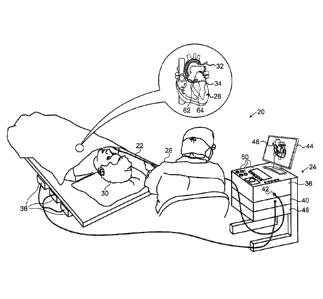
[0005] Figure 1 is a schematic pictorial illustration of a zero-
drift detection and correction system for a pressure-sensitive
catheter, in accordance with an embodiment of the present
invention;
[0006] Figure 2 is a schematic side view showing details of the
distal portion of the pressure-sensitive catheter, in accordance
with an embodiment of the present invention;
[0007] Figure 3 is a graph showing zero-drift of the pressure
sensitive catheter in accordance with an embodiment of the
present invention; and
[0008] Figure 4 is a flow diagram that schematically illustrates a
method of zero-drift detection and correction for the pressure-
sensitive catheter, in accordance with an embodiment of the
present invention.
DETAILED DESCRIPTION OF EMBODIMENTS
OVERVIEW
[0009] Various diagnostic and therapeutic procedures, such as
cardiac ablation and intracardiac electrical mapping, use an
invasive probe, such as a catheter, whose distal tip is fitted
4
i
1
CA 02756479 2011-10-28
. ..
with at least one electrode.
The electrode is typically
operated when the probe is pressed against a body cavity
surface. In these procedures, it is usually important to
ascertain a force the distal tip is exerting on the body cavity
surface. Therefore, some catheters comprise force sensors for
measuring the force between the probe and intra-body tissue,
such as the endocardium.
[0010]
To accurately measure a force exerted by the distal tip on
the endocardium, the force sensor is typically calibrated to a
"zero level," also referred to herein as a baseline.
In
embodiments of the present invention, the baseline is determined
from measurements generated by the force sensor when the distal
tip has minimal contact with any surface (and therefore there is
essentially no effective force exerted on the distal tip). Once
the baseline is identified, the measurements from the force
sensor can be used to provide a value of the force exerted.
[0011]
Since force sensors in catheters typically rely on analog
components, the sensors are susceptible to a "baseline drift,"
where the baseline may change due to factors including, but not
limited to, temperature and aging (i.e., of the analog
components). The baseline drift may result in an incorrect zero
level of the force sensor, thereby introducing inaccuracy into
the evaluated forces when the distal tip engages the intra-body
tissue. In order to ensure accurate force values, embodiments
of the present invention provide methods and systems for
detecting and correcting the baseline drift of a force sensor
disposed in a catheter. In some embodiments, the measurements
from the force sensor are monitored during an intracardiac
procedure (i.e., while the catheter is inside a heart of a
patient).
During the procedure, upon detecting that the
measurements are within a predefined noise threshold (i.e., the
i
1
CA 02756479 2011-10-28
measurements are relatively stable) for a specified duration,
then the catheter is assumed to be out of contact with the
endocardial tissue, and a current baseline is calculated using
the measurements collected during the specified duration.
[0012]
On the other hand, when the measurements vary by more than
the predetermined noise threshold, the catheter may be assumed
to be in contact with the endocardial tissue. The measurements
received in these cases, i.e., when the measurements vary, may
be used to give a value of the force exerted on the sensor.
[0013] Embodiments of the present invention enable automatic
calibration of a force sensor in a dynamic system.
In some
embodiments the force sensor can be automatically recalibrated
whenever a change is detected in the baseline, even if the
change is detected during an intracardiac procedure. Detecting
and correcting the baseline drift in the force sensor enables a
catheterization system to measure force with improved accuracy
and reliability.
SYSTEM DESCRIPTION
[0014]
Figure 1 is an illustration of a medical system 20 that
uses zero-drift detection and correction, in accordance with an
embodiment of the invention.
System 20 may be based, for
example, on the CARTOTm system, produced by Biosense Webster Inc.
(Diamond Bar, California). System 20 comprises a probe 22, such
as a catheter, and a control console 24. In the embodiment
described hereinbelow, it is assumed that probe 22 is used for
diagnostic or therapeutic treatment, such as for mapping
electrical potentials in a heart 26 or performing ablation of
heart tissue.
Alternatively, probe 22 may be used, mutatis
mutandis, for other therapeutic and/or diagnostic purposes in
the heart or in other body organs.
6
,
t
CA 02756479 2011-10-28
, =
[0015]
An operator 28, such as a cardiologist, inserts probe 22
through the vascular system of a patient 30 so that a distal end
32 of probe 22 enters a chamber of heart 26.
Operator 28
advances probe 22 so that a distal tip 34 of probe 22 engages
endocardial tissue at a desired location or locations. Probe 22
is typically connected by a suitable connector at its proximal
end to console 24.
[0016]
Console 24 typically uses magnetic position sensing to
determine position coordinates of distal end 32 inside heart 26.
To determine the position coordinates, a driver circuit 36 in
console 24 drives field generators 38 to generate magnetic
fields within the body of patient 30.
Typically, field
generators 38 comprise coils, which are placed below the
patient's torso at known positions external to patient 30.
These coils generate magnetic fields in a predefined working
volume that contains heart 26.
A magnetic field sensor 62
within distal end 32 of probe 22 (sensor 62 is shown in more
detail in Figure 2) generates electrical signals in response to
these magnetic fields.
A signal processor 40 processes these
signals in order to determine the position coordinates of distal
end 32, typically including both location and orientation
coordinates.
The method of position sensing described
hereinabove is implemented in the above-mentioned CARTOTm system
and is described in detail in the patents and patent
applications cited above.
[0017]
Signal processor 40 typically comprises a general-purpose
computer, with suitable front end and interface circuits for
receiving signals from probe 22 and controlling the other
components of console 24.
Processor 40 may be programmed in
software to carry out the functions that are described herein.
The software may be downloaded to console 24 in electronic form,
7
,
t
CA 02756479 2011-10-28
=
over a network, for example, or it may be provided on non-
transitory tangible media, such as optical, magnetic or
electronic memory media.
Alternatively, some or all of the
functions of processor 40 may be carried out by dedicated or
programmable digital hardware components.
[0018]
An input/output (I/0) interface 42 enables console 24 to
interact with probe 22.
Based on the signals received from
probe 22 (via interface 42 and other components of system 20),
processor 40 drives a display 44 to present operator 30 with an
image 46 showing the position of distal end 32 in the patient's
body, as well as status information and guidance regarding the
procedure that is in progress.
[0019]
In the present embodiment, processor 40 monitors the signal
measurements received from a force sensor 64 within distal end
32 (force sensor 64 is shown in more detail in Figure 2) during
periods in which the catheter is believed to be out of contact
with the endocardium, and detects any baseline drift.
If a
baseline drift is detected, processor 40 can correct the signals
from the force sensor when distal tip 34 engages the endocardial
tissue, in order to make an accurate evaluation of the force
experienced by the sensor.
[0020]
Processor 40 stores data representing image 46 in a memory
48.
In some embodiments, operator 28 can manipulate image 46
using one or more input devices 50.
[0021]
Alternatively or additionally, system 20 may comprise an
automated mechanism (not shown) for maneuvering and operating
probe 22 within the body of patient 30.
Such mechanisms are
typically capable of controlling both the longitudinal motion
(advance/retract) of probe 22 and transverse motion
(deflection/steering) of distal end 32 of the probe.
In such
embodiments, processor 40 generates a control input for
8
,
1
CA 02756479 2011-10-28
. =
controlling the motion of probe 22 based on the signals provided
by the magnetic field sensor in the probe.
[0022]
Although Figure 1 shows a particular system configuration,
other system configurations can also be employed to implement
embodiments of the present invention, and are thus considered to
be within the spirit and scope of this invention. For example,
the methods described hereinbelow may be applied using position
transducers of types other than the magnetic field sensor
described above, such as impedance-based or ultrasonic position
sensors. The term "position transducer" as used herein refers
to an element mounted on probe 22 which causes console 24 to
receive signals indicative of the coordinates of the element.
The position transducer may thus comprise a receiver on the
probe, which generates a position signal to the control unit
based on energy received by the transducer; or it may comprise a
transmitter, emitting energy that is sensed by a receiver
external to the probe.
Furthermore, the methods described
hereinbelow may similarly be applied in therapeutic and
diagnostic applications using not only catheters, but also
probes of other types, both in the heart and in other body
organs and regions.
[0023]
Figure 2 is a schematic sectional view of distal end 32 of
probe 22, in accordance with an embodiment of the present
invention. Specifically, Figure 2 shows functional elements of
distal end 32 used for therapeutic and/or diagnostic activity.
An electrode 60 (e.g., an ablation electrode) at distal tip 34
of the probe is typically made of a metallic material, such as a
platinum/iridium alloy or another suitable material.
Alternatively, multiple electrodes (not shown) along the length
of the probe may be used for this purpose.
9
,
[0024]
Position sensor 62 transmits a signal to console 24 that is
indicative of the location coordinates of distal end 32.
Position sensor 62 may comprise one or more miniature coils, and
typically comprises multiple coils oriented along different
axes.
Alternatively, position sensor 62 may comprise either
another type of magnetic sensor, an electrode which serves as a
position transducer, or position transducers of other types,
such as impedance-based or ultrasonic position sensors.
Although Figure 2 shows a probe with a single position sensor,
embodiments of the present invention may utilize probes with
more than one position sensor.
[0025]
In an alternative embodiment, the roles of position sensor
62 and magnetic field generators 38 may be reversed. In other
words, driver circuit 36 may drive a magnetic field generator in
distal end 32 to generate one or more magnetic fields.
The
coils in generator 38 may be configured to sense the fields and
generate signals indicative of the amplitudes of the components
of these magnetic fields.
Processor 40 receives and processes
these signals in order to determine the position coordinates of
distal end 32 within heart 26.
[0026]
Force sensor 64 measures a force applied by distal tip 34
to the endocardial tissue of heart 26 by conveying a signal to
the console that is indicative of the force exerted by the
distal tip on the intra-body tissue.
In one embodiment, the
force sensor may comprise a magnetic field transmitter and
receiver connected by a spring in distal end 32, and may
generate an indication of the force based on measuring the
deflection of the spring. Further details of this sort of probe
and force sensor are described in U.S. Patent Application
Publications 2009/0093806 and 2009/0138007. Alternatively,
distal end 32 may comprise another type of force sensor.
CA 2756479 2018-03-22
ZERO-DRIFT DETECTION AND CORRECTION
[0027]
Figure 3 is a graph 70 plotting force (in grams) vs. time
(in seconds) for a signal 72 comprising measurements transmitted
by force sensor 64 during an intracardiac procedure, in
accordance with an embodiment of the present invention.
When
signal 72 is within a noise threshold AFmin over a specified
duration Tmax, distal tip 34 may be assumed to be out of contact
with the endocardial tissue. On the other hand, when signal 72
varies by more than ,AFmin, distal tip 34 may be assumed to be in
contact with the endocardial tissue.
[0028]
Noise threshold AFmin is typically set to a value greater
then a noise variation for force sensor 64. For example, AFmin
may be set to 3.0 grams if force sensor 64 has a noise variation
of 1.0 grams. In one embodiment, by way of example, the value of
AFmin is set to be equal to 3o, where o is the standard
deviation of the signal from sensor 64 when it is out of contact
with tissue. Those having ordinary skill in the art will be able
to define values of other noise thresholds, such as no where n
is a real number, or a threshold based on a peak-peak variation,
without undue experimentation, and all such thresholds are
assumed to be comprised within the scope of the present
invention.
[0029]
In one embodiment Tmax may be set to 2.5 seconds, which is
substantially longer than a single cardiac cycle for heart 26 (a
cardiac cycle is typically less than or equal to 1.0 seconds).
[0030]
During a time period 78, signal 72 varies outside the range
defined by AFmin (due to movement of heart 26), indicating that
11
CA 2756479 2018-03-22
CA 02756479 2011-10-28
distal tip 34 is probably in contact with the endocardial
tissue. However, during a time period 79 (equal to Tmax in the
example shown in graph 70), signal 72 varies within AFmin,
indicating that distal tip 34 is probably out of contact with
the endocardial tissue. The variation of the signal, during a
period Tmax, by an amount less than or equal to AFmin, is
indicative that there is effectively no force on sensor 64
during this period. The signals acquired during this period may
thus be used to formulate a baseline for the sensor, as is
explained in more detail by the flow diagram of Figure 4.
[0031]
In some embodiments, there may he tissue contact even when
signal 72 has a variation equal to or less than AFmin.
To
verify tissue contact when signal 72 has a variation equal to or
less than AFmin, processor 40 may apply a filter to isolate
particular frequencies of signal 72.
The filter, typically a
band-pass filter, is configured to pass signals whose frequency
approximates heart rate frequencies (i.e., in this case the
frequency of heart 26), and block other frequencies. The band-
pass filter can provide a more accurate analysis of signal 72
when distal tip 34 is in low level contact with a moving object
such as the endocardial tissue, by allowing comparison between a
level of the filtered signal with a predefined level of the
band-pass frequencies.
[0032]
Figure 4 is a flow diagram that schematically illustrates a
method of cardiac ablation using zero-drift detection and
correction, in accordance with an embodiment of the present
invention.
It will be understood that the flow diagram is
presented by way of example, and that embodiments of the present
invention are not limited to procedures involving cardiac
ablation. Rather, embodiments of the present invention may be
12
1
CA 02756479 2011-10-28
used wherever the baseline of a force sensor is to be determined
while the sensor is operating within a body.
[0033]
In an initial step 80, operator 30 using input devices 50,
sets the noise threshold AFmin, the specified duration Tmax, and
the predefined level of the band-pass frequencies referred to
above. Alternatively, AFmin, Tmax and the predefined level may
be defined in advance of the ablation procedure, and stored in
memory 48.
[0034]
After operator 30 positions probe 22 in a positioning step
82, processor 40, in a collecting step 84, collects measurements
from force sensor 64 for the specified duration Tmax.
In a
first comparison step 86, if the collected force measurements
are within AFmin, then in a filter step 87, processor 40 applies
a band-pass filter to filter the force sensor measurements by
isolating measurements within a specific frequency range, as
described supra.
In a second comparison step 88, if the
filtered force measurements do not indicate probe-tissue
contact, then the method continues to a baseline calculation
step 89.
The comparison performed in step 88, to evaluate if
contact is or is not indicated, typically comprises a comparison
of a level of the filtered force measurements with the pre-
defined level of band-pass frequencies defined in step 80. In
baseline calculation step 89, processor 40 calculates a new
baseline by averaging the collected force measurements (i.e.,
those that were collected during the specified duration).
Alternatively, processor 40 may calculate an alternative
function based on the collected force measurements to determine
the new baseline.
[0035]
In a third comparison step 90, if the new baseline differs
from a baseline currently associated with force sensor 64 (i.e.,
a previous baseline), then processor 40, in a recalibration step
13
i
CA 02756479 2011-10-28
92, recalibrates force sensor 64 by setting the zero level of
the force sensor to the new baseline, and the processor may
present a notification on display 44 informing operator 28 of
the automatic baseline change. Alternatively, the processor may
present a message on display 44 notifying operator 28 of a
baseline change. In this case the operator may be provided with
the option of retaining the previous baseline, or of
implementing the new baseline. The new baseline may be
implemented during later contact with the endocardial tissue.
[0036]
After recalibrating force sensor 64, processor 40, in a
prompting step 94, presents a notification on display 44 that
operator 28 may reposition probe 22, and the method returns to
step 82. Returning to step 90, if the baseline did not change,
then the method continues with step 94.
[0037]
Returning to steps 86 and 88, if either the collected force
measurements exceed AFmin (i.e., the collected measurements
varied by more than the predefined amount AFmin in step 86) or
the filtered force measurements indicate probe-tissue contact
(in step 88), then distal tip 34 is assumed to be experiencing a
non-zero force, typically because it is in contact with the
endocardial tissue (or another surface of a body cavity) and the
method proceeds to a force calculation step 96.
In step 96,
processor 40 subtracts the current baseline from the
measurements collected from force sensor 64 (i.e., during
contact between distal tip 34 and the endocardial tissue),
thereby providing an accurate measurement of the force that
distal tip 34 is exerting on the endocardial tissue.
In some
embodiments, processor 40 may present a notification on display
44 warning operator 28 not to implement calculation of a new
baseline when distal tip 34 is assumed to be experiencing a non-
zero force (e.g., during time period 78).
14
1
CA 02756479 2011-10-28
= [0038] In a fourth comparison step 98, if the calculated force is
within a defined range acceptable for ablation, then in an
ablation step 100, processor 40 presents a notification on
display 44 prompting operator 28 to perform an ablation at the
current probe position. Returning to step 98, if the calculated
force is not within the defined range, the method continues with
step 94. Finally, in a fifth comparison step 102, if there are
additional regions in heart 26 targeted for ablation, the method
continues with step 94 until the ablation procedure is complete.
[0039] The corresponding structures, materials, acts, and
equivalents of all means or steps plus function elements in the
claims below are intended to include any structure, material, or
act for performing the function in combination with other
claimed elements as specifically claimed.
The description of
the present disclosure has been presented for purposes of
illustration and description, but is not intended to be
exhaustive or limiting to the disclosure in the form disclosed.
Many modifications and variations will be apparent to those of
ordinary skill in the art without departing from the scope and
spirit of the disclosure.
The embodiment was chosen and
described in order to best explain the principles of the
disclosure and the practical application, and to enable others
of ordinary skill in the art to understand the disclosure for
various embodiments with various modifications as are suited to
the particular use contemplated.
[0040]
It will be appreciated that the embodiments described above
are cited by way of example, and that the present invention is
not limited to what has been particularly shown and described
hereinabove.
Rather, the scope of the present invention
includes both combinations and subcombinations of the various
features described hereinabove, as well as variations and
,
CA 02756479 2011-10-28
modifications thereof which would occur to persons skilled in
the art upon reading the foregoing description and which are not
disclosed in the prior art.
16