Note: Descriptions are shown in the official language in which they were submitted.
CATHETER WITH OPTICAL CONTACT SENSING
FIELD OF THE INVENTION
[0001] The
present invention relates generally to invasive
probes, and specifically to an optical contact sensing probe.
BACKGROUND
[0002] A wide
range of medical procedures involves placing
objects, such as sensors, tubes, catheters, dispensing
devices, and implants, within the body. Various
types of
sensors have been proposed for assessing the quality of
contact between a catheter and tissue in the body.
[0003] The
quality of catheter-tissue contact can be verified,
for example, by sensing actual physical contact and/or
proximity between the catheter and the tissue. U.S.
Patent
Application 12/816,492 describes a catheter with multiple
optical contact sensors integrated along its distal end. Each
optical contact sensor comprises a combination of at least one
optical emitter, such as a Light Emitting Diode (LED), and at
least one respective optical detector (such as a photodiode or
a phototransistor) in close proximity to the emitter. At
small distances from the tissue, the optical detector senses
optical radiation, which is emitted by the optical emitter and
reflected from the tissue. The
optical detector produces a
signal that is indicative of the sensed reflection. As the
optical contact sensor comes into physical contact with the
tissue, the signal will increase to a maximal level. The
signal produced by the optical detector thus gives an
indication of the quality of contact between the tissue and
the distal end of the catheter.
[0004] The
description above is presented as a general overview
of related art in this field and should not be construed as an
1
CA 2756627 2017-10-26
CA 02756627 2011-11-01
) =
admission that any of the information it contains constitutes
prior art against the present patent application.
SUMMARY OF THE INVENTION
[0005] An embodiment of the present invention provides a medical
probe, including:
[0006] a biocompatible sheath having proximal and distal ends,
and having at least one transparent strip between the proximal
end and the distal end; and
[0007] one or more functional elements positioned within the
biocompatible sheath.
[0008] Typically, each of the one or more functional elements
includes an optical contact sensor having an optical emitter,
and an optical detector in close proximity to the optical
emitter. The optical contact sensor may be configured to
detect proximity of the distal end to body tissue, and to
verify contact between the distal end and the body tissue. The
optical contact sensor typically faces the at least one
transparent strip. In one embodiment the probe includes one or
more electrodes disposed along the biocompatible sheath which
are configured to perform an ablation, and the optical contact
sensor is configured to provide an indication for controlling
the ablation. The optical contact sensor may be configured to
provide a further indication for assessing a quality of the
ablation.
[0009] There is further provided, according to an embodiment of
the present invention, a medical probe, including:
[0010] a biocompatible sheath having proximal and distal ends,
and having at least one transparent element;
[0011] a dielectric substrate which is inserted within the
biocompatible sheath;
[0012] one or more electronic components positioned on the
dielectric substrate; and
2
CA 02756627 2011-11-01
, 4
[0013] one or more printed wiring traces positioned on the
dielectric substrate and coupled to each of the one or more
electronic components.
[0014] The at least one transparent element may include a
transparent strip between the proximal end and the distal end
of the sheath. In a disclosed embodiment each of the one or
more electronic components includes an optical contact sensor
having an optical emitter, and an optical detector in close
proximity to the optical emitter. The optical contact sensor
typically faces the transparent strip. The dielectric
substrate may include a flexible printed circuit board
material. The one or more electronic components may be
positioned on an outer side of the dielectric substrate, and
the one or more printed wiring traces may be positioned on an
inner side of the dielectric substrate.
[0015] There is further provided, according to an embodiment of
the present invention, a method, including:
[0016] incorporating at least one transparent strip between
proximal and distal ends of a biocompatible sheath; and
[0017] positioning one or more functional elements within the
biocompatible sheath.
[0018] There is further provided, according to an embodiment of
the present invention, a method, including:
[0019] incorporating at least one transparent element between
proximal and distal ends of a biocompatible sheath;
[0020] inserting a dielectric substrate within the biocompatible
sheath;
[0021] positioning one or more electronic components on the
dielectric substrate;
[0022] positioning one or more printed wiring traces on the
dielectric substrate; and
[0023] coupling the one or more printed wiring traces to each of
the one or more electronic components.
3
CA 02756627 2011-11-01
BRIEF DESCRIPTION OF THE DRAWINGS
[0024] The
disclosure is herein described, by way of example
only, with reference to the accompanying drawings, wherein:
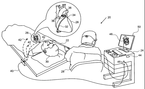
[0025] Figure
1 is a schematic, pictorial illustration of a
medical system implementing optical contact sensing, in
accordance with an embodiment of the present invention;
[0026] Figure
2A is a schematic, side view illustration of an
optical contact sensing probe With a transparent strip, in
accordance with an embodiment of the present invention;
[0027] Figure 2B is a schematic, cross-sectional view
illustration of a of the optical contact sensing probe with
the transparent strip, in accordance with an embodiment of the
present invention;
[0028] Figure
3A is a schematic side view illustration of the
optical contact sensing probe with an optoelectronic strip, in
accordance with an embodiment of the present invention;
[0029] Figure
3B is a schematic side view of the optoelectronic
strip, in accordance with an embodiment of the present
invention; and
[0030] Figure
3C is a schematic top-down view of an inner side
of the optoelectronic strip, in accordance with an embodiment
of the present invention.
DETAILED DESCRIPTION OF EMBODIMENTS
OVERVIEW
[0031] Various
diagnostic and therapeutic procedures, such as
intracardiac electrical mapping and cardiac ablation, use an
invasive probe, such as a catheter, whose distal tip is fitted
with at least one electrode. The
electrode is typically
operated when the probe is pressed against intra-body tissue.
In these procedures, it is usually important to ascertain the
proximity of the probe to a body cavity surface, and to
4
CA 02756627 2011-11-01
determine when the distal tip of the probe is in contact with
the body cavity surface.
[0032] In an
embodiment of the present invention, functional
elements of the probe are encased in a biocompatible sheath,
which incorporates one or more transparent strips between
proximal and distal ends of the probe. Aside
from the
transparent strip(s), the biocompatible sheath may be
otherwise opaque.
Functional elements, such as optical
contact sensors can be positioned within the sheath facing the
transparent strip (which serves as a window). In some
embodiments, multiple optical contact sensors may be
positioned within the sheath facing the same transparent strip
at different locations along the length of the probe.
[0033]
Embodiments of the present invention also provide an
optoelectronic strip, integrated into the probe, upon which
the multiple sensors are mounted. The
optoelectronic strip
comprises a long, narrow flexible dielectric substrate, such
as a flexible printed circuit board material, with the optical
contact sensors positioned on an outer side (of the
optoelectronic strip), and printed wiring traces positioned
along an inner side (of the optoelectronic strip) and coupled
to each of the sensors. In some embodiments, the strip may be
integrated longitudinally within the biocompatible sheath,
with the optical contact sensors facing the transparent strip
as described supra.
[0034]
Embodiments of the present invention, including the
biocompatible sheath incorporating the transparent strip and
the optoelectronic strip enable optical contact sensing probes
to be produced reliably and inexpensively.
SYSTEM DESCRIPTION
[0035] Figure
1 is a schematic, pictorial illustration of a
medical system 20 that implements optical proximity sensing,
in accordance with an embodiment of the present invention.
System 20 comprises an optical contact sensing probe 22, in
the present example a catheter, and a control console 24. In
the embodiment described hereinbelow, it is assumed that probe
22 is used for diagnostic or therapeutic treatment, such as
circumferentially mapping electrical potentials in a pulmonary
vein of a heart 26, or performing ablation of the vein tissue.
Alternatively, probe 22 may be used, mutatis mutandis, for
other therapeutic and/or diagnostic purposes in the heart or
in other body organs.
[0036] An
operator 28, such as a cardiologist, inserts probe 22
through the vascular system of a patient 30 so that a distal
end 32 of probe 22 enters a chamber of the patient's heart 26
(e.g., the left atrium).
Operator 28 advances probe 22 so
that a distal tip 34 (shown here in a "loop" or "lasso"
configuration) engages body tissue at desired locations (e.g.,
vein tissue in the left superior pulmonary vein). Distal tip
34 comprises electrodes 36 and optical contact sensors 38.
The configuration of optical contact sensor 38 is shown in
greater detail in Figure 2A below. Optical
contact sensors
are described, for example, in U.S. Patent Application
12/816,492. Probe
22 is typically connected by a suitable
connector at its proximal end to console 24.
[0037] Using
signals from the optical contact sensors fitted in
probe 22, console 24 determines the quality of contact between
distal tip 34 and the vein tissue. The
term "quality of
contact" refers to actual physical contact between the distal
tip and the tissue, as well as proximity of the distal tip to
the tissue. In the
example of Figure 1, console 24 is also
connected by a cable 40 to body surface electrodes, which
typically comprise adhesive skin patches 42. Console
24
determines position coordinates of probe 22 inside heart 26
based on the impedance measured between the probe and patches
6
CA 2756627 2017-10-26
42. Although system 20 measures position uses impedance-based
sensors, other position tracking techniques may be used (e.g.,
magnetic-based sensors).
Magnetic position tracking
techniques are described, for example, in U.S. Patents
5,391,199, 5,443,489, 6,788,967, 6,690,963,
5,558,091,
6,172,499 6,177,792.
Impedance-based position tracking
techniques are described, for example, in U.S. Patents
5,983,126, 6,456,864 and 5,944,022.
[0038] Console
24 comprises a processor 44, which typically
comprises a general-purpose computer, with suitable front end
and interface circuits for receiving signals from probe 22 and
controlling the other components of console 24. An
input/output (I/O) communications interface 46 enables console
24 to interact with probe 22 and patches 42. Based
on the
signals received from probe 22 and from patches 42, processor
44 produces and displays a map 48 showing the position of
distal tip 34 in the patient's body, the distance and/or
contact indication between the loop and the body tissue, as
well as status information and guidance regarding the
procedure that is in progress. Map 48
is presented to
operator 28 using a display 50. The position of probe 22 may
be superimposed on map 48 or on another image of heart 26.
PROBE WITH A TRANSPARENT STRIP
[0039] Figure
2A is a schematic, pictorial illustration of a
side view of optical contact sensing probe 22, and Figure 2B
is a schematic, pictorial illustration of a cross-section of
the probe, in accordance with an embodiment of the present
invention. Probe
22 comprises functional elements such as
optical contact sensor 38 and tubes 60, which are covered by a
biocompatible sheath 62. Sheath 62 is opaque to optical
7
CA 2756627 2017-10-26
radiation except for a transparent (to optical radiation)
strip 64, which is incorporated between a proximal end 66 and
distal end 32 of the probe. The proximal and distal ends of
the probe are respectively substantially the same as the
proximal and distal ends of the biocompatible sheath, so that
the terms proximal end 66 and distal end 32 also refer to the
corresponding ends of the sheath. Transparent strip 64 is also
referred to herein as window 64, and has a width 68.
[0040] In the configuration shown in Figure 2A, electrodes 36
are disposed along the length of distal end 32. Electrodes 36
are typically made of a metallic material, such as a
platinum/iridium alloy or another suitable material.
[0041] Optical contact sensor 38 is positioned within sheath 62
facing window 64, and is typically disposed symmetrically with
respect to the window. Optical contact sensor 38 comprises an
optical emitter 70 such as a light emitting diode (LED), and
an optical detector 72 such as a photodiode or a
phototransistor in close proximity to the optical emitter.
While the configuration of optical contact sensor 38 shown in
Figure 2A comprises one optical emitter 70 and one optical
detector 72, the optical contact sensor may be configured to
include more than one optical emitter and/or more than one
optical detector. Optical contact sensors incorporating
optical emitters 70 and optical detectors 72 are described,
for example, in U.S. Patent Application 12/816,492.
[0042] Width 68 of window 64 determines the extent of a field of
view 74 of the sensor in the azimuthal direction (i.e., how
far optical contact sensor 38 is able to "see" around probe
22). Window 64 may be configured to be narrow, if desired, to
ensure that optical contact sensor 38 is sensitive to contact
between probe 22 and body tissue only within a desired, narrow
angular range (i.e., field of view 74).
8
CA 2756627 2017-10-26
CA 02756627 2011-11-01
,
[0043] Window
64 may be created in the process of producing
biocompatible sheath 62 using a coextrusion process, which is
a variation of extrusion. During extrusion, an extruder melts
a material, which is then conveyed through a die configured to
give the final product (e.g., a tubular shaped sheath) a
desired profile. The die
is designed so that the molten
material evenly flows to the product's profile shape. To
produce sheath 62 with window 64, two extruders melt and
convey a steady volumetric throughput of different
biocompatible materials (i.e., for the opaque sheath and the
transparent strip, respectively) to a single die which
extrudes the materials into the form shown in Figure 2A.
Setting width 68 during the coextrusion process enables the
production of probe 22 with desired field of view 74.
[0044]
Although Figure 2A shows probe 22 along a straight probe
segment, the same sort of technique may be used in curved
elements, such as the curved end of a lasso catheter, as shown
in Figure 1. In the example shown in Figure 1, distal tip 34
comprises an adjustable loop fitted with electrodes 36 and
optical contact sensors 38. The
configuration of the loop
enables simultaneous mapping or ablation of circumferential
areas of tissue, such as a pulmonary vein.
[0045]
Additionally or alternatively, probe 22 can be produced
with multiple, parallel windows 64. For
example, in a
configuration with two windows 64 located at opposite ends of
a probe's diameter (as shown in Figure 3A below), separate
optical contact sensors 38 may be positioned and aligned
facing each of the two windows. Further
alternatively,
multiple optical contact sensors 38 may be positioned non-
symmetrically with respect to their windows, so that the
fields of view of the sensors, while encompassing
substantially the same angular width, have different angular
coverage. Configuring probe 22 with multiple windows 64 (and
corresponding optical contact sensors 38) enables
9
CA 02756627 2011-11-01
omnidirectional sensing in any direction orthogonal to an axis
of the probe, making it possible to sense contact along the
length of the probe, regardless of which side of the probe
makes contact with the tissue.
PROBE WITH AN OPTOELECTRONIC STRIP
[0046] Figure
3A is a schematic side view illustration of probe
22 with an optoelectronic strip 76, Figure 3B is a schematic
side view of the optoelectronic strip, and Figure 3C is a
schematic top-down view of an inner side 78 of the
optoelectronic strip, in accordance with embodiments of the
present invention. Probe 22 incorporates transparent
elements, which in the configuration shown in Figure 3A
comprise two windows 64 between proximal end 66 and distal end
32 of the probe. Probe 22 comprises optoelectronic strip 76
inserted longitudinally into the probe so that optical contact
sensors 38 positioned on an outer side 80 of the
optoelectronic strip face one of windows 64.
[0047]
Optoelectronic strip 76 comprises a long, narrow flexible
dielectric substrate 82, such as a flexible printed circuit
board material, with optical emitters 70 and optical detectors
72 positioned on outer side 80, and printed wiring traces 84
along inner side 78 that are coupled to each optical emitter
70 and optical detector 72.
Optoelectronic strip 76 may be
integrated longitudinally into probe 22 during production,
either along one side of the probe or wrapped around onto both
sides, as shown in Figure 3A.
Alternatively, a separate
optoelectronic strip 76 can be integrated longitudinally into
probe 22 for each window 64.
[0048] During
operation of the probe, optical emitters 70 emit
optical radiation, and optical detectors 72 convey signals to
processor 44 indicative of the optical radiation reflecting
off the body tissue. Based on the received signals, processor
CA 02756627 2011-11-01
, .
,
44 can determine the proximity of distal end 32 to the body
tissue, and can verify contact between the distal end and the
body tissue.
[0049]
As discussed supra, probe 22 may be used for ablating
vein tissue of heart 26.
During an ablation procedure,
electrodes 36 spaced along distal end 32 may emit energy,
which cauterizes a small amount of the vein tissue.
Since
cauterized and non-cauterized tissue typically have different
reflection properties, optical detectors 72 can be configured
to convey different signals, based on the different levels of
optical radiation reflecting off the cauterized and the non-
cauterized vein tissue.
Therefore, processor 44 may use
optical sensors 38 to control this and other ablation
procedures, as well as to assess a quality of the ablation
that has been performed.
[0050]
Although Figure 3A shows optical emitters 70 and optical
detectors 72 positioned on optoelectronic strip 76, electronic
components of other types may be positioned on the
optoelectronic strip, and are thus considered to be within the
spirit and scope of this invention.
Examples of electronic
components that can be positioned on optoelectronic strip 76
and coupled to printed wiring traces 84 include piezoelectric
transducers, capacitive sensors and pressure sensors of other
types.
[0051] Additionally, the optoelectronic strip described
hereinabove assumes that wiring traces 84 and the electronic
components (i.e., emitters 70 and detectors 72) are on
opposite sides of optoelectronic strip 76. In an alternative
embodiment, at least some traces 84 are on the same side (of
strip 76) as the electronic components.
[0052]
It will be appreciated that the embodiments described
above are cited by way of example, and that the present
invention is not limited to what has been particularly shown
and described hereinabove. Rather, the scope of the present
11
CA 02756627 2011-11-01
invention includes both combinations and subcombinations of
the various features described hereinabove, as well as
variations and modifications thereof which would occur to
persons skilled in the art upon reading the foregoing
description and which are not disclosed in the prior art.
12