Note: Descriptions are shown in the official language in which they were submitted.
CA 02756673 2011-10-26
x
DIGITAL AMPLIFICATION
The U.S. government retains certain rights in this invention by virtue
of its support of the underlying research, supported by grants CA 43460, CA
57345, and CA 62924 from the National Institutes of Health.
TECHNIC-Al-L FIELD OF THE INVENTION
This invention is related to diagnostic genetic analyses. In particular
it relates to detection of genetic changes and gene expression.
BACKGROUND OF THE INVENTION
In classical genetics, only mutations of the germ-line were considered
important for understanding disease. With the realization that somatic
mutations are the primary cause of cancer (1), and may also play a role in
aging (2,3), new genetic principles have arisen. These discoveries have
provided a wealth of new opportunities for patient management as well as for
basic research into the pathogenesis of neoplasia. However, many of these
opportunities hinge upon detection of a small number of mutant-containing
-- cells among a large excess of normal cells. Examples include the detection
of neoplastic cells in urine (4), stool (5,6), and sputum (7,8) of patients
with
cancers of the bladder, colorectum, and lung, respectively. Such detection has
been shown in some cases to be possible at a stage when the primary tumors
are still curable and the patients asymptomatic. Mutant sequences from the
DNA of neoplastic cells have also been found in the blood of cancer patients
(9-11). The detection of residual disease in lymph nodes or surgical margins
I
CA 02756673 2011-10-26
may be useful in predicting which :patients might benefit most from further
therapy (12-14). From a basic research standpoint, analysis of the early
effects of carcinogens is -often dependent on: the -ability to detect small
populations of mutant cells (15-17).
Because of the importance of this issue in.so many settings, many
useful techniques have been developed for the detection of mutations. DNA
sequencing is the gold standard for the detection of germ line mutations, but
is useful only when the fraction of mutated alleles is.. greater than -20%
(18,19). Mutant-specific oligonucleotides can sometimes be used to detect
mutations present in a minor proportion of the cells analyzed, but the signal
to noise ratio distinguishing mutant and wild-type (WT) templates is variable
(20-22) The use of mutant-specific primers or the digestion of polymerise
chain reaction (PCR) products with specific restriction endonucleases are
extremely sensitive methods for detecting such mutations, but it is difficult
to
quantitate the fraction of mutant molecules in the starting population with
these techniques (23-28). Other innovative approaches for the detection of
somatic mutations have been reviewed (29-32). A general problem with these
methods is that it is difficult or impossible to independently confirm the
existence of any mutations that are identified.
Thus there is a need in the art for methods foraccurately and
quantitatively detecting genetic sequences in mixed populations of sequences.
SUMMARY OF THE INVENTION
It is an aspect of the present invention to provide methods for
determining the presence of a selected genetic sequence in a population of
genetic sequences.
It is another aspect of the present invention to provide molecular
beacon probes useful in the method of the invention.
These and other aspects of~the invention are achieved by providing a
method for determining the presence of a selected genetic sequence in a
population of genetic sequences. A biological sample comprising nucleic acid
template molecules is diluted to form a set of assay samples. The template
molecules within the assay samples are amplified to forma population of
2
CA 02756673 2011-10-26
WO 01/09386 PCT/US00/20740
amplified molecules in the assay samples of the set. The amplified molecules
in the assay samples of the set are then analyzed to determine a first number
of assay samples which contain the selected genetic sequence and a second
number of assay samples which contain a reference genetic sequence. The
first number is then compared to the second number to ascertain a ratio which
reflects the composition of the biological sample.
Another embodiment of the invention is a method for determining the
ratio of a selected genetic sequence in a population of genetic sequences.
Template molecules within a set comprising a plurality of assay samples are
amplified to form a population of amplified molecules in each of the assay
samples of the set. The amplified molecules in the assay samples of the set
are analyzed to determine a first number of assay samples which contain the
selected genetic sequence and a second number of assay samples which
contain a reference genetic sequence. The first number is compared to the
second number to ascertain a ratio which reflects the composition of the
biological sample.
According to another embodiment of the invention, a molecular
beacon probe is provided. It comprises an oligonucleotide with a stem-loop
structure having a photoluminescent dye at one of the 5' or 3' ends and a
quenching agent at the opposite 5' or 3' end. The loop consists of 16 base
pairs and has a Tm of 50-51 C. The stem consists of 4 base pairs having a
sequence 5'-CACG-3'.
A second type of molecular beacon probe is provided in another
embodiment. It comprises an oligonucleotide with a stem-loop structure
having a photoluminescent dye at one of the 5' or 3' ends and a quenching
agent at the opposite 5' or 3' end. The loop consists of 19-20 base pairs and
has a T. of 54-56 C. The stem consists of 4 base pairs having a sequence 5'-
CACG-3'.
Another embodiment provides the two types of molecular beacon
probes, either mixed together or provided in a divided container as a kit.
3
CA 02756673 2011-10-26
In accordance with an aspect of the present invention, there is provided a
method for determining the ratio of a selected genetic sequence in a
population of
genetic sequences, comprising the steps of diluting nucleic acid template
molecules
in a biological sample to form a set comprising a plurality of assay samples;
amplifying the template molecules within the assay samples to form a
population of
amplified molecules in the assay samples of the set; analyzing the amplified
molecules in the assay samples of the set to determine a first number of assay
samples
which contain the selected genetic sequence and a second number of assay
samples
which contain a reference genetic sequence; comparing the first number to the
second
number to ascertain a ratio which reflects the composition of the biological
sample.
In accordance with another aspect of the present invention, there is provided
a
molecular beacon probe comprising: an oligonucleotide with a stem-loop
structure
having a photoluminescent dye at one of the 5' or 3' ends and a quenching
agent at
the opposite 5' or 3' end, wherein the loop consists of 16 base pairs, wherein
the loop
has a melting temperature (Tm) of 50-51 C and the stem consists of 4 base
pairs
having a sequence 5'-CACG-3'.
In accordance with another aspect of the present invention, there is provided
a
molecular beacon probe comprising: an oligonucleotide with a stem-loop
structure
having a photoluminescet dye at one of the 5' or 3' ends and a quenching agent
at the
opposite 5' or 3' end, wherein the loop consists of 19-20 base pairs, wherein
the loop
has a melting temperature (Tm) of 54-56 C and the stem consists of 4 base
pairs
having a sequence 5'-CACG-3'.
In accordance with another aspect of the present invention, there is provided
a
pair of molecular beacon probes comprising: a first molecular beacon probe
which is
an oligonucleotide with a stem-loop structure having a first photoluminescent
dye at
one of the 5' or 3' ends and a quenching agent at the opposite 5' or 3' end,
wherein
the loop consists of 16 base pairs having a melting temperature (Tm) of 50-51
C and
the stem consists of 4 base pairs having a sequence 5'-CACG-3'; and a second
molecular beacon probe which is an oligonucleotide with a stem-loop structure
having a second photoluminescent dye at one of the 5' or 3' ends and a
quenching
agent at the opposite 5' or 3' end, wherein the loop consists of 19-20 base
pairs
having a melting temperature (Tm) of 54-56 C and the stem consists of 4 base
pairs
having a sequence of 5'-CACG-3'; wherein the first and the second
photoluminescent
dyes are distinct.
3a
CA 02756673 2011-10-26
In accordance with a further aspect of the present invention there is provided
a
method for determining the ratio of a selected genetic sequence in a
population of
genetic sequences, comprising the steps of. amplifying template molecules
within a
set comprising a plurality of assay samples to form a population of amplified
molecules in each of the assay samples of the set; analyzing the amplified
molecules
in the assay samples of the set to determine a first number of assay samples
which
contain the selected genetic sequence and a second number of assay samples
which
contain a reference genetic sequence, wherein at least one-fiftieth of the
assay
samples in the set comprise a number (N) of molecules such that 1/N is larger
than
the ratio of selected genetic sequences to total genetic sequences required to
determine the presence of the selected genetic sequence; comparing the first
number
to the second number to ascertain a ratio which reflects the composition of
the
biological sample.
In accordance with a further aspect of the present invention there is provided
a
method for detecting a target nucleic acid, the method comprising the steps
of:
separating a sample in which a target nucleic acid is present in an amount
less than
about 20% relative to non-target nucleic acid in said sample, to form a
plurality of
assay samples;
amplifying said target nucleic acid in said assay samples;
hybridizing the amplification product to a first molecular beacon probe which
hybridizes to the target nucleic acid and to a second molecular beacon probe
which
hybridizes to the non-target nucleic acid, wherein each of the first and the
second
molecular beacon probes comprises a photoluminescent dye and a quenching agent
at
opposite 5' and 3' ends, wherein the photoluminescent dye on the first and
second
molecular beacon probes are different, and wherein the first molecular beacon
probe
further comprises a stem and a loop, wherein the stem comprises about 4 base
pairs
having a sequence 5' -CACG-3', and the loop comprises about 16 base pairs and
has
a T. of about 50-51 C; and
detecting the first and the second molecular beacon probes hybridized to the
target nucleic acid," thereby detecting the target nucleic acid.
In accordance with a further aspect of the present invention there is provided
a
method for detecting a target nucleic acid, the method comprising the steps
of.
3b
CA 02756673 2011-10-26
providing a sample comprising X% of a target nucleic acid, wherein X is less
than 100;
dividing the sample to produce a plurality of assay samples;
wherein the ratio of target to non-target nucleic acid in at least one of the
samples is
greater than X%;
amplifying the target nucleic acid to form an amplification product;
hybridizing the amplification product to a first molecular beacon probe which
hybridizes to the target nucleic acid and a second molecular beacon probe
which
hybridizes to the non-target nucleic acid, wherein each of the first and the
second
molecular beacon probes comprises a photoluminescent dye and a quenching agent
at
opposite 5' and 3' ends, wherein the photoluminescent dye on the first and
second
molecular beacon probes are different, and wherein the first molecular beacon
probe
further comprises a stem and a loop, wherein the stem comprises about 4 base
pairs
having a sequence 5' -CACG-3', and the loop comprises about 16 base pairs and
has
a TM of about 50-51 C; and
detecting the first and second molecular beacon probes hybridized to the
target nucleic acid, thereby detecting the target nucleic acid.
In accordance with a further aspect of the present invention there is provided
a
method for detecting a target nucleic acid in a population of non-target
nucleic acid
contained in a sample, the method comprising:
dividing a heterogeneous sample comprising target nucleic acid and non-
target nucleic acid to form a plurality of assay samples, wherein the
concentration of
non-target nucleic acid is at least 5-fold that of target nucleic acid in the
heterogeneous sample, and wherein at least one of the assay samples comprises
a
single molecule of the target nucleic acid molecule;
amplifying the single molecule of target nucleic acid to form an amplification
product;
hybridizing the amplification product to a first molecular beacon probe which
hybridizes to the target nucleic acid and a second molecular beacon probe
which
hybridizes to the non-target nucleic acid, wherein each of the first and the
second
molecular beacon probes comprises a photoluminescent dye and a quenching agent
at
opposite 5' and 3' ends, wherein the photoluminescent dye on the first and
second
molecular beacon probes are different, and wherein the first molecular beacon
probe
further comprises a stem and a loop, wherein the stem comprises about 4 base
pairs
3c
CA 02756673 2011-10-26
having a sequence 5' -CACG-3', and the loop comprises about 16 base pairs and
has
a T. of about 50-51 C; and
detecting the first and the second molecular beacon probes hybridized to the
target nucleic acid, thereby detecting the target nucleic acid.
In accordance with a further aspect of the present invention there is provided
a
method for detecting a target nucleic acid, the method comprising the steps
of.
separating a sample in which a target nucleic acid is present in an amount
less
than about 20% relative to non-target nucleic acid in said sample, to form a
plurality
of assay samples;
amplifying said target nucleic acid in said assay samples;
hybridizing the amplification products to a first molecular beacon probe which
hybridizes to the target nucleic acid and to a second molecular beacon probe
which
hybridizes to the non-target nucleic acid, wherein each of the first and the
second
molecular beacon probes comprises a photoluminescent dye and a quenching agent
at
opposite 5' and 3' ends, wherein the photoluminescent dye on the first and
second
molecular beacon probes are different, and wherein the second molecular beacon
probe further comprises a stem and a loop, wherein the stem comprises about 4
base
pairs having a sequence 5' -CACG-3', and the loop comprises about 19-20 base
pairs
and has a T. of about 54-56 C; and
detecting the first and the second molecular beacon probes hybridized to the
target nucleic acid, thereby detecting the target nucleic acid.
In accordance with a further aspect of the present invention there is provided
a
method for detecting a target nucleic acid, the method comprising the steps
of:
providing a sample comprising X% of a target nucleic acid, wherein X is less
than 100;
dividing the sample to produce a plurality of assay samples;
wherein the ratio of target to non-target nucleic acid in at least one of the
samples is
greater than X%;
amplifying the target nucleic acid to form an amplification product;
hybridizing the amplification products to a first molecular beacon probe which
hybridizes to the target nucleic acid and a second molecular beacon probe
which
hybridizes to the target nucleic acid and a second molecular beacon probe
which
hybridizes to the non-target acid, wherein each of the first and the second
molecular
probes comprises a photoluminescent dye and a quenching agent at opposite 5'
and 3'
3d
CA 02756673 2011-10-26
ends, wherein the photoluminescent dye on the first and second molecular
beacon
probes are different, and wherein the second molecular beacon probe further
comprises a stem and a loop, wherein the stem comprises about 4 base pairs
having a
sequence 5' -CACG-3', and the loop comprises about 19-20 base pairs and a Tm
of
about 54-56 C; and
detecting the first and second molecular beacon probes hybridized to the
target nucleic acid, thereby detecting the target nucleic acid.
In accordance with a further aspect of the present invention there is provided
a
method for detecting a target nucleic acid in a population of non-target
nucleic acid
contained in a sample, the method comprising:
dividing a heterogeneous sample comprising target nucleic acid and non-
target nucleic acid to form a plurality of assay samples, wherein the
concentration of
non-target nucleic acid is at least 5 fold that of target nucleic acid in the
heterogeneous sample, and wherein at least one of the assay samples comprises
a
single molecule of the target nucleic acid;
amplifying the single molecule of target nucleic acid to form an amplification
product;
hybridizing the amplification products to a first molecule beacon probe which
hybridizes to the target nucleic acid and a second molecular beacon probe
which
hybridizes to the non-target nucleic acid, wherein each of the first and the
second
molecular probes comprises a photoluminescent dye and a quenching agent at
opposite 5' and 3' ends, wherein the photoluminescent dye on the first and
second
molecular beacon probes are different, and wherein the second molecular beacon
probe further comprises a stem and a loop, wherein the stem comprises about 4
base
pairs having a sequence 5' -CACG-3', and the loop comprises about 19-20 base
pairs
and a T. of about 54-56 C; and
detecting the first and second molecular beacon probes hybridized to the
target nucleic acid, thereby detecting the target nucleic acid.
In accordance with a further aspect of the present invention there is provided
a
method for determining an allelic imbalance in a biological sample, comprising
the
steps of
amplifying template molecules within a set comprising a plurality of assay
samples to form a population of amplified molecules in each of the assay
samples of
the set, wherein the template molecules are obtained from a biological sample;
3e
CA 02756673 2011-10-26
analyzing the amplified molecules in the assay samples of the set to determine
a first number of assay samples which contain a selected genetic sequence on a
first
chromosome and a second number of assay samples which contain a reference
genetic sequence on a second chromosome, wherein between 0.1 and 0.9 of the
assay
samples yield an amplification product;
comparing the first number of assay samples to the second number of assay
samples to ascertain an allelic imbalance in the biological sample.
In accordance with a further aspect of the present invention there is provided
a method for determining an allelic imbalance in a biological sample,
comprising the
steps of:
distributing nucleic acid template molecules from a biological sample to form
a set comprising a plurality of assay samples;
amplifying the template molecules within the assay samples to form a
population of amplified molecules in the assay samples of the set;
analyzing the amplified molecules in the assay samples of the set to determine
a first number of assay samples which contain a selected genetic sequence on a
first
chromosome and a second number of assay samples which contain a reference
genetic sequence on a second chromosome;
comparing the first number of assay samples to the second number of assay
samples to ascertain an allelic imbalance between the first chromosome and the
second chromosome in the biological sample.
In accordance with a further aspect of the present invention there is provided
a
method for determining an allelic imbalance in a biological sample, comprising
the
steps of.
amplifying template molecules within a set comprising a plurality of assay
samples to form a population of amplified molecules in each of the assay
samples of
the set, wherein the template molecules are obtained from the biological
sample;
analyzing the amplified molecules in the assay samples of the set to determine
a first number of assay samples which contain a first allelic form of a marker
and a
second number of assay samples which contain a second allelic form of the
marker,
wherein between 0.1 and 0.9 of the assay samples yield an amplification
product;
comparing the first number to the second number to ascertain an allelic
imbalance in the biological sample; and
identifying an allelic imbalance in the biological sample.
3f
CA 02756673 2011-10-26
t
In accordance with a further aspect of the present invention there is provided
a
method for determining an allelic imbalance in a biological sample, comprising
the
steps of-
distributing nucleic acid template molecules from a biological sample to form
a set comprising a plurality of assay samples;
amplifying the template molecules within the assay samples to form a
population of amplified molecules in the assay samples of the set;
analyzing the amplified molecules in the assay samples of the set to determine
a first number of assay samples which contain a first allelic form of a marker
and a
second number of assay samples which contain a second allelic form of the
marker;
comparing the first number of assay samples to the second number of assay
samples to ascertain an allelic imbalance between the first allelic form and
the second
allelic form in the biological sample.
3g
CA 02756673 2011-10-26
The invention thus provides the art with the means to obtain
quantitative assessments of particular DNA or RNA sequences in mixed
populations of sequences using digital (binary) signals.
BRIEF DESCRIPTION OF THE DRAWINGS
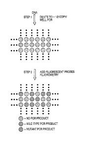
FIG. 1. Schematic of experimental design. (A) The basic two steps involved:
PCR on diluted DNA samples is followed by addition of fluorescent probes
which discriminate between WT and mutant alleles and subsequent
fluorometry. (B) Principle of molecular beacon analysis. In the stem-loop
configuration, fluorescence from a dye at the 5' end of the oligonucleotide
probe is quenched by a Dabcyl group at the 3' end. Upon hybridization to a
template, the dye is separated from the quencher, resulting in increased
fluorescence. Modified from Man-as et al.. (C) Oligonucleotide design.
Primers FI and RI are used to amplify the genomic region of interest. Primer
INT is used to produce single stranded DNA from the original PCR products
during a subsequent asymmetric PCR step (see Materials and Methods).
MB-RED is a Molecular Beacon which detects any appropriate PCR product,
whether it is WT or mutant at the queried codons. MB-GREEN is a
Molecular Beacon which preferentially detects the WT PCR product.
FIG. 2. Discrimination between WT and mutant PCR products by Molecular
20. Beacons. Ten separate PCR products, each generated from -50 genome
equivalents of DNA of cells containing the indicated mutations of c-Ki-Ras,
were analyzed with the Molecular Beacon probes described in the text.
Representative examples of the PCR products used for Molecular Beacon
analysis were purified and directly sequenced. In the cases with Glyl2Cys
and GIy12Arg mutations, contaminating non-neoplastic cells within the tumor
presumably accounted for the relatively low ratios. In the cases with
Glyl2Ser and Gly12Asp, there were apparently two or more alleles of mutant
c-Ki-Ras for every WT allele; both these tumors were aneuploid.
FIG. 3. Detecting Dig-PCR products with MB-RED. Specific Fluorescence
Units of representative wells from an experiment employing colorectal cancer
4
CA 02756673 2011-10-26
cells with Glyl2Asp or Glyl3Asp mutations of the c-Ki-Ras gene. Wells with
values >10,000 are shaded yellow. Polyacrylamide gel electrophoretic
analyses of the PCR products from selected wells are shown. Wells with
fluorescence values <3500 had no PCR product of the correct size while wells
with fluorescence values >I 0,000 SFU always contained PCR products of 129
bp. Non-specific products generated during the large number of cycles
required for Dig-PCR did not affect the fluorescence analysis. M 1 and M2 are
molecular weight markers used to determine the size of fragments indicated
on the left (in base pairs).
FIG. 4. Discriminating WT from mutant PCR products obtained in Dig-PCR.
RED/GREEN ratios were determined from the fluorescence of MB-RED and
MB-GREEN as described in Materials and Methods. The wells shown are the
same as those illustrated in Fig. 3. The sequences of PCR products from the
indicated wells were determined as described in Materials and Methods. The
wells with RED/GREEN ratios >3.0 each contained mutant sequences while
those with RED/GREEN ratios of -1.0 contained WT sequences.
FIG. 5. Dig-PCR of DNA from a stool sample. The 384 wells used in the
experiment are displayed. Those colored blue contained 25 genome
equivalents of DNA from normal cells. Each of these registered positive with
MB-RED and the RED/GREEN ratios were 1.0 +/- 0.1 (mean +/- 1 standard
deviation). The wells colored yellow contained no template DNA and each
was negative with MB-RED (i.e., fluorescence <3500 fluorescence units.).
The other 288 wells contained diluted DNA from the stool sample prepared
by alkaline extraction. (Rubeck et al., 1998, BioTechniques 25:588-592.)
Those registering as positive with MB-RED were colored either red or green,
depending on their RED/GREEN ratios. Those registering negative with
MB-RED were colored white. PCR products from the indicated wells were
used for automated sequence analysis.
DETAILED DESCRIPTION OF THE INVENTION
5
CA 02756673 2011-10-26
The method devised by the present inventors involves separately
amplifying small numbers of template molecules so that the resultant products
have a proportion of the analyte sequence which is detectable by the detection
means chosen. At its limit, single template molecules can be amplified so that
the products are completely mutant or completely wild-type (WT). The
homogeneity of these amplification products makes them trivial to distinguish
through existing techniques.
The method requires analyzing a large number of amplified products
simply and reliably. Techniques for such assessments were developed, with
the output providing a digital readout of the fraction of mutant alleles in
the
analyzed population.
The biological sample is diluted to a point at which a practically usable
number of the diluted samples contain a proportion of the selected genetic
sequence (analyte) relative to total template molecules such that the
analyzing
technique being used can detect the analyte. A practically usable number of
diluted samples will depend on cost of the analysis method. Typically it
would be desirable that at least 1/50 of the diluted samples have a detectable
proportion of analyte. At least 1/10, 1/5, 3/10, 2/5, 1/2, 3/5, 7/10, 4/5, or
9/10
of the diluted samples may have a detectable proportion of analyte. The
higher the fraction of samples which will provide useful information, the
more economical will be the dverall assay. Over-dilution will also lead to
a loss of economy, as many samples will be analyzed and provide no signal.
A particularly preferred degree of dilution is to a point where each of the
assay samples has on average one-half of a template. The dilution can be
performed from more concentrated samples. Alternatively, dilute sources of
template nucleic acids can be used. All of the samples may contain
amplifable template molecules. Desirably each assay sample prior to
amplification will contain less than a hundred or less than ten template
molecules.
Digital amplification can be used to detect mutations present at
relatively low levels in the samples to be analyzed. The limit of detection is
defined by the number of wells that can be analyzed and the intrinsic mutation
6
CA 02756673 2011-10-26
rate of the polymerase used for amplification. 384 well PCR plates are
commercially available and 1536 well plates are on the horizon, theoretically
allowing sensitivities for mutation detection at the -0.1% level. It is also
possible that Digital Amplification can be performed in microarray format,
potentially increasing the sensitivity by another order of magnitude. This
sensitivity may ultimately be limited by polymerase errors. The effective
error rate in PCR as performed under our conditions was <0.3%, i.e., in
control experiments with DNA from normal cells, none of 340 wells
containing PCR products exhibited RED/GREEN ratios >3Ø Any individual
mutation (such as a G- to C- transversion at the second position of codon 12
of c-Ki-ras) is expected to occur in <1 in 50 polymerase-generated mutants
(there are at least 50 base substitutions within or surrounding codons 12 and
13 that should yield high RED/GREEN ratios). Determining the sequence of
the putative mutants in the positive wells, by direct sequencing as performed
here or by any of the other techniques, provides unequivocal validation of a
prospective mutation: a significant fraction of the mutations found in
individual wells should be identical if the mutation occurred in vivo.
Significance can be established through rigorous statistical analysis, as
positive signals should be distributed according to Poisson probabilities.
Moreover, the error rate in particular Digital Amplification experiments can
be precisely determined through performance of Digital Amplification on
DNA templates from normal cells.
Digital Amplification is as easily applied to RT-PCR products
generated from RNA templates as it is to genomic DNA. For example, the
fraction of alternatively spliced or mutant transcripts from a gene can be
easily
determined using photoluminescent probes specific for each of the PCR
products generated. Similarly, Digital Amplification can be used to quantitate
relative levels of gene expression within an RNA population. For this
amplification, each well would contain primers which are used to amplify a
reference transcript expressed constitutively as well as primers specific for
the
experimental transcript. One photoluminescent probe would then be used to
detect PCR products from the reference transcript and a second
7
CA 02756673 2011-10-26
photoluminescent probe used for the test transcript. The number of wells in
which thetest transcript is amplified divided by the number, of wellsiin which
the reference transcript is amplified provides a quantitative measure of gene
expression. Another group of examples involves the investigations of allelic
status when two mutations are observed upon sequence analysis of a standard
DNA sample. To distinguish whether one variant is present in each allele (vs.
both occurring in one allele), cloning of PCR products is generally performed
The approach described here would simplify the analysis by eliminating the
need for cloning. Other potential applications of Digital Amplification are
listed in Table 1. When the goal is the quantitation of the proportion of two
relatively common alleles or transcripts rather than the detection of rare
TM
alleles, techniques such as those employing TaqMan and real time PCR
provide an excellent alternative to use of molecular beacons. Advantages of
real time PCR methods include their simplicity and the ability to analyze
multiple samples simultaneously. However, Digital Amplification may prove
useful for these applications when the expected differences are small, (e.g.,
only -2-fold, such as occurs with allelic imbalances (55))
The ultimate utility of Digital Amplification lies in its ability to
convert the intrinsically exponential nature of PCR to a linear one. It should
thereby prove useful for experiments requiring the investigation of individual
alleles, rare variants/mutations, or.quantitative analysis of PCR products.
In one preferred embodiment each diluted sample has on average one
half a template molecule. This is the same as one half of the diluted samples
having one template molecule. This can be empirically determined by
amplification. Either the analyte (selected genetic sequence) or the reference
genetic sequence can be used for this determination. If the analysis method
being used can detect analyte when present at a level of 20%, then one must
dilute such that a significant number of diluted assay samples contain more
than 20% of analyte. If the analysis method being used requires 100'/o analyte
to detect, then dilution down to the single template molecule level will be
required.
8
CA 02756673 2011-10-26
To achieve a dilution to approximately a single template molecule
level, one can dilute such that between 0.1 and 0.9 of the assay samples yield
an amplification product. More preferably the dilution will be to between 0.1
and 0.6, more preferably to between 0.3 and 0.5 of the assay samples yielding
an amplification product.
The digital amplification method requires analysis of a large number
of samples to get meaningful results. Preferably at least ten diluted assay
samples are amplified and analyzed. More preferably at least 15, 20, 25, 30,
40, 50, 75, 100, 500, or 1000 diluted assay samples are amplified and
analyzed. As in any method, the accuracy of the determination will improve
as the number of samples increases, up to a point. Because a large number of
samples must be analyzed, it is desirable to reduce the manipulative steps,
especially sample transfer steps. Thus it is preferred that the steps of
amplifying and analyzing are performed in the same receptacle. This makes
the method an in situ, or "one-pot" method.
The number of different situations in which the digital amplification
method will find application is large. Some of these are listed in Table 1. As
shown in the examples, the method can be used to find a tumor mutation in a
population of cells which is not purely tumor cells. As described in the
examples, a probe for a particular mutation need not be used, but diminution
in binding to a wild-type probe can be used as an indicator of the presence of
one or more mutations. Chromosomal translocations which are characteristic
of leukemias or lymphomas can be detected as a measure of the efficacy of
therapy. Gene amplifications are characteristic of certain disease states.
These can be measured using digital amplification. Alternatively spliced
forms of a transcript can be detected and quantitated relative to other forms
of the transcript using digital amplification on cDNA made from mRNA.
Similarly, using cDNA made from mRNA one can determine relative levels
of transcription of two different genes. One can use digital amplification to
distinguish between a situation where one allele carries two mutations and one
mutation is carried on each of two alleles in an individual. Allelic
imbalances
9
CA 02756673 2011-10-26
often result from a disease state. These can be detected using digital
amplification.
Biological samples which can be used as the starting material for the
analyses may be from any tissue or body sample from which DNA or mRNA
can be isolated. Preferred sources include stool, blood, and lymph nodes.
Preferably the biological sample is a cell-free lysate.
CA 02756673 2011-10-26
0
cd
L1.
~Ei
~, o o a) a it
co c*
o o y o u
4) 0 cc 0 0 o
Q " 0
N U p U p N X
0.4 0 o
to. a) ca 0 a) o y
0
(n
Fcd(
o cd
co avi F" y ~õ a po E
p o
4-4 A O O v vx J-, i ,.. 0
0 ca
y cOV ' O - S. O
9) U3 03
d E o E w w E v
a N
c)
"Itl d z o E.
0
0
O p >, '' y O O
P. 4) o, p, >
0
>C O
.a O k y U cis
ti. 4. >.
0 a) W
o v V o Ei 4) 4..
... Ad C O cn > o
= ca
cd O
c* 4)
04 =3
a) .C b ~J N O o0 0
W7
cd 4) &4 co
W U 0: o cd Q A E- O'
O v
y
4)
O _ cd O O
cd W >' b QO O -
.a
0 as
Q On cd ,. f1. cn 0 C.=
U3 O E 0 cd ed R7 w
0 u O N G 4) r
as U b C7 d h' U v d =o d
4
11
CA 02756673 2011-10-26
Molecular beacon probes according to the present invention can utilize
any photoluminescent moiety as a detectable moiety. Typically these are
dyes. Often these are fluorescent dyes. Photoluminescence is any process
in which a material is excited by radiation such as light, is raised to an
excited electronic or vibronic state, and subsequently re-emits that
excitation energy as a photon of light. Such processes include fluorescence,
which denotes emission accompanying descent from an excited state with
paired electrons (a "singlet" state) or unpaired electrons (a "triplet" state)
to a lower state with the same multiplicity, i.e., a quantum-mechanically"
"allowed" transition. Photoluminescence also includes phosphorescence
which denotes emission accompanying descent from an excited triplet or
singlet state to a lower state of different multiplicity, i.e., a quantum
mechanically "forbidden" transition. Compared to "allowed" transitions,
"forbidden" transitions are associated with relatively longer excited state
lifetimes.
The quenching of photoluminescence may be analyzed by a variety of
methods which vary primarily in terms of signal transduction. Quenching
may be transduced as changes in the intensity of photoluminescence or as
changes in the ratio of photoluminescence intensities at two different
wavelengths, or as changes in photoluminescence lifetimes, or even as
changes in the polarization (anisotropy) of photoluminescence. Skilled
practitioners will recognize that instrumentation for the measurement of
these varied photoluminescent responses are known. The particular
ratiometric methods for the analysis of quenching in the instant examples
should not be construed as limiting the invention to any particular form of
signal transduction. Ratiometric measurements of photoluminescence
intensity can include the measurement of changes in intensity,
photoluminescence lifetimes, or even polarization (anisotropy).
Although the working examples demonstrate the use of molecular
beacon probes as the means of analysis of the amplified dilution samples,
other techniques can be used as well. These include sequencing, gel
electrophoresis, hybridization with other types of probes, including
12
CA 02756673 2011-10-26
TagManTM (dual-labeled fluorogenic) probes (Perkin Elmer Corp./Applied
Biosystems, Foster City, Calif), pyrene-labeled probes, and other
biochemical assays.
The above disclosure generally describes the present invention. A
more complete understanding can be obtained by reference to the following
specific examples which are provided herein for purposes of illustration
only, and are not intended to limit the scope of the invention.
EXAMPLE I
Step 1: PCR amplifications. The optimal conditions for PCR described
in this section were determined by varying the parameters described in the
Results. PCR was performed in 7 ul volumes in 96 well polypropylene
PCR plates (Marsh Biomedical Products, Rochester, NY). The
composition of the reactions was: 67 mM Tris, pH 8.8, 16.6 mM NH,SO,.
6.7 mM MgC12, 10 mM P-mercaptoethanol, 1 mM dATP, 1 mM dCTP, I
mM dGTP, 1 mM TTP, 6% DMSO, I uM primer F 1, 1 uM primer R1, 0.05
units/ul Platinum Taq polymerase (Life Technologies, Inc.), and "one-half
genome equivalent" of DNA. To determine the amount of DNA
corresponding to one-half genome equivalent, DNA samples were serially
diluted and tested via PCR. The amount that yielded amplification
products in half the wells, usually -1.5 pg of total DNA, was defined as
"one-half genome equivalent" and used in each well of subsequent Digital
Amplification experiments. Fifty ul light mineral oil (Sigma M-3516) was
added to each well and reactions performed in a HybAid Thermal cycler
at the following temperatures: denaturation at 94 for one min; 60 cycles
of 94 for 15 sec, 55 for 15 sec., 70 for 15 seconds; 70 for five minutes.
Reactions were read immediately or stored at room temperature for up to
36 hours before fluorescence analysis.
13
CA 02756673 2011-10-26
EXAMPLE 2
Step 2: Fluorescence analysis. 3.5 ul of a solution with the following
composition was added to each well: 67 mM Tris, pH 8.8, 16.6 mM
NH4SO4 6.7 mM MgCl, Z10 mM (3-mercaptoethanol, 1 mM dATP, 1 mM
dCTP, 1 mM dGTP, 1 mM TTP, 6% DMSO, 5 uM primer INT, I uM
MB-GREEN, I uM MB-RED, 0.1 units/ul Platinum Taq polymerise. The
plates were centrifuged for 20 seconds at 6000 g and fluorescence read at
excitation/emission wavelengths of 485 nm/530 nm for MB-GREEN and
530 mm/590 nm for MB-RED. The fluorescence in wells without template
was typically 10,000 to 20,000 fluorescence "units", with about 75%
emanating from the fluorometer background and the remainder from the
MB probes. The plates were then placed in a thermal cycler for asymmetric
amplification at the following temperatures: 94 for one minute; 10 - 15
cycles of 94 for 15 sec, 55 for 15 sec., 70 for 15 seconds; 94 for one
minute; and 60 for five minutes. The plates were then incubated at room
temperature for ten to sixty minutes and fluorescence measured as
described above. Specific fluorescence was defined as the difference in
fluorescence before and after the asymmetric amplification. RED/GREEN
ratios were defined as the specific fluorescence of MB-RED divided by
20. that of MB-GREEN. RED/GREEN ratios were normalized to the ratio
exhibited by the positive controls (25 genome equivalents of DNA from
normal cells, as defined above in Example 1). We found that the ability of
MB probes to discriminate between WT and mutant sequences under our
conditions could not be reliably determined from experiments in which
they were tested by hybridization to relatively short complementary single
stranded oligonucleotides, and that actual PCR products had to be used for
validation.
EXAMPLE -a
Oligonucleotides and DNA sequencing. Primer Fl:
5'-CATGTTCTAATATAGTCACATTTTCA-3 ; Primer R1:
5'-TCTGAATTAGCTGTATCGTCAAGG-3'; Primer INT:
14
CA 02756673 2011-10-26
5'-TAGCTGTATCGTCAAGGCAC-3'; MB-RED:
5'-Cy3-CACGGGCCTGCTGAAAATGACTGCGTG-Dabcyl-3';
M B - G R E E N
5'-Fluorescein-CACGGGAGCTGGTGGCGTAGCGTG-Dabcyl-3'.
Molecular Beacons (33,34) were synthesized by Midland Scientific and
other oligonucleotides were synthesized by Gene Link (Thornwood, NY).
All were dissolved at 50 uM in TE (10 mM Tris, pH 8.0/ 1 mM EDTA)
and kept frozen and in the dark until use. PCR products were purified
using QlAquick PCR purification kits (Qiagen). In the relevant
experiments described in the text, 20% of the product from single wells
was used for gel electrophoresis and 40% was used for each sequencing
reaction. The primer used for sequencing was
5'-CATTAT ITVFATTATAAGGCCTGC-3'. Sequencing was performed
using fluorescently-labeled ABI Big Dye terminators and an ABI 377
automated sequencer.
F Ax MPLE 4
Principles underlying experiment. The experiment is outlined in Fig.
IA. First, the DNA is diluted into multiwell plates so that there is, on
average, one template molecule per two wells, and PCR is performed.
Second, the individual wells are analyzed for the presence of PCR products
of mutant and WT sequence using fluorescent probes.
As the PCR products resulting from the amplification of single
template molecules should be homogeneous in sequence, a variety of
standard techniques could be used to assess their presence. Fluorescent
probe-based technologies, which can be performed on the PCR products
"in situ" (i.e., in the same wells) are particularly well-suited for this
application (31, 33-40). We chose to explore the utility of one such
technology, involving Molecular Beacons (MB), for this purpose (33,34).
MB probes are oligonucleotides with stem-loop structures that contain a
fluorescent dye at the 5' end and a quenching agent (Dabcyl) at the 3' end
CA 02756673 2011-10-26
(Fig. 1B). The degree of quenching via fluorescence energy resonance
transfer is inversely proportional to the 6`h power of the distance between
the Dabcyl group and the fluorescent dye. After heating and cooling, MB
probes reform a stem-loop structure which quenches the fluorescent signal
from the dye (41). If a PCR product whose sequence is complementary
to the loop sequence is present during the heating/cooling cycle,
hybridization of the MB to one strand of the PCR product will increase the
distance between the Dabcyl and the dye, resulting in increased
fluorescence.
A schematic of the oligonucleotides used for Digital Amplifications
shown in Fig. IC. Two unmodified oligonucleotides are used as primers
for the PCR reaction. Two MB probes, each labeled with a different
fluorophore, are used to detect the PCR products. MB-GREEN has a loop
region that is complementary to the portion of the WT PCR product that is
queried for mutations. Mutations within the corresponding sequence of the
PCR product should significantly impede its hybridization to the MB probe
(33,34). MB-RED has a loop region that is complementary to a different
portion of the PCR product, one not expected to be mutant. It thus should
produce a signal whenever a well contains a PCR product, whether that
product is WT or mutant in the region queried by MB-GREEN. Both MB
probes are used together to simultaneously detect the presence of a PCR
product and its mutational status.
Practical Considerations. Numerous conditions were optimized to
define conditions that could be reproducibly and generally applied. As
outlined in Fig. IA, the first step involves amplification from single
template molecules. Most protocols for amplification from small numbers
of template molecules use a nesting procedure, wherein a product resulting
from one set of primers is used as template in a second reaction employing
internal primers. As many applications of digital amplification are
expected to require hundreds or thousands of separate amplifications, such
16
CA 02756673 2011-10-26
nesting would be inconvenient and could lead to contamination problems.
Hence, conditions were sought that would achieve robust amplification
without nesting. The most important of these conditions involved the use
of a polymerase that was activated only after heating (44,45) and optimized
concentrations of dNTP's, primers, buffer components, and temperature.
The conditions specified in Examples 1-3 were defined after individually
optimizing each of these components and proved suitable for amplification
of several different human genomic DNA sequences. Though the time
required for PCR was not particularly long (-2.5 hr), the number of cycles
used was high and excessive compared to the number of cycles required
to amplify the "average" single template molecule. The large cycle number
was necessary because the template in some wells might not begin to be
amplified until several PCR cycles had been completed. The large number
of cycles ensured that every well (not simply the average well) would
generate a substantial and roughly equal amount of PCR product if a
template molecule were present within it.
The second step in Fig IA involves the detection of these PCR
products. It was necessary to considerably modify the standard MB probe
approach in order for it to function efficiently in Digital Amplification
applications. Theoretically, one separate MB probe could be used to
detect each specific mutation that might occur within the queried sequence.
By inclusion of one MB corresponding to WT sequence and another
corresponding to mutant sequence, the nature of the PCR product would be
revealed. Though this strategy could obviously be used effectively in some
situations, it becomes complex when several different mutations are
expected to occur within the same queried sequence. For example, in the
c-Ki-Ras gene example explored here, twelve different base substitutions
resulting in missense mutations could theoretically occur within codons 12
and 13, and at least seven of these are observed in naturally-occurring
human cancers. To detect all twelve mutations as well as the WT sequence
with individual Molecular Beacons would require 13 different probes.
Inclusion of such a large number of MB probes would raise the background
17
CA 02756673 2011-10-26
fluorescence and cost of the assay. We therefore attempted to develop a
single probe that would react with WT sequences better than any mutant
sequence within the queried sequence. We found that the length of the
loop sequence, its melting temperature, and the length and sequence of the
stem were each important in determining the efficacy of such probes.
Loops ranging from 14 to 26 bases and stems ranging from 4 to 6 bases, as
well as numerous sequence variations of both stems and loops, were tested
during the optimization procedure. For discrimination between WT and
mutant sequences (MB-GREEN probe), we found that a 16 base pair loop,
of melting temperature (Tm) 50-51 , and a 4 bp stem, of sequence
5'-CACG-3', were optimal. For MB-RED probes, the same stem, with a
19-20 bp loop of Tm 54-56 , proved optimal. The differences in the loop
sizes and melting temperatures between MB-GREEN and MB-RED probes
reflected the fact that only the GREEN probe is designed to discriminate
between closely related sequences, with a shorter region of homology
facilitating such discrimination.
Examples of the ratios obtained in replicate wells containing DNA
templates from colorectal tumor cells with mutations of c-Ki-Ras are
shown in Fig. 2. In this experiment, fifty genome equivalents of DNA
were added to each well prior to amplification. Each of six tested mutants
yielded ratios of RED/GREEN fluorescence that were significantly in
excess of the ratio obtained with DNA from normal cells (1.5 to 3.4 in the
mutants compared to 1.0 in normal DNA; p < 0.0001 in each case,
Student's t-Test). The reproducibility of the ratios can be observed in this
figure. Direct DNA sequencing of the PCR products used for fluorescence
analysis showed that the RED/GREEN ratios were dependent on the
relative fraction of mutant genes within the template population (Fig. 2).
Thus, the DNA from cells containing one mutant c-Ki-Ras allele per every
two WT c-Ki-Ras allele yielded a RED/GREEN ratio of 1.5 (Glyl2Arg
mutation) while the cells containing three mutant c-Ki-Ras alleles per WT
allele exhibited a ratio of 3.4 (Glyl2Asp). These data suggested that wells
18
CA 02756673 2011-10-26
containing only mutant alleles (no WT) would yield ratios in excess of 3.0,
with the exact value dependent on the specific mutation.
Though this mode is the most convenient for many applications, we
found it useful to add the MB probes after the PCR-amplification was
complete (Fig. 1). This allowed us to use a standard multiwell plate
fluorometer to sequentially analyze a large number of multiwell plates
containing pre-formed PCR products and bypassed the requirement for
multiple real time PCR instruments. Additionally, we found that the
fluorescent signals obtained could be considerably enhanced if several
cycles of asymmetric, linear amplification were performed in the presence
of the MB probes. Asymmetric amplification was achieved by including
an excess of a single internal primer (primer INT in Fig. 1 C) at the time of
addition of the MB probes.
EXAMPLE 5
Analysis of DNA from tumor cells. The principles and practical
considerations described above were illlustrated with DNA from two
colorectal cancer cell lines, one with a mutation in c-Ki-Ras codon 12 and
the other in codon 13. Representative examples of the MB-RED
fluorescence values obtained are shown in Fig. 3. There was a clear
biphasic distribution, with "positive" wells yielding values in excess of
10,000 specific fluorescence units (SFU, as defined in Materials and
Methods) and "negative" wells yielding values less than 3500 SFU. Gel
electrophoreses of 127 such wells demonstrated that all positive wells, but
no negative wells, contained PCR products of the expected size (Fig. 3).
The RED/GREEN fluorescence ratios of the positive wells are shown in
Fig. 4. Again, a biphasic distribution was observed. In the experiment
with the tumor containing a Gly 12Asp mutation, 64% of the positive wells
exhibited RED/GREEN ratios in excess of 3.0 while the other 36% of the
positive wells exhibited ratios ranging from 0.8 to 1.1. In the case of the
tumor with the G1y13Asp mutation, 54% of the positive wells exhibited
19
CA 02756673 2011-10-26
RED/GREEN ratios >3.0 while the other positive wells yielded ratios
ranging from 0.9 to 1.1. The PCR products from 16 positive wells were
used as sequencing templates (Fig. 4). All the wells yielding a ratio in
excess of 3.0 were found to contain mutant c-Ki-Ras fragments of the
expected sequence, while WT sequence was found in the other PCR
products. The presence of homogeneous WT or mutant sequence
confirmed that the amplification products were usually derived from single
template molecules. The ratios of WT to mutant PCR products determined
from the Digital Amplificationassay was also consistent with the fraction
of mutant alleles inferred from direct sequence analysis of genomic DNA
from the two tumor lines (Fig. 2).
Digital Analysis of DNA from stool. As a more practical example, we
analyzed the DNA from stool specimens of colorectal cancer patients. A
representative result of such an experiment is illustrated in Fig. 5. From
previous analyses of stool specimens from patients whose tumors
contained c-Ki-Ras gene mutations, we expected that 1% to 10% of the
c-Ki-Ras genes purified from stool would be mutant. We therefore set up
a 384 well Digital Amplificationexperiment. As positive controls, 48 of
the wells contained 25 genome equivalents of DNA (defined in Materials
and Methods) from normal cells. Another 48 wells served as negative
controls (no DNA template added). The other 288 wells contained an
appropriate dilution of stool DNA. MB-RED fluorescence indicated that
102 of these 288 experimental wells contained PCR products (mean +/- s.d.
of 47,000 +/- 18,000 SFU) while the other 186 wells did not (2600 +/-
1500 SFU). The RED/GREEN ratios of the 102 positive wells suggested
that five contained mutant c-Ki-Ras genes, with ratios ranging from 2.1 to
5.1. The other 97 wells exhibited ratios ranging from 0.7 to 1.2, identical
to those observed in the positive control wells. To determine the nature
of the mutant c-Ki-Ras genes in the five positive wells from stool, the PCR
products were directly sequenced. The four wells exhibiting RED/GREEN
ratios in excess of 3.0 were completely composed of mutant c-Ki-Ras
sequence (Fig. 5). The sequence of three of these PCR products revealed
CA 02756673 2011-10-26
G1yl2A1a mutations (GGT to GCT at codon 12), while the sequence of the
fourth indicated a silent C to T transition at the third position of codon 13.
This transition presumably resulted from a PCR error during the first
productive cycle of amplification from a WT template. The well with a
ratio of 2.1 contained a -1:1 mix of WT and GlyI2A1a mutant sequences.
Thus 3.9% (4/102) of the c-Ki-Ras alleles present in this stool sample
contained a Glyl2Ala mutation. The mutant alleles in the stool
presumably arose from the colorectal cancer of the patient, as direct
sequencing of PCR products generated from DNA of the cancer revealed
the identical Gly 12A1a mutation (not shown).
References
1. Vogelstein & Kinzler, (1998) The Genetic Basis of Human Cancer
(MCGraw-Hill, Toronto).
2. Lee et at., (1997) Free Radical Biol Med 22, 1259-69.
3. Ozawa, T. (1997) Physiol Rev 77, 425-64.
4. Sidransky et al., (1991) Science 252, 706-09.
5. Sidransky et al., (1992) Science 256, 102-05.
6. Smith-Ravin et al., (1995) Gut 36, 81-6.
7. Mills et al., (1995) J. Natl Cancer Inst 87, 1056-60.
8. Mao et al. (1994) Cancer Res 54, 1634-37.
9. Brossart et al., (1995) Caner Res 55, 4065-68.
10. Tada et al. (1993) Cancer Res 53, 2472-74.
11. Nawroz et al. (1996) Nature Med 2, 1035-37.
12. Hayashi et al., (1994) Cancer Res 54, 3853-56.
13. Sidransky, (1997) Science 278, 1054-59.
14. Koch et al. (1994) Arch Otolaryngol Head Neck Surg 120, 943-47.
15. Kumar et al., (1990) Science 248, 1101-04.
21
CA 02756673 2011-10-26
16. Jonason et at., (1996) Proc Natl Acad Sci USA 93, 14025-29.
17. Ananthaswamy et at. (1999) J Invest Dermatol 112, 763-68.
18. Bar-Eli et al. (1989) Blood 73, 281-83.
19. Collins et at. (1989) Blood 73, 1028-32.
20. Saiki et al. (1986) Nature 324, 163-66.
21. Bos et al. (1987) Nature 327, 293-97.
22. Dicker et al. (1990) Genes Chromosomes Cancer 1, 257-69.
23. Haque et al. (1998) Diagn Mol Pathol 7, 248-52.
24. Haliassos et at. (1989) Nucleic Acids Res 17, 8093-99.
25. Chen et al. (1997) Anal Biochem 244, 191-94.
26. Cha et al. (1992) PCR Methods Appl 2, 14-20.
27. Jiang et al. (1989) Oncogene 4, 923-28.
28. Kahn et at. (1991) Oncogene 6, 1079-83.
29. Kahn et al. (1995) Methods Enzymol 255, 452-64.
30. Laken et al., (1998) Nature Biotechnol 16, 1352-56.
31. Whitcombe et at. (1998) Curr Opin Biotechnol 9, 602-08.
32. Day et al. (1999) Nucleic Acids Res 27, 1820-18.
33. Tyagi et al. (1998) Nature Biotechnol 16, 49-53.
34. Tyagi et al. (1996) Nat Biotechnol 14, 303-08.
35. Lee et at. (1993) Nucleic Acids Res 21, 3761-66.
36. Chiang et at. (1996) Genome Res 6, 1013-26.
37. Heid et al. (1996) Genome Res 6, 986-94.
38. Paris et at. (1998) Nucleic Acids Res 26, 3789-93.
39. Gibson et at. (1997) Clin Chem 43, 1336-41.
22
CA 02756673 2011-10-26
40. Chen et al. (1999) Genome Res 9, 492-98.
41. Szollosi et al. (1998) Cytometry 34, 159-79.
42. Cortopassi et al. (1992) Mutat Res 277, 239-49.
43. Monckton et al. (1991) Genomics 11, 465-67.
44. Chou et al. (1992) Nucleic Acids Res 20, 1717-23.
45. Kellogg et al. (1994) Biotechniques 16, 1134-37.
46. Li et al. (1988) Nature 335, 414-17:
47. Schmitt et al. (1994) Forensic Sci Int 66, 129-4 1.
48. Navidi et al. (1991) Hum Reprod 6, 836-49.
49. Zhang et al. (1992) Proc Natl Acad Sci USA 89, 5847-5 1.
50. Jeffreys et at. (1995) Electrophoresis 16, 1577-85.
51. Ruano et at. (1990) Proc Natl Acad Sci USA, 6296-300.
52. Sidransky et at. (1992) Nature 355, 846-47.
53. Parsons et al. (1995) Science 268, 738-40.
54. Lizardi et al. (1998) Nature Genet 19, 225-32.
55. Vogelstein (1989) Science 244, 207-11.
56. Marras et al. (1999) Genet Anal 14, 151-56.
23