Note: Descriptions are shown in the official language in which they were submitted.
- I -
CANNULA WITH INTEGRATED CAMERA AND ILLUMINATION
CROSS-REFERENCE TO RELATED APPLICATIONS
[0001] This application claims priority to co-pending U.S. provisional
patent application
Ser. No. 61/164,215, titled "Cannula with embedded camera and illumination,"
filed on March
27, 2009, and U.S. provisional patent application Ser. No. 61/261,910, titled
"Shape Memory
Alloy Group, applications including SMA Clips, closures and Endoscope
steering," filed on
Nov. 17, 2009.
TECHNICAL FIELD
[0002] The present invention relates generally to cannulas with
integrated imaging and
illumination devices, more particularly to those configured with a deployable
portion.
BACKGROUND
[0003] In minimally invasive surgery, there are often several small
incisions made into the
body to insert surgical tools, insufflation devices, endoscopes, or other
viewing devices.
Surgeons are now doing procedures in a manner that minimizes the number of
incisions,
possibly to only one, referred to as Single Port Incision or Single Port
Access (SPA).
Surgeons are also using natural orifices, such as the mouth, to provide access
for procedures
using no incision or only incisions internal to the body.
[0004] The advantages sought by surgeons by reducing the number of
incision points to as
few as possible is to lessen trauma to the patient, reduce the incidence of
infection, improve
recovery time, and decrease cosmetic damage.
[0005] The 'eduction of incision locations will change the way that
surgeons and their
teams work. There may no longer be room around the access point to accommodate
multiple
surgeons who would normally hold and adjust instruments around the surgical
field. A single
surgeon may need to control all of the instruments for the procedure through
one access point.
[0006] For example, endoscopic surgical procedures performed through a
tubular cannula
have evolved over the years. Presently, surgeons are performing endoscopic
procedures in
any hollow viscus of the torso body area after the region is insufflated.
Typically, multiple
narrow cannulas are each inserted through individual small entrance wounds
(i.e., ports) in the
CA 2756787 2018-07-30
- 2 -
skin, in order to accommodate various instruments, as well as varying viewing
angles. To
accomplish their insertion, separate trocars are used in conjunction with the
cannulas to
puncture the body cavity. A trocar is a guide placed inside the cannula with
either a pointed
cutting blade or blunt tip, depending on whether it is used to puncture the
skin or enter through
a separately made incision. Once the cannula is inserted, the trocar is
removed, leaving the
hollow cannula in place for use during the procedure.
[0007] The entry and deployment of imaging and/or lighting components can
aid surgical
procedures, such as endoscopic procedures. Examples of tubular cannula with
deployable
imaging and/or lighting components are described in U.S. Patent No. 5,166,787
to Trion, U.S.
Application Publication No. 2009/0275799 to Saadat et al., U.S. Application
Publication No.
2009/0259097 to Thompson, and U.S. Application Publication. No. 2008/0065099
to Cooper
et al.
[0008] There is, therefore, a need in the art for a surgical apparatus
assembly combining
trocar cannula, with imaging and illumination capabilities, in order to
minimize the number of
openings in the body per procedure.
SUMMARY OF THE INVENTION
[0009] Prior art surgical instruments lack the ability to protect the
optics of both imagers
and illumination during insertion and lack the ability to obtain a viewing
angle that is offset
from the cannula axis. One purpose of the present invention is to make it
easier to control the
access, imaging, and instrument use during minimally invasive surgery, when
using fewer
incisions than typically necessary. By combining the cannula, imaging, and
illumination, a
single device can take the place of several, thereby allowing more efficient
and more easily
controlled access.
[0010] In one embodiment of the invention, a surgical apparatus includes
combinations of
trocar, cannula, and imaging and illumination components. In this embodiment,
such
combinations provide the surgeon with improved viewing of the surgical cavity.
Alternative
embodiments allow for reduced number of incisions on a patient.
[0011] The apparatus also allows for the development of improved surgical
methods
including reduced number of incisions, improved imaging of the surgical
cavity, and/or
improved performance of surgical effectiveness.
CA 2756787 2018-07-30
=
- 3 -
[0012] In one aspect, the invention relates to a cannula assembling
comprising a
proximal end and a distal end adapted to be inserted into a body cavity and
defining a
longitudinal axis; a tubular element forming a lumen extending from the
proximal end to the
distal end. The cannula assembly includes a deployable portion of the assembly
rotatable about a
single axis transverse to the longitudinal axis, wherein the deployable
portion is coupled to and
movable with respect to the distal end so as to transition between a closed
position and an open
position. The cannula assembly includes an electronic component comprising an
image
transmission component mounted to the deployable portion of the assembly,
wherein when the
deployable portion is in the closed position, the electronic component: (i) at
least partially blocks
the lumen; (ii) is enclosed in an interior of the assembly so as to be
protected from contact at
least when the distal end of the assembly is inserted into the body; and (iii)
is adapted to provide
a forward view.
[0013] In an embodiment of the foregoing aspect, the open position
includes a range of
positions. In another embodiment, the electronic component is disposed
remotely from the lumen
when the deployable portion is in the open position. In yet another
embodiment, the deployable
portion includes a wall portion of the tubular element. In still another
embodiment, the cannula
assembly also includes a removable trocar adapted to fit into the lumen when
the deployable
portion is in the closed position. In an embodiment, the deployable portion
forms a pointed tip at
the distal end of the tubular element when the deployable portion is in the
closed position. In
another embodiment, the pointed tip includes an optically transparent
material, so as to project an
image through the pointed tip onto the electronic component when the
deployable portion is in
the closed position.
[0014] In yet another embodiment of the above aspect, the electronic
component can be
an image transmission component, an illumination component, and combinations
thereof. In still
another embodiment, the image transmission component can be a charge-coupled
device camera,
a complementary metal oxide semiconductor imaging device, and a fiber optic
cable. In an
embodiment, the illumination component can be a light source and a fiber optic
cable. In another
embodiment, the light source can be a light emitting diode, an organic light
emitting diode, a
filament lamp, an electroluminescent source, and a laser source.
[0015] In still another embodiment of the above aspect, the deployable
portion of the
tubular element transitions between the open position and the closed position
via a hinge
CA 2756787 2018-07-30
-3a-
arrangement. In an embodiment, the hinge arrangement is disposed in the
tubular element. In
another embodiment, the hinge arrangement includes a pivot. In yet another
embodiment, the
hinge arrangement is disposed on a circumference of the tubular element. In
still another
embodiment, the hinge arrangement includes a circumferential hinge. In an
embodiment, the
hinge arrangement is disposed on an exterior of the tubular element. In
another embodiment, the
hinge arrangement includes at least one four-bar linkage.
CA 2756787 2018-07-30
CA C27567872011-&9-26
WO 2010/111629 PCT/US2010/028881
- 4 -
[0016] In yet another embodiment of the foregoing aspect, the cannula
assembly also
includes an actuation mechanism configured to transition the deployable
portion between the
closed position and the open position. In another embodiment, the actuation
mechanism
includes a knob disposed near the proximal end, at least one link coupled to
the knob, and a
hinge arrangement coupled to the at least one link and the deployable portion.
such that
rotation of the knob moves the deployable portion between the open position
and the closed
position.
[0017] In another aspect, the invention relates to a method of using a
cannula assembly
including inserting a tubular element with a lumen into a body cavity, such
that the tubular
.. element has a proximal end and a distal end, and actuating a deployable
portion of the tubular
element about the distal end from a closed position to an open position, such
that an electronic
component mounted to the deployable portion is at least partially disposed in
the lumen when
the deployable portion is in the closed portion and the lumen is substantially
free from
obstruction when the deployable portion is in the open position.
[0018] In an embodiment of the foregoing aspect, the method of using a
cannula assembly
also includes using the electronic component to view a portion of the body
cavity beyond the
distal end. In another embodiment, the method also includes passing at least
one of a surgical
tool and a second electronic component through the lumen beyond the distal end
when the
deployable portion is in the open position. In yet another embodiment, the
method of using a
cannula assembly also includes first introducing an insertion cannula into the
body cavity such
that the tubular element is inserted through the insertion cannula. In an
embodiment, the
method also includes withdrawing the cannula assembly from the body cavity
through the
insertion cannula such that a force exerted on the cannula assembly by the
insertion cannula
moves the deployable portion to the closed position.
BRIEF DESCRIPTION OF THE DRAWINGS
[0019] Other features and advantages of the present invention, as well as
the invention
itself, can be more fully understood from the following description of the
various
embodiments, when read together with the accompanying drawings, in which:
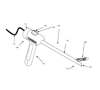
[0020] FIG. 1 depicts a schematic perspective view of a cannula assembly
in a closed
position, according to an embodiment of the present invention;
CA C27567872011-&9-26
WO 2010/111629 PCT/US2010/028881
- 5 -
[0021] FIG. 2 depicts a schematic perspective view of the cannula
assembly of FIG. 1 in
one of its open positions;
[0022] FIG. 3 depicts a close-up schematic perspective view of a tip
section of the cannula
assembly of FIG. 2;
[0023] FIG. 4 depicts a close-up schematic perspective view of another
embodiment of the
tip section of a cannula assembly in one of its open positions, in accordance
with an
embodiment of the present invention;
[0024] FIGS. 5A and 5B depict schematic cross-sectional and schematic
partial sectional
perspective views of a tip section of a cannula assembly in which an imaging
component is
configured for forward viewing while the cannula assembly is in the closed
position, in
accordance with an embodiment of the present invention;
[0025] FIG. 6 depicts a schematic perspective close-up view of another
embodiment of a
tip section of a cannula assembly in one of its open positions, according to
an embodiment of
the present invention;
[0026] FIGS. 7A and 7B depict schematic cross-sectional close-up and
schematic
perspective views of an actuating mechanism connected to a deployable portion
of a cannula
assembly in the open and closed positions, in accordance with an embodiment of
the present
invention;
[0027] FIG. 8 depicts a schematic perspective close-up view of a tip
section of a cannula
.. assembly in one of its open positions, according to another embodiment of
the present
invention; and
[0028] FIGS. 9A, 9B and 9C depict schematic perspective and schematic
cross-sectional
views of another tip embodiment of the apparatus with the capability of
forward viewing while
closed and having a removable trocar, according to an embodiment of the
present invention.
DETAILED DESCRIPTION
[0029] To provide an overall understanding of the invention, certain
illustrative
embodiments will now be described, including apparatus and methods for
displaying images.
However, it will be understood by one of ordinary skill in the art that the
systems and methods
described herein may be adapted and modified as is appropriate for the
application being
addressed and that the systems and methods described herein may be employed in
other
CA C27567872011-&9-26
WO 2010/111629 PCT/US2010/028881
- 6 -
suitable applications. All such adaptations and modifications are to be
considered within the
scope of the invention.
[0030] FIGS. 1 and 2 depict schematic perspective views of an embodiment
of the cannula
assembly 100 in closed and open positions, respectively. In one embodiment,
the cannula
assembly 100 includes a tubular element 110 fonaing a lumen 202. A proximal
end 114 of the
tubular element 110 can be adapted for manipulation by the surgeon or
clinician, and a distal
end 116 can be adapted for insertion into a body cavity. A housing 108 with a
handle 104 can
be attached near or at the proximal end 114 for manipulation by the surgeon or
clinician. In
alternative embodiments, the tubular element 110 can form a variety of cross-
sectional shapes,
e.g., generally round or cylindrical, ellipsoidal, triangular, square,
rectangular, and D-shaped
(in which one side is flat).
[0031] All or parts of the distal end of the cannula assembly 100 are
capable of being
positioned into a closed position 102 for insertion and extraction either
directly into the body
cavity or through another insufflating cannula. When closed, the distal end
assembly forms a
pointed tip, such as a trocar capable of puncturing the patient's skin. In
another embodiment,
the lumen 202 of the tubular element 110 is can be fitted with a retractable
and/or removable
trocar, such as that depicted in FIGS. 9A-9C and described further
hereinbelow. In one
embodiment, the trocar is made of solid, non-transparent material; whereas, in
another
embodiment all or parts of the trocar are made of optically transparent or
optically
transmissive material.
[0032] One or more portions of the distal end 116 of the tubular element
110 may be
designed to open once inserted into the body cavity. In one embodiment, as
depicted in FIG.
3, at least one deployable portion 204 of the tubular element 110 has an
adjustable angle of
deployment based on the operation of the opening adjustment means 106, i.e.,
an actuation
mechanism. For example, the adjustment means 106 can move the deployable
portion 204
between a closed position and an open position. Alternatively, the adjustment
means 106 can
incrementally move the deployable portion 204 between a closed position and
any number of
open positions. In additional or alternative embodiments, the deployable
portion 204 houses
an electronic component, which is at least partially disposed in the lumen
when in the closed
position. In an alternative embodiment, all electronic components are housed
within the walls
of the tubular element. When the deployable portion 204 is moved to at least
one open
position, the lumen 202 is substantially free from obstruction due to the
electronic components
CA C27567872011-&9-26
WO 2010/111629 PCT/US2010/028881
- 7 -
being moved out of the lumen, such that various instruments, e.g., surgical
tools or other
electronic components, can be passed through the lumen and used during the
operation or
surgical procedure.
[0033] The electronics components include one or more image transmission
components
304, in combination with one or more illumination components 305. In one
embodiment, the
image transmission component 304 may be a charge-coupled device (CCD) camera,
a
complementary metal oxide semiconductor (CMOS) imaging device, and/or an
imaging fiber
optic cable, and their ancillary optics and electronic drivers for power,
communication and
other functions.
[0034] Optically, one or more of the image transmission components 304 may
also image
across the spectrum, including those portions invisible to the human eye, such
as infrared and
ultra-violet. In one embodiment, two image transmission components may be
configured to
capture stereoscopic images (in still and/or in motion). In one embodiment,
one or more of the
image transmission components 304 may be configured with any of a combination
of fixed
optics, adaptive optics, and/or active optics. Adaptive and active optics can
be capable of
focusing and/or zooming onto the image or target area.
[0035] In one embodiment, the one or more image transmission components
304 are
capable of capturing both motion and still images, and transmitting them to
the surgeon or
operator through wired or wireless communication means 118 housed within or
connected to
the housing 108, handle 104, lumen 202 and/or the tubular element 110 wall.
Such
communications means 118 may include electrical signals, such as analog and/or
digital, or a
fiber optic communication system.
[0036] The illumination component 305 may be one or more light or
illumination sources
306, 308, and their ancillary electronic drivers 310. In one embodiment, the
illumination
sources 306, 308 are Light Emitting Diodes (LED), organic LED (OLED),
illumination fiber
optic, filament lamps, electroluminescent and/or laser sources. In one
embodiment, the
illumination component 305 is tailored to work closely in both optical and
spectrum
characteristics with the image transmission component 304, with the
illumination area, level,
and homogeneity being optimized. In one example, this may mean the
illumination level is
controlled by the surgeon or clinician; whereas, in another the image
transmission component
Automated Gain Control (AGC) is correlated with the illumination level of the
illumination
component 305.
CA C27567872011-&9-26
WO 2010/111629 PCT/US2010/028881
- 8 -
[0037] In one embodiment, as depicted in FIGS. 7A and 7B, the adjustment
means 106
may include a knob 701, which is connected to a rotational wheel 702, and
links 704, 706
traveling through the lumen, along the length of the tubular element 110. In
an alternative
embodiment, a push rod can be used in lieu of a knob 701. In another
alternative embodiment,
.. the links 704, 706 travel through one or more longitudinal apertures formed
in the wall of the
tubular element 110. Turning the knob 701 causes rotation of the wheel 702 in
one direction,
which pulls on one end of the link 706 and transfers force to its other end,
which is connected
to a downstream portion 712 of a hinge 714, and opens the deployable portion
204. A partial
turn of the knob 701 can, for example, move the deployable portion 204 into
any intermediate
.. open position. An equivalent, but opposite turn of the knob 701 pulls on
link 704, which is
connected to an upstream portion 710 of the hinge 714, and closes the
deployable portion 204.
The links 704 may be stiff or flexible elements, such as bars, rods, cables,
wires, etc.
Alternatively, a nut and lead screw combination may be used. Instead of the
knob 701, a lever
can be used. Similarly, instead of the knob 701, a spring-loaded release
mechanism can
.. actuate the deployable portion 204. In an alternative embodiment, the
deployable portion 204
can be actuated with a magnetic system. In this configuration, the deployable
portion 204 is
fitted with a magnet (e.g., a permanent magnet or a ferromagnetic target). A
complementary
magnet (e.g., a permanent magnet or a electromagnet target) external to the
body or patient is
used to interact with the magnet on the deployable portion 204, such that an
operator can open
and close the deployable portion 204 by moving the external magnet about and
in relation to
the deployable portion 204.
[0038] The hinge arrangements for the opening/closing of the deployable
portions 204
may be accomplished in a number of ways. In one embodiment, one or more of the
deployable portions 204 transition between a closed and a number of open
positions via a
hinge arrangement. The hinge arrangement may include a hinge disposed within a
wall of the
tubular element 110, e.g., all or partially within the lumen 202, around a
pivot point, on a
circumference of the tubular element 110, e.g., a circumferential hinge,
and/or on an exterior
of the tubular element 110. Alternatively, the hinge arrangement may include
at least one
four-bar linkage.
[0039] In an alternative embodiment, the lumen 202 is kept clear by passing
the links 704,
706 through a recess along or an aperture formed inside the tubular element
110 wall. In an
alternative embodiment, the adjustment means 106 include electro-mechanical
actuation of
CA C27567872011-&9-26
WO 2010/111629 PCT/US2010/028881
- 9 -
switches, operated by the surgeon or clinician, that drive one or more motors.
The motors or
actuators may be located either within the proximal end 114 (e.g., non-
deployable portions) of
the tubular element 110, or within the deployable portion 204. Alternatively,
the deployable
portion 204 can be moved via a pneumatic or fluidic actuator.
[0040] In an alternative embodiment, the hinge connecting the deployable
portion 204 to
the distal end 116 can include Shape Memory Alloy (SMA) materials with or
without an
assisted heating element. In one embodiment, using a material such as Nitinol
(located within
the lumen 202, tubular element 110 and/or the deployable portion 204), any
deployable
portion 204 can be closed at room temperature (e.g., 25 C), and deploy at the
temperature less
than that expected within the body cavity (e.g., less than 37 C). In an
alternative
embodiment, the assisted heating element can be controlled by the surgeon or
clinician. The
voltage for the assisted heating element can be transmitted along the tubular
element 110
walls. The assisted heating element is used to place the SMA material into the
deployable
temperature range once within the body cavity; for example, increasing the
voltage will
increase the temperature of the SMA material, which transitions the deployable
portion 204 to
one or more of its open positions. Removing or decreasing the voltage (and
hence the heat),
makes the deployable portion 204 transition to its closed position.
[0041] In one embodiment, as depicted in FIG. 6, the link 612 is a single
element, e.g., a
tape or rod, made of metal or other non-buckling configuration, which when
pushed towards
the distal end by the surgeon via operation of the knob 701, causes it to
extend towards the
distal end of the lumen 604. The extending pressure forces the hinge
arrangement 608, 614 on
the deployable portion 602 to flip and to rotate into one or more open
positions, causing the
formerly distal facing portion 610 to now face towards the proximal end. In
this
configuration, the imaging component 606 and illumination component 616 face
the area of
interest. The angle of opening of the deployable portion 602 may be adjusted
by the amount
of link 612 fed into the tubular element 110 through the rotation of the knob
701 or other
structural adjustment mechanism. This arrangement allows for the image
component 606 and
illumination components 616 to occupy almost or all of the lumen 604 when
closed, and to
leave the lumen 604 substantially open and available for instrument
insertion/operation and/or
removal of both instruments and body samples when open. In addition, this
arrangement
protects any image or illumination components when closed, while allowing the
same degree
of triangulation by adjustment of either the opening angle for the deployable
portion 602
CA C27567872011-&9-26
WO 2010/111629 PCT/US2010/028881
- 10 -
and/or the image component 606 and illumination component 616. Similar hinge
arrangements and adjustment means to those described herein may also be used.
[0042] In an alternative embodiment, as depicted in FIG. 8, the hinge
arrangement is a
four-bar linkage with arms 802, 803 that are attached to a deployable portion
804. In this
arrangement, the deployable portion 804 can be similarly actuated as described
above. For
example, one or more links passing internally through the wall of the tubular
element 110
connect to the arms 802, 803. Rotation of the knob or a push of a push rod
raises the
deployable potion 804 above the tubular element 110. Imaging component 606 and
illumination component 616 are housed in the deployable portion 804.
[0043] In an alternative embodiment, as depicted in FIG. 4, the tubular
element 110 can
include a plurality of deployable portions. e.g., two or three deployable
portions. For example,
as shown in FIG. 4, one embodiment includes two deployable portions, one
deployable portion
205 with electronic components (e.g., one or more image transmission
components and/or one
or more illumination components), the other deployable portion 314 without
electronic
components. Alternatively, both deployable portions 205, 314 can include
electronic
components. One or both of the deployable portions 205, 314 can open in
response to user
input. The number of deployable portions is only limited by the ability to
divide the
circumference of the tubular element 110, in either homogeneous or dissimilar
sized portions.
One or more such deployable portions may be formed, using any combination of
the
adjustment means discussed herein. This gives the surgeon or clinician
additional freedom
from cannula interference in the area where the surgery or operation is taking
place.
[0044] In one embodiment, the deployable portion 204 containing
electronic component is
actuated with mechanical links, while the other deployable portion 314, with
no electronic
component, uses SMA means for deployment. This would allow the fine
pointing/triangulation for the deployable portion 204 with electronic
components, and a
simpler, less precise adjustment mechanism for the other deployable portion
314. In another
embodiment, a complementary set of electronics is housed in each deployable
portion 204,
314, providing system redundancy selectable by the surgeon or operator. In
another
embodiment, the tubular element 110 can include three deployable portions, one
containing
image transmission components, the other containing illumination components,
and the last
one having no electronic component. Alternatively, at least one or more of an
image
CA C27567872011-&9-26
WO 2010/111629 PCT/US2010/028881
- 11 -
transmission component and an illumination component can be disposed on each
of the
deployable portions.
[0045] The deployable portion(s) 204 of the tubular element 110 is
configured to move
from the closed position aligned with the tubular element 110 (i.e., at zero
degrees) into an
infinite number of open positions from zero to 180 degrees relative to the
centerline axis
defined by the tubular element 110. This provides the surgeon or operator with
the ability to
effectively "triangulate" one or more of the field of views of the image
transmission
component and the illumination component. As may be seen in FIG. 4, adjusting
the angle a
of the opening of the deployable portion 204 relative to the axis 402 of the
tubular element
110, causes the direction of view 404, e.g., of the image transmission
component or
illumination component, to be adjusted without movement of the cannula. This
allows the
view to be changed slightly, without reverting to the need to move the
cannula. During a
procedure, moving the cannula may affect an instrument's position, vis-a-vis
the organ or
body structure being operated on. In use, the cannula may be rotated so that
the image
transmission component and the illumination component cover more fields of
view. The
rotation of the cannula can be tracked to keep the image in one orientation.
In various
embodiments, an accelerometer, an encoder (e.g., mechanical or optical), or
other suitable
feedback element disposed in the cannula assembly can communicate with control
electronics
in the video output of the image transmission component to rotate the image
before it is
displayed to the operator, in order to maintain the image in the same
orientation. The
feedback element may have a fixed reference point to indicate a preferred
orientation, such as
a vertical or up orientation. The fixed reference point of the feedback
element corresponds to
a particular reference point or orientation of the image transmission
component. This may be
the same orientation, such as an up orientation. Rotation of the cannula can
be accomplished
automatically without user-intervention or the rotation can be controlled by
the operator.
Within the image transmission component, well known and understood imaging
features may
be implemented, including electro-optic image stabilization and others.
[0046] A fail-safe design feature of the cannula assembly results from
the hinge
arrangement for the deployable portion(s) 204 being located at a point
upstream of the distal
end 116. The deployable portion 204 can be closed upon extraction of the
cannula assembly
through the force exerted on it during withdrawal through an external
insertion cannula 200.
CA C27567872011-&9-26
WO 2010/111629 PCT/US2010/028881
- 12 -
In this configuration, the deployable portion 204 is moved to the closed
position without
operation of the adjustment means 106.
[0047] All or part of the distal end 116 of the tubular element 110 may
be formed from an
optically transparent material as a trocar, pointed tip, or any suitably
shaped frontal form. In
combination with a deployable or removable mirror occupying all or part of the
interior
volume of the lumen 202, the surgeon or operator would be able to see a
forward view beyond
the cannula assembly when the deployable portion 204 is at or near the closed
position. In an
alternative embodiment, a prism can be used in lieu of a mirror.
[0048] FIGS. 5A and 5B illustrate an embodiment of the cannula assembly
100 in which
to all or part of the trocar is made of optically transmissive or optically
transparent materials.
When the deployable portion 204 is in a closed position 500, the image
transmission
component 508 is capable of imaging through the distal end 502 of the cannula
assembly via
an optical path 512. which travels through a window 510, light pipe, or
similar optically
transmissive medium on the distal end 502.
[0049] Within the lumen, a mirror assembly, which can be one or more
suitably reflective
surfaces 506, can be placed at suitable angle(s) to permit the forward view.
The reflective
surface 506 forms a connection 504 (e.g., a rod) through the lumen 202 of the
cannula
assembly 100 to the proximal end 114, allowing the mirror assembly to be
extracted once the
deployable portion 204 is opened, if necessary.
[0050] As depicted in FIGS. 9A, 9B and 9C, a cannula assembly 900 has the
capability of
forward viewing while the deployable portion 204 is in the closed position. In
addition, the
cannula assembly 900 can include a removable trocar. The distal end forms a
combination of
two portions. One is a deployable portion 204 (with external surface 902) and
the other is a
retractable portion 904. The deployable portion 204 may be made from any
suitable material,
while the retractable portion 904 is preferably made from an optically
transmissive or optically
transparent material. An optically transmissive or optically transparent
material provides a
window, that in combination with the mirror assembly inside the lumen (and
similar to those
described above), allows the imaging transmission component to view forward
while the
deployable portion 204 is in the closed position. Upon deployment, retractable
portion 904 is
retracted through the lumen by the surgeon or operator.
CA C27567872011-&9-26
WO 2010/111629 PCT/US2010/028881
- 13 -
[0051] In one embodiment, additional illumination sources are placed
within indentations
in the external surface 904 facing the distal end. Such illumination sources
would minimize
reflections from the optically transmissive portions of the trocar coming back
to the image
transmission components. In an alternative embodiment, power for the
illumination sources is
provided by energy storage components placed within the cannula assembly,
e.g., a battery in
the handle, minimizing the interfacing to the rest of the cannula assembly.
The surgeon can
activate the illumination sources at the time of insertion of the cannula
assembly. The
illumination source may include any of the illumination sources described
above. The energy
storage component may be batteries or super-capacitors. The energy storage
component can
be attached to the rod 504 or can be connected to the illumination sources.
[0052] Various embodiments and features of the present invention have
been described in
detail with a certain degree of particularity. The utilities thereof can be
appreciated by those
skilled in the art. It should be emphasized that the above-described
embodiments of the
present invention merely describe possible examples of the implementations to
set forth a
clear understanding of the principles of the invention, and that numerous
changes, variations,
and modifications can be made to the embodiments described herein without
departing from
the spirit and scope of principles of the invention. Also, such variations and
modifications are
intended to be included herein within the scope of the present invention, as
set forth in the
appended claims. The scope of the present invention is defined by the appended
claims, rather
.. than the forgoing description of embodiments. Accordingly, what is desired
to be secured by
Letters Patent is the invention as defined and differentiated in the following
claims, and all
equivalents
[0053] What is claimed is: