Note: Descriptions are shown in the official language in which they were submitted.
WO 2010/115011 PCT/US2010/029648
MODULAR GASTROINTESTINAL PROSTHESES
CROSS-REFERENCE TO RELATED APPLICATION
[0001] This application claims the benefit under 35 U.S.C. 119 of U.S.
Provisional
Application 61/211,853, filed on April 3, 2009, entitled "Modular Systems for
Intra-
Luminal Therapies within Hollow Body Organs," which is incorporated herein by
reference in its entirety for all purposes.
TECHNICAL FIELD
[0002] This invention relates to prosthetic implants placed within the
gastrointestinal
system, including the stomach, the esophagus and the intestines. In
particular, it relates to
implant systems having components implantable and removable using endoscopic
techniques, for treatment of obesity, diabetes, reflux, and other
gastrointestinal conditions.
BACKGROUND
[0003] Bariatric surgery procedures such as sleeve gastrectomy, the Rouen-Y
gastric
bypass (RYGB) and the bileo-pancreatic diversion (BPD) are surgical procedures
to
modify food intake and/or absorption within the gastrointestinal system to
effect weight
loss in obese patients. These procedures affect metabolic processes within the
gastrointestinal system, by either short-circuiting certain natural pathways
or creating
different interaction between the consumed food, the digestive tract, its
secretions and the
neurohormonal system regulating food intake and metabolism. In the last few
years there
has been a growing clinical consensus, that obese diabetic patients who
undergo bariatric
surgery see a remarkable resolution of their Type-2 Diabetes Mellitus (T2.DM)
soon after
the procedure. The remarkable resolution of diabetes after RYGB and BPD
typically
occurs too fast to be accounted for by weight loss alone, suggesting that
there may be a
direct impact on glucose homeostasis. The mechanism of this resolution of T2DM
is not
well understood, and it is quite likely that multiple mechanisms are involved.
[0004] One of the drawbacks of bariatric surgical procedures is that they
require fairly
invasive surgery, with potentially serious complications and long patient
recovery periods.
In recent years, there is an increasing amount of ongoing effort to develop
minimally
invasive procedures to mimic the effects of bariatric surgery using minimally
invasive
procedures. One such procedure involves the use of gastrointestinal implants
that modify
transport and absorption of food and organ secretions. For example, U.S.
Patent 7,476,256
describes an implant having a tubular sleeve with an anchor having barbs.
While these
1
WO 2010/115011 PCT/US2010/029648
implants may be delivered endoscopically, the implants offer the physician
limited
flexibility and are not readily removable or replaceable, as the entire
implant is subject to
tissue in-growth after implantation. Moreover, stents with active fixation
means, such as
barbs that penetrate in to the surrounding tissue, may potentially cause
tissue necrosis and
erosion of the implants through the tissue, which can lead to serious
complications such as
systemic infection.
SUMMARY
[0005] According to various embodiments, the present invention is a modular
intra-
luminal implant systems for treating metabolic disorders such as obesity and
diabetes,
which provides far more flexible therapy alternatives than single devices to
treat these
disorders. These implant systems include components that can be selectively
added or
removed to mimic a variety of bariatric surgical procedures with a single
basic construct.
The fundamental building blocks of the system include anchoring implants that
are placed
within the GI system or some instances around particular organs. These low-
profile
implants are designed for long-term performance with minimal interference with
normal
physiological processes. Features of these anchoring implants allow them to
act as docking
stations for therapy implants designed for achieving certain metabolic
modification goals.
By using a combination of anchoring implants with corresponding replaceable
tubular
elements that dock with them, it is possible to design therapies with
particular metabolic
modification goals or those that mimic currently practiced bariatric surgical
procedures.
This allows the physician to customize the therapy to the patient at the time
of the initial
procedure but also allows the flexibility to alter the therapy during the life-
time of the
patient by replacing individual components.
[0006] According to some embodiments, the modular systems of the invention
includes a anchoring implant portion (docking element) including an expandable
structure
(e.g., a low profile stent or ring or fabric/elastomeric cuff) anchored within
the esophagus,
the gastro-esophageal junction, the pyloric junction, the duodenum or the
jejunum and
may have sleeve or graft extensions. The stents may be balloon expandable or
self-
expanding and anchor against the tissue with radial force. The rings could be
made of self-
expanding Nitinol and anchor to the tissue by entrapment of the tissue within
the ring
elements or by radial force. The cuffs could be either sutured or stapled or
permanently or
reversibly attached by other mechanical means to the tissue. The anchoring
implant
2
WO 2010/115011 PCT/US2010/029648
includes or is adapted to receive (e.g., endoscopically) features that enable
docking
functionality. The docking functionality of the stent, ring or cuff, for
example, could take
the form of magnetic elements, hooks, mating mechanical elements or structures
(such as
the stent braid or mesh) that are integral to the framework of the stent, ring
or cuff or the
sleeve or graft extension. The system also could be such that the docking
functionality is
not integral to the stent, ring or cuff but is introduced later by attaching
other elements
such as magnets, hooks, mating mechanical elements etc to the framework of the
stent,
ring, cuff or to the sleeve/graft extension of the above implants. Therapeutic
implants,
such as tubular sleeves or stent grafts are adapted to be reversibly attached
to the
anchoring implants. These therapeutic implants will have corresponding
features (e.g.,
magnets, hooks, mechanical elements) to enable docking to the anchoring
implants, so that
the therapeutic implants can be reversibly attached to the anchoring implants.
In some
embodiments, the tubular implants will not be in contact with tissue to
minimize or
prevent tissue in-growth and facilitate easy removal with endoscopic
instrumentation after
long-term implantation.
[0007] According to various embodiments, the anchoring or docking implants
comprise stems or covered stents (stent grafts) that promote tissue in-growth
without
penetrating into the tissue. Such stents may include, for example, a self-
expanding laser
cut stent with non-penetrating struts that engage the wall of the GI tract or
a self-
expanding stent braided with a Dacron type fabric covering of the right
porosity would
promote tissue in-growth and aid fixation.
[0008] According to various embodiments, the anchoring or docking implants
comprise a double braided stent (e.g., having a spacing between the braids of
0.5 to 5.0
mm). This embodiment is optimized such that the outer braid could be securely
anchored
within tissue, but the tissue would not grow into the inner braid, which can
then be used to
anchor the replaceable implant.
[0009] According to various embodiments, the anchoring or docking implants are
specifically designed to be constrained at certain anatomic locations. Such
designs, for
example, may include a double-flange shaped or dumbbell-shaped implants placed
at the
pyloric junction or barrel shaped stents placed within the duodenal bulb.
[0010] According to various embodiments, the replaceable therapeutic implants
that
dock to the anchoring implants take the form of long tubes that can
selectively channel the
flow of food and secretions from organs (e.g., the stomach, gall bladder,
intestines and
3
WO 2010/115011 PCT/US2010/029648
pancreas) to various destinations within the digestive tract. This diversion
and bypass of
food and organ secretions (e.g., insulin and incretin from the pancreas and
bile from the
gall bladder) could then be controlled by adjusting the design features of the
system where
the implants are placed within the GI tract. The implants could also include
restrictive
stoma type elements or anti-reflux valves. To divert food and secretions from
the first part
of the intestine, for example, an anchoring implant can be placed within the
duodenal bulb
or at the pyloric junction. Then, a thin tube about 1-2 feet in length with a
funnel shaped
proximal end and a rigid ring shaped distal end can be introduced into the
proximal
duodenum and docked to the permanent implant. It would be possible to later
remove this
by endoscopic means by simple undocking it from the anchoring implant. To
restrict
passage of food, a restrictive element such as one created by a tapered
stepped tube or a
stent or a stent graft can become the docking element and be reversibly
attached to the
docking station.
[0011] According to various embodiments, the docking means may include
engaging/disengaging mechanical shape memory and super-elastic elements,
attractive/repulsive and levitating magnetic mechanisms, loop-hoop fastener
technologies
etc.. The systems may be deployed with functional docking components or those
components would be attached to the permanent implants under endoscopic visual
guidance. The docking means is designed so that the therapeutic implants can
be easily
deployed and securely affixed to the anchoring implants. According to various
embodiment, the engaging elements of the docking system are arranged so that
they do not
impinge on the surrounding tissue, nor would be later covered with tissue
layers. This
facilitates disengaging the tubular sleeve elements from the stent with simple
magnetic
instruments or grasper type endoscopic instruments or funnel shaped retrieval
basket
catheters or using a draw-string type mechanism.
[0012] According to some embodiments, the anchoring element is integrated with
a
therapy component.
[0013] According to various embodiments, the present invention is a method of
treating gastro-esophageal reflux disease (GERD) including placing a low-
profile implant
within the stomach, the esophagus, the intestine or at internal junctions of
these organs or
around these organs, and securely attaching to the implant other gastro-
intestinal implants
that permit bypass of food and organ secretions from one site within the
gastro-intestinal
tract to other sites within the gastro-intestinal tract.
4
WO 2010/115011 PCT/US2010/029648
[0014] While multiple embodiments are disclosed, still other embodiments of
the
present invention will become apparent to those skilled in the art from the
following
detailed description, which shows and describes illustrative embodiments of
the invention.
Accordingly, the drawings and detailed description are to be regarded as
illustrative in
nature and not restrictive.
BRIEF DESCRIPTION OF THE DRAWINGS
[0015] FIG. 1 is a cross sectional view of a portion of the digestive tract in
the body.
A docking element is implanted in the duodenal bulb and a tubular implant
(sleeve) is
attached to the docking element and extended into the duodenum to the ligament
of treitz.
[0016] FIG. 2 is a cross sectional view of a portion of the digestive tract in
the body.
An endoscope is inserted into the mouth, passing through the esophagus in to
the stomach
and the end of the scope is pointed to allow viewing of the pylorus.
[0017] FIG. 3 is a drawing of a typical endoscope used for diagnostic and
therapeutic
procedures in the gastro intestinal (GI) tract.
[0018] FIG. 4A is a drawing of an over the wire sizing balloon that can be
used to
measure the diameter of the pylorus, duodenal bulb, esophagus, pyloric antrum
or other
lumen in the GI tract.
[0019] FIG. 4B is a drawing of a monorail sizing balloon that can be used to
measure
the diameter of the pylorus, duodenal bulb, esophagus, pyloric antrum or other
lumen in
the GI tract.
[0020] FIG. 5 is a sectional view of a portion of the digestive tract in the
body. An
endoscope is inserted into the GI tract up to the pylorus. A sizing balloon is
inserted
through the working channel and into the area of the duodenal bulb. The
balloon is
inflated to measure the diameter of the duodenal bulb.
[0021] FIG. 6A is a drawing of a stent that can used as a docking element.
[0022] FIG. 6B is a drawing of a stent that can used as a docking element that
has a
polymer covering on the inside and outside.
[0023] FIG. 7 is a tubular implant that can be used to bypass the stomach,
duodenum
or other intestinal lumen.
[0024] FIG. 8 is a drawing of a delivery catheter for the docking element and
tubular
implant.
WO 2010/115011 PCT/US2010/029648
[0025] FIG. 9A is a cross sectional view of a portion of the digestive tract
in the body.
A delivery catheter with a docking element and tubular implant loaded onto the
catheter
are loaded onto an endoscope. The endoscope is then advanced through the
esophagus,
stomach and into the duodenal bulb.
[0026] FIG. 9B is a cross sectional view of a portion of the digestive tract
in the body.
A delivery catheter with a docking element and tubular implant loaded onto are
loaded
onto an endoscope. The endoscope is then advanced through the esophagus,
stomach and
into the duodenal bulb. The outer sheath of the delivery catheter is retracted
to partially
deploy the docking element into the duodenal bulb.
[0027] FIG. 10 is a drawing showing the docking element fully deployed into
the
duodenal bulb. The delivery catheter and endoscope has been has been removed
to show
clarity
[0028] FIG. 11 is a drawing showing the endoscope and delivery catheter
advanced
through the docking element into the duodenum up to the ligament of treitz.
[0029] FIG. 12 is a drawing showing the endoscope and delivery catheter
advanced
through the docking element into the duodenum up to the ligament of treitz.
The outer
sheath of the delivery catheter is retracted to partially expose the tubular
implant.
[0030] FIG. 13 is a drawing showing the endoscope and delivery catheter
advanced
through the docking element into the duodenum up to the ligament of treitz.
The outer
sheath of the delivery catheter is retracted to partially expose the tubular
implant. A
balloon catheter is inserted through the working channel of the endoscope to
the area of
the partially exposed tubular implant. The balloon is inflated to temporarily
secure the
tubular implant to the duodenum.
[0031] FIG. 14 is a continuation of FIG. 13 where the outer sheath is
retracted further
to unsheath the tubular implant up to the duodenal bulb.
[0032] FIG. 15 is a continuation of Fig 14 where the endoscope has been
withdrawn
to the duodenal bulb. The balloon on the balloon catheter is then deflated and
the balloon
catheter is withdrawn to the duodenal bulb. The balloon is then re-inflated to
open up and
secure the proximal end of the tubular implant to the inside diameter of the
docking
element.
[0033] FIG. 16 is a drawing of an alternative device and method for deploying
the
proximal end of the tubular element.
6
WO 2010/115011 PCT/US2010/029648
[0034] FIG. 17A is a cross sectional view of a portion of the digestive tract
in the
body. A docking element is implanted in the esophagus at the gastro-esophageal
junction.
The docking element serves as an anti-reflux valve.
[0035] FIG 17B is a cross sectional view of a portion of the digestive tract
in the
body. A docking element is implanted in the esophagus at gastro-esophageal
junction. The
docking element serves as a restrictive stoma.
[0036] FIG. 18 is a cross sectional view of a portion of the digestive tract
in the body.
A docking element is implanted in the esophagus at gastro-esophageal junction.
The
docking element serves as an anti-reflux valve.
[0037] FIG. 19A is a stented sleeve with a stent used to hold open the sleeve.
The
sleeve located from the duodenal bulb to the ligament of treitz.
[0038] FIG. 19B is a stented sleeve with a stent used to hold open the sleeve.
The
sleeve located from the pylorus to the ligament of treitz.
[0039] FIG. 20 is a stented sleeve with a stent used to hold open the sleeve.
The
sleeve is located from the stomach antrum to the ligament of treitz.
[0040] FIG. 21A is a sectional view of a portion of the digestive tract in the
body. A
docking element is implanted in the esophagus at the gastro-esophageal
junction. A
docking element and tubular implant is implanted in the duodenum also.
[0041] FIG. 21B is a sectional view of a portion of the digestive tract in the
body. A
docking element is implanted in the esophagus at the gastro-esophageal
junction. A
docking element and tubular sleeve is implanted in the duodenum also. A third
implant
element bypasses the stomach.
[0042] FIG. 22A is a sectional view of a portion of the digestive tract in the
body. A
docking element is implanted in the esophagus at the gastro-esophageal
junction. A second
docking element and tubular implant is implanted from the esophageal implant
to the
ligament of treitz.
[0043] FIG. 22B is a sectional view of a portion of the digestive tract in the
body. A
docking element is implanted in the esophagus at gastro-esophageal junction. A
docking
element and tubular implant is implanted from the esophageal implant to the
duodenal
bulb.
[0044] FIG. 23A is a sectional view of a portion of the digestive tract in the
body. A
docking element and tubular implant is implanted in the esophagus at the
gastro-
esophageal junction. The modular implant has an anti-reflux valve. A second
docking
7
WO 2010/115011 PCT/US2010/029648
station and tubular implant is placed in the duodenal bulb and extends to the
ligament of
treitz. A third docking station and tubular implant connects the esophageal
implant and the
duodenal implant.
[0045] FIG. 23B is a sectional view of a portion of the digestive tract in the
body. A
docking element and tubular implant is implanted in the esophagus at the
gastro-
esophageal junction. The modular implant has an-anti reflux valve. A second
docking
station and tubular implant is placed in the pylorus and extends to the
ligament of treitz. A
third docking station and tubular implant connects the esophageal implant and
the
duodenal implant at the pylorus.
[0046] FIG. 24 is a sectional view of a portion of the digestive tract in the
body. A
docking element and tubular implant is implanted in the esophagus at gastro-
esophageal
junction. The modular implant has an-anti reflux valve. A second docking
station and
tubular implant is placed in the pyloric antrum and extends to the ligament of
treitz. A
third docking station and tubular implant connects the esophageal implant and
the
duodenal implant at the pyloric antrum.
[0047] FIG. 25 is a drawing of a delivery catheter with a docking element
loaded onto
it.
[0048] FIG. 26 is a drawing of a delivery catheter with the endoscope inserted
through inner diameter of the delivery catheter.
[0049] FIG. 27 is a drawing of a delivery catheter which is designed to be
inserted
through the working channel of the endoscope.
[0050] FIG. 28 is a drawing of a delivery catheter with a docking element and
tubular
implant loaded onto it.
[0051] FIGS. 29-35 show a variety of stents that can be used as a docking
element.
[0052] FIG. 36A is a drawing of a stent that can be used as a docking element.
[0053] FIG. 36B is a drawing of a stent that can be used as a docking element.
[0054] FIGS. 37-39 show docking elements.
[0055] FIG. 40A is an expandable ring that can attached to a sleeve to form a
tubular
implant.
[0056] FIG. 40B is an expandable ring that can attached to a sleeve to form a
tubular
implant.
[0057] FIG. 40C is an expandable ring that can attached to a sleeve to form a
tubular
implant.
8
WO 2010/115011 PCT/US2010/029648
[0058] FIG. 41 is a tubular implant that uses an expandable ring as in FIG.
40A, 40B
or 40C as an anchoring means.
[0059] FIG. 42 is a tubular implant that uses an expandable ring as in FIG.
40A, 40B
or 40C as an anchoring means. The tubular implant is placed and secured within
a docking
element.
[0060] FIG. 43 is a tubular implant that uses an expandable ring as in FIG.
40A, 40B
or 40C as an anchoring means. The tubular implant is expanded and secured
within the
docking element.
[0061] Fig 44 is a drawing of a docking element which uses hook and loop to
secure
the tubular implant to docking element.
[0062] FIG. 45A is a drawing of a tubular implant that has magnets in the wall
to
allow attachment to another tubular implant or to a docking element.
[0063] FIG. 45B is a drawing of a tubular implant that has magnets in the wall
to
allow attachment to another tubular implant or to a docking element, it has a
female
receptacle to allow attachment to a docking element or other tubular implant.
[0064] FIGS. 46A and 46B show tubular implants.
[0065] FIGS. 47A and 47B show tubular implants in which the sleeve has
longitudinal or circumferential pleats, respectively.
[0066] FIGS. 48A and 48B show tubular implants or sleeves with a magnetic
attachment means.
[0067] FIG. 49 is a drawing of a tubular implant or sleeve with barbs to
attach to
attach to tissue or to a docking element.
[0068] FIG. 50A is a drawing of a tubular implant or sleeve with pockets to
insert
magnets to allow attachment to a docking element or to another tubular
implant.
[0069] FIG. 50B is a drawing of a tubular implant or sleeve with hooks to
attach
docking element or another tubular implant.
[0070] FIG. 51A is a conical or tapered shaped docking element or tubular
implant.
[0071] FIG. 51B is a docking element or tubular implant with a stepped
diameter.
[0072] FIG. 52 is a tubular implant that has hook and loop (velcro) attachment
means
to attach to a docking element or another tubular implant.
[0073] FIG. 53A is an over the wire balloon catheter for delivering and
expanding
balloon expandable stents for a docking element.
9
WO 2010/115011 PCT/US2010/029648
[0074] FIG. 53B is a rapid exchange balloon catheter for delivering and
expanding
balloon expandable stents for a docking element.
[0075] FIG. 54 shows a docking element design with a single-braided or laser-
cut
design placed at the pyloric junction.
[0076] FIG. 55 shows another docking element designed where the stomach side
of
docking element is more disk-like .
[0077] FIG.s 56 and 57 show docking elements of FIG. 55 and FIG. 56 covered
with
fabric or polymer sheets in areas where they contact tissue.
[0078] FIG. 58 shows a different design of the docking element placed within
the
pylorus, where two metallic elements (one on the stomach side and one on the
duodenal
side) are connected by a flexible sleeve element
[0079] FIG. 59 depicts the docking element of FIG. 58 where the flexible
sleeve
element has expanded with the opening of the pyloric valve.
[0080] FIG. 60 depicts another docking element design incorporating a flexible
sleeve
element.
[0081] FIG. 61 depicts a tubular implant which can be reversibly attached to
various
compatible docking elements described elsewhere such as those shown in FIG.s
54
through FIG. 58.
[0082] FIG. 62 shows delivery of the tubular implant of FIG. 61 close to the
docking
element of FIG. 54.
[0083] FIG. 63 depicts the docking element and the tubular element mated
together
upon release from the delivery catheter
[0084] FIG. 64 shows where the tubular element is now attached to the docking
element of FIG. 58
[0085] FIG. 65 shows a situation where the tubular element is attached to the
docking
element of FIG. 58 on the stomach portion of the docking element.
[0086] FIGS. 66-78 show schematic views of various stages of an implantation
method according to embodiments of the invention.
[0087] While the invention is amenable to various modifications and
alternative
forms, specific embodiments have been shown by way of example in the drawings
and are
described in detail below. The intention, however, is not to limit the
invention to the
particular embodiments described. On the contrary, the invention is intended
to cover all
WO 2010/115011 PCT/US2010/029648
modifications, equivalents, and alternatives falling within the scope of the
invention as
defined by the appended claims
DETAILED DESCRIPTION
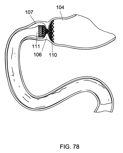
[0088] FIG. 1 is a schematic, sectional view of an embodiment of the invention
implanted in a portion of a human digestive tract. As a person ingests food,
the food enters
the mouth 100, is chewed, and then proceeds down the esophagus 101 to the
lower
esophageal sphincter at the gastro-esophageal junction 102 and into the
stomach 103. The
food mixes with enzymes in the mouth 100 and in the stomach 103. The stomach
103
converts the food to a substance called chyme. The chyme enters the pyloric
antrum 104
and exits the stomach 103 through the pylorus 106 and pyloric orifice 105. The
small
intestine is about 21 feet long in adults. The small intestine is comprised of
three sections.
The duodenum 112, jejunum 113 and ileum (not shown). The duodenum 112 is the
first
portion of the small intestine and is typically 10-12 inches long. The
duodenum112 is
comprised of four sections: the superior, descending, horizontal and
ascending. The
duodenum 112 ends at the ligament of treitz 109. The papilla of vater 108 is
the duct that
delivers bile and pancreatic enzymes to the duodenum 112. The duodenal bulb
107 is the
portion of the duodenum which is closest to the stomach 103.
[0089] As shown in FIG. 1, a docking or anchoring element 110 is implanted in
the
duodenal bulb 107 and a tubular or therapy implant 111 is attached to the
docking element
and extended into the duodenum 112 to the ligament of treitz 109. In this
embodiment,
magnets 135 on the docking element 110 and magnets 136 on the tubular implant
111 are
magnetically attracted to each other and thereby secure the docking element
110 to the
therapy implant 111. According to various exemplary embodiments, the anchoring
element 110 includes an expandable structure (e.g., a stent or ring) adapted
for anchoring
within the duodenal bulb and has a diameter of between about 20 and about 40
mm in its
unrestrained expanded configuration. In these embodiments, the magnets 135 on
the
docking or anchoring element 110 serve as a docking feature for releasably
coupling with
the magnets 136 of the tubular implant 111.
[0090] FIG. 2 is a schematic view of a portion of the digestive tract in a
human body.
An endoscope 114 has been inserted through the mouth 100, esophagus 101, the
gastro-
esophageal junction 102 and into the stomach 103. The endoscope 114 further
extends
into the pyloric antrum 104 to allow visualization of the pylorus 106.
11
WO 2010/115011 PCT/US2010/029648
[0091] FIG. 3 is a drawing of an endoscope 114. Endoscopes 114 are commonly
used
for diagnostic and therapeutic procedures in the gastrointestinal (GI) tract.
The typical
endoscope 114 is steerable by turning two rotary dials 115 to cause deflection
of the
working end 116 of the endoscope. The working end of the endoscope 116 or
distal end,
typically contains two fiber bundles for lighting 117, a fiber bundle for
imaging 118
(viewing) and a working channel 119. The working channel 119 can also be
accessed on
the proximal end of the endoscope. The light fiber bundles and the image fiber
bundles are
plugged into a console at the plug in connector 120. The typical endoscope has
a working
channel, for example, having a diameter in the 2 to 4 mm diameter range. It
may, for
example having a working channel having a diameter in the 2.6 to 3.2 mm range.
The
outside diameter of the endoscopes are typically in the 8 to 12 mm diameter
range
depending on whether the endoscope is for diagnostic or therapeutic purposes.
[0092] FIG. 4A is a partial sectional view of an over the wire sizing balloon
121 that
is used to measure the diameter of the pylorus 106, duodenal bulb 107,
esophagus 102,
pyloric antrum 104 or other lumen in the GI tract. The sizing balloon is
composed of the
following elements: a proximal hub 122, a catheter shaft 124, a distal balloon
component
125, radiopaque marker bands 126, a distal tip 127, a guide wire lumen 128,
and an
inflation lumen 129. The distal balloon component 125 can be made, for
example, from
silicone, silicone polyurethane copolymers, latex, nylon 12, PET (Polyethylene
terphalate)
Pebax (polyether block amide), polyurethane, polyethelene, polyester elastomer
or other
suitable polymer. The distal balloon component 125 can be molded into any
desired shape,
including for example a cylindrical shape, a dog bone shape, or a conical
shape. The distal
balloon component 125 can be made compliant or noncompliant. The distal
balloon
component 125 can be bonded to the catheter shaft 124 with glue, heat bonding,
solvent
bonding, laser welding or any suitable means. The catheter shaft can be made
from
silicone, silicone polyurethane copolymers, latex, nylon 12, PET (Polyethylene
terphalate)
Pebax (polyether block amide), polyurethane, polyethylene, polyester elastomer
or other
suitable polymer. Section A-A (shown at the top portion of FIG. 4A) is a cross
section of
the catheter shaft 124. The catheter shaft 124 is shown as a dual lumen
extrusion with a
guide wire lumen 128 and an inflation lumen 129. The catheter shaft 124 can
also be
formed from two coaxial single lumen round tubes in place of the dual lumen
tubing. The
balloon is inflated by attaching a syringe (not shown) to luer fitting side
port 130. The
sizing balloon accommodates a guidewire through the guidewire lumen from the
distal tip
12
WO 2010/115011 PCT/US2010/029648
127 through the proximal hub 122. The sizing balloon 121 can be filled with a
radiopaque
dye to allow visualization and measurement of the size of the anatomy with a
fluoroscope.
In the embodiment of FIG. 4A, the sizing balloon 121 has two or more
radiopaque marker
bands 126 located on the catheter shaft to allow visualization of the catheter
shaft and
balloon position. The marker bands 126 also serve as fixed known distance
reference point
that can be measured to provide a means to calibrate and determine the balloon
diameter
with the use of the fluoroscope. The marker bands can be made from tantalum,
gold,
platinum, platinum iridium alloys or other suitable material.
[0093] FIG 4B is a partial sectional view of a rapid exchange sizing balloon
134 that
is used to measure the diameter of the pylorus 106, duodenal bulb 107,
esophagus 102,
pyloric antrum 104 or other lumen in the GI tract. The sizing balloon is
composed of the
following elements: a proximal luer 131, a catheter shaft 124, a distal
balloon component
125, radiopaque marker bands 126, a distal tip 127, a guide wire lumen 128,
and an
inflation lumen 129. The materials of construction will be similar to that of
the sizing
balloon 121 of FIG. 4A. The guide wire lumen 128 does not travel the full
length of the
catheter, it starts at the distal tip 127 and exist out the side of the
catheter at distance
shorter that that the shorter that the overall catheter length. A guide wire
132 is inserted
into the balloon catheter to illustrate the guidewire path through the sizing
balloon 134. As
shown in FIG. 4B, the sizing balloon catheter shaft changes section along its
length from a
single lumen at section B-B 133 to a dual lumen at section A-A at 124.
[0094] FIG. 5 is a schematic view of a portion of the digestive tract in the
body. An
endoscope 114 is inserted into the GI tract up to the pylorus 106. A sizing
balloon 121 is
inserted through the working channel 119 of the endoscope and into the area of
the
duodenal bulb 107. The sizing balloon 121 is inflated with contrast agent. The
diameter of
the duodenal bulb 107 is measured with a fluoroscope.
[0095] FIG. 6A shows various views of a stent that can used as a docking or
anchoring element. The stents of this invention can be comprised, for example,
of any one
or more of the following materials: Nickel titanium alloys (Nitinol),
Stainless steel alloys:
304, 316L, BioDur 108 Alloy, Pyromet Alloy CTX-909, Pyromet Alloy CTX-3,
Pyromet Alloy 31, Pyromet Alloy CTX- 1, 21Cr-6Ni-9Mn Stainless, 21Cr- 6Ni-
9Mn
Stainless, Pyromet Alloy 350, 18Cr-2Ni-12Mn Stainless, Custom 630 (17Cr-4Ni)
Stainless, Custom 465 Stainless, Custom 455 Stainless Custom 450 Stainless,
Carpenter 13-8 Stainless, Type 440C Stainless, Cobalt chromium alloys- MP35N,
Elgiloy,
13
WO 2010/115011 PCT/US2010/029648
L605, Biodur Carpenter CCM alloy, Titanium and titanium alloys, Ti-6A1-4V/ELI
and
Ti-6A1-7Nb, Ti-15Mo Tantalum, Tungsten and tungsten alloys, Pure Platinum,
Platinum-
Iridium alloys, Platinum - Nickel alloys, Niobium, Iridium, Conichrome, Gold
and Gold
alloys. The stent may also be comprised of the following absorbable metals:
Pure Iron and
magnesium alloys. The stent may also be comprised of the following plastics:
Polyetheretherketone (PEEK), polycarbonate, polyolefin's, polyethylene's,
polyether
block amides (PEBAX), nylon 6, 6-6, 12, Polypropylene, polyesters,
polyurethanes,
polytetrafluoroethylene (PTFE) Poly(phenylene sulfide) (PPS), poly(butylene
terephthalate) PBT, polysulfone, polyamide, polyimide, poly(pphenylene oxide)
PPO,
acrylonitrile butadiene styrene (ABS), Polystyrene, Poly(methyl methacrylate)
(PMMA),
Polyoxymethylene (POM), Ethylene vinyl acetate , Styrene acrylonitrile resin,
Polybutylene. The stent may also be comprised of the following absorbable
polymeres:
Poly (PGA), Polylactide (PLA), Poly( -caprolactone), Poly(dioxanone)
Poly(lactide-
coglycolide). Stent 137 stent according to various embodiments is laser cut
from a round
tubing or from a flat sheet of metal. The flat representation of the stent
circumference is
shown in item 138. The flat representation of an expanded stent is shown in
item 139. The
end view of the stent is shown 141. Magnets 140 are attached to the stent on
the outside
diameter. The magnets may be attached to the stent by use of a mechanical
fastener, glue,
suture, welding, snap fit or other suitable means. The stent can be either
balloon
expandable or self expanding. The magnets may be located in middle of the
stent or at the
ends of the stent. Suitable materials for the magnets include: neodymium-iron-
boron [Nd-
Fe-B], samarium-cobalt [Sm-Co], alnico, and hard ferrite [ceramic] or other
suitable
material. In some embodiments, the magnets are encapsulated in another metal
(e.g.,
titanium) or polymer to improve corrosion resistance and biocompatibility.
[0096] FIG. 6B shows various views of a stent that can used as a docking or
anchoring element. Stent 142 may be laser cut from a round tubing or from a
flat sheet of
metal. The flat representation of the stent circumference is shown in item
143. The flat
representation of an expanded stent is shown in item 144. The end view of the
stent is
shown 145. Permanent magnets 140 are attached to the stent on the outside
diameter. This
stent is a covered stent. The stent covering is not shown on items 142, 143 or
144. The
covering are shown on the end view which shows stent 145. Stent may have an
outside
covering 146, inside covering 147 or both. Suitable materials for the covering
include but
are not limited to: silicone, polyether block amides (PEBAX), polyurethanes,
silicone
14
WO 2010/115011 PCT/US2010/029648
polyurethane copolymers, nylon 12, polyethylene terphalate (PET), Goretex
ePTFE,
Kevlar , Spectra, Dyneena, polyvinyl chloride (PVC), polyethylene or polyester
elastomers. The coverings may be dip coated onto the stent or they may be made
as a
separate tube and then attached to the stent by adhesives or mechanical
fasteners such as
suture, rivets or by thermal bonding of the material to the stent or another
layer. The
covering may also have drugs incorporated into the polymer to provide for a
therapeutic
benefit. The covering 146 or 147 may also be of biologic origin. Suitable
biologic
materials include but are not limited to: Amnion, Collagen Type I, II, III,
IV, V, VI -
Bovine, porcine, ovine, placental tissue or placental veins or arteries and
small intestinal
sub- mucosa.
[0097] FIG. 7 is a tubular therapy implant that can be used to bypass the
stomach 103,
duodenum 112 or other intestinal lumens (e.g., a portion or all of the
jejunum). The tubular
implant is made of a thin wall tube 148 and a series of magnets 140 attached
to the inside
of the thin wall tube. According to other embodiments, the magnets 140 may be
attached
to the outside of the tube 148. According to various embodiments, the magnets
140 are
disposed about a circumference of the tube 148 such that the location of the
magnets
correspond to locations of corresponding magnets located on the anchoring or
docking
element. The tubular implants of this invention may be comprised, for example,
of the
following materials: silicone, polyether block amides (PEBAX), polyurethanes,
silicone
polyurethane copolymers, Nylon, polyethylene terphalate (PET), Goretex ePTFE,
Kevlar,
Spectra, Dyneena, polyvinyl chloride (PVC), polyethylene, polyester elastomers
or other
suitable materials. The thin wall tube length 149 may range from 1 inch in
length up to 5
feet in length. The thickness of the thin walled tube will typically be in the
range of 0.000 1
inches to 0.10 inches. The diameter of the tubular implant will range from
typically 25 to
35 mm, but may also range anywhere from 5 mm to 70 mm in diameter.
[0098] Exemplary tubular elements for performing intra-luminal
gastrointestinal
therapies, e.g., treating metabolic disorders, which may be used with the
system of present
invention include, for example, those elements disclosed in any of U.S.
Patents 4,134,405;
4,314,405; 4,315,509; 4,641,653; 4,763,653; and 5,306,300, each of which is
hereby
incorporated by reference in its entirety.
[0099] FIG. 8 is a schematic view of a delivery catheter for a delivering a
self
expanding docking or anchoring element 110 and tubular or therapy implant 111,
according to various embodiments of the invention. The delivery catheter is
constructed
WO 2010/115011 PCT/US2010/029648
with a central lumen 150 sufficiently large to allow the catheter to loaded be
over the
outside diameter of the endoscope 114. The delivery catheter consists of an
outer catheter
151 and an inner catheter 152. To load the tubular implant onto the delivery
catheter, the
outer sheath handle 153 is retracted towards the inner catheter handle 154
until distance
155 (between the outer handle 153 and inner handle 154) is relatively small.
The tubular
implant 111 is then compressed around the inner catheter, and the outer sheath
is partially
closed by advancing the outer sheath handle 153 away from the inner sheath
handle 154.
When the tubular implant is completely (or sufficiently) covered by the outer
sheath or
catheter 151, the loading process is complete for the tubular implant. The
delivery catheter
also has a space on the inner catheter 151 for the docking or anchoring
implant 110 to be
loaded. As shown in FIG. 8, the anchoring implant 110 is compressed around the
distal
portion of the inner catheter 152. The outer sheath handle 153 is then
advanced distally
until it completely (or sufficiently) covers and retains the anchoring
implant. In one
embodiment, the tubular or therapy implant 111 is compressed over the inner
catheter and
the outer catheter is placed over the outside (left to right in Figure 8) of
the tubular implant
111.
[00100] As further shown in FIG. 8, according to exemplary embodiments, a
stent
retainer 159 is attached to the inner catheter. The stent retainer 159 acts to
prevent the
stent (e.g., the anchoring or docking implant 110) from releasing from the
delivery
catheter prematurely during deployment. The stent retainer is fastened to the
inner
catheter. The stent retainer 159 can be made from metal or plastic and can be
made
radiopaque by making from it from a radiopaque material such as tantalum. The
stent
retainer has a complementary shape that holds the tips on the stent and does
not allow the
stent to move distally or forward until the outer sheath 151 is fully
retracted to the stent
retainer 159.
[00101] The catheter has a side port 156 which allows the space between the
inner and
outer sheaths to be flushed with saline. The outer sheath 151 and inner sheath
152 may be
made from made from a simple single layer polymer extrusion such as from
polyethylene
or PTFE. The outer sheath may also be constructed in the following manner. The
sheath
inner diameter surface is constructed of a thin wall PTFE liner 157. A layer
of
reinforcement 158 is placed over the PTFE liner, the reinforcement is
preferably either a
braid of wire or a coil of wire. The wire cross section can be either round or
rectangular.
The preferred material for the wire is a metal such as 316 or 304 stainless
steel or Nitinol
16
WO 2010/115011 PCT/US2010/029648
or other suitable material. The wire diameters are typically in the .0005 inch
to .010 inch
diameter range. The outer jacket material is preferably reflowed into the
reinforcement
layer by melting the material and flowing it into the spaces in between the
braided wire or
the coil wires.
[00102] FIGS. 9A-16 shows a series of steps in the implantation of the
apparatus
herein disclosed, according to an exemplary embodiment. FIG. 9A is a schematic
view of
a portion of the digestive tract in the body. A delivery catheter with a
docking element 110
and tubular implant 111 loaded onto the catheter are loaded over the outside
of an
endoscope. The endoscope is then advanced through the esophagus, stomach, such
that a
distal portion is located in the pylorus or the duodenal bulb. FIG. 9B is a
schematic view
of a portion of the digestive tract in the body. As shown, a delivery catheter
with a docking
element 110 and tubular implant 111 loaded onto the catheter are loaded onto
an
endoscope. The endoscope is then advanced through the esophagus, stomach and
into the
duodenal bulb. The outer sheath or catheter 151 is then retracted by moving
outer handle
153 towards inner handle 154 to deploy the docking or anchoring element 110.
FIG. 10 is
a schematic view of a portion of the digestive tract in the body. The drawing
shows the
docking element 110 fully deployed into the duodenal bulb 107. The delivery
catheter and
endoscope have been has been removed to show clarity.
[00103] FIG. 11 is a schematic view showing the delivery catheter (of FIG. 9),
wherein
the docking element is fully deployed, further advanced into the duodenum 112
until the
distal end of the delivery catheter is disposed at or near the ligament of
treitz 109. Next,
as shown in FIG. 12, the outer sheath 151 of the delivery catheter is
retracted slightly (e.g.,
1-3 centimeters) to expose the distal portion of the tubular implant 111.
Also, the tubular
implant 111 is advanced forward slightly (e.g., 1-5 centimeters), such that a
sufficient
amount of the distal end of the tubular implant 111 is disposed beyond the
distal most
portion of both the inner sheath 152 and the outer sheath 151. In some
embodiments, this
is accomplished by use of a third intermediate sleeve to apply a distal force
to the tubular
implant 111. In other embodiments, after deploying the anchoring element, the
physician
removes the endoscope from the patient, loads the tubular implant with a
sufficient
amount extending distally, then advances the endoscope to the appropriate
locations and
deploys the tubular implant 111.
[00104] Then, in FIG. 13, a sizing balloon 121 has been inserted through the
working
channel 119 on endoscope 114. The sizing balloon 121 is advanced slightly
(e.g., 1-2
17
WO 2010/115011 PCT/US2010/029648
inches) beyond the distal end of the endoscope 114 but still inside of the
tubular implant
111. The sizing balloon 121 is then inflated with saline or contrast agent to
generate
sufficient radial force to hold the tubular implant 111 in place in the
duodenum 112 near
the ligament of treitz 109.
[00105] Next, as shown in FIG. 14, the outer sheath 151 is retracted further
to expose
much or most (e.g., all but 1-3 centimeters) of the tubular implant 111. The
outer sheath
151 end is now located at or near the pylorus 106. Then, a shown in FIG. 15,
the distal
end of the endoscope 114 has been pulled back to the pyloric orifice 105 and
the sizing
balloon 121 has been deflated and repositioned at a location near the proximal
end of the
tubular implant 111. The sizing balloon 121 is then reinflated to force or
urge the
proximal end of the tubular implant 111 into contact with the docking element
110, such
that the magnets 140 on the tubular sleeve are now in contact with the magnets
140 on the
docking element. The magnetic attraction between the magnets 140 secures the
tubular
implant 111 to the docking element 110. The endoscope 114 is then removed and
the
procedure is complete.
[00106] FIG. 16 shows an alternative embodiment for securing the proximal end
of the
tubular implant 111 to the docking element 110. As shown, according to various
embodiments, a Nitinol conical and tubular shaped forceps 160 are attached to
the inner
catheter near the proximal end of where the tubal implant is loaded on the
delivery
catheter. The Nitinol forceps 160 are configured to have an elastic memory in
the open
state. When the outer sheath 151 is full retracted the conical forceps open
and in turn urge
open the proximal end of the tubular implant 111 to seat the magnets on the
tubular
implant 111 to the magnets on the docking station 110.
[00107] At some point during or after implantation of the docking element 110
or the
tubular implant 111, the physician may wish to remove one or both components.
Either or
both components may be readily removed using any of a number of techniques
generally
known in the art. One such technique for removing or extracting the stent or
stent-like
portion of the docking element 110 or the tubular implant 111 involves use of
a retrieval
hook and a collapsing sheath or overtube. One such exemplary system is
disclosed in EP
1 832 250, which is hereby incorporated by reference in its entirety. Other
removal or
extraction systems are disclosed, for example in each of U.S. Publication
2005/0080480,
U.S. Patent 5,474,563, and U.S. Patent 5,749,921, each of which is hereby
incorporated by
reference in its entirety.
18
WO 2010/115011 PCT/US2010/029648
[00108] FIG. 17A is a schematic view of a portion of the digestive tract in
the body. A
docking element 160 is implanted in the esophagus at gastro-esophageal
junction 102. The
docking element serves as an anti-reflux valve when the tube 161 is compressed
flat by
pressure in the stomach 103. FIG 17B is a schematic view of a portion of the
digestive
tract in the body. A docking element 162 is implanted in the esophagus at
gastro-
esophageal junction 102. The docking element 162 has a neck or narrow portion
having an
inside diameter less than the diameter of the native gastro-esophageal
junction. Due to
this reduced diameter, the docking element 162 serves as a restrictive stoma.
FIG 18 is a
schematic view of a portion of the digestive tract in the body. A docking
element 164 is
implanted in the esophagus at gastro-esophageal junction 102. A tubular
implant 165 is
attached to the docking element 164. The tubular implant can have bi-leaflet
reflux valve
166, a tri-leaflet reflux valve 167, a quad-leaflet reflux valve 168, a penta-
leaflet reflux
valve 169, a six-leaflet reflux valve 170 or seven-leaflet reflux valve.
[00109] FIG. 19A is a schematic view showing an alternative embodiment of the
invention, wherein a docking element is not used but a stented sleeve 171 is
used. A stent
is used to hold open the sleeve and anchor it. The sleeve extends from a
proximal end in or
near the duodenal bulb 107 to a distal end at or near the ligament of treitz
109. Those of
skill in the art will understand that, in the stented-sleeve construct above,
the stent and the
sleeve could be mechanically pre-attached, such as by sutures or other
chemical and
mechanical bonding in which case the expansion of the stent results in
anchoring of the
stented sleeve structure on to the tissue. On the other hand, the stent could
also reside
freely within the sleeve at its end and when expanded could press the sleeve
against the
tissue to anchor it. All the stents and delivery catheters herein disclosed
may also be used
to deliver and anchor a stented sleeve or deliver a stent within a sleeve to
anchor it on to
surrounding tissue.
[00110] FIG. 19B is an alternative embodiment of the invention wherein a
docking
element is not used but a stented sleeve 172 is used. A stent is used to hold
open the sleeve
and anchor it. As shown, in this embodiment, the sleeve extends from a
proximal end at or
near the pylorus 106 to a distal end at or near the ligament of treitz 109.
Those of skill in
the art will understand that in the stented-sleeve construct above the stent
and the sleeve
could be mechanically pre-attached, such as by sutures or other chemical and
mechanical
bonding in which case the expansion of the stent results in anchoring of the
stented sleeve
structure on to the tissue. On the other hand the stent could also reside
freely within the
19
WO 2010/115011 PCT/US2010/029648
sleeve at its end and when expanded could press the sleeve against the tissue
to anchor it.
All the stents and delivery catheters herein disclosed may also be used to
deliver and
anchor a stented sleeve or deliver a stent within a sleeve to anchor it on to
surrounding
tissue.
[00111] FIG. 20 is an alternative embodiment of the invention wherein a
docking
element is not used but a stented sleeve 172 is used. A stent is used to hold
open the sleeve
and anchor it. As shown, in this embodiment, the sleeve extends from a
proximal end in
the pyloric antrum 104 to a distal end at or near the ligament of treitz 109.
Those of skill in
the art will understand that in the stented-sleeve construct above the stent
and the sleeve
could be mechanically pre-attached, such as by sutures or other chemical and
mechanical
bonding in which case the expansion of the stent results in anchoring of the
stented sleeve
structure on to the tissue. On the other hand the stent could also reside
freely within the
sleeve at its end and when expanded could press the sleeve against the tissue
to anchor it.
All of the stents and delivery catheters herein disclosed may also be used to
deliver and
anchor a stented sleeve or deliver a stent within a sleeve to anchor it on to
surrounding
tissue.
[00112] FIG. 21A shows an embodiment of the invention wherein a first docking
(or
anchoring) element 174 or a stented sleeve is implanted in the gastro-
esophageal junction
102 and a second docking (or anchoring) element 175 or stented sleeve is
implanted in the
duodenal bulb 107. FIG. 21B shows an embodiment of the invention wherein a
first
docking element 174 or a stented sleeve is implanted in the gastro-esophageal
junction
102, a second docking element 175 or stented sleeve in the duodenal bulb 107,
and a third
docking element and tubular implant 176 is implanted to bypass the stomach
from 174 to
175.
[00113] FIG. 22A is an alternative embodiment of the invention wherein a first
docking element 178 is implanted in the gastro-esophageal junction 102, a
second docking
element 177 and tubular implant is implanted extending from the docking
element 178 to a
distal end at or near the ligament of treitz. FIG. 22B is an alternative
embodiment of the
invention wherein a first docking element 178 is implanted in the gastro-
esophageal
junction 102, a second docking element 179 and tubular implant is implanted
from the 178
docking element to the duodenal bulb 107.
[00114] FIG. 23A is an alternative embodiment of the invention wherein a first
docking element 180, having an anti-reflux valve, is implanted in the gastro-
esophageal
WO 2010/115011 PCT/US2010/029648
junction 102, a second docking element 181 and tubular implant is implanted
from the
duodenal bulb 107 to a location at or near the ligament of treitz. A third
docking element
182 and tubular implant is implanted from the docking element 180 to the
docking
element 181. FIG. 23B is an alternative embodiment of the invention wherein a
first
docking element 180 with an anti-reflux valve is implanted in the gastro-
esophageal
junction 102, a second docking element 183 and tubular implant is implanted
from a the
pylorus 106 to the ligament of treitz. A third docking element 184 and tubular
implant is
implanted from the 183 docking to the 184 docking element.
[00115] FIG. 24 is an alternative embodiment of the invention wherein a first
docking
element 185 with an anti-reflex valve is implanted in the gastro-esophageal
junction 102, a
second docking element 186 and tubular implant is implanted from the pyloric
antrum 104
to the ligament of treitz. A third docking element and tubular implant 187 is
implanted
from the docking element 185 to the docking element 186. As shown, the implant
187
includes a stent or stent-like anchoring element, which is adapted for
delivery in a
compressed configuration and to engage the first docking element 185 in an
expanded
configuration.
[00116] FIG. 25 is a schematic view of a delivery catheter for a self
expanding docking
element 110, according to embodiments of the invention. As shown in FIG. 25,
the
catheter is preloaded with the docking element but not the tubular implant.
The delivery
catheter is constructed with a central lumen 150 sufficiently large to allow
the catheter to
loaded be over the outside diameter of an endoscope. The delivery catheter
consists of an
outer catheter 151 and an inner catheter 152. To load the tubular implant onto
the delivery
catheter the outer sheath handle 153 is retracted towards the inner catheter
handle 154
until distance 155 is sufficiently small. Once the tubular implant is loaded
over the inner
catheter, the outer sheath is partially closed by advancing the outer sheath
handle away
from the inner sheath handle 154. The outer sheath 151 is then advanced
further until the
tubular implant is completely (or sufficiently) covered by the outer sheath.
[00117] The delivery catheter also has a space on the inner catheter for the
modular
implant 110 to be loaded. Attached to the inner catheter is a stent retainer
159. The
purpose of the stent retainer 159 is to prevent the stent from releasing from
the delivery
catheter prematurely during deployment. The stent retainer is fastened to the
inner
catheter. The stent retainer 159 can be made from metal or plastic and can be
made
radiopaque by making from it from a radiopaque material such as tantalum. The
stent
21
WO 2010/115011 PCT/US2010/029648
retainer has a complementary shape that holds the tips on the stent and does
not allow the
stent to move distally or forward until the outer sheath 151 is fully
retracted to the stent
retainer 159. The catheter has a side port 156 which allows the space between
the inner
and outer sheaths to be flushed with saline. The outer sheath 151 and inner
sheath 152 may
be made from made from a simple single layer polymer extrusion such as from
polyethylene or PTFE. The outer sheath may also be constructed in the
following manner.
The sheath inner diameter surface is constructed of a thin wall PTFE liner
157. A layer of
reinforcement 158 is placed over the PTFE liner, the reinforcement is
preferably either a
braid of wire or a coil of wire. The wire cross section can be either round or
rectangular.
The preferred material for the wire is a metal such as 316 or 304 stainless
steel or Nitinol
or other suitable material. The wire diameters are typically in the .0005 inch
to .010 inch
diameter range. The outer jacket material is preferably reflowed into the
reinforcement
layer by melting the material and flowing it into the spaces in between the
braided wire or
the coil wires.
[00118] FIG. 26 is a schematic view showing the delivery catheter for the
apparatus
disclosed loaded over an endoscope. FIG. 27 is a schematic view of an
alternative
delivery catheter for a self expanding docking element 110, tubular implant
111 or for
both 110 and 111 on the same catheter. The delivery catheter is constructed
with a smaller
outside diameter to allow the catheter to be inserted through the working
channel of the
endoscope 114. The delivery catheter consists of an outer catheter 151 and an
inner
catheter 152. Attached to the inner catheter is a stent retainer 159. The
purpose of the stent
retainer 159 is to prevent the stent from releasing from the delivery catheter
prematurely
during deployment. The stent retainer is fastened to the inner catheter. The
stent retainer
159 can be made from metal or plastic and can be made radio-opaque by making
from it
from a radio-opaque material such as tantalum. The stent retainer has a
complementary
shape that holds the tips on the stent and does not allow the stent to move
distally or
forward until the outer sheath 151 is fully retracted to the stent retainer
159.
[00119] The catheter has a side port 156 which allows the space between the
inner and
outer sheaths to be flushed with saline. The outer sheath 151 and inner sheath
152 may be
made from made from a simple single layer polymer extrusion such as from
polyethylene
or PTFE. The outer sheath may also be constructed in the following manner. The
sheath
inner diameter surface is constructed of a thin wall PTFE liner 157. A layer
of
reinforcement 158 is placed over the PTFE liner, the reinforcement is
preferably either a
22
WO 2010/115011 PCT/US2010/029648
braid of wire or a coil of wire. The wire cross section can be either round or
rectangular.
The preferred material for the wire is a metal such as 316 or 304 stainless
steel or Nitinol
or other suitable material. The wire diameters are typically in the .0005 inch
to .010 inch
diameter range. The outer jacket material is preferably reflowed into the
reinforcement
layer by melting the material and flowing it into the spaces in between the
braided wire or
the coil wires. The outside diameter of this catheter will range typically
from 1 mm to 4
mm. The catheter can be constructed to be an over the wire catheter or a rapid
exchange
catheter. For a rapid exchange design the guidewire will enter the central
lumen of the
distal end of the catheter and exit at point 188. For an over the wire
catheter design the
guidewire will enter the central lumen of the distal end of the catheter and
exit at point
189.
[00120] FIG. 28 is a schematic view of an alternative embodiment drawing of a
delivery catheter for a self expanding docking element 110 and tubular implant
111. As
shown in FIG. 28, the tubular implant is located distal to the docking
element. The
delivery catheter could also be used for delivery of a stented sleeve
construct where the
sleeve and stent are integrated together into one implant. The delivery
catheter is
constructed with a central lumen 150 large enough to allow the catheter to
loaded be over
the outside diameter of the endoscope 114. The delivery catheter consists of
an outer
catheter 151 and an inner catheter 152. To load the tubular implant onto the
delivery
catheter, the outer sheath handle 153 is retracted towards the inner catheter
handle 154
until distance 155 is a sufficiently small. The outer sheath is then partially
closed by
advancing the outer sheath handle away from the inner sheath handle 154. The
outer
sheath 151 is then further advanced until the tubular implant is completely
(or sufficiently)
covered by the outer sheath. The delivery catheter also has a space on the
inner catheter
for the modular implant 110 to be loaded. Attached to the inner catheter is a
stent retainer
159. The purpose of the stent retainer 159 is to prevent the stent from
releasing from the
delivery catheter prematurely during deployment. The stent retainer is
fastened to the inner
catheter. The stent retainer 159 can be made from metal or plastic and can be
made radio-
opaque by making from it from a radioopaque material such as tantalum. The
stent retainer
has a complementary shape that holds the tips on the stent and does not allow
the stent to
move distally or forward until the outer sheath 151 is fully retracted to the
stent retainer
159.
23
WO 2010/115011 PCT/US2010/029648
[00121] The catheter has a side port 156 which allows the space between the
inner and
outer sheaths to be flushed with saline. The outer sheath 151 and inner sheath
152 may be
made from made from a simple single layer polymer extrusion such as from
polyethylene
or PTFE. The outer sheath may also be constructed in the following manner. The
sheath
inner diameter surface is constructed of a thin wall PTFE liner 157. A layer
of
reinforcement 158 is placed over the PTFE liner, the reinforcement is
preferably either a
braid of wire or a coil of wire. The wire cross section can be either round or
rectangular.
The preferred material for the wire is a metal such as 316, 304 stainless
steel, Nitinol or
other suitable material. The wire diameters are typically in the .0005 inch to
.010 inch
diameter range. The outer jacket material is preferably reflowed into the
reinforcement
layer by melting the material and flowing the melted polymer into the spaces
in between
the braided wire or the coiled wires.
[00122] FIG. 29 is a drawing of a stent that can used as a docking element.
Stent 137
stent is preferably laser cut from a round metal tubing or from a flat sheet
of metal. The
flat representation of the stent circumference is shown in item 138. The flat
representation
of an expanded stent is shown in item 139. The end view of the stent is shown
141.
Magnets 140 are attached to the stent on the inside diameter. The magnets may
be attached
to the stent by use of a mechanical fastener, glue, suture, welding, snap fit
or other suitable
means. The stent can be either balloon expandable or self expanding. The
magnets may be
located in middle of the stent or at the ends of the stent. Suitable materials
for the magnets
include, for example, neodymium-iron-boron [Nd-Fe-B], samarium-cobalt [Sm-Co],
alnico, and hard ferrite [ceramic] or other suitable material. The stent may
be balloon
expanded or self expanding.
[00123] FIG. 30 is a drawing of a stent that can be used as a docking or
anchoring
element 110. The stent can be laser cut from metal tubing or from a flat sheet
of metal.
The stent can also be braided or woven from round or flat wire. As shown in
FIG. 30, the
stent has a double-layer mesh construction and it can a have separation
between the two
layers to allow other mechanical elements attached to mating tubular implant
to
mechanically interlock with the stent without exerting any anchoring force
against the
tissue.
[00124] In the picture shown, the stent has a narrowed diameter in the
midpoint of the
length this will provide for the stent to anchor more securely in anatomical
locations such
as the pylorus 106. According to other embodiments, the stent has a
cylindrical or other
24
WO 2010/115011 PCT/US2010/029648
shape of double layer construction like a dumbbell shape. The mesh of the
stent may be
left open or it may be covered with a suitable material previously disclosed
in this
application. Magnets or other mechanical means for attachment of a tubular
implant may
be incorporated as disclosed in this application. The stent may be balloon
expanded or self
expanding. The mesh of the stent may be left open or it may be covered with a
suitable
material previously disclosed in this application. While the preferred
embodiment of the
above stent is a double-layer mesh construction, other single or multi-layer
constructs
which create hollow space within the structure to permit interlocking with
other tubular
implants could also be used. The space between the two mesh layers of the
stent also help
prevent or minimize tissue in-growth reaching the second (i.e., inner) layer
of the stent and
likewise from reaching an tubular or therapy implant coupled to the inner
layer of the
stent. Preventing or minimizing such tissue in-growth facilitates safe and
easy removal (or
replacement) of any such tubular or therapy implant.
[00125] FIG. 31A is a drawing of a stent that can be used as a docking or
anchoring
element. The stent can be braided from round or flat wire. As depicted in FIG.
31A, the
stent is in the expanded state. The mesh of the stent may be left open or it
may be covered
with a suitable material, as previously disclosed in this application. The
stent may be
balloon expanded or self expanding. The mesh of the stent may be left open or
it may be
covered with a suitable material previously disclosed in this application.
FIG. 31B is a
drawing of a stent that can be used as a docking element. The stent can be
braided from
round or flat wire. As depicted in FIG. 31 B, the stent is in the expanded
state. The stent
may include magnets 140 attached to the stent. The magnets may be on the
inside
diameter, outside diameter, both the inside or outside diameter or
incorporated into the
wall. The magnets can be used as a means to attach a tubular implant such as
111. The
mesh of the stent may be left open or it may be covered with a suitable
material previously
disclosed in this application. The stent may be balloon expanded or self
expanding. The
mesh of the stent may be left open or it may be covered with a suitable
material, as
previously disclosed in this application.
[00126] FIG. 32A is a drawing of a stent that can be used as a docking or
anchoring
element. The stent may be laser cut from round metal tubing or from a flat
sheet of metal.
The central portion of the stents diameter may be set to a smaller diameter to
provide
increased resistance to stent migration. The stent may be balloon expanded or
self
expanding. The mesh of the stent may be left open or it may be covered with a
suitable
WO 2010/115011 PCT/US2010/029648
material previously disclosed in this application. FIG. 32B is a drawing of a
stent that can
be used as a docking element. The stent may be laser cut from round metal
tubing or from
a flat sheet of metal. The central portion of the stents diameter may be
shaped to an hour
glass shape to provide increased resistance to stent migration. As shown in
FIG. 32B, the
stent has hoops 190 at the end of the stent. The hoops may be used to
interlock with a stent
retainer 159 on the inner catheter 152 to prevent premature deployment for the
sheath is
full y retracted. Radiopaque markers 191 can be attached to the end of the
stent to increase
the radiopacity of the stent. A metal insert may be pressed or swaged into the
hoops 190.
The insert may be made from a high atomic density material such as tantalum,
gold,
platinum or iridium. The insert may take form of a disk or sphere and may be
plastically
deformed to fill the hoop cavity. The stent may be balloon expanded or self
expanding.
The mesh of the stent may be left open or it may be covered with a suitable
material
previously disclosed in this application.
[00127] FIG. 33A is a drawing of a stent that can be used as a docking
element. Stent
is preferably laser cut from round metal tubing or from a flat sheet of metal.
The stent may
be balloon expanded or self expanding. The mesh of the stent may be left open
or it may
be covered with a suitable material previously disclosed in this application.
FIG. 33B is a
drawing of a stent that can be used as a docking element. Stent is preferably
laser cut from
round metal tubing or from a flat sheet of metal. The stent may be balloon
expanded or
self expanding. The mesh of the stent may be left open or it may be covered
with a
suitable material previously disclosed in this application.
[00128] FIG. 34A is a drawing of a coil stent that can be used as a docking
element.
Stent is preferably made from round or flat wire. The stent is preferably self
expanding,
but may be made to be balloon expandable. The stent also may be laser cut into
a coil from
tubing. The preferred material for the stent is Nitinol. The mesh of the stent
may be left
open or it may be covered with a suitable material previously disclosed in
this application.
The stent has a hoop192 at each end of the coil. The stent can be wound down
onto a
catheter by inserting a pin into the hoops on each end of the stent and
rotating the pins in
opposite directions to cause the stent to wind down onto the catheter. FIG.
34B is a
drawing of a coil stent that can be used as a docking element. The stent is
preferably made
from round or flat wire. The stent is preferably self expanding, but may be
made to be
balloon expandable. The stent also may be laser cut into a coil from tubing.
The preferred
material for the stent is Nitinol. The mesh of the stent may be left open or
it may be
26
WO 2010/115011 PCT/US2010/029648
covered with a suitable material previously disclosed in this application. The
stent has a
hoop 192 at each end of the coil. The stent can be wound down onto a catheter
by inserting
a pin into the hoops on each end of the stent and rotating the pins in
opposite directions to
cause the stent to wind down onto the catheter. The stent has magnets 140 and
the coil of
the stent. The magnets can be used as an attachment means to a tubular
implant.
[00129] FIG. 35 is a drawing of a coil stent that can be used as a docking
element. The
stent is preferably made from wire or sheet Nitinol metal. Several stents in
series adjacent
to each other can be used to form the docking element.
[00130] FIG. 36A is a drawing of a stent that can be used as a docking
element. Stent
is preferably laser cut from round metal tubing or from a flat sheet of metal.
The stent is
shaped to a conical shape to provide increased resistance to stent migration
and to more
closely fit the anatomy. The stent may be balloon expanded or self expanding.
The mesh
of the stent may be left open or it may be covered with a suitable material
previously
disclosed in this application. FIG. 36B is a drawing of a stent that can be
used as a docking
element. Stent is preferably laser cut from round metal tubing or from a flat
sheet of metal.
The stent is shaped to a have a stepped diameter to provide increased
resistance to stent
migration and to more closely fit the anatomy. The stent may be balloon
expanded or self
expanding. The mesh of the stent may be left open or it may be covered with a
suitable
material previously disclosed in this application.
[00131] FIG. 37 shows schematic views of a docking element. The docking
element is
composed of three primary components: A stent 194, a sleeve material 193 and
magnets
140. The stent can be self expanding or balloon expandable. The sleeve can be
any
suitable material, as was previously disclosed in this application. The
magnets may be
attached to the sleeve by adhesive or mechanical fasteners such as rivets,
screws, suture or
mechanical interlocking.
[00132] FIG. 38 shows schematic views of a docking element. The docking
element is
composed of four primary components: A stent 194, a sleeve material 193, radio-
opaque
markers 196 and pockets 195. The stent can be self expanding or balloon
expandable. The
sleeve can be made from any suitable material, as was previously disclosed in
this
application. The pockets 195 are like small sleeves that are created in the
sleeve material
194. The pockets 195 may be made by sewing or by the use of a mechanical
fastener. The
pockets 195 form receptacles to hold magnets or other fasteners that will be
delivered to
the pocket, such that the docking element may be assembled in-situ. This
design allows
27
WO 2010/115011 PCT/US2010/029648
much larger magnetic or mechanical fastening elements to be incorporated into
the
docking element. A guide wire may be inserted into the pockets and the magnets
or
fasteners can be advanced over the guide wire into the pocket under endoscopic
guidance.
The sleeve may have holes 197cut into it to allow some fluid transfer through
the docking
element if desired.
[00133] FIG. 39 is a drawing of a docking element. The docking element is
composed
of four primary components: A stent 194, a sleeve material 193, radio-opaque
markers 196
and hooks 198. The stent can be self expanding or balloon expandable. The
sleeve can be
made from any suitable material as was previously disclosed in this
application. The hooks
198 are made from metal or plastic and are attached by adhesive, mechanical
means or
integrated into the sleeve material. The hooks serve as a docking feature for
coupling with
a corresponding feature on a tubular implant. The sleeve may have holes 197 in
it to allow
some fluid transfer through the docking element if desired.
[00134] FIGS. 40A-40C show expandable rings that can be attached to a sleeve
to
form a tubular implant 111. The rings can be made of metal or plastic and can
be self
expanding or balloon expandable. In various embodiments, the rings are made of
Nitinol.
The expandable rings serve as coupling feature that operate to releasably
couple the
tubular implant 111 to a docking feature on the docking or anchoring element
110.
[00135] FIG. 41 is a drawing of a tubular implant. The implant is composed of
sleeve
material 193, expandable ring 199, and a radiopaque marker 196. The sleeve can
be any
suitable material as was previously disclosed in this application and the
expandable ring
can be of any suitable design as disclosed in FIGS. 40A-40C. Holes 197 can be
cut into
the sleeve to allow drainage through the sleeve. The expandable ring can be
fastened to the
sleeve by mechanical fasteners such as suture, wire, clips, or by adhesive or
other suitable
means. FIG. 42 is drawing of a tubular implant with expandable ring 199 and
sleeve
material 193 placed expanded and anchored to a docking or anchoring
element(such as,
for example, the anchoring element shown in FIG. 30). FIG. 43 is drawing of a
tubular
implant with expandable ring 199 and sleeve material 193 placed expanded and
anchored
to a docking element. The docking element is a modification to FIG. 30. The
docking
element has the two layers of braid or material, but is it cylindrical without
the hour glass
shape of FIG. 30. In both FIGS 42 and 43 the coupling feature of the tubular
implant is
configured to releasably couple to the inner portion of the stent (i.e., the
docking feature)
of the docking or anchoring element.
28
WO 2010/115011 PCT/US2010/029648
[00136] FIG. 44 shows a docking element composed of three primary components:
A
stent 194, a sleeve material 193 and hook and loop fastener (velcro) 200 or
201. The stent
can be self expanding or balloon expandable. The hook and loop fastener may be
sewn or
glued onto the sleeve material. The tubular implant that fastens to the
docking element of
this construction must have the hook fastener if the docking station has the
loop fastener or
vice-versa.
[00137] FIG. 45A is a drawing of a tubular implant. The tubular implant is
designed to
attach to another tubular implant or to a docking station by a magnetic
attachment means.
The tubular implant has magnets 140 embedded in the wall. Alternatively, the
magnets
could be located on either or both of the inner and outer walls. The magnets
provide for an
end-to-end connection method between components. FIG. 45B shows a tubular
implant
with a complementary end or female component to match with the male component
of
FIG. 45A.
[00138] FIGS. 46A shows a basic sleeve that is to be used as a component of a
docking
station, tubular implant, or for extending a tubular implant. The sleeve has
radio-opaque
markers 196 and may have holes in the sleeve 197 to allow some fluid flow thru
the sleeve
if required. FIG. 46B shows a basic sleeve that is to be used as a component
of a docking
station, tubular implant, or for extending a tubular implant. The sleeve has
magnetic
particles or ferromagnetic material 140 incorporated into the sleeve to allow
attachment of
the sleeve to a magnetic docking station or tubular implant.
[00139] FIG. 47A shows a basic sleeve that is to be used as a component of a
docking
station, tubular implant, or for extending a tubular implant. The sleeve has
magnetic
particles or ferromagnetic material 140 incorporated into the sleeve to allow
attachment of
the sleeve to a magnetic docking station or tubular implant. The sleeve also
has
longitudinal pleats 202 in the surface to allow it to collapse in diameter
more uniformly
and may help to reduce the loaded profile. The longitudinal pleats may be over
the entire
length or only a portion of the diameter or length. FIG. 47B shows a basic
sleeve that is to
be used as a component of a docking station, tubular implant, or for extending
a tubular
implant. The sleeve also has pleats around the circumference 203. The
circumferential
pleats will allow the tubular implant or sleeve to bend easier without
kinking.
[00140] FIG. 48A shows a tubular implant designed to attach to another tubular
implant or to a docking station by a magnetic attachment means. The tubular
implant has
magnets 140 on the outside diameter. FIG. 48B shows a tubular implant designed
to attach
29
WO 2010/115011 PCT/US2010/029648
to another tubular implant or to a docking station by a magnetic attachment
means. The
tubular implant has magnets 140 in the wall thickness.
[00141] FIG. 49 shows a tubular implant that is constructed with a sleeve 193
material,
and set of barbed hooks 204. Hook 204 has 2 barbs per hook, hook 205 has one
barb per
hook, hook 206 has no barbs, hook 207 and 208 have different bend angles. The
modular
implant can attach to a docking element or directly to the anatomy or to
another sleeve.
[00142] FIG. 50A shows a basic sleeve with pockets 195. The basic sleeve may
be
used as part of a docking station or tubular implant. FIG. 50B shows a basic
sleeve with
hooks 198. The sleeve may be used as part of a docking station or tubular
implant. FIG.
51A is a basic sleeve with a conical diameter. The sleeve may be used as part
of a docking
station or tubular implant. FIG. 51B is a basic sleeve with a stepped
diameter. The simple
sleeve may be used as part of a docking station or tubular implant. FIG. 52 is
a basic
sleeve with hook and loop fastener (Velcro) on the outside diameter. The
sleeve may be
used as part of a docking station or tubular implant.
[00143] FIG. 53A is a balloon catheter for delivery of stents for docking
elements or
stented sleeves. The catheter is an over the wire design. FIG. 53B is a
balloon catheter for
delivery of stents for docking elements or stented sleeves. The catheter is of
rapid
exchange design.
[00144] FIG. 54 shows an enlarged view of the gastro-intestinal anatomy of the
junction between the stomach and the duodenum, including the pyloric antrum
104, the
pylorus 106, and the duodenal bulb 107. A soft, braided docking or anchoring
element
209 is placed at the pyloric junction (i.e., extending across the pylorus). As
shown in FIG.
54, the docking element is a variant of the element shown in FIG. 42 using a
single braid.
As shown, the docking element 209 is shaped such that it does not exert radial
forces on
the stomach wall or the duodenal wall for anchoring. It is retained within the
pyloric
junction due to its shape, which has an outer diameter larger than the maximum
outer
diameter of the pyloric orifice. As shown in FIG. 54, the docking element 209
includes a
proximal portion (i.e., the portion located in the pyloric antrum 106), a
distal portion (i.e.,
the portion located in the duodenal bulb 107, and a neck portion adapted to
extend through
the pylorus 106. According to various embodiments, the proximal and distal
portion are
shaped such that each has an unconstrained diameter of between about 15 and
about 25
millimeters, and the neck portion has an unconstrained diameter of between
about 5 and
about 15 millimeters. In some embodiments, the ratio of the diameter of the
proximal
WO 2010/115011 PCT/US2010/029648
portion to the diameter of the neck portion is between about 1.2 and about 5.
According to
various embodiments, the neck portion is formed with an unconstrained diameter
smaller
than a maximum diameter of the native pylorus, such that the neck portion
operates to
restrict flow from the stomach into the duodenum (i.e., to function as a
restrictive stoma).
In other embodiments, the neck portion is formed with an unconstrained
diameter larger
than a maximum diameter of the native pylorus, such that the neck portion does
not
restrict flow from the stomach into the duodenum (i.e., through the pylorus).
[00145] FIG. 55 shows another docking or anchoring element 210 having an
alternate
shape. In this instance, the proximal portion of the anchoring element 210
(i.e., the portion
located on the pyloric antrum side) is more disk-like and serve as a
pronounced
anchoring/retaining flange for the device. In some embodiments, the anchoring
element
210 has a maximum or unconstrained diameter slightly larger than an internal
diameter of
the pyloric antrum, such that the docking element 210 exerts a slight radial
force on the
wall of the pyloric antrum. In other embodiments, the unconstrained shape is
such that the
anchoring element 210 does not exert a radial force on the wall of the pyloric
antrum. To
minimize or prevent abrasive injury to tissue and tissue in-growth, and to
provide for ease
of replacement exemplary embodiments of the docking elements 209 and 210 could
be
covered with flexible woven fabric or nonwoven, extruded polymeric material
used in
synthetic medical grafts such as polyurethane, silicone, ePTFE, etc. FIGS. 56
and 57
show exemplary covered embodiments where the docking element includes a
covering
211.
[00146] According to various embodiments, one or both of the proximal portion
and
the distal portion of the anchoring element are sized or shaped such that at
least a portion
of the anchoring element has an unconstrained diameter larger than the
diameter of the
corresponding anatomical organ (e.g., the pyloric antrum or the duodenal
bulb), such that
when implanted the anchoring element exerts a radial force upon the wall of
the organ.
[00147] FIG. 58 shows a different design of the docking element, where the
docking
element 213 now consists of separate proximal (i.e., stomach side) and distal
(i.e.,
duodenal side) metallic braided elements connected by a flexible sleeve
(tubular) element
212. The flexible element 212 could be constructed of materials such as
silicone,
polyurethane, ePTFE, etc., which are resistant to stomach acid, enzymes and
intestinal
juices. The flexible element 212 is provides minimal interference to the
opening and
closing of the pyloric valve. Figure 58 depicts the sleeve element in a
somewhat
31
WO 2010/115011 PCT/US2010/029648
compressed state (hence the drawing showing wrinkles to the sleeve 212. FIG.
59 depicts
the same docking element 213 where the pylorus 106 is now fully open and the
sleeve
element 212 is an expanded state. FIG. 60 depicts another docking element 214
where the
flexible sleeve element 212 is attached to other docking structures such as
the docking
element 210 shown in FIG. 55. According to various embodiments, the flexible
element
212 has an outer diameter substantially similar to the maximum diameter of the
native
pylorus. The flexible element 212, for example, may have a diameter of between
about 5
and about 15 millimeters. According to other embodiments, the diameter of the
flexible
element 212 is set somewhat smaller than the maximum diameter of the pylorus,
such that
the flexible element 212 acts to restrict flow from the stomach into the
duodenum.
According to various embodiments, the neck portion is attached to the proximal
and distal
stent portions by a sewing technique.
[00148] FIG. 61 depicts a tubular implant 215, which is a variant of the
tubular implant
of FIG. 41. Here, the flexible sleeve portion is more stepped in shape, such
as is shown in
the tubular implant in Figure 51 B. The stepped portion of the tubular implant
can serve the
purpose of acting like a restrictive element for food passage, depending on
the choice of
dimensions of the inlet and outlet. The tubular element also has ring-like
anchoring or
coupling features 199 attached to its proximal end similar to the tubular
element of Figure
41.
[00149] FIG. 62 depicts the ring like anchoring elements 199 of the tubular
implant
215 of Figure 61 constrained in a delivery catheter 216 as it is being
withdrawn close to
the docking element. FIG. 63 depicts the docking element and the tubular
implant 215
mated together upon release from the delivery catheter. By withdrawing the
delivery
catheter while the tubular element is anchored in place, the ring like
anchoring elements
are released from the delivery catheter and expand to their unconstrained set
shape and
diameter. Upon such expansion, the fingers or protrusions of the coupling
feature 199
engage the distal portion of the docking element. In these embodiments, the
distal portion
of the docking element is sized and shaped such that the protrusion of the
coupling feature
may extend through the openings (i.e., docking features) in the proximal
portion, such that
the coupling feature 199 of the tubular implant engages the docking or
anchoring element.
In addition to providing an anchoring function by resisting forces directed
toward the
pylorus or stomach, the distal portion of the docking element 209 further
provides some
32
WO 2010/115011 PCT/US2010/029648
amount of structural support to the tubular implant 215, which help resist
kinking, binding
or twisting of the tubular implant.
[00150] FIG. 64 shows the tubular implant 215 attached to the docking element
213
using the same steps as outlined in FIGS. 62 and 63. FIG. 65 shows a variant
of the same
concept where the tubular element 215 is now attached to the stomach side of
the docking
element 213. Here, the delivery catheter will have to withdrawn through the
pylorus before
activating the release of the ring element.
[00151] While each of FIGS. 63-65 show a modular system in which a tubular
implant
is removably or releasably coupled with a docking or anchoring element,
according to
other embodiments, the tubular implant is structurally integrated with the
docking or
anchoring element (e.g., such as is shown in FIGS. 19-20). The tubular implant
and
docking element may be integrated using a variety of techniques, including for
example
adhesive bonding, mechanical fastening, sewing, and overmolding. Likewise,
according
to some embodiments, portions of the system are modular while other portions
are
integrally formed. For example, according to exemplary embodiments, the
anchoring
element and tubular implant located within the duodenum are integrally formed
and the
docking element and tubular implant located at the gastro-esophageal junction
and within
the stomach are modular.
[00152] FIGS. 66-78 show schematic views of various stages of an implantation
method according to embodiments of the invention. FIG. 66 shows the initial
stage of a
minimally invasive method of implanting any of the various embodiments
disclosed
herein. As shown, the physician has advanced (e.g., endoscopically) a delivery
system
300 to the pyloric antrum 104. The delivery system 300, according to some
embodiments,
includes an endoscope for visualization and a dual catheter system for
securing the
prostheses in a collapsed configuration. According to some embodiments, the
delivery
system 300 includes each of the components shown in and described with
reference to
FIG. 8.
[00153] As shown in FIG. 67, the physician has successfully guided the
delivery
system 300 through the pylorus 106, such that a tip of the delivery system is
located within
the duodenal bulb 107. Next, as shown in FIG. 68, the physician has actuated
the delivery
system 300 (e.g, by retracting an outer sheath or catheter), so as to release
a distal portion
of the docking or anchoring element 110 in the duodenal bulb. As shown, the
physician
advances the delivery system 300 a sufficient distance to allow the distal
portion to fully
33
WO 2010/115011 PCT/US2010/029648
expand within the duodenal bulb 107 and a neck portion of the anchoring
element 110 to
expand within the opening of the pylorus 106. Then, as shown in FIG. 69, the
delivery
system 300 is further actuated to effect release of a proximal portion of the
anchoring
element 110 with the pyloric antrum 104. As shown, at this stage, the
anchoring element
110 is fully disengaged from the delivery system. As shown in FIG. 70, the
anchoring
element is implanted across the pylorus 106, such that the proximal portion of
the
anchoring element engages the proximal surface of the pylorus and the distal
portion
engages the distal surface of the pylorus.
[00154] Next, as shown in FIG. 71, the delivery system 300, which holds the
tubular or
therapy element 111 in a collapsed configuration, is advanced across the
pylorus 106 into
the duodenal bulb 107. The delivery system 300, as shown in FIG. 72, is then
advanced
further down the duodenum (and, as desired, the jejunum), until the tip
reaches the desired
distal most implant location. Then, as shown in FIG. 73, the physician
actuates the
delivery system 300 (e.g., by retracting an outer catheter), to release a
distal portion of the
therapy element 111 with the duodenum (or jejunum). Next, as shown in FIGS. 74-
76, the
delivery system is further retracted such that the therapy element 111 is
further released
from the delivery system 300. As shown in FIGS. 77-78, the therapy element 111
is fully
released from the delivery system 300 and has engaged the docking element 110.
[00155] Various modifications and additions can be made to the exemplary
embodiments discussed without departing from the scope of the present
invention. For
example, while the embodiments described above refer to particular features,
the scope of
this invention also includes embodiments having different combinations of
features and
embodiments that do not include all of the described features. Accordingly,
the scope of
the present invention is intended to embrace all such alternatives,
modifications, and
variations as fall within the scope of the claims, together with all
equivalents thereof
34