Note: Descriptions are shown in the official language in which they were submitted.
CA 02757544 2011-10 03
WO 2010/120525 PCT/US2010/029409
1
TITLE OF THE INVENTION
INTERWOVEN MULTI-APERTURE COLLIMATOR FOR 3-DIMENSIONAL
RADIATION IMAGING APPLICATIONS
CROSS-REFERENCE TO A RELATED APPLICATION
This application claims the benefit under 35 U.S.C. 119(e) of U.S. Provisional
Application
No. 61/165,653 filed on April 1, 2009, the content of which is incorporated
herein in its
entirety.
STATEMENT OF GOVERNMENT LICENSE RIGHTS
The present invention was made with government support under contract number
DE-AC02-
98CH10886 awarded by the U.S. Department of Energy. The United States
government may
have certain rights in this invention.
BACKGROUND
1. FIELD OF THE INVENTION
[0001] This invention relates to the field of radiation imaging. In
particular, this
invention relates to an interwoven multi-aperture collimator for 3-dimensional
radiation
imaging applications.
II. BACKGROUND OF THE RELATED ART
[0002] Improvements in X-ray and gamma-ray detectors have revolutionized the
potential of radiation imaging applications. Radiation imaging applications
may range
anywhere from astronomy to national security and nuclear medicine
applications, among
others. Gamma cameras, for example, have been widely used for nuclear medical
imaging to
diagnose disease by localizing abnormal tissue (e.g., cancerous tissue) inside
the human
body.
[0003] Generally, nuclear medical imaging uses radiation emitters in the 20-
1500 keV
range because at these energies most of the emitted rays are sufficiently
penetrating to
transmit through a patient even if the radiation is generated deep within the
patient's body.
One or more detectors are used to detect the emitted radiation from a specific
part of the
CA 02757544 2011-10 03
WO 2010/120525 PCT/US2010/029409
2
imaged object, and the information collected from the detector(s) is processed
to calculate the
position of origin of the emitted radiation within the body organ or tissue
under study.
Radioactive tracers, generally used in nuclear medical imaging, emit radiation
in all
directions. Because it currently is not possible to focus radiation at very
short wavelengths
through the use of conventional optical elements, collimators are used in
nuclear medical
imaging. A collimator is a radiation absorbing device that is placed in front
of a scintillation
crystal or solid state detector to allow only radiation aligned with
specifically designed
apertures to pass through to the detector. In this manner a collimator guides
radiation from a
specific part of the imaged object onto a specific area of a detector. In most
applications, the
choice of collimator represents a trade-off between sensitivity (the amount of
radiation
recorded), the resolution (how well the trajectory of a particular ray of
radiation from the
object to the detector is resolved) and the size of the field-of-view (the
maximum size of the
object to be imaged).
[0004] FIG. 1A illustrates an example of a conventional radiation imaging
system
100. Radiation imaging system 100 includes a radiation detection device 40
coupled via a
communication network 50 to a signal processing unit 60 and then to an image
analysis and
display unit 70. Radiation detection device 40 includes the collimator 42 and
a detector
module 45. Collimator 42 is fabricated of a radiation absorbing material
(usually lead, but
may include other absorbing materials such as tungsten or gold), and includes
a plurality of
closely arranged apertures A, e.g., parallel holes or pinholes. Detector
module 45 is arranged
parallel to collimator 42, and includes a plurality of radiation detector
elements 44. Radiation
detector elements 44 are arranged in a one- or two-dimensional array atop a
mounting frame
board 46. The axes of apertures A in the collimator 42 are perpendicular to
the surface plane
of the radiation detector module 45, and often designed and positioned such
that each one of
the apertures A is aligned in correspondence with each radiation detector
element 44. In
some cases, the apertures may not be precisely aligned with each detector
element. For
example, there may be multiple apertures aligned perpendicularly to a single
detector
element, or a single aperture may be aligned perpendicularly with multiple
detector elements.
In other cases, there may be a honeycomb-like collection of collimators
positioned
perpendicularly to, but in a manner that they do not precisely match, the
arrangement of the
detector elements. In each of the above-mentioned cases, a perpendicular
orientation of the
apertures with respect to the detector elements is selected to advantageously
maximize the
field-of-view of a radiation detection device.
CA 02757544 2011-10 03
WO 2010/120525 PCT/US2010/029409
3
[0005] In the conventional imaging system of FIG. IA, imaging system 100
allows
for an object 20 placed at a predetermined distance p from the radiation
detection device to be
imaged. In some arrangements, object 20 may be placed at a position between a
radiation
source (not shown) and the radiation detection device 40. A radioactive
isotope chemically
included in a tracer molecule is administered to a subject of interest (object
20). The
radioactive isotope concentrated in a target area 10, e.g., damaged tissue,
decays and emits
radiation beams 30 with a characteristic energy. The emitted radiation beams
30 traverse the
object 20 and, if not absorbed or scattered by body tissue, for example, the
beams 30 exit the
object 20 along a straight-line trajectory. Collimator 42 blocks/absorbs
radiation beams that
are not parallel to the axes of apertures A. Radiation beams 30 parallel to
aperture A are
detected by the radiation detector elements 44 of radiation detection module
45. The
radiation detected at detector module 45 is transmitted to the signal
processing unit 60 via
communication network 50 in a known manner. Signal processing unit 60
processes the
information corresponding to the detected radiation and sends it digitally to
the image
analysis and display unit 70. The resultant image taken with imaging system
100 is a
projection of object 20 onto the surface plane of detector module 45. The main
drawback of
this conventional system is that only a single two-dimensional (2-D)
projection of the
radiation within the imaged object can be obtained at any given time.
[0006] Several techniques have been developed to overcome this drawback. A
first
known approach used in commercial imaging applications, such as computerized
tomography
(CT), single photon emission computed tomography (SPECT), position emitted
tomography
(PET), and scintimammography, relies on the use of a plurality of detector
modules
strategically placed around the object of interest, or the use of a single
detector module
orbiting around the object of interest.
[0007] FIG. 1B illustrates a conventional CT system including a radiation
source 15
in correspondence with a single radiation detection device 40 orbiting around
an object of
interest 20. In this case, radiation detection device 40 includes, for
example, a parallel-hole
collimator 42 and a detector module 45. Radiation detection device 40 records
a first 2-D
image of object 20 while the detector is motionless in a first position
(Position 1). Then, the
radiation detection device 40 in correspondence with radiation source 15
rotates by a few
degrees to successive positions and records a series of corresponding
successive 2-D images.
Depending on the type of imaging application, the arrangement of FIG. 1B would
require any
CA 02757544 2011-10 03
WO 2010/120525 PCT/US2010/029409
4
number of n positions and corresponding n number of 2-D images necessary for
accurate
imaging.
[0008] FIG. 1C illustrates a conventional PET system where a plurality of
radiation
detection devices 40a through 40f are arranged around an object 20, e.g., a
human body,
including a radioisotope tracer 10, so as to obtain a plurality of
corresponding a through f 2-D
images from different angles. Radiation detection devices 40a through 40f may
be
configured in a manner similar to the examples of FIGs. 1A and 1B, so that
each radiation
detection device includes, for example, a parallel-hole collimator 42 and
corresponding
detector module 45. In the arrangement of FIG. 1C, the number of radiation
detectors and
corresponding 2-D images captured would also be determined by type of imaging
application
required.
[0009] In either of the above-described cases, the data obtained from a large
set of 2-
D images can be used to reconstruct a three-dimensional (3-D) image
tomographically.
However, both of these approaches result in bulky and processing-intensive
systems that can
only be used for external diagnosis of the body. These systems cannot be used
very close to
the human body, or internally to human organs, e.g., in a trans-rectal probe
for detecting
prostate cancer, or in mammography for breast cancer, since it is not possible
to rotate around
the prostate or to position an array of detectors around the prostate when
viewing the gland
using a trans-rectal probe.
[0010] Another approach is to use a non-uniform collimator. FIG. 1D
illustrates one
possible configuration of radiation imaging devices using a non-uniform
collimator, such as
those disclosed in U.S. Patents Nos. 4,659,935, 4,859,852, and 6,424,693. FIG.
1D illustrates
a radiation detector 40 configured to obtain a plurality of different but
simultaneous 2-D
images of object 20. The different 2-D images are produced by groups of
apertures H
designed to simultaneously guide radiation beams 30 to two or more sections of
radiation
detection device 40. Thus, the basic idea in this type of device is to divide
a collimator into
two or more sections, and give the apertures H in each section of the
collimator different slant
angles with respect to the surface plane of the collimator. As illustrated in
FIG. 1D, apertures
H on section 42A of the collimator may have a slant angle towards the right,
while apertures
H in section 42B may have a slant angle towards the left with respect to the
collimator's
surface plane. With a collimator such as that illustrated in FIG. 1D, the two
or more
CA 02757544 2011-10 03
WO 2010/120525 PCT/US2010/029409
simultaneous images of different views of a given object are obtained by using
a single
radiation detector 40 and without having to move the detector.
[0011] When used on the human body, however, the non-uniform collimator
approach presents at least two drawbacks. A first issue is that the radiation
detection device
40 cannot be used very close to the object being imaged because the field-of-
view (FOV), as
illustrated by the shaded area on FIG. 1D, becomes increasingly smaller as the
detection
device 40 approaches the object. The time required to obtain a complete image
of the object
increases considerably as the object is positioned further away from the
radiation detector. A
second issue is that in order to take an image of the entire object at one
time, i.e., in a single
shot, the size of detector's surface plane must be at least twice the size of
the object to be
imaged. Thus, the overall size of the radiation detection device becomes
larger. As a result,
the non-uniform collimator approach is impractical for imaging applications
where
operational space is limited and the size of the radiation detection device is
required to be
small, e.g., viewing of the object through a body cavity such as rectal,
vaginal or esophageal.
[0012] In view of the foregoing challenges encountered in the conventional
radiation
imaging systems, it is highly desirable to develop a new collimator and
collimation technique
that would enable fast 3-D radiation imaging while maintaining an object of
interest at the
closest possible distance from a small-sized detector.
SUMMARY
[0013] In accordance with the present invention, an interwoven multi-aperture
collimator for 3-dimensional radiation imaging applications is disclosed. The
collimator
comprises a collimator body configured to absorb and collimate radiation beams
emitted from
a radiation source within a field-of-view of the collimator. The collimator
body has a surface
plane disposed closest to the radiation source. A plurality of apertures is
disposed in a two-
dimensional grid throughout the surface plane of the collimator body. The
plurality of
apertures is divided into groups such that each group of apertures defines
respective views of
an object to be imaged. A first group of apertures is formed by interleaving
or alternating
rows of the grid; a second group of apertures is formed by the rows of
apertures adjacent to
the rows of the first group. The apertures of the first group have respective
longitudinal axes
aligned along a first orientation angle with respect to the surface plane; and
the apertures of
the second group have respective longitudinal axes aligned along a second
orientation angle
CA 02757544 2011-10 03
WO 2010/120525 PCT/US2010/029409
6
with respect to the surface plane such that the apertures of the first group
are interwoven with
the apertures of the second group.
[0014] In addition, the plurality of apertures may be further divided into a
third group.
The third group of apertures defines respectively a third view of an object to
be imaged. The
third group of apertures is formed by further interleaving or alternating rows
of the grid
located between the rows of apertures of the first and second groups. The
apertures within
the third group have longitudinal axes aligned along a third orientation angle
with respect to
the surface plane such that the apertures of the third group are interwoven
with the apertures
of the first and second groups.
[0015] In addition, the plurality of apertures may be further divided into a
fourth,
fifth, sixth, seventh, eighth, ninth and so on and so forth group. Each
additional group of
apertures defines respectively an additional view of an object to be imaged.
Each additional
group of apertures is formed by further interleaving or alternating rows of
the grid located
between the rows of apertures of the earlier groups, e.g., for forth group, it
would be first,
second, and third groups. The apertures within this additional group have
longitudinal axes
aligned along a further desirable orientation angle with respect to the
surface plane such that
the apertures of these groups are interwoven with the apertures of the earlier
groups, e.g.,
first, second, and third groups.
[0016] Preferably, in the multi-aperture collimator, the apertures in the
first group are
orthogonal to the surface plane of the collimator body, while the apertures of
the second
group are slanted to a predetermined angle with respect to the surface plane
of the collimator
body. Alternatively, the apertures in the first group may be slanted to a
first direction with
respect to the surface plane, while the apertures of the second group may be
slanted to a
second direction with respect to the surface plane. When the plurality of
apertures is divided
into three groups, the apertures of the first group are slanted to a first
predetermined angle
with respect to the surface plane, the apertures of the second group are
slanted to a second
predetermined angle with respect to the surface plane, and the apertures of
the third group are
perpendicular to the surface plane of said collimator body.
[0017] The plurality of apertures may preferably be pinholes or parallel
holes. The
plurality of apertures may be formed by directly machining holes in a solid
plate of radiation-
absorbing material, laterally arranging septa of radiation-absorbing material
so as to form
predetermined patterns of radiation guiding conduits or channels, or
vertically stacking
CA 02757544 2011-10 03
WO 2010/120525 PCT/US2010/029409
7
multiple layers of radiation-absorbing material with each layer having
predetermined aperture
cross-sections and/or aperture distribution patterns. The plurality of
apertures may have a
geometric cross-section defined by at least one of a circle, a parallelogram,
a hexagon, a
polygon, or combinations thereof.
[0018] The plurality of apertures disposed in the two-dimensional grid may be
arranged such that rows of the grid are perpendicular to columns of the grid,
or the rows of
the grid may be offset from each other so as to form a honeycomb-like
structure.
[0019] The present invention also discloses a radiation imaging device
configured to
perform three-dimensional radiation imaging. The radiation imaging device
comprises an
interwoven multi-aperture collimator as described above, and a radiation
detection module
designed in accordance with a pixilated detector design, an orthogonal strip
design, or a
mosaic array arrangement of single individual detectors.
[0020] The interwoven multi-aperture collimator of the present invention
addresses
imaging applications where a compact radiation detector is required and an
object of interest
can be positioned close to, or even in contact with, a radiation detection
device's surface
plane. For example, the object may be positioned within zero to a few inches
from the
collimator's surface plane. Other unique aspects of the interwoven multi-
aperture collimator
of this invention are that it allows for the design of compact radiation
detection devices, e.g.,
gamma cameras, of sizes comparable to the size of the object of interest, and
enables swift
and efficient imaging with superior sensitivity and spatial resolution.
[0021] One example of an application where such a compact design may be
desirable
is the construction of radiation detection probes for prostate cancer
detection. When used in
prostate gland imaging, the compact size of the radiation detection device and
the ability to
use it very closely to the object of interest are particularly desirable not
only for the patients'
comfort, but also for more accurately pinpointing of damaged or unhealthy
tissue. In
addition, positioning the detection device within zero to a few inches from
the object of
interest can advantageously produce high-quality images, and the greater
sensitivity results in
shorter image collection times and less radioactive tracer injected into
patients, as compared
to radiation detection devices that are used external to the patient's body.
[0022] In accordance with the present invention, a method of radiation imaging
in a
patient is disclosed. The method comprises the steps of (a) defining a
predetermined target
location in an object of interest, (b) positioning an interwoven multi-
aperture collimator of
CA 02757544 2011-10 03
WO 2010/120525 PCT/US2010/029409
8
the present invention near the target location, (c) collimating the radiation
emitted from the
radiation source by an interwoven multi-aperture collimator in the field of
view of said
interwoven multi-aperture collimator into at least two views of the target
location, where, the
view of the target location is defined by a plurality of apertures disposed in
a two-
dimensional grid throughout a collimator body, (d) detecting the radiation
that passes through
the interwoven multi-aperture collimator by a radiation detection module, and
(e) processing
the information recorded by the radiation detection module to produce a
desired image based
on the defined angle of the apertures in the interwoven multi-aperture
collimator. In another
embodiment of the present invention, the method of radiation imaging comprises
collimating
radiation from the target location by an interwoven multi-aperture collimator
in the field of
view of said interwoven multi-aperture collimator into a first and a second
view of the target
location. The first and second views of the target location are defined,
respectively, by a first
group and a second group of apertures disposed throughout the collimator body.
The first
group of apertures is formed by interleaving the rows of apertures, and the
second group of
apertures is formed by rows of apertures adjacent to the rows of the first
group. The
apertures within the first group have respective longitudinal axes aligned
along a first
orientation angle with respect to the surface plane. Whereas, the apertures
within the second
group have respective longitudinal axes aligned along a second orientation
angle with respect
to the surface plane such that the apertures of the first group are interwoven
with the
apertures of the second group. In yet another embodiment of the present
invention, the
method of radiation imaging further comprises collimating the radiation
emitted from the
radiation source by the interwoven multi-aperture collimator into a third view
of the target
location. In still another embodiment of the present invention, the method of
radiation
imaging further comprises collimating the radiation emitted from the radiation
source by the
interwoven multi-aperture collimator into a fourth, a fifth, a sixth and so on
view of the target
location.
BRIEF DESCRIPTION OF THE DRAWINGS
[0023] FIG. 1A illustrates a conventional prior art radiation imaging system
for
explaining the imaging principle thereof.
[0024] FIG. 1B illustrates a configuration of a conventional prior art CT
system in
which a radiation detection device in correspondence with a radiation source
rotates around the imaged object.
CA 02757544 2011-10 03
WO 2010/120525 PCT/US2010/029409
9
[0025] FIG. 1C illustrates a conventional prior art PET system where multiple
radiation detection devices are arranged around the object.
[0026] FIG. 1D illustrates a configuration of a conventional prior art non-
uniform
collimator.
[0027] FIG. 2 illustrates one embodiment of an interwoven multi-aperture
collimator
including two groups of apertures with cross sectional views along the center
of adjacent rows of apertures, in accordance with the present invention.
[0028] FIGs. 3A and 3B illustrate exemplary distributions of apertures on the
surface
of the interwoven multi-aperture collimator.
[0029] FIGs. 4A and 4B illustrate exemplary field-of-view arrangements in two
different embodiments of an interwoven multi-aperture collimator with two
groups of apertures interwoven with each other.
[0030] FIGs. 5A, 5B and 6 illustrate further embodiments of the interwoven
multi-
aperture collimator.
[0031] FIG. 7 illustrates an exemplary embodiment of a radiation imaging
device
using an interwoven multi-aperture collimator with an orthogonal strip
detector.
[0032] FIG. 8 illustrates an exemplary embodiment of a radiation imaging
device
using an interwoven multi-aperture collimator with an array of single detector
elements.
[0033] FIG. 9 illustrates an exemplary embodiment of a radiation imaging
device
using and interwoven multi-aperture collimator with a pixilated detector.
DETAILED DESCRIPTION
[0034] In the interest of clarity in describing the embodiments of present
invention,
the following terms and acronyms are defined as set forth below.
DEFINITIONS
2-D: two-dimensional: generally directed to 2-D imaging,
3-D: three-dimensional: generally directed to 3-D imaging,
CA 02757544 2011-10 03
WO 2010/120525 PCT/US2010/029409
aperture: generally refers to a conduit or channel fabricated or constructed
in the body
of a collimator for guiding radiation from an object of interest to a
detecting
element. Thus, "aperture" may also be referred to as a pinhole, parallel hole,
a
radiation guide, or the like.
CT: computed tomography,
FOV: field of view
keV: kilo-electron volt (a unit of energy equal to one thousand electron
volts),
object: refers to an article, organ, body part or the like either in the
singular or plural
sense,
PET: positron emission tomography,
septa: thin walls or partitions forming conduits or channels for guiding
radiation,
SPECT: single photon emission computed tomography.
[0035] In the following description of the various examples, reference is made
to the
accompanying drawings where like reference numerals refer to like parts. The
drawings
illustrate various embodiments in which an interwoven multi-aperture
collimator for 3-D
radiation imaging applications may be practiced. It is to be understood,
however, that those
skilled in the art may develop other structural and functional modifications
without departing
from the scope of the instant disclosure.
1. STRUCTURE OF AN INTERWOVEN MULTI-APERTURE COLLIMATOR
[0036] FIG. 2 illustrates one exemplary embodiment, in accordance with the
present
invention, of an interwoven multi-aperture collimator with cross-sectional
views through the
centers of adjacent rows of apertures. Referring to FIG. 2, radiation
detection device 200
includes a multi-aperture collimator 210 and a detector module 220. Multi-
aperture
collimator 210 comprises a radiation-absorbing collimator body having a
surface plane 205
disposed closest to a radiation source (not shown) and includes a plurality of
apertures P
arranged throughout the collimator body.
[0037] FIG. 3A illustrates one possible arrangement in which the plurality of
apertures P are arranged on the surface plane 205 of the collimator body in an
orthogonal
two-dimensional grid of rows and columns. In an orthogonal two-dimensional
grid
CA 02757544 2011-10 03
WO 2010/120525 PCT/US2010/029409
11
arrangement, the apertures in the collimator are organized in rows and
columns, which are
aligned with each other such that an imaginary line R traveling across the
center of a row of
apertures would be perpendicular to an imaginary line C traveling across the
center of a
column of apertures. In other words, rows and columns are orthogonal to each
other.
Alternatively, as shown in FIG. 3B, the plurality of apertures may be arranged
in a succession
of rows adjacent to each other, but each row is offset from the adjacent one
by a
predetermined angle s, so as to form honeycomb-like structure. In a honeycomb-
like
structure, since the rows are offset from each other, no orthogonal columns of
apertures
would be formed. Accordingly, in an offset arrangement, an imaginary line R
traveling
across the center of a row of apertures would form an angles with an imaginary
line X
traveling transversely through the center of a corresponding aperture in an
adjacent row. In
either case, the plurality of apertures is selectively divided into at least
two groups (L Group
and R Group).
[0038] Referring again to FIG. 2, a first group of apertures 201 (L Group) is
formed
by alternating (interleaving) rows of apertures in the grid. A cross-sectional
view I-I across
the center of a row of apertures of the first group is illustrated on the top-
left side of FIG. 2,
as designated by reference numeral 201a. In this first group, the apertures
have longitudinal
axis 222 that are arranged in a first orientation angle 0 (e.g., slanted to
the left in FIG. 2) with
respect to the collimator's surface plane 205.
[0039] Similarly, a second group of apertures 202 (R Group) is formed by
alternating
(interleaving) the rows of apertures adjacent to those of the first group. A
cross-sectional
view II-II across the center of a row of apertures of the second group is
illustrated on the
bottom-left side of FIG. 2, as designated by reference numeral 202a. In the
second group, the
apertures have respective longitudinal axis 222 that are arranged in a second
orientation angle
0 (e.g., slanted to the right in FIG. 2) with respect to the collimator's
surface plane 205. The
angle R may or may not be equal to the angle 0 depending on the requirements
of a specific
application.
[0040] As a result of the above-described arrangement, the rows of apertures
from
these two groups are interwoven with each other. Specifically, all of the
apertures in the rows
of the first group 201 are arranged in a first orientation angle 0, while all
of the apertures in
the rows of the second group are arranged in a second orientation angle R, and
the rows of the
first group and the rows of the second group are alternatingly interleaved
with each other.
CA 02757544 2011-10 03
WO 2010/120525 PCT/US2010/029409
12
Within the first group 201 and the second group 202 all of the apertures P are
parallel. More
specifically, within each group, each of the axes 222 of the plurality of
apertures P is parallel
to all others.
[0041] In a preferred embodiment, the collimator body having a surface plane
205 of
collimator 210 may be fabricated from a radiation-absorbing material known as
the "high-Z"
materials that have high density and moderate-to-high atomic mass. The
examples of such
materials include, but not limited to, lead (Pb), tungsten (W), gold (Au),
molybdenum (Mo),
and copper (Cu). The selection of the radiation-absorbing material and the
thickness of the
radiation-absorbent material should be determined so as to provide efficient
absorption of the
incident radiation, and would normally depend on the type of incident
radiation and the
energy level of the radiation when it strikes the surface plane of the
collimator. The type of
incident radiation and the energy level of the radiation depends on the
particular imaging
application, e.g., medical or industrial, or may be designed to be used in any
of several
different applications by using a general purpose radiation-absorbing
material. In one
embodiment, applicable to industrial and/or medical applications, the incident
radiation is
emitted by an external radiation source or device that generates X-rays. In
medical
application, for instance, in one embodiment, Indium-111 (111In; 171 keV and
245 keV) and
Technetium-99m (99mTc; 140 keV) are used as a radioactive tracer for imaging
of prostate or
brain cancer. In such applications, it is envisioned that the collimator 210
may be fabricated
from tungsten, lead, or gold. In another embodiment as applicable to medical
applications,
Iodine-131 (13 11; 364 keV) is used as a radioactive tracer for imaging and/or
as a radioactive
implant seed for treatment of thyroid cancer. In such applications, it is
envisioned that the
collimator 210 may be fabricated from tungsten, lead, or gold. In yet another
embodiment as
applicable to medical applications, Iodine-125 (125I; 27-36 keV) and Palladium-
103 (103Pd; 21
keV) are used as a radioactive implant seed for treatment of the early stage
prostate cancer,
brain cancer, and various melanomas. In such applications, it is envisioned
that the collimator
210 may be fabricated from copper, molybdenum, tungsten, lead, or gold. In one
preferred
embodiment, the collimator 210 is fabricated from copper. In another preferred
embodiment,
the collimator 210 is fabricated from tungsten. In yet another preferred
embodiment, the
collimator 210 is fabricated from gold. The collimator body defining the
surface plane 205
may be fabricated of a solid layer of radiation-absorbing material of a
predetermined
thickness, in which the plurality of apertures may be machined in any known
manner
according to optimized specifications. For example, a solid layer of radiation-
absorbing
CA 02757544 2011-10 03
WO 2010/120525 PCT/US2010/029409
13
material of a predetermined thickness may be machined in a known manner, e.g.,
using
precision lasers, a collimator with the appropriate aperture parameters and
aperture
distribution pattern may be readily achieved.
[0042] The collimator body containing the plurality of apertures may also be
fabricated by laterally arranging septa of radiation-absorbing material so as
to form
predetermined patterns of radiation-guiding conduits or channels. In addition,
the collimator
body having a plurality of apertures may be manufactured by vertically
stacking multiple
layers of radiation-absorbing material with each layer having predetermined
aperture cross-
sections and distribution patterns so as to collectively form radiation-
guiding conduits or
channels. For example, multiple layers of lead, gold, tungsten, or the like
may be vertically
stacked to provide enhanced absorption of stray and scattered radiation to
thereby ensure that
only radiation with predetermined wavelengths is detected. In the case of
vertically stacking
multiple layers, the collimator may be formed by stacking repetitive layers of
the same
radiation-absorbing material, or by stacking layers of different radiation-
absorbing materials.
[0043] In the interwoven multi-aperture collimator 210, the aperture
parameters such
as aperture diameter and shape, aperture material, aperture arrangement,
number of apertures,
focal length, and acceptance angle(s) are not limited to specific values, but
are to be
determined subject to optimization based on required system performance
specifications for
the particular system being designed, as will be understood by those skilled
in the art.
Extensive patent and non-patent literature providing optimal configurations
for apertures such
as pinholes and parallel holes is readily available. Examples of such
documentation are U.S.
Patent No. 5,245,191 to Barber et al., entitled Semiconductor Sensor for Gamma-
Ray
Tomographic Imaging System, and non-patent literature article entitled
"Investigation of
Spatial Resolution and Efficiency Using Pinholes with Small Pinhole Angle," by
M. B.
Williams, A. V. Stolin and B. K. Kundu, IEEE TNS/MIC 2002, each of which is
incorporated
herein by reference in its entirety.
[0044] Referring back to FIG. 2, in order to reduce the overall size of a
radiation
detection device, collimator 210 is adapted to be positioned substantially
parallel to detector
module 220 such that collimator 210 may be preferably positioned close to, or
even in contact
with, detector module 220. Detector module 220 is arranged with respect to
collimator 210
so as to align each axis 222 of aperture P with the center of a corresponding
detector element
225, as illustrated in the cross-sectional views I-I and II-II of FIG. 2. In
this manner, the
CA 02757544 2011-10 03
WO 2010/120525 PCT/US2010/029409
14
detector module 220 including a two-dimensional array of detector elements 225
is also
virtually divided into two groups. As a result, the rows of the two groups of
detector
elements 225 are also interleaved in a manner similar to the rows of the
collimator 210.
[0045] The interwoven multi-aperture collimator illustrated in FIG. 2 provides
several
features distinguishing it from those conventionally known heretofore. For
example, this
collimator allows for the simultaneous imaging of an object from at least two
different views,
while maintaining the object of interest very close to, or even in contact
with, the radiation
detection device 200. Thus, the overall size of the radiation detection
device, e.g., gamma
ray camera, can be effectively reduced. The specific arrangement of this
interwoven multi-
aperture collimator is considered particularly significant to radiation
imaging applications
where the radiation detecting device is required to be positioned close to the
object of interest
and the size of the detector is required to be small. Moreover, when the
apertures in the
interwoven multi-aperture collimator of the present invention are designed in
the form of
pinholes, an interwoven multi-pinhole collimator offers increased sensitivity
without
sacrificing spatial resolution. Specifically, an interwoven multi-aperture
collimator as
disclosed herein allows for the imaging of large FOVs with relatively small
but high-
resolution radiation detectors.
[0046] The above-described embodiment of FIG. 2 of the present invention is
directed, among other things, to balancing the tradeoff between efficiency and
spatial
resolution by reducing the distance between the object and the radiation
detection device, so
that a radiation detection device may be positioned close to, or even in
contact with, the
object of interest.
[0047] FIGs. 4A and 4B illustrate the collimation process and advantages
thereof
obtained with different embodiments of the interwoven multi-aperture
collimator of the
present invention. The interweaving of the groups of apertures A may be
complete or partial
depending upon the desired application. "Complete" interweaving means that all
of the holes
in one group of apertures sit in the area covered by the other group of
apertures, except
perhaps for the apertures on the edges of the collimator body. If some (not
all) of the
apertures in one group sit beyond the area covered by another group, the
apertures is
"partially" interwoven.
[0048] FIG. 4A illustrates a radiation detection device 400 including an
interwoven
multi-aperture collimator in which two groups of apertures are completely
interwoven. As
CA 02757544 2011-10 03
WO 2010/120525 PCT/US2010/029409
can be appreciated from FIG. 4A, by "completely" interweaving a first group of
apertures
arranged along a first orientation angle with a second group of apertures
disposed along a
second orientation angle, two different fields of view are defined, L VIEW by
a first group of
apertures and R VIEW by a second group of apertures. Because of the complete
interwoven
arrangement of the aperture groups, two fields of view are overlapped with
each other at the
surface of the collimator. Thus, a relatively wide FOV is readily achieved
near the
collimator, allowing the detection device 400 to be positioned very close to
the object of
interest and to image the entire object 20 simultaneously from at least two
different
orientation angles. This arrangement dramatically increases the sensitivity
and the efficiency
of radiation detection device 400.
[0049] FIG. 4B illustrates a radiation detection device 401 in which the
interwoven
multi-aperture collimator is designed so that only part of the apertures are
interwoven. In the
embodiment of FIG. 4B, even if the two groups of apertures are only partially
interwoven,
radiation detection device 401 placed at a distance substantially close to an
object 20 allows
for imaging the entire object with optimal imaging sensitivity and resolution.
In the
arrangement as illustrated in FIG. 4B, since the two groups of the apertures
are only partially
interwoven with each other, the FOV is effectively extended along the
direction
perpendicular to the detector module. Thus, in comparison with the
"completely" interwoven
configuration of FIG. 4A, this configuration allows imaging objects that are
located further
away from the detector device while still maintaining enhanced sensitivity and
efficiency in
the radiation detection device. In addition, by only partially interweaving
the two groups of
apertures, different degrees of imaging resolution can be obtained. For
example, the section
of the radiation detection device 401 where the two groups of apertures are
interwoven (i.e.,
where the FOV of the first group overlaps the FOV of the second group) would
provide
higher imaging resolution than the sections where the two groups of apertures
are not
interwoven. Thus, selective imaging resolution may be achieved.
[0050] As illustrated in the embodiment of FIGs. 4A and 4B, by alternatingly
interweaving at least two groups of apertures, the overall size of the
detector may be
effectively reduced to a size comparable to the size of the object or region
of interest. In
contrast, the prior art of FIG. 1D requires detector modules of at least twice
the size of the
object of interest. As a result, it is evident from the foregoing description
that at least one
embodiment of the interwoven multi-aperture collimator of the present
invention addresses
CA 02757544 2011-10 03
WO 2010/120525 PCT/US2010/029409
16
the needs of radiation imaging applications where a compact radiation detector
may be used
very close to, or even in contact with, the object of interest.
[0051] FIGs. 5A and 5B illustrate further embodiments of the present
invention,
which are based on modifications of the embodiment described in FIG. 2.
Elements and
structures already described in reference to FIG. 2 are now omitted. FIG. 5A
illustrates a
multi-aperture collimator 500 having a surface plane 505 in which a plurality
of apertures P is
arranged in rows offset from each other, and divided into a first group 501 (L
Group) and a
second group 502 (R Group). The two groups are interwoven in a manner similar
to the
groups of apertures in the collimator of FIG. 2. However, the apertures P in
the embodiment
of FIG. 5A are designed such that the geometric cross-section of each aperture
is defined by a
parallelogram. For example, in the embodiment of FIG. 5A, the geometric cross-
section of
each aperture may be defined by a rectangle or a square. An aperture of a
rectangular or
square cross-section may be advantageous in facilitating the alignment of each
aperture with
the corresponding radiation detecting element or pixel (not shown) to thereby
improve
detection efficiency. For example, in a multi-aperture collimator 500 designed
in a pattern
generally mimicking the grid-like arrangement of rows and columns, as well as
the cross-
sectional shapes, of an array of detector elements, the surface of each
radiation detecting
element would be optimally exposed to only radiation passing along the desired
paths from a
given radiation region of interest from an imaged object. Specifically,
matching the
geometric cross-section of each aperture to the geometrical shape of each
detecting element
would lead to more efficient radiation detection. The geometrical cross-
section of each group
of apertures is not limited to the above-described structures. For example, in
addition to the
above-described, apertures with geometrical cross-sections defined by a
hexagon or other
polygon, or combinations thereof are considered to be within the scope of the
present
invention.
[0052] FIG. 5B illustrates another modification of the embodiment shown in
FIG. 2.
In the embodiment of FIG. 513, the first and second groups of apertures are
interwoven
similarly to that of the first embodiment. Specifically, the rows of apertures
from the first
group 511 and those of the second group 512 are alternatingly interwoven with
each other.
The apertures in the first group 511 are arranged with a first orientation
angle CO, which is
orthogonal to the surface plane of the collimator, while the apertures in the
second group 512
are arranged with a second orientation angle 1 (e.g., slanted to a
predetermined angle) with
respect to the surface plane of the collimator. This particular embodiment may
be
CA 02757544 2011-10 03
WO 2010/120525 PCT/US2010/029409
17
advantageous in obtaining different magnifications from each different imaging
view. For
example, depending upon the object's distance from the radiation detection
device, an image
obtained by the first group 511 (orthogonal to the object) may produce an
actual size image,
while an image obtained by the second group 512 (slanted to a predetermined
angle) may be
designed to produce an image with a predetermined level of magnification.
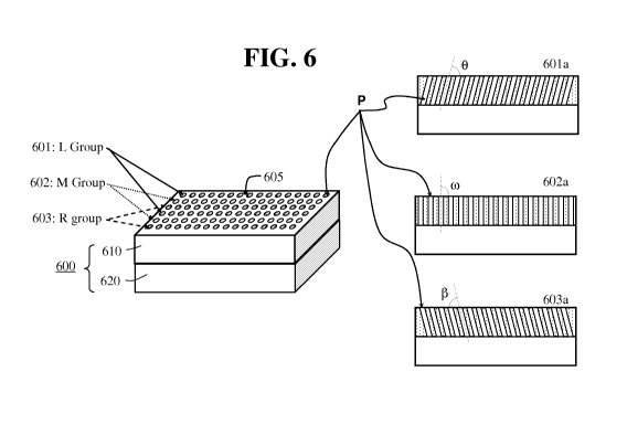
[0053] FIG. 6 illustrates a further modification to the embodiment shown in
FIG. 2.
In accordance with the embodiment of FIG. 6, a radiation detection device 600
includes a
multi-aperture collimator 610 and a detector module 620. Multi-aperture
collimator 610 has
a surface plane 605. A plurality of apertures, e.g., pinholes or parallel
holes, is disposed
throughout the collimator body. The plurality of apertures is selectively
divided into three
groups, and each group is interwoven with the others in a manner similar to
the embodiment
of FIG. 2. The apertures of a first group 601 (L Group), configured to define
a left imaging
view, are arranged with a first orientation angle 0 with respect to the
surface plane 605 of the
collimator. Respectively, a second group 602 (M Group) and a third group (R
Group),
configured to define corresponding middle and right imaging views, may have
corresponding
angles co and R with respect to the surface plane 605 of the collimator. Cross-
sectional views
across a row of apertures in the first, second, and third groups are
represented by reference
numerals 601a, 602a and 603a, respectively.
[0054] In the embodiment of FIG. 6, within the first group 601, second group
602,
and third group 603 all of the apertures P are parallel. More specifically,
within each group,
each of the axes of the plurality of apertures P is parallel to all others.
This particular
embodiment may be advantageous in obtaining further views and/or magnification
levels that
may be useful in obtaining more accurate image reconstruction while
maintaining a compact
size in the detector module. For example, first group 601 may be used for
imaging at a first
predetermined level of magnification, the second group 602 may be utilized for
non-
magnification imaging, e.g., real size imaging, and the third group 603 may be
used for
imaging from different angle and at another predetermined level of
magnification. In other
words, each of the groups may be designed for imaging at a predetermined level
of
magnification, in accordance with the optimized sensitivity and resolution
requirements of a
given system.
CA 02757544 2011-10 03
WO 2010/120525 PCT/US2010/029409
18
II. EXAMPLES OF INTERWOVEN MULTI-APERTURE COLLIMATOR
APPLICATIONS
[0055] FIG. 7 illustrates one possible configuration of a radiation detection
device
700 including an interwoven multi-aperture collimator 710 and a radiation
detector module
720 for 3-D imaging applications. The multi-aperture collimator 710 having a
surface plane
705 includes a 2-D grid of apertures P. The apertures in the grid may be
arranged
orthogonally or in a honeycomb-like arrangement as illustrated in FIGs. 3A and
3B,
respectively. The grid is divided into at least two groups of apertures that
are interwoven and
arranged in accordance with any of the above-described embodiments, or
equivalents thereof.
Detection module 720 may include solid-state detectors or scintillator
detectors configured to
detect radiation beams incoming from an object of interest (not shown) and
transmitted
through the interwoven multi-aperture collimator 710.
[0056] Scintillator detectors include a sensitive volume of a luminescent
material
(liquid or solid) that is viewed by a device that detects the gamma ray-
induced light
emissions (usually a photomultiplier (PMT) or photodiode). The scintillation
material may
be organic or inorganic. Examples of organic scintillators are anthracene and
p-Terphenyl,
but it is not limited thereto. Some common inorganic scintillation materials
are sodium
iodide (Nal), cesium iodide (CsI), zinc sulfide (ZnS), and lithium iodide
(Lil), but it is not
limited thereto. Bismuth germanate (Bi4Ge3O12), commonly referred to BGO, has
become
very popular in applications with high gamma counting efficiency and/or low
neutron
sensitivity requirements. In most clinical SPECT systems, thallium-activated
sodium iodide,
NaI(Tl), is a commonly used scintillator.
[0057] Solid-state detectors include semiconductors that provide direct
conversion of
detected radiation energy into an electronic signal. The gamma ray energy
resolution of these
detectors is dramatically better than that of scintillation detectors. Solid-
state detectors may
comprise a crystal, typically having either a rectangular or circular cross-
section, with a
sensitive thickness selected on the basis of the radiation energy region
relevant to the
application of interest. Solid-state detectors such as cadmium zinc telluride
(CdZnTe or
CZT), cadmium manganese telluride (CdMnTe or CMT), Si, Ge, amorphous selenium,
among others, have been proposed and are well suited for radiation imaging
applications in
which the interwoven multi-aperture collimator may be applied.
CA 02757544 2011-10 03
WO 2010/120525 PCT/US2010/029409
19
[0058] The detector module 720 of FIG. 7 may be based on an orthogonal strip
design. An orthogonal strip detector may be double-sided, as proposed by J.C.
Lund et al. in
"Miniature Gamma-Ray Camera for Tumor Localization", issued by Sandia National
Laboratories (August 1997) which is incorporated by reference herein in its
entirety.
Alternatively, the detector module 720 may be based on an array of single
detector elements
or pixilated detectors.
[0059] In the example of FIG. 7, detector module 720 represents one possible
configuration of a double-sided orthogonal strip design. In the double-sided
orthogonal strip
design, rows and columns of parallel electrical contacts (strips) are placed
at right angles to
each other on opposite sides of a piece of semiconductor wafer. Radiation
detection on the
detector plane is determined by scoring a coincidence event between a column
and a row.
More specifically, when radiation beams emitted from an object of interest
traverse apertures
P of collimator 710, only the radiation beams substantially parallel to the
axis of the aperture
P arrive at a crossing of a column and a row, to thereby generate a signal.
Readout
electronics 750 transmit the received signals to processing and analyzing
equipment in a
known manner.
[0060] Using the orthogonal strip design reduces the complexity of the readout
electronics considerably. In general, to read out an array of N2 detecting
elements only
requires 2 x N channels of readout electronics (750 in FIG. 7), as opposed to
N2 channels
required for an array of NxN individual pixels. The single-sided orthogonal
strip detector
operates on a charge sharing principle using collecting contacts organized in
rows and
columns on only one side of the detector, e.g., the anode surface of a
semiconductor detector.
A single-sided strip detector requires even fewer electronic channels than a
double-sided one.
For example, whereas double-sided detectors require that electrical contacts
be made to the
strips on both sides, single-sided (coplanar) ones use collecting contacts
arranged only on one
side of the detector. Because of the simplicity in design and reduced
complexity of the
readout electronics, detector modules of orthogonal strip design are
considered particularly
advantageous to the application of the various embodiments of the interwoven
multi-aperture
collimator of this invention. However, the applications of the interwoven
multi-aperture
collimator are not limited thereto.
[0061] FIG. 8 illustrates another exemplary application of the interwoven
multi-
aperture collimator. In the embodiment of FIG. 8, a radiation detection device
800 includes
CA 02757544 2011-10 03
WO 2010/120525 PCT/US2010/029409
an interwoven multi-aperture collimator 810 and a detector module 820.
Detector module
820, in this embodiment, includes an array of single detection elements 825.
Radiation
beams (not shown) substantially parallel to the axis of apertures P traverse
collimator 810 and
are detected by individual detection elements 825. Here, the single detection
element 825
may be based on scintillator plus photon-sensing devices or semiconductor
detectors with
various configurations including but not limited to planar detector or the so-
called Frisch-grid
detector design, as proposed by A. E. Bolotnikov et al. in "Optimization of
virtual Frisch-
grid CdZnTe detector designs for imaging and spectroscopy of gamma rays",
Proc. SPIE,
6706, 670603 (2007), which is incorporated by reference herein in its
entirety. Readout
electronics 850 transmit the detected signal to processing and analyzing
equipment in a
known manner.
[0062] FIG. 9 illustrates a further example of a radiation imaging device 900,
including an interwoven multi-aperture collimator 910 and a detector module
920. The
interwoven multi-aperture collimator may be designed in accordance with any of
the
embodiments described in reference to FIGs. 2-6 of the present invention. The
detector
module 920 includes a pixilated detector with a plurality of sensing
electrodes 925, which are
arranged in correspondence with the plurality of apertures P of collimator
910. Here, the
pixilated detector is a semiconductor detector with a common electrode on one
side and an
array of sensing electrodes on the other side. Readout electronics 950
transmit the detected
signal to processing and analyzing equipment in a manner similar to the
examples of FIGs. 7
or 8.
[0063] All publications and patents mentioned in the above specification are
herein
incorporated by reference. Various modifications and variations of the
described interwoven
multi-pinhole collimator will be apparent to those skilled in the art without
departing from the
scope and spirit of the invention. Although the disclosure has been described
in connection
with specific preferred embodiments, it should be understood that the
invention as claimed
should not be unduly limited to such specific embodiments. Indeed, those
skilled in the art
will recognize, or be able to ascertain using no more than routine
experimentation, many
equivalents to the specific embodiments of the invention described herein.
Such equivalents
are intended to be encompassed by the following claims.