Note: Descriptions are shown in the official language in which they were submitted.
CA 02757554 2011-10 03
WO 2010/115072 PCT/US2010/029738
TISSUE ANCHORS AND MEDICAL DEVICES FOR RAPID DEPLOYMENT OF TISSUE
ANCHORS
FIELD
[0001] The present invention relates generally to medical devices, and more
particularly relates to tissue anchors for closing perforations in tissue.
BACKGROUND
[0002] Perforations in bodily walls may be naturally occurring, or formed
intentionally
or unintentionally. In order to permanently close these perforations and allow
the tissue to
properly heal, numerous medical devices and methods have been developed
employing
sutures, adhesives, clips, staples and the like. Many of these devices
typically employ one
or more sutures, the strands of which must be brought together and fixed in
place in order to
close the perforation, and thereafter cut and removed from within the
patient's body.
[0003] Manually tying suture strands together to close a perforation can be
very
complex and time consuming. For example, a significant level of skill and
coordination is
required by the medical professional, especially when the perforation and
sutures are
difficult to access within the body, such as in endoscopic or laparoscopic
procedures. The
numerous difficulties with manually tying and cutting sutures are well
documented. In order
to address these and other issues of manual suture tying and cutting, various
automatic
suture tying systems have been developed. Unfortunately, such automatic
systems are
often complex and costly, difficult to use, and limited to use in certain
situations.
BRIEF SUMMARY
[0004] The present invention provides tissue anchors, as well as related
devices and
methods, for closing perforations in bodily walls. The tissue anchors are
simple and reliable
in use, facilitate perforation closure, and are adaptable to a variety of
perforation closure
situations. One embodiment of a tissue anchor, structured for engagement with
a
tensioning member, for closing a perforation, constructed in accordance with
the teachings
of the present invention, generally comprises a crossbar and a strand. The
crossbar has
first and second opposing ends and defines a longitudinal axis. The strand is
connected to
the crossbar at a location between the opposing ends. The strand has a length
in the range
of about 5 mm to about 50 mm extending from a distal end connected to the
crossbar to a
1
CA 02757554 2011-10 03
WO 2010/115072 PCT/US2010/029738
proximal end having a first connector. The strand and its first connector
project away from
the longitudinal axis.
[0005] Another embodiment of the present invention includes a medical device
for
closing a perforation. The medical device generally comprises a set of tissue
anchors and
an elongate tensioning member. Each tissue anchor includes a crossbar and a
strand. The
crossbar has first and second opposing ends and defines a longitudinal axis.
The strand is
connected to the crossbar at a location between the opposing ends and projects
away from
the longitudinal axis. A distal end of the strand is connected to the crossbar
and a proximal
end of the strand includes a first connector. The elongate tensioning member
is structured
to selectively engage and disengage the first connector. The strands are
capable of being
tensioned and fixed together for closing the perforation.
[0006] According to more detailed aspects of this embodiment of the medical
device,
the strand has a length in the range of about 20 mm to about 30 mm.
[0007] A method for closing a perforation in a bodily wall of a patient is
also provided
in accordance with the teachings of the present invention. A medical device,
such as the
device described above, is provided. Each tissue anchor is passed through the
bodily wall
adjacent the periphery of the perforation such that the crossbar of each
tissue anchor is on a
distal side of the bodily wall and the first connector of each tissue anchor
is on a proximal
side of the bodily wall for selectively engaging and disengaging the elongate
tensioning
member. The elongate tensioning member engages with the first connector of
each strand
and the elongate tensioning member is manipulated to position the strands
close to one
another. The strands are secured together on the proximal side of the bodily
wall. The
elongate tensioning member disengages from the first connector and is removed
from within
the patient.
[0008] A method of securing a set of anchors under tension is also provided in
accordance with further teachings of the present invention. Each anchor
includes a
crossbar and a strand connected to the cross bar. The strand has a distal end
connected to
the crossbar. A proximal end of the strand includes a first connector. An
elongate
tensioning member connects to the first connector of each strand within a
lumen of a tubular
member. The tubular member is moved relative to the elongate tensioning member
to
position the elongate tensioning member and the set of anchors distally beyond
a distal end
of the tubular member. The elongate tensioning member is manipulated to
tension the
strands together and the strands are then secured together while under
tension. The
elongate tensioning member is then disengaged from the first connector of the
secured
strands.
2
CA 02757554 2011-10 03
WO 2010/115072 PCT/US2010/029738
BRIEF DESCRIPTION OF THE DRAWINGS
[0009] The accompanying drawings incorporated in and forming a part of the
specification illustrate several aspects of the present invention, and
together with the
description serve to explain the principles of the invention. In the drawings:
[0010] FIG. la is a front view of one embodiment of a tissue anchor
constructed in
accordance with the teachings of the present invention;
[0011] FIG. lb is a front view of yet another embodiment of a tissue anchor
constructed in accordance with the teachings of the present invention;
[0012] FIG. 2a is front view of one embodiment of a medical device constructed
in
accordance with the teachings of the present invention;
[0013] FIG. 2b is a front view of another embodiment of a medical device
constructed in accordance with the teachings of the present invention;
[0014] FIG. 2c is a front view of yet another embodiment of a medical device
constructed in accordance with the teachings of the present invention;
[0015] FIG. 3 is a cross-sectional view of tissue anchors, in accordance with
the
teachings of the present invention, shown closing a perforation;
[0016] FIG. 4 is a plan view, partially in cross-section, of a medical
delivery device
constructed in accordance with the teachings of the present invention; and
[0017] FIGS. 5-9 depict steps in a method for using a medical device in
accordance
with the teachings of the present invention.
DETAILED DESCRIPTION OF THE INVENTION
[0018] Turning now to the figures, FIG. 1 a depicts a tissue anchor 20
constructed in
accordance with the teachings of the present invention. The anchor 20 is
utilized for closing
a perforation 10 in a bodily wall 12 (FIGS. 5-7). The anchor 20 may also be
used for
apposing tissue, for example, in gastroesophageal reflux disease (GERD)
therapy, or
bariatric surgery in which an anastamosis is formed, or for use in other
procedures. The
anchor 20 generally includes a crossbar 24 having opposing ends 26 and 28 and
defining a
longitudinal axis 22. A flexible strand 30 is connected to the crossbar 24 at
a location
between the opposing ends 26 and 28 of the crossbar 24. The strand 30 includes
a distal
end 32 connected to the crossbar 24 and extending away from the longitudinal
axis 22 of
the crossbar 24 to a proximal end 34 which terminates in a connector 36,
discussed in
further detail below.
3
CA 02757554 2011-10 03
WO 2010/115072 PCT/US2010/029738
[0019] The crossbar 24 is preferably elongated, but may take any form suitable
for
closing the perforation 10 in the bodily wall 12, including rods, tubes, disc
shapes or other
elongated or planar shaped members. The crossbar 24 is preferably formed of a
tubular
cannula, although the crossbar 24 may be a solid cylinder, a metal bar, a
plastic molded
piece, or any stock materials. The strand 30 is preferably formed from a
flexible suture
material, although the strand 30 can have other constructions such as a metal
wire,
including single filament and multi-filament wires, and wound and braided
wires, plastic
strings, rope and the like.
[0020] It will be recognized by those skilled in the art that the strand 30
may be
secured to the crossbar 24 using any now known or hereinafter developed
attachment
means, including mechanical fasteners, adhesives or various welding or
soldering
techniques. In one preferred construction, the crossbar 24 is formed of a
cannula having an
opening formed therethrough between opposing ends 26 and 28 and the distal end
32 of the
strand 30 is received within the opening in the cannula and crimped in place.
Alternatively,
the strand 30 may be unitarily and integrally formed with the crossbar 24 as a
single piece.
Accordingly, the entire tissue anchor 20 may be formed of a single plastic or
metal material,
and most preferably a resorbable material. For example, the anchor 20,
including the
crossbar 24 and strand 30, may be injection molded of a permanent material,
such as nylon,
or a resorbable material. The material of the anchor 20 could also be made
radiopaque or
echogenic, e.g., by embedding particles within the plastic or selecting a
suitable material
having inherent or formed radiopaque or echogenic properties.
[0021] As used herein, the term "resorbable" refers to the ability of a
material to be
absorbed into a tissue and/or body fluid upon contact with the tissue and/or
body fluid. A
number of resorbable materials are known in the art, and any suitable
resorbable material
can be used. Examples of suitable types of resorbable materials include
resorbable
homopolymers, copolymers, or blends of resorbable polymers. Specific examples
of suitable
resorbable materials include poly-alpha hydroxy acids such as polylactic acid,
polylactide,
polyglycolic acid (PGA), or polyglycolide; tri- methlyene carbonate;
polycaprolactone; poly-
beta hydroxy acids such as polyhydroxybutyrate or polyhydroxyvalerate; or
other polymers
such as polyphosphazines, polyorgano- phosphazines, polyanhydrides,
polyesteramides,
poly- orthoesters, polyethylene oxide, polyester-ethers (e.g., poly-
dioxanone) or polyamino
acids (e.g., poly-L-glutamic acid or poly-L-lysine). There are also a number
of naturally
derived resorbable polymers that may be suitable, including modified
polysaccharides, such
as cellulose, chitin, and dextran, and modified proteins, such as fibrin and
casein.
4
CA 02757554 2011-10 03
WO 2010/115072 PCT/US2010/029738
[0022] The strand 30 preferably has a length in the range of about 5 mm to
about 50
mm, and most preferably about 20 mm to about 30 mm. The strand 30 preferably
has a
diameter less than about 50% of a diameter of the crossbar 24, and most
preferably less
than about 35%. The strand 30 preferably has a diameter in the range of about
0.20 mm to
about 0.35 mm, and most preferably about 0.254 mm. The crossbar 24 preferably
has a
diameter in the range of about 0.5 mm to about 1.0 mm, and most preferably
about 0.8 mm.
The crossbar 24 typically has a length in the range of about 3.0 mm to about
10.0 mm. The
crossbar 24 and/or the strand 30 may be coated with a low-friction material
such as known
plastic or hydrophilic coatings.
[0023] As illustrated in FIG. la, the connector 36 most preferably takes the
form of
an eyelet or closed loop at the proximal end 34 of the strand 30. The closed
loop connector
36 preferably has a diameter in the range of about 0.5 mm to about 1.5 mm. The
connector
36 can have an alternative shape, such as that depicted in FIGS. 1b. FIG. lb
depicts an
embodiment of a tissue anchor 220 in accordance with the teachings of the
present
invention and having a description similar to that of FIG. la, and in which
similar
components are denoted by similar reference numerals increased by 200. In this
embodiment, the connector 236 takes the form of a J-shaped hook.
[0024] The connector 36 is structured to receive a tensioning member 38 (FIGS.
2a-
c) for aiding in deployment of the anchor 20. The connector 36 may be formed
from a
flexible suture material, metal wire, plastic, rope or any suitable resorbable
material. As
shown in FIG. la, the closed loop connector 36 is preferably formed integrally
with the
strand 30. The closed loop connector 36 is flexible and is capable of
stretching or adjusting
its shape and orientation when tensioned by the tensioning member 38.
Alternatively, the
connector 36 may be formed from a different material than the strand 30, may
have a
greater thickness than the strand 30, and may be secured to the strand 30
using any
suitable means, including mechanical fasteners, tying, bonding, welding, or
adhesives. For
example, in order for the J-shaped hook connector 236 of FIG. lb to maintain
its shape
when tensioned by a tensioning member, the connector 236 is preferably made of
a
stronger, more rigid material than the strand 230 such as a rigid plastic or
metal material. In
addition, the connector 36 preferably has a smooth, rounded shape so that the
anchor 20 is
atraumatic to other surrounding tissues.
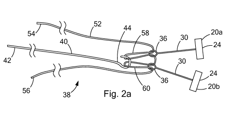
[0025] Turning now to FIGS. 2a-c, a pair of tissue anchors 20a and 20b is
depicted
with various embodiments of tensioning members 38, 138, and 238 configured to
selectively
engage and disengage the connectors 36. As depicted in FIG. 2a, the tensioning
member
38 includes a suture 52 in combination with an elongate holding member 40. The
suture 52
CA 02757554 2011-10 03
WO 2010/115072 PCT/US2010/029738
and elongate holding member 40 of FIG. 2a are shown in use with anchors 20a
and 20b
having connectors 36 in the form of a closed loop, as depicted in the
embodiment of FIG.
1 a.
[0026] The suture 52 includes first and second ends 54 and 56 which are
preferably
located and maintained outside of the body. The first and second ends 54 and
56 can be
fixed within a handle (not shown) at a proximal end of the tissue anchor
delivery device
(FIG. 4), for example, by a tuohy borst connector or clamp. The suture 52 is
preferably pre-
loaded through the closed loop connectors 36 such that a section of the suture
52 is pulled
through each of the closed loops connectors 36 and maintained by the elongate
holding
member 40. Thus, the suture 52 preferably passes through each closed loop
connectors 36
twice to form folded over looped sections 58 and 60. These folded over looped
sections 58
and 60 are of a sufficient length to provide adequate slack in the suture 52
during distal
advancement and separate positioning of the tissue anchors 20a and 20b.
[0027] As depicted in FIG. 2a, the suture 52 extends from the first end 54 and
is
folded over to form the first looped section 58; the suture 52 extends from
the first looped
section 58 and is folded over to form the second looped section 60; and the
suture 52
extends from the second looped section 60 to the second end 56. The first
looped section
58 is slidably received within the closed loop connector 36 of the first
anchor 20a of the pair
of anchors and the second looped section 60 is slidably received within the
closed loop
connector 36 of the second anchor 20b of the pair of anchors. The elongate
holding
member 40 includes a proximal end 42 and a distal end 44 terminating with a
connector 46
structured to selectively engage and disengage the first and second looped
sections 58 and
60 of the suture 52. The suture 52 may be of a single filament or multi-
filament
constructions and preferably has a diameter in the range of about 0.20 mm to
about 0.35
mm.
[0028] It will be recognized by those skilled in the art that the connector 46
may take
on any suitable shape or form suitable for selective engagement and
disengagement with
the looped sections 58 and 60. Preferably, the connector 46 at the distal end
44 of the
elongate member 40 is hook shaped, e.g., a J-shaped hook as shown.
[0029] As depicted in the alternate embodiment of FIG. 2b, the tensioning
member
138 includes a suture 152 having first and second ends 154 and 156 which
extend through
the tissue anchor delivery device and are located and maintained outside of
the body
proximate a proximal end of the delivery device for manipulation by the
physician. In this
embodiment, the suture 152 extends from the first end 154 through the first
and second
closed loop connectors 36 of the pair of anchors 20a and 20b, respectively.
The tensioning
6
CA 02757554 2011-10 03
WO 2010/115072 PCT/US2010/029738
member 138 including the suture 152 may be used in conjunction with anchors
having
connectors other than the closed loop connectors 36 illustrated FIG. 2b. For
example, the
suture 152 may selectively engage hooked connectors, such as the J-shaped hook
connector 236 illustrated in FIG. 1 b, in which the suture 152 would be
threaded through the
hooked connector 236. The suture 152 may be of a single filament or multi-
filament
constructions.
[0030] As depicted in the alternate embodiment of FIG. 2c, the tensioning
member
238 includes an elongate holding member 240 having a proximal end 242 and a
distal end
244 terminating with a connector 246 structured to selectively engage and
directly
disengage the first and second closed loop connectors 36 of the pair of
anchors 20a and
20b, respectively. Preferably, the connector 246 at the distal end 244 of the
elongate
member 240 is a J-shaped hook. The elongate holding member 240 having the
connector
246 may be used in conjunction with anchors having connectors other than the
closed loop
connectors 36 illustrated in FIG. 2c. For example, the connector 246 of the
holding member
240 may selectively engage a hook shaped connector, such as the J-shaped
hooked
connector 236 of FIG. 1 b. It will be recognized by those skilled in the art
that the connector
246 may take on any suitable shape or form suitable for selective engagement
and
disengagement with the various connectors 36. For example, the connector 246
could be a
ringlet or closed loop, while the connectors 36 could be hook shaped (see FIG.
1 b). These
and other variations will be readily apparent to the skilled artisan.
[0031] Turning now to FIGS. 3 and 4, the tissue anchors 20 are preferably
deployed
as a set of anchors including at least two anchors 20a and 20b. The tensioning
member 38
aids in positioning the anchors 20a and 20b during delivery of the anchors 20a
and 20b
through tissue of the bodily wall 12 and is removed thereafter, as will be
described in more
detail below. As best seen in FIG. 3, the crossbars 24 of the anchors 20a and
20b are
positioned on a distal side of the bodily wall 12, while the majority of the
strands 30
connected to the crossbars 24, including the connectors 36, are positioned on
a proximal
side of the bodily wall. After the anchors 20a and 20b are positioned through
the tissue of
the bodily wall 12, the strands 30 of the anchors 20a and 20b are brought
together, via the
tensioning member 38, and fixed in place, via a suture lock 62, in order to
close the
perforation 10. Thereafter, the tensioning member 38 is removed. Thus, rather
than
employing a separate suture or a plurality of individual sutures to fix the
anchors to the
bodily wall (which must be fixed in place in order to close the perforation,
and thereafter
manually cut and removed) the anchors 20a and 20b are fixed together directly,
via the
strands 30 and the suture lock 62. Thus, the construction of the anchors 20a
and 20b,
7
CA 02757554 2011-10 03
WO 2010/115072 PCT/US2010/029738
including strands 30 with connectors 36, alleviates the many difficulties
associated with
manually cutting sutures used to fix the anchors together.
[0032] Referring to FIG. 4, a medical delivery device 100 for employing the
anchors
20a and 20b and the lock 62, in accordance with the teachings of the present
invention,
includes a needle 86 having a needle lumen 88 sized to slidably receive the
tissue anchors
20a and 20b. Preferably, a resorbable spacer member 50 is positioned between
the
anchors 20a and 20b within the needle lumen 88 near the distal end 89 of the
needle 86.
The needle 86 includes a needle slot 87 sized to receive the strands 30 of the
anchors 20a
and 20b. The medical delivery device 100 may include a set of more than two
anchors and
more than one spacer members. The longitudinal length of the needle slot 87 is
dependent
upon the number of anchors and the length of the crossbars and the number of
spacer
members positioned between the anchors and the length of the spacer members
such that
the needle slot 87 is capable of receiving the strands 30 connected to each of
the crossbars
24 positioned within the needle lumen 88. A pusher 98 is slidably received
within the needle
lumen 88 to engage the proximal-most anchor, the anchor 20b in FIG. 4, and
deploy the
anchors 20a and 20b, and the spacer member 50 positioned therebetween, from
the
delivery device 100.
[0033] The medical device 100 further includes an inner sheath 90 having a
lumen
92 sized to slidably receive the needle 86 and an outer sheath 94 having a
lumen 96 sized
to slidably receive the inner sheath 90. The strands 30 of the anchors 20a and
20b extend
away from the longitudinal axis 22 of the crossbars 24 of the anchors 20a and
20b, through
the needle slot 87 and proximally within the outer sheath lumen 96. A
tensioning member
38, in accordance with the teachings of the present invention, is slidably
received within the
outer sheath lumen 96 to selectively engage and disengage the connectors 36 of
the
strands 30 of the anchors 20a and 20b. While the tensioning member 38 of FIG.
2a,
including the suture 52 and the elongate holding member 40, is illustrated as
part of the
medical delivery device 100 in FIGS. 4-9, the tensioning members 138 or 238 of
FIGS. 2b
and 2c, respectively, may be employed as part of the medical delivery device
100. Similarly,
the anchor 220 of FIG. lb having a J-shaped connector 236 may be delivered
using the
medical delivery device 100 in accordance with the teachings of the present
invention.
[0034] The inner and outer sheaths 90 and 94 are preferably formed of a
plastic
such as polytetrafluorethylene (PTFE), expanded polytetrafluorethylene
(EPTFE),
polyethylene ether ketone (PEEK), polyvinylchloride (PVC), polycarbonate (PC),
polyamide
including nylon, polyimide, polyurethane, polyethylene (high, medium or low
density), or
elastomers such as Santoprene , including multi-layer or single layer
constructions with or
8
CA 02757554 2011-10 03
WO 2010/115072 PCT/US2010/029738
without reinforcement wires, coils or filaments. The needle 86, inner and
outer sheaths 94
and 90, the pusher 98, and the tensioning member 38, including the suture 52
and the
holding member 40, are preferably elongated structures that are flexible,
allowing navigation
within a patient's body such as during endoscopic or laparoscopic procedures.
As such, a
suitable handle or control mechanism will be connected to the proximal ends of
the needle
86, sheaths 90 and 94, and pusher 98 for relative translation of these
components by the
medical professional, as is known in the art.
[0035] Preferably, the medical device 100 further includes an over-the-needle
suture
lock 62 for fixing the strands 30 of the anchors 20a and 20b after delivery of
the anchors 20a
and 20b through the bodily wall 12. An over-the-needle suture lock 62, in
accordance with
the teachings of the present invention, allows the strands 30 of the set of
anchors 20a and
20b to be preloaded within the suture lock 62 during delivery of the anchors
20a and 20b
through the bodily wall 12. The suture lock 62 generally includes a locking
pin or plug 64
and a retaining sleeve 66 which cooperate to fix the strands 30 of the anchors
20a and 20b
relative to tissue of the bodily wall 12 for closing the perforation 10 in the
bodily wall 12. The
retaining sleeve 66 and plug 64 may have a circular cross-section, or any
other cross-
sectional shapes including triangular, square, etc.
[0036] As best seen in FIGS. 4-9, the retaining sleeve 66 generally includes a
tubular body 68 having an interior surface 69 defining an interior passageway
70. A
peripheral rim 72 is formed at a distal end of the tubular body 68, and
defines a shoulder 74
which is used for placement of the retaining sleeve 68, as will be discussed
in further detail
herein. Generally, the retaining sleeve 68 receives the strands 30 of the
anchors 20a and
20b within the interior passageway 70. The strands 30 are then fixed in place
using the plug
64, which is designed to fit within the passageway 70 and pinch or compress
the strands 30
of the anchors 20a and 20b. It will also be recognized that the plug 64 may
have many
configurations (e.g. regular or irregular shapes), and constructions (e.g.
cast, molded,
machined, wound (such as with wire), etc.) so long as a portion of the plug 64
cooperates
with the retaining sleeve 66 to fix the strands 30. Preferably, the plug 64
and the retaining
sleeve 66 are formed from stainless steel or any other suitable metal or
plastic material
known in the art.
[0037] As best seen in FIGS. 4-9, the plug 64 generally includes a main body
76
having an interior surface 77 defining an interior passageway 78 sized to
slidably receive the
needle 86. The main body 76 includes a grip 80 and a stop 82, each extending
radially from
the main body 76. In the illustrated embodiment, the grip 80 is formed at a
distal end of the
plug 64, although it could be moved proximally along the length of the main
body 76. The
9
CA 02757554 2011-10 03
WO 2010/115072 PCT/US2010/029738
grip 80 defines an annular edge 79 that is used to engage the strands 30 of
the anchors 20a
and 20b. The stop 82 is longitudinally spaced from the grip 80 and is used to
control the
position of the plug 64 within the retaining sleeve 66. The stop 82 generally
includes a
proximally facing surface 83 defining a shoulder 84 which is used to position
the plug 64.
The stop 82 is positioned relative to the grip 80 to prevent the grip 80 from
passing
completely through the internal passageway 70 of the retaining sleeve 66.
While the length
of the strands 30 of the tissue anchors 20 is preferably between about 5 mm
and 50 mm,
the length of the strands 30 should be such that the connectors 36 of the
strands 30 exit the
suture lock 62 to provide sufficient engagement of the strands 30 between the
plug 64 and
retaining sleeve 66.
[0038] As depicted in FIGS. 4-7 the inner sheath 90 is sized and positioned to
abut
the shoulder 84 of the plug 64 and the outer sheath 94 is sized and positioned
to abut the
shoulder 74 of the retaining sleeve 66. Translation of the inner sheath 90
relative to the
outer sheath 94 causes the plug 64 to slide over-the-needle 86 and to be
received within the
passageway 78 of the retaining sleeve 66 to engage the strands 30 of the
anchors 20a and
20b between the main body 76 of the plug 64 and the interior surface 77 of the
retaining
sleeve 66 to fix the strands 30.
[0039] Further details of the needle assembly and the over-the-needle suture
lock 62
may be found in U.S. Provisional Application No. 61/166,361 entitled "Medical
Devices,
Systems, and Methods for Rapid Deployment and Fixation of Tissue Anchors" to
Ducharme,
the entire contents of which are incorporated by reference herein.
[0040] The medical device 100 may be sized to be used through an accessory
channel of an endoscope or alongside an endoscope, or in combination with
other devices
used in conjunction with endoscopes, for example, endoscopic suction devices
or fluid
injection devices.
[0041] A method of closing the perforation 10, in accordance with the
teachings
present invention, includes passing each tissue anchor 20a and 20b through the
tissue of
the bodily wall 12 adjacent the periphery of the perforation 10, as shown in
FIGS. 5-7.
Preferably, the anchors are sequentially positioned around the perforation 10.
As shown,
the anchors 20a and 20b are positioned on opposing sides of the perforation
10. As
previously noted, a plurality of anchors including more than the two anchors
20a and 20b
may be sequentially positioned around the perforation 10.
[0042] As illustrated in FIG. 5, the medical delivery device 100, in
accordance with
the teachings of the present invention, is delivered to a position proximate
the tissue of the
bodily wall 12. The anchors 20a and 20b are disposed within the needle lumen
88 at the
CA 02757554 2011-10 03
WO 2010/115072 PCT/US2010/029738
distal end 89 of the needle 86 and a resorbable spacer member 50 is disposed
between the
anchors 20a and 20b. Spaces between the spacer member 50 and the anchors 20a
and
20b have been shown for clarity, but the spacer member 50 and the anchors 20a
and 20b
would generally be abutting end-to-end within the needle lumen 88. The strands
30 of the
anchors 20a and 20b are received within the needle slot 87 and project away
from the
longitudinal axis 22 of the anchor crossbars 24. Preferably, the needle 86 is
slidably
received within the inner sheath lumen 92, and the tensioning member 38 is pre-
engaged
with the connectors 36 of the anchor strands 30 extending from the needle
lumen 88, prior
to being slidably received within the outer sheath lumen 96. Thus, the needle
86 within the
inner sheath 90 and the tensioning member 38 are preferably pre-engaged and
loaded into
the outer sheath 94 together and advanced toward a distal end 95 of the outer
sheath 96
prior to positioning of the medical delivery device 100. Thus, as depicted in
FIGS. 4-7, the
tensioning member 38 is disposed within the outer sheath lumen 96 and is in
selective
engagement with the connectors 36 of the strands 30 of the anchors 20a and
20b.
[0043] The method illustrated in FIGS. 5-7 depicts a medical delivery device
100
including, as an example, the tensioning member 38 of FIGS. 2a. In this
example, once
positioned near the distal end 95 of the outer sheath 96, the elongate holding
member 40
maintains its hold on the connectors 36 of the anchor strands 30 via the first
and second
looped sections 58 and 60 of the suture 52. The first and second ends 54 and
56 of the
suture 52 are preferably maintained within a proximal handle (not shown) of
the device 100
while the physician maintains a hold of and manipulates the elongate holding
member 40.
The folded over looped sections 58 and 60 are preferably of a sufficient
length to provide
adequate slack in the suture 52 during separate positioning of the tissue
anchors 20a and
20b.
[0044] As illustrated in FIG. 5-6, the needle 86 is deployed through the
tissue of the
bodily wall 12 by translating the needle 86 relative to the inner and outer
sheaths 90 and 94.
The distal-most tissue anchor, the anchor 20a in FIGS. 4-5, is then deployed
from the
needle 86 by translating the anchor 20a relative to the needle 86 so that the
anchor 20a
exits the needle lumen 88. As shown in FIGS. 4-5, the anchors 20a and 20b, and
the
spacer member 50 positioned therebetween, are shown aligned within the needle
lumen 88
along a longitudinal axis of the needle lumen 88 such that the pusher 98 may
be slidably
received within the inner sheath lumen 92 and used to engage and press on the
proximal-
most anchor, anchor 20b in FIGS. 4-7, which will in turn transmit force
through the spacer
member 50 and the distal-most anchor 20a, thus advancing the distal-most
anchor 20a out
11
CA 02757554 2011-10 03
WO 2010/115072 PCT/US2010/029738
of the needle lumen 88. As the anchor 20a exits the needle lumen 88, the
strand 30 of the
anchor 20a is released from the needle slot 87.
[0045] Accordingly, it will be recognized that a large number of tissue
anchors and
spacer members may be employed within the medical device 100, and the
longitudinal
length of needle slot 87 can be sized to accommodate any number of anchor
strands 30. In
this manner, the medical device 100 need not be withdrawn to be reloaded. The
method
may therefore include withdrawing the needle 86 from the bodily wall by
translating the
needle 86 proximally, and then repeating the steps of translating the needle
86 through the
tissue 12 and deploying a tissue anchor therethrough.
[0046] Turning to FIG. 7, the needle 86 is retracted back through the bodily
wall 12
by translating the needle 86 proximally, repositioned at a different position
about the
perforation 10, and redeployed back through the tissue of the bodily wall 12
by translating
the needle 86 relative to the inner and outer sheaths 90 and 94. The pusher 98
is then
further advanced distally to deploy the spacer member 50 and the proximal
anchor 20b,
wherein the strand 30 of the anchor 20b is released from within the needle
slot 87. The
spacer member 50 is then resorbed within the patient's body.
[0047] After the anchors 20a and 20b are deployed on the distal side of the
bodily
wall 12, the needle 86 is retracted back through to the proximal side of the
bodily wall 12
and removed from within the inner sheath lumen 92. The elongate holding member
40 is
used to tension the strands 30 of the anchors 20a and 20b to bring the strands
30 together
to close the perforation 10. Preferably, the elongate holding member 40 is
retracted,
applying a pulling force on the first and second looped sections 58 and 60 of
the suture 52,
which in turn applies a pulling force on the connectors 36, thus tensioning
the strands 30 of
the anchors 20a and 20b to reduce the distance between the anchors 20a and 20b
and
compress the bodily wall 12 around the perforation 10, as depicted in FIGS. 8
and 9.
[0048] As best seen in FIG. 9, the strands 30 of the anchors 20a and 20b are
secured to maintain the compression of the bodily wall 10, such as through the
use of a
suture lock. Preferably, the stands 30 are fixed through the use of an over-
the-needle
suture lock 62, in accordance with the teachings of the present invention,
described in detail
above and illustrated in FIGS. 4-9. Alternatively, other suture locks may be
employed to fix
the strands 30, such as the suture locks disclosed in copending U.S. Patent
Application
Nos. 12/125,525 and 12/191,001, the disclosures of which are incorporated
herein by
reference in their entirety. It will be recognized that any now known or
future developed
method for securing the strands 30 of the anchors 20a and 20b may be employed,
such as
knotting, tying, clamps, rivets and the like. After the anchors 20a and 20b
have effectively
12
CA 02757554 2011-10 03
WO 2010/115072 PCT/US2010/029738
closed the perforation 10 in the bodily wall 12 and the stands 30 are secured,
the delivery
device 100, including the tensioning member 38, is removed from within the
patient. In the
example in FIGS. 8-9, the holding member 40 is unhooked from the first and
second looped
sections 58 and 60 and the suture 52 is removed by opening or releasing the
tuohy borst
connector or clamp of the proximal handle of the delivery device and pulling
one end of the
suture 52 while releasing the opposing end of the suture. In this manner, the
suture 52
slides through the closed loop connectors 36 as it is removed from the
patient.
[0049] It will be recognized by those skilled in the art that, while the
methods
described above generally include placing the tissue anchors in tissue through
an internal
bodily lumen, it will be recognized that the devices and methods may be used
on any layer
of material (e.g. fabrics, cloth, polymers, elastomers, plastics and rubber)
that may or may
not be associated with a human or animal body and a bodily lumen. For example,
the
devices and methods disclosed herein can find use in laboratory and industrial
settings for
placing devices through one or more layers of material that may or may not
find application
to the human or animal body, and likewise closing holes or perforations in
layers of material
that are not bodily tissue. Some examples include sewing or stitching and
related
manufacturing, working with synthetic tissues, connecting polymeric sheets,
animal studies,
and post-mortem activities.
[0050] The foregoing description of various embodiments of the invention has
been
presented for purposes of illustration and description. It is not intended to
be exhaustive or
to limit the invention to the precise embodiments disclosed. Numerous
modifications or
variations are possible in light of the above teachings. The embodiments
discussed were
chosen and described to provide the best illustration of the principles of the
invention and its
practical application to thereby enable one of ordinary skill in the art to
utilize the invention in
various embodiments and with various modifications as are suited to the
particular use
contemplated. All such modifications and variations are within the scope of
the invention as
determined by the appended claims when interpreted in accordance with the
breadth to
which they are fairly, legally, and equitably entitled.
13