Note: Descriptions are shown in the official language in which they were submitted.
CA 02757574 2014-06-13
Inhibitors of Bacterial Type III Secretion System
Cross-Reference to Priority Applications
This application claims priority to US Provisional Appin. No. 61/212,021 filed
April 6,
2009, US Provisional Appin. No. 61/304,305 filed February 12, 2010, and US
Provisional
Appin. No. 61/304,978 filed February 16, 2010.
Field of the Invention
This invention is in the field of therapeutic drugs to treat bacterial
infection and disease.
In particular, the invention provides organic compounds that inhibit the type
III secretion
system of one or more bacterial species.
Background of the Invention
The bacterial type III secretion system (T3SS) is a complex multi-protein
apparatus that
facilitates the secretion and translocation of effector proteins from the
bacterial cytoplasm
directly into the mammalian cytosol. This complex protein delivery device is
shared by over
species of Gram-negative human pathogens, including Salmonella spp., Shigella
flexneri,
15 Pseudomonas aeruginosa, Yersinia spp., enteropathogenic and
enteroinvasive Escherichia coli,
and Chlamydia spp. (23, 25, 43). In the opportunistic pathogen P. aeruginosa,
the T3SS is the
major virulence factor contributing to the establishment and dissemination of
acute infections
(19). Four T3SS effectors have been identified in P. aeruginosa strains ¨
ExoS, ExoT, ExoY,
and ExoU. ExoS and ExoT are bifunctional proteins consisting of an N-terminal
small G-
protein activating protein (GAP) domain and a C-terminal ADP ribosylation
domain; ExoY is
an adenylate cyclase; and ExoU is a phospholipase [reviewed in (11)]. In
studies with strains
producing each effector separately, ExoU and ExoS contributed significantly to
persistence,
dissemination, and mortality while ExoT produced minor effects on virulence in
a mouse lung
infection model, and ExoY did not appear to play a major role in the
pathogenesis of P.
aeruginosa (51). While not a prototypical effector toxin, flagellin (FliC) may
also be injected
into the cytoplasm of host cells from P. aeruginosa via the T3SS machinery
where it triggers
1
CA 02757574 2011 10 03
WO 2010/118046 PCT/US2010/030120
activation of the innate immune system through the nod-like receptor NLRC4
inflammasome (13, 33).
The presence of a functional T3SS is significantly associated with poor
clinical
outcomes and death in patients with lower respiratory and systemic infections
caused by P.
aeruginosa (48). In addition, T3SS reduces survival in P. aeruginosa animal
infection
models (49), and is required for the systemic dissemination of P. aeruginosa
in a murine
acute pneumonia infection model (56). T3SS appears to contribute to the
development of
severe pneumonia by inhibiting the ability of the host to contain and clear
bacterial
infection of the lung. Secretion of T355 toxins, particularly ExoU, blocks
phagocyte-
mediated clearance at the site of infection and facilitates establishment of
an infection (9).
The result is a local disruption of an essential component of the innate
immune response,
which creates an environment of immunosuppression in the lung. This not only
allows P.
aeruginosa to persist in the lung, but it also facilitates superinfection with
other species of
bacteria.
While several antibacterial agents are effective against P. aeruginosa, the
high rates
of mortality and relapse associated with serious P. aeruginosa infections,
even in patients
with hospital-acquired pneumonia (HAP) receiving antibiotics active against
the causative
strain, reflect the increasing incidence of drug-resistant strains and
highlights the need for
new therapeutic agents (10, 46, 52). Conventional bacteriostatic and
bactericidal
antibiotics appear insufficient to adequately combat these infections, and new
treatment
approaches such as inhibitors of P. aeruginosa virulence determinants may
prove useful as
adjunctive therapies (58).
The potential for T3SS as a therapeutic target has prompted several groups to
screen for inhibitors of T355 in various bacterial species, including
Salmonella
typhimurium, Yersinia pestis, Y. pseudotuberculosis, and E. coli [reviewed in
(5, 25)].
However, only a single screen for inhibitors of P. aeruginosa T355 inhibitors
has been
reported, and it yielded specific inhibitors of one of the T355 effectors,
ExoU (27) rather
than inhibitors of the T355 machinery. High levels of sequence conservation
among
various proteins comprising the T355 apparatus suggest that inhibitors of T355
in one
species may also be active in related species. Broad spectrum activity of T355
inhibitors
identified in a screen against Yersinia has been demonstrated in Salmonella,
Shigella, and
Chlamydia (22, 57, 59).
2
CA 02757574 2014-06-13
Clearly, needs remain for new, potent inhibitors of bacterial T3SS of P.
aeruginosa and
other bacterial species.
Summary of the Invention
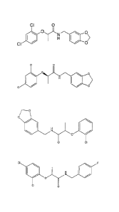
Various embodiments of this invention provide a compound of the formula:
CI 0
CI 1101
40 0>
0
0
ON 0
=
CI
f-------o
1401
0
0 CI
CA 02757574 2013-08-22
CI *ON
CI 0
0
H _______________________________
N
______________________________________ 0 CI
BrCi
I \
ON
0
0
0/¨\CI
i
HN
0 ___________________________________________ 0
3a
CA 02757574 2014-06-13
=
CI
oi
No
\ I
CI
or
CI
HN ____________________________________
F
Various embodiments of this invention provide a compound of the formula:
CI 0
=
oN 0
>
CI
Or 0
3b
CA 02757574 2014-06-13
Various embodiments of this invention provide a compound of the formula:
ci 0
0
0)
CI =
Various embodiments of this invention provide use of a compound as described
above, to
inhibit a bacterial type III secretion system.
Various embodiments of this invention provide use of a compound as described
above in
treatment of a Gram-negative bacterial infection or in manufacture of a
medicament for such
treating.
Various embodiments of this invention provide use of a compound of the
formula:
ci
=
for treatment of a Gram-negative bacterial infection. The use may be in
manufacture of a
medicament for such treating.
The invention addresses the above problems by providing new bacterial type III
secretion
system (T3SS) inhibitor compounds. To identify T3SS inhibitory compounds
described herein, a
cell-based bioluminescent reporter assay was developed and employed as a high
throughput
primary screen to identify putative inhibitors of the P. aeruginosa T3SS from
libraries of thousands
of organic compounds. The putative T3SS inhibitor compounds ("hits") from the
high throughput
primary screen were then qualified through a series of secondary assays.
Accordingly, a T3SS
inhibitor described herein inhibits T3SS-mediated secretion of a bacterial
exotoxin (effector) from a
bacterial cell. More preferably, a T3SS inhibitor compound described herein
inhibits T3SS-
mediated secretion of an effector from a bacterial cell and also inhibits T3SS-
mediated
translocation of the effector from the bacterial cell to a host cell (e.g.,
human or other animal cell).
3c
CA 02757574 2013-08-22
In a preferred embodiment, a T3SS inhibitor compound described herein inhibits
the
T3SS in a bacterium of the genus Pseudomonas, Yersinia, or Chlamydia.
In another embodiment, a T3SS inhibitor compound described herein inhibits the
T3SS
of Pseudomonas and the T3SS of a bacterium of at least one other genus.
Preferably, the
inhibition target Pseudomonas bacterium is P. aeruginosa. Preferably, the
other bacterial
genus susceptible to T3SS inhibition by compound(s) of the invention is
Yersinia or
Chlamydia. A preferred inhibition target species of Yersinia is Y. pestis. A
preferred inhibition
target species of Chlamydia is C. trachomatis.
The present invention provides several specific bacterial T3SS inhibitor
compounds,
listed below by structure, manufacturer's designation, and chemical name:
CI 0
0\
/
CI 0
compound 1 (ChemBridge 5690431; Microbiotix MBX 1641; racemate)
N-(benzo[d][1,3]dioxo1-5-ylmethyl)-2-(2,4-dichlorophenoxy)propanamide
3d
CA 02757574 2011 10 03
WO 2010/118046 PCT/US2010/030120
H 0
le N,(
N-N
0 S-4
N
compound 2 (TimTec 7803985)
2-(6-oxo-5,6-dihydrothiazolo[3,2-b][1,2,4]triazol-5-y1)-N-phenylacetamide
0
H
0 N ./"/'' N
0 0 0
compound 3 (ChemBridge 7817424)
N-(2,3-dihydrobenzo[b][1,4]dioxin-6-y1)-2-(4-ethy1-3-oxomorpholin-2-
yl)acetamide
0
H
F'
NI.r=LN
F
0 ())
compound 4 (ChemBridge 7836532)
2-(4-ethyl-3-oxomorpholin-2-y1)-N-(4-fluorophenyl)acetamide
0 F
H
40
0 F F
compound 5 (ChemBridge 5251671)
(E)-2,2,3,3-tetrafluoropropyl 4-oxo-4-(p-tolylamino)but-2-enoate
4
CA 02757574 2011-10-03
WO 2010/118046 PCT/US2010/030120
0
H
is NI.r)-L
NH2
0
compound 6 (ChemBridge 5268081)
Nl-phenylfumaramide
CI 0
H
N1,r)-LN H2
0
compound 7 (ChemBridge 5278959)
10 N1-(2-chlorophenyl)fumaramide
0
1 0 N 11
0
compound 8 (ChemBridge ST026942)
2-(2,4-dimethylpheny1)-4,7-dimethy1-3a,4,7,7a-tetrahydro-1H-4,7-epoxyisoindole-
1,3(2H)-
dione
5
CA 02757574 2011 10 03
WO 2010/118046 PCT/US2010/030120
0 NO2
1 0 N 411
0
compound 9 (TimTec ST002413)
(3aS,4R,7R,7aR)-4-methy1-2-(2-nitropheny1)-3a,4,7,7a-tetrahydro-1H-4,7-
epoxyisoindole-
1,3(2H)-dione
02N
.Os
\ I
S \
N
OH
it Ni r-N
LI
compound 10 (TimTec 7741077)
1-(6-ethylbenzo[d]thiazol-2-y1)-3-hydroxy-5-(4-nitropheny1)-4-(thiophene-2-
carbony1)-1H-
pyrrol-2(5H)-one
S
1101 N
N
N
1
compound 11 (ChemBridge 7828938)
(4-(dimethylamino)phenyl)(4-methylpiperazin-1-y1)methanethione
6
CA 02757574 2011 10 03
WO 2010/118046
PCT/US2010/030120
CI 0
0 041406...7õ.. N
0:>
CI
MBX 1684 (Microbiotix; R-stereoisomer of MBX 1641, supra)
(R)-N-(benzo[d][1,3]dioxo1-5-ylmethyl)-2-(2,4-dichlorophenoxy)propanamide
CI o
H
1 1 oN
401
CI o
6375680 (ChemBridge)
2-(2,4-dichlorophenoxy)-N-(4-methoxybenzyl)propanamide
/----o
o
10 H
N 0
o ci
9153915 (ChemBridge)
N-(1,3-benzodioxo1-5-ylmethyl)-2-(2-chlorophenoxy)propanamide
7
CA 02757574 2011-10-03
WO 2010/118046
PCT/US2010/030120
CI F
H
1 1 oN
140
CI 0
6380194 (ChemBridge)
2-(2,4-dichlorophenoxy)-N-(4-fluorobenzyl)propanamide
CI
HN 4*
li o ci
6109233 (ChemBridge)
2-(2,4-dichlorophenoxy)-N-(4-methylbenzyl)propanamide
CI
H
I ON
101
Cl 0 0
6374948 (ChemBridge)
2-(2,4-dichlorophenoxy)-N-(2-methoxybenzyl)propanamide
8
CA 02757574 2011-10-03
WO 2010/118046 PCT/US2010/030120
II I-
5HN ______________________ /0
F 0 CI
9101768 (ChemBridge)
2-(2-chlorophenoxy)-N-(2-fluorobenzyl)propanamide
ci0 a
/
0
H
N 10
o
5685325 (ChemBridge)
2-(2,4-dichlorophenoxy)-N-(2-furylmethyl)propanamide
Br 0 CI
H I \
o
7945429 (ChemBridge)
2-(4-bromo-2-chlorophenoxy)-N-(2-furylmethyl)propanamide
H,so CI
N
4111 o 0
ci
6467504 (ChemBridge)
N-[2-(1-cyclohexen-l-yl)ethyl]-2-(2,4-dichlorophenoxy)propanamide
9
CA 02757574 2011-10-03
WO 2010/118046
PCT/US2010/030120
CI
r.õ.õ.0
\
0 O. HN =
0
6116488 (ChemBridge)
N-(1,3-benzodioxo1-5-ylmethyl)-2-(4-chloro-2-methylphenoxy)propanamide
ci ci
0
1
401
ON
0
6468028 (ChemBridge)
N-benzy1-2-(2,4-dichlorophenoxy)-N-methylpropanamide
ci
=
HN _______ 4
(:)i
CI 0 CI
CI
7271715 (ChemBridge)
N-(3,4-dichlorobenzy1)-2-(2,4-dichlorophenoxy)propanamide
CA 02757574 2011 10 03
WO 2010/118046
PCT/US2010/030120
CI 40
N
H
ON
CI 0
6372013 (ChemBridge)
2-(2,4-dichlorophenoxy)-N-(4-pyridinylmethyl)propanamide
o
s
No 0
\ I H
CI
7290938 (ChemBridge)
2-(4-chloro-2-methylphenoxy)-N-(2-thienylmethyl)propanamide
N3
1 _____________ o
N / \
V HN
0 CI
.
ci
8804126 (ChemBridge)
2-(2,4-dichlorophenoxy)-N-[(1,3-dimethy1-1H-pyrazol-4-y1)methyl]propanamide
11
CA 02757574 2011 10 03
WO 2010/118046
PCT/US2010/030120
CI
Fini __ o 0
F
= 0
7306705 (ChemBridge)
2-(4-chloro-2-methylphenoxy)-N-(4-fluorobenzyl)propanamide
N
\ OH
10 N
H
40
HO
6430631 (ChemBridge)
2-((1H-benzo[d]imidazol-2-yl)methyl)benzene-1,4-diol
ci
= ______________________ H N /11 =
0 C I
7247834 (ChemBridge)
N-(1-(bicyclo[2.2.1]heptan-2-yl)ethyl)-2-(2,4-dichlorophenoxy)propanamide
12
CA 02757574 2014-06-13
A =
N-N
Nco 0 40
F5054-0019 (Life Chemicals)
1-(indolin-l-y1)-2-(4-(5-methy1-1,3,4-oxadiazol-2-y1)phenoxy)ethanone
The foregoing compounds were identified by assays showing specific inhibition
of the
T3SS of P. aeruginosa. Selected compounds were additionally tested for
inhibition of
Chlamydia trachomatis and Yersinia pestis and showed effective inhibition,
indicating that a
T3SS inhibitor compound according to this invention can be an effective
inhibitor of many
bacterial type III secretion systems, acting across species within a genus and
across genera of
bacteria having type III secretion systems.
T3SS inhibitory properties discovered for the compounds of the invention are
set forth
in Table 3, Table 4, Table 5, and Tables 6A-6Q, infra. Inhibitor compounds
were identified as
inhibiting T3SS effector transcription by at least 15% at a concentration of
50 M using a
transcriptional reporter assay or by exhibiting at least 50% inhibition of
effector secretion at a
concentration of 100 pM or less (IC50 < 100 M) in an effector secretion
assay. The
compounds listed above showed T3SS-specific inhibition in Psuedomonas of
greater than 15%
using an exoT-lux transcriptional reporter construct transferred into
Pseudomonas aeruginosa
PA01 (reporter strain MDM852, described herein) and/or showed an IC50 of less
than 100 M
for T3SS as measured in an assay of T3SS-mediated secretion of an effector
toxin-13-lactamase
reporter fusion protein assay described herein using P. aeruginosa strain
MDM973
(PAK/pUCP24GW-lacfl-lacP0-exoS::blaM) (Table 1). Compounds inhibiting effector
transcription by less than 15% or with an IC50 greater than 100 f.tM are not
generally useful as
T3SS inhibitors in the compositions and methods described herein.
In a particularly preferred embodiment, a T3SS inhibitor compound useful in
the
compositions and methods described herein has an IC50 of less than 100 p.M as
measured in a
13
CA 02757574 2014-06-13
T3SS-mediated effector toxin-13-lactamase reporter fusion protein secretion
assay described
herein (or comparable assay) and also has a relatively low cytotoxicity toward
human cells,
such as a CC50 value of greater than or equal to 100 pM (CC50> 100 M) as
measured in a
standard cytotoxicity assay as described herein or as employed in the
pharmaceutical field for
antibiotics. Such standard cytotoxicity assays may employ any human cell
typically employed
in cytotoxicity assays for antibiotics, including but not limited to, Chinese
hamster ovary
(CHO) cells, HeLa cells, Hep-2 cells, human embryonic kidney (HEK) 293 cells,
293T cells,
and the like.
Even more preferably, a T3SS inhibitor compound described herein has an IC50
value <
25 jtM as measured in a T3SS-mediated effector toxin-13-lactamase reporter
fusion protein
secretion assay as described herein or in a comparable assay.
In yet another embodiment, a T3SS inhibitor compound described herein has a
sufficiently high minimal inhibitory concentration (MIC) to indicate that it
inhibits T3SS
specifically.
In a particularly preferred embodiment of the invention, a T3SS inhibitor
compound is a
phenoxyacetamide inhibitor that blocks T3SS-mediated secretion and
translocation of one or
more toxin effectors from cells of P. aeruginosa. More preferably, a
phenoxyacetamide T3SS
inhibitor of the invention is MBX 1641 (racemic mixture), which is the
designation of re-
synthesized phenoxyacetamide T3SS inhibitor compound 1 obtained from the
screening and
validation protocol described herein, and that has the structure
CI 0
0 N 0>
C I 0
and properties shown in Tables 3,
4 and 6A. Even more preferably, the phenoxyacetamide T3SS inhibitor compound
is the R-
isomer of MBX 1641, designated MBX 1684, which has the structure
14
CA 02757574 2014-06-13
CI 0
10, 0
0)
CI
and properties shown in Tables 4 and 6A, below.
In another embodiment, a T3SS inhibitor compound useful in the compositions
and
methods described herein is selected from the group of inhibitor compounds
consisting of
MBX 1641 (compound 1, Table 6A), MBX 1684 (R-isomer of MBX 1641) (see, Table
6A),
compound 3 (see, Table 3), compound 4 (see, Table 3), compound 5685325 (see,
Table 6B),
compound 6380194 (see, Table 6B), compound 6430631 (see, Table 5), compound
7247834
(see, Table 5), compound F5054-0019 (see, Table 5), and combinations thereof.
The T3SS compounds described herein are useful as antibacterial or
bacteriostatic
agents and may be used to treat bacterial infections. Accordingly, an
individual infected with
or exposed to bacterial infection, especially Pseudomonas, Yersinia or
Chlamydia infection,
may be treated by administering to the individual in need an effective amount
of a compound
according to the invention, e.g., administering one or more of the following
compounds:
CI 0
0
>
C I 0
N
NN
0
CA 02757574 2011-10-03
WO 2010/118046
PCT/US2010/030120
0
H
N
N
0 0
0 1.1
0
HyyL
N N
F401 0 0)
5
0 F
Er\i'LOF
401 0 F F
0
H
NI.r)-LN H2
lel 0
CI 0
H
NI.r).LN H2
S 0
0
1 0 N .
0
16
CA 02757574 2011-10-03
WO 2010/118046
PCT/US2010/030120
0 NO2
1 0 N 11
0
02N
. 0
S
\I
S \
N
OH
N ,-,
1/4../
S
401 N
N
N
I
CI 0
0 0...
N
0 0)
H
0
CI
CI o
H
1 I ON
CI o
17
CA 02757574 2011-10-03
WO 2010/118046
PCT/US2010/030120
r----o
O
0 H
N 0
0 CI
CI F
H
0 N
CI
o
CI
HNI 40
II 0 Cl
CI
H
1035 * oN
CI 0
0
HN
0 .
li
F _____________________________ 0 ci
18
CA 02757574 2011-10-03
WO 2010/118046 PCT/US2010/030120
CI a
/ 0
H
0
0
Br 0 CI
H I \
....N
0 0
0
H Cl
N.,C) 0
el o
CI
CI
0
4
\ HN
0 40 0
a 0 a
1
N
0
0
19
CA 02757574 2011-10-03
WO 2010/118046
PCT/US2010/030120
CI
4
HN 0
CI . 0 CI
CI
ci
N
H
1 1 0 "/
a o
o
s o
\ I HN 10
CI
N3
1 _______________________________ 0
\
HN ______________________________
______________________________________ 0 CI
.
Cl
20
CA 02757574 2011 10 03
WO 2010/118046
PCT/US2010/030120
CI
HNI 410.
F\/ 0
10N
\ OH
N .H
HO
CI
= ______________________ HN /1C) =
0 ci
0
N N-N
Nc .0 0 4 1 6
/
0
21
CA 02757574 2011 10 03
WO 2010/118046 PCT/US2010/030120
Use of one or more or a combination of the above compounds to treat infection
by
bacteria having a type III secretion system is contemplated herein.
Especially, use of one
or more or a combination of the above compounds to treat Pseudomonas, Yersinia
or
Chlamydia infection is contemplated herein. In particular, use of one or more
or a
combination of the above compounds for the treatment of Pseudomonas
aeruginosa,
Yersinia pestis, or Chlamydia trachomatis infections is advantageously carried
out by
following the teachings herein.
The present invention also provides pharmaceutical compositions containing one
or
more of the T3SS inhibitor compounds disclosed herein and a pharmaceutically
acceptable
carrier or excipient. The use of one or more of the T3SS inhibitor compounds
in the
preparation of a medicament for combating bacterial infection is disclosed.
A T3SS inhibitor compound or combination of T3SS inhibitor compounds
described herein may be used as a supporting or adjunctive therapy for the
treatment of
bacterial infection in an individual (human or other animal). In the case of
an individual
with a healthy immune system, administration of a T3SS inhibitor compound
described
herein to inhibit the T3SS of bacterial cells in or on an individual may be
sufficient to
permit the individual's own immune system to effectively clear or kill
infecting or
contaminating bacteria from the tissue of the individual. Alternatively, a
T3SS inhibitor
compound described herein may be administered to an individual in conjunction
(i.e., in a
mixture, sequentially, or simultaneously) with an antibacterial agent, such as
an antibiotic,
an antibody, or immunostimulatory agent, to provide both inhibition of T3SS
and inhibition
of growth of invading bacterial cells.
In yet another embodiment, a composition comprising a T3SS inhibitor or a
combination of T3SS inhibitors described herein may also comprise a second
agent (second
active ingredient, second active agent) that possesses a desired therapeutic
or prophylactic
activity other than that of T3SS inhibition. Such a second active agent
includes, but is not
limited to, an antibiotic, an antibody, an antiviral agent, an anticancer
agent, an analgesic
(e.g., a non-steroidal anti-inflammatory drug (NSAID), acetaminophen, an
opioid, a COX-
2 inhibitor), an immunostimulatory agent (e.g., a cytokine), a hormone
(natural or
synthetic), a central nervous system (CNS) stimulant, an antiemetic agent, an
anti-
histamine, an erythropoietin, a complement stimulating agent, a sedative, a
muscle relaxant
22
CA 02757574 2011 10 03
WO 2010/118046 PCT/US2010/030120
agent, an anesthetic agent, an anticonvulsive agent, an antidepressant, an
antipsychotic
agent, and combinations thereof
Compositions comprising a T3SS inhibitor described herein may be formulated
for
administration to an individual (human or other animal) by any of a variety of
routes
including, but not limited to, intravenous, intramuscular, subcutaneous, intra-
arterial,
parenteral, intraperitoneal, sublingual (under the tongue), buccal (cheek),
oral (for
swallowing), topical (epidermis), transdermal (absorption through skin and
lower dermal
layers to underlying vasculature), nasal (nasal mucosa), intrapulmonary
(lungs),
intrauterine, vaginal, intracervical, rectal, intraretinal, intraspinal,
intrasynovial,
intrathoracic, intrarenal, nasojejunal, and intraduodenal.
Brief Description of the Drawings
Figure 1. Characterization of bioluminescent and chromogenic reporter strains
for
identification of T3SS inhibitors. Figure lA shows luminescence (relative
light units,
RLU) from a chromosomal transcriptional fusion of exoT to the P. luminescens
luxCDABE
operon in wild-type (strain MDM852) or ApscC (strain MDM1355) P. aeruginosa
PA01
cells. Overnight cultures were diluted at time zero to A600 -0 .025 and
induced (+ 5 mM
EGTA) or not induced (no added EGTA). RLU values were measured in 96-well
opaque
microplates throughout a 320 minute time course. Black diamonds, 1, MDM852 + 5
mM
EGTA; white diamonds, 0, MDM852 with no added EGTA); black triangles A,
MDM1355 + 5 mM EGTA; white triangles, ,A,, MDM1355 with no added EGTA. See,
Example 2, for details.
Figure 1B shows luminescence (RLU) from five 384-well microplates containing
reporter strain MDM852 in a high throughput screen for T355 inhibitors. RLU
values are
shown at 200 minutes for 160 negative controls (white squares, o, fully
induced by EGTA)
in positions 1-160, for 160 positive controls (black triangles, A, no
induction by EGTA) in
positions 1,761-1,920, and for 1,600 samples (black circles, *) in positions
161-1,760. Six
samples were designated as hits because their RLU values displayed z-scores >4
(i.e., >4
standard deviations below the average sample value, denoted as a horizontal
line at 6,084
RLU). Compound 1 at position 443 was the most potent hit (z-score = 10). See,
Example
2 for details.
Figure 1C shows detection of secretion of the effector toxin-13-lactamase
fusion
protein ExoSH3LA from P. aeruginosa strains MDM973 (PAK) and MDM974 (PAK
23
CA 02757574 2011 10 03
WO 2010/118046 PCT/US2010/030120
ApscC) carrying pUCP24GW-lac1Q-lacP0-exoS' -blaM, as measured by hydrolysis of
nitrocefin. A490 values are plotted vs. time for MDM973 in the presence (black
squares,.)
and absence (white squares, o) of 5 mM EGTA and for strain MDM974 in the
presence
(black circles, *) and absence (white circles, 0) of 5 mM EGTA. See, Example 2
for
details.
Figure 2 shows an evaluation of inhibition of type III and type II secretion
in P.
aeruginosa. P. aeruginosa ExoS-secreting strain PAKATY was grown under T355
inducing conditions (LB+5 mM EGTA) for 3 hours in the presence of the
indicated
concentrations of compounds. Culture medium (1 ml) was concentrated in SDS-
PAGE
sample buffer, separated by 12.5% SDS-PAGE, and stained with Coomassie Blue.
The
positive control, DMS0+EGTA, was treated with 5 mM EGTA but not inhibitors,
and the
negative control, DMSO-EGTA, was treated with neither EGTA nor inhibitors.
Identity
and molecular weight of protein markers are as follows: porcine myosin (200K),
E. coli f3-
galactosidase (116K), rabbit muscle phosphorylase B (97K), bovine albumin
(66K),
ovalbumin (45K), and bovine carbonic anhydrase (29K). Figure 2A shows analysis
of
secreted proteins from cells treated with EGTA and five validated T3 SS
inhibitors
(compounds 1, 3, 4, 8, and 9 in Table 3). The band corresponding to 49K ExoS
is marked
by the arrow. See, Example 3 for details.
Figure 2B shows an analysis of secreted proteins from cells treated with EGTA
and
serial dilutions of T355 inhibitor compound 1. The band corresponding to 49K
ExoS is
marked by the arrow. See, Example 3 for details
Figure 2C shows the effects of T355 inhibitors (compounds 1, 3, 4, and 9) on
type
II secretion of elastase. P. aeruginosa PA14 cells were grown in LB medium for
16 h in
the presence of 50 [LM of the indicated compounds. As controls, PA14 and PA14
xcpQ::Tn
cells were grown in LB in the presence of the equivalent concentration of
DMSO, and
PA14 was grown in the presence of 50 [iM of a type II secretion inhibitor
(compound
7941790, ChemBridge Corporation). Culture medium corresponding to equivalent
numbers of cells was harvested by centrifugation and incubated with shaking
for 6 hours
with Congo Red-elastin. Digested soluble Congo Red was measured by A495 in two
independent assays and plotted (grey and black bars). See, Example 3 for
details.
Figure 3 shows results of an analysis of inhibition of T355-mediated effects
on
mammalian cells incubated with P. aeruginosa cells in culture. Figure 3A shows
24
CA 02757574 2011 10 03
WO 2010/118046 PCT/US2010/030120
concentration-dependent rescue of CHO cells from ExoU cytotoxicity by T3SS
inhibitor
MBX 1641 (re-synthesized compound 1). ExoU-secreting P. aeruginosa strain
PAKASTYexoU was mixed with CHO cells at an MOI of 5 in the presence of MBX
1641
(black circles, *) or the known ExoU inhibitor pseudolipasin (black squares,
N) (27) at
__ various concentrations as indicated. Percent (%) cytotoxicity is calculated
as the % of
LDH released from cells intoxicated with P. aeruginosa +I¨ inhibitor as
compared to LDH
released from intoxicated cells that were not treated with inhibitor. The
effects of
pseudolipasin (white squares, o) and MBX 1641 (white circles, o) are also
shown in the
absence of P. aeruginosa cells in order to evaluate the inherent cytotoxicity
of the
__ compounds themselves. See, Example 4, for details.
Figure 3B shows that the T355 inhibitor MBX 1641 relieves the ExoT block of
HeLa cell internalization of P. aeruginosa. HeLa cells were infected with P.
aeruginosa
PAK strains secreting ExoT (PAKAexoS) (bars 3 and 4) or deficient in T355
(PAKApscC)
(bars 1 and 2) at an MOI of 10. MBX 1641 was added at 50 [tM to half the wells
__ containing each strain (bars 1 and 3). After 2 hours, cultures were treated
with gentamicin
(50 [tg/m1) for an additional 2 hours. HeLa cells were lysed with Triton non-
ionic
detergent, and serial dilutions were plated to determine the number of P.
aeruginosa cells
(colony-forming units, CFU) that had been protected from gentamicin by
internalization.
The CFU/ml of P. aeruginosa cells from lysed HeLa cells were determined in
triplicate and
__ plotted as the average +/- the standard deviation. See, Example 4, for
details.
Figure 3C shows that MBX 1641, but not compound 3, inhibits the growth of C.
trachomatis L2 cells in Hep-2 cells in culture. Confluent monolayer Hep-2
cells were
infected with L2 at an MOI of 0.5 and treated with compounds (50 uM) (bar 3, +
MBX
1641) (bar 4, + compound 3), followed by sonication and measurement of IFUs on
HeLa
__ monolayers. Experiments were done in triplicate, and averages +/-standard
deviation are
shown. Chloramphenicol (Cm, bar 2) was used at 200 jig/ml as a positive
control.
Compound diluent (DMSO, bar 3) was used as a negative control. Bar 3, cultures
treated
with MBX 1641. Bar 4, cultures treated with compound 3. See, Example 5, for
details.
Figure 3D shows concentration-dependence of the inhibition of C. trachomatis
L2
__ growth in Hep-2 cells by MBX 1641. See, Example 5, for details.
Figure 4 shows inhibition of T355-mediated secretion of effector-13-lactamase
fusion proteins by two bacterial species. In Figure 4A, cells growing under
T355-inducing
CA 02757574 2011 10 03
WO 2010/118046 PCT/US2010/030120
conditions were treated for 3 hours with MBX 1641, and13-lactamase activity
was
measured by cleavage of nitrocefin as AA490/min. The rate of nitrocefin
cleavage as a
fraction of that of the untreated control is plotted versus the compound
concentration.
Bacterial species and effector I3LA fusions were as follows: P. aeruginosa
ExoSH3LA
(black squares, 0), Y. pestis YopE-I3LA (white circles, 0). See, Example 5,
for details.
Figure 4B shows the effects of MBX 1641 and its R- and S-enantiomers on ExoS'-
13LA secretion from P. aeruginosa. Concentration-dependence for MBX 1641 and
its two
stereo isomers, MBX 1684 (R-enantiomer) and MBX 1686 (S-enantiomer) were
determined by the rate of nitrocefin cleavage by secreted ExoSH3LA and
calculated as the
fraction of cleavage in the absence of inhibitor. Racemic mixture MBX 1641
(black
diamonds, #), R-enantiomer MBX 1684 (white triangles, A), and S-enantiomer MBX
1686
(white squares, o).
Figure 5 shows an evaluation of the effects of MBX 1641 on bacterial and
mammalian cell growth. Figure 5A shows a determination of the minimal
inhibitory
concentration of MBX 1641 for P. aeruginosa. P. aeruginosa PA01 cells were
grown in
the presence of the indicated concentrations of MBX 1641 (black circles, 0) or
tetracycline
(white triangles, A) for 16 hours in clear 96-well microplates, and the A600
was
determined. The A600 as a fraction of that of DMSO-treated control cells is
plotted. See,
Example 6.
Figure 5B shows the growth rate of P. aeruginosa cells treated with MBX 1641.
P.
aeruginosa PA01 cells were grown in the presence of three different
concentrations of
MBX 1641 for 5 hr in clear 96-well microplates, and the A600 was measured
periodically as
indicated as a measure of cell density. MBX 1641 was present at 100 [iM (small
white
squares, o), 50 [iM (large white squares, ), or 25 [iM (white circles, 0), or
cells were
treated with an equivalent concentration (2%) of DMSO only (white triangles,
A). See,
Example 6.
Figure 5C shows HeLa cell cytotoxicity of MBX 1641 compared to the antibiotic
novobiocin. HeLa cells were cultured in VP-SFM medium without serum in the
presence
of the indicated concentrations of MBX 1641 (black circles, 0) or novobiocin
(white
triangles, A) for 3 days, and cytotoxicity was determined by the ability of
remaining live
cells to reduce a vital tetrazolium salt stain. Results are plotted as the
percentage of
26
CA 02757574 2014-06-13
cytotoxicity relative to DMSO-treated and Triton X-100 non-ionic detergent
lysed control cells.
See, Example 6.
Figure 6 shows plots of AA490/min. (slope) versus time (min.) for secretion of
ExoSt-
13LA fusion protein over time in cultures of P. aeruginosa strain MDM973 grown
under T3SS
inducing conditions. As a control, a separate culture of the same cells was
grown without
induction of T3SS (black squares, s). After 2.5 hours, compound 1 was added at
50 [IM to one
portion of the T3SS-induced cells. Simultaneously, the 13LA chromogenic
substrate nitrocefin
was added to portions of all three cultures, and the A490 was recorded over
time (minutes).
Every 15 minutes, another portion of all three cultures was withdrawn,
nitrocefin was added,
and slopes were determined. The slope of A490 versus time (AA-490/min.) is
proportional to the
amount of ExoSt-I3LA secreted into and accumulating in the culture medium.
Secretion of
ExoSt-PLA fusion protein in culture of cells grown under T3SS induction
without addition of
inhibitor (black circles, 0). Secretion of ExoSi-f3LA fusion protein in
culture of cells grown
under T3SS induction with addition of inhibitor (black triangles, A). See,
Example 7 for
details.
Figure 7 shows plots of percent (%) cytotoxicity versus log of concentration
of T3SS
inhibitor compounds in studies of the ability of each of two T3SS inhibitor
compounds (analogs
of compound 1) to rescue CHO cells from ExoU cytotoxicity. The log of
concentration of each
inhibitor (0/1) is plotted on the x-axis versus percent (%) cytotoxicity on
the y-axis. %
cytotoxicity is calculated as the % of LDH (lactate dehydrogenase) released
from cells
intoxicated with P. aeruginosa +I- inhibitor as compared to LDH released from
cells lysed with
Triton X-100 non-ionic detergent. Plots include % cytotoxicity in the presence
of P.
aeruginosa (black diamonds, *) as well as in the absence of P. aeruginosa
(black squares, 0).
Figure 7A shows plots T3SS inhibitor compound 5685325 (ChemBridge
Corporation). Figure
7B shows plots for T3SS inhibitor compound 638014 (ChemBridge Corporation).
See,
Example 8 for details.
Detailed Description of the Invention
The invention provides organic compounds that inhibit a bacterial type III
secretion
system ("T3SS") that secretes and translocates bacterially produced effectors
(also referred to
27
CA 02757574 2014-06-13
as effector toxins, exotoxins, cytotoxins, bacterial toxins) from the
bacterial cell into animal
host cells. Effectors translocated into a host's cells can effectively
inactivate the host immune
response, such as by killing phagocytes and thereby disabling the host's
innate immune
response. The T3SS is thus a critical virulence factor in establishing
bacterial infections in an
individual (human or other animal) and is particularly critical to P.
aeruginosa opportunistic
infections of human patients with compromised immune systems or that otherwise
have been
made susceptible to infection by bacteria such as P. aeruginosa.
In order that the invention may be more clearly understood, the following
abbreviations
and terms are used as defined below.
Abbreviations for various substituents (side groups, radicals) of organic
molecules are
those commonly used in organic chemistry. Such abbreviations may include
"shorthand" forms
of such substituents. For example, "Ac" is an abbreviation for an acetyl
group, "Ar" is an
abbreviation for an "aryl" group, and "halo" or "halogen" indicates a halogen
radical (e.g., F,
Cl, Br, I). "Me" and "Et" are abbreviations used to indicate methyl (CH3-) and
ethyl (CH30-12-
) groups, respectively; and "OMe" (or "Me0") and "OEt" (or "Et0") indicate
methoxy (CH30-)
and ethoxy (CH3CH20-), respectively. Hydrogen atoms are not always shown in
organic
molecular structures or may be only selectively shown in some structures, as
the presence and
location of hydrogen atoms in organic molecular
28
CA 02757574 2011 10 03
WO 2010/118046 PCT/US2010/030120
structures are understood and known by persons skilled in the art. Likewise,
carbon atoms
are not always specifically abbreviated with "C", as the presence and location
of carbon
atoms, e.g., between or at the end of bonds, in structural diagrams are known
and
understood by persons skilled in the art. Minutes are commonly abbreviated as
"min";
hours are commonly abbreviated as "hr" or "h".
A composition or method described herein as "comprising" one or more named
elements or steps is open-ended, meaning that the named elements or steps are
essential,
but other elements or steps may be added within the scope of the composition
or method.
To avoid prolixity, it is also understood that any composition or method
described as
"comprising" (or which "comprises") one or more named elements or steps also
describes
the corresponding, more limited composition or method "consisting essentially
of' (or
which "consists essentially of') the same named elements or steps, meaning
that the
composition or method includes the named essential elements or steps and may
also
include additional elements or steps that do not materially affect the basic
and novel
characteristic(s) of the composition or method. It is also understood that any
composition
or method described herein as "comprising" or "consisting essentially of' one
or more
named elements or steps also describes the corresponding, more limited, and
closed-ended
composition or method "consisting of' (or "consists of') the named elements or
steps to the
exclusion of any other unnamed element or step. In any composition or method
disclosed
herein, known or disclosed equivalents of any named essential element or step
may be
substituted for that element or step. It is also understood that an element or
step "selected
from the group consisting of' refers to one or more of the elements or steps
in the list that
follows, including combinations of any two or more of the listed elements or
steps.
The terms "bacterial type III secretion system inhibitor", "bacterial T3SS
inhibitor",
"bacterial T3SS inhibitor compound", and "T3SS inhibitor compound" as used
herein are
interchangeable and denote compounds exhibiting the ability to specifically
inhibit a
bacterial type III secretion system by at least 15% at a concentration of 50
[tM, for example
as measured in a T3SS effector transcriptional reporter assay or the ability
to inhibit a
bacterial T3SS, for example as measured in a T3SS-mediated effector toxin
secretion
assay.
29
CA 02757574 2011 10 03
WO 2010/118046 PCT/US2010/030120
In the context of therapeutic use of the T3SS inhibitor compounds described
herein,
the terms "treatment", "to treat", or "treating" will refer to any use of the
T3SS inhibitor
compounds calculated or intended to arrest or inhibit the virulence or the
T3SS-mediated
effector secretion or translocation of bacteria having type III secretion
systems. Thus,
treating an individual may be carried out after any diagnosis indicating
possible bacterial
infection, i.e., whether an infection by a particular bacterium has been
confirmed or
whether the possibility of infection is only suspected, for example, after an
individual's
exposure to the bacterium or to another individual infected by the bacterium.
It is also
recognized that while the inhibitors of the present invention affect the
introduction of
effector toxins into host cells, and thus block or decrease the virulence or
toxicity resulting
from infection, the inhibitor compounds are not necessarily bacteriocidal or
effective to
inhibit growth or propagation of bacterial cells. For this reason, it will be
understood that
elimination of the bacterial infection will be accomplished by the host's own
immune
system or immune effector cells, or by introduction of antibiotic agents.
Thus, it is
contemplated that the compounds of the present invention will be routinely
combined with
other active ingredients such as antibiotics, antibodies, antiviral agents,
anticancer agents,
analgesics (e.g., a non-steroidal anti-inflammatory drug (NSAID),
acetaminophen, opioids,
COX-2 inhibitors), immunostimulatory agents (e.g., cytokines or a synthetic
immunostimulatory organic molecules), hormones (natural, synthetic, or semi-
synthetic),
central nervous system (CNS) stimulants, antiemetic agents, anti-histamines,
erythropoietin, agents that activate complement, sedatives, muscle relaxants,
anesthetic
agents, anticonvulsive agents, antidepressants, antipsychotic agents, and
combinations
thereof
The meaning of other terms will be understood by the context as understood by
the
skilled practitioner in the art, including the fields of organic chemistry,
pharmacology, and
microbiology.
The invention provides specific organic compounds that inhibit the T3SS of
Pseudomonas aeruginosa. Putative T3SS inhibitors ("hits") were initially
identified in
screening libraries of organic molecules with a P. aeruginosa cell-based
luminescent
reporter assay (P. aeruginosa MDM852 (PA01::pGSV3-exo T-luxCDABE, Table 1).
Most
(e.g., greater than 80%) of the initial hits were subsequently eliminated by
requiring
inhibition of exo T-regulated bioluminescence at a level that was at least two-
fold greater
CA 02757574 2014-06-13
than inhibition of bioluminescence from the non-T3SS regulated lux P.
aeruginosa strain
MDM1156 (PAO-Lac/pUCP24GW-/acP0-/uxCDABE, see Table 1). The remaining
compounds
were evaluated for inhibition of T3SS-mediated secretion of an effector toxin-
B-lactamase fusion
protein (ExoSi-BLA) using P. aeruginosa strain MDM973 (PAK/pUCP24GW-/ac/Q-
/acP0-
exoS::blaM, Table 1). See, Examples 1 and 2, below for details of screening
and validation of
T3SS inhibitors.
A bacterial T3SS inhibitor compound useful in the compositions and methods of
the
invention has a structure of a compound in any of Table 3, Table 5, and Table
6A, 6B, or 6C. The
compounds preferably have an 1050 less than 100 M, preferably less than 25
M, as measured in
an assay for T3SS-mediated secretion of an effector toxin, e.g, such as by
performing the ExoS'43-
lactamase fusion protein (ExoSt-BLA) assay described in the examples, infra,
using P. aeruginosa
strain MDM973 (PAKIpUCP24GW-lac12-lacP0-exoS::blaM) as shown in Table 1 or
comparable
assay. Compounds with 1050 greater than 100 M are not generally useful as
T3SS inhibitors in the
compositions and methods described herein for administration to humans and
other animals.
A T355 inhibitor compound that is particularly useful in the compositions and
methods
described herein has an 1050 of less than 100 M as measured in an assay for
T3SS-mediated
secretion of an effector toxin-P-lactamase fusion protein (ExoS1-3LA) using P.
aeruginosa strain
MDM973 (PAKIpUCP24GW-lac12-lacP0-exoS::blail/1) described herein or a
comparable assay
and also has a relatively low cytotoxicity toward human cells, such as a CC50
value of greater than
or equal to 100 M as measured in a standard cytotoxicity assay as described
herein or as employed
in the pharmaceutical field for antibiotics. Such standard cytotoxocity assays
may employ Chinese
hamster ovary (CHO) cells, HeLa cells, Hep-2 cells, human embryonic kidney
(HEK) 293 cells,
293T cells, or other standard mammalian cell lines (61, 62).
The T3SS is the major virulence factor contributing to the establishment and
dissemination
of many acute bacterial infections but, with the possible exception of
Chlamydia spp., does not
appear to be essential for development or growth of the bacterial cells.
Preferably, a T3SS inhibitor
compound for use in compositions and methods of the invention also has a
minimal inhibitory
concentration (MIC) that is sufficiently high as to indicate that the
inhibitor is not promiscuous but
acts specifically on T3SS. Accordingly, a preferred T3SS inhibitor compound or
combination of
T3SS inhibitor compounds described herein is particularly useful as a
supporting or adjunctive
therapy for the treatment of bacterial infections in an individual (e.g.,
human or other animal). For
31
CA 02757574 2014-06-13
example, a T3SS inhibitor compound may be administered to inhibit the T3SS of
infecting bacterial
cells, and another active agent, such as an antibiotic, may also be
administered to inhibit growth of
the infecting or potentially infecting bacterial cells in the individual. In
an alternative treatment, a
T3SS inhibitor compound may be administered to an individual to inhibit the
T3SS of infecting or
potentially infecting bacterial cells and thereby support or enable the
individual's own immune
system to more effectively kill and/or clear infecting bacteria from the
tissues of the individual.
A particularly preferred T3SS inhibitor compound described herein is a
phenoxyacetamide
inhibitor that blocks T3SS-mediated secretion and translocation of one or more
toxin effectors from
cells of P. aeruginosa. Such a phenoxyacetamide T3SS inhibitor was identified
as compound 1 in
Table 3 and as MBX 1641 in Table 6A. MBX 1641 is a racemic mixture. The R-
isomer of MBX
1641, designated "MBX 1684" (Table 6A) is an even more potent inhibitor of
T3SS than the
racemate. In contrast, the S-isomer, designated "MBX 1686" (Table 6A) is
considerably less
active, having an 1050 greater than 100 M, and thus is not preferred for use
in compositions and
methods of the invention. See, Table 4 and Table 6A.
A T3SS inhibitor compound useful in the compositions and methods includes a
compound
selected from MBX 1641 (compound 1) (see, e.g., Table 3, Table 4, Tables 6A to
6Q), MBX 1684
(R-isomer of MBX 1641) (see, e.g., Table 3, Table 4, Tables 6A to 6Q),
compound 3 (see, e.g.,
Table 3), compound 4 (see, e.g., Table 3), compound 5685325 (see, e.g., Table
4, Tables 6A to 6Q),
compound 6380194 (see, e.g., Table 4, Tables 6A to 6Q), compound 6430631 (see,
Table 5),
compound 7247834 (see, Table 5), compound F5054-0019 (see, Table 5) and
combinations thereof.
Compositions and Methods
The T3SS inhibitor compounds described herein are organic compounds that can
be ordered
from suppliers such as ChemBridge Corporation (San Diego, CA, USA), Life
Chemicals Inc.
(Burlington, ON, Canada) and Timtec LLC (Newark, DE, USA). T3SS inhibitor
compounds as
described herein may also be synthesized using established chemistries, and
suitable synthesis
schemes for the compounds disclosed herein are discussed in Examples 12-14.
Most of the
compounds described herein are produced or obtained as racemic mixtures of
stereoisomers. As is
demonstrated herein for compound 1 (MBX 1641, Table 6A), racemates may be
resolved to
separate optical isomers, and one of the isomers may prove to be inactive as a
T3SS inhibitor. See,
Example 12. We demonstrated that the R-stereoisomer of the MBX 1641 racemate
(i.e., compound
MBX 1648, Table 6A) was active as a T3SS inhibitor whereas the S-isomer was
not. While we
have determined that MBX 1648 is an active isomer, the resolution of any
racemic T3SS inhibitor
32
CA 02757574 2014-06-13
compounds disclosed herein into its component isomers, and determination of
whether one or both
of the optical isomers is an active inhibitor, will be a matter of routine for
those skilled in the art.
Therefore, reference to inhibitory racemates herein is also a disclosure of
the active isomers having
the same chemical structure, which may be confirmed by routine
experimentation.
Unless otherwise indicated, it is understood that description of the use of a
T3SS inhibitor
compound in a composition or method also encompasses the embodiment wherein a
combination of
two or more T3SS inhibitor compounds are employed as the source of T3SS
inhibitory activity in a
composition or method of the invention.
Pharmaceutical compositions according to the invention comprise a T3SS
inhibitor
compound as described herein, or a pharmaceutically acceptable salt thereof,
as the "active
ingredient" and a pharmaceutically acceptable carrier (or "vehicle"), which
may be a liquid, solid,
or semi-solid compound. By "pharmaceutically acceptable" is meant that a
compound or
composition is not biologically, chemically, or in any other way, incompatible
with body chemistry
and metabolism and also does not adversely affect the T3SS inhibitor or any
other component that
may be present in a composition in such a way that would compromise the
desired therapeutic
and/or preventative benefit to a patient. Pharmaceutically acceptable carriers
useful in the
invention include those that are known in the art of preparation of
pharmaceutical compositions and
include, without limitation, water, physiological pH buffers, physiologically
compatible salt
solutions (e.g., phosphate buffered saline), and isotonic solutions.
Pharmaceutical compositions of
the invention may also comprise one or more excipients, i.e., compounds or
compositions that
contribute or enhance a desirable property in a composition other than the
active ingredient.
Various aspects of formulating pharmaceutical compositions, including examples
of various
excipients, dosages, dosage forms, modes of administration, and the like are
known
33
CA 02757574 2011 10 03
WO 2010/118046 PCT/US2010/030120
to those skilled in the art of pharmaceutical compositions and also available
in standard
pharmaceutical texts, such as Remington's Pharmaceutical Sciences, 18th
edition, Alfonso
R. Gennaro, ed. (Mack Publishing Co., Easton, PA 1990), Remington: The Science
and
Practice of Pharmacy, Volumes 1 & 2, 19th edition, Alfonso R. Gennaro, ed.,
(Mack
Publishing Co., Easton, PA 1995), or other standard texts on preparation of
pharmaceutical
compositions.
Pharmaceutical compositions may be in any of a variety of dosage forms
particularly suited for an intended mode of administration. Such dosage forms,
include, but
are not limited to, aqueous solutions, suspensions, syrups, elixirs, tablets,
lozenges, pills,
capsules, powders, films, suppositories, and powders, including inhalable
formulations.
Preferably, the pharmaceutical composition is in a unit dosage form suitable
for single
administration of a precise dosage, which may be a fraction or a multiple of a
dose that is
calculated to produce effective inhibition of T355.
A composition comprising a T355 inhibitor compound (or combination of T355
inhibitors) described herein may optionally possess a second active ingredient
(also
referred to as "second agent", "second active agent") that provides one or
more other
desirable therapeutic or prophylactic activities other than T3 SS inhibitory
activity. Such a
second agent useful in compositions of the invention includes, but is not
limited to, an
antibiotic, an antibody, an antiviral agent, an anticancer agent, an analgesic
(e.g., a non-
steroidal anti-inflammatory drug (NSAID), acetaminophen, an opioid, a COX-2
inhibitor),
an immunostimulatory agent (e.g., a cytokine or a synthetic immunostimulatory
organic
molecule), a hormone (natural, synthetic, or semi-synthetic), a central
nervous system
(CNS) stimulant, an antiemetic agent, an anti-histamine, an erythropoietin, a
complement
stimulating agent, a sedative, a muscle relaxant agent, an anesthetic agent,
an
anticonvulsive agent, an antidepressant, an antipsychotic agent, and
combinations thereof
Pharmaceutical compositions as described herein may be administered to humans
and other animals in a manner similar to that used for other known therapeutic
or
prophylactic agents, and particularly as used for therapeutic aromatic or
multi-ring
antibiotics. The dosage to be administered to an individual and the mode of
administration
will depend on a variety of factors including age, weight, sex, condition of
the patient, and
genetic factors, and will ultimately be decided by an attending qualified
healthcare
provider.
34
CA 02757574 2011 10 03
WO 2010/118046 PCT/US2010/030120
Pharmaceutically acceptable salts of T3SS inhibitor compounds described herein
include those derived from pharmaceutically acceptable inorganic and organic
acids and
bases. Examples of suitable acids include hydrochloric, hydrobromic, sulfuric,
nitric,
perchloric, fumaric, maleic, malic, pamoic, phosphoric, glycolic, lactic,
salicylic, succinic,
toluene-p-sulfonic, tartaric, acetic, citric, methanesulfonic, formic,
benzoic, malonic,
naphthalene-2-sulfonic, tannic, carboxymethyl cellulose, polylactic,
polyglycolic, and
benzenesulfonic acids.
The invention may also envision the "quaternization" of any basic
nitrogen-containing groups of a compound described herein, provided such
quaternization
does not destroy the ability of the compound to inhibit T3SS. Such
quaternization may be
especially desirable to enhance solubility. Any basic nitrogen can be
quaternized with any
of a variety of compounds, including but not limited to, lower (e.g., C1-C4)
alkyl halides
(e.g., methyl, ethyl, propyl and butyl chloride, bromides, and iodides);
dialkyl sulfates (e.g.,
dimethyl, diethyl, dibutyl and diamyl sulfates); long chain halides (e.g.,
decyl, lauryl,
myristyl and stearyl chlorides, bromides and iodides); and aralkyl halides
(e.g., benzyl and
phenethyl bromides).
For solid compositions, conventional nontoxic solid carriers may be used
including,
but not limited to, mannitol, lactose, starch, magnesium stearate, sodium
saccharin, talc,
cellulose, glucose, sucrose, and magnesium carbonate.
Pharmaceutical compositions may be formulated for administration to a patient
by
any of a variety of parenteral and non-parenteral routes or modes. Such routes
include,
without limitation, intravenous, intramuscular, intra-articular,
intraperitoneal, intracranial,
paravertebral, periarticular, periostal, subcutaneous, intracutaneous,
intrasynovial,
intrasternal, intrathecal, intralesional, intratracheal, sublingual,
pulmonary, topical, rectal,
nasal, buccal, vaginal, or via an implanted reservoir. Implanted reservoirs
may function by
mechanical, osmotic, or other means. Generally and particularly when
administration is
via an intravenous, intra-arterial, or intramuscular route, a pharmaceutical
composition may
be given as a bolus, as two or more doses separated in time, or as a constant
or non-linear
flow infusion.
A pharmaceutical composition may be in the form of a sterile injectable
preparation, e.g., as a sterile injectable aqueous solution or an oleaginous
suspension. Such
preparations may be formulated according to techniques known in the art using
suitable
CA 02757574 2011 10 03
WO 2010/118046 PCT/US2010/030120
dispersing or wetting agents (e.g., polyoxyethylene 20 sorbitan monooleate
(also referred to
as "polysorbate 80"); TWEENO 80, ICI Americas, Inc., Bridgewater, New Jersey)
and
suspending agents. Among the acceptable vehicles and solvents that may be
employed for
injectable formulations are mannitol, water, Ringer's solution, isotonic
sodium chloride
solution, and a 1,3-butanediol solution. In addition, sterile, fixed oils may
be
conventionally employed as a solvent or suspending medium. For this purpose, a
bland
fixed oil may be employed including synthetic mono- or diglycerides. Fatty
acids, such as
oleic acid and its glyceride derivatives are useful in the preparation of
injectables, as are
natural pharmaceutically-acceptable oils, including olive oil or castor oil,
especially in their
polyoxyethylated versions.
A T3SS inhibitor described herein may be formulated in any of a variety of
orally
administrable dosage forms including, but not limited to, capsules, tablets,
caplets, pills,
films, aqueous solutions, oleaginous suspensions, syrups, or elixirs. In the
case of tablets
for oral use, carriers, which are commonly used include lactose and corn
starch.
Lubricating agents, such as magnesium stearate, are also typically added. For
oral
administration in a capsule form, useful diluents include lactose and dried
cornstarch.
Capsules, tablets, pills, films, lozenges, and caplets may be formulated for
delayed or
sustained release.
Tablets and other solid or semi-solid formulations may be prepared that
rapidly
disintegrate or dissolve in an individual's mouth. Such rapid disintegration
or rapid
dissolving formulations may eliminate or greatly reduce the use of exogenous
water as a
swallowing aid. Furthermore, rapid disintegration or rapid dissolve
formulations are also
particularly useful in treating individuals with swallowing difficulties. For
such
formulations, a small volume of saliva is usually sufficient to result in
tablet disintegration
in the oral cavity. The active ingredient (a T3 SS inhibitor described herein)
can then be
absorbed partially or entirely into the circulation from blood vessels
underlying the oral
mucosa (e.g., sublingual and/or buccal mucosa), or it can be swallowed as a
solution to be
absorbed from the gastrointestinal tract.
When aqueous suspensions are to be administered orally, whether for absorption
by
the oral mucosa or absorption via the gut (stomach and intestines), a
composition
comprising a T355 inhibitor may be advantageously combined with emulsifying
and/or
suspending agents. Such compositions may be in the form of a liquid,
dissolvable film,
36
CA 02757574 2011 10 03
WO 2010/118046 PCT/US2010/030120
dissolvable solid (e.g., lozenge), or semi-solid (chewable and digestible). If
desired, such
orally administrable compositions may also contain one or more other
excipients, such as a
sweetener, a flavoring agent, a taste-masking agent, a coloring agent, and
combinations
thereof
The pharmaceutical compositions comprising a T3SS inhibitor as described
herein
may also be formulated as suppositories for vaginal or rectal administration.
Such
compositions can be prepared by mixing a T355 inhibitor compound as described
herein
with a suitable, non-irritating excipient that is solid at room temperature
but liquid at body
temperature and, therefore, will melt in the appropriate body space to release
the T355
inhibitor and any other desired component of the composition. Excipients that
are
particularly useful in such compositions include, but are not limited to,
cocoa butter,
beeswax, and polyethylene glycols.
Topical administration of a T355 inhibitor may be useful when the desired
treatment involves areas or organs accessible by topical application, such as
the epidermis,
surface wounds, or areas made accessible during surgery. Carriers for topical
administration of a T355 inhibitor described herein include, but are not
limited to, mineral
oil, liquid petroleum, white petroleum, propylene glycol, polyoxyethylene
polyoxypropylene compounds, emulsifying wax, and water. Alternatively, a
topical
composition comprising a T355 inhibitor as described herein may be formulated
with a
suitable lotion or cream that contains the inhibitor suspended or dissolved in
a suitable
carrier to promote absorption of the inhibitor by the upper dermal layers
without significant
penetration to the lower dermal layers and underlying vasculature. Carriers
that are
particularly suited for topical administration include, but are not limited
to, mineral oil,
sorbitan monostearate, polysorbate 60, cetyl esters wax, cetearyl alcohol, 2-
octyldodecanol,
benzyl alcohol, and water. A T355 inhibitor may also be formulated for topical
application
as a jelly, gel, or emollient. Topical administration may also be accomplished
via a dermal
patch.
Persons skilled in the field of topical and transdermal formulations are aware
that
selection and formulation of various ingredients, such as absorption
enhancers, emollients,
and other agents, can provide a composition that is particularly suited for
topical
administration (i.e., staying predominantly on the surface or upper dermal
layers with
minimal or no absorption by lower dermal layers and underlying vasculature) or
37
CA 02757574 2011 10 03
WO 2010/118046 PCT/US2010/030120
transdermal administration (absorption across the upper dermal layers and
penetrating to
the lower dermal layers and underlying vasculature).
Pharmaceutical compositions comprising a T3SS inhibitor as described herein
may
be formulated for nasal administrations, in which case absorption may occur
via the
mucous membranes of the nasal passages or the lungs. Such modes of
administration
typically require that the composition be provided in the form of a powder,
solution, or
liquid suspension, which is then mixed with a gas (e.g., air, oxygen,
nitrogen, or a
combination thereof) so as to generate an aerosol or suspension of droplets or
particles.
Inhalable powder compositions preferably employ a low or non-irritating powder
carrier,
such as melezitose (melicitose). Such compositions are prepared according to
techniques
well-known in the art of pharmaceutical formulation and may be prepared as
solutions in
saline, employing benzyl alcohol or other suitable preservatives, absorption
promoters to
enhance bioavailability, fluorocarbons, and/or other solubilizing or
dispersing agents
known in the art. A pharmaceutical composition comprising a T355 inhibitor
described
herein for administration via the nasal passages or lungs may be particularly
effective in
treating lung infections, such as hospital-acquired pneumonia (HAP).
Pharmaceutical compositions described herein may be packaged in a variety of
ways appropriate to the dosage form and mode of administration. These include
but are not
limited to vials, bottles, cans, packets, ampoules, cartons, flexible
containers, inhalers, and
nebulizers. Such compositions may be packaged for single or multiple
administrations
from the same container. Kits may be provided comprising a composition,
preferably as a
dry powder or lyophilized form, comprising a T355 inhibitor and preferably an
appropriate
diluent, which is combined with the dry or lyophilized composition shortly
before
administration as explained in the accompanying instructions of use.
Pharmaceutical
composition may also be packaged in single use pre-filled syringes or in
cartridges for
auto-injectors and needleless jet injectors. Multi-use packaging may require
the addition of
antimicrobial agents such as phenol, benzyl alcohol, meta-cresol, methyl
paraben, propyl
paraben, benzalconium chloride, and benzethonium chloride, at concentrations
that will
prevent the growth of bacteria, fungi, and the like, but that are non-toxic
when
administered to a patient.
Consistent with good manufacturing practices, which are in current use in the
pharmaceutical industry and which are well known to the skilled practitioner,
all
38
CA 02757574 2011 10 03
WO 2010/118046 PCT/US2010/030120
components contacting or comprising a pharmaceutical composition must be
sterile and
periodically tested for sterility in accordance with industry norms. Methods
for
sterilization include ultrafiltration, autoclaving, dry and wet heating,
exposure to gases such
as ethylene oxide, exposure to liquids, such as oxidizing agents, including
sodium
hypochlorite (bleach), exposure to high energy electromagnetic radiation
(e.g., ultraviolet
light, x-rays, gamma rays, ionizing radiation). Choice of method of
sterilization will be
made by the skilled practitioner with the goal of effecting the most efficient
sterilization
that does not significantly alter a desired biological function of the T3SS
inhibitor or other
component of the composition.
Additional embodiments and features of the invention will be apparent from the
following non-limiting examples.
Examples
Example 1. Materials and Methods for Identification and Characterization of
T3SS
Inhibitors.
Strains, plasmids, and growth media.
Bacterial strains and plasmids used for assays are described in Table 1,
below. All P.
aeruginosa strains were derivatives of PA01 (21), PAK (1), or PA14 (45). E.
coli TOP10
(Invitrogen), E. coli DB3.1 (GATEWAY host, Invitrogen), E. coli SM10 (7), and
E. coli
S17-1 (ATCC 47055) were used as hosts for molecular cloning. Luria-Bertani
(LB)
medium (liquid and agar) was purchased from Difco. LB was supplemented with 30
ug/m1
gentamicin (LBG) with or without 1 mM isopropyl-13-D-thiogalactopyranoside
(IPTG) and
5 mM EGTA (LBGI and LBGIE, respectively).
Table 1: Strains and Plasmids
Reference or
Strain Genotype/Features
Source
P. aeruginosa:
MDM852 PA01::pGSV3-`exoT'-luxCDABE This
study
MDM1355 PA01 ApscC::pGSV3-` exoT'-luxCDABE This
study
MDM973 PAK/pUCP24GW-lac/Q-lacP0-exoS::blaM This
study
MDM974 PAK ApscCIpUCP24GW-lacP-lacP0-exoS::blaM This
study
MDM1156 PAO-LAC/pUCP24GW-lacP0-1uxCDABE This
study
PAKAC PAK ApscC; T355 defective (28)
PAKAS PAK AexoS; secretes ExoT as its only cytotoxic T355
effector (28)
PAKASTYexoU PAK AexoS::miniCTX-exoU-spcU; secretes ExoU as its only
(28)
cytotoxic T3 SS effector
PAKATY PAK AexoT AexoY; secretes ExoS as its only T355 effector
(28)
MDM1387 PA14 xcpQ::MrT7; (aka, PAMr_nr_mas_02_2:H7) (29)
defective in type II secretion
39
CA 02757574 2011 10 03
WO 2010/118046 PCT/US2010/030120
Y pestis:
JG153/pMM85 KIM Apgm pPCP1- pCD1
/pHSG576 yopE::blaM (31, 44)
The Y. pestis reporter strain was kindly provided by Dr. Jon Goguen (U.
Massachusetts
Medical School). Plasmid pGSV3-Lux was kindly provided by Dr. Donald Woods (U.
Calgary).
PCR and Primers.
Synthetic oligonucleotide primers (from Operon, Inc.) were designed using the
published
genome sequence for P. aeruginosa (53) and web-based PRIMER3 (Whitehead
Institute)
(Table 2). Primers were used at 10 ILIM in PCR amplifications with FAILSAFE
polymerase (Epicentre), Buffer G (Epicentre), and 4% DMSO for P. aeruginosa
chromosomal DNA templates.
Table 2. Primers Used
# Primer Name Primer Sequence
1 exoT-F+EcoRI TACTACGAATTCCCAGGAAGCACCGAAGG (SEQ ID NO:1)
2 exoT-R+EcoRI CATTACGAATTCCTGGTACTCGCCGTTGGTAT (SEQ ID NO:2)
3 exoT-out-F TAGGGAAAGTCCGCTGTTTT (SEQ ID NO:3)
4 luxC-R CCTGAGGTAGCCATTCATCC (SEQ ID NO:4)
5 exoS-F+GWL TACAAAAAAGCAGGCTAGGAAACAGACATGCATATTCAATCG
CTTCAG (SEQ ID NO:5)
6 exoS(234)-R ATCTTTTACTTTCACCAGCGTTTCTGGGTGACCGTCGGCCGATA
CTCTGCT (SEQ ID NO:6)
7 BLA-F CACCCAGAAACGCTGGTGAA (SEQ ID NO:7)
8 BLA-R+GWR TACAAGAAAGCTGGGTTTGGTCTGACAGTTACCAATGC (SEQ
ID NO:8)
9 GW-attB1 GGGGACAAGTTTGTACAAAAAAGCAGGCT (SEQ ID NO:9)
10 GW-attB2 GGGGACCACTTTGTACAAGAAAGCTGGGT (SEQ ID NO:10)
11 lux-F+GWL TACAAAAAAGCAGGCTAGGAAACAGCTATGACGAAGAAGATC
AGTTTTATAATTAACGGCCAGGTTGAAATC (SEQ ID NO:11)
12 lux-R+GWR TACAAGAAAGCTGGGTGTTTTCCCAGTCACGACGTT (SEQ ID
NO:12)
Screening compounds.
Compounds screened in this study were purchased from ChemBridge (San Diego,
CA) and
Timtec (Newark, DE), diluted in 96-well master plates at 2.5 mM in DMSO, and
stored at
¨20 C.
Luciferase transcriptional reporter screen.
A transcriptional fusion of the Photorhabdus luminescens lux operon (luxCDABE)
to effector gene exoT (P A0044) was constructed by inserting an internal
fragment of the
CA 02757574 2013-08-22
exoT gene (712 bp generated by PCR with primers exoT-F+EcoRI / exoT-R+EcoRI,
Table 2,
above) into EcoRI-cut reporter plasmid pGSV3-lux-Gm (37) as described
previously (35). The
resulting plasmid was introduced into E. coli SMI 0 cells and transferred into
P. aeruginosa PA01
and PA01 ApscC cells by conjugation (35) to generate recombinant reporter
strains MDM852 and
MDM1355, respectively. Insertion at the exoT chromosomal locus was confirmed
by PCR with a
primer outside of the cloned locus (exoT-out-F) and a primer within the /uxC
gene (luxC-R) (Table
2, above).
For inhibitor screening, compound master plates were thawed at room
temperature on the
day of the screen, and 1 1 of compound (final 45 M compound and 1.8% DMSO)
was added to
the 384-well opaque black screening plates using a ScicloneTM ALH 3000 liquid
handling robot
(Caliper, Inc.) and a Twister JJTM Microplate Handler (Caliper, Inc.).
Reporter strain MDM852 was
grown at 37 C in LBGI to 0D600 ¨0.025 - 0.05, transferred into microplates
(50 p1/well) containing
test compounds and EGTA (5 I of 0.1M stock solution), which were covered with
a translucent
gas-permeable seal (Abgene, Inc., Cat. No. AB-0718). Control wells contained
cells with fully
induced T3SS (EGTA and DMSO, columns 1 and 2) and uninduced T3SS (DMSO only,
columns
23 and 24). Plates were incubated at room temperature for 300 min. Then,
luminescence was
measured in an Envision MultilabelTM microplate reader (PerkinElmer) (Figures
IA and 1B). The
screening window coefficient, Z'-factor (60), defined as the ratio of the
positive and negative
control separation band to the signal dynamic range of the assay, averaged 0.7
for the screen. All
screening data, including the z-score, and confirmation and validation data
were stored in one
central database (CambridgeSoft's ChemOfficeTM 11.0). Validated hits were re-
ordered from the
vendor and confirmed to be >95% pure and to be of the expected mass by LC-MS
analysis.
Compounds for SAR analysis were ordered from ChemBridge Corporation (San
Diego, CA).
Effector-f3-lactamase (OLA) secretion assays.
(a) P. aeruginosa. A gene encoding an ExoS'-0-1actamase (13LA) fusion protein
(comprised
of 234 codons of P. aeruginosa effector ExoS fused to the TEM-113-lactamase
gene lacking
secretion signal codons) was constructed by splicing by overlap extension PCR
(SOE-PCR) (4)
using primers 5-10 (Table 2, above), sequence confirmed, cloned into /acP-
containing
GATEWAY vector pUCP24GW (36) behind the lac promoter, and introduced into P.
aeruginosa
by electroporation (3). Secretion of fusion proteins was
41
CA 02757574 2013-08-22
=
detected by measuring the hydrolysis of the chromogenic 13-lactamase substrate
nitrocefin in clear
96-well microplates in a modification of a previously described assay (27).
Cells of strain
MDM973 (PAK/pUCP24GW-exoS::b/aM) were sub-cultured in the morning from
overnight
growths in LBG into 0.1 ml of LBGIE with or without test compounds and grown
for 150 min.
Nitrocefin (100 ps/m1 final) was added, and A490 measurements taken every
minute for 15 min in a
Victor3V 1420 Multilabel HIS CounterTM (PerkinElmer). Slopes were calculated
as a relative
measure of the quantity of the effector-PLA fusion protein secreted and were
absolutely dependent
on induction with IPTG, EGTA, and the presence of a functional pscC gene in
the P. aeruginosa
cells (Fig. 1C). Typical signal:background ratios were 6-10.
(b) Yersinia pestis. Attenuated Y. pestis strain JG 153 (gift of Jon Goguen,
U. of
Massachusetts Medical School, Worcester, MA) carrying plasmid pMM85
(yopE::blaM) was
grown in LB +20 g/m1 chloramphenicol at 30 C to prevent T3SS induction and
loss of the pCD1
plasmid encoding T3SS. To induce T3SS, cells were shifted from 30 C to 37 C
and EGTA was
added to 1 mM final concentration. Cell culture (0.1 ml) was added to clear 96-
well microplates
containing test compound and incubated for 3 hours at 37 C. Nitrocefin was
added (100 g/m1
final), and A490 measurements were taken every minute for 10 minutes in an
Envision Multilabel
microplate reader (PerkinElmer). Slopes were plotted vs. the inhibitor
concentration to determine
IC50 values.
Counter screen for inhibition of bioluminescence of /ac-promoted lwcCDABE.
The complete Photorhabdus luminescens luxCDABE locus was amplified from pGSV3-
lux (37) by
PCR with Phusion polymerase (NEB, Beverly, MA) and primers lux-F+GWL and lux-
R+GWR,
followed by a second PCR with primers GW-attB1 and GW-attB2 to provide the
full Gateway
recognition sequence (Table 2). The ¨5.8 kb product was gel-purified and
inserted into pDONR221
with BPClonase enzyme (Invitrogen), and then into pUCP24GW (36) with
LRClonase enzyme
(Invitrogen). The resulting pUCP24GW-lacP0-1uxCDABE plasmid was introduced
into the P.
aeruginosa PAO-LAC strain carrying one chromosomal copy of the lac repressor,
lacIQ, at the
phiCTX locus (20) by electroporation, selecting for gentamicin-resistance (3).
To measure the
effects of T3SS inhibitors on lac-promoted luciferase production, the
resulting strain MDM1156
was subcultured from overnight LBG growths into LBGI at an A600-0.05 and grown
for 3 h in the
presence or absence of inhibitors at 50 M. The percent inhibition by
compounds of
42
CA 02757574 2011 10 03
WO 2010/118046 PCT/US2010/030120
RLU produced by lac-promoted vs. exoT-promoted luciferase was calculated and
used as
an indication of the T3SS-selectivity of the screening hits.
Detection of inhibition of T3SS-mediated ExoS secretion into culture broths
P. aeruginosa strain PAKATY, which produces the ExoS, but not the ExoT or
ExoY T3SS effectors, was grown overnight in LB and treated essentially as
described
previously (28). Bacteria were subcultured 1:1,000 in LB supplemented with 5
mM EGTA
and grown for 3 h at 37 C with aeration in the presence or absence of
inhibitors at the
indicated concentrations. Bacteria were sedimented by centrifugation at 3,220
x g for 15
min at 4 C. Culture supernatant was collected, and proteins were concentrated
by
precipitation with 12.5% trichloroacetic acid followed by washing with acetone
or by
ultrafiltration. Proteins were resuspended according to original culture
density (A6(0,
separated by sodium dodecyl sulfate-polyacrylamide gel electrophoresis (12.5%
SDS-
PAGE), and stained with Coomassie blue. Stained gel image files were processed
with
ImageJ software (ver. 1.42q, NIH) by subtracting the background, inverting the
image, and
integrating the density of each band.
Inhibition of P. aeruginosa ExoU-dependent CHO cell killing.
Rescue of CHO cells from T355 mediated cytotoxicity of translocated effector
protein ExoU was measured using a lactate dehydrogenase (LDH) release assay as
previously reported (28) except that infection with P. aeruginosa was carried
out for 2 h in
the absence of gentamicin. Percent cytotoxicity (% LDH release) was calculated
relative to
that of the uninfected control, which was set at 0% LDH release, and that of
cells infected
with P. aeruginosa unprotected by test compound (100% LDH release). LDH
released
from unprotected, infected cells reached at least 80% of the value obtained
from complete
lysis with 1% Triton X-100 in the 2 h timeframe of this experiment.
Pseudolipasin, which
acts by direct inhibition of the ExoU phospholipase, was used as control
inhibitor (27).
Gentamicin protection assays of bacterial internalization.
Experiments were carried out using a modification of a previously published
method (18). A total of 2 x 10⁵ HeLa cells were seeded into each well of a 12-
well plate
containing 2 ml per well of MEM supplemented with 10% FCS and incubated at 37
C in
5% of CO2 for 24 h. After two washes with PBS, 1 ml of MEM containing 1% FCS
was
added to the HeLa cells. MBX 1641 was added to half the wells at 50 [iM final
concentration (DMSO at 0.2% final). P. aeruginosa strains PAKAC (negative
control) and
43
CA 02757574 2013-08-22
=
PAKAS (positive control) were grown overnight in LB medium at 37 C with
shaking, diluted
1:1,000 in the morning and grown to an *Moo of 0.3 (-108 cells/m1). Bacteria
were washed in
PBS, resuspended in 1 ml of MEM, and added to the HeLa cells at an MOT of 10
in the
presence or absence of MBX 1641. Infected HeLa cells were incubated at 37 C
in 5% CO2 for
2 h. After two washes with PBS, 1 ml of MEM containing 50 ,g/m1 gentamicin was
added, and
cells were incubated for an additional 2h. After three washes with PBS, the
cells were lysed in
PBS containing 0.25% Triton X100TM, and dilutions were plated on LB-agar
plates to count
the number of bacteria internalized within HeLa cells.
Elastase secretion assay.
The effect of test compounds on type II-mediated secretion of elastase from P.
aeruginosa was determined by a modification of a previously described method
(42). P.
aeruginosa PA14 cells were cultured from a starting density of A600-0.05 for
16 h to saturation
in LB in the presence or absence of test compound at 50 pM. Cells were removed
by
centrifugation in a microfuge, and 0.2 ml of cleared supernatant was added to
0.4 ml of a
suspension of elastin-Congo Red (5 mg/ml, Sigma) in buffer consisting of 0.1 M
Tris-HC1, pH
7.4 and 1 mM CaCl2 in capped microfuge tubes. Tubes were incubated at 37 C
with shaking
for 6 h. Then, 0.4 ml of buffer consisting of 0.7 M sodium phosphate (pH 6.0)
was added,
tubes were centrifuged in a microfuge to remove undigested elastin-Congo Red,
and A495 of the
cleared supernatants was measured. Readings were normalized to the original
cell density
(0D600), and % inhibition of elastase secretion was determined relative to
untreated PA14 (no
inhibition control) and to untreated type II secretion defective PA14
xcpQ::MrT7 (29) (strain
MDM1387, Table 1) (complete inhibition control).
Chlamydia trachomatis growth inhibition assay.
Inhibition of the growth of Chlamydia trachomatis L2 strain by compounds was
measured in 24-well plates essentially according to the method of Wolf et al.
(59). Confluent
monolayer Hep-2 cells were infected with L2 at an MOI of 0.5 and treated with
compounds at
indicated concentrations for 48 h. Then cultures were collected and sonicated.
Entire lysates
were used for counting inclusion forming units (IFUs) as a measurement of
production of
Chlamydia progeny elementary bodies (EBs) by re-plating onto fresh HeLa
monolayers. An
uninhibited control (DMSO only) and a complete
44
CA 02757574 2014-06-13
inhibition control (chloramphenicol, 200 g/ml) were included. Experiments
were done in
triplicate.
Minimum Inhibitory Concentration (MIC).
MIC determination was done by the broth microdilution method described in the
CLSI
(formerly NCCLS) guidelines (39) and expressed in M to facilitate comparisons
with 1050 and
CC50 values.
Determination of Mammalian Cytotoxicity.
The cytotoxic concentration (CC50) of compound versus cultured mammalian cells
(HeLa,
ATCC CCL-2; American Type Culture Collection, Manassas, VA) was determined as
the
concentration of compound that inhibits 50% of the conversion of MTS to
formazan (32). Briefly,
96-well plates were seeded with HeLa cells at a density of 4x10³ per well in
VP-SFM medium
without serum (14), in the presence or absence of serial dilutions of a
compound dissolved in
DMSO. Following incubation for 3 days at 37 C in VP-SFM, cell viability was
measured with the
vital tetrazolium salt stain 3-(4,5-dimethylthiazol-2-y1)-2,5
diphenyltetrazolium bromide according
to the manufacturer's instructions (Promega, Madison, Wisconsin). Values were
determined in
duplicate using dilutions of inhibitory compound from 100 M to 0.2 M.
Chemistry.
The organic compounds identified as T3SS inhibitors herein were obtained
mainly from
commercial sources. A series of phenoxyacetamide compounds was synthesized for
closer study of
compound 1 (Table 3), which was designated MBX 1641 when resynthesized by us.
Additional
phenoxyacetamides designated MBX1685, MBX1684, and MBX1686 (Table 6A), which
are
related to screening hit MBX 1641, were all prepared from 2,4-dichlorophenol.
Alkylation of 2,4-
dichlorophenol with ethyl 2-bromo-2-methylpropanoate (K2CO3, CH3CN) provided
ethyl 2-(2,4-
dichlorophenoxy)-2-methylpropanoate, which was hydrolyzed (KOH, Et0H) and
coupled (HOAT,
EDCI, DMF, DIPEA) (2) with 3,4-methylenedioxybenzylamine to provide MBX1685.
Mitsunobu
coupling (34) of 2,4-dichlorophenol with (9-ethyl 2-hydroxypropanoate (PPh3,
DIAD, THF)
provided ethyl (R)-2-(2,4-dichlorophenoxy)propanoate, which was hydrolyzed
(Li0H-H20,
CH3CN-H20) and then coupled as above with 3,4-methylenedioxybenzylamine to
give MBX 1684.
The corresponding S-enantiomer (MBX1686) was prepared in precisely the same
fashion, but using
methyl (R)-2-hydroxypropanoate with 2,4-dichlorophenol in the Mitsunobu
coupling protocol. Hit
compound MBX 1641 and the desmethyl analog compound MBX 1668 (Table 6A) were
prepared
directly from commercially available 2-(2,4-dichlorophenoxy) propanoic acid
and 2,4-
CA 02757574 2014-06-13
dichlorophenoxyacetic acid, respectively, by coupling with 3,4-
methylenedioxybenzylamine as
described above.
Additional details of synthesis and synthesis schemes for other classes of
compounds
disclosed herein are presented in Examples 12-14.
Example 2. Identification and Validation of Inhibitors of P. aeruginosa T3SS.
A P. aeruginosa cell-based bioluminescent reporter screen (luciferase
transcriptional
reporter screen, described above) for the identification of T3SS inhibitors
was constructed in an
analogous fashion to that described previously in Yersinia (24). Due to tight
coupling of T3SS
gene regulation in P. aeruginosa with type III secretion of the negative
regulator ExsE, reduced
type III secretion capability results in decreased expression of all T3SS
operons (47, 54). P.
aeruginosa strains were constructed carrying a transcriptional fusion of the
T3SS effector gene
exoT to the luxCDABE operon of Photorhabdus luminescens and their luminescence
production
under T3SS-inducing and repressing conditions evaluated. When Ca ++ levels
remain high, e.g., no
EGTA addition (55), or a key component of the T3SS assembly is deleted, e.g.,
the pscC gene
encoding the secreton component of T3SS (27), T3SS is not functional and
luminescence is
significantly reduced as compared to wild-type cells grown in low levels of
free Ca (addition of 5
mM EGTA) (see, Fig. 1A). The application of the wild-type transcriptional
fusion strain was
optimized for screening in 384-well microplates, and about 80,000 discrete
chemical compounds
were screened at 50 p,M to identify inhibitors of T3SS. Screening results are
shown graphically for
five representative 384-well assay plates in Fig. 1B. The substantial signal-
to-background ratio
(>20) and the very modest coefficients of variation (standard
deviation/average signal) for samples,
positive, and negative controls (all <10%) are representative of those
observed in the entire screen.
A total of 331 compounds (0.4% of the library) were detected as primary hits
due to inhibition of
RLU values at least 4 standard deviations below the sample average (z-score
>4; solid line in Fig.
1B), and over 60% of them (208 compounds) were confirmed as inhibitors when re-
tested in the
same assay in triplicate. However, over 80% of these putative inhibitors were
eliminated by
requiring that they inhibit luminescence from the exoT-lux screening strain >2-
fold more
46
CA 02757574 2011 10 03
WO 2010/118046 PCT/US2010/030120
potently than from a non-T3SS regulated lux strain (/ac-regulated luxCDABE in
strain
MDM1156). The absence of T3SS-specificity observed for most screening hits is
likely
the result of the many non-T3SS related mechanisms capable of reducing
luminescence
(e.g., inhibition of growth, energy metabolism, transcription, or
translation).
Validation of inhibitors of P. aeruginosa T3SS-mediated secretion.
The remaining T3SS-selective hits were evaluated directly for inhibition of
T3SS-
mediated secretion. Measurements were carried out using a cellular assay
consisting of an
effector-reporter fusion protein. Codons for the type III secretion signals
(8) and the GAP
domain of P. aeruginosa ExoS (17) were fused to the TEM113-lactamase gene
lacking its
secretion signal. The construct was cloned into the exogenously replicating
plasmid
pUCP24GW, resulting in the production of ExoSH3LA fusion protein under lac
regulation
in P. aeruginosa cells. In this assay, secreted13-lactamase activity is
detected by hydrolysis
of the 13-lactamase chromogenic substrate nitrocefin, resulting in increased
A490. Signal
generation is dependent on the presence of EGTA and IPTG, and is eliminated in
T355-
defective ApscC mutant cells (Fig. 1C). Almost all (41 of 43) of the T355-
selective
inhibitors identified in the transcriptional fusion reporter assays also
inhibited secretion of
the effector-reporter fusion protein by at least 50% when added at a
concentration of 50 [tM
during induction of T355 and the effector fusion. No inhibition was observed
when
compounds were added after induction at the time of chromogenic substrate
addition,
indicating that the compounds inhibit the appearance of extracellular13-
lactamase rather
than 13-lactamase catalysis itself
Finally, the inhibitors were evaluated for potency of ExoSH3LA fusion protein
secretion inhibition (IC50) and counter-screened for cytotoxicity (CC50),
yielding 5
additionally validated T355 inhibitors with IC50 values < 25 [iM and CC50
values >100 [iM
(Table 3). These five inhibitors (compounds 1, 3, 4, 8, and 9) exhibited no
detectable MIC
(MIC >100 [tM) vs. P. aeruginosa, and did not inhibit the growth rate of P.
aeruginosa
cells (data not shown), confirming that they are not reducing luminescence or
13-lactamase
secretion by inhibiting bacterial cell growth or viability. These five
hypervalidated T3 SS
inhibitors can be categorized into three structural classes, indicated in
Table 3 as series A
(phenoxyacetamides, compound 1), B (malic diamides, compounds 3 and 4), and D
(N-
phenyl maleimide adducts, compounds 8 and 9).
47
CA 02757574 2011 10 03
WO 2010/118046 PCT/US2010/030120
Table 3. Validated T3SS Inhibitors.t
(i)
RLU ExoS'-
yopE-
-2
a 45
Select Serum r3LA HeLa 0050/ r3LA
o (i) Structure -ivitya Effectb I C5oc
0050d 1050e 1050f
CI 0
1 A --)L,1 7 3.7 12.5
102 8.1 22
2 B 0 s_V 2.6 2 12 37 3.1 6.1
0
3 B C =
4.1 4.8 20 >100 >5.0 16
0
0
H 011
4 B = r\IM 3.9 4.2 13 100 6.2 6
F
0 F
C =1\11-rOF 4.5 2.6 22 28 1.3
0
0
6 C=L,NH2 3.6 1.5 19 35 1.8
0
0
FyNH2
7 C w ).L 4.7 1.5 17 40 2.4
0
0
8 D 10) N 9.2 3.1 15 >100 >6.7
103
0
0 NO2
9 D N 2.4 4.7 21 >100 >4.8 51
0
02N
'Os
n.a. \ I 3.8 1.8 3 16 5.3 4.1
sy-N \ OH
N 0
11 n.a. Na 22 1 19 18 0.9 19
48
CA 02757574 2011-10-03
WO 2010/118046 PCT/US2010/030120
1-All 1050 and 0050 values are presented in pM units.
a % inhibition of exoT-lux RLU / % inhibition of lac-lux RLU, both at 50 pM
compound.
b % inhibition of exoT-lux RLU in the absence of serum / % inhibition of exoT-
lux RLU in the
presence of 10% fetal calf serum, both at 50 pM compound.
c Compound concentration at which secretion of ExoS'-8LA fusion protein from
P.
aeruginosa strain MDM973 is reduced by 50%.
d Compound concentration at which the viability of HeLa cells cultured in
serum-free
medium is reduced by 50%.
e Selectivity of T355 inhibition as measured by the ratio of potency of the
compound in the
HeLa cell viability assay vs the T355 inhibition assay.
f Compound concentration at which secretion of YopE-8LA fusion protein from Y.
pestis
strain JG153/pMM85 is reduced by 50%.
Example 3. Inhibition of T3SS-Mediated Secretion of Native Effectors.
To confirm that inhibitors identified by the cell-based reporter assays
inhibit T355-
mediated secretion of natural effectors, conditioned culture media, obtained
from P.
aeruginosa PAKATY, an ExoS-secreting strain, exposed to each of the five T355
inhibitors at 50 iuM during growth for 3 hours under T355 inducing conditions,
were
concentrated and the secreted effectors were visualized on SDS-PAGE (Figure
2A). All
five compounds inhibited the secretion of ExoS from P. aeruginosa cells by at
least 75%.
Compounds 1,3, and 4 completely inhibited the secretion of ExoS (marked by an
arrow in
Figure 2A) from P. aeruginosa PAKATY cells when present at 50 iuM during T355
induction. Compounds 8 and 9 reduced the amount of secreted effector
significantly, but
not completely at the 50 iuM concentration.
The concentration-dependence of inhibition of native ExoS secretion was
examined
in detail for compound 1 and was found to be very similar to that observed in
the ExoS'-
13LA inhibition assay (IC50 of ¨12.5 [tM) (Figure 2B). The inhibitory effect
appeared
specific for type III secretion, since members of all three structural classes
failed to inhibit
type II-mediated elastase secretion when added to type II secretion-competent
P.
aeruginosa PA14 cells at 50 [iM (Figure 2C). Control inhibitor 7941790
(ChemBridge
Corporation) reduced elastase secretion to the level observed in a type II
deficient PA14
strain carrying a transposon insertion in the secreton gene xcpQ while the
three series of
T355 inhibitors had no detectable effect.
Example 4. Inhibition of T355-Mediated Effects on Mammalian Cells.
To assess their effects on T355-mediated translocation of effectors, five
specific
inhibitors of type III secretion, i.e., compounds 1,3, 4, 8, and 9 (see Table
3), were tested
in a cellular activity assay for T355 effector translocation into mammalian
cells (27). The
49
CA 02757574 2011 10 03
WO 2010/118046 PCT/US2010/030120
compounds were added to CHO cells simultaneously with addition of P.
aeruginosa ExoU-
producing cells to determine whether the inhibitors were capable of blocking
CHO cell
death due to the cytotoxic activity of translocated ExoU. Only compound 1 was
capable of
reproducibly rescuing CHO cells from the ExoU-secreting P. aeruginosa cells
(Fig. 3A),
and its potency in this assay (IC50 ¨15 M) was similar to its potencies in
the ExoSH3LA
assay (Table 3) and in the inhibition of secretion of native ExoS (Fig. 2B).
These results
demonstrate that the phenoxyacetamide compound 1 not only blocks T3SS-mediated
secretion of effectors from P. aeruginosa into culture medium, but also blocks
translocation of effectors into mammalian cells.
Rescue from ExoU cytotoxicity by compound 1 was limited somewhat due to
cytotoxicity of the compound itself in the absence of P. aeruginosa cells
which reaches
about 30% at 25 0/1 (Fig. 3A, open circles) and 50% at 75 0/1 (not shown).
This CCso
value is somewhat lower than the values obtained with HeLa cells (102 M in
Table 3, and
see Fig. 5C, below) and 293T cells (110 M, data not shown) in the absence of
serum. The
difference probably reflects the facts that three different cell types were
employed, and that
the CHO cells were under stress due to the sudden reduction in serum levels
from 10% to
1% just prior to infection with P. aeruginosa cells. In any case, there is a
clear margin of
efficacy for compound 1 in this CHO rescue experiment. A known ExoU inhibitor,
pseudolipasin (27), also rescued CHO cells from ExoU toxicity with a similar
potency.
Compound 1 was re-synthesized and the resulting compound, designated MBX 1641,
exhibited the same T3SS inhibition potency and selectivity as the original
compound 1.
ExoS and ExoT appear to block uptake of P. aeruginosa cells by both epithelial
and
phagocytic cells in culture, suggesting that the T3SS may function as a
virulence factor by
preventing phagocytic cell clearance of P. aeruginosa cells during infection
(6, 15).
Inhibition of T3SS-mediated secretion and translocation of ExoS or ExoT by
mutation
results in increased internalization of bacteria (6, 15, 18, 50). MBX 1641 was
tested to
determine if its T3SS inhibition would facilitate the internalization of P.
aeruginosa cells
by HeLa cells in culture. Addition of the compound at 50 [tM to HeLa cells
simultaneously with the addition of ExoT-producing P. aeruginosa cells at a
multiplicity of
infection of 10 resulted in a stimulation of internalization of bacterial
cells by over 11-fold
as measured by protection of bacteria from gentamicin (compare bar 3 (+ MBX
1641) with
bar 4 (untreated) in Figure 3B). In the presence of MBX 1641, the number of
internalized
CA 02757574 2011 10 03
WO 2010/118046 PCT/US2010/030120
P. aeruginosa ExoT-secreting cells (bar 3 of Figure 3B) increased to nearly
the number of
T3SS-deficient ApscC cells taken up by HeLa (bar 2 of Figure 3B). As expected,
MBX
1641 had no significant effect on the already high levels of uptake of a T3SS-
deficient
ApscC mutant strain (compare bars 1 (+ MBX 1641) and 2 (untreated), Figure
3B).
Example 5. Bacterial Spectrum of Activity.
The intracellular pathogen Chlamydia trachomatis expresses a T3SS thought to
be
responsible for injecting effectors into the host cytosol (23). Recently,
Yersinia T3SS
inhibitors INP0007 and INP0400, both members of an acylated hydrazone series
(40), were
demonstrated to arrest growth of C. trachomatis in mammalian cell hosts (38,
59),
suggesting that T3SS plays an essential role in the Chlamydia development
cycle. MBX
1641 (re-synthesized compound 1) and compound 3 (Table 3) were tested for the
ability to
block the growth of C. trachomatis L2 in Hep-2 cells. The results reveal that
MBX 1641,
but not compound 3, significantly reduced the growth of C. trachomatis when
added at 50
[iM (compare bar 3 (+ MBX 1641) with bar 4 (compound 3) of Figure 3C). In
addition,
MBX 1641 exhibited a concentration-dependent effect on C. trachomatis growth
in Hep-2
cells (Figure 3D). These results suggest that MBX 1641 is capable of
inhibiting T355 in
Chlamydia.
The ability of MBX 1641 to inhibit the T355 of Yersinia pestis was also
examined.
As shown in Fig. 4A, MBX 1641 inhibits T355-dependent secretion of a YopE-I3LA
effector fusion protein from attenuated Y pestis strain JG153 (white circles
in Fig. 4A)
with a potency about 3-fold poorer (IC50 ¨38 [tM) than that observed for its
inhibition of
ExoSH3LA secretion from P. aeruginosa (black squares in Fig. 4A). It is
interesting to
note that the other four validated T3SS inhibitors of P. aeruginosa type III
secretion also
inhibit Y pestis T355-mediated secretion (Table 3), consistent with the fact
that the
structural components of these two TTS systems share considerable sequence
homology
(23).
Example 6. Preliminary Structure-Activity Relationship (SAR) for
Phenoxyacetamide
T355 Inhibitors.
Results described above demonstrate that MBX 1641 inhibits both T355-mediated
secretion and translocation. In addition, it does so with minimal effects on
the extent (see,
Fig. 5A) and rate (see, Fig. 5B) of growth of P. aeruginosa cells and on the
viability of
HeLa cells (see, Fig. 5C), yielding a favorable selectivity index (CC50/IC50)
of
51
CA 02757574 2014-06-13
approximately 10. To explore the structure-activity relationships of the
phenoxyacetamide
series represented by MBX 1641, a total of 114 analogs were purchased
(ChemBridge
Corporation) and assayed for T3SS inhibition at a single concentration (50 M)
(structures
included in catalog in Tables 6A to 6Q). 1050 values were determined for
several key analogs
by using the ExoS'-f1LA assay (Table 4). The results indicate that very few
alterations are
acceptable on ring A, but there is considerable flexibility in the
substituents tolerated on ring B.
Results also suggest that the linker region cannot be lengthened by one
methylene unit, but a
tertiary amine is tolerated with some loss of activity. The discovery of
inhibitory analogs in
series A supports the validity of this chemotype as a T3SS inhibitor and
provides a basis for
further optimization of the potency of this class of inhibitors.
Further SAR studies focused on the single stereocenter of MBX 1641 (* in Table
4),
which is a racemic mixture. Since pure enantiomers were not available for
purchase, the two
stereoisomers, MBX 1684 (R-isomer) and MBX 1686 (S-isomer) were synthesized.
Also, to
evaluate the effect of eliminating the stereocenter, analogs of MBX 1641
lacking the methyl
group at the stereocenter in the linker region (MBX 1668) and containing two
methyl groups at
the stereocenter (MBX 1685) were synthesized. The concentration-dependent
inhibition of
T3SS by these compounds was measured in the ExoSi-j3LA reporter assay, and the
results
unambiguously establish the importance of the stereocenter for T3SS inhibitory
activity. Only
the R-isomer was active, and it was almost twice as potent as the racemic
mixture (see, Fig. 4B
and Table 4,1050 ¨6 M for MBX 1684 vs. ¨10 M for MBX 1641). Both analogs
lacking the
stereocenter, the desmethyl, and dimethyl compounds, were inactive (IC50
values >100 M,
Table 4), as was the S-isomer MBX 1686 (Fig. 4B).
52
CA 02757574 2011-10-03
WO 2010/118046
PCT/US2010/030120
Table 4. Preliminary Structure-Activity Relationships
0
0
Ring A Ring B
( Linker ,
IC50(PM)
(ExoS'-
6LA Stereo- Linker
Vendor ID assay) center Ring A Modification Ring B
MBX 1641 10 racemic 2,4-dichlorophenyl none
3,4-methylenedioxyphenyl
MBX 1684 6 R-isomer 2,4-dichlorophenyl none
3,4-methylenedioxyphenyl
MBX 1686 >100 S-isomer 2,4-dichlorophenyl none
3,4-methylenedioxyphenyl
MBX 1668 >100 none 2,4-dichlorophenyl
desmethyl 3,4-methylenedioxyphenyl
MBX 1685 >100 none 2,4-dichlorophenyl
dimethyl 3,4-methylenedioxyphenyl
6109233 5 racemic 2,4-dichlorophenyl none 4-
methylphenyl
6380194 9 racemic 2,4-dichlorophenyl none 4-
fluorophenyl
6375680 10 racemic 2,4-dichlorophenyl none 4-
methoxyphenyl
6374948 12 racemic 2,4-dichlorophenyl none 2-
methoxyphenyl
6468028 21 racemic 2,4-dichlorophenyl N-methyl
phenyl
5685325 25 racemic 2,4-dichlorophenyl none
furan-2-y1
6374984 45 racemic 2,4-dichlorophenyl none
pyridine-2-y1
6372013 59 racemic 2,4-dichlorophenyl none
pyridine-4-y1
8804126 61 racemic 2,4-dichlorophenyl none
1,3-dimethylpyrazol-4-y1
constrained 1,2,3,4-
7229146 100 racemic 2,4-dichlorophenyl tert-
amine tetrahydroisoquinoline
6467504 >100 racemic 2,4-dichlorophenyl +CH2
2-cyclohexen-1-ylmethyl
7271715 >100 racemic 2,4-dichlorophenyl none 3,4-
dichlorophenyl
7314595 >100 racemic 2,4-dichlorophenyl +CH2 2-
chlorophenyl
9153915 23 racemic 2-chlorophenyl none
3,4-methylenedioxyphenyl
2-methy1-4-
6116488 98 racemic chlorophenyl none
3,4-methylenedioxyphenyl
7339628 >100 racemic 2-fluorophenyl none
3,4-methylenedioxyphenyl
7303859 >100 racemic 3-chlorophenyl none
3,4-methylenedioxyphenyl
53
CA 02757574 2014-06-13
Example 7. Kinetics of inhibition of T3SS by compound 1.
In order to determine how rapidly compound 1 is capable of inhibiting T3SS,
the
following experiment was conducted. P. aeruginosa cells carrying the ExoS1-PLA
fusion
protein (strain MDM973) were grown under T3SS inducing conditions. As a
control, a
separate culture of the same cells was grown without induction for T3SS. After
2.5 hours,
compound 1 was added at 50 M to one portion of the T3SS induced cells.
Simultaneously,
nitrocefin was added to portions of all three cultures, and the A490 resulting
from cleavage of
nitrocefin by ExoS1-PLA was recorded. Every 15 minutes, another portion of all
three cultures
was withdrawn, nitrocefin was added, and slopes were determined. The slope of
a plot of A490
versus time in minutes (AA490/min.) is proportional to the amount of ExoS'-PLA
secreted into
and accumulating in the culture medium. A plot of the slope (AA490/min.)
versus time of assay
(Figure 6) indicates that compound 1 inhibited T3SS-mediated secretion of
ExoS'-PLA by 50%
within 15 minutes and 100% within 45 minutes of addition to the culture. Such
rapid kinetics
rule out effects on gene expression as the primary mechanism and indicate that
these
compounds inhibit T3SS directly. As expected, induced cells in the absence of
compound 1
continued to secrete ExoSt-PLA, while uninduced cells secreted no detectable
ExoSI-PLA. See,
Figure 6.
Example 8. Inhibition of T3SS-mediated effector translocation by analogs of
compound 1.
Two analogs of compound 1, compound 5685325 (ChemBridge Corporation, Table 6B)
and compound 6380194 (ChemBridge Corporation, Table 6B), also rescued CHO
cells from
intoxication by ExoU translocated by P. aeruginosa cells through the T3SS
(Figure 7). These
results demonstrate that other members of the phenoxyacetamide chemotype not
only block
T3SS-mediated secretion of effectors from P. aeruginosa into culture medium,
but also block
translocation of effectors into mammalian cells. Rescue from ExoU cytotoxicity
by the analogs
was limited somewhat due to cytotoxicity of the compounds in the absence of P.
aeruginosa
cells, which reached about 8% and 42% at 25 viM for the two analogs,
respectively. These
results for compound 5685325 also indicate that modifications to the compound
1 scaffold are
capable of reducing the inherent cytotoxicity while still providing potent
inhibition of T3SS-
mediated translocation of effector toxin ExoU.
54
CA 02757574 2011 10 03
WO 2010/118046 PCT/US2010/030120
Example 9. Results and Conclusions from Examples 1-8.
In the above studies (Examples 1-8), a bioluminescent cellular reporter screen
and
multiple secondary assays were employed to identify and validate new selective
inhibitors
of P. aeruginosa T3SS-mediated secretion. One of the compounds (compound 1,
from
screen; re-synthesized designation MBX 1641) is also an inhibitor of T3SS-
mediated
translocation. Selected compounds 1, 3, 4, 8, and 9 display minimal
cytotoxicity (CCso
>100 M) and moderate potency (IC50 values <15 M) and exhibit no significant
effects on
the extent or rate of growth of P. aeruginosa cells, nor do they inhibit the
type II secretion
system as determined by measurements of secreted elastase. The compounds
represent 3
different chemotypes (series A, B, and D, Table 3), but series A and B appear
to be
structurally related and contain a stereocenter, which was demonstrated to be
critical for
activity for series A. Compound 1 (MBX 1641) in series A reproducibly inhibits
both
T3 SS-mediated secretion and translocation and was an effective antagonist in
three
mammalian cell assays which depend on T355 intoxication of CHO cells by ExoU-
producing P. aeruginosa, blockage by P. aeruginosa of HeLa cell
internalization, and
growth of C. trachomatis in Hep-2 cells. The potency and selectivity of
inhibitors in series
A suggest that this class of T3 SS inhibitors is suitable for further chemical
optimization to
produce a clinically useful inhibitor. Table 3 also provides data on compounds
8 and 9 of
chemotype series D as well as singleton compounds 10 and 11.
It is unclear why compounds 3, 4, 8, and 9, which are validated inhibitors of
T355-
mediated secretion, failed to inhibit T355-mediated translocation as measured
by rescue of
CHO cells from ExoU intoxication (Example 4). Most secretion inhibitors would
be
expected to inhibit translocation since many aspects of T3 SS-mediated
secretion are also
required for translocation. At least four possible explanations could account
for this
discrepancy. First, the inhibitors may interact with the T3 SS apparatus at a
site that is
inaccessible when the P. aeruginosa needle is docked to the mammalian cell
membrane.
Second, inherent cytotoxicity of the inhibitors may preclude our ability to
detect rescue of
CHO cells from ExoU-mediated cytotoxicity. Some cytotoxicity was evident even
in the
successful inhibition by MBX 1641, and it limited our ability to achieve
complete rescue of
CHO cells. While the four secretion inhibitors do not appear to be more
cytotoxic than
MBX 1641, even subtle increases in cytotoxicity may be sufficient to mask CHO
cell
rescue in this assay. Third, the secretion inhibitors may bind extensively to
serum proteins
CA 02757574 2011 10 03
WO 2010/118046 PCT/US2010/030120
and be unavailable for activity in the mammalian cell-based translocation
assay. In fact,
compounds 3, 4, and 9 do display greater loss of activity in the presence of
serum than does
compound 1 (MBX 1641) ("serum effect", Table 3). A fourth formal possibility
is that the
inhibitors may block T3SS induced by low Ca ' ' but not by mammalian cell
contact.
However, the speed with which the inhibitors function seems to preclude action
at the level
of transcription regulation (see below).
The phenoxyacetamide MBX 1641 does not appear to be related structurally to
any
of the T3SS inhibitors reported previously. Results have been described for
T3SS inhibitor
screens in Yersinia pseudotuberculosis (24, 41), Y. pestis (44),
enteropathogenic
Escherichia coli (EPEC) (16), Salmonella typhimurium (12), and P. aeruginosa
(27). All
have utilized cell-based assays, both for direct identification of compounds
active against
whole cells and because the complexity of the molecular machine renders
biochemical
screens of component parts of T3SS particularly challenging. The only
previously
described screen for P. aeruginosa T3SS inhibitors was based on the reducing
potential of
remaining live CHO cells and consequently could detect inhibitors of any step
in the
secretion, translocation, and toxin activity leading to mammalian cell death
(27). The
validated inhibitors identified in the screen were shown to inhibit the ExoU
toxin directly
rather than the T3SS process itself. However, one series of hits described in
that study
displays structural similarity to MBX 1641. Two compounds in that series,
5929052 and
5925831 (see Supplemental Table 2 in (27)), failed to exhibit detectable
inhibition in the
ExoSH3LA assay described here (IC50 values >100 uM; unpublished results). The
absence
of detectable inhibition is not surprising since those compounds were
identified as ExoU
inhibitors and since they lack the stereocenter demonstrated to be crucial for
T355-
inhibitory activity of MBX 1641 (e.g., see desmethyl analog in Table 4). It is
particularly
interesting to compare the previously reported inhibitors of Y.
pseudotuberculosis and Y.
pestis T355 to the inhibitors identified in this study because the Pseudomonas
T355
proteins exhibit more sequence similarity to those of Yersinia than to those
of any other
genus (23). Two Y. pseudotuberculosis T355 inhibitors, compounds 8 and 11
described in
Nordfeth et al., Infect. Immun., 73: 3104-3114 (2005) (41), were present in
our screening
collection. While they do inhibit P. aeruginosa T355 moderately, they failed
to inhibit the
exoT-lux primary reporter screen with sufficient potency to be selected as
primary hits
(unpublished observations). One Y. pestis T355 inhibitor (compound 2 described
in Pan et
56
CA 02757574 2011 10 03
WO 2010/118046 PCT/US2010/030120
al., Antimicrob. Agents Chemother., 53: 385-392 (2009) (44)), was also present
in our
screening collection, and it proved to be a potent inhibitor of P. aeruginosa
T3SS in the
primary and secondary screens applied here (IC50 <10 0/1 in the ExoSH3LA
assay), but
was not pursued due to high serum protein binding. The ability of 3 different
Yersinia
T3SS inhibitors to block P. aeruginosa T3SS is consistent with the high
sequence
homology observed for T3SS components in the two genera and with the ability
of the five
P. aeruginosa T3SS inhibitors described in this study to inhibit Y. pestis
T3SS-mediated
secretion.
The molecular target(s) of these P. aeruginosa T3SS inhibitors is not known;
however, the results described here provide some evidence that these compounds
specifically inhibit the activity of the T3SS apparatus. First, the data show
that the
compounds are not simply inhibiting one of the effector toxins because they
specifically
affected the secretion or the translocation of three different effectors --
ExoS (SDS-PAGE),
ExoT (HeLa cell internalization), and ExoU (rescue of CHO cells). Second, the
inhibitors
do not affect the extent (MIC) or rate of growth of P. aeruginosa cells.
Third, the
compounds do not appear to be general inhibitors of gene expression or
virulence gene
expression because they demonstrate differential effects on the generation of
luminescence
by strains carrying exoT-lux and lac-lux transcriptional fusions, and they do
not inhibit
production or secretion of another virulence factor, elastase, which utilizes
the type II
secretion mechanism. Fourth, inhibition of ExoSH3LA secretion by MBX 1641 is
equally
potent when measured in a multiple efflux-pump knock-out strain -- P.
aeruginosa strain
PA0397 (26) (provided by Dr. Herbert Schweizer, Colorado State Univ.)
(unpublished
observations). This suggests that T355 inhibitors are not effluxed and/or do
not need to
enter P. aeruginosa cells to act, and the latter possibility is more likely
since few small
molecules enter and are retained in P. aeruginosa cells (30). Fifth, MBX 1641
acts equally
potently to block ExoSH3LA secretion whether administered during or after the
2.5 hour
EGTA induction of T355, suggesting that the compound is not blocking T355 gene
expression or assembly of the type III apparatus (unpublished observations).
Finally, the
strict requirement for the R-isomer configuration at the stereocenter of the
phenoxyacetamide series indicates that the inhibitor is interacting with a
specific target or
targets and is not acting by a promiscuous non-specific mechanism. The
observed
57
CA 02757574 2014-06-13
spectrum of activity against T3SS in three bacterial species points to a
conserved target, but the
sequence conservation is high across species among many of the T3SS gene
products.
In addition to establishing the importance of the stereocenter in the linker
region of the
phenoxyacetamide series (Series A), the initial SAR described here provides
some clear
directions for improving the potency of the inhibitor. The low tolerance for
alterations to ring
A (Table 4) suggests that this region of the molecule together with the
stereocenter is involved
in important contacts with the target. Further chemical optimization of these
regions may
provide improved potency. By contrast, the considerable tolerance demonstrated
for various
substituents on ring B (Table 4) suggests that few target contacts are made on
that side of the
compounds, perhaps providing a location for a tethered photoreactive group for
target
identification or for other modifications to provide ADME benefits.
The results of the foregoing examples show that MBX 1641 is capable of
inhibiting the
T3SS of three different bacterial species: P. aeruginosa, Y. pestis, and C.
trachomatis. Multiple
different assays demonstrate the inhibition of P. aeruginosa T3SS while
inhibition of T3SS in
the other two species is based on a single assay in each case. Nevertheless,
effector-p-
lactamase fusion proteins appear to be reliable reporters of T3SS function. In
the absence of a
manipulable genetic system in Chlamydia, it has not been possible to firmly
establish the
essentiality of the T3SS for intracellular growth. The possibility that MBX
1641 is arresting C.
trachomatis growth by mechanisms other than T3SS inhibition cannot be ruled
out, but the
compound has not demonstrated promiscuous behavior in a variety of assays and
does not
appear to be overtly cytotoxic or to block gene expression.
From the foregoing, compound 1641 and its R-stereoisomer are seen to be potent
and
selective inhibitors which block both T3SS-mediated secretion and
translocation of P.
aeruginosa effectors. The absolute requirement for the R-stereoisomer
indicates that the
phenoxyacetamides (structure series A, Table 3; see Tables 6A to 6Q) target a
specific
component required for type III secretion. The structure-activity
relationships demonstrated
here suggest approaches to optimize this compound series to achieve higher
potency and
reduced cytotoxicity. Such optimized compounds could be evaluated in animal
models either
alone or in combination with antibiotics to determine their benefit in
potential therapeutic
applications.
58
CA 02757574 2014-06-13
Example 10. Summary of compound 1 (MBX 1641) analogs.
Tables 6A to 6Q provide a summary catalog of MBX 1641 (re-synthesized compound
1) and 117 analogs that were characterized for T3SS inhibitory activity.
Sixteen additional
analogs of MBX 1641 (in addition to its R-stereoisomer, MBX 1684) were
discovered to be
specific T3SS inhibitors. See Tables 6A to 6C.
Example 11. Additional validated T3SS inhibitor compounds.
Screening of an additional library using the methods and assays described
above
identified additional validated T3SS inhibitors. See Table 5.
Table 5. Additional Validated T3SS Inhibitors!
ExoS'-
RLU- f3LA
Series Structure Sa SEb IC5oc Vendord
N\ OH
H =
HO 643063
n.a. C14H12N202 4.2 1.6 22.3 1
CI
= HN
0 CI
724783
A C181-123C12NO2 5.6 3.4 16.7 4
N-N
Nco 0 40
0
F5054-
A C19F1,7N303 10.8 2.1 22.3 0019
I All IC50 and CC50 values are presented in pM units.
a RLU Selectivity = % inhibition of exoT-lux RLU / % inhibition of lac-lux
RLU, both at 50 pM
compound
b Serum Effect = % inhibition of exoT-lux RLU in the absence of serum / `)/0
inhibition of
exoT-lux RLU in the presence of 10% fetal calf serum, both at 50 pM compound
c Compound concentration at which secretion of ExoS'-f3LA fusion protein from
P.
aeruginosa strain MDM973 is reduced by 50%
d ChemBridge, except F5054-0019 from Life Chemicals
59
CA 02757574 2011 10 03
WO 2010/118046 PCT/US2010/030120
Example 12. Synthesis of phenoxyacetamide T3SS inhibitor compound 1 and
analogs
(chemotype A).
This example provides a synthetic scheme for the phenoxyacetamide compounds
such as the T355 inhibitor compound 1 identified in the screening and
validation protocol
described above and for selected analogs of chemotype A. See, Table 3, above.
As noted
above, the re-synthesized version of compound 1 was designated MBX 1641. "MBX"
designations of compounds are the same as those described in the above
description and
examples. The other compound numbers are only relevant to the description of
the specific
synthetic schemes and protocols provided below.
Synthetic Schemes
Phenoxyacetamides can be synthesized using well-established chemistry from
commercially available starting materials. The compounds 2a (MBX 1668, "des-
methyl"
analog of MBX 1641; see Table 4, above) and the validated T355 inhibitor 2b
(MBX
1641, racemic mixture, re-synthesized version of compound 1 in Table 3, above)
are made
(Scheme 1, below) in one step from the corresponding commercial phenoxyacetic
acids
(la, lb) and piperonylamine using common peptide coupling reagents.
Scheme 1
piperonylamine
CI 0 EDCI CI 0
40 OyLOH _________________________________
R HON
DIPEA J. is Oy-N
R H 0
0 >
CI CH2C12/DMF CI 0
la R = H 2a R = H
lb R = Me 2b R = Me
The gem-dimethyl analog 7 (MBX 1685, Table 4) is synthesized similarly,
starting
from the commercially available a-bromoester 4 and 2,4-dichlorophenol 3. Thus,
base-
promoted displacement of the bromo group provides the intermediate ester 5,
which is then
saponified to the acid 6. Peptide coupling of this acid with piperonylamine
produces the
desired compound 7 (designated MBX 1685, "dimethyl" analog of MBX 1641).
CA 02757574 2011-10-03
WO 2010/118046 PCT/U52010/030120
Scheme 2
CI CI 0
0
K2CO3
is OH 0 0*LOEt
OEt
+ Br CH3CN
CI CI 5
3 4
1 KOH
Et0H
piperonylamine
CI 0 EDCI CI 0
lei ON 00 HOAt
H
o> DIPEA 0 ()*OH
CI
CH2Cl2/DMF ClC 6
Synthesis of optically pure analogs of compounds 2b (i.e., ha and 11b, below)
begins from the commercially available (5)-ethyl lactate (Scheme 3).
Displacement of the
hydroxy group of the lactate with dichlorophenol under Mitsunobu conditions
proceeds
with inversion of configuration at the chiral center to provide the (R)-ester
9a.
Saponification of the ester, followed by peptide coupling as before, provides
the validated
T355 inhibitor compound ha as a single enantiomer, designated MBX 1684, which
is the
R-isomer of MBX 1641.
Scheme 3
CI DIAD CI 0
0
40 OH
HOJLOEt PPh3
0 0).L0Et
_
+ THF
=
CI CI 9a
3 8a
1 KOH
Et0H
piperonylamine
CI 0 CI 0
EDCI
s ON 0 0 HOAt
CI 0 0j-LOH
H
o> DIPEA 11a CH2Cl2/DMF CI
10a
61
CA 02757574 2011 10 03
WO 2010/118046 PCT/US2010/030120
The other enantiomer (compound 11b, designated MBX 1686, which is the S-
isomer of MBX 1686) is produced in the same way beginning from (R)-ethyl
lactate
(Scheme 4).
Scheme 4
CI DIAD CI 0
0
0 OHPPh3 0j-L
OEt v.- 40 i OEt
+ HO THF
CI CI 9b
3 8b
I KOH
Et0H
CI 0 piperonylamine CI 0
EDCI
j. 0j=
0 HOAt
0 0 11 101 o> 101
. OH
DIPEA
CI CI
lib CH2Cl2/DMF
10b
Synthetic protocols according to the above synthetic schemes
1. Synthesis of N-[3,4-(methylenedioxy)benzy1]-2-(2,4-
dichlorophenoxy)acetamide
(compound 2a in Scheme 1, designated MBX 1668, "des-methyl" analog of MBX
1641).
To a solution of 2-(2,4-dichlorophenoxy)acetic acid (la; 1.0 g, 4.52 mmol), 1-
hydroxy-7-azabenzotriazole (0.62 g, 5.0 mmol, 1.1 eq), and N-(3-
dimethylaminopropy1)-
N1-ethylcarbodiimide hydrochloride (0.87 g, 5.0 mmol, 1.1 eq) in dry DMF (25
mL) was
added piperonylamine (0.81 mL, 6.5 mmol, 1.2 eq). The solution was stirred at
room
temperature for 30 minutes. Diisopropylethylamine (2.35 mL, 13.5 mmol, 3.0
eq.) was
then added, and the solution was stirred at room temperature for 16 hour. The
reaction was
poured into water (250 mL) and refrigerated. The resulting precipitated solids
were
filtered, rinsed with water, and dried. The solid was then subjected to
chromatography on
silica gel with 15%-40% Et0Ac/hexane. Product-containing fractions were pooled
and
evaporated to yield 0.84 g (53%) of compound 2a (MBX 1668) as a white powder:
Rf 0.38
(50% Et0A/hexanes); mp 117-119 C; MS (ESI) m/z 353.9 [M+H]+; 1H NMR (CDC13) 6
7.39-7.38 (d, 1H), 7.23-7.23 (dd, 1H), 7.00 (s, 1H), 6.85-6.82 (d, 1H), 6.78-
6.76 (m, 3H),
5.95 (s, 2H), 4.55 (s, 2H), 4.65-4.45 (d, 2H).
62
CA 02757574 2011 10 03
WO 2010/118046 PCT/US2010/030120
2. Synthesis of N- [3,4-(methylenedioxy)benzy1]-2-(2,4-
dichlorophenoxy)propanamide
(compound 2b in Scheme 1, designated MBX 1641), a validated T355 inhibitor.
To a solution of 2-(2,4-dichlorophenoxy)propionic acid (compound lb; 1.275 g,
5.45 mmol), 1-hydroxy-7-azabenzotriazole (0.82 g, 6.0 mmol, 1.1 eq), and N-(3-
dimethylaminopropy1)-N1-ethylcarbodiimide hydrochloride (1.15 g, 6.0 mmol, 1.1
eq) in
dry DMF (25 mL) was added piperonylamine (0.81 mL, 6.5 mmol, 1.2 eq). The
solution
was stirred at room temperature for 30 minutes. Diisopropylethylamine (2.84
mL, 16.4
mmol, 3.0 eq.) was then added, and the solution was stirred at room
temperature for 16 h.
The reaction was poured into a mixture of 10% aq. citric acid (200 mL) and
Et0Ac (300
mL). The organic layer was washed with 10% aqueous citric acid, water,
saturated
aqueous NaHCO3, water, then brine. The organic solution was then dried over
Na2504, and
concentrated on a rotary evaporator. The concentrated solution was triturated
with hexanes
(x3) to give a precipitate. The solid was collected by filtration to yield
1.84 g (92%) of
compound 2b as a white powder: Rf 0.52 (50% Et0Ac/hexanes); mp 120-121 C; MS
(ESI) m/z 367.9 [M+H] '; 1H NMR (CDC13) 6 7.38-7.37 (d, 1H), 7.20-7.17 (dd,
1H), 6.91
(s, 1H), 6.86-6.83 (d, 1H), 6.76-6.68 (m, 3H), 5.95 (s, 2H), 4.76-4.69 (q,
1H), 4.40-4.37 (m,
2H), 1.65-1.63 (d, 3H).
3. Synthesis of ethyl 2-(2,4-dichlorophenoxy)-2-methylpropionate (intermediate
ester
compound 5 in Scheme 2).
A suspension of ethyl 2-bromo-2-methylpropanoate (compound 4; 2.19 mL, 15.4
mmol), 2,4-dichlorophenol (compound 3, 3.0 g, 18.4mmol, 1.2 eq), and K2CO3
(3.05 g,
22.1mmol, 1.2 eq) in acetonitrile (25 mL) was refluxed for 16 hours. The
suspension was
filtered through Celite, and the solids rinsed with acetonitrile. The filtrate
was evaporated
to yield a thick, pale yellow oil which was used without further purification:
1H NMR
(CDC13) 6 7.37 (d, 1H), 7.10 (dd, 1H), 6.86 (d, 1H), 4.25 (q, 2H), 1.60 (s,
6H), 1.27 (t, 3H).
3. Synthesis of 2-(2,4-dichlorophenoxy)-2-methylpropionic acid (compound 6 in
Scheme
2).
To a solution of KOH (2.48 g, 44.2 mmol, 15 eq) in H20 (12 mL) was added a
solution of ethyl 2-(2,4-dichlorophenoxy)-2-methylpropionate (compound 5; 0.80
g, 2.89
mmol) in Et0H (12 mL). The solution was stirred at room temperature for 4
hours, then
excess Et0H was removed under vacuum. The remaining aqueous solution was
washed
with Et0Ac (10 mL), then acidified with concentrated aqueous HC1. The aqueous
mixture
63
CA 02757574 2011 10 03
WO 2010/118046 PCT/US2010/030120
was then extracted with Et0Ac (10 mL), and the organic extract dried over
Na2SO4,
filtered, and evaporated to yield a thick, colorless oil which was used
without further
purification: 1H NMR (CDC13) 6 7.42-7.41 (d, 1H), 7.19-7.15 (dd, 1H), 7.04-
7.01 (d, 1H),
1.64 (s, 6H).
4. Synthesis of N- [3,4-(methylenedioxy)benzy1]-2-(2,4-dichlorophenoxy)2-
methylpropanamide (compound 7 in Scheme 2, designated MBX 1685, "dimethyl"
analog
of MBX 1641).
To a solution of 2-(2,4-dichlorophenoxy)-2-methylpropionic acid (compound 6;
136 mg, 0.55 mmol), 1-hydroxy-7-azabenzotriazole (82 mg 0.60 mmol, 1.1 eq),
and N-(3-
dimethylaminopropy1)-N1-ethylcarbodiimide hydrochloride (115 mg, 0.60 mmol,
1.1 eq) in
dry DMF (3 mL) was added piperonylamine (0.81 mL, 0.65 mmol, 1.2 eq). The
solution
was stirred at room temperature for 30 minutes. Diisopropylethylamine (0.28
mL, 16.4
mmol, 3.0 eq.) was then added, and the solution was stirred at room
temperature for 16
hours. The reaction was poured into 10% aq. citric acid (20 mL) and extracted
with Et0Ac
(30 mL x 3). The combined organic extracts were dried over Na2504, and
evaporated to
provide a residue which was subjected to chromatography on silica gel with 20%
Et0Ac/hexane. The fractions were pooled and evaporated to yield 101 mg (48%)
of
compound 7 as an ivory-colored solid: Rf 0.60 (50% Et0Ac-Hexanes); mp 92-94
C; MS
(ESI) m/z 382.0 [M+H] '; lti NMR (CDC13) 6 7.38 (d, 1H), 7.26 (s, 1H), 7.13
(d, 1H), 6.94
(d, 1H), 6.76 (m, 3H), 5.95 (s, 2H), 4.41 (s, 2H), 1.57 (s, 6H).
5. Synthesis of ethyl (R)-2-(2,4-dichlorophenoxy)propionate (compound 9a in
Scheme 3).
To a solution of 2,4-dichlorophenol (compound 3; 3.0 g, 18.4 mmol), ethyl (5)-
lactate (compound 8a; 2.39 g, 20.2 mmol, 1.1 eq), and triphenylphosphine (7.23
g, 27.6
mmol, 1.5 eq) in anhydrous THF (50 mL), diisopropylazodicarboxylate (5.46 g,
27.6
mmol, 1.5 eq) was added dropwise. The reaction mixture was stirred 16 hours at
room
temperature. The solvent was evaporated and the resulting residue was
subjected to
chromatography on silica gel with 2-5% Et0Ac/hexanes. Product-containing
fractions
were pooled and evaporated to yield a pale yellow oil which was used without
further
purification: 1H NMR (CDC13) 6 7.31 (d, 1H), 7.06 (dd, 1H), 6.75 (d, 1H), 4.70
(q, 1H),
4.21 (dq, 2H), 1.62 (d, 3H), 1.20 (t, 3H).
6. Synthesis of (R)-2-(2,4-dichlorophenoxy)propionic acid (compound 10a in
Scheme 3).
64
CA 02757574 2011 10 03
WO 2010/118046 PCT/US2010/030120
To a solution of KOH (2.48 g, 44.2 mmol, 15 eq) in H20 (12 mL) was added ethyl
(R)-2-(2,4-dichlorophenoxy)propionate (compound 9a; 0.80 g, 3.05 mmol) in Et0H
(12
mL) at room temperature. The solution was stirred for 4 hours, and then
acidified with
concentrated aqueous HC1 (pH 3). The resulting solid was filtered, rinsed with
water, and
dried to yield a white powder which was used without further purification.
7. Synthesis of N-[3,4-(methylenedioxy)benzy1]-(R)-2-(2,4-
dichlorophenoxy)propanamide
(compound ha in Scheme 3, a validated T355 inhibitor, designated MBX 1684,
which is
the R-isomer of MBX 1641).
To a solution of (R)-2-(2,4-dichlorophenoxy)propionic acid (compound 10a; 128
mg, 0.55 mmol), 1-hydroxy-7-azabenzotriazole (82 mg, 0.60 mmol, 1.1 eq), and N-
(3-
dimethylaminopropy1)-N'-ethylcarbodiimide hydrochloride (115 mg, 0.60 mmol,
1.1 eq) in
dry DMF (3 mL) was added piperonylamine (0.81 mL, 0.65 mmol, 1.2 eq). The
solution
was stirred at room temperature for 30 minutes. Diisopropylethylamine (0.28
mL, 1.7
mmol, 3.0 eq) was then added, and the solution was stirred at room temperature
for 16
hours. The reaction was poured into 10% aqueous citric acid (20 mL) and
extracted with
Et0Ac (30 mL x 3). The combined organic extracts were dried over Na2504, and
evaporated to provide a residue which was subjected to chromatography on
silica gel with
20% Et0Ac/hexane. The fractions were pooled and evaporated to yield 91 mg
(45%) of
compound ha (designated MBX 1684) as a white solid: Rf 0.52 (50% Et0Ac-
Hexanes);
mp 136-138 C; MS (ESI) m/z 368.0 [M+H] '; 1H NMR (CDC13) 6 7.37 (d, 1H), 7.19
(dd,
1H), 6.91 (s, 1H), 6.85 (d, 1H), 6.76-6.68 (m, 3H), 5.95 (s, 2H), 4.73 (q,
1H), 4.39 (m, 2H),
1.64 (d, 3H).
8. Synthesis of N-[3,4-(methylenedioxy)benzy1]-(S)-2-(2,4-
dichlorophenoxy)propanamide
(compound llb in Scheme 4, designated MBX-1686, which is the S-isomer of MBX
1641).
The synthesis of compound llb was carried out in precisely the same manner as
that of compound 11a, except ethyl (R)-lactate was used in the initial step of
the sequence.
The product was obtained as 98 mg (50%) of white powder: Rf 0.52 (50% Et0Ac-
Hexanes); mp 140-142 C; MS (ESI) m/z 367.9 [M+H] '; 1H NMR (CDC13) 6 7.37 (d,
1H),
7.19 (dd, 1H), 6.91 (s, 1H), 6.85 (d, 1H), 6.76-6.68 (m, 3H), 5.95 (s, 2H),
4.73 (q, 1H), 4.39
(m, 2H), 1.64 (d, 3H).
CA 02757574 2011-10-03
WO 2010/118046
PCT/US2010/030120
Example 13. Synthesis of morpholinone compounds.
This example provides a synthetic scheme for morpholinone compounds, such as
the validated T3SS inhibitor compounds 3 and 4 in Table 3, above. The compound
numbers in Scheme 5, below, are only relevant to the description of the
specific synthetic
scheme.
Synthetic Scheme 5
Morpholinone molecules of the general type 16 can be assembled beginning from
the commercially available ethyl fumaryl chloride (12) and a commercially
available or
easily synthesized mono-alkylated ethanolamine. The resulting mono-amide 13 is
then
cyclized under the influence of base to provide the substituted morpholinone
14. This is
then saponified to the corresponding acid and peptide coupling is used to
introduce the
amide functionality needed for the target molecule.
Scheme 5
0 RN 0 0
CI )HrOEt H RN, )HrOEt K2003
R,N)=HrOEt
),.. ).--
12 0 pyridine 0 DMF (õ0
0
CH2Cl2 H 60 C
14
OH 13
1 NaOH
Et0H
H20
0
H2NR y.r
R,NiHrNHR' R ,N
OH
'
.4 ___________________________________________________________
0 0 peptide
coupling Lõ0 0
16 15
Example 14. Synthesis of fused succinimide compounds.
This example provides a synthetic scheme for fused succinimide compounds, such
as the validated T355 inhibitor compounds 8 and 9 in Table 3, above. The
compound
numbers in Scheme 6, below, are only relevant to the description of the
specific synthetic
scheme.
66
CA 02757574 2013-08-22
Attorney Docket No.: MBX-306.3 PCT
Synthetic Scheme 6
Fused succinimide compounds of the general type 21 are synthesized from a
fused
succinic anhydride (20) that is made via the Diels-Alder cyclization of 2,5-
dimethylfuran (17)
and maleic anhydride (18). The intermediate 19 is reduced by hydrogenation to
provide 20,
which is then reacted with amines at high temperature to provide the target
analogs 21.
Scheme 6
0
1
0.4:).0 f 4" A 1 1
0 0
17 18
19 0
1 H2, Pd/C
Et0H
0 0
H2N-R
0 N¨R = ____________________________________ 0 0
xylene
0 reflux 0
21 20
In case of conflict, the present specification, including definitions, will
control. In
addition, the materials, methods, and examples are illustrative only and not
intended to be
limiting.
Obvious variations to the disclosed compounds and alternative embodiments of
the
invention will be apparent to those skilled in the art in view of the
foregoing disclosure. All
such obvious variants and alternatives are considered to be within the scope
of the invention as
described herein.
67
CA 02757574 2014-06-13
Tables 6A to 6Q. Summary catalog of structures and selected properties of MBX
1641 (re-
synthesized compound 1) and 117 analogs that were characterized for T3SS
inhibitory activity.
The first five compounds listed in Table 6A were resynthesized and tested at
Microbiotix, Inc.
(Worcester, MA); these compounds are identified by MBX-numbers. The rest of
the
compounds appearing in these tables were ordered from ChemBridge Corporation
(San Diego,
CA); each of these compounds is identified by the ChemBridge catalog
designation. The
ChemBridge compounds are listed in descending order of determined percentage
T3SS
inhibition in the exoT-lux primary reporter screen described herein; IC50 ( M)
values for
inhibition of T3SS-mediated secretion of ExoS effector were also determined
for some
compounds using the ExoS'-13LA fusion protein secretion assay described
herein. Compounds
discovered to have an average percentage T3SS inhibition of 15% or greater
(for example,
>15% inhibition of exoT-lux at 50 tiM as shown in this figure) and/or an IC50
value of 100 M
or less (for example, IC50 <100 pM in the ExoS'-f3LA secretion assay as shown
in this figure)
are considered specific T3SS inhibitors of this invention.
67a
CA 02757574 2014-06-13
, . .
Table 6A
c c Zni
.-. 0
E
L>.6
-cs
c
a ai
0 t.=
Structure 8 -0 .t `E; 2
46 2 611-E -1
Cl 0
CI --._ 0
140 0 [NI 140 o) MBX 1641 368 95% (1 5) 15%(2.9)
10
Cl 0
401 11 duk0)
CI 1W 0 MBX 1684 368 98.6% n.d. 6
Cl 0
riii, 0)
a W 0 MBX 1686 368 <3% n.d.
>100
Cl 0
.., 0)
0 0 111
CI IW 0 MBX 1668 353 <3% n.d.
>100
Cl , 0
*I 0 x1.11 le 0
>
CI 0 MBX 1685 383 , <3% n.d.
>100
Cl f&
H 0
W 0N
C I 0 6375680 354 91% (12.8) 7% (1.6)
10
r-o
o alb,
H5
S N --,,,, 0 1/10
0 Cl 9153915 334 91%(14) 17%(3) 23
67h
CA 02757574 2014-06-13
Table 6B
0 0
7-1 4., V. 2.)
.4c2 ra .40 0
"CS X L) = ¨ 0 .2
v, 4-=
cu c >cri c
o
6 1. (51
>'4-O C<
<
Structure 3 < Qv 1.n cc 1.r,
CI fai
FIN 0
CI 0 6380194 342
90%(12.7) 6%(1.3) 9
CI
0 =
= HN
0 CI
6109233 338 87%(12.2) 5%(1.1) 5
CI Ai
1-N-1
CI 0 C) 6374948 354
80%(11.2) 8%(1.7) 12
0 =
HN
0 CI
9101768 308 77%(11.8) 17%(3.1)
CI = CI
5685325 314 74%(10.5) 13%(2.9) 25
Br CI
0
7945429 359 73% (10.2) 7% (1.5)
OCI
0
CI 6467504 342 63%(8.9) 18%(4.1) >100
CI
roo. HN
0 6116488 348 61% (8.6) 9% (2.2) 98
67c
CA 02757574 2014-06-13
Table 6C
C
o .2
LLI
CS 0 _12 0
a X õ9 4-r. (LA
8 0 .42
ow
0
õa;
Ec ___ x ra _a - =
0W >4-2) Q >
Structure u72 E
aoLna'46Ln Unca
CI CI
11\1 *
6468028 338 57%(8.1) -1%(0) 21
CI
0 =
= HN
CI 0 CI
CI 7271715 393 31%(4.4) 12%(2.8) >100
CI H (N
=
CI 0 6372013 325 21%(3) 6%(1.5) 59
r\lo
CI 7290938 310 18%(2.6) 7%(1.8)
1\
N 0 CI
=
CI 8804126 342 17% (2.6) 15% (2.7) 61
Cl
= HN
0 =
F
7306705 322 16% (2.3) 19% (4.1)
67d
CA 02757574 2014-06-13
Table 6D
o o>5,
"
:12 eli 0 :C2 0 4_ 0
-a tJ 0
x es vl 0 4.,
C C l==1õ1 X C
13)
ØE 5 g c.= <
Structure 13 < Li" < c. in LT
CI
N / =
/ 0 CI 7229146 350 11%(1.5) 9%(2) 100
CI H
o =
7350222 305 11%(1.5) 9%(1.8)
C!=F
0 7329325 322 10%
(1.4) 12% (2.6)
CISH
o
CI 0 6374984 325
10%(1.4) 9%(1.9) 45
CI
0 =
FN
/I
0
\--N 7256767 305 9%
(1.3) 7% (1.3)
0
0 F
0 9082307 317 9%(1.4) 24%(4.3)
CI AI CI
0 6455980 342 9%
(1.3) 10% (2.4)
0
CI 9077891 320 9% (1.4) 12%
(2.2)
67e
CA 02757574 2014-06-13
Table 6E
-07 = -
h. U
13
X ns w, 0 0
c__=1.Z4 xr,14
g- ct
Structure C.J ¨ < 0 Lfl < 0
0
CI
* 0
NH,
0 7408089 383
9%(1.2) 10%(2.3)
CI al
0 0
o
7329995 294 9%(1.2) 11%(2.2)
CI
/ =
H N 0
N - )
0
7251622 305 8%(1.1) 10%(2.5)
CI i& r0
CI 0 6377124 361
8%(1.1) 9%(2.1)
=
CI
11-\110
0 0
0 9040226 280
8%(1.2) 9%(1.7)
CI la
0
CI 0 6735480 347
6%(0.9) 9%(2.1)
no
0, H
00
0 9157203 349 6%
(0.9) 15% (2.7)
67f
CA 02757574 2014-06-13
Table 6F
õ 5
-0 :2 :2 (5 0 0
c c r,4 x
0 7
EC sp Er) sp <
Structure 8 -0 =-=: 46 :7; .1 tn. 6" -
1E --I
CI = CI
H
0 =
N
0 CI 7314595 373 6%(0.8) 18%(4.1) >100
0 = CI
HN
Cd
7301641 291 5%(0.7) 11%(2.7)
41--\ 0 =
HN
0 F 9085280 291 5%(0.8) 16%(3)
0¨\
CI *I-N4
o 7409826 362 5%(0.6) 7%(1.7)
* H
o 7400395 284 4%(0.7) 14%(2.4)
H
= N¨Z4,
0 =
6184035 285 4%(0.6) 10%(2.2)
CI
N I N
CI 6373847 325 4% (0.5) 3% (0.5)
67g
CA 02757574 2014-06-13
Table 6G
Zni
3 es 0 3 0 LU
A=
-0 = 0 =
x no in ,2
c=i cx cl)
0- V..-= ser- E =:cti
E cu ,a3 <
Structure 8 73 "Ei "E
a 41, sc5/Y
CI 7249916 338 3%(0.4)
12%(2.6)
0
/NO
da, 0
CI CI 8894355 389 3%(0.5)
12%(2.2)
CI
(
/ HN 0
=
5701205 305 3%(0.4) 5%(0.9)
0
HN
= 0 416171416 269 3%(0.4) 31%(6.7)
HO
0
0 HN
CI 8815129 348 3%(0.4)
16%(2.8)
HN 0
CI
0
7313338 291 2%(0.3) 5%(1.3)
CI .01
0/
0 7323677 334 2%
(0.2) 11% (2.6)
67h
CA 02757574 2014-06-13
Table 6H
0
eu` c
C
m-4= z ftc.i sji)
g6 `7 -(2 0 c):2
<
Structure < Ln `4 Ln al
0
H\N
0 Br
CI 9087433 384 1%(0.1) 14%(2.5)
CI
CI = 0 CI
HN
\O 8815418 359 0%(0.1) 28%(5.1)
HO
0 CI
0 HN \ =
O
\ 8815417 334 0%(0) 9%(1.7)
\ 0 CI
HN
0
5699983 280 0%(0) 13%(2.5)
H
N '
o 7403018 284 0% (-
0.1) 12% (2.7)
0
¨0 II
\0
0
0 8817481 360 0%(-0.1) 19%(3.4)
0
\O
CI 8815601 344 -1%(-
0.1) 18%(3.2)
67i
CA 02757574 2014-06-13
Table 61
o .2 1-6
" "LUC
ft3 8 ..ct 8
-0 :E ,õ :2 Rs o ,,, 0
C a a x a
w ¨
Structure 8 75 ar LS) c t'S <
C
C'S
0
)
ON 0
0 7337117 348 -1%(-
0.1) 13%(3.1)
= CI
N
0
HN¨
N-0 7994174 372 -1%(-
O.2) 8%(1.3)
\ =
S HN 0 F
9093717 297 -1%(-0.2) 13%(2.4)
0
0 F
9146843 317 -1%(-0.2) 20%(3.5)
C I igh
WI 0
0 0 7346733 334 -2% (-
0.2) 12% (2.8)
/-0
0
0
7339628 317 -2%(-0.2) 16%(3.8) >100
67]
CA 02757574 2014-06-13
Table 6J
o
-47.'
o 0
Eu. V.
ss 0 o
L., 4-
õ, rcs y) 0 0
CL.
cu C.2r4j c EL2
E = 6 ,514
EC<
Structure kJ OLfl==t o
*
0
0 7320598 305 -2%(-
0.2) 5%(1.4)
CJ
So
o CI 7631617 324 -
2% (-0.3) 19% (4.4)
NJ L0
a
NI
o
9004603 358 -2% (-0.3) 17% (2.9)
\ /0 =
HN
0 CI 9092469 309 -3% (-
0.4) 19% (3.5)
H CI
o 9008665 305 -3% (-0.4) 19% (3.2)
0
la"
1 H
CI 9072749 291 -3%(-
0.4) 21%(3.7)
0 =H CI
0 0
7303859 334 -3%(-0.4) 19%(3.9) >100
67k
CA 02757574 2014-06-13
. ,
Table 6K
a a 'CI%
13 Q
",... LU C
-0 .-_e" X IAL) a ro , , ,L ; 0 0
= 47,
c , _
o
t"6 g" "> - T = --c; c<
Structure t..) 1:3 < o L 6 n < `4 2 Li .E
CI
)N -N
4 0(H
......,..õ7-L----N
0 \ 11.
0 7999188 372 -3%(-
0.5) 13%(2.4)
0
N
=O i-i 1101
0
0 9011236 315 -3% (-
0.4) 14% (2.4)
---
F 0
0....õ...----...N---
0 7321995 287 -4% (-
0.5) 14% (2.8)
H NI
0 = CI
. 0
7282756 304 -4% (-0.6) 17% (4)
Br i=
i b
I W 0 N O
0 7778780 360 -4% (-
0.6) 3% (0.8)
CI dit 0
o N
H ' ,
N 6449914 305 -4% (-
0.6) 1% (0.7)
0
= N -'() O
F 9079184 299 -5%(-
0.6) 23%(4.1)
671
CA 02757574 2014-06-13
Table 6L
C a
.2 c-T;
:6 f1:5 ,j3 .6
-0 x Va a
CL.
a) 144x a ;12
o'
Structure
¨ -2
Structure 8 -0 ct L`;-_ <
1 cc 46 C
0
0 10 ill
CI
H N
2 0 7225800 383 -5%(-0.6)
1%(O.3)
F-0
0, H
1101
0 9077001 313 -5%(-
0.7) 21%(3.7)
0¨c
N
F
9113794 320 -5%(-0.7) 17%(3.1)
0
0
>
0
0 6428685 299 -5%(-0.7) 7%(1.6)
0
N
0 CI
7270639 334 -5%(-0.8) 13%(3.1)
fah
0 7775867 311 -5%(-
0.8) 11%(2.6)
Br
0
ON >
0 7780547 378 -5%(-0.8) 6%(1.6)
67m
CA 02757574 2014-06-13
Table 6M
-- .29
4., 5 5 C
..1:2 ia cs
CU
LE X IAL1 r0 v1Lj 0" 0
C C 2 r,;j cxrj
cu
o
Zii=-= 6 ..751
t5.1
g k7cp L7
ct 2:2.5
Structure c.) <o
a
o 0, 9014209 334 -5%(-
O.8) 15%(2.6)
0
H
7902425 312 -6% (-0.8) 43% (9)
0
<00 40/
7948406 375 -6%(-0.9) 10%(1.8)
CI
FN =/o
0
6122947 294 -6%(-0.9) 5%(1.1)
CI
110
H N
CI 11/ 0
CI 7219850 373 -6%(-
0.9) 11%(2.5)
0
N)
CI 9017965 280 -7%(-1) 19%(3.3)
al 0
01-11
o 7395214 327 -7%(-1) 5%(1)
67n
CA 02757574 2014-06-13
=
Table 6N
C tni
2 .2 73--,
o-
ns 0 .13 0
C C =r44 C X144
C
W
t=
CL = 6
E cut.5 g- <
Structure 1:3 < 40 lel < OLfl
Br,
o CI 7779883 369
-7%(-1.1) 7%(1.5)
0
KS._rN)0 * CI
\ I H 7280215 296 -8%(-
1.2) 18%(4)
== CI
o
CI 7955075 354 -8%(-
1.1) 5%(-0.8)
0
=N 9069386 284 -8%(-1.3) 15%(2.7)
00)
ON
0 7756759 329 -8%(-1.2) 5%(1.3)
110 H
CI ON
0 7636682 320 -9%(-
1.2) 11%(2.6)
CI *
0 6436224 291 -9% (-
1.2) 12% (2.6)
0
9059903 283 -10% (-1.5) 16% (2.8)
67o
CA 02757574 2014-06-13
. .
Table 60
C a Zni
0
112 14 c
. ,
1E X .E4 113 iµg 8 0
c
CG)
Structure-.it). FY, .146 r71
0
% i 0 [1 )- 0
0 CI
H NµW
2 0 7294617 369 -10% (-1.4) 1% (0.4)
...,- O 1_4 ip
0=1\i
0 CI 7748599 320 -10%(-1.5) 4%(1.1)
C
OOH
CI H I
0
ON
0
CI 0 0 6382074 398 -11%(-1.5) 5%(1.4)
O
H * F
/ ON
0 7769146 303 -11%(-1.5) 10%(2.2)
H
0 = CI
1\1/) / N
¨
0 6126135 291 -11%(-1.6)
3%(0.6)
0--\
0
. ON
H 0
0 7412712 327 -12%(-1.6) 4%(1.2)
Br 0O H
N5
0 (k
7782126 364 -12%(-1.7) 3%(0.9)
67p
CA 02757574 2014-06-13
Table 6P
C a
o o 0
*=*=; 7-1 F2
ns 0 o I.UC
-CW
CS
cxr;.1
o
Structure 8 1:5 < 4 c
C < Lel
0
CI
0 CI 7967267 441 -14% (-2) -2% (-0.2)
-N
0 \
CI =
o 7956758 372 -15%(-2) -4%(-0,6)
Br NFI
o CI 7956175 383
-15%(-2.1) 1%(0.3)
CI 0H
o 7447885 305 -16% (-2.2) 11%(2.6)
CI =
0 Nq
0
o 7980573 387 -17%(-2.4) 4%(1)
0
Sa
7893929 346 -17% (-2.4) 8% (1.9)
no
0 gal
11,111
0 7968820 313 -17%(-
2.4) 3%(0.9)
67q
CA 02757574 2014-06-13
Table 6Q
C a
242 ¨ 0
2.) a.14
WC
0
-0 x :E rts 0
C
c c x
.2 u
0
(7) === u <
Structure "r3 < ci 46 an-E
0 = a
= HN
CI 0
Cl 7229422 359 -18%
(-2.6) 25% (5.5)
o 7969489 270 -19%
(-2.6) 3% (0.6)
= CI
HN 0
0
7975109 388 -19%(-2.7) 0% (0.1)
0 \
0
F o
F F
0 7949586 367 -21%(-
2.9) 12% (2.6)
CI 0o H
0 7437956 319 -24%
(-3.4) -4%(-0.6)
67r
CA 02757574 2011 10 03
WO 2010/118046 PCT/US2010/030120
References Cited in Text
1. Bradley, D. E. 1974. The adsorption of Pseudomonas aeruginosa pilus-
dependent
bacteriophages to a host mutant with nonretractile pili. Virology 58:149-63.
2. Carpino, L. A. 1993. 1-Hydroxy-7-azabenzotriazole. An Efficient Peptide
Coupling
Additive. J. Am. Chem. Soc. 115:4397-98.
3. Choi, K. H., A. Kumar, and H. P. Schweizer. 2006. A 10-min method for
preparation of highly electrocompetent Pseudomonas aeruginosa cells:
application
for DNA fragment transfer between chromosomes and plasmid transformation. J
Microbiol Methods 64:391-7.
4. Choi, K. H., and H. P. Schweizer. 2005. An improved method for rapid
generation
of unmarked Pseudomonas aeruginosa deletion mutants. BMC Microbiol 5:30.
5. Clatworthy, A. E., E. Pierson, and D. T. Hung. 2007. Targeting
virulence: a new
paradigm for antimicrobial therapy. Nat Chem Biol 3:541-8.
6. Cowell, B. A., D. Y. Chen, D. W. Frank, A. J. Vallis, and S. M.
Fleiszig. 2000.
ExoT of cytotoxic Pseudomonas aeruginosa prevents uptake by corneal epithelial
cells. Infect Immun 68:403-6.
7. de Lorenzo, V., and K. N. Timmis. 1994. Analysis and construction of
stable
phenotypes in gram-negative bacteria with Tn5- and Tn10-derived
minitransposons.
Methods Enzymol 235:386-405.
8. Derouazi, M., B. Toussaint, L. Quenee, 0. Epaulard, M. Guillaume, R.
Marin,
and B. Polack. 2008. High-yield production of secreted active proteins by the
Pseudomonas aeruginosa type III secretion system. Appl Environ Microbiol
74:3601-4.
9. Diaz, M. H., C. M. Shaver, J. D. King, S. Musunuri, J. A. Kazzaz, and A.
R.
Hauser. 2008. Pseudomonas aeruginosa induces localized immunosuppression
during pneumonia. Infect Immun 76:4414-21.
10. El Solh, A. A., G. Choi, M. J. Schultz, L. A. Pineda, and C. Mankowski.
2007.
Clinical and hemostatic responses to treatment in ventilator-associated
pneumonia:
role of bacterial pathogens. Crit Care Med 35:490-6.
11. Engel, J., and P. Balachandran. 2009. Role of Pseudomonas aeruginosa
type III
effectors in disease. Curr Opin Microbiol 12:61-6.
68
CA 02757574 2011 10 03
WO 2010/118046 PCT/US2010/030120
12. Felise, H. B., H. V. Nguyen, R. A. Pfuetzner, K. C. Barry, S. R.
Jackson, M. P.
Blanc, P. A. Bronstein, T. Kline, and S. I. Miller. 2008. An inhibitor of gram-
negative bacterial virulence protein secretion. Cell Host Microbe 4:325-36.
13. Franchi, L., T. Eigenbrod, R. Munoz-Planillo, and G. Nunez. 2009. The
inflammasome: a caspase-l-activation platform that regulates immune responses
and
disease pathogenesis. Nat Immunol 10:241-7.
14. Frazzati-Gallina, N. M., R. L. Paoli, R. M. Mourao-Fuches, S. A. Jorge,
and C.
A. Pereira. 2001. Higher production of rabies virus in serum-free medium cell
cultures on microcarriers. J Biotechnol 92:67-72.
15. Garrity-Ryan, L., B. Kazmierczak, R. Kowal, J. Comolli, A. Hauser, and
J. N.
Engel. 2000. The arginine finger domain of ExoT contributes to actin
cytoskeleton
disruption and inhibition of internalization of Pseudomonas aeruginosa by
epithelial
cells and macrophages. Infect Immun 68:7100-13.
16. Gauthier, A., M. L. Robertson, M. Lowden, J. A. Ibarra, J. L. Puente,
and B. B.
Finlay. 2005. Transcriptional inhibitor of virulence factors in
enteropathogenic
Escherichia coli. Antimicrob Agents Chemother 49:4101-9.
17. Goehring, U. M., G. Schmidt, K. J. Pederson, K. Aktories, and J. T.
Barbieri.
1999. The N-terminal domain of Pseudomonas aeruginosa exoenzyme S is a
GTPase-activating protein for Rho GTPases. J Biol Chem 274:36369-72.
18. Ha, U., and S. Jin. 2001. Growth phase-dependent invasion of
Pseudomonas
aeruginosa and its survival within HeLa cells. Infect Immun 69:4398-406.
19. Hauser, A. R. 2009. The type III secretion system of Pseudomonas
aeruginosa:
infection by injection. Nat Rev Microbiol 7:654-65.
20. Hoang, T. T., A. J. Kutchma, A. Becher, and H. P. Schweizer. 2000.
Integration-
proficient plasmids for Pseudomonas aeruginosa: site-specific integration and
use
for engineering of reporter and expression strains. Plasmid 43:59-72.
21. Holloway, B. W., V. Krishnapillai, and A. F. Morgan. 1979. Chromosomal
genetics of Pseudomonas. Microbiol Rev 43:73-102.
22. Hudson, D. L., A. N. Layton, T. R. Field, A. J. Bowen, H. Wolf-Watz, M.
Elofsson, M. P. Stevens, and E. E. Galyov. 2007. Inhibition of type III
secretion in
Salmonella enterica serovar Typhimurium by small-molecule inhibitors.
Antimicrob
Agents Chemother 51:2631-5.
69
CA 02757574 2011 10 03
WO 2010/118046 PCT/US2010/030120
23. Hueck, C. J. 1998. Type III protein secretion systems in bacterial
pathogens of
animals and plants. Microbiol Mol Biol Rev 62:379-433.
24. Kauppi, A. M., R. Nordfelth, H. Uvell, H. Wolf-Watz, and M. Elofsson.
2003.
Targeting bacterial virulence: inhibitors of type III secretion in Yersinia.
Chem Biol
10:241-9.
25. Keyser, P., M. Elofsson, S. Rosell, and H. Wolf-Watz. 2008. Virulence
blockers
as alternatives to antibiotics: type III secretion inhibitors against Gram-
negative
bacteria. J Intern Med 264:17-29.
26. Kumar, A., K. L. Chua, and H. P. Schweizer. 2006. Method for regulated
expression of single-copy efflux pump genes in a surrogate Pseudomonas
aeruginosa
strain: identification of the BpeEF-OprC chloramphenicol and trimethoprim
efflux
pump of Burkholderia pseudomallei 1026b. Antimicrob Agents Chemother
50:3460-3.
27. Lee, V. T., S. Pukatzki, H. Sato, E. Kikawada, A. A. Kazimirova, J.
Huang, X.
Li, J. P. Arm, D. W. Frank, and S. Lory. 2007. Pseudolipasin A is a specific
inhibitor for phospholipase A2 activity of Pseudomonas aeruginosa cytotoxin
ExoU.
Infect Immun 75:1089-98.
28. Lee, V. T., R. S. Smith, B. Tummler, and S. Lory. 2005. Activities of
Pseudomonas aeruginosa effectors secreted by the Type III secretion system in
vitro
and during infection. Infect Immun 73:1695-705.
29. Liberati, N. T., J. M. Urbach, S. Miyata, D. G. Lee, E. Drenkard, G.
Wu, J.
Villanueva, T. Wei, and F. M. Ausubel. 2006. An ordered, nonredundant library
of
Pseudomonas aeruginosa strain PA14 transposon insertion mutants. Proc Natl
Acad
Sci U S A 103:2833-8.
30. Lomovskaya, 0., M. S. Warren, A. Lee, J. Galazzo, R. Fronko, M. Lee, J.
Blais,
D. Cho, S. Chamberland, T. Renau, R. Leger, S. Hecker, W. Watkins, K.
Hoshino, H. Ishida, and V. J. Lee. 2001. Identification and characterization
of
inhibitors of multidrug resistance efflux pumps in Pseudomonas aeruginosa:
novel
agents for combination therapy. Antimicrob Agents Chemother 45:105-16.
31. Marketon, M. M., R. W. DePaolo, K. L. DeBord, B. Jabri, and 0.
Schneewind.
2005. Plague bacteria target immune cells during infection. Science 309:1739-
41.
CA 02757574 2011 10 03
WO 2010/118046 PCT/US2010/030120
32. Marshall, N. J., C. J. Goodwin, and S. J. Holt. 1995. A critical
assessment of the
use of microculture tetrazolium assays to measure cell growth and function.
Growth
Regul 5:69-84.
33. Miao, E. A., R. K. Ernst, M. Dors, D. P. Mao, and A. Aderem. 2008.
Pseudomonas aeruginosa activates caspase 1 through Ipaf. Proc Natl Acad Sci U
S
A 105:2562-7.
34. Mitsunobu, 0. 1981. The use of diethyl azodicarboxylate and
triphenylphosphine in
synthesis and transformation of natural products. Synthesis 1981:1-28.
35. Moir, D. T., M. Di, R. A. Moore, H. P. Schweizer, and D. E. Woods.
2008.
Cellular reporter screens for inhibitors of Burkholderia pseudomallei targets
in
Pseudomonas aeruginosa. Trans R Soc Trop Med Hyg 102 Suppl 1:S152-62.
36. Moir, D. T., M. Di, T. Opperman, H. P. Schweizer, and T. L. Bowlin.
2007. A
high-throughput, homogeneous, bioluminescent assay for Pseudomonas aeruginosa
gyrase inhibitors and other DNA-damaging agents. J Biomol Screen 12:855-64.
37. Moore, R. A., S. Reckseidler-Zenteno, H. Kim, W. Nierman, Y. Yu, A.
Tuanyok, J. Warawa, D. DeShazer, and D. E. Woods. 2004. Contribution of gene
loss to the pathogenic evolution of Burkholderia pseudomallei and Burkholderia
mallei. Infect Immun 72:4172-87.
38. Muschiol, S., L. Bailey, A. Gylfe, C. Sundin, K. Hultenby, S.
Bergstrom, M.
Elofsson, H. Wolf-Watz, S. Normark, and B. Henriques-Normark. 2006. A
small-molecule inhibitor of type III secretion inhibits different stages of
the
infectious cycle of Chlamydia trachomatis. Proc Natl Acad Sci U S A 103:14566-
71.
39. NCCLS. 1997. Approved standard M7-A4. Methods for dilution
antimicrobial
susceptibility tests for bacteria that grow aerobically, 4th ed. National
Committee for
Clinical Laboratory Standards, Wayne, PA.
40. Negrea, A., E. Bjur, S. E. Ygberg, M. Elofsson, H. Wolf-Watz, and M.
Rhen.
2007. Salicylidene acylhydrazides that affect type III protein secretion in
Salmonella
enterica serovar typhimurium. Antimicrob Agents Chemother 51:2867-76.
41. Nordfelth, R., A. M. Kauppi, H. A. Norberg, H. Wolf-Watz, and M.
Elofsson.
2005. Small-molecule inhibitors specifically targeting type III secretion.
Infect
Immun 73:3104-14.
71
CA 02757574 2011 10 03
WO 2010/118046 PCT/US2010/030120
42. Ohman, D. E., S. J. Cryz, and B. H. Iglewski. 1980. Isolation and
characterization
of Pseudomonas aeruginosa PAO mutant that produces altered elastase. J
Bacteriol
142:836-42.
43. Pallen, M. J., S. A. Beatson, and C. M. Bailey. 2005. Bioinformatics,
genomics
and evolution of non-flagellar type-III secretion systems: a Darwinian
perspective.
FEMS Microbiol Rev 29:201-29.
44. Pan, N. J., M. J. Brady, J. M. Leong, and J. D. Goguen. 2009. Targeting
type III
secretion in Yersinia pestis. Antimicrob Agents Chemother 53:385-92.
45. Rahme, L. G., E. J. Stevens, S. F. Wolfort, J. Shao, R. G. Tompkins,
and F. M.
Ausubel. 1995. Common virulence factors for bacterial pathogenicity in plants
and
animals. Science 268:1899-902.
46. Rello, J., D. Mariscal, F. March, P. Jubert, F. Sanchez, J. Valles, and
P. Coll.
1998. Recurrent Pseudomonas aeruginosa pneumonia in ventilated patients:
relapse
or reinfection? Am J Respir Crit Care Med 157:912-6.
47. Rietsch, A., I. Vallet-Gely, S. L. Dove, and J. J. Mekalanos. 2005.
ExsE, a
secreted regulator of type III secretion genes in Pseudomonas aeruginosa. Proc
Natl
Acad Sci U S A 102:8006-11.
48. Roy-Burman, A., R. H. Savel, S. Racine, B. L. Swanson, N. S. Revadigar,
J.
Fujimoto, T. Sawa, D. W. Frank, and J. P. Wiener-Kronish. 2001. Type III
protein secretion is associated with death in lower respiratory and systemic
Pseudomonas aeruginosa infections. J Infect Dis 183:1767-74.
49. Schulert, G. S., H. Feltman, S. D. Rabin, C. G. Martin, S. E. Battle,
J. Rello,
and A. R. Hauser. 2003. Secretion of the toxin ExoU is a marker for highly
virulent
Pseudomonas aeruginosa isolates obtained from patients with hospital-acquired
pneumonia. J Infect Dis 188:1695-706.
50. Shaver, C. M., and A. R. Hauser. 2006. Interactions between effector
proteins of
the Pseudomonas aeruginosa type III secretion system do not significantly
affect
several measures of disease severity in mammals. Microbiology 152:143-52.
51. Shaver, C. M., and A. R. Hauser. 2004. Relative contributions of
Pseudomonas
aeruginosa ExoU, ExoS, and ExoT to virulence in the lung. Infect Immun 72:6969-
77.
72
CA 02757574 2011 10 03
WO 2010/118046 PCT/US2010/030120
52. Silver, D. R., I. L. Cohen, and P. F. Weinberg. 1992. Recurrent
Pseudomonas
aeruginosa pneumonia in an intensive care unit. Chest 101:194-8.
53. Stover, C. K., X. Q. Pham, A. L. Erwin, S. D. Mizoguchi, P. Warrener,
M. J.
Hickey, F. S. Brinkman, W. 0. Hufnagle, D. J. Kowalik, M. Lagrou, R. L.
Garber, L. Goltry, E. Tolentino, S. Westbrock-Wadman, Y. Yuan, L. L. Brody,
S. N. Coulter, K. R. Folger, A. Kas, K. Larbig, R. Lim, K. Smith, D. Spencer,
G.
K. Wong, Z. Wu, I. T. Paulsen, J. Reizer, M. H. Saier, R. E. Hancock, S. Lory,
and M. V. Olson. 2000. Complete genome sequence of Pseudomonas aeruginosa
PA01, an opportunistic pathogen. Nature 406:959-64.
54. Urbanowski, M. L., G. L. Lykken, and T. L. Yahr. 2005. A secreted
regulatory
protein couples transcription to the secretory activity of the Pseudomonas
aeruginosa
type III secretion system. Proc Natl Acad Sci U S A 102:9930-5.
55. Vallis, A. J., T. L. Yahr, J. T. Barbieri, and D. W. Frank. 1999.
Regulation of
ExoS production and secretion by Pseudomonas aeruginosa in response to tissue
culture conditions. Infect Immun 67:914-20.
56. Vance, R. E., A. Rietsch, and J. J. Mekalanos. 2005. Role of the type
III secreted
exoenzymes S, T, and Y in systemic spread of Pseudomonas aeruginosa PA01 in
vivo. Infect Immun 73:1706-13.
57. Veenendaal, A. K., C. Sundin, and A. J. Blocker. 2009. Small-molecule
type III
secretion system inhibitors block assembly of the Shigella type III secreton.
J
Bacteriol 191:563-70.
58. Veesenmeyer, J. L., A. R. Hauser, T. Lisboa, and J. Rello. 2009.
Pseudomonas
aeruginosa virulence and therapy: evolving translational strategies. Crit Care
Med
37:1777-86.
59. Wolf, K., H. J. Betts, B. Chellas-Gery, S. Hower, C. N. Linton, and K.
A. Fields.
2006. Treatment of Chlamydia trachomatis with a small molecule inhibitor of
the
Yersinia type III secretion system disrupts progression of the chlamydial
developmental cycle. Mol Microbiol 61:1543-55.
60. Zhang, J. H., T. D. Chung, and K. R. Oldenburg. 1999. A Simple
Statistical
Parameter for Use in Evaluation and Validation of High Throughput Screening
Assays. J Biomol Screen 4:67-73.
73
CA 02757574 2011 10 03
61. Miret, S., de Groene, E M., and Klaffke, W. 2006. Comparison of In
Vitro
Assays of Cellular Toxicity in th eHuman Hepatic Cell Line HepG2. J Biomol
Screen 11(2):184-193.
62. Peternel, L., Kotnik, M., Rezelj, A., and Urleb, U. 2009. Comparison of
3
Cytotoxicity Screening Assays and Their Application to the Selection of Novel
Antibacterial Hits. J Biomol Screen 14(2):142-150.
SEQUENCE LISTING IN ELECTRONIC FORM
This description contains a sequence listing in electronic form in ASCII text
format. A copy of the sequence listing in electronic form is available from
the Canadian
Intellectual Property Office.
74