Note: Descriptions are shown in the official language in which they were submitted.
CA 02758118 2016-11-09
64181-362
UTERINE ELECTRICAL STIMULATION SYSTEM AND METHOD
RELATED APPLICATIONS
[0001] This application claims priority to United States Patent Application
Nos.
61/167,465 filed on April 7, 2009 and 61/250,802 filed on October 12, 2009.
BACKGROUND
[0002] Postpartum hemorrhage, which is a significant source of maternal
morbidity
and mortality in modern obstetrics, occurs in up to 18 percent of births
(1,2). Even with
appropriate management, approximately 3-4 percent of vaginal deliveries result
in severe
postpartum hemorrhage in the United States (3), which can result in occult
myocardial
ischemia, dilutional coagulopathy, and death (4). While sudden death can occur
from
rapid and uncontrolled postpartum hemorrhage because of brisk blood loss, many
deaths
are the result of ineffective management of continuous low-level bleeding (5).
In less-
developed countries and in rural areas of the United States, maternal
hemorrhage is a
greater issue. For example, in Zimbabwe, hemorrhage is responsible for 25
percent of
maternal deaths. Approximately 125,000 women per year die worldwide due to
postpartum hemorrhage (6).
[OM] Uterine atony causes more than 90 percent of cases of postpartum
hemorrhage
(5). Uterine atony is a loss of tone in the uterine musculature postpartum,
resulting in the
failure of uterine muscles to contract tonically and stop postpartum bleeding.
This may be
related to the inability of myometrial cells in some patients to act properly
as pacemakers
for tonic contractions after delivery (7), or may be related to changes in
threshold or
resting potentials brought on by the delivery process or by administration of
medications
(8).
[0004] Normally, contraction of the uterine muscle compresses the
vessels and reduces
blood flow after delivery. This increases coagulation, which prevents
bleeding. However,
lack of uterine muscle contractions can cause an acute postpartum hemorrhage.
Many
factors can contribute to the loss of uterine muscle tone, including
overdistention of the
uterus, multiple gestations, polyhydramnios, fetal macrosomia, prolonged
labor, oxytocin
1
81661836
augmentation of labor, grand multiparity (having given birth 5 or more times),
precipitous
labor (labor lasting less than 3 hours), magnesium sulfate treatment of
preeclampsia,
chorioamnionitis, halogentated anesthetics, and uterine leiomyomata (9).
[0005] Current treatments for preventing blood loss during uterine
atony and/or
uterine rupture include radical procedures such as surgery, manual massage,
which is often
minimally effective, and drugs, such as oxytocin, prostaglandins, and ergot
alkyloids.
Oxytocin and other drug treatment is a common global application, however it
is not well
controlled and can have dangerous side effects for the mother.
SUMMARY
[0006] Some embodiments of the invention provide a method for treating
insufficient
uterine contractions after labor and delivery. The method may include
generating electrical
stimulating current signals at a frequency greater than or equal to about 5.0
Hertz and
applying the electrical stimulating current signals to one of a cervix, a
vagina, and a uterus to
produce uterine tonic contractions.
[0007] Some embodiments of the invention provide a system for treating
insufficient
uterine contractions in a patient after labor and delivery. The system may
include a control
module which performs at least one of preprogrammed stimulation tasks and user-
defined
stimulation tasks and a current source controlled by the control module to
produce stimulating
current at a frequency greater than about 5.0 Hertz. The system may also
include one or more
stimulation electrodes coupled to one of a uterus, a cervix, a vaginal wall,
and an abdominal
wall of the patient to provide the stimulating current to the patient in order
for the patient to
produce tonic uterine contractions.
[0007a] Some embodiments of the invention provide a system for
treating insufficient
uterine contractions in a patient after labor and delivery, the system
comprising: a control
module to perform at least one of preprogrammed stimulation tasks and user-
defined
stimulation tasks including a combination of parameters configured to produce
tonic uterine
contractions, wherein pre-recorded uterine electrical traces obtained from
normal contracting
2
CA 2758118 2018-05-30
81661836
patients after labor and delivery are saved digitally within memory of the
control module; a
current source controlled by the control module to produce biphasic,
sinusoidal, tonic uterine
muscle contraction stimulating current at a frequency greater than 5.0 Hertz
with patterns
identical to the pre-recorded uterine electrical traces; one or more
stimulation electrodes
configured to be coupled to one of a uterus, a cervix, a vaginal wall, and an
abdominal wall of
the patient; and wherein the control module is configured to control the
current source to
deliver stimulating current to the one or more electrodes during uterine atony
after labor and
delivery to provide the stimulating current to the patient in order for the
patient to produce
tonic uterine contractions that are initiated by the stimulating current and
are global uterine
contractions sustained for a time period after the stimulating current has
been stopped.
DESCRIPTOIN OF THE DRAWINGS
[0008] FIG. 1 illustrates different types of observable uterine
contractile events.
[0009] FIG. 2 is a graph illustrating a measured power of contracting
uterine muscles
at different action potential frequencies.
[0010] FIG. 3 is a graph illustrating forces exerted by contracting uterine
muscles over
time when stimulating current is applied at different frequencies.
2a
CA 2758118 2017-07-21
CA 02758118 2011-10-06
WO 2010/118178
PCT/US2010/030302
[0011] FIG. 4 is a schematic of an in vitro setup for stimulating uterine
tissue and
measuring resulting contractile activity.
[0012] FIG. 5 is a graph illustrating a contractile recording of rat
uterine tissue when
varying frequency in applied stimulation current.
[0013] FIG. 6 is a graph illustrating a contractile recording of human
uterine tissue,
when varying frequency in applied stimulation current.
[0014] FIG. 7 is a graph illustrating a contractile recording of human
uterine tissue,
when varying train duration in applied stimulation current.
[0015] FIG. 8 is another graph illustrating contractile recordings of human
uterine
tissue, including a control trace and a test trace, when varying train
duration in applied
stimulation current.
[0016] FIG. 9 is a another graph illustrating contractile recordings of
human uterine
tissue, when varying frequency outside conventional parameters in applied
stimulation
current, in accordance with one embodiment of the invention.
[0017] FIG. 10 is a schematic view of a system according to one embodiment
of the
invention.
[0018] FIG. 11 is a front cross-sectional view of a uterus.
[0019] FIG. 12A is a side cross-sectional view of a uterus normally
contracting post-
partum.
[0020] FIG. 12B is a side cross-sectional view of a ruptured uterus, which
is not
contracting post-partum due to uterine atony.
[0021] FIG. 12C is a side cross-sectional view of a ruptured uterus being
stimulated by
the system of FIG. 10.
DETAILED DESCRIPTION
[0022] Before any embodiments of the invention are explained in detail, it
is to be
understood that the invention is not limited in its application to the details
of construction
and the arrangement of components set forth in the following description or
illustrated in
3
CA 02758118 2011-10-06
WO 2010/118178
PCT/US2010/030302
the following drawings. The invention is capable of other embodiments and of
being
practiced or of being carried out in various ways. Also, it is to be
understood that the
phraseology and terminology used herein is for the purpose of description and
should not
be regarded as limiting. The use of "including," "comprising," or "having" and
variations
thereof herein is meant to encompass the items listed thereafter and
equivalents thereof as
well as additional items. Unless specified or limited otherwise, the terms
"mounted,"
"connected," "supported," and "coupled" and variations thereof are used
broadly and
encompass both direct and indirect mountings, connections, supports, and
couplings.
Further, "connected" and "coupled" are not restricted to physical or
mechanical
connections or couplings. Where appropriate, the terms "stimulation" and
"stimulated"
are understood to refer to electrical stimulation and electrically stimulated,
respectively.
[0023] The
following discussion is presented to enable a person skilled in the art to
make and use embodiments of the invention. Various modifications to the
illustrated
embodiments will be readily apparent to those skilled in the art, and the
generic principles
herein can be applied to other embodiments and applications without departing
from
embodiments of the invention. Thus, embodiments of the invention are not
intended to be
limited to embodiments shown, but are to be accorded the widest scope
consistent with the
principles and features disclosed herein. The following detailed description
is to be read
with reference to the figures, in which like elements in different figures
have like reference
numerals. The figures, which are not necessarily to scale, depict selected
embodiments
and are not intended to limit the scope of embodiments of the invention.
Skilled artisans
will recognize the examples provided herein have many useful alternatives and
fall within
the scope of embodiments of the invention.
[0024] Some
embodiments of the invention provide a system and method of treating
uterine atony by administering electrical stimulation to the uterus. The
electrical
stimulation to the uterus can result in uterine muscle contractile activity,
which can aid in
decreasing and/or stopping uterine bleeding.
[0025] There are
several different types of observable uterine contractile events. As
shown in FIG. 1, some uterine contractile events can include spontaneous
phasic
contractions (spontaneous contractions which are short in duration and occur
without
outside stimulation), short stimulated phasic contractions (stimulated
contractions which
are shorter in duration and stop at or before the time stimulation is
stopped), long
4
CA 02758118 2016-11-09
64181-362
stimulated phasic contractions (stimulated contractions which are longer in
duration and
stop immediately after the time stimulation is stopped), and tonic
contractions (sustained
contractions which persist long after stimulation is stopped). During labor
and delivery,
the human uterus exhibits spontaneous phasic contractions that produce
associated
electrical action potential frequencies in the range of 0.0 Hertz (Hz) to
about 3.0 Hz. In
addition, to a lesser degree, the human uterus also exhibits spontaneous
phasic
contractions during menstrual cycles in non-pregnant women. As shown in FIG.
2,
electrical power output of uterine spontaneous phasic contractions is mostly
concentrated
at less than 1.0 Hz. Very little electrical power is observed in higher
frequencies than the
above-described range.
[0026] Current stimulation systems are used for stimulating the uterine tissue
with
similar frequencies as those seen naturally, using an external power source to
induce
contractions in laboring women who experience insufficient contractions to
adequately deliver a baby. For example, United States Patent No. 6,356,777,
specifies
the use of electrical stimulating frequencies in the 0.0 Hz to about 5.0 Hz
range for
controlling phasic contractions. The uterus responds favorably to such
electrical
stimulation signals by exhibiting stimulated phasic contractions, like those
occurring
naturally during labor and delivery, as shown in FIG. 3.
[0027] FIG. 3
illustrates uterine muscle activity over time when a stimulation current
is applied. As shown in FIG. 3, uterine muscle action returns to baseline
immediately
after the current is switched off when using frequencies up to about 5 Hz. In
some
instances, the maximal contractile activity begins to fall well before the
current is turned
off, which is indicative of stimulated phasic contractile activity. The
stimulated phasic
contractile activity shown in FIG. 3 can be considered short stimulated phasic
contractions, as the stimulation duration is substantially small (e.g., less
than about 3
minutes) and the stimulation frequency lies within the conventional uterine
stimulation
frequency range. In some embodiments, short stimulated phasic contractions can
be
specified as having a minimal duration time of about 30 seconds and a maximum
duration
time of about 3 minutes. Uterine muscle stimulation within these established
ranges and
the resulting phasic contractile activity are not thought to be useful for
stopping uterine
blood loss in the case of uterine rupture and postpartum hemorrhage.
CA 02758118 2011-10-06
WO 2010/118178
PCT/US2010/030302
[0028] FIG. 4
illustrates an in vitro setup 10 for stimulating uterine tissue and
measuring resulting contractile activity. The setup includes one or more
strips 12 (i.e.,
strips of uterine muscle tissue) outfitted with a plurality of stimulation
electrodes 14 at
each end (i.e., through suturing) isolated in a bath 16 of Kreb's solution.
Electrode lead
wires 18 are Teflon-coated so as to act as insulation from the Krebs solution
to prevent
shorting of electrical current. The setup 10 also includes a source 20 for
providing
electrical stimulation with varying parameters. Tension force of the strips
are recorded
using a transducer (e.g., force gauge 21) and a computer obtains force data
sensed by the
transducer for analysis and display. The following paragraphs describe force
data
obtained from setups similar to that described with reference to FIG. 4, using
tissue of
pregnant patients in labor or after delivery.
[0029] FIG. 5
illustrates resulting force data from a test strip 12 of rat uterine tissue,
when varying the stimulation current frequency (at 1 Hz, 2 Hz, 3 Hz, and 5
Hz), with
stimulation voltage and train duration fixed. Each frequency tested produced a
visible
contractile response, resulting in short stimulated phasic contractions. FIG.
6 illustrates
resulting force data from a test strip 12 of human uterine tissue, with
stimulation current
frequency varied (at 1 Hz, 2 Hz, and 5 Hz), with stimulation voltage and train
duration
fixed. Each frequency tested produced a short stimulated phasic contraction.
FIG. 7
illustrates resulting force data from a test strip 12 of human uterine tissue,
with stimulation
current train duration varied (at 1 second, 2 seconds, 3 seconds, 5 seconds,
and 10
seconds), with stimulation voltage and frequency fixed. No noticeable response
was seen
from 1-second and 2-second train durations. However, train durations of 3
seconds, 5
seconds, and 10 seconds produced short stimulated phasic contractions. The
short
stimulated phasic contractions shown in FIGS. 5-7, while useful for inducing
or
augmenting labor in women whose uterine function is insufficient for
successful labor and
delivery, are not useful for stopping blood loss during uterine atony and
postpartum
hemorrhage.
[0030] FIG. 8
illustrates resulting force data from test and control strips 12 of human
myometrial tissue that were obtained from a term patient (39 weeks gestation)
who
demonstrated insufficient contractile activity during labor. Electrical
stimulation at about
volts in pulses of about 2 Hz were applied to the test strip 12. The pulses
were run for
a 5 minute duration (period 1), a 10 minute duration (period 2), and a 20
minute duration
6
CA 02758118 2011-10-06
WO 2010/118178
PCT/US2010/030302
(period 3). FIG. 8 shows spontaneous phasic contractile activity in the
control strip 12
(top trace, no outside electrical stimulation provided), and spontaneous
phasic contractile
activity as well as stimulated phasic contractile activity in the test strip
12 (bottom trace,
outside electrical stimulation provided by the source 20). The test strip 12
produced
stimulated phasic contractile activity during period 1, period 2, and period 3
as a result of
direct electrical stimulation of the test tissue. The duration of the
stimulated phasic
contractile activity was in direct proportion to the duration of the
electrical stimulation
current applied, and when the electrical stimulation current was turned off,
the test strip
force measurement returned fully to baseline, illustrating complete relaxation
of the tissue.
[0031] The
stimulated phasic contractile activity shown in FIG. 8 can be considered
long stimulated phasic contractions, as the stimulation duration is longer
than about 3
minutes and the stimulation frequency lies within the conventional uterine
stimulation
frequency range. In some embodiments, long stimulated phasic contractions may
be
substantially effective for reducing bleeding during postpartum hemorrhage and
uterine
atony, however, the amount of electrical energy required, and the length of
time that the
uterine tissue is exposed to such energy, may be too large to be of practical
value in other
embodiments.
[0032] FIG. 9
illustrates resulting force data from two test strips 12 of human uterine
tissue, with electrical stimulation frequencies varied (at 6 Hz, 10 Hz, 20 Hz)
and with
electrical stimulation current pulse train duration varied (at 60 seconds, 120
seconds, 300
seconds, 1200 seconds). Spikes shown in FIG. 9 indicate uterine muscle
contractions.
The spikes labeled "P" indicate initial preparatory contractions. The spikes
labeled "S"
indicate spontaneous uterine phasic contractions. The solid bars under the
long spikes
indicate the time periods during which electrical stimulation currents were
applied to the
uterine muscles. These time durations of electrical stimulation are indicated
above the
long spikes (in seconds) following the letter "E". While frequencies greater
than or equal
to about 5.0 Hz lie outside of the established range of frequencies normally
associated
with uterine electrical activity, they are capable of producing a muscle
response in the
form of sustained uterine contractions. These contractions can be considered
tonic
contractions (a type not observed during labor and delivery or using
electrical stimulation
on the uterus within established frequencies). As shown in FIG. 9, these tonic
contractions
remain forceful well after the treatment has stopped (i.e., after the applied
electrical
7
CA 02758118 2011-10-06
WO 2010/118178
PCT/US2010/030302
current has been turned off). In some embodiments, these tonic contractions
(i.e., forceful
and sustained contractions) or tetanic contractions (i.e., tonic contractions
which remain
maximally, or near-maximally, forceful) can be very useful for stopping blood
loss during
uterine atony and uterine rupture.
[0033] Tonic
contractile events are not possible to achieve using conventional
electrical stimulation parameters (i.e., 0.0 Hz to about 5.0 Hz), which only
seem capable
of producing phasic contractions of the type observed during labor and
delivery. Also,
presently available drugs and systems, including oxytocin, are not capable of
producing
sustained, forceful contractions after treatment with them has completed. In
some
embodiments, only tonic contractions, achieved using frequencies at or above
about 5.0
Hz, can be useful for contracting the uterus during critical bleeding in women
with uterine
atony and/or uterine rupture. These types of contractions can help reduce the
bleeding to
allow doctors enough time to stabilize the patient with other methods (e.g.,
to suture the
uterus if needed without having to perform more radical surgery, like a
hysterectomy), or
can help stop the bleeding completely on their own.
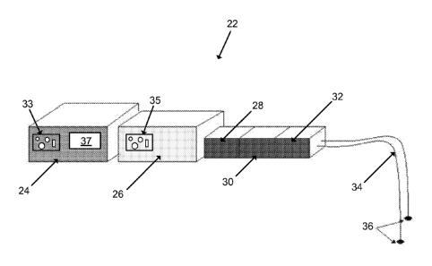
[0034] FIG. 10
illustrates a system 22 according to one embodiment of the invention.
The system 22 can stimulate uterine muscles into tonic contractions using
frequencies
greater than about 5.0 Hz. The system 22 can be used to stimulate muscles of
the uterus in
a way that does not affect other organs and can be accurately regulated and
controlled,
unlike oxytocin or other conventionally-used drugs. The system 22 can be used
on a
patient, such as a female post-partum, and can be controlled by a user, such
as a physician
or medical staff member. For example, the system 22 can input innocuous
electrical
pulses into the patient's uterus with sufficient effect to incite postpartum
tonic or tetanic
contractions in order to help treat uterine atony and postpartum hemorrhage.
In some
embodiments, the system 22 can include a control module 24, a current source
26, an
isolation unit 28, a constant maximum current unit 30, a biphasic converter
32, a set of
lead wires 34, and a set of electrodes 36.
[0035] The control
module 24 can contain computing capability, software, and
memory. The controlling module 12 can be set using interface controls 33, such
as dials,
switches and/or auxiliary inputs, to perform preprogrammed stimulation tasks,
including
commanding the current source 26 to output stimulation current of selected
frequency,
amplitude, pulse width, and train duration automatically for selected periods
of time. The
8
CA 02758118 2011-10-06
WO 2010/118178
PCT/US2010/030302
control module 24 can also be operated manually by the user, in which the user
can
determine and set one or more output stimulation currents of desired
frequencies,
amplitudes, pulse widths, and train durations as needed spontaneously (i.e.,
in real time or
in near-real time). For example, the control module 24, can be operated
automatically or
manually to produce a stimulation current which can cause tonic or tetanic
contractions of
the patient's uterine muscle and the user has the capability to adjust the
stimulation current
parameters (i.e., frequencies, amplitudes, pulse widths, and/or train
durations) in real time
or near-real time during observation of the patient's uterus.
[0036] In one
embodiment, the control module 24 can automatically or manually
operate multiple stimulation outputs of the current source 26 independently or
in unison
with varying or similar current frequencies, amplitudes, pulse widths, and
train durations.
As a result, the control module 24 can provide stimulation currents directly
to the uterus or
through various organs, such as the cervix, vaginal wall and/or abdominal wall
separately,
simultaneously, or sequentially, or can provide stimulation currents to
various parts of the
uterus separately, simultaneously, or sequentially.
[0037] In one
embodiment, pre-recorded uterine electrical traces, obtained from
normally-contracting patients and saved digitally, can be stored in the
control module 24
to be used, in turn, as the electrical current trace patterns for commanding
the current
source 26 to output identical stimulation current to patients with abnormal
uterine activity,
such as patients with insufficient or absent contractile activity during
postpartum
hemorrhage. In addition, artificially generated current traces, saved
digitally, with known
frequencies, amplitudes, pulse widths, and train durations, can be stored in
the control
module 24 to be used as the electrical current trace patterns for commanding
the current
source 26 to output identical stimulation current to patients with abnormal
uterine activity
during postpartum hemorrhage.
[0038] In another
embodiment, the control module 24 can automatically regulate and
modify the electrical current output produced by the current source 26 based
on input from
electrical contractile activity of the patient's uterus, which can be
transmitted to the
control module 24 via pick-up wires, a signal conditioner, and/or after-
conditioning wires
(not shown). The control module 24 can regulate and modify the produced
electrical
current by changing the electrical stimulation pulse-width, current amplitude,
pulse train
duration, and/or the pulse frequency according to a pre-programmed algorithm.
9
CA 02758118 2011-10-06
WO 2010/118178
PCT/US2010/030302
[0039] In some
embodiments, the control module 24 can include a display 37, such as
a video display, a digital display, light-emitting diode (LED) display, etc.,
to display the
stimulation output currents produced for the user to read or assess. The
control module 24
can be coupled to the current source 26 by wires, direct electrical coupling,
or another
suitable coupling. For example, in one embodiment, the control module 24 can
communicate with the current source 26 via a wireless connection, such as
Bluctooth*).
[0040] The current
source 26 can generate the output stimulation current. In one
embodiment, the electrical stimulation cunent settings can be adjusted at the
current
source 26 by the user using interface controls 35, such as dials, switches or
other settings.
In another embodiment, the electrical stimulation settings can be controlled
by the control
module 24 (e.g., as preprogrammed settings or by the user using the interface
controls 33,
as described above), and output to the current source 26. As described above,
in some
embodiments, the current source 26 can output multiple electrical stimulation
currents
either directly to the uterus or indirectly to the uterus via the cervix, the
vaginal wall
and/or the abdominal wall separately, simultaneously, or sequentially, as
commanded by
the control module 24, or the current source 26 can output multiple electrical
stimulation
currents to various locations of the uterus separately, simultaneously, or
sequentially.
[0041] In some
embodiments, there can be a constant two-way communication
between the current source 26 and the control module 24, so that the current
source 26 can
receive commands from the control module 24 and the control module 24 can
receive
actual output current values from the current source 26.
[0042] In some
embodiments, the current source 26 can be capable of generating an
output current between about 0.01 milliamperes and about 40.00 milliamperes
(with
possible voltages between about 0.0001 volts and about 100 volts). Pulse
widths of the
current can be adjusted between about 0.1 millisecond and about 1000
milliseconds.
Frequencies of the current can be adjusted from about 0.1 Hertz to about 30 Hz
or greater.
Pulse train durations can be adjusted from about 1 second to about 10,000
seconds. In
addition, output currents can be sinusoidal so as to reduce tissue damage and
maximize
effect (10). In one embodiment, the current source 26 can produce a maximal
"jolt" of
uterine electrical stimulation energy equivalent to between about 1 Joule and
about 120
Joules of electrical energy in a short duration between about 1 millisecond
and about 1000
milliseconds. Further, the electrical stimulation current output from the
current source 26
CA 02758118 2011-10-06
WO 2010/118178
PCT/US2010/030302
can be sensed, measured, or detected by either the current source 26 or the
control module
24 and can be automatically shut off if current values are determined to be
dangerous or
outside prescribed, programmed, or set values.
[0043] The
isolation unit 28 can prevent ground loop currents from affecting the
patient. In one embodiment, isolation is accomplished through optical
isolation. In other
embodiments, induction or other methods of isolation can be used by the
isolation unit 28.
[0044] The constant
maximum current unit 30 can allow the user to regulate the
amount of maximum current that the patient's uterus receives. The constant
maximum
current unit 30 can prevent tissue damage due to extreme current fluctuations
as tissue
resistance varies (11), and can be set (either in a discrete or continuous
fashion) to or
between values well below human threshold for human feeling (e.g., about 0.01
milliamperes) and values uncomfortable for humans (e.g., about 10
milliamperes). In one
example, the constant maximum stimulation current can be set at a value which
maximizes
current input without damaging tissue and with minimal discomfort to the
patient (e.g.,
about 4 milliamperes).
[0045] The biphasic
converter 32 can alternate the polarity of current pulses produced
by the current source 26 after having moved through the isolation unit 28 and
the constant
maximum current unit 30 in order to further prevent adverse effects on the
patient's
tissues. The biphasic converter 32 can insure that the total energy delivered
at the tissue
site, as integrated over time, has a net value of zero. This can reduce the
possibility of
heating and subsequent damage to the patient's tissues (11, 12).
[0046] The lead
wires 34 can transmit the output current from the biphasic converter
32 to the electrodes 36. In one embodiment, the lead wires 34 can be similar
to those
manufactured by Advantage Medical Cables. In some embodiments, the system 22
can
include between one and ten lead wires 34. For example, different lead wires
34 can carry
different types or strengths of currents that incite, induce, or augment a
tonic contraction at
different times in different parts of the uterus, as preprogrammed or set by
the user (e.g., to
stimulate various parts of the patient's uterus separately, simultaneously,
and/or
sequentially).
[0047] FIG. 11
illustrates a patient's uterus 38, ovaries 40, fallopian tubes 42, a uterine
body (or intrauterine cavity) 44, a cervix 46, a vagina 48, a fundus 50 (i.e.,
top portion) of
11
CA 02758118 2011-10-06
WO 2010/118178
PCT/US2010/030302
the uterus, and a distal portion 52 of the uterus. The electrodes 36 can be
attached to or
near the uterus 38 in a specific orientation and at specific locations that
will have the best
effect upon uterine contractility for the patient, as determined by the user.
In one example,
the electrodes 36 can be placed upon the vaginal wall 48 and/or the cervix 46.
In another
example, the electrodes 36 can be placed at locations across the fundal 50 and
distal
portions 52 of the uterus 38. Also, the electrodes 36 can be mounted
externally to the
patient's abdominal surface.
[0048] The
electrodes 36 can be attached to the patient's abdominal surface and/or
uterus 38 using biocompatible glue or tissue adhesive, or by suction or other
self-affixing
electrodes. In one embodiment, the electrodes 36 can be standard silver
chloride (AG2C1)
electrodes, EEG electrodes, suction electrodes, or needle electrodes. In
some
embodiments, the system 22 can include between one and ten electrodes 36
(e.g., equal to
the number of lead wires 34). Different electrodes 36 can be positioned at
various
locations in or around the patient's uterus 38, where some or each of the
electrodes 36
causes tonic and/or phasic effects according to the electrical stimulus
applied through
them. For example, one or several electrodes 36 can act as a local pacemaker
for eliciting
contractions, while one or several other electrodes 36 can cover one or many
different
portions of the uterus 38 for eliciting global tonic or tetanic contractions.
In addition, in
some embodiments, the electrodes 36 can consist of platinum-iridium metals, so
as to
reduce the possibility of tissue lesions (12).
[0049] FIGS. 12A-
12C illustrate a patient's uterus 38 in three different conditions.
FIG. 12A shows a naturally contracting uterus 38 post-partum. Forceful and
spontaneous
tonic contractions can prevent blood loss. FIG. 12B shows a uterus 38 which is
not
contracting postpartum due to uterine atony. The lack of tonic contractile
activity allows
the uterus to bleed out, threatening the life of the patient. FIG. 12C shows
the uterus 38
with atony and uterine rupture treated effectively (i.e., forcefully
contracted) using
electrical tonic stimulation. As shown in FIG. 12C the uterus 38 has been
outfitted with
electrodes 36 (trans-vaginally) so that the system 22 can output stimulated
current (i.e.,
through the lead wires 34) for tonic activity using electrical frequencies
greater than or
equal to about 5 Hz. The artificially-stimulated tonic contractions can help
reduce, stop
and/or manage the blood loss. In one embodiment, the stimulated current can be
output to
12
CA 02758118 2016-11-09
64181-362
the patient for a duration greater than about 10 seconds. In some embodiments,
the pulse
train durations can be up to about 30 minutes long.
[0050] In addition,
the system 22 can be used in conjunction with other devices,
methods, systems, and treatments for postpartum hemorrhage, uterine atony, and
bleeding
or coagulation problems, including but not limited to oxytocin,
prostaglandins,
misoprostol, prepidil, ergot allcyloids, tamponades, balloon tamponades,
sponges, clamps,
manual uterine massage and manipulation, sutures, bio-compatible adhesives,
cauterization, and/or pharmaceutical coagulants.
[0051] It will be
appreciated by those skilled in the art that while the invention has
been described above in connection with particular embodiments and examples,
the
invention is not necessarily so limited, and that numerous other embodiments,
examples,
uses, modifications and departures from the embodiments, examples and uses are
intended
to be encompassed by the claims attached hereto. To the extent that specific
materials are
mentioned, it is merely for purposes of illustration and is not intended to
limit the
invention. One skilled in the art may develop equivalent means or reactants
without the
exercise of inventive capacity and without departing from the scope of the
invention.
[0052] Unless defined otherwise, technical and scientific terms used herein
have the
same meaning as commonly understood by one of ordinary skill in the art to
which
this invention belongs. Singleton et al., Dictionary of Microbiology and
Molecular
Biology 3"1 ed., J. Wiley & Sons (New York, NY 2001); March, Advanced Organic
Chemistry Reactions, Mechanisms and Structure 5th ed., J. Wiley & Sons (New
York,
NY 2001); and Sambrook and Russel, Molecular Cloning: A Laboratory Manual 3rd
ed., Cold Spring Harbor Laboratory Press (Cold Spring Harbor, NY 2001),
provide
one skilled in the art with a general guide to many of the terms used in the
present
application.
REFERENCES
[0053] 1. The Prevention
and Management of Postpartum Haemorrhage:
Report of Technical Working Group, Geneva 3-6 July 1989. Geneva: World Health
Organization, 1990.
13
CA 02758118 2011-10-06
WO 2010/118178 PCT/US2010/030302
[0054] 2. Elbourne DR,
Prendiville WJ, Carroli G, Wood J, McDonald S.
Prophylactic use of oxytocin in the third stage of labour. Cochrane Database
Syst Rev
2001 ;(4):CD001808.
[0055] 3. Bais JM,
Eskes M, Pel M, Bonsel GJ, Bleker OP. Postpartum
haemorrhage in nulliparous women: incidence and risk factors in low and high
risk
women. A Dutch population-based cohort study on standard (>= 500 mL) and
severe (>=
1000 mL) postpartum haemorrhage. Eur J Obstet Gynecol Reprod Biol 2004;115:166-
72.
[0056] 4. Reyal F,
Deffarges J, Luton D, Blot P, Oury JF, Sibony 0. Severe post-
partum hemorrhage: descriptive study at the Robert-Debre Hospital maternity
ward
[French]. J Gynecol Obstet Biol Reprod (Paris) 2002;31:358-64.
[0057] 5. Norris TC.
Management of postpartum hemorrhage. Am Fam
Physician. 1997 Feb 1;55(2):635-40.
[0058] 6. Fawcus, S,
Mbizvo, M, Lindmark, G, Nystrom, L. A community-based
investigation of maternal mortality from obstetric haemorrhage in rural
Zimbabwe.
Maternal Mortality Study Group. Trop Doct. 1997 Jul;27(3):159-63.
[0059] 7. Sultatos LG.
Mechanisms of drugs that affect uterine motility. J Nurse
Midwifery. 1997 Jul-Aug;42(4):367-70.
[0060] 8. Alexander E.
Weingarten, MD, Jeffrey I. Korsh, MD, George G.
Neuman, MD, and Steven B. Stern, MD. Postpartum Uterine Atony after
Intravenous
Dantrolene. Anesth Analg 1987; 66:269-270.
[0061] 9. Hacker,
Neville, J. G. Moore, and Joseph Gambone. Essentials of
Obstetrics and Gynecology. 4th ed. Vol. 1. Philadelphia: Elsevier Inc., 2004.
151.
[0062] 10. Bennie SD,
Petrofsky JS, Nisperos J, Tsurudome M, Laymon M. Eur J
Appl Physiol. 2002 Nov;88(1-2):13-9. Epub 2002 Sep 10. Toward the optimal
waveform
for electrical stimulation of human muscle.
[0063] 11. DeLisa, Joel
A.; Gans, Bruce M.; Walsh, Nicolas E.; Bockenek,
William L.; Frontera, Walter R.; Gerber, Lynn H.; Geiringer, Steve R.; Pease,
William S.;
Robinson, Lawrence R.; Smith, Jay; Stitik, Todd P.; Zafonte, Ross D. Physical
Medicine
14
CA 02758118 2011-10-06
WO 2010/118178 PCT/US2010/030302
and Rehabilitation: Principles and Practice. 4th edition. 2004. Lippincott
Williams &
Wilkins (LWW): Chapter 66.
[0064] 12. Piallat B,
Chabardes S, Devergnas A, Torres N, Allain M, Ban-at E,
Benabid AL. Monophasic but not biphasic pulses induce brain tissue damage
during
monopolar high-frequency deep brain stimulation. Neurosurgery. 2009
Jan;64(1):156-62;
discussion 162-3.