Note: Descriptions are shown in the official language in which they were submitted.
CA 02758201 2011-10-06
WO 2010/115629
PCT/EP2010/002205
AMATOXIN-ARMED THERAPEUTIC CELL SURFACE BINDING COMPONENTS DESIGNED FOR
TUMOUR THERAPY
FIELD OF THE INVENTION
The invention relates to tumour therapy. In one aspect, the present invention
relates to
conjugates of a toxin and a target-binding moiety, e.g. an antibody, which are
useful in the
treatment of cancer. In particular, the toxin is an amatoxin, and the target-
binding moiety is
preferably directed against tumour-associated antigens. In particular, the
amatoxin is
conjugated to the antibody by linker moieties. In particular the linker
moieties are covalently
bound to functional groups located in positions of the amatoxin proved as
preferred positions
for the attachment of linkers with respect to optimum antitumor activity. In a
further aspect
the invention relates to pharmaceutical compositions comprising such target-
binding moiety
toxin conjugates and to the use of such target-binding moiety toxin conjugates
for the
preparation of such pharmaceutical compositions. The target-binding moiety
toxin conjugates
and pharmaceutical compositions of the invention are useful for the treatment
of cancer.
BACKGROUND OF THE INVENTION AND STATE OF THE ART
Antibody therapy has been established for the targeted treatment of patients
with cancer,
immunological and angiogenic disorders. The use of antibody-drug conjugates
(ADC), i. e.
immunoconjugates, for the local delivery of cytotoxic or cytostatic agents, i.
e. drugs to kill or
inhibit tumor cells in the treatment of cancer theoretically allows targeted
delivery of the drug
moiety to tumors, and intracellular accumulation therein, where systemic
administration of
these unconjugated drug agents may result in unacceptable levels of toxicity
to normal cells as
well as the tumor cells sought to be. Maximal efficacy with minimal toxicity
is sought
thereby. Efforts to design and refine ADC have focused on the selectivity of
monoclonal
antibodies (mAbs) as well as drug-linking and drug-releasing properties. Both
polyclonal
antibodies and monoclonal antibodies have been reported as useful in these
strategies.
Amatoxins
Amatoxins are cyclic peptides composed of 8 amino acids. They can be isolated
from
Amanita phalloides mushrooms or prepared from the building blocks by
synthesis. Amatoxins
specifically inhibit the DNA-dependent RNA polymerase II of mammalian cells,
and thereby
CA 02758201 2011-10-06
wo 2010/115629 2
PCT/EP2010/002205
also the transcription and protein biosynthesis of the affected cells.
Inhibition of transcription
in a cell causes stop of growth and proliferation. Though not covalently
bound, the complex
between amanitin and RNA-polymerase II is very tight (KD = 3nM). Dissociation
of arnanitin
from the enzyme is a very slow process what makes recovery of an affected cell
unlikely.
When the inhibition of transcription lasts too long, the cell will undergo
programmed cell
death (apoptosis).
Conjugates of amatoxins and target-binding moieties
Earlier patent application EP 1 859 811 Al (published November 28, 2007) by
the
inventors describes conjugates, in which P-amanitin is coupled to albumin or
to the
monoclonal antibodies HEA125, OKT3, and PA-1. Furthermore, the inhibitory
effect of these
conjugates on the proliferation of breast cancer cells (MCF-7), Burkitt's
lymphoma cells
(Raji), and T-lymphoma cells (Jurkat) was studied.
Epithelial cell adhesion molecule (EpCAM) antigen
Epithelial cell adhesion molecule (EpCAM, CD326) is one of the best-studied
target
antigens on human tumors (Trzpis et al., 2007; Baeuerle and Gires, 2007). It
represents a type
I membrane glycoprotein of 314 amino acids with an apparent molecular weight
of 40 kDa
(Balzar et al., 1999). It is overexpressed in the majority of adenocarcinomas
(Winter et al.,
2003; Went et al., 2004). In particular, EpCAM expression is enhanced in node-
positive
breast cancer, epithelial ovarian cancer, cholangiocarcinoma, pancreatic
adenocarcinoma and
squamous cell head and neck cancer. Increased EpCAM expression is indicative
for a poor
prognosis in breast and gallbladder carcinomas (Gastl et al., 2000; Varga et
al., 2004; Spizzo
et al., 2002; Spizzo et al., 2004). Importantly, EpCAM is expressed by tumor
initiating or
cancer stem cells in mammary, colorectal and pancreatic carcinomas (Al-Hajj et
al., 2003;
Dalerba et al., 2007; Li et al., 2007).
EpCAM-specific monoclonal antibodies have been used as a diagnostic tool for
the
detection of rare circulating tumor cells in cancer patients (Allard et al.,
2004; Nagrath et al.,
2007). A couple of engineered anti-EpCAM antibodies are currently investigated
in clinical
studies.
HER2 antigen
HER2 (Her2/neu; ErbB2), a receptor tyrosine kinase with an apparent molecular
weight of
185 kDa is overexpressed in about 25-30% of human breast cancers and gastric
cancers. This
overexpression, which is often due to amplification of the receptor-encoding
gene, generally
CA 02758201 2011-10-06
3
wo 2010/115629
PCT/EP2010/002205
represents a poor prognosis, often involving progressive disease in the years
after the initial
diagnosis is made.
Monoclonal antibody therapy has been established for the targeted treatment of
patients with Her2/neu-positive cancers. HERCEPTIN (trastuzumab) is a
recombinant DNA-
derived humanized monoclonal antibody that selectively binds with high
affinity in a cell-
based assay (Kd = 5 nM) to the extracellular domain of the human epidermal
growth factor
receptor 2 protein, HER2 (ErbB2). Trastuzumab is an IgG1 kappa antibody that
contains
human framework regions with the complementarity-determining regions of a
murine
antibody (4D5) that binds to HER2. Trastuzumab binds to the HER2 antigen and
thus inhibits
the growth of cancerous cells. Because trastuzumab is a humanized antibody, it
minimizes
any HAMA response in patients. Trastuzumab has been shown, in both in vitro
assays and in
animals, to inhibit the proliferation of human tumor cells that overexpress
HER2.
Trastuzumab is a mediator of antibody-dependent cellular cytotoxicity, ADCC.
HERCEPTIN is clinically active in patients with ErbB2-overexpressing
metastatic breast
cancers that have received extensive prior anti-cancer therapy. Although
HERCEPTIN is a
breakthrough in treating patients with ErbB2-overexpressing breast cancers
that have received
extensive prior anti-cancer therapy, the majority of the patients in this
population fail to
respond or respond only poorly to HERCEPTIN treatment. Therefore, there is a
significant
clinical need for developing further HER2-directed cancer therapies for those
patients with
HER2-overexpressing tumors or other diseases associated with HER2 expression
that do not
respond, or respond poorly, to HERCEPTIN treatment.
TECHNICAL PROBLEMS UNDERLYING THE PRESENT INVENTION
There was a need in the prior art for target-binding moiety toxin conjugates
that exert
their toxic effects to target cells or tissues at much lower concentration so
that the conjugates
may be administered at lower concentrations and harmful side effects to non-
target cells are
minimized. Furthermore, there was a need in the prior art for the treatment of
other types of
cancer, particularly those being therapy resistant, or poorly responding to
actual tumour
therapies.
The present invention fulfils these and other needs. For example, the
inventors found
out in the experiments underlying the present invention that very effective
target-binding
moiety toxin conjugates, in particular antibody amatoxin conjugates, can be
constructed by
choosing particular linkage points in the amatoxin part of the conjugate and
by choosing
particular linker compounds. Such target-binding moiety toxin conjugates are
very effective
CA 02758201 2011-10-06
4
WO 2010/115629
PCT/EP2010/002205
in that they exert their toxic activity to the target cells at very low
concentrations (IC50 of
=about 5x10-12 M) as well as by being highly specific for their target cells.
Without wishing to
be bound by a particular theory, these advantages might be explained in that
the linkage
between the target-binding moiety and the amatoxin or, if present, between the
linker and the
amatoxin is efficiently cleaved inside the target cell and to a much lesser
degree outside the
cell.
The above overview does not necessarily describe all problems solved by the
present
invention.
SUMMARY OF THE INVENTION
In a first aspect the present invention relates to a target-binding moiety
toxin conjugate
comprising: (i) a target-binding moiety; (ii) at least one amatoxin; and (iii)
optionally a linker
L2; wherein the at least one amatoxin is connected to the target-binding
moiety or, if present,
to the linker L2 via the 6' C-atom of amatoxin amino acid 4.
In a second aspect the present invention relates to a target-binding moiety
toxin
conjugate comprising: (i) a target-binding moiety; (ii) at least one amatoxin;
and (iii)
optionally a linker L3; wherein the at least one amatoxin is connected to the
target-binding
moiety or, if present, to the linker L3 via the 8 C-atom of amatoxin amino
acid 3.
In a third aspect the present invention relates to a target-binding moiety
toxin
conjugate comprising: (i) a target-binding moiety; (ii) at least one amatoxin;
and (iii)
optionally a linker Li; wherein the at least one amatoxin is connected to the
target-binding
moiety or, if present, to the linker Li via the y C-atom of amatoxin amino
acid 1.
In a fourth aspect the present invention relates to the target-binding moiety
toxin
conjugate according to the first, the second, or the third aspect for use in
medicine.
In a fifth aspect the present invention relates to the target-binding moiety
toxin
conjugate according to the first, the second, the third or the fourth aspect
for the treatment of
cancer or of an autoimmune disease in a patient, wherein the cancer is
preferably selected
from the group consisting of pancreatic cancer, cholangiocarcinoma, breast
cancer, colorectal
cancer, lung cancer, prostate cancer, ovarian cancer, stomach cancer, kidney
cancer, head and
neck cancer, brain tumors, childhood neoplasms, soft tissue sarcomas,
epithelial skin cancer,
malignant melanoma, leukemia, and malignant lymphoma and wherein the
autoimmune
disease is preferably selected from the group consisting of Ankylosing
Spondylitis, Chagas
disease, Crohns Disease, Dermatomyositis, Diabetes mellitus type 1,
Goodpasture's
syndrome, Graves' disease, Guillain-Barre syndrome (GBS), Hashimoto's disease,
CA 02758201 2011-10-06
wo 2010/115629
PCT/EP2010/002205
Hidradenitis suppurativa, Idiopathic thrombocytopenic purpura, Lupus
erythematosus, Mixed
Connective Tissue Disease, Myasthenia gravis, Narcolepsy, Pemphigus vulgaris,
Pernicious
anaemia, Psoriasis, Psoriatic Arthritis, Polymyositis, Primary biliary
cirrhosis, Relapsing
polychondritis, Rheumatoid arthritis, Schizophrenia, Sjogren's syndrome,
Temporal arteritis,
5 Ulcerative Colitis, Vasculitis Wegener's granulomatosis, in particular
Rheumatoid arthritis.
In a sixth aspect the present invention relates to a pharmaceutical
composition
comprising at least one type of target-binding moiety toxin conjugate
according to the first,
the second, and/or the third aspect and further comprising one or more
pharmaceutically
acceptable diluents, carriers, excipients, fillers, binders, lubricants,
glidants, disintegrants,
adsorbents; and/or preservatives.
This summary of the invention does not necessarily describe all features of
the
invention.
BRIEF DESCRIPTION OF THE DRAWINGS
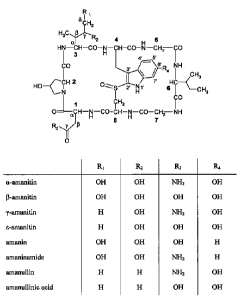
Fig. 1 shows the structural formulae of different amatoxins. The numbers in
bold type
(1 to 8) designate the standard numbering of the eight amino acids forming the
amatoxin. The
standard designations of the atoms in amino acids 1, 3 and 4 are also shown
(Greek letters a
to y, Greek letters a to 8, and numbers from l' to 7', respectively).
Fig. 2 shows a comparison of the binding affinities of huHEA125-Ama and
huHEA125 to target cells by a binding competition analysis. EpCAM-expressing
Co1o205
cells were incubated with a fixed amount of directly FITC-labeled mouse HEA125
antibody.
Binding to target cells was analyzed by flow cytometry. Competition of binding
with
increasing amounts of huHEA125-Ama or huHEA125 revealed a very similar
affinity
towards the target antigen.
Fig. 3 shows the surface expression of EpCAM antigen on various carcinoma cell
lines detected by indirect immunofluorescence: Fig. 3A Capan-1; Fig. 3B
Colo205; Fig. 3C
OZ; and Fig. 3D MCF-7. The grey-shaded histograms on the left side of each
diagram show
the results obtained with control antibody Xolaira; the histograms having a
white area on the
right side of each diagram show the results obtained with antibody huHEA125.
The
abbreviation FL1-H stands for "fluorescence 1 height" which means the
intensity of
fluorescence 1, i.e. the green channel for FITC.
Fig. 4 shows the binding of huHEA125-Amanitin and huHEA125-Phalloidin
conjugates to MCF-7 breast cancer cells analyzed by flow cytometry. The
abbreviation FL1-
CA 02758201 2011-10-06
wo 2010/115629 6
PCT/EP2010/002205
H stands for "fluorescence 1 height" which means the intensity of fluorescence
1, i.e. the
green channel for FITC.
A: bold histogram, huHEA125-Amanitinl; shaded histogram, huHEA125; dotted
histogram,
Xolair (negative control);
= 5
B: bold histogram, huHEA125-Amanitin4; shaded histogram, huHEA125; dotted
histogram,
Xolair (negative control);
C: bold histogram, huHEA125-a-Phalloidin; shaded histogram, huHEA125; dotted
histogram,
Xolair (negative control).
= Fig. 5 shows a comparison of the inhibition of MCF-7 cell proliferation
caused by the
conjugate huHEA125-Amanitinl, the non-binding control conjugate Xolair-
Amanitinl, and
free Amanitin.
Fig. 6 shows a comparison of the inhibition of MCF-7 cell proliferation caused
by the
conjugate huHEA125-Amanitin4, the conjugate alpha-phalloidin-huHEA125, and
free
Amanitin.
Fig. 7 shows a comparison of the inhibition of Capan-1 cell proliferation
caused by
conjugate huHEA125-Amanitin3, Amanitin-armed control antibody Xolair , and
free
Amanitin.
Fig. 8 shows a comparison of the inhibition of Co1o205 cell proliferation
caused by
conjugate huHEA125-Amanitin3, Amanitin-armed control antibody Xolair , and
free
Amanitin.
Fig. 9 shows a comparison of the inhibition of MCF-7 cell proliferation caused
by
conjugate huHEA125-Amanitin3, Amanitin-armed control antibody Xolair , and
free
Amanitin.
Fig. 10 shows a comparison of the inhibition of OZ cell proliferation caused
by
conjugate huHEA125-Amanitin3, Amanitin-armed control antibody Xolair , and
free
Amanitin.
Fig 11 A to D show a comparison of the inhibition on cell proliferation
exerted by
various a-amanitin conjugates at different amanitin concentrations using the
Her2/neu
positive cell lines SKOV-3, SKBR-3 and NCI-N87 as well as the Her2/neu
negative cell line
MDA-MB231.
Fig. 12 shows the antitumor activity of various ct-amanitin conjugates at two
different
concentrations (A: 30 ilg/kg and B: 150 lag/kg body weight) in an in vivo SKOV-
3 xenograft
tumor model.
CA 02758201 2016-01-20
7
=
DETAILED DESCRIPTION OF THE INVENTION
Definitions
Before the present invention is described in detail below, it is to be
understood that
this invention is not limited to the particular methodology, protocols and
reagents described
herein as these may vary. It is also to be understood that the terminology
used herein is for the
purpose of describing particular embodiments only. Unless defined otherwise,
all technical
and scientific terms used herein have the same meanings as commonly understood
by one of
ordinary skill in the art. The scope of the claims should not be limited to
the illustrative
embodiments, but should be given the broadest interpretation consistent with
the description
as a whole.
Preferably, the terms used herein are defined as described in "A multilingual
glossary
of biotechnological terms: (IUPAC Recommendations)", Leuenberger, H.G.W,
Nagel, B. and
Ko1bl, H. eds. (1995), Helvetica Chimica Acta, CH-4010 Basel, Switzerland).
Throughout this specification and the claims which follow, unless the context
requires
otherwise, the word "comprise", and variations such as "comprises" and
"comprising", will be
understood to imply the inclusion of a stated integer or step or group of
integers or steps but
not the exclusion of any other integer or step or group of integer or step.
Several documents are cited throughout the text of this specification. Nothing
herein is
to be construed as an admission that the invention is not entitled to antedate
such disclosure
by virtue of prior invention.
The term "target-binding moiety", as used herein, refers to any molecule or
part of a
molecule that can specifically bind to a target molecule or target epitope.
Preferred target-
binding moieties in the context of the present application are (i) antibodies
or antigen-binding
fragments thereof; (ii) antibody-like proteins; and (iii) nucleic acid
aptamers. "Target-binding
moieties" suitable for use in the present invention typically have a molecular
mass of at least
15 kDa, at least 20 kDa, at least 30 kDa or of at least 40 kDa or more.
As used herein, an "antibody toxin conjugate" refers to a target-binding
moiety toxin
conjugate in which the target-binding moiety is an antibody or antigen-binding
fragment
thereof according to above alternative (i).
As used herein, an "antibody-like protein toxin conjugate" refers to a target-
binding
moiety toxin conjugate in which the target-binding moiety is an antibody-like
protein
according to above alternative (ii).
CA 02758201 2011-10-06
wo 2010/115629 8
PCT/EP2010/002205
As used herein, an "aptamer conjugate" refers to a target-binding moiety toxin
conjugate in which the target-binding moiety is a nucleic acid aptamer
according to above
alternative (iii).
In the context of the present application the terms "target molecule" and
"target
epitope", respectively, refers to an antigen and an epitope of an antigen,
respectively, that is
specifically bound by a target-binding moiety, preferably the target molecule
is a tumour-
associated antigen, in particular an antigen or an epitope which is present on
the surface of
one or more tumour cell types in an increased concentration and/or in a
different steric
configuration as compared to the surface of non-tumour cells or an antigen
preferentially
expressed on cells involved in autoimmune diseases, examples of such antigens
are
Immunoglobulin G Fc-part, Thyreotropin-receptor, Type IV Collagen, Proteinase
3, DNA
Topoisomerase I, Placoglobin. Preferably, said antigen or epitope is present
on the surface of
one or more tumour cell types but not on the surface of non-tumour cells.
Preferably the term "tumour associated antigen" comprises all substances,
which elicit
an immune response against a tumour. Particular suitable substances are those
which are
enriched in a tumour cell in comparison to a healthy cell. These substances
are preferably
present within and/or are accessible on the outside of the tumour cell. If the
tumour antigen is
only present within a tumour cell, it will still be accessible for the immune
system, since the
antigen or fragments thereof will be presented by the MHC system at the
surface of the cell.
In a preferred aspect tumour antigen is almost exclusively present on and/or
in the tumour cell
and not in a healthy cell of the same cell type.
Suitable tumour antigens can be identified, for example, by analyzing the
differential
expression of proteins between tumour and healthy cells of the same cell type
using a
microarray-based approach (Russo et al., Oncogene. 2003,22 : 6497-507), by PCR-
or
microarray-based screening for tumor specific mutated cellular genes (Heller,
Armu. Rev.
Biomed. Eng. 2002, 4: 129-53) or by serological identification of antigens by
recombinant expression cloning (SEREX; Tureci et al., Mol Med Today. 1997,3 :
342-349).
The skilled artisan is aware of a large number of substances which are
preferentially or
exclusively present on and/or in tumor cell, which include for example,
oncogenes like, for
example truncated epidermal growth factor, folate binding protein,
melanoferrin,
carcinoembryonic antigen, prostate-specific membrane antigen, HER2-neu and
certain sugar
chains like, for example, epithelial mucins.
It is preferred that tumour antigens are selected, which elicit a strong
immune
response, preferentially a MHC class I immune response. Antigens eliciting a
strong immune
CA 02758201 2011-10-06
9
wo 2010/115629
PCT/EP2010/002205
response will induce at least 1%, preferably at least 5%, more preferably at
least 10% and
most preferably at least 15% IFN-y-producing CD8+ T or CD4+ T cells isolated
from mice
previously immunized with the antigen, upon challenge with the antigen and/or
will induce
preferably at least 5%, and most preferably at least 15% of B-cells cells
isolated from mice
previously immunized with the antigen, upon challenge with the antigen to
proliferate.
Antigens fulfilling these criterions are candidates for use in therapeutic
and/or prophylactic
cancer vaccines.
In a particular preferred embodiment the tumour antigen is selected from the
group
consisting of T-cell or B-cell-defined cancer-associated antigens belonging to
unique gene
products of mutated or recombined cellular genes, in particular cyclin-
dependent kinases (e.g.
CDC2, CDK2, CDK4), pl5Ink4b, p53, AFP, P-catenin, caspase 8, p53, Bcr-abl
fusion product,
MUM-1 MUM-2, MUM-3, ELF2M, HSP70-2M, HST-2, KIAA0205, RAGE, myosin/m, 707-
AP, CDC27/m, ETV6/AML, TEL/Amll, Dekcain, LDLR/FUT, Pml-RARa, TEL/AMLI;
Cancer-testis (CT) antigens, in particular NY-ESO- 1, members of the MAGE-
family
(MAGE-Al, MAGE-A2, MAGE-A3, MAGE-A4, MAGE-A6, MAGE-10, MAGE-12),
BAGE, DAM-6, DAM-10, members of the GAGE- family (GAGE-1, GAGE-2, GAGE-3,
GAGE-4, GAGE-5, GAGE-6, GAGE-7B, GAGE- 8), NY-ESO-1, NA-88A, CAG-3, RCC-
associated antigen G250; Tumour virus antigens, in particular human papilloma
virus (HPV) -
derived E6 or E7 oncoproteins, Epstein Barr virus EBNA2-6, LMP-1, LMP-2;
overexpressed
or tissue-specific differentiation antigens, in particular gp77, gp100, MART-
1/Melan-A, p53,
tyrosinase, tyrosinase-related protein (TRP-1 and TPR-2), PSA, PSM, MC1R ;
widely
expressed antigens, in particular ART4, CAMEL, CEA, CypB, EpCAM, HER2/neu,
hTERT,
hTRT, ICE, Muc 1 , Muc2, PRAME RU1, RU2, SART-1, SART-2, SART-3, and WT1; and
fragments and derivatives thereof. Particular preferred tumour antigens are
antigens derived
from HER-2 and EpCAM. In the context of this section the term fragment refers
to C-
terminally and/or N-terminally deleted proteins, which comprise at least one
epitope which
can be specifically bound by a target-binding moiety.
The term "antibody or antigen binding fragment thereof", as used herein,
refers to
immunoglobulin molecules and immunologically active portions of immunoglobulin
molecules, i.e. molecules that contain an antigen binding site that
immunospecifically binds
' an antigen. Also comprised are immunoglobulin-like proteins that are
selected through
techniques including, for example, phage display to specifically bind to a
target molecule, e.g.
to the target protein EpCAM or Her2. The immunoglobulin molecules of the
invention can be
of any type (e.g., IgG, IgE, IgM, IgD, IgA and IgY), class (e.g., IgGl, IgG2,
IgG3, IgG4,
CA 02758201 2011-10-06
wo 2010/115629 10
PCT/EP2010/002205
IgAl and IgA2) or subclass of immunoglobulin molecule. "Antibodies and antigen-
binding
fragments thereof" suitable for use in the present invention include, but are
not limited to,
polyclonal, monoclonal, monovalent, bispecific, heteroconjugate,
multispecific, human,
humanized (in particular CDR-grafted), deinununized, or chimeric antibodies,
single chain
- 5 antibodies (e.g. scFv), Fab fragments, F(ab1)2 fragments, fragments
produced by a Fab
expression library, diabodies or tetrabodies (Holliger P. et al., 1993),
nanobodies, anti-
idiotypic (anti-Id) antibodies (including, e.g., anti-Id antibodies to
antibodies of the
invention), and epitope-binding fragments of any of the above.
In some embodiments the antigen-binding fragments are human antigen-binding
antibody fragments of the present invention and include, but are not limited
to, Fab, Fab' and
F(ab')2, Fd, single-chain Fvs (scFv), single-chain antibodies, disulfide-
linked Fvs (dsFv) and
fragments comprising either a VL or VH domain. Antigen-binding antibody
fragments,
including single-chain antibodies, may comprise the variable domain(s) alone
or in
combination with the entirety or a portion of the following: hinge region, CL,
CH1, CH2, and
CH3 domains. Also included in the invention are antigen-binding fragments also
comprising
any combination of variable domain(s) with a hinge region, CL, CH1, CH2, and
CH3
domains.
Antibodies usable in the invention may be from any animal origin including
birds and
mammals. Preferably, the antibodies are from human, rodent (e.g. mouse, rat,
guinea pig, or
rabbit), chicken, pig, sheep, goat, camel, cow, horse, donkey, cat, or dog
origin. It is
particularly preferred that the antibodies are of human or murine origin. As
used herein,
"human antibodies" include antibodies having the amino acid sequence of a
human
immunoglobulin and include antibodies isolated from human immunoglobulin
libraries or
from animals transgenic for one or more human immunoglobulin and that do not
express
endogenous immunoglobulins, as described for example in U.S. Patent No.
5,939,598 by
Kucherlapati & Jakobovits.
The term "antibody-like protein" refers to a protein that has been engineered
(e.g. by
mutagenesis of loops) to specifically bind to a target molecule. Typically,
such an antibody-
like protein comprises at least one variable peptide loop attached at both
ends to a protein
scaffold. This double structural constraint greatly increases the binding
affinity of the
antibody-like protein to levels comparable to that of an antibody. The length
of the variable
peptide loop typically consists of 10 to 20 amino acids. The scaffold protein
may be any
protein having good solubility properties. Preferably, the scaffold protein is
a small globular
protein. Antibody-like proteins include without limitation affibodies,
anticalins, designed
CA 02758201 2011-10-06
wo 2010/115629 11
PCT/EP2010/002205
ankyrin repeat proteins (for review see: Binz et al. 2005) and proteins with
ubiquitine based
scaffolds. Antibody-like proteins can be derived from large libraries of
mutants, e.g. be
panned from large phage display libraries and can be isolated in analogy to
regular antibodies.
Also, antibody-like binding proteins can be obtained by combinatorial
mutagenesis of
surface-exposed residues in globular proteins.
The term "nucleic acid aptamer" refers to a nucleic acid molecule that has
been
engineered through repeated rounds of in vitro selection or SELEX (systematic
evolution of
ligands by exponential enrichment) to bind to a target molecule (for a review
see: Brody and
Gold, 2000). The nucleic acid aptamer may be a DNA or RNA molecule. The
aptamers may
contain modifications, e.g. modified nucleotides such as 2'-fluorine-
substituted pyrimidines.
The term "amatoxin" includes all cyclic peptides composed of 8 amino acids as
isolated from the genus Amanita and described in ref. (Wieland, T. and
Faulstich H., 1978);
further all chemical derivatives thereof; further all semisynthetic analogs
thereof; further all
synthetic analogs thereof built from building blocks according to the master
structure of the
natural compounds (cyclic, 8 amino acids), further all synthetic or
semisynthetic analogs
containing non-hydroxylated amino acids instead of the hydroxylated amino
acids, further all
synthetic or semisynthetic analogs, in which the thioether sulfoxide moiety is
replaced by a
sulfide, sulfone, or by atoms different from sulfur, e.g. a carbon atom as in
a carbaanalog of
amanitin.
Functionally, amatoxins are defined as peptides or depsipeptides that inhibit
mammalian RNA polymerase II. Preferred amatoxins are those with a functional
group (e.g. a
carboxylic group, an amino group, a hydroxy group, a thiol or a thiol-
capturing group) that
can be reacted with linker molecules or target-binding moieties as defined
above. Amatoxins
which are particularly suitable for the conjugates of the present invention
are a-amanitin, 13-
amanitin, y-amanitin, s-amanitin, amanin, amaninamide, amanullin, and
amanullinic acid as
shown in Fig. 1 as well as salts, chemical derivatives, semisynthetic analogs,
and synthetic
analogs thereof. Particularly preferred amatoxins for use in the present
invention are a-
, amanitin, 13-amanitin, and amaninamide.
As used herein, a "chemical derivative" (or short: a "derivative") of a
compound refers
to a species having a chemical structure that is similar to the compound, yet
containing at
least one chemical group not present in the compound and/or deficient of at
least one
chemical group that is present in the compound. The compound to which the
derivative is
compared is known as the "parent" compound. Typically, a "derivative" may be
produced
from the parent compound in one or more chemical reaction steps.
CA 02758201 2011-10-06
wo 2010/115629 12
PCT/EP2010/002205
As used herein, an "analog" of a compound is structurally related but not
identical to
the compound and exhibits at least one activity of the compound. The compound
to which the
analog is compared is known as the "parent" compound. The afore-mentioned
activities
include, without limitation: binding activity to another compound; inhibitory
activity, e.g.
enzyme inhibitory activity; toxic effects; activating activity, e.g. enzyme-
activating activity. It
is not required that the analog exhibits such an activity to the same extent
as the parent
compound. A compound is regarded as an analog within the context of the
present
application, if it exhibits the relevant activity to a degree of at least 1%
(more preferably at
least 5%, more preferably at least 10%, more preferably at least 20%, more
preferably at least
30%, more preferably at least 40%, and more preferably at least 50%) of the
activity of the
parent compound. Thus, an "analog of an amatoxin", as it is used herein,
refers to a
compound that is structurally related to any one of a-amanitin, P-amanitin, y-
amanitin, E-
amanitin, amanin, amaninamide, amanullin, and amanullinic acid as shown in
Fig. 1 and that
exhibits at least 1% (more preferably at least 5%, more preferably at least
10%, more
preferably at least 20%, more preferably at least 30%, more preferably at
least 40%, and more
preferably at least 50%) of the inhibitory activity against mammalian RNA
polymerase II as
compared to at least one of a-amanitin, P-amanitin, y-amanitin, E-amanitin,
amanin,
amaninamide, amanullin, and amanullinic acid. An "analog of an amatoxin"
suitable for use
in the present invention may even exhibit a greater inhibitory activity
against mammalian
RNA polymerase II than any one of a-amanitin, P-amanitin, y-amanitin, E-
amanitin, amanin,
amaninamide, amanullin, or amanullinic acid. The inhibitory activity might be
measured by
determining the concentration at which 50% inhibition occurs (IC50 value). The
inhibitory
activity against mammalian RNA polymerase II can be determined indirectly by
measuring
the inhibitory activity on cell proliferation. A suitable assay for measuring
inhibition of cell
proliferation is described in Example 3.
A "semisynthetic analog" refers to an analog that has been obtained by
chemical
synthesis using compounds from natural sources (e.g. plant materials,
bacterial cultures, or
cell cultures) as starting material. Typically, a "semisynthetic analog" of
the present invention
has been synthesized starting from a compound isolated from a mushroom of the
Amanita
family. In contrast, a "synthetic analog" refers to an analog synthesized by
so-called total
synthesis from small (typically petrochemical) building blocks. Usually, this
total synthesis is
carried out without the aid of biological processes.
A "linker" in the context of the present application refers to a molecule that
increases
the distance between two components, e.g. to alleviate steric interference
between the target-
CA 02758201 2011-10-06
wo 2010/115629 13
PCT/EP2010/002205
binding moiety and the amatoxin, which may otherwise decrease the ability of
the amatoxin
to interact with RNA polymerase II. The linker may serve another purpose as it
may facilitate
the release of the amatoxin specifically in the cell being targeted by the
target binding moiety.
It is preferred that the linker and preferably the bond between the linker and
the amatoxin on
one side and the bond between the linker and the antibody on the other side is
stable under the
physiological conditions outside the cell, e.g. the blood, while it can be
cleaved inside the cell,
in particular inside the target cell, e.g. cancer cell or immune cell. To
provide this selective
stability the linker may comprise functionalities that are preferably pH-
sensitive to generate
pH-sensitive linkers as described, e.g. in S. Fletcher, M. R. Jorgensens and
A. D. Miller; Org.
Lett. 2004, 6(23), pp 4245-4248, or protease sensitive to generate protease
sensitive linkers as
described, e.g. in L. DA Ibsen, Blood 2003, 102, 1458-65 or Francisco JA,
Cerreny CG,
Meyer DL, Nat. Biotechnol 2003, 21, 778-84. Alternatively, the bond linking
the linker to the
target binding moiety may provide the selective stability. Preferably a linker
has a length of at
least 1, preferably of 1-20 atoms length (e.g. 1, 2, 3, 4, 5, 6, 7, 8, 9, 10,
11, 12, 13, 14, 15, 16,
17, 18, 19, or 20 atoms) wherein one side of the linker has been reacted with
the amatoxin
and, the other side with a target-binding moiety. In the context of the
present invention, a
linker preferably is a C1-20-alkyl, C1_20-heteroalkyl, C2_20-alkenyl, C2.20-
heteroalkenyl, C2-20-
alkynyl, C2_20-heteroalkynyl, cycloalkyl, heterocycloalkyl, aryl, heteroaryl,
aralkyl, or a
heteroaralkyl group, optionally substituted. The linker may contain one or
more structural
elements such as amide, ester, ether, thioether, disulfide, hydrocarbon
moieties and the like.
The linker may also contain combinations of two or more of these structural
elements. Each
one of these structural elements may be present in the linker more than once,
e.g. twice, three
times, four times, five times, or six times. In some embodiments the linker
may comprise a
disulfide bond. It is understood that the linker has to be attached either in
a single step or in
two or more subsequent steps to the amatoxin and the target binding moiety. To
that end the
linker to be will carry two groups, preferably at a proximal and distal end,
which can (i) form
a covalent bond to a group, preferably an activated group on an amatoxin or
the target
binding-peptide or (ii) which is or can be activated to form a covalent bond
with a group on
an amatoxin. Accordingly, if the linker is present, it is preferred that
chemical groups are at
the distal and proximal end of the linker, which are the result of such a
coupling reaction, e.g.
an ester, an ether, a urethane, a peptide bond etc. The presence of a "linker"
is optional, i.e.
the toxin may be directly linked to a residue of the target-binding moiety in
some
embodiments of the target-binding moiety toxin conjugate of the present
invention. It is
preferred that the linker is connected directly via a bond to the targeting
moiety, preferably at
CA 02758201 2011-10-06
wo 2010/115629 14
PCT/EP2010/002205
its terminus. If the target-binding moiety comprises free amino, carboxy or
sulfhydryl groups,
e.g. in the form of Asp, Glu, Arg, Lys, Cys residues, which may be comprised
in a
polypeptide, then it is preferred that the linker is coupled to such a group.
As used herein, a first compound (e.g. an antibody) is considered to
"specifically bind"
to a second compound (e.g. an antigen, such as a target protein), if it has a
dissociation
constant KD to said second compound of 100 p,M or less, preferably 50 1.1M or
less, preferably
30 pLM or less, preferably 20 11M or less, preferably 10 11M or less,
preferably 5 piM or less,
more preferably 1 ptM or less, more preferably 900 nM or less, more preferably
800 nM or
less, more preferably 700 nM or less, more preferably 600 nM or less, more
preferably 500
nM or less, more preferably 400 nM or less, more preferably 300 nM or less,
more preferably
200 nM or less, even more preferably 100 nM or less, even more preferably 90
nM or less,
even more preferably 80 nM or less, even more preferably 70 nM or less, even
more
preferably 60 nM or less, even more preferably 50 nM or less, even more
preferably 40 nM or
less, even more preferably 30 nM or less, even more preferably 20 nM or less,
and even more
preferably 10 nM or less.
As used herein, a "patient" means any mammal or bird who may benefit from a
treatment with the target-binding moiety toxin conjugates described herein.
Preferably, a
"patient" is selected from the group consisting of laboratory animals (e.g.
mouse or rat),
domestic animals (including e.g. guinea pig, rabbit, chicken, pig, sheep,
goat, camel, cow,
horse, donkey, cat, or dog), or primates including human beings. It is
particularly preferred
that the "patient" is a human being.
As used herein, "treat", "treating" or "treatment" of a disease or disorder
means
accomplishing one or more of the following: (a) reducing the severity of the
disorder; (b)
limiting or preventing development of symptoms characteristic of the
disorder(s) being
treated; (c) inhibiting worsening of symptoms characteristic of the
disorder(s) being treated;
(d) limiting or preventing recurrence of the disorder(s) in patients that have
previously had the
disorder(s); and (e) limiting or preventing recurrence of symptoms in patients
that were
previously symptomatic for the disorder(s).
As used herein, "administering" includes in vivo administration, as well as
administration directly to tissue ex vivo, such as vein grafts.
An "effective amount" is an amount of a therapeutic agent sufficient to
achieve the
intended purpose. The effective amount of a given therapeutic agent will vary
with factors
such as the nature of the agent, the route of administration, the size and
species of the animal
to receive the therapeutic agent, and the purpose of the administration. The
effective amount
CA 02758201 2011-10-06
wo 2010/115629 15
PCT/EP2010/002205
in each individual case may be determined empirically by a skilled artisan
according to
established methods in the art.
"Pharmaceutically acceptable" means approved by a regulatory agency of the
Federal
or a state government or listed in the U.S. Pharmacopeia or other generally
recognized
pharmacopeia for use in animals, and more particularly in humans.
Embodiments of the Invention
The present invention will now be further described. In the following passages
different aspects of the invention are defined in more detail. Each aspect so
defined may be
combined with any other aspect or aspects unless clearly indicated to the
contrary. In
particular, any feature indicated as being preferred or advantageous may be
combined with
any other feature or features indicated as being preferred or advantageous.
In a first aspect the present invention is directed to a target-binding moiety
toxin
conjugate comprising: (i) a target-binding moiety; (ii) an amatoxin; and (iii)
optionally a
linker L2; wherein the amatoxin is connected to the target-binding moiety or,
if present, to the
linker L2 via the 6' C-atom of amatoxin amino acid 4 (see Fig. 1). In
preferred amatoxins
usable in the first aspect said amino acid 4 is 2'-sulfur-substituted
tryptophan or 2'-sulfur-
substituted 6' -hydroxy-tryptophan.
In a preferred embodiment of the first aspect the amatoxin is connected to the
target-
binding moiety or, if present, to the linker L2 via an oxygen atom bound to
the 6' C-atom of
amatoxin amino acid 4. It is further preferred that the amatoxin is connected
to the target-
binding moiety or, if present, to the linker L2 via an ether linkage (i.e.
amatoxin-O-L2 or
amatoxin-O-target-binding moiety). In these embodiments, it is preferred that
amino acid 4 is
6'-hydroxy-tryptophan.
In preferred embodiments of the first aspect the linker L2 is present and the
conjugate
has the following structure: amatoxin-6'C-0-L2-C(0)-NH-target-binding moiety.
In a second aspect the present invention is directed to a target-binding
moiety toxin
conjugate comprising: (i) a target-binding moiety; (ii) an amatoxin; and (iii)
optionally a
linker L3; wherein the amatoxin is connected to the target-binding moiety or,
if present, to the
linker L3 via the 8 C-atom of amatoxin amino acid 3 (see Fig. 1). In preferred
amatoxins
usable in the second aspect said amino acid 3 is isoleucine, y-hydroxy-
isoleucine or 7,8-
dihydroxy-isoleucine.
In a preferred embodiment of the second aspect the amatoxin is connected to
the
target-binding moiety or, if present, to the linker L3 via an oxygen atom
bound to the 8 C-
.
CA 02758201 2011-10-06
wo 2010/115629 16
PCT/EP2010/002205
atom of amatoxin amino acid 3. It is further preferred that the amatoxin is
connected to the
target-binding moiety or, if present, to the linker L3 via an ester linkage
preferably in the form
of an amatoxin-O-C(0)-L3-target binding boiety or an amatoxin-O-C(0)-target-
binding
moiety, more preferably an amatoxin-SC-0-C(0)-L3-target-binding moiety or an
amatoxin-
8C-0-C(0)-target-binding moiety, i.e. an amatoxin-8CH2-0-C(0)-L3-target-
binding moiety
or an amatoxin-8CH2-0-C(0)-target-binding moiety; an ether linkage preferably
in the form
of an amatoxin-O-L3 or an amatoxin-O-target-binding moiety preferably an
amatoxin-6C-0-
L3-target binding moiety or an amatoxin-SC-0-target binding moiety, more
preferably an
amatoxin-8CH2-0-L3-target binding moiety or an amatoxin-SCH2-0-target binding
moiety;
or a urethane linkage preferably in the form of an amatoxin-O-C(0)-NH-L3 or
amatoxin-O-
C(0)-NH-target-binding moiety, preferably an amatoxin-6C-0-C(0)-NH-L3-target-
binding
moiety or an amatoxin-K-O-C(0)-NH-target-binding moiety, i.e. an amatoxin-6CH2-
0-
C(0)-NH-L3-target-binding moiety or an amatoxin-oCH2-0-C(0)-NH-target-binding
moiety.
In these embodiments, it is preferred that amino acid 3 is 705-dihydroxy-
isoleucine.
In preferred embodiments of the second aspect the linker L3 is present and the
conjugate has one of the following structures: (i) amatoxin-SC-0-C(0)-L3-C(0)-
NH-target-
binding moiety; (ii) amatoxin-SC-0-L3-C(0)-NH-target-binding moiety; or (iii)
amatoxin-
8C-0-C(0)-NH-L3-C(0)-NH-target-binding moiety, i.e. (i) amatoxin-SCH2-0-C(0)-
L3-
C(0)-NH-target-binding moiety; (ii) amatoxin-SCH2-0-L3-C(0)-NH-target-binding
moiety;
or (iii) amatoxin-SCH2-0-C(0)-NH-L3-C(0)-NH-target-binding moiety.
In a third aspect the present invention is directed to a target-binding moiety
toxin
conjugate comprising: (i) a target-binding moiety; (ii) an amatoxin; and (iii)
optionally a
linker Li; wherein the amatoxin is connected to the target-binding moiety or,
if present, to the
linker Li via the y C-atom of amatoxin amino acid 1 (see Fig. 1). In preferred
amatoxins
usable in the third aspect said amino acid 1 is asparagine or aspartic acid.
In a preferred embodiment of the third aspect the amatoxin is connected to the
target-
binding moiety or, if present, to the linker Li via a nitrogen atom bound to
the y C-atom of
amatoxin amino acid 1. It is further preferred that the amatoxin is connected
to the target-
binding moiety or, if present, to the linker Li via an amide linkage (i.e.
amatoxin-C(0)-NH-
Li or amatoxin-C(0)-NH-target-binding moiety; the C-atom in the aforementioned
C(0)-
moiety is the y C-atom of amatoxin amino acid 1). In these embodiments, it is
preferred that
amino acid 1 is asparagine.
In preferred embodiments of the third aspect the linker Li is present and the
conjugate
has the following structure: amatoxin-yC(0)-NH-L1-C(0)-NH-target-binding
moiety. In this
CA 02758201 2011-10-06
wo 2010/115629 17
PCT/EP2010/002205
context it is preferred that the amide on the target-binding moiety side of
the conjugate is the
product of a reaction with a free amino group that was present in the target-
binding moiety.
In preferred embodiments of the first, the second, or the third aspect the
target-binding
moiety is connected to the amatoxin or, if present, to the linker Li, L2, or
L3 via an amino
group present in the target-binding moiety.
In preferred embodiments of the first, the second, or the third aspect the
amatoxin is
selected from a-amanitin, 0-amanitin, y-amanitin, e-amanitin, amanin,
amaninamide,
amanullin, or amanullinic acid (all shown in Fig. 1), as well as salts,
chemical derivatives,
semisynthetic analogs, and synthetic analogs thereof. Particularly preferred
amatoxins are a-
amanitin, 13-amanitin, and amaninamide, as well as salts, chemical
derivatives, semisynthetic
analogs, and synthetic analogs thereof.
The target binding moiety is in preferred embodiments a protein, in particular
an
antibody. Proteins and in particular antibodies will comprise several amino
acids, which allow
the coupling of amatoxins. Preferred amino acids have free amino, hydroxy, or
carbonyl-
groups, including Lys, Gin, Glu, Asp, Asn, Thr, and Ser. Accordingly, it is
possible to couple
more than one amatoxin molecules to one protein molecule. An increase of the
number of
amatoxins per molecule will also increase the toxicity. Accordingly, in a
preferred
embodiment the ratio of protein to amatoxin is between 1 protein molecule to
between 1 and
15 amatoxin molecules, preferably 1,2, 3, 4, 5, 6, 7, 8, 9, 10, 11, 12, 13,
14, or 15. For the
purpose of the calculation of the ratio in case of dimmers like IgGs the
dimmer is considered
as one molecule. Similar ratios are preferred, if the target binding moiety is
not a protein.
In preferred embodiments of the first, the second, or the third aspect the
linker Li, L2,
or L3 has above indicated meaning and preferred meanings. In further preferred
embodiments
of the first, the second, or the third aspect the linker Li, L2, or L3
comprises a disulfide bond.
In preferred embodiments of the first, the second, or the third aspect the
linker Li, L2,
or L3 has a length of 1 to 20 atoms, e.g. 1, 2, 3, 4, 5, 6, 7, 8, 9, 10, 11,
12, 13, 14, 15, 16, 17,
18, 19, or 20 atoms. The length of the linker is defined as the shortest
connection - as
measured by the number of atoms or bonds - between the toxin moiety and the
target-binding
moiety.
In preferred embodiments of the first, the second, or the third aspect the
target-binding
moiety specifically binds to an epitope that is present on a tumour cell. It
is particularly
preferred that the target-binding moiety specifically binds to an epitope of T-
cell- or B-Cell-
defined cancer-associated antigen belonging to unique gene products of mutated
or
recombined cellular genes, in particular cyclin-dependent kinases (e.g. CDC2,
CDK2,
CA 02758201 2011-10-06
wo 2010/115629 18
PCT/EP2010/002205
CDK4), p15Ink4b, p53, AFP, 13-catenin, caspase 8, p53, Bcr-abl fusion product,
MUM-1
MUM-2, MUM-3, ELF2M, HSP70-2M, HST-2, KIAA0205, RAGE, myosin/m, 707-AP,
CDC27/m, ETV6/AML, TEL/Amll, Dekcain, LDLR/FUT, Pml-RARa, TEL/AMLI; Cancer-
testis (CT) antigens, in particular NY-ESO- 1, members of the MAGE-family
(MAGE-Al,
MAGE-A2, MAGE-A3, MAGE-A4, MAGE-A6, MAGE-10, MAGE-12), BAGE, DAM-6,
DAM-10, members of the GAGE- family (GAGE-1, GAGE-2, GAGE-3, GAGE-4, GAGE-5,
GAGE-6, GAGE-7B, GAGE- 8), NY-ESO-1, NA-88A, CAG-3, RCC-associated antigen
G250; Tumour virus antigens, in particular human papilloma virus (HPV) -
derived E6 or E7
oncoproteins, Epstein Barr virus EBNA2-6, LMP-1, LMP-2; overexpressed or
tissue-specific
differentiation antigens, in particular gp77, gp100, MART-1/Melan-A, p53,
tyrosinase,
tyrosinase-related protein (TRP-1 and TPR-2), PSA, PSM, MC1R ; widely
expressed
antigens, in particular ART4, CAMEL, CEA, CypB, EpCAM, HER2/neu, hTERT, hTRT,
ICE, Mud, Muc2, PRAME RU1, RU2, SART-1, SART-2, SART-3, and WT i; and
fragments
and derivatives thereof. Particular preferred tumour antigens are antigens
derived from the
HER-2 and EpCAM proteins.
In preferred embodiments of the first, the second, or the third aspect the
target-binding
moiety is selected from the group consisting of: (i) antibody or antigen-
binding fragment
thereof; (ii) antibody-like protein; and (iii) nucleic acid aptamer. In
preferred embodiments
the antibody or the antigen-binding fragment thereof is selected from a
diabody, a tetrabody, a
nanobody, a chimeric antibody, a deimmunized antibody, a humanized antibody or
a human
antibody. In preferred embodiments the antigen binding fragment is selected
from the group
consisting of Fab, F(ab')2, Fd, Fv, single-chain Fv, and disulfide-linked Fvs
(dsFv). In
preferred embodiments the antibody or the antigen binding fragment thereof
comprises (a)
either the membrane-bound form of the heavy chain of huHEA125 (SEQ ID NO: 1)
or the
soluble form of the heavy chain of huHEA125 (SEQ ID NO: 2); and/or (b) the
light chain of
- huHEA125 (SEQ ID NO: 11).
In preferred embodiments of the first, the second, or the third aspect the
target-binding
moiety toxin conjugate comprises (i) an antibody or an antigen binding
fragment thereof
specifically binding to epithelial cell adhesion molecule (EpCAM), wherein the
antibody or
an antigen binding fragment thereof comprises: (a) the heavy chain of
huHEA125, wherein
the heavy chain is selected from the group consisting of: (al) the membrane-
bound form of
the heavy chain according to SEQ ID NO: 1, wherein the variable domain of the
heavy chain
VH as shown in SEQ ID NO: 3 comprises between 0 and 10 (e.g. 0, 1, 2, 3, 4, 5,
6, 7, 8, 9, or
10) amino acid exchanges, between 0 and 10 (e.g. 0, 1, 2, 3, 4, 5, 6, 7, 8, 9,
or 10) amino acid
CA 02758201 2011-10-06
wo 2010/115629 19
PCT/EP2010/002205
deletions and/or between 0 and 10 (e.g. 0, 1, 2, 3, 4, 5, 6, 7, 8, 9, or 10)
amino acid additions
positioned in the framework regions of VH, and wherein the constant domain of
the heavy
chain as shown in SEQ ID NO: 26 comprises between 0 and 10 (e.g. 0, 1, 2, 3,
4, 5, 6, 7, 8, 9,
or 10) amino acid exchanges, between 0 and 10 (e.g. 0, 1, 2, 3, 4, 5, 6, 7, 8,
9, or 10) amino
acid deletions and/or between 0 and 10 (e.g. 0, 1, 2, 3, 4, 5, 6, 7, 8, 9, or
10) amino acid
additions; and (a2) the soluble form of the heavy chain according to SEQ ID
NO: 2, wherein
the variable domain of the heavy chain VH as shown in SEQ ID NO: 3 comprises
between 0
and 10 (e.g. 0, 1, 2, 3, 4, 5, 6, 7, 8, 9, or 10) amino acid exchanges,
between 0 and 10 (e.g. 0,
1, 2, 3, 4, 5, 6, 7, 8, 9, or 10) amino acid deletions and/or between 0 and 10
(e.g. 0, 1, 2, 3, 4,
5, 6, 7, 8, 9, or 10) amino acid additions positioned in the framework regions
of VH, and
wherein the constant domain of the heavy chain as shown in SEQ ID NO: 27
comprises
between 0 and 10 (e.g. 0, 1, 2, 3, 4, 5, 6, 7, 8, 9, or 10) amino acid
exchanges, between 0 and
10 (e.g. 0, 1, 2, 3, 4, 5, 6, 7, 8, 9, or 10) amino acid deletions and/or
between 0 and 10 (e.g. 0,
1, 2, 3, 4, 5, 6, 7, 8, 9, or 10) amino acid additions; and (b) the light
chain of huHEA125
according to SEQ ID NO: 11, wherein the variable domain of the light chain VL
as shown in
SEQ ID NO: 12 comprises between 0 and 10 (e.g. 0, 1, 2, 3, 4, 5, 6, 7, 8, 9,
or 10) amino acid
exchanges, between 0 and 10 (e.g. 0, 1, 2, 3, 4, 5, 6, 7, 8, 9, or 10) amino
acid deletions and/or
between 0 and 10 (e.g. 0, 1, 2, 3, 4, 5, 6, 7, 8, 9, or 10) amino acid
additions positioned in the
framework regions of VL, and wherein the constant domain of the light chain CL
as shown in
SEQ ID NO: 28 comprises between 0 and 10 (e.g. 0, 1, 2, 3, 4, 5, 6, 7, 8, 9,
or 10) amino acid
exchanges, between 0 and 10 (e.g. 0, 1, 2, 3, 4, 5, 6, 7, 8, 9, or 10) amino
acid deletions and/or
between 0 and 10 (e.g. 0, 1, 2, 3, 4, 5, 6, 7, 8, 9, or 10) amino acid
additions; (ii) an amatoxin;
and (iii) optionally a linker Li, L2, or L3.
In preferred embodiments of the first, the second, or the third aspect the
target-binding
moiety toxin conjugate comprises: (a) the heavy chain of huHEA125, wherein the
heavy
chain is selected from the group consisting of: (al) the membrane-bound form
of the heavy
chain according to SEQ ID NO: 1, wherein the variable domain of the heavy
chain VH as
shown in SEQ ID NO: 3 comprises between 0 and 10 (e.g. 0, 1, 2, 3, 4, 5, 6, 7,
8, 9, or 10)
amino acid exchanges, between 0 and 10 (e.g. 0, 1, 2, 3, 4, 5, 6, 7, 8, 9, or
10) amino acid
deletions and/or between 0 and 10 (e.g. 0, 1, 2, 3, 4, 5, 6, 7, 8, 9, or 10)
amino acid additions
positioned in the framework regions of VH; and (a2) the soluble form of the
heavy chain
according to SEQ ID NO: 2, wherein the variable domain of the heavy chain VH
as shown in
SEQ ID NO: 3 comprises between 0 and 10 (e.g. 0, 1, 2, 3, 4, 5, 6, 7, 8, 9, or
10) amino acid
exchanges, between 0 and 10 (e.g. 0, 1, 2, 3, 4, 5, 6, 7, 8, 9, or 10) amino
acid deletions and/or
CA 02758201 2011-10-06
wo 2010/115629 20
PCT/EP2010/002205
between 0 and 10 (e.g. 0, 1, 2, 3, 4, 5, 6, 7, 8, 9, or 10) amino acid
additions positioned in the
framework regions of VH; and (b) the light chain of huHEA125 according to SEQ
ID NO:
11, wherein the variable domain of the light chain VL as shown in SEQ ID NO:
12 comprises
between 0 and 10 (e.g. 0, 1, 2, 3, 4, 5, 6, 7, 8, 9, or 10) amino acid
exchanges, between 0 and
10 (e.g. 0, 1, 2, 3, 4, 5, 6, 7, 8, 9, or 10) amino acid deletions and/or
between 0 and 10 (e.g. 0,
1, 2, 3, 4, 5, 6, 7, 8, 9, or 10) amino acid additions positioned in the
framework regions of VL.
In preferred embodiments of the first, the second, or the third aspect the
target-binding
moiety toxin conjugate comprises: (a) the heavy chain of huHEA125, wherein the
heavy
chain is selected from the group consisting of: (al) the membrane-bound form
of the heavy
chain according to SEQ ID NO: 1, wherein the variable domain of the heavy
chain VH as
shown in SEQ ID NO: 3 comprises between 0 and 10 (e.g. 0, 1, 2, 3, 4, 5, 6, 7,
8, 9, or 10)
amino acid exchanges, amino acid deletions and/or amino acid additions
positioned in the
framework regions of VH, and wherein the constant domain of the heavy chain as
shown in
SEQ ID NO: 26 comprises between 0 and 10 (e.g. 0, 1, 2, 3, 4, 5, 6, 7, 8, 9,
or 10) amino acid
exchanges, amino acid deletions and/or amino acid additions; and (a2) the
soluble form of the
heavy chain according to SEQ ID NO: 2, wherein the variable domain of the
heavy chain VH
as shown in SEQ ID NO: 3 comprises between 0 and 10 (e.g. 0, 1, 2, 3, 4, 5, 6,
7, 8, 9, or 10)
amino acid exchanges, amino acid deletions and/or amino acid additions
positioned in the
framework regions of VH, and wherein the constant domain of the heavy chain as
shown in
SEQ ID NO: 27 comprises between 0 and 10 (e.g. 0, 1, 2, 3, 4, 5, 6, 7, 8, 9,
or 10) amino acid
exchanges, amino acid deletions and/or amino acid additions; and (b) the light
chain of
huHEA125 according to SEQ ID NO: 11, wherein the variable domain of the light
chain VL
as shown in SEQ ID NO: 12 comprises between 0 and 10 (e.g. 0, 1, 2, 3, 4, 5,
6, 7, 8, 9, or 10)
amino acid exchanges, amino acid deletions and/or amino acid additions
positioned in the
framework regions of VL, and wherein the constant domain of the light chain CL
as shown in
SEQ ID NO: 28 comprises between 0 and 10 (e.g. 0, 1, 2, 3, 4, 5, 6, 7, 8, 9,
or 10) amino acid
exchanges, amino acid deletions and/or amino acid additions.
In preferred embodiments of the first, the second, or the third aspect the
target-binding
moiety toxin conjugate comprises: (a) the heavy chain of huHEA125, wherein the
heavy
chain is selected from the group consisting of: (al) the membrane-bound form
of the heavy
chain according to SEQ ID NO: 1, wherein the variable domain of the heavy
chain VH as
shown in SEQ ID NO: 3 comprises between 0 and 10 (e.g. 0, 1, 2, 3, 4, 5, 6, 7,
8, 9, or 10)
amino acid exchanges positioned in the framework regions of VH, and wherein
the constant
domain of the heavy chain as shown in SEQ ID NO: 26 comprises between 0 and 10
(e.g. 0,
CA 02758201 2011-10-06
wo 2010/115629 21
PCT/EP2010/002205
1, 2, 3, 4, 5, 6, 7, 8, 9, or 10) amino acid exchanges; and (a2) the soluble
form of the heavy
chain according to SEQ ID NO: 2, wherein the variable domain of the heavy
chain VH as
shown in SEQ ID NO: 3 comprises between 0 and 10 (e.g. 0, 1, 2, 3, 4, 5, 6, 7,
8, 9, or 10)
amino acid exchanges positioned in the framework regions of VH, and wherein
the constant
domain of the heavy chain as shown in SEQ ID NO: 27 comprises between 0 and 10
(e.g. 0,
1, 2, 3, 4, 5, 6, 7, 8, 9, or 10) amino acid exchanges; and (b) the light
chain of huHEA125
according to SEQ ID NO: 11, wherein the variable domain of the light chain VL
as shown in
SEQ ID NO: 12 comprises between 0 and 10 (e.g. 0, 1, 2, 3, 4, 5, 6, 7, 8, 9,
or 10) amino acid
exchanges positioned in the framework regions of VL, and wherein the constant
domain of
the light chain CL as shown in SEQ ID NO: 28 comprises between 0 and 10 (e.g.
0, 1, 2, 3, 4,
5, 6, 7, 8, 9, or 10) amino acid exchanges.
Within further preferred embodiments of the first, the second, or the third
aspect the
target-binding moiety comprises the heavy chain of huHEA125 (membrane-bound
form, SEQ
ID NO: 1) and/or the light chain of huHEA125 (SEQ ID NO: 11). In one
embodiment of the
first, the second, or the third aspect, the heavy chain of huHEA125 and/or the
light chain of
huHEA125 each comprise independently from each other up to 20 (e.g. 1, 2, 3,
4, 5, 6, 7, 8, 9,
10, 11, 12, 13, 14, 15, 16, 17, 18, 19 or 20) amino acid exchanges, deletions,
or additions,
wherein these amino acid exchanges, deletions, or additions may be positioned
in the constant
domains of the heavy chain and/or in the constant domain of the light chain
and/or in the
framework regions of the variable domain of the heavy chain and/or in the
framework regions
of the variable domain of the light chain. In a particularly preferred
embodiment of the first,
the second, or the third aspect, the antibody is a complete IgG antibody
comprising two heavy
chains of huHEA125 (SEQ ID NO: 1) and two light chains of huHEA125 (SEQ ID NO:
11),
wherein one heavy chain is connected to one light chain via a disulfide
linkage and wherein
the heavy chains are connected to each other by one or two (preferably two)
disulfide
linkages.
Within further preferred embodiments of the first, the second, or the third
aspect the
target-binding moiety comprises the heavy chain of huHEA125 (soluble form, SEQ
ID NO:
2) and/or the light chain of huHEA125 (SEQ ID NO: 11). In one embodiment of
the first, the
second, or the third aspect, the heavy chain of huHEA125 and/or the light
chain of
huHEA125 each comprise independently from each other up to 20 (e.g. 1, 2, 3,
4, 5, 6, 7, 8, 9,
10, 11, 12, 13, 14, 15, 16, 17, 18, 19 or 20) amino acid exchanges, deletions,
or additions,
. wherein these amino acid exchanges, deletions, or additions may be
positioned in the constant
domains of the heavy chain and/or in the constant domain of the light chain
and/or in the
CA 02758201 2011-10-06
wo 2010/115629 22
PCT/EP2010/002205
framework regions of the variable domain of the heavy chain and/or in the
framework regions
of the variable domain of the light chain. In a particularly preferred
embodiment of the first,
the second, or the third aspect, the antibody is a complete IgG antibody
comprising two heavy
chains of huHEA125 (SEQ ID NO: 2) and two light chains of huHEA125 (SEQ ID NO:
11),
wherein one heavy chain is connected to one light chain via a disulfide
linkage and wherein
the heavy chains are connected to each other by one or two (preferably two)
disulfide
linkages.
In a fourth aspect the present invention is directed to the target-binding
moiety toxin
conjugate according to the first, the second, or the third aspect for use in
medicine.
In a fifth aspect the present invention is directed to the target-binding
moiety toxin
conjugate according to the first, the second, the third or the fourth aspect
for the treatment of
cancer or an autoimmune disease in a patient, wherein the cancer is preferably
selected from
the group consisting of pancreatic cancer, cholangiocarcinoma, breast cancer,
colorectal
cancer, lung cancer, prostate cancer, ovarian cancer, stomach cancer, kidney
cancer, head and
neck cancer, brain tumors, childhood neoplasms, soft tissue sarcomas,
epithelial skin cancer,
malignant melanoma, leukemia, and malignant lymphoma and wherein the
autoimmune
disease is preferably selected from the group consisting of Ankylosing
Spondylitis, Chagas
disease, Croluis Disease, Dermatomyositis, Diabetes mellitus type 1,
Goodpasture's
syndrome, Graves' disease, Guillain-Barre syndrome (GBS), Hashimoto's disease,
Hidradenitis suppurativa, Idiopathic thrombocytopenic purpura, Lupus
erythematosus, Mixed
Connective Tissue Disease, Myasthenia gravis, Narcolepsy, Pemphigus vulgaris,
Pernicious
anaemia, Psoriasis, Psoriatic Arthritis, Polymyositis, Primary biliary
cirrhosis, Relapsing
polychondritis, Rheumatoid arthritis, Schizophrenia, Sjogren's syndrome,
Temporal arteritis,
Ulcerative Colitis, and Vasculitis Wegener's granulomatosis, in particular
Rheumatoid
arthritis.
In a sixth aspect the present invention is directed to a pharmaceutical
composition
comprising at least one type of the target-binding moiety toxin conjugate
according to the
= first, the second, or the third aspect and further comprising one or more
pharmaceutically
acceptable diluents, carriers, excipients, fillers, binders, lubricants,
glidants, disintegrants,
adsorbents; and/or preservatives. It is envisioned that the pharmaceutical
composition may
comprise two or more different target-binding moiety toxin conjugates.
Preferably the target
binding moieties bind to different targets.. In particular in tumour therapy
it has be recognized
that it may be advantageous to administer two or more target-binding moieties
directed
= against two different targets on the same tumour cell thereby increasing
the likelihood that all
CA 02758201 2011-10-06
wo 2010/115629 23
PCT/EP2010/002205
tumour cells are killed by the administration of the therapeutic and
decreasing the likelihood
of development of resistance.
It is particularly preferred that the pharmaceutical composition of the
seventh aspect or
as prepared in the sixth aspect can be used in the form of systemically
administered
medicaments. These include parenterals, which comprise among others
injectables and
infusions. Injectables are formulated either in the form of ampoules or as so
called ready-for-
use injectables, e.g. ready-to-use syringes or single-use syringes and aside
from this in
puncturable flasks for multiple withdrawal. The administration of injectables
can be in the
form of subcutaneous (s.c.), intramuscular (i.m.), intravenous (i.v.) or
intracutaneous (i.c.)
application. In particular, it is possible to produce the respectively
suitable injection
formulations as a suspension of crystals, solutions, nanoparticular or a
colloid dispersed
systems like, e.g. hydrosols.
Injectable formulations can further be produced as concentrates, which can be
dissolved or dispersed with aqueous isotonic diluents. The infusion can also
be prepared in
form of isotonic solutions, fatty emulsions, liposomal formulations and micro-
emulsions.
Similar to injectables, infusion formulations can also be prepared in the form
of concentrates
for dilution. Injectable formulations can also be applied in the form of
permanent infusions
both in in-patient and ambulant therapy, e.g. by way of mini-pumps.
It is possible to add to parenteral drug formulations, for example, albumin,
plasma,
expander, surface-active substances, organic diluents, pH-influencing
substances, complexing
substances or polymeric substances, in particular as substances to influence
the adsorption of
the target-binding moiety toxin conjugates of the invention to proteins or
polymers or they
. can also be added with the aim to reduce the adsorption of the target-
binding moiety toxin
conjugates of the invention to materials like injection instruments or
packaging-materials, for
example, plastic or glass.
The target-binding moiety toxin conjugates of the invention can be bound to
microcarriers or nanoparticles in parenterals like, for example, to finely
dispersed particles
based on poly(meth)acrylates, polylactates, polyglycolates, polyamino acids or
polyether
urethanes. Parenteral formulations can also be modified as depot preparations,
e.g. based on
the "multiple unit principle", if the target-binding moiety toxin conjugates
of the invention are
introduced in finely dispersed, dispersed and suspended form, respectively, or
as a suspension
of crystals in the medicament or based on the "single unit principle" if the
target-binding
= moiety toxin conjugate of the invention is enclosed in a formulation,
e.g. in a tablet or a rod
which is subsequently implanted. These implants or depot medicaments in single
unit and
CA 02758201 2011-10-06
wo 2010/115629 24
PCT/EP2010/002205
multiple unit formulations often consist of so called biodegradable polymers
like e.g.
polyesters of lactic acid and glycolic acid, polyether urethanes, polyamino
acids,
poly(meth)acrylates or polysaccharides.
Adjuvants and carriers added during the production of the pharmaceutical
compositions of the present invention formulated as parenterals are preferably
aqua sterilisata
(sterilized water), pH value influencing substances like, e.g. organic or
inorganic acids or
bases as well as salts thereof, buffering substances for adjusting pH values,
substances for
isotonization like e.g. sodium chloride, sodium hydrogen carbonate, glucose
and fructose,
tensides and surfactants, respectively, and emulsifiers like, e.g. partial
esters of fatty acids of
polyoxyethylene sorbitans (for example, Tween ) or, e.g. fatty acid esters of
polyoxyethylenes (for example, Cremophore), fatty oils like, e.g. peanut oil,
soybean oil or
castor oil, synthetic esters of fatty acids like, e.g. ethyl oleate, isopropyl
myristate and neutral
oil (for example, Miglyol ) as well as polymeric adjuvants like, e.g.
gelatine, dextran,
polyvinylpyrrolidone, additives which increase the solubility of organic
solvents like, e.g.
propylene glycol, ethanol, N,N-dimethylacetamide, propylene glycol or complex
forming
substances like, e.g. citrate and urea, preservatives like, e.g. benzoic acid
hydroxypropyl ester
and methyl ester, benzyl alcohol, antioxidants like e.g. sodium sulfite and
stabilizers like e.g.
EDTA.
When formulating the pharmaceutical compositions of the present invention as
suspensions in a preferred embodiment thickening agents to prevent the setting
of the target-
binding moiety toxin conjugates of the invention or, tensides and
polyelectrolytes to assure
the resuspendability of sediments and/or complex forming agents like, for
example, EDTA
are added. It is also possible to achieve complexes of the active ingredient
with various
polymers. Examples of such polymers are polyethylene glycol, polystyrol,
carboxymethyl
cellulose, Pluronics or polyethylene glycol sorbit fatty acid ester. The
target-binding moiety
toxin conjugates of the invention can also be incorporated in liquid
formulations in the form
of inclusion compounds e.g. with cyclodextrins. In particular embodiments
dispersing agents
can be added as further adjuvants. For the production of lyophilisates
scaffolding agents like
marmite, dextran, saccharose, human albumin, lactose, PVP or varieties of
gelatine can be
used.
In a further aspect the present invention is directed to a method of treating
cancer, or
an autoimmune disease, wherein the cancer is preferably selected from
pancreatic cancer,
cholangiocarcinoma, breast cancer, colorectal cancer, lung cancer, prostate
cancer, ovarian
cancer, stomach cancer, kidney cancer, head and neck cancer, brain tumors,
childhood
CA 02758201 2011-10-06
WO 2010/115629 25
PCT/EP2010/002205
neoplasms, soft tissue sarcomas, epithelial skin cancer, malignant melanoma,
leukemia, or
malignant lymphoma and wherein the autoimmune disease is preferably selected
from the
group consisting of Ankylosing Spondylitis, Chagas disease, Crohns Disease,
Dermatomyositis, Diabetes mellitus type 1, Goodpasture's syndrome, Graves'
disease,
Guillain-Barre syndrome (GBS), Hashimoto's disease, Hidradenitis suppurativa,
Idiopathic
thrombocytopenic purpura, Lupus erythematosus, Mixed Connective Tissue
Disease,
Myasthenia gravis, Narcolepsy, Pemphigus vulgaris, Pernicious anaemia,
Psoriasis, Psoriatic
Arthritis, Polymyositis, Primary biliary cirrhosis, Relapsing polychondritis,
Rheumatoid
arthritis, Schizophrenia, Sjogren's syndrome, Temporal arteritis, Ulcerative
Colitis, and
Vasculitis Wegener's granulomatosis, in a patient in need thereof, comprising
administering to
the patient an effective amount of a target-binding moiety toxin conjugate as
defined in the
first, the second, or the third aspect.
EXAMPLES
In the following, the invention is explained in more detail by non-limiting
examples:
Example 1: Materials and Methods
1.1 Chimeric antibody huHEA125
Several years ago, the inventors have established a hybridoma cell line
secreting the
anti-EpCAM mouse monoclonal antibody HEA125 (Moldenhauer et al., 1987; Momburg
et
al., 1987). Using molecular biology techniques this hybridoma line was
reconstructed to
produce a chimeric version of the antibody consisting of the mouse variable
domains hooked
up to human kappa constant light chain and human IgG1 constant heavy chain.
The resulting
antibody huHEA125 binds to EpCAM-expressing cells with high affinity (Kd =
2.2x10⁻⁹ M)
and high specificity. The gene sequences and the amino acid sequences of
huHEA125
immunoglobulin are shown below:
huHEA125 heavy chain
Peptide sequence heavy chain, membrane bound form (IGHV/IGHD/IGHRIGHG1; IGHG1
is
underlined) (SEQ ID NO: 1):
EVKLLE SGGGLVQPGGS LKLS CAAS GFDF SRFWMTWVRQAPGKGLEW I GE I NLDS ST I
NYTPSLKDKF I I SRDNAKNTLFLQMS KVRS EDTALYYC S RG I SMDYWGQGTSVTVS SA
STKGP SVF PLAP S S KSTSGGTAALGCLVKDYF PE PVTVSWNSGALTSGVHTF PAVLQS
CA 02758201 2011-10-06
wo 2010/115629 26
PCT/EP2010/002205
SGLYS LS SVVTVP S S S LGTQTY I CNVNHKPSNTKVDKKVE PKS CDKTHTC P PC PAPEL
LGGP SVFLFP PKPKDTLM I SRTPEVTCVVVDVSHEDPEVKFNWYVDGVEVHNAKTKPR
EEQYNSTYRVVSVLTVLHQDWLNGKEYKCKVSNKALPAP I EKT I SKAKGQPREPQVYT
L PP SRDELTKNQVSLTCLVKGFYP SDIAVEWESNGQPENNYKTT P PVLDSDGS FFLYS
KLTVDKSRWQQGNVF S CSVMHEALHNHYTQKS LS LS PGLQLDETCAEAQDGELDGLWT
TITIFI SLFLLSVCYSAAVTLFKVKW I FSSVVELKQTLVPEYKNMIGQAP
Peptide sequence heavy chain, secreted form (SEQ ID NO: 2):
EVKLLE SGGGLVQPGGSLKLS CAASGFDF SRFWMTWVRQAPGKGLEW I GE I NLDS ST I
NYTPSLKDKF I I SRDNAKNTLFLQMSKVRSEDTALYYCSRGI SMDYWGQGTSVTVS SA
STKGPSVFPLAPS S KSTSGGTAALGCLVKDYFPE PVTVSWNSGALTSGVHTFPAVLQS
SGLYS LS SVVTVPS S S LGTQTY I CNVNHKPSNTKVDKKVEPKSCDKTHTCPPCPAPEL
LGGP SVFLF P PKPICDTLM I SRTPEVTC'VVVDVSHEDPEVKFNWYVDGVEVHNAKTKPR
EEQYNSTYRVVSVLTVLHQDWLNGKEYKCIKVSNKALPAP I EKT I SKAKGQPREPQVYT
LP PSRDELTKNQVSLTCLVKGFYPSDIAVEWESNGQPENNYKTTP PVLDSDGS FFLYS
KLTVDKS RWQQGNVF S CSVMHEALHNHYTQKS LS LS PGK
Peptide sequence (IGHV/IGHD/IGHJ = VH domain; the framework regions FR1, FR2,
FR3
and FR4 are underlined) (SEQ ID NO: 3):
EVKLLE S GGGLVQPGGS LKLS CAASGFDFS RFWMTWVRQAPGKGLEW I GE I NLDS S T I
NYTPSLKDKF I I SRDNAKNTLFLQMS KVRS EDTALYYCSRG I SMDYWGQGTSVTVSS
Nucleic acid sequence (annotated according to the IMGT-nomenclature,
IGHV/IGHD/IGHJ;
IGHD underlined; IGHJ doubly underlined):
FR1 (SEQ ID NO: 4):
GAAGTGAAGCTTCTCGAGTCTGGAGGTGGCCTGGTGCAGCCTGGAGGATCCCTGAAAC
TCTCCTGTGCAGCCTCA
CDR1 (SEQ ID NO: 5):
GGATTCGATTTTAGTAGATTCTGG
FR2 (SEQ ID NO: 6):
ATGACTTGGGTCCGGCAGGCTCCAGGGAAAGGGCTAGAATGGATTGGAGAA
CDR2 (SEQ ID NO: 7):
ATTAATCTAGATAGCAGTACGATA
FR3 (SEQ ID NO: 8):
AACTATACGCCATCTCTAAAGGATAAATTCATCATCTCCAGGGACAACGCCAAAAATA
CGCTGTTCCTGCAAATGAGCAAAGTGAGATCTGAGGACACAGCCCTTTATTACTGT
CDR3 (SEQ ID NO: 9):
TCAAGAGGTATTTCTATGGACTAC
FR4 (SEQ ID NO: 10):
TGGGGTCAGGGAACCTCAGTCACCGTCTCCTCA
huHEA125 light chain
CA 02758201 2011-10-06
wo 2010/115629 27
PCT/EP2010/002205
Peptide sequence light chain (IGKV/IGKJ/IGKC; IGKC is underlined) (SEQ ID NO:
11):
D I LLTQS PAI LSVSPGERVSFSCRASQS I GI SLHWYQQRP SDS PRLL I KYASES I SGI
PSRFSGSGSGTDFTLS INSVESEDIADYYCQQSNIWPTTFGAGTKLELKRTVAAPSVF
I FP PSDEQLKSGTASVVCLLNNFYPREAKVQWKVDNALQSGNSQESVTEQDSKDS TYS
- 5 LS S TLTLS KADYEKHKVYACEVTHQGLS S PVTKSFNRGEC
Peptide sequence (IGKV/IGKJ = VL domain; the framework regions FR1, FR2, FR3
and FR4
are underlined) (SEQ ID NO: 12):
DI LLTQSPAI LSVS PGERVSFSCRASQS I GI SLHWYQQRP SDS PRLL I KYASES I SGI
PSRFSGSGSGTDFTLS INSVESEDIADYYCQQSNIWPTTFGAGTKLELK
Nucleic acid sequence (annotated according to the IMGT-nomenclature,
IGKV/IGKJ; IGKV
is underlined; IGKJ is doubly underlined):
FR1 (SEQ ID NO: 13):
GACATCTTGCTGACTCAGTCTCCAGCCATCCTGTCTGTGAGTCCAGGAGAAAGAGTCA
GTTTCTCCTGCAGGGCCAGT
CDR1 (SEQ ID NO: 14):
CAGAGCATTGGCATAAGT
FR2 (SEQ ID NO: 15):
TTACACTGGTATCAGCAAAGACCAAGTGATTCTCCAAGGCTTCTCATAAAG
CDR2 (SEQ ID NO: 16):
TATGCTTCT
FR3 (SEQ ID NO: 17):
GAGTCAATCTCTGGGATCC CTTCCAGGTT TAGTGGCAGTGGATCAGGGACAGATT T TA
CTCTTAGCATCAACAGTGTGGAGTCTGAAGATATTGCAGAT TAT TACTGT
CDR3 (SEQ ID NO: 18:
CAACAAAGTAATATCTGGCCAACCACG
FR4 (SEQ ID NO: 19):
TTCGGTGCTGGGACCAAGCTGGAGCTGAAA
1.2 Control antibody Xolair
The control antibody Xolair (Omalizumab, human IgG1 antibody directed against
human IgE immunoglobulin) was produced by Novartis, Germany.
1.3 Carcinoma cell lines
CA 02758201 2011-10-06
wo 2010/115629 28
PCT/EP2010/002205
The following carcinoma cell lines were used for growth inhibition studies
with
huHEA125-amatoxin conjugates:
Capan-1 pancreatic adenocarcinoma
MCF-7 human breast adenocarcinoma (derived from pleural effusion)
Co1o205 colon cancer metastasis
OZ cholangiocarcinoma
The following carcinoma cell lines were used for growth inhibition studies or
mouse
xenograft studies with HERCEPTIN-amatoxin conjugates:
SKOV-3 ovarian carcinoma
SK-BR-3 breast adenocarcinoma
NCI-N87 gastric carcinoma
MDA-MB231 breast carcinoma
Cells were obtained from the American Type Culture Collection (Manassas, USA).
1.4 Synthesis of Amanitin derivatives with linker at amino acid 1
1.4.1 Synthesis of Di-t-butyloxycarbonyl-hexamethylenediamine
Thirty g of t-butyloxycarbonylazide was dissolved in 50 ml of 1.4-dioxan and
added
dropwise to 12 g of hexamethylenediamine dissolved in 60 ml of 1,4-dioxane at
0 C. After
20h at RT diethylether was added and the precipitate isolated in a Buchner
funnel.
Recrystallized from methanol/water.
1.4.2 Synthesis of t-Butyloxycarbonyl-hexamethylenediamine hydrochloride
12.9 g of di-t-butyloxycarbonyl-hexamethylenediamine was suspended in 100 ml
of
diethylether containing HC1 (2N) and stirred magnetically for 3h at RT. The
precipitate
formed was isolated and thoroughly washed with diethylether yielding a first
fraction of the
product. Addition of another 100 ml of diethylether containing HC1 (2N) yields
another
fraction of the product, which is pure after several washings with
diethylether. Yield ca. 3 g.
1.4.3 Synthesis of P-Amanitin-(t-buty1oxy-carbony1)-hexamethylenediamide (I)
CA 02758201 2011-10-06
wo 2010/115629 29 PCT/EP2010/002205
HO HO
0 0
H
HeThr H H
bocõ..N...........INH,
0 N N o
0 HeTh(N N
H H
0 HN 0
(_ chlorisobutylformate
N HO N .1 0 0 N HO N 0
NH Et3N/DMF
o M_
=\l'i s) ji.,....,,MH
N
0 H y Iii
0 0
0
0
OHHN.............,,,,........,.....,..--,.,N)t,
H
13-amanitin (I)
20 mg of dried 13-amanitin (22 mol) was dissolved in 0.3 ml of dried
dimethylform-
amide (DMF), and 0.005 ml of triethylamine was added. Under magnetic stirring,
the reaction
mixture was cooled to -18 C (ice/NaC1) and after 10 min 0.164 ml of a mixture
of 0.1 ml of
chloroisobutylformate and 1.0 ml of DMF (110 [tmol, 5 eq.) was added. The
reaction was
allowed to proceed for 20 min at -18 C. Fifty-five mg (220 la.mol, 10 eq.) of
t-butyloxy-
carbonyl-hexamethylenediamine hydrochloride and 0.005 ml of triethylamine were
dissolved
in 0.3 ml of DMF, added to the reaction and stirred for lh at RT.
The reaction mixture was applied to 4 tic silica plates (20 x 20 cm) and
developed in
chloroform/methanol/water (65:25:5). The product was identified in the u.v.
light (RF = 0.49),
scraped off and extracted with methanol. Yield 11.5 mg. Recovery of P-amanitin
by the same
procedure was 7.5 mg.
1.4.4 Synthesis of p-Amanitin-hexamethylenediamide (II)
HO HO
'''' '
HO 0 HO ' ''' 0
H H
NN
HN ri...,"\r0
HN
do , i i i i i i . 0 HN < 0 H
0 HN
TFA , 0 )
IN HO MP N 0 0 HO .... NH0 10H
NH\ 0- 01 (¨
0V N y===<: ..)c....õ N H
H
0 0
0
0
HN.õ....õ,,,,õ........õ,,,,,...N.)..,0 4,.....
HN.,.....õ....,,,....õ,..õ.õ/...õ
NH,
H
(11) (II)
CA 02758201 2016-01-20
4.54 mg (4.05 mol) 13-Amanitin-(t-butyloxy-carbonyl)-hexamethylenediamide (I)
was
stirred at room temperature in 250 p.1 trifluoroacetic acid. After 2 minutes
the excess TFA was
evaporated at 20 C and the remaining solid coevaporated 2 times with lml
acetonitrile and
methanol. The crude amine was dissolved in 10000 dmso and prified on a LaPrep-
HPLC*:
5 column: Kromasil* 100-C18, 10 pm, 250x20 mm, with methanol/water ( 0.05%
TFA ), flow:
26 ml/min, detection at 2=-295 nm. Solvent A: 95% water: 5% methanol 0.05%
trifluoroacetic
acid. Solvent B: 10% water: 90% methanol 0.05% trifluoroacetic acid. Gradient:
0-5 min
100% A; 5-20 25 min 0% A; 25-27 min 100% A ; 27-35 min 100% A. The fractions
with the
same retention time were collected and the solvents evaporated.
10 4.0 mg (70% yield) of a white foam. MS: 1019 M+H;
1.4.5 Synthesis of 11-Amanitin-hexamethy1enediamido-suberoyl-HERCEPTIN and p-
Amanitin-hexamethylenediamido-dithio[bis-propionatel-HERCEPTIN
15 1.33 mg of B-amanitin-hexamethylendiamide (II) was dissolved in 144 p.1
molecular
sieve dried DMF. 16.0 1 solution of DSS (disuccinimidyl suberate; 3.7 mg
DSS/100 1 DMF)
or 16.0 p,1 solution of DSP (dithiobis(succinimidyl) propionate; 3.4 mg
DSP/100 1 DMF) and
3.7 pl triethylamine have been added respectively. Reaction was performed over
night at RT.
Reaction products have been precipitated by 2x 30m1 dried diethylether and
resolubilized in
20 133 1 dried DMF. 133 pl of each DMF solution was added to 2.25m1
HERCEPTIN solution
(2 mg/ml in PBS). Reaction was performed over night at RT on a rotating
shaker. Isolation of
the antibody-conjugates B-amanitin-hexamethylenediamido-suberoyl-HERCEPTIN and
0-
amanitin-hexamethylenediamido-suberoyl-HERCEPTIN was performed by separation
of
macromolecular components on a G25-gelfiltration column.
1.4.6 Synthesis of fi-Amanitin-hexamethylenediamido-suberoyl-Xolair
The B-amanitin conjugate with the control antibody Xolair (2mg/m1) was
prepared
according to the huHEA125 conjugate. Ratio toxin : IgG was ca 1: 1.
1.4.7 Synthesis of 13-Amanitin-hexamethylenediamido-suberoyl-huHEA125
10 mg of (I) (9.0 pmol) were treated with 0.2 ml of trifluoroacetic acid for 2
min at
RT. The acid was removed in vacuo, and the residue dissolved in 0.2 ml of DMF.
After the
addition of 0.010 ml of triethylamine, 9.0 mg of disuccinimidylsuberate (DSS)
(27 mop in
0.1 ml of DMF was added and reacted for 2.5 h at RT. The reaction product was
precipitated
with diethylether, centrifuged, and the pellet dissolved in 0.2 ml of DMF.
Half of this solution
*Trademark
CA 02758201 2016-01-20
31
was added to 8 mg of huHEA125 in 4 ml of PBS. The mixture was rotated slowly
for 16 h at
C, and the toxin-antibody conjugate was separated from unreacted amanitin and
N-
hydroxy-succinimide on a Sephadexs G25 column (100 x 2 cm) developed with PBS.
5 1.4.8 Synthesis of I3-Amanitin-N-hydroxysuccinimide ester (I)
mg of dried f3-amanitin (11 mot) was dissolved in 0.1 ml of dry
dimethylformamide (DMF). To this solution 8 mg of N-hydroxysuccinimide (70
mop in
0.02 ml of DMF was added, followed by 4 mg of dicyclohexylcarbodiimide (20
mop in 0.02
ml of DMF. The mixture was allowed to react for 16 h at RT, and the solution
separated from
10 crystallized dicyclohexylurea. f3-Amanitin-N-hydroxysuccinimide ester
was precipitated by
the addition of 10 ml of diethylether, and the precipitate isolated by
centrifugation. The pellet
was macerated with another 10 ml of ether and centrifuged again. Purification
was not
necessary, because the following step allowed separation and recovery of
unreacted 16-
amanitin.
1.4.9 Synthesis of 13-Amanitin-huHEA125 (huHEA125-Amanitinl)
The precipitate of (I) was dissolved in 0.2 ml of DMF, added to 4 ml of
huHEA125 (2
mg/ml) in PBS and rotated slowly over night at 5 C. Applied to a Sephadex G25
column (100
x 2cm) developed with PBS, the reaction product was separated from unreacted
13-amanitin
and N-hydroxysuccinimide. The toxin load was ca. 1 amanitin per IgG molecule.
1.5 Synthesis of Amanitin huHEA conjugate with linker at amino acid 4
1.5.1 Synthesis of a-Amanitin-6'-(t-butyl-acetate) (I)
Twenty mg of a-amanitin (22 fimol) was dissolved in 0.4 ml of dry
dimethylformamide (DMF), and 1.5 eq. (33 timol) of 0.5M sodium ethylate were
added under
magnetic stirring. Immediately, 18 ill (5.5 eq., 120 p.mol, 23.4 mg, d = 1.3)
of t-butyl
bromoacetate (mwt. 195) was added and allowed to react for 10 min. The
reaction mixture
was applied to 2 silica tic plates (20cm x 20cm, Merck HF254) and developed in
chloroform/methanol/water (65:25:4).The product (RF = 0.41) was detected in
u.v. light,
scraped off and eluted with methanol. Yield: 55%.
1.5.2 Synthesis of a-Amanitin-6'-acetyl-(t-butyloxycarbonyI)-ethylene diamide
(H)
*Trademark
CA 02758201 2011-10-06
WO 2010/115629 32 PCT/EP2010/002205
Five mg (5 mop of (I) were reacted with 0.2 ml of trifluoroacetic acid for 2
min, and
the acid was removed in vacuo. The residue was dissolved in 0.2 ml DMF, and
0.005 ml of
triethylamine was added. Under magnetic stirring, the solution was brought to -
18 C
(ice/NaC1) and 3.4 mg (25 gmol, 5 eq.) of isobutylchloroformate was added. The
reaction was
allowed to proceed at -18 C for 20 min., and 9.8 mg (50 mol, 10 eq.) of t-
butyloxycarbonyl-
ethylenediamine hydrochloride dissolved in 0.1 ml DMF and 0.006 ml
triethylamine were
added. The reaction mixture was stirred for 1 h at RT. The product was
precipitated with dry
diethylether, and the residue developed on a silica tic plate as described
above. (RF = 0.28).
Yield: 85%.
1.5.3 Synthesis of a-Amanitin-6'-acetylethylene-diamido-suberoyl-
huHEA125
(huHEA125-Amanitin4)
Four mg (3.6 gmol) of (II) was dissolved in 0.2 ml of trifluoroacetic acid for
2 min
and evaporated in vacuo. The residue was dissolved in 0.2 ml of dry DMF, 0.005
ml of
triethylamine added, and reacted with 3 mg (8.2 grnol, 2.3 eq.) of
disuccinimidyl suberate
(DSS) under magnetic stirring for 2.5 h at RT. The amanitin derivative was
precipitated with
dry diethylether, centrifuged, macerated with ether again, and centrifuged.
Dissolved in 0.15
ml of DMF it was added to 5 ml of huHEA125 (2mg/m1) in PBS and rotated slowly
over
night at 5 C. Developed on a Sephadex G25 column (100 x 2 cm) with PBS the
antibody
amanitin conjugate was separated from unreacted amanitin derivative and by-
products. The
ratio toxin: antibody was 3Ø
, 1.6 Synthesis of Amanitin Herceptin conjugates with linker at amino
acid 4
1.6.1 Synthesis of 6"0-(NH-boc-6-aminohexyl )-a-amanitin (1)
O
H0 H
). ,,,,,
,,
HO 0 H HO 0
N.1(..01.0rNs...........,.......,,,,,...õ....õBr H
N
HN N
. HN Nr
0 (Fisl
0
HO, ahriH \ . .. 0_ HN
d
S
N HO MP N c 0 0 c K-t-OBu / DMS0 .)7/
H
\ õ 0- HN
S,
01111111 N \ 00 C
s,' NH
01µ1')IN,,...k..NH
HN 0 N
H H
0 0
0 o 0
NH,
)< NH,
a-amanitin (1)
CA 02758201 2011-10-06
33
wo 2010/115629
PCT/EP2010/002205
Under argon 30.00 mg (32.6 mol) of vacuum dried a-amanitin was dissolved in
1000
1 dry dimethyl sulfoxide (DMSO). 73.18 mg (261.2 pl, 6 eq.) NH-boc-
aminohexylbromide
(Flulca 89171) and 3.66 mg (32.6 mol) potassium tert.-butylate was added.
After 90 minutes
at room temperature the reaction mixture was acidified to pH= 4 with acetic
acid and diluted
with 40 ml diethylether. The solid was collected and taken up in 1000 p.1
methanol. The
methanol solution was diluted with 1000 1 water. The solution was purified on
a LaPrep-
HPLC: column: Kromasil 100-C18, 10 p.m, 250x20 mm, with methanol/water ( 0.05%
TFA ),
flow: 26 ml/min, detection at X=295 nm. Solvent A: 95% water: 5% methanol
0.05%
trifluoroacetic acid. Solvent B: 10% water: 90% methanol 0.05% trifluoroacetic
acid.
Gradient: 0-5 min 100% A; 5-20 min 0% A; 20-25 min 0% A; 25-27 min 100% A; 27-
30
min 100% A. The fractions with the same retention time were collected and the
solvents
evaporated.
9.9 mg (27% yield) of a white powder. MS: 1118 M+H; 1140 M+Na
1.6.2 Synthesis of 6"-0-(-6-aminohexyl )-a-amanitin (2)
HO c, HO 0
H H
N N
HNN/*0
0 H I
\ 0_ HN
e
040
N
N Sr TFA o 0
'C)
H I
0 N
N Co '
H H
HT 0 H2N 0
H H
0 8
0 0
0----0
) NH2 NH,
(1) (2)
9.90 mg (8.85 pmol) 6"-(-NH-boc-6-aminohexyl+a-amanitin (compound (1)) was
dissolved in 250 1 trifluoroacetic acid. The reaction mixture was stirred
under argon at
ambient temperature. After 2 minutes the acid was removed in vacuum at 20 C
and the
residue dried. The crude a-amanitin ether was purified on a LaPrep-HPLC:
column: Kromasil
100-C18, 10 m, 250x20 mm, with methanol/water (0.05% TFA ), flow: 26 ml/min,
detection
at X=295 nm. Solvent A: 95% water: 5% methanol 0.05% trifluoroacetic acid.
Solvent B: 10%
water: 90% methanol 0.05% trifluoroacetic acid. Gradient: 0-5 min 100% A; 5-25
min 50%
A; 25-30 min 0% A; 30-35 min 0% A ; 35-40min 100% A, 40-45 min 100% A. The
fractions
with the same retention time were collected and the solvents evaporated.
CA 02758201 2011-10-06
34
WO 2010/115629
PCT/EP2010/002205
9.10mg (99% yield) of a white powder. MS: 1019 M+H+; 1041 M+Na+
1.6.3 Synthesis of a-amanitin-Herceptin conjugates (3) and (4)
2.0 mg of compound (2) was dissolved in 113 1 molecular sieve dried DMF. 21.8
1
solution of DSS (disuccinimidyl suberate; 3.7 mg DSS/100 1 DMF) or 23.9 I
solution of
DSP (dithiobis(succinimidyl) propionate; 3.7 mg DSP/100 1 DMF) and 5.7 1
triethylamine
have been added respectively. Reaction was performed over night at RT.
Reaction products
have been precipitated by 2x 30m1 dried diethylether and resolubilized in 200
IA dried DMF.
59 I (DSS) or 173 ul (DSP) of the DMF solutions were added to 6.0m1 antibody
solution (2
mg/ml in PBS). Reaction was performed over night at RT on a rotating shaker.
Isolation of
the antibody-conjugates (3) and (4) was performed by separation of
macromolecular
components on a G25-gelfiltration column.
?Ft
H3CN
CH
CH ""CH2OH
HN-CH-CO-NH-TH-CO-NH-CH2-TO
H2C NH
TOOH
I /CH3
HC-HC
I \-CH3
HOCN 1 NH 0
CH2
0C-r-N-CO-CH-NH-CO-CH2- -NH
HERCEPTIN
CH2-CONH2 0
(3)
?H
H3C
CH
CH "CH2OH
HN-CH-CO-NH
-TH-CO-NH-CH2-TO
H2C NH
OOH
I /CH3
100 HO-HC
I \CH3
-
CL=T NH 0
I:?CN
TH2 10
0
0C-TH-N-CO-OH-NH-CO-CH2- -NH
'''71(NH--.....HERCEPTIN
NH
CH2-CONH2 0
H
(4)
1.7 Synthesis of Amanitin Herceptin conjugates with linker at amino acid
4
1.7.1 Synthesis of 6"-0-(5-0-t-butyl-carboxypenty1)-a-amanitin (5)
CA 02758201 2011-10-06
wo 2010/115629 PCT/EP2010/002205
HO OH
\
HO''''
0 0
H H
N
HN N
Br(CH2)5CO2t-Bu
FNii/y
HN Nr
HO
,_:: HO CI 0 0 . H
0
\ 0
HN (____ K-0- HOtBu / DMS0 =
, 0
z) / N N 1 0 i '''' N 40 N\ 1- H H
0õ. ).L...........
N NH
0
N H 0 0 N
H
00 0
NH2 NH2
a-amanitin (5)
Under argon 17.07 mg (18.6 mop of vacuum dried a-amanitin was dissolved in
1000
5
I dry dimethyl sulfoxide (DMSO). 60.1 ul (18.6 mot, 1 eq.) potassium-tert-
butanolate as a
3.09 M solution in DMSO was added at once. After the addition of the base 38
I (148.6
mop of 6-bromoheptanoic acid-tert-butylester was added. The reaction mixture
was stirred
for 8 hours. After 8, 11, 23, 34, 50 and 52h additional amounts of potassium-
tert-butanolate (
60.1;11 ) and 6-bromoheptanoic acid-tert-butylester (38 I ) was added. After
56h the reaction
10
mixture was quenched with 100 1 of a 0.3M solution of acetic acid in DMSO. The
volatiles of
the reaction mixture were removed at 40 C and 8 mbar. The crude amanitin ether
was purified
on a LaPrep-HPLC: column: Kromasil 100-C18, 10 p.m, 250x20 mm, with
methanol/water(
0.05% TFA ), flow: 26 ml/min, detection at X---295 nm. Solvent A: 95% water:
5% methanol
0.05% trifluoroacetic acid. Solvent B: 10% water: 90% methanol 0.05%
trifluoroacetic acid.
15
Gradient: 0-5 min 100% A; 5-20 min 0% A; 20-25 min 0% A; 25-27 min 100% A; 27-
35
min 100% A. The fractions with the same retention time (20.2 min) were
collected and the
solvent evaporated.
17.88 mg (53% yield) of a white powder. MS: 1089 M+H+; 1111 M+Na+
20 1.7.2 Synthesis of 6"-0-
(carboxypenty1)-a-amanitin (6)
OH OH
/
HO ''' 0 HO-----jc. H 0
H
N N
'''
HN
N'''r 0
HN rl
, do TFA o
o
0 , 0
\ .õ O-
S
0
I
0 N 1 0 0
N
H NH
HO N 0
0 HO "" = N .1 0
0 H H
µ.õ )1.............
NY`N
H
NH
0 0
NH, NH,
CA 02758201 2011-10-06
wo 2010/115629 36
PCT/EP2010/002205
(5) (6)
14.84 mg (13.64nunol) 6' carboxypentyl+a-amanitin (compound (5)) was dissolved
under argon in 250 1 trifluoro acetic acid (TFA). The reaction mixture was
stirred for 2
minutes and evaporated to dryness at 20 C. The residue was co-evaporated 2
times with lml
methanol. The remaining solid was purified on a LaPrep-HPLC: column: Kromasil
100-C18,
um, 250x20 mm, with methanol/water (0.05% TFA), flow: 26 ml/min, detection at
X.=295
nm. Solvent A: 95% water: 5% methanol 0.05% trifluoroacetic acid. Solvent B:
10% water:
90% methanol 0.05% trifluoroacetic acid. Gradient: 0-5 min 100% A; 5-20 min 0%
A; 20-40
10 min 0% A. The fractions with the same retention time were collected and
evaporated.
7.05 mg (50% yield) of a white powder. MS: 1033 M+H+; 1056 M+Na+
1.7.3 Synthesis of a-amanitin-Herceptin conjugate (7)
10.0 mg of compound (6) was dissolved in 100 !Al molecular sieve dried DMF.
80.0 ul
_15 solution of N-hydroxysuccinimide (7.4 mg N-OH-Succ/80 1 DMF) and 80.0
1 solution of
DCCi (N,N-dicyclohexylcarbodimide; 3.4 mg DCCi/80u1 DMF) was added. Reaction
was
performed over night at RT. Reaction product was precipitated by 2x 30 ml
dried diethylether
and resolubilized in 800 1 dried DMF. 266 IA of the DMF solution was added to
5.0 ml
antibody solution (6mg/m1 in PBS). Reaction was performed over night at RT on
a rotating
shaker. Isolation of the antibody-conjugate (7) was performed by separation of
macromolecular components on a G25-gelfiltration column.
H9
H3CN
IH s'CH2OH
HN-CH-CO-NH-CH-CO-NH-CH2-CO
H2C
NH
CO
CH3
CH /
2-12
'S NH 0
CN
TH2
0C-TH-NH-CO-CH-NH-CO-CH2- -NH
CH2-CONH2
NH¨HERCEPTIN
(7)
CA 02758201 2011-10-06
WO 2010/115629 37
PCT/EP2010/002205
1.8 Synthesis of Amanitin huHEA conjugate with linker at amino acid 3
1.8.1 Synthesis of a-Amanitin-glutarate
3.0 mg (3.3 mop of a-amanitin, dried in vacuo over P4010 was dissolved in
0.25 ml
of dry pyridine and reacted with 0.9 mg (79 mop glutaric anhydride in 0.1 ml
pyridine for
24h at RT in the dark. The peptide was precipitated by addition of 7 ml of dry
diethylether,
centrifuged, and the solid washed a second time with diethylether and
centrifuged.
By way of this reaction an a-amanitin derivative is obtained wherein R1= -OH
(in
Fig. 1) is replaced by R1 = -0-C(0)-(CH2)3-COOH.
1.8.2 Synthesis of a-Amanitin-glutaric acid N-hydroxysuccinimidate
3.4 mg of a-amanitin glutarate (3.3 mop was dissolved in 0.05 ml of dry
dimethylformamide (DMF), and 2.4 mg (7 eq.) of N-hydroxy-succinimide dissolved
in 0.01
ml of DMF were added. After the addition of 1.2 mg of dicyclohexylcarbodiimide
in 0.01 ml
of DMF the reaction was allowed to proceed for 16 h at RT. The solution was
separated from
the crystals formed, and the peptide precipitated by the addition of 4 ml of
dry diethylether.
After centrifugation, the pellet was washed with another 4 ml of ether and
centrifuged. The
solid was dissolved in 0.1 ml of dimethylformamide and immediately used for
the reaction
with the antibody solution.
1.8.3 Synthesis of a-Amanitin-glutarate-huHEA125 (huHEA125-Amanitin3)
0.1 ml of the solution of 3.0 mg of a-amanitin-glutaric acid N-
hydroxysuccinimidate
was added to 10 mg of hu-HEA125 antibody in 5 ml of PBS and reacted under slow
rotation
at 5 C in the dark. After 16h the solution was applied to a Sephadex G25
column (120 x 1.5
cm) equilibrated with PBS, and the protein fraction collected. Amanitin load
was determined
spectrophotometrically from the absorption difference at 310 nm of the protein
solution
against a blank containing the same concentration of the native antibody,
using the molar
extinction coefficient for amatoxins of 13.500 cm-I'M-1. Ratio a-amanitin: IgG
of this
preparation was ca. 8.
1.9 Synthesis of Amanitin Herceptin conjugates with linker at amino acid
3
1.9.1 Synthesis of 8-04 NH-boc-6-aminohexylcarbamoyl )-a-amanitin (8)
CA 02758201 2011-10-06
wo 2010/115629 38 PCT/EP2010/002205
0
H
HO....õ,0,y.,.N.õ......õ.õ....õ....õõ...õNõ,k,0,,,k..õ..
0 H
HO "'d 0
H H
N/
N N y
HN tii,õ==="\r,0
0 HN
H
OCN(CHNHCO2t-BU
A
HO
N HO N 1 0 0 cat.o-
H FNi 0 o
______________________________________________________________ d 40 .
, õ) HO N 1 0
NH
= N NH
0 N
0 H
0 o.0
NH2 NH2
a-amanitin (8)
cat.: dibutyl dilaurylstannate n-bu2Sn[OCO(CH2)10CH3]2
Under argon 13.43 mg (14.6 umol) vacuum dried a-amanitin was dissolved in 1000
ul
dry dimethyl formamide (DMF). 7.08 mg (29.2 mop NH-Boc-6-isocyanato
aminohexane
and 18.46 mg (29.2umo1) di-Butyl dilaurylstannate was added and the reaction
mixture stirred
at ambient temperature. After 23 hours additional 13.43 mg (14.6 umol) NH-Boc-
6-
isocyanatoaminohexane was added. After 52 hours the reaction mixture was
hydrolyzed with
200 1 methanol and evaporated to dryness. The residue was dissolved in 1200 1
DMSO and
purified on a LaPrep-HPLC:column: Kromasil 100-C18, 10 um, 250x20 mm, with
methanol/water (0.05% TFA), flow: 26 ml/min, detection at 2=295 nm. Solvent A:
95% water
: 5% methanol. Solvent B: 5% water: 95% methanol. Gradient: 0-5 min 100% A; 5-
20 min
0% A; 20-25 min 0% A; 25-27 min 100% A; 27-35 min 100%A. The fractions with
the same
retention time were collected and the solvents evaporated.
9.06 mg (53% yield) of a white solid. MS: 1161 M+H+; 1183 M
1.9.2 Synthesis of 8-0-(6-aminohexylcarbamoy1)-a-amanitin (9)
0 H
H NH,
..,õØr- -Nõ.......,õ,-.õ......õ.
0 H
H
HO .. 0
0
N
N HN
N/y
HN 110
TEA , ce/
0
1-1¨(---N
NHO, HO N 1 0 0¨(--
..........,,,A
ON[,i1
0 N 0
H 0
0
oy..-
NH,
NH,
(8) (9)
CA 02758201 2011-10-06
WO 2010/115629 39
PCT/EP2010/002205
9.06 mg (7.8 mop compound (8) was dissolved in 250 I trifluoroacetic acid
and
stirred for 2 minutes at ambient temperature. The reaction mixture was
evaporated to dryness
and the residue koevaporated 2 times with 1.5 ml acetonitrile. The solid was
purified on a
LaPrep-HPLC: column: Kromasil 100-C18, 10 11111, 250x20 mm, with
acetonitrile/water,
flow: 26 ml/min, detection at 21/4.=295 nm. Solvent A: 95% water: 5%
acetonitrile. Solvent B:
5% water: 95% acetonitrile. Gradient: 0-5 min 100% A; 5-20 min 0% A; 20-25 min
0% A;
25-27 min 100% A; 27-35 mm 100% A. The fractions with the retention time
between 12-17
min were collected and evaporated to a white solid.
8.75 mg (95% yield). MS: 1061 M+H+; 1083 M+Na+
1.9.3 Synthesis of a-amanitin-Herceptin conjugates
2.0 mg of compound (9) was dissolved in 113 I molecular sieve dried DMF. 21.8
I
solution of DSS (disuccinimidyl suberate; 3.7 mg DSS/100 1 DMF) or 23.9 1
solution of
DSP (dithiobis(succinimidyl) propionate; 3.7 mg DSP/100 I DMF) and 5.7 I
triethylamine
was added respectively. The reaction was performed over night at RT. Reaction
products were
precipitated by 2x 30 ml dried diethylether and resolubilized in 200 I dried
DMF. 122 I
(DSS) or 176 I (DSP) of the DMF solutions were added to 6.0 ml of a solution
of Her-2
specific Herceptin antibody (2 mg/ml in PBS). The reaction was performed over
night at RT
on a rotating shaker. The isolation of the antibody-conjugate (10) and (11),
respectively, was
performed by separation of macromolecular components on a G25-gelfiltration
column.
HO 0 0
H3 NH
C.N --CH NH¨HERCEPTIN
--CH2-0 NH
co H
0
H2C NH
I /CH3
0:z...1 NH 11101 HrISCH2-CH3
HC\;)CN OH
CH2
0C-TH N _______________________________________ CO-CH-NH-CO-CH2---NH
CH2--CONH2
(10)
CA 02758201 2011-10-06
WO 2010/115629 40
PCT/EP2010/002205
Pi? 0 0
,
-cH2-0 !NH
0
HN¨CH¨CO¨N---OH¨c0¨NH¨CH2-10
H I
H2C NH
I /CH3
O.. / 1110 1¨HC.
CH2¨CH3
NH OH
HC?CN ?0
TH2
0C¨TH¨N¨CO¨CH¨NH¨CO¨CH2¨NH
CH2¨CONH2
(11)
1.10 Synthesis of Amanitin Herceptin conjugates with linker at amino acid 3
1.10.1 Synthesis of 8-04 5-0-t-butyl-carboxypentylcarbamoyl )-a-amanitin (12)
0
HO \ 0
1C)
HOC
0
0 HO 0
HN =r NH
Nr
0 FiN¶_ OCN(CH2)5CO2t¨BU HN
0
.õ 0¨
HO "" N H0 µ1111 N 0 0 cat. HO "" N HO N 0
H ssõ.
NH H H so=
ONYN)NH
0 0
0 0
NH2 NH,
a-amanitin (12)
cat.: dibutyl dilaurylstannate n-bu2Sn[OCO(CH2)10CH3]2
Under argon 30.76 mg (33.5 mop vacuum dried a-amanitin was dissolved in
1000111
dry dimethyl formamide (DMF). 14.28 mg (13.83 I, 66.9 mo1) isocyanatohexanoic
acid-
tert-butylester and 42.28 mg (40.26 1, 66.9 mol) dibutyll dilaurylstannate
was added. After
23 hours stirring at room temperature additional isocyanato ester (13.83 1)
was added and the
reaction mixture was quenched with methanol after 33 hours. The reaction
mixture was
evaporated to dryness and the remaining solid was dissolved in DMSO and
purified on a
LaPrep-HPLC: column: Kromasil 100-C18, 10 m, 250x20 mm, with methanol/water
(0.05%
TFA), flow: 26 ml/min, detection at k=295 nm. Solvent A: 95% water: 5%
methanol 0.05%
trifluoroacetic acid. Solvent B: 10% water: 90% methanol 0.05% trifluoroacetic
acid.
Gradient: 0-5 min 100% A; 5-20 min 0% A; 20-25 min 0% A; 25-27 min 100% A; 27-
35 min
CA 02758201 2011-10-06
wo 2010/115629 41 PCT/EP2010/002205
100%A. The fractions with the same retention time were collected and the
solvents
evaporated.
17.95 mg (47% yield) of a powder. MS: 1133 M+H+; 1155 M+Na+
1.10.2 Synthesis of 8-0-(carboxypentylcarbamoyl )-a-amanitin (13)
o
0
orl"............õ.-õ,....õ.....ji,
o NH II
L. 8 OH
HO ''''
HO ' 0
0 H
H
N
HN N/0 0 HN(N
N.....0
H
d 0 H
HN
TEA
______________________________________________ . o
HN
(_____
H H ,õ.= ..õ jc.
0 Li NH
NH
N nsii
H 0
0 0
0
N
NH2 H2
(12) (13)
17.95 mg (15.9 p.mol) tert-butylester (Compound (12)) was dissolved in 500 p.1
trifluoro
acetic acid (TFA) and stirred for 2 minutes at ambient temperature. Excess
trifluoro acetic
aceid was removed in vacuum and the remaining solid was coevaporated two times
with 1.5
ml acetonitrile. The free carboxylic derivative (13) was purified on a LaPrep-
HPLC: column:
Kromasil 100-C18, d=10 mm, 10 p.m, 250x20 mm, with acetonitril/water, flow:
26m1/min,
detection at k=295nm. Solvent A: 95% water : 5% acetonitrile. Solvent B: 5%
water: 95%
acetonitrile. Gradient: 0-5 min 100% A; 5-20 min 0% A; 20-25 min 0% A; 25-27
min 100%
A; 27-35 min 100%A. The fractions with the same retention time 12-17min were
collected
and the solvents evaporated.
11.34 mg (66% yield) of a white slid. MS: 1076 M+H ; 1098 M+Na+
1.10.3 Synthesis of Synthesis of Herceptin-a-amanitin conjugate
10.0 mg HDP compound (13) was dissolved in 100 IA molecular sieve dried DMF.
80.0 1 solution of N-hydroxysuccinimide (7.4 mg N-OH-Succ/80 1 DMF) and 80.0
pl
solution of DCCi (N,N-dicyclohexylcarbodimide; 3.4 mg DCCi/80 1 DMF) were
added. The
reaction was performed over night at RT. The reaction product was precipitated
by 2x 30 ml
dried diethylether and resolubilized in 800 p.1 dried DMF. 266 1 of the DMF
solution was
added to 5.0 ml antibody solution (6 mg/ml in PBS). Reaction was performed
over night at
:
CA 02758201 2011-10-06
WO 2010/115629 42
PCT/EP2010/002205
RT on a rotating shaker. The isolation of the antibody-conjugate (14) was
performed by
separation of macromolecular components on a G25-gelfiltration column.
H? 0
H3CN NH¨HERCEPTIN
CH --CH2-0 N H
0
HN-CH-CO-NH-CH-CO-NH-CH2-TO
H2C NH
CO
/CH3
/
CH2-CH3
C NH S OH
HC) N
TH2 cj
0C-TH-NH-CO-CH-NH-CO-CH2-NH
CH2-CONH2
(14)
1.11 Synthesis of Amanitin Herceptin conjugates with linker at amino acid 3
1.11.1 Synthesis of a-Amanitin-glutarate
3.0 mg (3.3 mot) of a-amanitin, dried in vacuo over 134010 was dissolved in
0.25 ml
of dry pyridine and reacted with 0.9 mg (79 mop glutaric anhydride in 0.1 ml
pyridine for
24h at RT in the dark. The peptide was precipitated by addition of 7 ml of dry
diethylether,
centrifuged, and the solid washed a second time with diethylether and
centrifuged.
By way of this reaction an a-amanitin derivative is obtained wherein R1 = -OH
(in Fig. 1) is
replaced by R1 = -0-C(0)-(CH2)3-COOH.
1.11.2 Synthesis of a-Amanitin-glutaric acid N-hydroxysuccinimidate
3.4 mg of a-amanitin glutarate (3.3 mop was dissolved in 0.05 ml of dry
dimethylformamide
(DMF), and 2.4 mg (7 eq.) of N-hydroxy-succinimide dissolved in 0.01 ml of DMF
were
added. After the addition of 1.2 mg of dicyclohexylcarbodiimide in 0.01 ml of
DMF the
reaction was allowed to proceed for 16 h at RT. The solution was separated
from the crystals
formed, and the peptide precipitated by the addition of 4 ml of dry
diethylether. After
centrifugation, the pellet was washed with another 4 ml of ether and
centrifuged. The solid
was dissolved in 0.1 ml of dimethylformamide and immediately used for the
reaction with the
antibody solution.
CA 02758201 2011-10-06
43
wo 2010/115629
PCT/EP2010/002205
1.11.3 Synthesis of a-Amanitin-glutarate-Herceptin (15)
0.1 ml of the solution of 3.0 mg of a-amanitin-glutaric acid N-
hydroxysuccinimidate was
added to 10 mg of Herceptin antibody in 5 ml of PBS and reacted under slow
rotation at 5 C
in the dark. After 16h the solution was applied to a Sephadex G25 column (120
x 1.5 cm)
equilibrated with PBS, and the protein fraction collected. Amanitin load was
determined
spectrophotometrically from the absorption difference at 310 nm of the protein
solution
against a blank containing the same concentration of the native antibody,
using the molar
extinction coefficient for amatoxins of 13.500 cm-EM-1. Ratio a-amanitin: IgG
of this
preparation was about 4.
?H 0 0
H3CN
CH --CH2-0
HN¨CH¨CO¨N¨CH¨CO¨NH¨CH2¨TO
H
TOOH H2C NH
I 1CH3
/N H
.3
H H
?
HCCN OH 0
TH2
0C¨TH¨N¨CO¨CH¨NH¨CO¨CH2--NH
CH2¨CONH2
(15)
1.12. Synthesis of Aminophalloidin (APHD)-suberoyl-huHEA 125
Aminophalloidin was prepared from mono-tosylphalloidin by reaction with
methanolic ammonia. Conjugation of aminophalloidin with huHEA125 was performed
in
analogy to the reaction described in 1.5.3.
Example 2: Binding studies
CA 02758201 2011-10-06
44
wo 2010/115629
PCT/EP2010/002205
2.1 Binding Competition Analysis
Binding of conjugate huHEA125-amanitin3 vs. non-conjugated huHEA125 antibody
was analyzed in a competition experiment by flow cytometry. The a-amanitin-
huHEA125
conjugate was synthesized as described above in sections 1.6.1 to 1.6.3.
Co1o205 target cells (colon cancer metastasis) were washed twice in FACS
buffer
(Dulbecco's PBS with 1% heat-inactivated fetal calf serum and 0.1% sodium
azide) counted
and adjusted to 2x10⁷ cells per ml. Fifty p.1 of cell suspension was given to
each well of a 96
well U-bottom microtiter plate to which 50 .t1/well of FITC-labeled huHEA125
antibody was
pipetted. Serial dilutions of amanitin-huHEA125 or huHEA125 ranging from
400p.g/m1 to
1 Ong/ml final dilution were added in triplicates in a volume of 50 l/well
and incubated for
1 h on ice. Subsequently, the plate was centrifuged (2 min at 2000 rpm) and
the supernatant
was removed from the cells. Cells were re-suspended in 150 p.1 of FACS buffer
and
centrifuged again. After two washing steps by centrifugation, cells were taken
up in
100 l/well of propidium iodide solution (1 pg/ml in FACS buffer) allowing
discrimination of
dead cells. Analysis was performed on a FACScan cytometer (Becton and
Dickinson,
Heidelberg, Germany) using CellQuest software.
As shown in Figure 2 competition of binding to target cells with increasing
amounts
of huHEA125-amanitin conjugate or unmodified huHEA125 antibody revealed a
comparable
binding strength over the whole concentration range from 1Ong/m1 to 400 g/m1
competing
antibody or antibody conjugate. Therefore, the conjugation procedure did not
significantly
alter the affinity of huHEA125-amanitin to the target cells.
2.2
Surface expression of EpCAM antigen on various carcinoma cell lines detected
by indirect immunofluorescence
Cell lines Capan-1, Co1o205, OZ, and MCF-7 were first incubated with either
huHEA125 or Xolair . After washing, binding of the primary antibody was
visualized by
FITC-labelled F(ab')2 goat anti-human IgG (H+L) as second step reagent. The
results are
shown in Fig. 3A (Capan-1), Fig. 3B (Colo205), Fig. 3C (OZ), and Fig. 3D (MCF-
7). The
grey-shaded histograms in the left side of each diagram show the results
obtained with control
antibody Xolaire; the histograms having a white area in the right side of each
diagram show
the results obtained with antibody huHEA125.
CA 02758201 2011-10-06
wo 2010/115629
PCT/EP2010/002205
2.3 Binding of huHEA125-amanitin and huHEA125-phalloidin conjugates to
MCF-7
breast cancer cells
Binding of huHEA125-amanitin and huHEA125-phalloidin conjugates versus non-
conjugated huHEA125 antibody was analyzed by flow cytometry. MCF-7 target
cells were
5 washed twice in FACS buffer (Dulbecco's PBS with 1 % heat-inactivated
fetal calf serum and
0.1 % sodium azide) counted and adjusted to 2x10⁷ cells per ml. Fifty 1 of
cell suspension
was given to each well of a 96 well U-bottom microtiter plate. Immunotoxins
huHEA125-
amanitinl, huHEA125-amanitin4 and huHEA125-phalloidin as well as unconjugated
huHEA125 antibody were added at a concentration of 1 g/ml in a volume of 100
[t1 per well
10 and incubated for 1 h on ice. The plate was centrifuged (2 min at 2000
rpm) and the
supernatant was removed from the cells. Cells were re-suspended in 150 1 of
FACS buffer
and centrifuged again. Subsequently, 100 I of FITC-labeled F(ab')2 goat anti-
human IgG
(H+L) secondary antibody was added per well and incubated again for 1 h on
ice. After two
washing steps by centrifugation, cells were taken up in 100 l/well of
propidium iodide
15 solution (1 g/m1 in FACS buffer) allowing discrimination of dead cells.
Analysis was
performed on a FACScan cytometer (Becton and Dickinson, Heidelberg, Germany)
using
CellQuest software.
As shown in Figure 4 the binding capacity of immunotoxins to target cells was
only
moderately reduced by the conjugation procedure. When compared with the non-
modified
20 huHEA125 antibody showing a mean fluorescence intensity (MFI) of 1094,
conjugation with
amanitinl decreased binding to MFI 730, conjugation with amanitin4 resulted in
a MFI of
905, whereas coupling to alpha-phalloidin reduced MFI to 604. These values
were obtained
with identical antibody amounts of conjugates.
Example 3: Specific growth inhibition of carcinoma cells by immunoconjugates
composed of huHEA125 antibody and amanitin at different binding sites
3.1 Proliferation assay
Inhibition of cell growth by amanitin-IgG conjugates was determined by
incorporation
of [311]-thymidine. Serial dilutions of amanitin-huHEA125 and amanitin in
complete medium
(RPMI 1640 supplemented with 10% heat-inactivated FCS, 2 mM L-glutamine and 1
mM
sodium pyruvate) ranging from 2x10⁻⁵ M to 6x10-13 M were prepared in
triplicates in a
volume of 100 1 in the wells of a 96 well flat-bottom tissue culture
microtiter plate. In each
CA 02758201 2016-01-20
46
well, cells were added in 50 I at a density of 5x10⁴ per ml in the
experiments with
huHEA125-Amanitinl and huHEA125-Amanitin4 and at a density of 2x10⁴ per ml in
the
experiments with huHEA125-Amanitin3. Plates were incubated in a
humidifiectatmosphere at
37 C and 5% CO2 for 72 or 96 h. At 20 h before the end of the assay, 1 Ci of
[3H]-
thymidine was added. Subsequently, plates were processed with a Tomtec cell
harvester and
the incorporated radioactivity was determined by liquid scintillation counting
(Wallac
Betaplate* Liquid Scintillation Counter, PerkinElmer Life and Analytical
Sciences) and given
as cpm.
3.2 Comparison of inhibition of carcinoma cell proliferation caused by
conjugates
using different linkage sites in the amanitin moiety
Three examples of growth inhibition induced by different amanitin-IgG
conjugates are
depicted in Figures 5, 6, and 9. In all three experiments MCF-7 cells were
used. Figure 5
shows a comparison of huHEA125-Amanitinl with the non-binding control Xolair-
Amanitin1
and with free Amanitin. In the experiment outlined in Figure 6 huHEA125-
Amanitin4 was
compared with an alpha-phalloidin huBEA125 conjugate and with free Amanitin.
Figure 9
shows a comparison of huHEA125-Amanitin3 with Amanitin-armed control antibody
Xolair
and with free Amanitin.
The IC50 of conjugates huHEA125-amanitinl and huHEA125-amanitin4 were both
approximately 5x10-12 M (Figures 5 and 6) and the IC50 of conjugate huHEA125-
amanitin3
was approximately 2x10-12 M (Figure 9). In contrast, the phalloidin-huHEA125
preparation
exhibited virtually no effect at least at the dose levels tested (Fig. 6). In
accordance with our
previous findings, the IC50 of Amanitin alone is in the range of 10-7 M
(Figures 5, 6, and 9).
3.3 Comparison of inhibition of carcinoma cell proliferation for different
carcinoma
cell lines
Four examples of growth inhibition tested in four different carcinoma cell
lines are
depicted in Figures 7, 8, 9, and 10. In all four experiments, the conjugate
huHEA125-
Amanitin3 was used.
In case of the pancreatic carcinoma cell line Capan-1 the huHEA125-Amanitin3
immunotoxin induced growth arrest at amanitin concentrations of 1x10-11 to
3x10⁻¹ M as
depicted in Figure 7.
*Trademark
CA 02758201 2011-10-06
47
wo 2010/115629
PCT/EP2010/002205
In case of the colon cancer cell line Co1 205 the huHEA125-Amanitin3
immunotoxin
induced growth arrest at amanitin concentrations of 1x10'2 to 4x10⁻¹ I M as
depicted in
Figure 8.
In case of the breast cancer cell line MCF-7 the huHEA125-Amanitin3
immunotoxin
induced growth arrest at amanitin concentrations of 1x10² to 1x10-11 M as
depicted in
Figure 9.
In case of the cholangiocarcinoma cell line OZ the huHEA125-Amanitin3
immunotoxin induced growth arrest at amanitin concentrations of 1x10-11 to
6x10⁻¹ M as
depicted in Figure 10.
Example 4: Specific growth inhibition of carcinoma cells by immunoconjugates
composed of Herceptin antibody and amanitin at different binding sites and
using
different linking chemistry
Inhibition of cell growth by amanitin-Herceptin conjugates was determined by
in vitro
BrdU incorporation as described in Current Protocols in Immunology 1 (see
chapter 7.10.
Coligan, J. E. et al., eds.) John-Wiley & Sons, New York). Compounds (3), (4),
(7), (10),
(11), (14), non-conjugated Herceptin and a-amanitin as such were incubated for
72 h and 120
h, respectively, with three tumor cell lines expressing Her2/neu in high
concentration, namely,
SKOV-3, SK-BR-3 and NCI-N87 and one Her2/neu negative cell line MDA-MB231. Non
conjugated Herceptin showed no cytotoxicity on any cell line while the various
amanitin
conjugates showed a marked toxicity on the Her2/neu positive cell lines with
an EC50 in the
pico- to nanomolar range, no siginifcant toxicity was observed on the Her2/neu
negative cell
line. (See Figures 11A to 11D). The indicated molar concentration is indicated
on the basis of
the entire amanitin comprised in the respective conjugate.
Example 5: In vivo Xenograft Tumor Model
A mouse tumor xenograft model, wherein 2.5 x 10& SKOV-3 ovarial carcinoma
cells
are implated sub-cutaneously (s.c.) into SCID mice and allowed to grow for 10
days. After 10
days a single dose of 30 pig/kg body weight (see Figure 12A) or at 150 jig/kg
body weight
(see Figure 12B) of various a-amanitin-Herceptin conjugates (Compounds (15),
(3), (4) ,
(10), (11), and (7)) and non-conjugated Herceptin (Control) were administered
intravenously.
A clear concentration dependent reduction of tumor growth was observed.
Conjugates (7),
(10) and (15) led to full tumor remission within the period of observation,
i.e. 87 days from
the initiation of the experiment.
CA 02758201 2011-10-06
wo 2010/115629 48
PCT/EP2010/002205
REFERENCES
Al-Hajj M., Wicha M.S., Benito-Hernandez A., Morrison S.J., Clarke M.F.
Prospective
identification of tumorigenic breast cancer cells. Proc. Natl. Acad. Sci. USA
100(7),
3983-3988 (2003)
Allard W.J., Matera J., Miller M.C., Repollet M., Connelly M.C., Rao C., Tibbe
A.G., Uhr
J.W., Terstappen L.W. Tumor cells circulate in the peripheral blood of all
major
carcinomas but not in healthy subjects or patients with nonmalignant diseases.
Clin.
Cancer Res. 10(20), 6897-6904 (2004)
Baeuerle P.A. and Gires 0. EpCAM (CD326) finding its role in cancer. Br. J.
Cancer 96(3),
417-423 (2007)
Balzar M., Winter M.J., de Boer C.J., Litvinov S.V. The biology of the 17-1A
antigen (Ep-
CAM). J. Mol. Med. 77(10), 699-712 (1999)
Binz H.K., Amstutz P., Pluckthun A. Engineering novel binding proteins from
nonimmunoglobulin domains. Nat Biotechnol. 23(10):1257-1268 (2005)
Brody E.N. and Gold L., Aptamers as therapeutic and diagnostic agents. J.
Biotechnol.
74(1):5-13 (2000)
Dalerba P., Dylla S.J., Park I.K., Liu R., Wang X., Cho R.W., Hoey T., Gurney
A., Huang
E.H., Simeone D.M., Shelton A.A., Parmiani G., CasteIli C., Clarke M.F.
Phenotypic
characterization of human colorectal cancer stem cells. Proc. Natl. Acad. Sci.
USA
104(24), 10158-10163 (2007)
Gastl G., Spizzo G., Obrist P., Dtinser M., Mikuz G. Ep-CAM overexpression in
breast cancer
as a predictor of survival. Lancet 356(9246), 1981-1982 (2000)
Holliger P., Prospero T., Winter G. "Diabodies": small bivalent and bispecific
antibody
fragments. Proc. Natl. Acad. Sci. U.S.A. 90(14), 6444-6448 (1993)
Leuenberger, H.G.W, Nagel, B. and KOlbl, H. eds. "A multilingual glossary of
biotechnological terms: (IUPAC Recommendations)", Helvetica Chimica Acta, CH-
4010 Basel, Switzerland), 1995
Li C., Heidt D.G., Dalerba P., Burant C.F., Zhang L., Adsay V., Wicha M.,
Clarke M.F.,
Simeone D.M. Identification of pancreatic cancer stem cells. Cancer Res.
67(3), 1030-
1037 (2007)
Moldenhauer G., Momburg F., Moller P., Schwartz R., Hammerling G.J. Epithelium-
specific
surface glycoprotein of Mr 34,000 is a widely distributed human carcinoma
marker.
Br. J. Cancer 56(6), 714-721 (1987)
Momburg F., Moldenhauer G., Hammerling G.J., Moller P. Immunohistochemical
study of
the expression of a Mr 34,000 human epithelium-specific surface glycoprotein
in
normal and malignant tissues. Cancer Res. 47(11), 2883-2891 (1987)
Nagrath S., Sequist L.V., Maheswaran S., Bell D.W., Irimia D., Ulkus L., Smith
M.R., Kwak
E.L., Digumarthy S., Muzikansky A., Ryan P., Balis U.J., Tompkins R.G., Haber
D.A., Toner M. Isolation of rare circulating tumour cells in cancer patients
by
microchip technology. Nature 450(7173), 1235-1239 (2007)
Spizzo G., Obrist P., Ensinger C., Theurl I., Danser M., Ramoni A., Gunsilius
E., Eibl G.,
Mikuz G., Gastl G. Prognostic significance of Ep-CAM AND Her-2/neu
overexpression in invasive breast cancer. Int. J. Cancer 98(6), 883-888 (2002)
Spizzo G., Went P., Dirnhofer S., Obrist P., Simon R., Spichtin H., Maurer R.,
Metzger U.,
von Castelberg B., Bart R., Stopatschinskaya S., Kochli O.R., Haas P., Mross
F.,
Zuber M., Dietrich H., Bischoff S., Mirlacher M., Sauter G., Gastl G. High Ep-
CAM
expression is associated with poor prognosis in node-positive breast cancer.
Breast
Cancer Res. Treat. 86(3), 207-213 (2004)
CA 02758201 2011-10-06
49
wo 2010/115629
PCT/EP2010/002205
Trzpis M., McLaughlin P.M., de Leij L.M., Harmsen M.C. Epithelial cell
adhesion molecule:
more than a carcinoma marker and adhesion molecule. Am. J. Pathol. 171(2), 386-
395
(2007)
Varga M., Obrist P., Schneeberger S., Miihlmann G., Felgel-Farnholz C., Fong
D., Zitt M.,
Brunhuber T., Schafer G., Gastl G., Spizzo G. Overexpression of epithelial
cell
adhesion molecule antigen in gallbladder carcinoma is an independent marker
for poor
survival. Clin. Cancer Res. 10(9), 3131-3136 (2004)
Went P.T., Lugli A., Meier S., Bundi M., Mirlacher M., Sauter G., Dirnhofer S.
Frequent
EpCam protein expression in human carcinomas. Hum. Pathol. 35(1), 122-128,
2004
Wieland, T. and Faulstich H. Amatoxins, phallotoxins, phallolysin, and
antamanide: the
biologically active components of poisonous Amanita mushrooms. CRC Crit. Rev.
Biochem. 5(3), 185-260 (1978)
Winter M.J., Nagtegaal I.D., van Krieken J.H., Litvinov S.V. The epithelial
cell adhesion
molecule (Ep-CAM) as a morphoregulatory molecule is a tool in surgical
pathology.
Am. J. Pathol. 163(6), 2139-2148 (2003)
SEQUENCE LISTING - FREE TEXT INFORMATION
SEQ ID NO: 1: chimeric antibody huHEA125, heavy chain, membrane-bound
form
SEQ ID NO: 2: chimeric antibody huHEA125, heavy chain, secreted form
SEQ ID NO: 3: chimeric antibody huHEA125, heavy chain, VH domain
, SEQ ID NO: 4: chimeric antibody huHEA125, heavy chain, FR1 segment
SEQ ID NO: 5: chimeric antibody huHEA125, heavy chain, CDR1 segment
SEQ ID NO: 6: chimeric antibody huHEA125, heavy chain, FR2 segment
SEQ ID NO: 7: chimeric antibody huHEA125, heavy chain, CDR2 segment
SEQ ID NO: 8: chimeric antibody huHEA125, heavy chain, FR3 segment
SEQ ID NO: 9: chimeric antibody huHEA125, heavy chain, CDR3 segment
SEQ ID NO: 10: chimeric antibody huHEA125, heavy chain, FR4 segment
SEQ ID NO: 11: chimeric antibody huHEA125, light chain
SEQ ID NO: 12: chimeric antibody huHEA125, light chain, VL domain
SEQ ID NO: 13: chimeric antibody huHEA125, light chain, FR1 segment
SEQ ID NO: 14: chimeric antibody huHEA125, light chain, CDR1 segment
SEQ ID NO: 15: chimeric antibody huHEA125, light chain, FR2 segment
SEQ ID NO: 16: chimeric antibody huHEA125, light chain, CDR2 segment
SEQ ID NO: 17: chimeric antibody huHEA125, light chain, FR3 segment
SEQ ID NO: 18: chimeric antibody huHEA125, light chain, CDR3 segment
SEQ ID NO: 19: chimeric antibody huHEA125, light chain, FR4 segment
SEQ ID NO: 20: chimeric antibody huHEA125, heavy chain, CDR1 domain
SEQ ID NO: 21: chimeric antibody huHEA125, heavy chain, CDR2 domain
SEQ ID NO: 22: chimeric antibody huHEA125, heavy chain, CDR3 domain
SEQ ID NO: 23: chimeric antibody huHEA125, light chain, CDR1 domain
SEQ ID NO: 24: chimeric antibody huHEA125, light chain, CDR2 domain
SEQ ID NO: 25: chimeric antibody huHEA125, light chain, CDR3 domain
SEQ ID NO: 26: chimeric antibody huHEA125, heavy chain, constant
domain,
membrane bound form
SEQ ID NO: 27: chimeric antibody huHEA125, heavy chain, constant
domain, secreted
form
SEQ ID NO: 28: chimeric antibody huHEA125, light chain, constant domain