Note: Descriptions are shown in the official language in which they were submitted.
CA 02758229 2011-10-07
WO 2010/118524 PCT/CA2010/000569
SENSITIZING AGENTS FOR CANCER THERAPY, METHODS OF USE
AND METHODS FOR THE IDENTIFICATION THEREOF
FIELD OF THE INVENTION
This invention relates to the field of cancer therapy and specifically
sensitizing agents
for cancer therapy, including, but not limited to, radio and chemotherapy.
Also
described herein is the novel target UROD (uroporphyrinogen decarboxylase),
the
down-regulation or inhibition of which results in increased sensitivity to
cancer
therapies.
BACKGROUND
Head and neck cancer (HNC) is the eighth most common cancer worldwide, with an
estimated annual global incidence of approximately 650,000 cases and -90,000
deaths
attributed to this disease per year [1]. HNC comprises a diverse group of
tumor types
arising from the upper aerodigestive tract, including the lip, nasal and oral
cavities,
sinuses, pharynx, larynx, and other sites in this anatomical region [2]. The
vast majority
of HNC diagnoses (>90%) are of squamous epithelial cell origin (oral cavity,
pharynx,
larynx), and are thus termed head and neck squamous cell carcinomas (HNSCC)
[2].
Nasopharyngeal carcinoma (NPC) is a less common distinct HNC in that >90% of
cases harbor latent Epstein-Barr virus [3]. At the time of diagnosis, -30-40%
of HNC
patients typically have localized disease, >50% have associated regional
disease, and
-10% harbor distant metastases. In addition to the anatomic and molecular
heterogeneity of HNC, most patients present with locally advanced disease,
and/or
suffer from other co-morbidities, rendering HNC particularly challenging to
treat.
Despite the advances in therapeutic options over the recent few decades,
treatment
toxicities and overall clinical outcomes have remained disappointing [4]. For
all sites
and stages in the head and neck region, 5-year survival rates average -50%
[5].
Radiation therapy (RT) remains the primary curative modality for HNC. Even the
most
effective RT regimens achieve local control rates of 45-55%, with disease-free
survival
1
CA 02758229 2011-10-07
WO 2010/118524 PCT/CA2010/000569
rates of only 30-40% for patients with locally advanced head and neck squamous-
cell
carcinomas (HNSCC) [6]. Furthermore, standard RT administering the maximal
tolerable dose, limited by the surrounding critical normal tissues, yet is
still associated
with significant morbidity. Thus, the development of novel strategies to
enhance tumor
cell killing, while minimizing damage to surrounding normal cells, is critical
to
improving the therapeutic ratio of RT. The benefits of chemotherapy or
molecularly-
targeted agents combined with RT for HNC is strongly supported through the
results
from randomized trials and meta-analyses [7, 8]. However, these results remain
modest;
meta-analyses have documented concurrent RT with chemotherapy to offer an
absolute
survival advantage of only 4.5% at 5 years [7]. The 5-year overall survival
rate of
HNSCC patients treated with both RT and Cetuximab is still only 45.6% [8],
underscoring a continued need for further improvement.
Novel molecular therapies for HNC have been developed and evaluated, ranging
from
adenovirus-mediated gene therapy [9-11] to anti-sense oligonucleotide (ASO)
approaches involving systemically delivered Bcl-2 ASO combined with local
tumor RT
[12]. More recently, a rapid, cell-based phenotype-driven high-throughput
screen
(HTS) was developed for the large-scale identification of novel HNC
cytotoxics,
preferably with radiosensitizing activities [13, 14].
Ionizing radiation (IR) induces a myriad of physico-chemical changes at the
cellular
and molecular level [15], most of which have not yet been clearly elucidated,
suggesting the existence of many unidentified radiosensitizing targets.
SUMMARY OF INVENTION
In accordance with one aspect, there is provided a method for sensitizing a
subject with
cancer to a cancer therapy comprising administering to the subject a
sensitizing amount
of an agent that downregulates or inhibits UROD.
Preferably, the cancer is a head and neck cancer and the cancer therapy is one
of
radiation therapy and chemotherapy.
2
CA 02758229 2011-10-07
WO 2010/118524 PCT/CA2010/000569
In accordance with a further aspect, there is provided a method for
sensitizing a subject
with cancer to a cancer therapy comprising downregulating or inhibiting UROD
in
cancer cells of the subject.
In accordance with a further aspect, there is provided use of an agent that
downregulates or inhibits UROD for sensitizing a subject to a cancer therapy.
In accordance with a further aspect, there is provided use of an agent that
downregulates or inhibits UROD in the preparation of a medicament for
sensitizing a
subject to a cancer therapy.
In accordance with a further aspect, there is provided a compound for
sensitizing a
subject with cancer to a cancer therapy comprising a UROD inhibitor or UROD
downregulator.
In accordance with a further aspect, there is provided a method for
identifying an agent
that sensitizes a subject with cancer to a cancer therapy comprising screening
for a
compound that downregulates or inhibits UROD.
In accordance with a further aspect, there is provided a method of
prognosticating a
survival outcome to a cancer therapy of a subject with cancer comprising:
providing a sample comprising cancer cells from the subject; and
determining the level of UROD expression and/or activity in the cancer cells;
wherein a relatively low level of UROD expression and/or activity compared to
a control is correlated with an improved clinical outcome in response to
cancer
therapy.
In accordance with a further aspect, there is provided a method of diagnosing
a subject
with cancer comprising:
providing a sample from the subject; and
3
CA 02758229 2011-10-07
WO 2010/118524 PCT/CA2010/000569
assaying the level of UROD expression and/or activity in the sample;
wherein a relatively high level of UROD expression and/or activity compared to
a control is correlated with cancer.
In accordance with a further aspect, there is provided a kit for diagnosing a
cancer in or
prognosticating a survival outcome to a cancer therapy of a subject with the
cancer,
comprising an assay for UROD expression and/or activity along with
instructions for
use.
In accordance with a further aspect, there is provided a method for
sensitizing a subject
with cancer to a cancer therapy comprising elevating the intracellular iron in
cancer
cells of the subject.
In accordance with a further aspect, there is provided a method for
sensitizing a subject
with cancer to a cancer therapy comprising administering an agent that
elevates
intracellular iron.
In accordance with a further aspect, there is provided a use of an agent that
elevates the
intracellular iron in cancer cells for sensitizing a subject to a cancer
therapy.
In accordance with a further aspect, there is provided a use of an agent that
elevates the
intracellular iron in cancer cells in the preparation of a medicament for
sensitizing a
subject to a cancer therapy.
In accordance with a further aspect, there is provided a compound for
sensitizing a
subject with cancer to a cancer therapy comprising an elevator of
intracellular iron.
BRIEF DESCRIPTION OF THE FIGURES
Embodiments of the invention may best be understood by referring to the
following
description and accompanying drawings. In the drawings:
4
CA 02758229 2011-10-07
WO 2010/118524 PCT/CA2010/000569
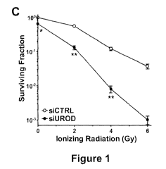
Figure 1 shows the identification of UROD as a novel radiosensitizing target
via a
siRNA-based high-throughput screen. (A) Preliminary screen of the Human
siGENOME Druggable (6080 genes) and Protein Kinase (800 genes) siRNA Libraries
at 2 Gy in transfected FaDu (human hypopharyngeal squamous cell cancer) cells.
(B)
67 target sequences with potential radiosensitizing effects (>50% reduction in
surviving
fraction at 2 Gy vs. 0 Gy) were identified. Targets that decreased the
surviving fraction
by >30% in the absence of IR were not considered (grey box). Known
radiosensitizing
targets (grey circles); UROD (black circle); scrambled siRNA control (black
triangle).
(C) Clonogenic survival curves of FaDu cells transfected with scrambled
control
siRNA (siCTRL) or UROD siRNA (siUROD) for 48 h, then irradiated (0-6 Gy).
Colonies were counted 12 days post-IR. *p<0.05 and **p<0.01, siCTRL vs. siUROD
for each IR dose. (D) As in (C), but FaDu cells were transfected with a range
of siRNA
concentrations (0-60 nM), combined with IR (0-6 Gy) for Chou-Talalay
combination
index analyses. (E) Relative UROD mRNA levels in FaDu cells transfected with
siCTRL or siUROD for 24, 48, and 120 h, as measured by qRT-PCR. **p<0.01,
siCTRL vs. siUROD. (F) UROD protein expression was detected by immunoblotting
at
24-72 h post-transfection. (G) FaDu cells were co-transfected with siRNA
(siCTRL or
siUROD) and plasmid DNA (empty vector control, pVector or siRNA-resistant
rescue
plasmid, pUROD) for 48 h, and then irradiated (4 Gy). Apoptotic fractions were
assessed by flow cytometry 72 h post-IR. **p<0.01, siCTRL-pVector vs. siUROD-
pVector or siUROD-pUROD IR. Each datum represents the mean SEM from three
independent experiments.
Figure 2 shows that the radio sensitizing effect of UROD knockdown is
independent of
porphyrin accumulation. (A) Heme biosynthetic pathway. ALA, 6-aminolevulinic
acid;
CPOX, coproporphyrinogen oxidase; PPOX, protoporphyrinogen oxidase; Fe, iron.
(B)
Porphyrin synthesis in mock-, siCTRL-, or siUROD-transfected FaDu cells was
artificially induced with ALA (500 M, 4 h) prior to porphyrin extraction at
24 h post-
transfection. Porphyrin levels were quantified spectrofluorometrically and
normalized
to total cell number. Representative spectral scans (575-750 nm) are shown.
**p<0.01,
siUROD vs. siCTRL or untreated ALA. (C) Fluorescent microscopy images of
transfected cells ALA (500 M, 1 h). Mitochondria and nuclei were stained
with
5
CA 02758229 2011-10-07
WO 2010/118524 PCT/CA2010/000569
MitoTracker Green and Hoechst 33342, respectively. Intracellular porphyrin
excited
with a wavelength of -400 nm emits red fluorescence at a peak of -635 nm.
Scale bar,
m. (D) ALA-treated (250-1000 M, 4 h) and siCTRL- or siUROD-transfected (48
h-transfection) FaDu cells were irradiated (4 Gy), then cell viability was
assessed 96 h
5 later via MTS assay. **p<0.01, siCTRL vs. siUROD IR; untreated vs. ALA
IR. In
all cases, each datum represents the mean SEM from three independent
experiments.
Figure 3 shows that UROD down-regulation promotes radiation-induced
cytotoxicity.
(A) Flow cytometric DNA content analyses of siCTRL- or siUROD-transfected FaDu
cells at 12-72 h post-IR (4 Gy). Representative histograms with gates for cell
cycle
10 distributions are shown. *p<0.05 and **p<0.01, siCTRL vs. siUROD IR at
each time
point. (B) Flow cytometric analyses of cellular y-H2AX expression levels in
transfected
FaDu cells at 0-240 min post-IR (4 Gy). **p<0.01, siCTRL vs. siUROD at each
time
point. (C) Representative images of y-H2AX nuclear foci formation in siCTRL-
and
siUROD-transfected FaDu cells 30 min post-IR. Scale bar, 10 m. (D) Flow
cytometric
analyses of caspase 9, 8, and 3 activation in siCTRL or siUROD-transfected
FaDu cells
at 12-48 h post-IR (4 Gy). *p<0.05 and **p<0.01, siCTRL vs. siUROD IR at
each
time point. (E) A PM depolarization was quantified by flow cytometry 48 h post-
IR in
transfected FaDu cells. **p<0.01, siCTRL vs. siUROD IR. Each datum
represents the
mean SEM from three independent experiments.
Figure 4 shows that siUROD-mediated radiosensitization enhances cellular
oxidative
stress. (A) Intracellular superoxide anions in siCTRL- or siUROD-transfected
FaDu
cells at 3-72 h post-IR (4 Gy) were detected by flow cytometry with
dihydroethidium
(DE). *p<0.05 and **p<0.01, siCTRL vs. siUROD IR at each time point. (B)
Overall
ROS levels in transfected FaDu cells were measured with CM-H2DCFDA at 3-72 h
post-IR (4 Gy). *p<0.05 and **p<0.01, siCTRL vs. siUROD IR at each time
point.
(C) Superoxide radical levels in two transfected normal head and neck
epithelial cells
(NOP, normal oropharyngeal; NOE, normal oral epithelial) 72 h post-IR (4 Gy).
**p<0.01, normals vs. FaDu at 72 h post-IR. (D) Overall ROS levels in
transfected
NOP and NOE cells 72 h post-IR (4 Gy). *p<0.05 and **p<0.01, normals vs. FaDu
at
72 h post-IR. (E) Cell viability of siCTRL or siUROD-transfected FaDu, NOP,
and
6
CA 02758229 2011-10-07
WO 2010/118524 PCT/CA2010/000569
NOE cells at 96 h post-lR (2 Gy) via MTS assay. **p<0.01, siCTRL vs. siUROD
IR.
(F) FaDu cells were transfected with siCTRL or siUROD and irradiated under
normoxia (21% 02) or hypoxia (0.2% 02). Apoptotic fractions were assessed by
now
cytometry 72 h post-IR. *p<0.05 and **p<0.01, normoxic vs. hypoxic treatments.
(G)
Relative mRNA expression of a panel of genes involved in cellular oxidative
stress
responses in siCTRL- or siUROD-transfected FaDu cells 48 h post-IR. Relative
fold
changes represent average ACt values normalized to those of (3-actin, then
compared to
siCTRL-transfected cells. **p<0.01, siCTRL vs. siUROD IR. Each datum
represents
the mean SEM from three independent experiments.
Figure 5 shows that UROD knockdown induces intracellular iron accumulation.
(A)
Ferrous (Fe2+) and ferric (Fe3+) iron staining of siCTRL or siUROD-transfected
FaDu
cells at 48 h post-IR (4 Gy). Scale bar, 50 m. (B) Quantification of
intracellular Fe2+
and Fe 3+ levels from (A). Deep-purple areas and total area of cultured cells
were
measured. The ratio (% area) was calculated by dividing the sum of deep-purple
areas
by the sum of the total area from sections. *p<0.05 and **p<0.01, siCTRL vs.
siUROD
IR. (C) FaDu cells transfected with siCTRL or siUROD for 24 h were treated
with
deferoxamine (DFO; 5 MM), and then irradiated (4 Gy) 24 h later. Apoptotic
fractions
were assessed by flow cytometry 72 h post-IR. **p<0.01, - DFO vs. + DFO
treatments.
Each datum represents the mean SEM from at least two independent
experiments.
Figure 6 shows the in vivo efficacy of UROD knockdown plus irradiation in HNC
models. (A) Mock, siCTRL, or siUROD-transfected FaDu cells were implanted into
the
left gastrocnemius muscle of SCID mice, followed immediately by local RT (4
Gy).
Each treatment group comprised of 9 mice. ***p<0.001, siUROD vs. mock or
siCTRL
RT. (B) FaDu tumors were established in SCID mice; once TLDs reached -8 mm,
mice were randomly assigned to siCTRL, siUROD, siCTRL-plus-RT, or siUROD-plus-
RT. Mice were intraperitoneally-injected with 600 pmol of jetPEI-complexed
siRNA
thrice a week for up to 2 weeks (white arrows). Local tumor RT (4 Gy) was
delivered
on days 5 and 13 post IP-injections (grey arrows). Each treatment group
comprised of
>_5 mice. ***p<0.001, siUROD vs. siCTRL + RT. (C) UROD knockdown was assessed
in FaDu tumors 24 h after the last treatment as described in (B). Excised
tumors were
7
CA 02758229 2011-10-07
WO 2010/118524 PCT/CA2010/000569
subjected to immunoblotting for UROD expression. Western blots were quantified
and
relative fold changes in UROD protein levels were determined by normalizing to
corresponding GAPDH loading controls, then compared to siCTRL-treated tumors.
(D)
UROD knockdown in tumors (black arrows) was also verified by
immunohistochemistry. (E) Minimal differences in the average mice body weights
for
each treatment group from (B) indicated that the systemic siUROD-plus-local RT
regimen was well-tolerated. Each datum represents the mean SEM from at least
two
independent experiments.
Figure 7 shows the clinical relevance of UROD in human cancers. (A) Cell
viability
assessment of siCTRL or siUROD-transfected cancer cells at 96 h post-IR (2 Gy)
via
MTS assay. Human HNC (FaDu, C666-1, UTSCC-8, UTSCC-42a), cervix (SiHa, ME-
180), breast (T47D), lung (A549), and prostate (DU-145) cancer cell lines.
**p<0.01,
siCTRL vs. siUROD IR. (B) Relative UROD mRNA expression in UTSCC-42a cells
transfected with UROD-expressing plasmid (pUROD) or empty vector control
(pVector) for 48 h, determined via qRT-PCR. ***p<0.001, pVector vs. pUROD. (C)
UTSCC-42a cells transfected with pUROD or pVector for 48 h were irradiated (2
Gy).
Apoptotic fractions were assessed by flow cytometry 72 h post-IR.
Representative
histogram of cell cycle distribution is shown. ***p<0.001, pUROD vs. pVector +
IR.
(D) Total RNA was extracted from 38 HNSCC patient tumor biopsies and 5 normal
laryngeal and tonsillar epithelial tissues, and assessed for relative levels
of UROD
mRNA expression. Fold change was determined by normalizing to (3-actin levels,
and
comparing to the average from normal tissues. Solid line, mean fold change.
*p<0.05,
tumor vs. normal tissues. (E) Kaplan-Meier plot of disease-free survival (DFS)
for the
HNSCC patients from (D); trichotomized based on interquartile range (low,
medium,
vs. high levels of UROD mRNA expression). DFS was defined as absence of
relapse or
death, calculated from the time of diagnosis. Median follow-up time was 6.9
years
(range 2.3-10.8 yrs). (F) Cell viability assessment of irradiated (2 Gy)
primary normal
human fibroblasts (MRCS, GM05757) and untransformed fibroblasts from PCT
patients (GM01482, GM00977, GM00961, GM01041) 96 h post-IR via MTS assay.
*p<0.05, MRCS vs. PCT fibroblasts. (G) siCTRL- or siUROD-transfected FaDu
cells
were treated with increasing doses of Cisplatin (0.01-0.25 AM), 5-FU (1-2.5
AM), or
8
CA 02758229 2011-10-07
WO 2010/118524 PCT/CA2010/000569
Paclitaxel (PTX) (0.1 M) for 24 h, then assessed for cell viability 96 h
later.
***p<0.001 and *p<0.05, siCTRL drug vs. siUROD drug. Each datum represents
the mean SEM from three independent experiments.
DETAILED DESCRIPTION
In the following description, numerous specific details are set forth to
provide a
thorough understanding of the invention. However, a person skilled in the art
would
understand when the invention may be practiced without certain specific
details. Some
methods herein have been described as a series of steps and a person skilled
in the art
will also understand that the steps may be performed in any logical order
unless the
context dictates otherwise.
Head and neck cancer (HNC) is a challenging disease due to its heterogeneity
and
complexity, often resulting in poor survival rates. Radiation therapy (RT)
remains the
primary curative modality for HNC. Even the most effective RT regimens
however,
achieve local control rates of 45-55%, with disease-free survival rates of
only 30-40%.
Thus, the development of novel strategies to enhance tumor cell killing, while
minimizing damage to the surrounding normal tissues, is critical to improving
cure
rates with RT.
A siRNA-based high-throughput screen (HTS) was developed for the large-scale
identification of novel genes that will selectively sensitize HNC cells to
radiation. The
preliminary screen identified 188 target sequences with potential
radiosensitizing
effects; the validity of the screen was corroborated by the identification of
known
radiosensitizing targets (e.g. ATM, ATR, Aurora-A kinase). To confirm the
initial HTS
results, FaDu cells (human hypopharyngeal squamous cell cancer) were
transfected
with the 188 siRNAs RT, and those that were cytotoxic without RT were
eliminated,
leaving 67 potential `hits'. Targets reducing surviving fraction by >50% at 2
Gy
relative to their un-irradiated counterparts were selected. Corroboration of
siRNA-
mediated mRNA and protein knockdown were assessed using qRT-PCR and Western
blotting, respectively.
9
CA 02758229 2011-10-07
WO 2010/118524 PCT/CA2010/000569
A key regulator of the heme biosynthetic pathway, uroporphyrinogen
decarboxylase
(UROD), was thus identified as a potent radiosensitizer. Increased heme
biosynthesis
has previously been reported in tumor tissues with up-regulation of several
regulatory
proteins, including UROD. The broad applicability of this radiosensitization
was
exhibited in other HNC cell lines (nasopharyngeal and laryngeal squamous
cancers), as
well as other cancer models (cervix, breast, lung, and prostate carcinomas);
no
radiosensitization was observed in normal oral cavity or oropharyngeal
epithelial cells.
Functional validation studies and in vitro characterization of mechanisms for
radiosensitization were examined. These studies suggest an effect mediated by
tumor-
selective enhancement of cellular oxidative stress via perturbation of iron
homeostasis
and increased reactive oxygen species (ROS) production. In vivo validation
studies
such as tumor formation assays and treatment of established HNC xenograft
models
were also evaluated. The clinical relevance of UROD down-regulation in head
and
neck cancer was also demonstrated.
UROD knockdown has significant implications in the management of human
cancers.
Its over-expression is able to prognosticate for radiation resistance, thereby
potentially
allowing selection of cancer patients who would be suitable for siUROD
radiosensitization. The therapeutic application of this approach is broad, and
effective
in the selective enhancement of radiation-induced cytotoxicity in cancer
tissues, with
no toxicity observed in normal tissues. Furthermore, there is a naturally
occurring state
of porphyria cutanea tarda (PCT), which is non-lethal; hence a "temporary"
state of
PCT would have minimal consequences to cancer patients during the few weeks of
RT
and/or chemotherapy. This discovery uncovers the translational significance of
iron
homeostasis and dysregulation within the context of tumor radiosensitization,
warranting further investigations into this important biological process.
Therefore, in accordance with one aspect, there is provided a method for
sensitizing a
subject with cancer to a cancer therapy comprising administering to the
subject a
sensitizing amount of an agent that downregulates or inhibits UROD.
Preferably, the
cancer is a head and neck cancer and is selected from the group consisting of
cancers
CA 02758229 2011-10-07
WO 2010/118524 PCT/CA2010/000569
originating from the lip, nasal and oral cavities, sinuses, pharynx, larynx,
and other sites
in this anatomical region.
In an embodiment, the cancer is selected from the group consisting of
hypopharyngeal
carcinoma, nasopharyngeal carcinoma, laryngeal carcinoma, lung adenocarcinoma,
cervical carcinoma, prostate carcinoma and mammary adenocarcinoma.
In accordance with a further aspect, there is provided a method for
sensitizing a subject
with cancer to a cancer therapy comprising downregulating or inhibiting UROD
in
cancer cells of the subject.
In accordance with a further aspect, there is provided use of an agent that
downregulates or inhibits UROD for sensitizing a subject to a cancer therapy.
In accordance with a further aspect, there is provided use of an agent that
downregulates or inhibits UROD in the preparation of a medicament for
sensitizing a
subject to a cancer therapy.
In accordance with a further aspect, there is provided a compound for
sensitizing a
subject with cancer to a cancer therapy comprising a UROD inhibitor or UROD
downregulator.
In accordance with a further aspect, there is provided a method for
identifying an agent
that sensitizes a subject with cancer to a cancer therapy comprising screening
for a
compound that downregulates or inhibits UROD.
In accordance with a further aspect, there is provided a method of
prognosticating a
survival outcome to a cancer therapy of a subject with cancer comprising:
providing a sample comprising cancer cells from the subject; and
determining the level of UROD expression and/or activity in the cancer cells;
11
CA 02758229 2011-10-07
WO 2010/118524 PCT/CA2010/000569
wherein a relatively low level of UROD expression and/or activity compared to
a control is correlated with an improved clinical outcome in response to
cancer
therapy.
In accordance with a further aspect, there is provided a method of diagnosing
a subject
with cancer comprising:
providing a sample from the subject; and
assaying the level of UROD expression and/or activity in the sample;
wherein a relatively high level of UROD expression and/or activity compared to
a control is correlated with cancer.
In accordance with a further aspect, there is provided a kit for diagnosing a
cancer in or
prognosticating a survival outcome to a cancer therapy of a subject with the
cancer,
comprising an assay for UROD expression and/or activity along with
instructions for
use.
In accordance with a further aspect, there is provided a method for
sensitizing a subject
with cancer to a cancer therapy comprising elevating the intracellular iron in
cancer
cells of the subject.
In accordance with a further aspect, there is provided a method for
sensitizing a subject
with cancer to a cancer therapy comprising administering an agent that
elevates
intracellular iron.
In accordance with a further aspect, there is provided a use of an agent that
elevates the
intracellular iron in cancer cells for sensitizing a subject to a cancer
therapy.
In accordance with a further aspect, there is provided a use of an agent that
elevates the
intracellular iron in cancer cells in the preparation of a medicament for
sensitizing a
subject to a cancer therapy.
12
CA 02758229 2011-10-07
WO 2010/118524 PCT/CA2010/000569
In accordance with a further aspect, there is provided a compound for
sensitizing a
subject with cancer to a cancer therapy comprising an elevator of
intracellular iron.
In preferable embodiments, the cancer therapy is radiation therapy. In one
embodiment,
the radiation therapy is therapy using ionizing radiation. In another
embodiment, the
radiation therapy is therapy using non-ionizing radiation and is preferably
photodynamic therapy.
In other embodiments, the cancer therapy is chemotherapy. Preferably, the
chemotherapy uses Cisplatin, 5-FU or Paclitaxel.
In some embodiments, the agent is any one of an siRNA, antisense
oligonucleotide,
miRNA, aptamer, protein, shRNA and small molecule, that downregulates or
inhibits
UROD or a modified version of any of the foregoing.
The term "radiation therapy" is used interchangeably with the term
"radiotherapy". In
some embodiments, the radiation is one of x-ray and gamma ray. For example,
but not
by way of limitation, x-ray radiation can be administered; in particular, high-
energy
megavoltage (radiation of greater that 1 MeV energy) can be used for deep
tumors, and
electron beam and orthovoltage x-ray radiation can be used for skin cancers.
Gamma
ray emitting radioisotopes, such as radioactive isotopes of radium, cobalt and
other
elements may also be administered to expose tissues to radiation. However, any
radiation therapy protocol can be used depending upon the type of cancer to be
treated.
Radiation therapy as used herein includes both ionizing and non-ionizing
radiation.
Non-ionizing radiation may be used, for example, in connection with
photodynamic
therapy ("PDT") and PDT-photosensitizing agents.
The term "chemotherapy" refers to the use of drugs to treat cancer. A
"chemotherapeutic agent" is used to connote a compound or composition that is
administered in the treatment of cancer. Some examples of chemotherapeutic
agents
include, but are not limited to, antibiotic chemotherapeutics such as,
Doxorubicin,
Daunorubicin, Mitomycin (also known as mutamycin and/or mitomycin-C),
Actinomycin D (Dactinomycin), Bleomycin, Plicomycin; plant alkaloids such as
Taxol,
13
CA 02758229 2011-10-07
WO 2010/118524 PCT/CA2010/000569
Vincristine, Vinblastine; miscellaneous agents such as Cisplatin, VP16, Tumor
Necrosis Factor; alkylating agents such as, Carmustine, Melphalan (also known
as
alkeran, L-phenylalanine mustard, phenylalanine mustard, L-PAM, or L-
sarcolysin, is a
phenylalanine derivative of nitrogen mustard), Cyclophosphamide, Chlorambucil,
Busulfan (also known as myleran), Lomustine; and other agents for example,
Cisplatin
(CDDP), Carboplatin, Procarbazine, Mechlorethamine, Camptothecin, Ifosfamide,
Nitrosurea, Etoposide (VP16), Tamoxifen, Raloxifene, Estrogen Receptor Binding
Agents, Gemcitabine, Navelbine, Farnesyl-protein transferase inhibitors,
Transplatinum, 5-Fluorouracil, and Methotrexate, Temazolomide (an aqueous form
of
DTIC), or any analog or derivative variant of the foregoing.
As used herein, "UROD" refers to Uroporphyrinogen decarboxylase enzyme or gene
as
the context dictates. UROD is an enzyme in the heme biosynthetic pathway,
catalyzing
the decarboxylation of uroporphyrinogen to form coproporphyrinogen and four
molecules of carbon dioxide.
The term "oligonucleotide" as used herein refers to a nucleic acid molecule
comprising
from about 1 to about 100 nucleotides, more preferably from 1 to 80
nucleotides, and
even more preferably from about 4 to about 35 nucleotides. This may include
nucleic
acid molecules of variable length that correspond either to the sense strand
or to the
non-coding strand of a target nucleic acid sequence.
"Antisense oligonucleotides" (AON) are complementary to a region of a target
gene
and are capable of hybridizing to the target gene sequence and inhibiting gene
expression. Gene expression is inhibited through hybridization of an AON to a
specific
messenger RNA (mRNA) sense target according to the Watson-Crick base pairing,
typically in which adenosine and thymidine (uracil in mRNA) or guanosine and
cytidine interact through hydrogen bonding. Without being bound to any theory,
two
mechanisms are generally thought to account for these effects, the first being
hybridization with impaired translation of targeted mRNA, the second being the
induction of RNase H or similar enzymes with associated degradation of target
mRNA.
14
CA 02758229 2011-10-07
WO 2010/118524 PCT/CA2010/000569
Oligonucleotide compounds in accordance with the present invention also
include
siRNAs (small interfering RNAs) and the RISCs (RNA-induced silencing
complexes)
containing them that result from the RNAi (RNA interference) approach. The
RNAi
approach is a tool for the inhibition of target gene expression. RNAi is based
on an
ancient anti-viral defense mechanism in lower eukaryotes. It is induced by
double-
stranded RNA and its processing to typically 21-23 nt siRNAs, which cause the
degradation of homologous endogenous mRNA after hybridizing to the target mRNA
in a single stranded fashion with the assistance of the RISC complex. The way
in which
RNAi inhibits target gene expression remains to be fully elucidated, but
presently,
RNAi serves as an attractive choice approach to generate loss-of-function
phenotypes
across a broad spectrum of eukaryotic species, such as nematodes, flies,
plants, fungi
and mammals..
Oligonucleotide compounds in accordance with the present invention also
include
microRNA (miRNA). "MicroRNA" are single-stranded RNA molecules, typically of
about 21-23 nucleotides in length, which regulate gene expression in a
hybridization
dependent manner. Typically, miRNAs are encoded by genes that are transcribed
from
DNA but not translated into protein (non-coding RNA); instead they are
processed
from primary transcripts known as pri-miRNA to short stem-loop structures
called pre-
miRNA and finally to functional miRNA. Mature miRNA molecules are partially
complementary to one or more messenger RNA (mRNA) molecules, typically at the
3'end of the mRNA, and their main function is to downregulate gene expression.
As used herein, the term "aptamer," e.g., RNA aptamer or DNA aptamer, includes
single-stranded oligonucleotides that bind specifically to a target molecule.
Aptamers
are selected, for example, by employing an in vitro evolution protocol called
systematic
evolution of ligands by exponential enrichment. Aptamers bind tightly and
specifically
to target molecules; most aptamers to proteins bind with a Kd (equilibrium
dissociation
constant) in the range of 1 pM to 1 nM. Aptamers and methods of preparing them
are
described in, for example, E. N. Brody et al. (1999) Mol. Diagn. 4:381-388.
In one embodiment, the subject aptamers can be generated using SELEX, a method
for
generating very high affinity receptors that are composed of nucleic acids
instead of
CA 02758229 2011-10-07
WO 2010/118524 PCT/CA2010/000569
proteins. See, for example, Brody et al. (1999) Mol. Diagn. 4:381-388. SELEX
offers a
completely in vitro combinatorial chemistry alternative to traditional protein-
based
antibody technology. Similar to phage display, SELEX is advantageous in terms
of
obviating animal hosts, reducing production time and labor, and simplifying
purification involved in generating specific binding agents to a particular
target PET.
An "amino acid" is a monomer unit of a peptide, polypeptide, or protein. There
are
twenty amino acids found in naturally occurring peptides, polypeptides and
proteins, all
of which are L-isomers. The term also includes analogs of the amino acids and
D-
isomers of the protein amino acids and their analogs.
A "protein" is any polymer consisting essentially of any of the 20 amino
acids.
Although "polypeptide" is often used in reference to relatively large
polypeptides, and
"peptide" is often used in reference to small polypeptides, usage of these
terms in the
art overlaps and is varied. The term "protein" as used herein refers to
peptides, proteins
and polypeptides, unless otherwise noted.
The term "small molecule" is a term of the art and includes molecules that are
less than
about 1000 molecular weight or less than about 500 molecular weight. Exemplary
small molecule compounds which can be screened for activity include, but are
not
limited to, peptides, peptidomimetics, nucleic acids, carbohydrates, small
organic or
inorganic molecules, and natural product extract libraries.
The term "downregulate" is used herein to refer to at least partial inhibition
or
knockdown of the expression of a gene or activity of the protein that it
encodes. For
example, in some embodiments, an antisense oligonucleotide, siRNA or miRNA
compound exhibiting complementarity to UROD downregulates or inhibits
expression
of UROD in a hybridization dependent manner. In another embodiment an aptamer,
protein or small molecule downregulates or inhibits UROD protein activity by
binding
thereto.
As used herein, the term "screening" or "to screen" refers to a process in
which a large
number of potentially useful agents are processed in the methods of the
invention.
16
CA 02758229 2011-10-07
WO 2010/118524 PCT/CA2010/000569
Without limitation, screening may refer to an assay of members having a
desired
activity or function from a library such as small molecule, aptamer, protein
and nucleic
acid libraries. For example, in some embodiments, potential antisense
oligonucleotides,
siRNAs and/or miRNAs exhibiting complementarity to UROD are screened/processed
in order to identify species that downregulate or inhibit expression of UROD
in a
hybridization dependent manner. In another embodiment aptamers, proteins
and/or
small molecules are screened/processed to identify species that downregulate
or inhibit
UROD protein activity by binding thereto.
The term "sensitizing amount" means a sufficient amount of an agent to provide
the
desired sensitizing effect. For example, in some embodiments, "sensitizing
amount"
means that dose of agent effective to increase the sensitivity of cancerous
cells or
tumour to radiation therapy or chemotherapy.
The term "prognosticating" as used herein means predicting or identifying the
clinical
outcome group that a subject belongs to according to the subject's similarity
to a
control group or control profile.
The term "diagnosing" means judging, predicting, assessing and/or evaluating
as well
as identifying and characterizing, including screening, whether a person is
susceptible
of or suffers from cancer, including, but not limited to head and neck
cancers.
The term "sample" as used herein refers to any fluid, cell or tissue sample
from a
subject, which can be assayed, for example, for UROD expression or activity.
As used herein, the term "control" refers to a specific value or dataset that
can be used
to prognosticate, diagnose or classify the value e.g. expression level of UROD
obtained
from the test sample associated with an outcome class (e.g. high vs. low
survival or
tumour vs. normal cells). A person skilled in the art will appreciate that the
comparison
between the expression of UROD in the test sample and the expression of UROD
in the
control will depend on the control used. In some embodiments, the control
comprises
an UROD expression profile from multiple samples in order to dichotomize the
control
values into different outcome classes (e.g. high vs. low survival or tumour
vs. normal
17
CA 02758229 2011-10-07
WO 2010/118524 PCT/CA2010/000569
cells). As such, when a test sample is compared to the UROD expression
profile, the
test sample can be placed in one of the outcome classes based on UROD
expression.
EXAMPLES
MATERIALS AND METHODS
Cell Lines
FaDu, A549, SiHa, ME-180, T47D, DU-145, and MRC5 cells were obtained from the
American Type Culture Collection (Manassas, VA). Normal human oropharyngeal
(NOP) and oral epithelial (NOE) cells were purchased from Celprogen (San
Pedro,
CA). Untransformed fibroblasts from familial porphyria cutanea tarda (type II)
patients
(GM01482, GM00977, GM00961, GM01041) and GM05757 (primary normal human
skin) fibroblasts were obtained from Coriell Institute (Camden, NJ). All cell
lines were
cultured according to the manufacturer's specifications. C666-1
undifferentiated
nasopharyngeal cancer cells [16] were maintained in RPMI 1640 supplemented
with
10% fetal bovine serum (Wisent, Quebec, Canada) and antibiotics (100 mg/L
penicillin
and 100 mg/L streptomycin). UTSCC-8 and -42a laryngeal squamous cell cancer
cells
were a gift from R. Grenman (Turku, Finland) and maintained as previously
described
[17]. All cells were maintained in 5% C02, 21% 02, and 95% humidity at 37 C
unless
otherwise stated.
Patient Samples
Thirty-eight formalin-fixed paraffin-embedded (FFPE) tissue biopsies from
locally
advanced HNSCC patients (Stage III or IV; oropharynx, hypopharynx, or larynx
primary SCC subsites), who participated in a randomized clinical study of two
RT
fractionation regimens [18] were utilized with Institutional Research Ethics
Board
approval. FFPE samples were macro-dissected for regions of invasive SCC (>70%
malignant epithelial cell content). Five normal human larynx and tonsillar
FFPE tissues
were purchased from Asterand (Detroit, MI). Total tumor RNA was extracted with
RecoverAll Total Nucleic Acid Isolation Kit for FFPE (Ambion, Austin, TX) as
specified by the manufacturer.
18
CA 02758229 2011-10-07
WO 2010/118524 PCT/CA2010/000569
Reagents
Cisplatin, 5-fluorouracil, Paclitaxel, S-aminolevulinic acid hydrochloride,
and
deferoxamine mesylate salt were obtained from Sigma-Aldrich (St. Louis, MO).
All
compounds were dissolved and/or diluted in complete media.
BrdU-Based siRNA High-Throughput Screen
The Human siGENOME Druggable and Protein Kinase siRNA Libraries (Dharmacon,
Lafayette, CO) were provided by the Samuel Lunenfeld Research Institute (SLRI)
HTS
Robotics Facility (Toronto, Canada). Automation of the 96-well siRNA
transfection
and bromodeoxyuridine (BrdU) cell proliferation assay (Exalpha Biologicals,
Shirley,
MA) were performed using the BioMek FX (Beckman Coulter, Fullerton, CA),
SpectraMax Plus384 microplate reader (Molecular Devices, Sunnyvale, CA), and
SLRI
robotics platform.
Working stock solutions of siRNA were prepared in Opti-MEM I reduced-serum
media
(Invitrogen, Carlsbad, CA). Reverse transfections (final concentration of 40
nM
siRNA) were performed with Lipofectamine 2000 (Invitrogen) as specified by the
manufacturer. Columns 1 and 2 of each plate contained siRNA targeting DNA
ligase
IV (LIG4 siGENOME SMARTpool; Dharmacon), serving as the positive
radiosensitizing control, and scrambled negative siRNA control (ON-TARGETplus
Non-Targeting Pool; Dharmacon), respectively. Twenty-four h post-transfection,
100
L of complete media was added to each well, then cells were irradiated using a
137Cs
unit (Gammacell 40 Extractor; MDS Nordion, Ottawa, Canada) at a dose rate of
0.84
Gy/min. Cells were incubated for an additional 72 h, at which time, BrdU
(Exalpha
Biologicals) was added to each well. After 24 h, cells were monitored for BrdU
incorporation on a SpectraMax Plus384 microplate reader according to the
manufacturer's specifications.
Transfections
siRNAs targeting UROD (Hs UROD 2/8 HP GenomeWide siRNAs) and a scrambled
control (AllStars Negative Control siRNA) were purchased from Qiagen
(Valencia,
CA). A plasmid vector containing the protein-coding sequence of UROD
(Hs UROD_IM_1 QlAgene Expression Construct) and an empty vector control (pQE-
19
CA 02758229 2011-10-07
WO 2010/118524 PCT/CA2010/000569
TriSystem Vector) were also purchased from Qiagen. All transfections were
performed
in complete media without antibiotics using Lipofectamine 2000 and 40 n1\4 of
siRNA
and/or 1 g of plasmid DNA.
Product Catalogue Sequence
Number
Target: 5'-GACGGTGACATTGCAGGGCAA-3'
(SEQ ID NO. 1)
Hs_UROD_2 Sense 5'-CGGUGACAUUGCAGGGCAATT-3'
siRNA SI00008162 Strand: (SEQ ID NO. 2)
Anti-sense 5'-UUGCCCUGCAAUGUCACCGTC-3'
Strand: (SEQ ID NO. 3)
Target: 5'-CTCAAGTACCACTAACACAGA-3'
(SEQ ID NO. 4)
Hs_UROD_8 Sense 5'-CAAGUACCACUAACACAGATT-3'
siRNA S105034988 Strand: (SEQ ID NO. 5)
Anti-sense 5'-UCUGUGUUAGUGGUACUUGAG-3'
Strand: (SEQ ID NO. 6)
AllStars Negative 1027281 Proprietary sequence
Control siRNA
Hs_UROD_IM_1
QlAgene
Expression EIM0140882 (SEQ ID NO. 7)
Construct
Plasmid
pQE-TriSystem 33903 (SEQ ID NO. 8)
Vector
Quantitative Real-Time PCR (qRT-PCR)
Primers for PCR amplifications were designed using Primer3 software
(http://primer3.sourceforge.net). Total RNA from transfected cells was
harvested using
the RNeasy Mini Kit (Qiagen). Total RNA (1 g) was reverse-transcribed using
SuperScript II Reverse Transcriptase (Invitrogen) as specified by the
manufacturer.
qRT-PCR was performed using SYBR Green PCR Master Mix (Applied Biosystems,
Foster City, CA), and an ABI PRISM 7900 Sequence Detection System (Applied
Biosystems) with cycle parameters previously described [12]. Relative mRNA
levels
were calculated using the 2-mct method [19].
CA 02758229 2011-10-07
WO 2010/118524 PCT/CA2010/000569
Gene Forward Sequence Reverse Sequence SEQ
ID
(3-ACTIN 5'-CCCAGATCATGTTTGAGACCT-3' 5'-AGTCCATCACGATGCCAGT-3' 9/10
UROD 5'-AGGCCTGCTGTGAACTGACT-3' 5'-CCTGGGGTACAACAAGGATG-3' 11/12
SOD I 5'-AGGGCATCATCAATTTCGAG-3' 5'-ACATTGCCCAAGTCTCCAAC-3' 13/14
SOD2 5'-TTGGCCAAGGGAGATGTTAC-3' 5'-AGTCACGTTTGATGGCTTCC-3' 15/16
5'-CTCTTCGAGAAGTGCGAGGT-3' 5'-TCGATGTCAATGGTCTGGAA-3' 17/18
GPX1
FTMT 5'-ACGTGGCCTTGAACAACTTC-3' 5'-ATTCCAGCAACGACTGGTTC-3' 19/20
Western Blot Analysis
Total protein extracts from transfected cells were harvested and prepared for
immunoblotting as previously described [12]. Membranes were probed with anti-
UROD polyclonal (clone L-19; 1:300 dilution; Santa Cruz Biotechnology, Santa
Cruz,
CA) or anti-GAPDH monoclonal (1:15000 dilution; Abcam, Cambridge, MA)
antibodies, followed by secondary antibodies conjugated to horseradish
peroxidase
(1:2000 dilution; Abcam). GAPDH protein levels were used as loading controls.
Western blots were quantified with the Adobe Photoshop Pixel Quantification
Plug-In
(Richard Rosenman Advertising & Design, Toronto, Canada).
Colony Formation Assay
Cells were irradiated (0-6 Gy) 48 h post-transfection and harvested
immediately for
seeding (500-5000 cells/well in 6-well plates). Twelve days later, colonies
were fixed
in 70% ethanol, stained with 10% methylene blue, and colonies of >-50 cells
were
counted. Clonogenic survival curve data were utilized to evaluate the
interactive effects
of combinatorial therapies via the Chou-Talalay combination index method [20].
Radiosensitivity was also expressed in terms of the mean inactivation dose (D-
bar),
which represents the area under the survival curve [21]. Radiosensitization
was
expressed as an enhancement ratio, defined as the mean inactivation doses of
control to
treatment.
21
CA 02758229 2011-10-07
WO 2010/118524 PCT/CA2010/000569
Cell Viability Assay
The CellTiter 96 AQueoas One Solution Cell Proliferation Assay (3-(4,5-
dimethylthiazol-2-yl)-5-(3-carboxymethoxyphenyl)-2-(4-sulfophenyl)-2H-
tetrazolium,
inner salt MTS (Promega, Madison, WI) was used to detect cell viability
according to
the manufacturer's specifications.
Flow Cytometric Assays
Flow cytometric analyses were performed on a FACSCalibur Flow Cytometer (BD
Biosciences, San Jose, CA), equipped with FlowJo software (Tree Star, Ashland,
OR).
Cell cycle distributions, caspase activation, and mitochondrial membrane
potentials
were measured as previously described [17]. Intracellular ROS levels were
quantified
using the non-specific 5-(and 6-)chloromethyl-2',7'-dichlorodihydrofluorescein
diacetate (CM-H2DCFDA) dye, and the superoxide-selective dihydroethidium (DE)
dye as instructed by the manufacturer (Invitrogen).
y-H2AX Detection
Global cellular y-H2AX protein levels were quantified by flow cytometry using
the
H2AX Phosphorylation Assay Kit (Upstate Biotechnology, Lake Placid, NY) as
specified by the manufacturer. To image y-H2AX nuclear foci, cells transfected
on
cover slips were fixed with 2% paraformaldehyde (PFA)-0.2% Triton X-100, then
probed with anti-y-H2AX mouse monoclonal antibody (clone JBW301; Upstate
Biotechnology), followed by donkey anti-mouse Alexa 488 antibody (Invitrogen)
and
DAPI (4',6-diamidino-2-phenylindole; Invitrogen) for nuclear staining. Cells
were
imaged with an Olympus IX81 inverted microscope equipped with a 16-bit
Photometrics Cascade 512B EM-CCD camera (Roper Scientific, Tucson, AZ).
Hypoxia Treatment
Transfected cells were immediately exposed to a continuous flow of humidified
0.2%
02 with 5% CO2 and balanced N2 (Praxair, Ontario, Canada) in an In Vivo2 400
Hypoxia Chamber (Ruskinn Technology, Pencoed, UK). An OxyLite 4000 oxygen-
sensing probe (Oxford Optronix, Oxford, UK) was used to verify target 02
levels.
22
CA 02758229 2011-10-07
WO 2010/118524 PCT/CA2010/000569
Iron Histochemistry
Intracellular Fe 2+ and Fe 3+ were detected according to Perl's Prussian blue
and
Turnbull's blue staining protocols [22], respectively. Images were captured
with a
Nikon ECLIPSE E600 microscope equipped with a Nikon DXM1200F digital camera
(Nikon Instruments, Melville, NY) for quantitative analysis using SimplePCI
imaging
software (Hamamatsu, Sewickley, PA).
Porphyrin Detection
Transfected cells were treated with ALA (500 M) for 4 h. Cells were lysed
with
SOLVABLE (PerkinElmer, Waltham, MA), and intracellular porphyrin levels were
measured spectrofluorometrically using a SpectraMax Plus384 microplate reader
(excitation 405 nm, emission 635 nm). To visualize porphyrin accumulation,
transfected cells ALA were stained with MitoTracker Green FM (Invitrogen)
and
Hoechst 33342 (Invitrogen) as specified by the manufacturer. Live cells were
imaged
on a Zeiss LSM5 10 confocal microscope (Carl Zeiss Microlmaging).
In Vivo Tumor Model
All animal experiments utilized 6-8 week-old severe combined immunodeficient
(SCID) BALB/c female mice in accordance with the guidelines of the Animal Care
Committee, Ontario Cancer Institute, University Health Network (Toronto,
Canada).
TLDs and body weights were recorded thrice weekly; mice were euthanized by CO2
once TLDs reached -14 mm.
Tumor Formation Assay
Cells transfected with siCTRL or siUROD for 48 h were harvested and implanted
into
the left gastrocnemius muscle of SCID mice (2.5x10⁵ viable cells in 100 gL
growth
medium per mouse), followed immediately by administration of local tumor RT (4
Gy).
Mice were immobilized in a Lucite box and the tumor-bearing leg was exposed to
225
kV (13 mA) at a dose rate of 3.37 Gy/min (X-RAD 225C Biological X-Ray
Irradiator;
Precision X-Ray, North Branford, CT).
23
CA 02758229 2011-10-07
WO 2010/118524 PCT/CA2010/000569
Therapeutic Tumor Growth Assay
Cells were implanted into the left gastrocnemius muscle of SCID mice (2.5x10⁵
viable
cells in 100 gL). Once the TLDs reached an average of -8 mm, mice were
injected
intraperitoneally (IP) with 600 pmol of siRNA complexed to in vivo-jetPEI
(Polyplus-
Transfection, New York, NY), thrice a week for up to 2 weeks. siRNAs were
mixed
with in vivo-jetPEI following the manufacturer's specifications
(nitrogen/phosphate
ratio: 8). Local tumor RT (4 Gy) was delivered on days 5 and 13 post IP-
injections.
In Vivo Knockdown Validation
To assess the extent of UROD knockdown in vivo, mice were sacrificed 24 h
after the
last treatment described in Methods (Therapeutic Tumor Growth Assay). Tumors
were
excised, immediately fixed in 10% formalin for 48 h, 70% alcohol for an
additional 48
h, paraffin embedded, and then sectioned (5 m). Immunohistochemical analysis
was
performed using microwave antigen retrieval with anti-UROD polyclonal antibody
(clone B02; 1:500 dilution; Abnova, Walnut, CA) and Level-2 Ultra Streptavidin
Detection System (Signet Laboratories, Dedham, MA). For immunoblotting, tumors
were excised and immediately snap-frozen in liquid nitrogen. 30 mg of tumor
tissue
was lysed and homogenized as detailed elsewhere [23]; 30 g of protein was
analyzed
for UROD expression via immunoblotting as described above.
Statistical Analyses
All experiments were performed at least three independent times, with the data
presented as the mean SEM. Statistical differences between treatment groups
were
determined using the Student's t test and one-way ANOVA. The Ingenuity
Pathways
Analysis software (Ingenuity Systems, Redwood City, CA) was used to identify
functional biological networks from the HTS data. The right-tailed Fisher
Exact test
was employed to calculate p-values and scores (p-score = -log,o p-value),
indicating the
likelihood of genes being observed together in a network due to random chance.
24
CA 02758229 2011-10-07
WO 2010/118524 PCT/CA2010/000569
RESULTS
We have successfully developed an RNAi-based radiosensitizer HTS (Fig. IA, B),
and
identified a heretofore unreported novel radiosensitizing target for the
treatment of
human HNC. Uroporphyrinogen decarboxylase (UROD) is the fifth enzyme in the
heme biosynthetic pathway (Fig. 2A) that catalyses the decarboxylation of
uroporphyrinogen to coproporphyrinogen [24]. Our findings reveal a potentially
novel
function of UROD in tumor response to ionizing radiation, an established anti-
cancer
treatment modality. Clonogenic survival curves confirmed UROD down-regulation
to
significantly enhance the radiosensitivity of FaDu cells, a highly aggressive
radioresistant HNC cell line, in a dose-dependent and synergistic manner (Fig.
1C,D).
Corroboration of siRNA-mediated UROD knockdown was determined via qRT-PCR
and immunoblotting (Fig. 1E,F). To ensure this observation was not due to off-
target
effects, a rescue plasmid expressing target mRNA refractory to siRNA via
silent
mutations was utilized. Co-transfection of FaDu cells with siUROD and the
rescue
plasmid completely neutralized any siUROD-mediated effects, with or without IR
(Fig.
I G), further confirming a siUROD-specific process. In vivo, siUROD-plus-RT
dramatically reduced the tumor-forming capacity of FaDu cells (Fig. 6A), and
significantly delayed the growth of established tumors systematically treated
with
UROD siRNA plus local tumor RT (Fig. 6B); whilst maintaining a favorable
toxicity
profile (Fig. 6E; no significant difference in mice body weights with these
treatments).
UROD down-regulation was functionally validated by measuring overall changes
in
oxidized porphyrin levels. Spectrofluorometrically, porphyrin accumulation
with
siUROD alone was negligible (Fig. 2B); thus, FaDu cells were pre-treated with
S-
aminolevulinic acid (ALA) to artificially induce porphyrin synthesis. ALA-plus-
siUROD significantly increased intracellular porphyrin levels relative to ALA
alone or
siCTRL-treated cells. Similar observations were made via fluorescent
microscopy (Fig.
2C), reflecting the disruption of heme biosynthesis by siUROD. Since the
majority of
currently utilized photosensitizers in photodynamic therapy (PDT) are
porphyrin based
[25], it was of interest to compare the radiosensitizing effects of siUROD to
commonly
used photosensitizers. ALA-based PDT is a well established anti-cancer therapy
that
utilizes the heme precursor ALA, to induce accumulation of protoporphyrin IX
(PPIX)
CA 02758229 2011-10-07
WO 2010/118524 PCT/CA2010/000569
in neoplastic cells [26, 27]. When ALA-treated cells are exposed to visible
light, PPIX
become excited and induce ROS formation, leading to oxidative stress-mediated
cell
death. In this study, siUROD-plus-IR was dramatically more cytotoxic compared
to the
negligible effects of ALA-plus-IR (Fig. 2D), indicating that the effects of
siUROD
were independent of intracellular porphyrin accumulation (Fig. 2B,C), thus
distinct
from PDT.
Although PDT and our siUROD radiosensitizing strategy both exploit the heme
biosynthesis pathway to harnesses their anti-cancer effects, siUROD is
superior for
several reasons. Tumor hypoxia severely hampers PDT efficacy, since molecular
02 is
a prerequisite for the production of photo-induced singlet oxygen molecules
[28, 29].
However, siUROD-plus-IR retained radiosensitizing efficacy even under hypoxic
conditions (Fig. 4F). The applicability of PDT is further limited since the
light source
used to excite porphyrins and its derivatives occupy the visible spectrum,
which cannot
penetrate tissues >0.8 cm, restricting PDT to superficial lesions [30].
Moreover,
porphyrins cannot be excited by the high-energy photons of x-rays or y-rays
[31],
thereby accounting for the modest radiosensitizing efficacies of porphyrins
[30, 32, 33].
Thus, siUROD provides a clear therapeutic advantage with significant
sensitization by
y-rays, a mainstay in the standard anti-cancer therapeutic armamentarium.
The enhanced tumor radiosensitivity observed with UROD suppression (Fig. 1C)
was
mediated in part by G2-M cell cycle arrest (Fig. 3A), along with induction of
double-
strand DNA breaks (the most lethal type of DNA damage), reflected by increased
overall y-H2AX expression and nuclear foci formation in siUROD-plus-IR-treated
FaDu cells vs. IR alone (Fig. 3B,C). The significantly prolonged G2-M arrest
and
concomitant increase in the subG1 population suggested that the DNA damage
induced
by siUROD-plus-IR was more lethal than IR alone, thereby significantly
augmenting
apoptosis (Fig. 3A). The central role of apoptosis in siUROD-plus-IR-mediated
cytotoxicity was further evident by the induction of caspase activation (Fig.
3D) and
depolarization of the mitochondrial membrane potential (A VfM) (Fig. 3E), both
classical
hallmarks of apoptosis.
26
CA 02758229 2011-10-07
WO 2010/118524 PCT/CA2010/000569
Heme biosynthesis occurs within the cytoplasm and mitochondrion (Fig. 2A); the
latter
being a major source of intracellular free radicals [34]. Thus, to investigate
whether
siUROD mediated its radiosensitizing effects via perturbation of ROS
homeostasis,
intracellular levels of oxidants were measured. Mitochondrial superoxide anion
radicals, as well as other ROS species (hydrogen peroxide, hydroxyl radical,
peroxyl
radical, peroxynitrite anion), were significantly more prevalent in siUROD-
plus-IR vs.
IR- or siUROD-treated FaDu cells (Fig. 4A,B). Accordingly, anti-oxidants
involved in
maintaining cellular redox homeostasis, including superoxide dismutases (SOD1
and
SOD2), glutathione peroxidase (GPX1), and mitochondrial ferritin (FTMT) were
all
up-regulated in FaDu cells in response to siUROD-plus-IR (Fig. 4G). This
enhancement of ROS production appeared to be relatively tumor-specific (Fig.
4C,D),
translating into higher survival for normal vs. FaDu cells after siUROD IR
(Fig. 4E),
exposing a therapeutic window for tumor-selective radiosensitization.
Mitochondria are intimately involved in iron (Fe)-trafficking for heme
biosynthesis and
the formation of Fe-sulfur clusters [35]. These organelles, also being the
major source
of ROS production, have developed efficient mechanisms to segregate free Fe
from
ROS, thereby preventing the production of harmful hydroxyl radicals ('OH) via
Fenton-type reactions [36]. Accordingly, up-regulation of the Fe-sequestering
FTMT
anti-oxidant in siUROD IR treated cells (Fig. 4G) was associated with
markedly
elevated levels of intracellular ferrous (Fe2) and ferric (Fe 3) iron (Fig.
5A,B). The
central role of excess cellular Fe in mediating siUROD radiosensitization was
demonstrated by the significant suppression of siUROD-plus-IR-induced
apoptosis in
cells pre-treated with deferoxamine, a Fe-chelator, before IR (Fig. 5C). Thus,
the
novelty of our UROD discovery relates to the opportunity to perturb Fe
homeostasis as
the initiator of oxidative stress in tumor cells. When heme synthesis is
disrupted via
siUROD, large quantities of iron, which would normally be incorporated into
PPIX to
form heme, continue to be imported into the mitochondria. Upon IR, superoxide
and
hydroxyl radicals are formed [37], both of which can react with themselves to
form
H202, initiating the Fenton reaction and ultimately, enhancing oxidative
damage and
cell death.
27
CA 02758229 2011-10-07
WO 2010/118524 PCT/CA2010/000569
There is a paucity of literature surrounding UROD and cancer. Only a few
studies have
reported enhanced heme biosynthesis in human cancers, wherein increased UROD
activity was observed in breast tumors vs. normal tissues [38, 39]; the basis
for which
remained unclear. Our work represents the first such report in HNC, whereby
UROD
was markedly over-expressed in primary HNSCC vs. corresponding normal tissues
(Fig. 7D). A potential predictive value for UROD was also revealed, wherein
lower
levels of pre-treatment UROD expression appeared to correlate with improved
disease-
free survival (DFS) in HNSCC patients treated with RT (Fig. 7E); consistent
with the
notion that higher UROD levels conferred radioresistance, and supporting the
strategy
of reducing UROD to increase radiocurability. The possible role of UROD in
modulating tumor radioresponse was further supported by the reversal of the
radiosensitive phenotype of UTSCC-42a cells with exogenous UROD over-
expression
(Fig. 7B,C); thereby facilitating the selection of cancer patients who would
be
amenable to UROD-mediated radiosensitization.
The potential therapeutic application of siUROD in human cancers appears to be
quite
extensive. UROD down-regulation not only radiosensitized a wide range of solid
cancers while sparing normal cells (Fig. 7A and 4E), but also sensitized HNC
cells to
low doses of standard chemotherapeutic agents, such as Cisplatin, 5-
fluorouracil, and
Paclitaxel (Fig. 7G). Hence, siUROD could play a significant role in enhancing
the
outcome for both RT and chemotherapy in HNC patients, allowing lower treatment
doses to be administered without compromising cure. Furthermore, a naturally
occurring state of UROD deficiency is responsible for the clinical syndrome of
porphyria cutanea tarda (PCT), a chronic non-fatal disorder characterized by
elevated
cellular porphyrin and iron levels [24]. Thus, a transient development of
"PCT" during
the weeks of RT and/or chemotherapy should be well-tolerated. Evidence for
minimal
toxicity is provided by the few case reports wherein no significant increase
in toxicities
was observed when PCT-cancer patients underwent RT [40-42]. In our hands,
untransformed fibroblasts from familial PCT patients demonstrated minimal
cytotoxicity comparable to UROD-functional primary normal human fibroblasts
(Fig.
7F), corroborating our previous data that siUROD-mediated radiosensitization
is tumor
selective (Fig. 4E).
28
CA 02758229 2011-10-07
WO 2010/118524 PCT/CA2010/000569
In conclusion, the novel identification of down-regulating UROD has
significant
implications in the management of human cancers for several reasons. First,
its over-
expression is able to prognosticate for radiation resistance, thereby
potentially allowing
selection of cancer patients who would be suitable for siUROD
radiosensitization.
Second, the therapeutic application of this approach is broad and effective in
the tumor-
selective enhancement of radiation and chemotherapy efficacy. Third, there is
a
naturally occurring state of UROD deficiency that is non-lethal; hence, a
temporary
state of "PCT" would have minimal consequences to cancer patients during the
few
weeks of treatment. Finally, our discovery provides important insights into
the
translational significance of iron homeostasis and dysregulation in cancer.
Although preferred embodiments of the invention have been described herein, it
will be
understood by those skilled in the art that variations may be made thereto
without
departing from the spirit of the invention or the scope of the appended
claims. All
references described herein, including those listed on the following list, are
incorporated by reference.
29
CA 02758229 2011-10-07
WO 2010/118524 PCT/CA2010/000569
REFERENCES
1. Parkin DM, Bray F, Ferlay J, Pisani P. Global cancer statistics, 2002. CA
Cancer J Clin 2005; 55: 74-108.
2. Pai SI, Westra WH. Molecular pathology of head and neck cancer:
implications
for diagnosis, prognosis, and treatment. Annu Rev Pathol 2009; 4: 49-70.
3. Lo KW, To KF, Huang DP. Focus on nasopharyngeal carcinoma. Cancer Cell
2004; 5: 423-428.
4. Carvalho AL, Nishimoto IN, Califano JA, Kowalski LP. Trends in incidence
and prognosis for head and neck cancer in the United States: a site-specific
analysis of the SEER database. Int J Cancer 2005; 114: 806-816.
5. Chin D, Boyle GM, Porceddu S, Theile DR, Parsons PG, Coman WB. Head and
neck cancer: past, present and future. Expert Rev Anticancer Ther 2006; 6:
1111-1118.
6. Bourhis J, Overgaard J, Audry H, et al. Hyperfractionated or accelerated
radiotherapy in head and neck cancer: a meta-analysis. Lancet 2006; 368: 843-
854.
7. Pignon JP, le Maitre A, Maillard E, Bourhis J. Meta-analysis of
chemotherapy
in head and neck cancer (MACH-NC): an update on 93 randomised trials and
17,346 patients. Radiother Oncol 2009; 92: 4-14.
8. Bonner JA, Harari PM, Giralt J, et al. Radiotherapy plus cetuximab for
locoregionally advanced head and neck cancer: 5-year survival data from a
phase 3 randomised trial, and relation between cetuximab-induced rash and
survival. Lancet Oncol 2010; 11: 21-28.
9. Li JH, Shi W, Chia M, et al. Efficacy of targeted FasL in nasopharyngeal
carcinoma. Mol Ther 2003; 8: 964-973.
10. Yip KW, Li A, Li JH, et al. Potential utility of BimS as a novel apoptotic
therapeutic molecule. Mol Ther 2004; 10: 533-544.
11. Chia MC, Shi W, Li JH, et al. A conditionally replicating adenovirus for
nasopharyngeal carcinoma gene therapy. Mol Ther 2004; 9: 804-817.
12. Yip KW, Mocanu JD, Au PY, et al. Combination bcl-2 antisense and radiation
therapy for nasopharyngeal cancer. Clin Cancer Res 2005; 11: 8131-8144.
CA 02758229 2011-10-07
WO 2010/118524 PCT/CA2010/000569
13. Yip KW, Mao X, Au PY, et al. Benzethonium chloride: a novel anticancer
agent identified by using a cell-based small-molecule screen. Clin Cancer Res
2006; 12: 5557-5569.
14. Yip KW, Ito E, Mao X, et al. Potential use of alexidine dihydrochloride as
an
apoptosis-promoting anticancer agent. Mol Cancer Ther 2006; 5: 2234-2240.
15. Tannock IF, Hill RP, Bristow RG, Harrington L. The Basic Science of
Oncology. Fourth ed. Toronto: McGraw-Hill; 2005.
16. Cheung ST, Huang DP, Hui AB, et al. Nasopharyngeal carcinoma cell line
(C666-1) consistently harbouring Epstein-Barr virus. Int J Cancer 1999; 83:
121-126.
17. Ito E, Yip KW, Katz D, et al. Potential use of cetrimonium bromide as an
apoptosis-promoting anticancer agent for head and neck cancer. Mol Pharmacol
2009; 76: 969-983.
18. Cummings B, Keane T, Pintilie M, et al. Five year results of a randomized
trial
comparing hyperfractionated to conventional radiotherapy over four weeks in
locally advanced head and neck cancer. Radiother Oncol 2007; 85: 7-16.
19. Livak KJ, Schmittgen TD. Analysis of relative gene expression data using
real-
time quantitative PCR and the 2(-Delta Delta C(T)) Method. Methods 2001; 25:
402-408.
20. Chou TC, Talalay P. Quantitative analysis of dose-effect relationships:
the
combined effects of multiple drugs or enzyme inhibitors. Adv Enzyme Regul
1984; 22: 27-55.
21. Fertil B, Dertinger H, Courdi A, Malaise EP. Mean inactivation dose: a
useful
concept for intercomparison of human cell survival curves. Radiat Res 1984;
99: 73-84.
22. Carson FL. Histotechnology: A Self-Instructional Text. Second ed. Chicago:
American Society for Clinical Pathology; 1997.
23. Gillespie DL, Whang K, Ragel BT, Flynn JR, Kelly DA, Jensen RL. Silencing
of hypoxia inducible factor-lalpha by RNA interference attenuates human
glioma cell growth in vivo. Clin Cancer Res 2007; 13: 2441-2448.
24. Lambrecht RW, Thapar M, Bonkovsky HL. Genetic aspects of porphyria
cutanea tarda. Semin Liver Dis 2007; 27: 99-108.
31
CA 02758229 2011-10-07
WO 2010/118524 PCT/CA2010/000569
25. Berg K, Selbo PK, Weyergang A, et al. Porphyrin-related photosensitizers
for
cancer imaging and therapeutic applications. J Microsc 2005; 218: 133-147.
26. Kennedy JC, Pottier RH, Pross DC. Photodynamic therapy with endogenous
protoporphyrin IX: basic principles and present clinical experience. J
Photochem Photobiol B 1990; 6: 143-148.
27. Kennedy JC, Pottier RH. Endogenous protoporphyrin IX, a clinically useful
photosensitizer for photodynamic therapy. J Photochem Photobiol B 1992; 14:
275-292.
28. Moan J, Sommer S. Oxygen dependence of the photosensitizing effect of
hematoporphyrin derivative in NHIK 3025 cells. Cancer Res 1985; 45: 1608-
1610.
29. Mitchell JB, McPherson S, DeGraff W, Gamson J, Zabell A, Russo A. Oxygen
dependence of hematoporphyrin derivative-induced photoinactivation of
Chinese hamster cells. Cancer Res 1985; 45: 2008-2011.
30. Kulka U, Schaffer M, Siefert A, et al. Photofrin as a radiosensitizer in
an in
vitro cell survival assay. Biochem Biophys Res Commun 2003; 311: 98-103.
31. Evensen IF. The use of porphyrins and non-ionizing radiation for treatment
of
cancer. Acta Oncol 1995; 34: 1103-1110.
32. Schaffer M, Schaffer PM, Corti L, et al. Photofrin as a specific
radiosensitizing
agent for tumors: studies in comparison to other porphyrins, in an
experimental
in vivo model. J Photochem Photobiol B 2002; 66: 157-164.
33. Schaffer M, Ertl-Wagner B, Schaffer PM, et al. Porphyrins as
radiosensitizing
agents for solid neoplasms. Curr Pharm Des 2003; 9: 2024-2035.
34. Valko M, Leibfritz D, Moncol J, Cronin MT, Mazur M, Telser J. Free
radicals
and antioxidants in normal physiological functions and human disease. Int J
Biochem Cell Biol 2007; 39: 44-84.
35. Hower V, Mendes P, Torti FM, et al. A'general map of iron metabolism and
tissue-specific subnetworks. Mol Biosyst 2009; 5: 422-443.
36. Fenton HJH. Oxidation of tartaric acid in presence of iron. J Chem Soc
1894;
65: 899-910.
37. Tannock IF, Hill SA, Bristow RG, Harrington L. The Basic Science of
Oncology. Fourth ed. Toronto: McGraw-Hill; 2005.
32
CA 02758229 2011-10-07
WO 2010/118524 PCT/CA2010/000569
38. Navone NM, Polo CF, Frisardi AL, Andrade NE, Battle AM. Heme
biosynthesis in human breast cancer--mimetic "in vitro" studies and some heme
enzymic activity levels. Int J Biochem 1990; 22: 1407-1411.
39. Navone NM, Frisardi AL, Resnik ER, Battle AM, Polo CF. Porphyrin
biosynthesis in human breast cancer. Preliminary mimetic in vitro studies. Med
Sci Res 1988; 16: 61-62.
40. Maughan WZ, Muller SA, Perry HO. Porphyria cutanea tarda associated with
lymphoma. Acta Derm Venereol 1979; 59: 55-58.
41. Schaffer M, Schaffer PM, Panzer M, Wilkowski R, Duhmke E. Porphyrias
associated with malignant tumors: results of treatment with ionizing
irradiation.
Onkologie 2001; 24: 170-172.
42. Gunn GB, Anderson KE, Patel AJ, et al. Severe radiation therapy-related
soft
tissue toxicity in a patient with porphyria cutanea tarda: A literature
review.
Head Neck 2009.
33