Note: Descriptions are shown in the official language in which they were submitted.
CA 02758531 2016-09-06
INFLAMMATORY BOWEL DISEASE PROGNOSTICS
100011
10
BACKGROUND OF THE INVENTION
[0002] Inflammatory bowel disease (IBD), which occurs world-wide and afflicts
millions
of people, is the collective term used to describe three gastrointestinal
disorders of unknown
etiology: Crohn's disease (CD), ulcerative colitis (UC), and indeterminate
colitis (IC). IBD,
together with irritable bowel syndrome (IBS), will affect one-half of all
Americans during
their lifetime, at a Cost of greater than $2.6 billion dollars for IBD and
greater than $8 billion
dollars for IBS. A. primary determinant of these high medical costs is the
difficulty of
diagnosing digestive diseases and how these diseases will progress. The cost
of IBD and IBS
is compounded by lost productivity, with people suffering from these disorders
missing at
least 8 more days of work annually than the national average.
100031 Inflammatory bowel disease has many symptoms in common with irritable
bowel
syndrome, including abdominal pain, chronic diarrhea, weight loss, and
cramping, making
definitive diagnosis extremely difficult. Of the 5 million people suspected of
suffering from
IBD in the United States, only I million are diagnosed as having IBD. The
difficulty in
differentially diagnosing IBD and determining its outcome hampers early and
effective
treatment of these diseases. Thus, there is a need for rapid and sensitive
testing methods for
prognosticating the severity of IBD.
[00041 Although progress has been made in precisely diagnosing clinical
subtypes of IBD,
current methods for determining its prognosis are non-existent. Thus, there is
a need for
1
CA 02758531 2011-10-12
WO 2010/120814
PCT/US2010/030934
improved methods for prognosing an individual who has been diagnosed with IBD,
the
severity of the disease, and whether the individual will respond to therapy.
Since 70% of CD
patients will ultimately need a GI surgical operation, the ability to predict
those patients who
will need surgery in the future is important. The present invention satisfies
these needs and
provides related advantages as well.
BRIEF SUMMARY OF THE INVENTION
[0005] The present invention provides methods and systems to improve the
diagnosis of
inflammatory bowel disease (1:13D) and to improve the prognosis of IBD
progression and
complications. With the present invention, it is possible to predict outcome
of disease and
patients who will have a particular risk of disease complications and/or
progression to
surgery.
[0006] In one aspect, the present invention provides a method for aiding in
the prognosis of
inflammatory bowel disease (MD) in an individual diagnosed with IBD, the
method
comprising:
(a) analyzing a sample obtained from the individual to determine the presence,
level or genotype of one or more markers in the sample to obtain a marker
profile;
(b) applying a statistical analysis to the marker profile to obtain a
prognostic
profile for the individual; and
(c) comparing the prognostic profile for the individual to a prognostic model
to aid in the prognosis of IBD.
[0007] In particular embodiments, the methods utilize multiple serological,
protein, and/or
genetic markers to provide physicians with valuable prognostic insight.
[0008] In another aspect, the present invention provides a method for
predicting the
likelihood that an individual diagnosed with inflammatory bowel disease (IBD)
will respond
to an 1BD therapeutic agent, the method comprising:
(a) analyzing a sample obtained from the individual to determine the presence,
. level or genotype of one or more markers in the sample to obtain a
marker profile;
(h) applying a statistical analysis to the marker profile to obtain a
therapeutic
profile for the individual; and
(c) comparing the therapeutic profile for the individual to a therapeutic
model
to aid in the prediction of the likelihood that an individual diagnosed with
IBD will respond
to an IBD therapeutic agent.
2
CA 02758531 2011-10-12
WO 2010/120814
PCT/US2010/030934
[0009] In particular embodiments, the methods utilize multiple serological,
protein, and/or
genetic markers to provide physicians with valuable therapeutic insight.
[0010] In a related aspect, the present invention provides a method for
selecting a suitable
drug for the treatment of inflammatory bowel disease (IBD) in an individual,
the method
comprising:
(a) analyzing a sample obtained from the individual to determine the presence,
level or genotype of one or more markers in the sample to obtain a marker
profile;
(b) applying a statistical analysis to the marker profile to obtain a
therapeutic
- profile for the individual; and
(c) comparing the therapeutic profile for the individual to a therapeutic
model
to aid in the selection of a suitable drug for the treatment of IBD.
[0011] In particular embodiments, the methods utilize multiple serological,
protein, and/or
genetic markers to provide physicians with valuable therapeutic insight.
[0012] In a further aspect, the present invention provides a method for
predicting a
probability of disease complications and/or surgery in an individual diagnosed
with Crohn's
disease (CD), the method comprising:
(a) analyzing a sample obtained from the individual to determine the level or
genotype of one or more markers in the sample; and
(h) comparing the level or genotype of each of the markers to a reference
level
or genotype to predict the probability of disease complications and/or surgery
in an individual
diagnosed with CD.
[0013] In a related aspect, the present invention provides a method for
predicting a
probability of disease complications and/or surgery in an individual diagnosed
with Crohn's
disease (CD), the method comprising:
(a) analyzing a sample obtained from the individual to determine the presence,
level or genotype of one or more markers in the sample to obtain a marker
profile;
(b) applying a statistical analysis to the marker profile to obtain a
prognostic
profile for the individual; and
(c) comparing the prognostic profile for the individual to a prognostic model
to predict the probability of disease complications and/or surgery in an
individual diagnosed
with CD.
3
CA 02 75 8531 201 1-1 0-1 2
WO 2010/120814
PCT/US2010/030934
[0014] In particular embodiments, the methods utilize multiple serological,
protein, and/or
genetic markers to provide physicians with valuable prognostic insight into an
individual's
risk of developing Crohn's disease complications and/or needing surgery.
[0015] In certain aspects, the methods described herein can predict the
probability of
response, serve as a guide for selecting an initial therapy, serve as a guide
for selecting
aggressive or non-aggressive treatment (e.g., at the start of therapy or
anytime during a
therapeutic regimen), and serve as a guide for changing disease behavior.
[0016] Advantageously, by using a prognostic profile composed of multiple
markers (e.g.,
serological, protein, genetic, etc.) alone or in conjunction with statistical
analysis, the assay
methods and systems of the present invention provide prognostic value by
identifying
patients with a risk of complicated disease and/or surgery, as well as
assisting in determining
the rate of disease progression. In certain instances, the methods and systems
described
herein enable classification of disease severity along a continuum of IBD
subgroups rather
than merely as CD or UC. In other instances, the use of multiple markers
(e.g., serological,
protein, genetic, etc.) provide the ability to distinguish responders from non
responders.
[0017] In another aspect, the present invention provides a method for aiding
in the
diagnosis of inflammatory bowel disease (IBD), the method comprising:
(a) analyzing a sample obtained from the individual to determine the level or
genotype of one or more markers in the sample; and
(b) comparing the level or genotype of each of the markers to a reference
level
or genotype to aid in the diagnosis of IBD.
[0018] In a related aspect, the present invention provides a method for aiding
in the
diagnosis of inflammatory bowel disease (IBD), the method comprising:
(a) analyzing a sample obtained from the individual to determine the presence,
level or genotype of one or more markers in the sample to obtain a marker
profile; and
(b) applying a statistical analysis to the marker profile to aid in the
diagnosis
of IBD.
[0019] In certain embodiments, the methods further comprise comparing the
results from
the statistical analysis (i.e., diagnostic profile) to a reference (i.e.,
diagnostic model) to aid in
the diagnosis of IBD. In particular embodiments, the methods utilize multiple
serological,
protein, and/or genetic markers to provide physicians with valuable diagnostic
insight.
4
CA 02 75 8531 201 1-1 0-1 2
WO 2010/120814
PCT/US2010/030934
[0020] Other objects, features, and advantages of the present invention will
be apparent to
one of skill in the art from the following detailed description and figures.
BRIEF DESCRIPTION OF THE DRAWINGS
[0021] FIG. 1 illustrates a diagram of the pathophysiology of IBD.
[0022] FIG. 2 illustrates an exemplary embodiment of an IBD decision tree of
the present
invention in which the IBD prognostic panel described herein is used (1) to
predict disease
course and (2) to monitor and predict response to therapy.
[0023] FIG. 3 illustrates a disease classification system (DCS) according to
one
embodiment of the present invention.
[0024] FIG. 4 illustrates an exemplary laboratory report using grey scaling or
color for
visualization and magnitude of disease behavior and/or prognosis.
[0025] FIG. 5 illustrates another exemplary laboratory report using grey
scaling or color
for visualization and magnitude of disease behavior and/or prognosis.
[0026] FIG. 6 illustrates an exemplary laboratory report having potential for
adding disease
characteristics as well as assay, genetic, and predictive outcome markers,
which improves
diagnostic and prognostic capabilities.
[0027] FIG. 7 illustrates a radar chart for visualization of magnitude as an
indicator of
disease behavior and/or prognosis.
[0028] FIG. 8 illustrates serial quantitative biomarker measurements (SQBM) in
combination with 'weighting" in determination of the course of disease in
response to
treatmeni.
[0029] FIG. 9 illustrates separation of samples into normal, CD and UC based
on
concentration of SAA.
[0030] FIG. 10 illustrates separation of samples into normal, CD and UC based
on
concentration of CRP.
[0031] FIG. 11 illustrates CD patient distribution in the subgroups with QSS.
[0032] FIG. 12 illustrates Kaplan-Meier analysis based on serology biomarker
levels.
[0033] FIG. 13 illustrates Kaplan-Meier analysis based on serology activity
QSS.
5
CA 02758531 2011-10-12
WO 2010/120814
PCT/US2010/030934
[0034] FIG. 14 illustrates an exemplary anti-CBirl titration curve for one
embodiment of
the present invention.
[0035] FIG. 15 illustrates an exemplary anti-OmpC titration curve for one
embodiment of
the present invention.
[0036] FIG. 16 illustrates an exemplary calibration curve for 12.
[0037] FIG. 17 illustrates an exemplary calibration curve for 12 with
standards.
[0038] FIG. 18 illustrates an exemplary trending of standards using a nominal
calibration
curve.
[0039] FIG. 19 illustrates a diagram of percent complications.
[0040] FIG. 20 illustrates a diagram of percent surgery.
[0041] FIG. 21 illustrates that early identification of markers reduces risk.
[0042] FIG. 22 illustrates a diagram which shows complications with a single
marker.
[0043] FIG. 23 illustrates a diagram which shows surgery with a single marker.
[0044] FIG. 24 illustrates a diagram which shows percent surgery.
[0045] FIG. 25 illustrates a denaturing gel with three preparations of GST-I2
antigen.
[0046] FIG. 26 illustrates a diagram which shows the distribution of QSS
values for all
samples evaluated in Example 16.
, [0047] FIG. 27 illustrates a diagram which shows the distribution of QSS
values for
samples with non-complicated phenotypes as described in Example 16.
[0048] FIG. 28 illustrates a diagram which shows the distribution of QSS
values for
samples with complicated phenotypes as described in Example 16.
[0049] FIG. 29 illustrates a diagram which shows the distribution of durations
for all
samples evaluated in Example 16.
[0050] FIG. 30 illustrates a diagram which shows the durations for samples
with a
complication phenotype as described in Example 16.
[0051] FIG. 31 illustrates a diagram which shows the durations for samples
with a non-
complication phenotype as described in Example 16.
6
CA 02 75 853 1 201 1-1 0-1 2
WO 2010/120814
PCT/US2010/030934
[0052] FIG. 32 illustrates a wild-type logistic regression model which shows
the
probabilities predicted by the model for a range of QSS and duration values as
described in
Example 16.
[0053] FIG. 33 illustrates a sem-genetic logistic regression model which shows
the
probabilities predicted by the model for a range of QSS and duration values as
described in
Example 16.
[0054] FIG. 34 illustrates the correspondence of predicted (on the Y axis) and
actual
complications (on the X axis) as described in Example 16.
[0055] FIG. 35 illustrates an exemplary ROC curve generated using the
probabilities
reported by the cross-validation calculations described in Example 16.
[0056] FIG. 36 illustrates an exemplary ROC curve with lines drawn at 73%
sensitivity
and specificity as described in Example 16.
[0057] FIG. 37 illustrates quartile sum score (QSS) distributions by
complication status -
complicated and uncomplicated disease.
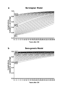
[0058] FIG. 38 illustrates predictions of the serological and sero-genetic
logistic regression
models. (A) The serological logistic regression model was constructed with QSS
and
duration of disease as predictors and complication status as the outcome. This
model was
used to predict probability of complication for a range of QSS (6-24) and
durations (1-40).
(B) The tern-genetic logistic regression model was constructed with QSS,
duration of
disease, and SNP13 mutation as predictors and complication status as the
outcome. This
model was used to predict probability of complication for a range of QSS (6-
24) and
durations (1-40), with SNP13 mutation present.
= = [0059] FIG. 39 illustrates a comparison of predicted and
observed rates of complication by
category (decile). Predictions were grouped into categories, and compared to
observed rates
of complications for each category. Number of patients in each category
prediction group
were: 0 in the 0-10% category; 13 in the >10-20% category; 49 in the >20-30%
category; 54
in the >30-40% category; 64 in the >40-50% category; 74 in the >50-60%
category; 83 in the
>60-70% category; 85 in the >70-80% category; 112 in the >80-90% category; 76
in the >90-
99% category; and 9 in the >99% category.
[0060] FIG. 40 illustrates a Receiver Operating Characteristic (ROC) curve for
cross-
validation predictions. Probabilities were generated using a leave-one-out
cross validation to
repeatedly generate a serological and sero-genetic logistic regression.
7
CA 02 75 8531 2011-1 0-1 2
WO 2010/120814 PCT/US2010/030934
[0061] FIG. 41 illustrates a diagram showing the velocity of quartile sum
score over time.
[0062] FIG. 42A illustrates a gel confirming the expression of the GST-I2
antigen. FIG.
42B illustrates a gel confirming the presence of the GST-I2 antigen in the
denatured sample
(DEN). FIG. 42C illustrates a gel confirming the presence of the GST-I2
antigen in the
filtered sample (FIL).
[0063] FIG. 43 illustrates a graph of a sample standard curve with controls as
described in
Example 20.
[0064] FIG. 44A illustrates an anti-I2 ELISA which utilizes a monoclonal
antibody
(McAb) against GST and a refolded GST-I2 antigen. FIG. 44B illustrates an anti-
I2 ELISA
which utilizes neutravidin and a biotinylated refolded GST-I2 antigen.
DETAILED DESCRIPTION OF THE INVENTION
I. Introduction
[0065] The present invention is based, in part, upon the surprising discovery
that the
accuracy of diagnosing or prognosing IBD or predicting response to an LBD
therapeutic agent
can be substantially improved by detecting the presence, level, or gdnotype of
certain markers
in a biological sample from an individual. As such, in one embodiment, the
present invention
provides diagnostic and prognostic platforms based on a serological and/or
genetic panel of
markers.
[0066] Figure 1 is an illustration of the pathophysiology of IBD, which
illustrates that in
certain instances, a patient has a genetic predisposition, a mucosal immune
system defect, a
luminal inflammation (increased immune response to enteric microbial
antigens), a barrier
function which is compromised, or a combination thereof. Figure 2 is an
illustration of an
IBD decision tree of the present invention.
[0067] The present invention provides methods and systems to improve the
diagnosis and
prognosis of UC and CD. In certain instances, the methods herein accurately
predict "IJC
like CD," a disease which is known to he very difficult to diagnose and
predict outcome. In
one aspect, the methods described herein utilize multiple serological,
protein, and/or genetic
markers, alone or in combination with one or more algorithms or other types of
statistical
analysis, to provide physicians valuable diagnostic or prognostic insight. In
some aspects, the
methods and systems of the present invention provide an indication of a
patient's projected
response to biological therapy. In other aspects, the methods and systems of
the present
invention utilize multiple markers (e.g., serological, protein, and/or
genetic) in conjunction
8
CA 02758531 2011-10-12
WO 2010/120814 PCT/US2010/030934
with statistical analysis (e.g., quartile analysis) to provide prognostic
value by identifying
patients with complicated disease or a risk of developing disease
complications (e.g., internal
stricturing or internal penetrating disease) and/or a need for surgical
intervention, while also
assisting in assessing the rate of disease progression. In certain other
instances, the methods
enable classification of disease severity along a continuum of IBD subgroups
rather than
merely as CD or UC. Moreover, the methods guide therapeutic decisions of
patients with
advanced disease. In further aspects, the use of multiple markers (e.g.,
serological, protein,
and/or genetic) provides the ability to distinguish responders from non-
responders and guides
initial therapeutic options (e.g., whether or not to prescribe aggressive
treatment), with the
potential to change disease behavior.
[0068] In certain instances, the methods and systems of the present invention
comprise a
step having a "transformation" or "machine" associated therewith, For example,
an ELISA
technique may be performed to measure the presence or concentration level of
many of the
markers described herein. An ELISA includes transformation of the marker,
e.g., an auto-
antibody, into a complex between the marker (e.g., auto-antibody) and a
binding agent (e.g.,
antigen), which then can be measured with a labeled secondary antibody. In
many instances,
the label is an enzyme which transforms a substrate into a detectable product.
The detectable
product measurement can be performed using a plate reader such as a
spectrophotometer. In
other instances, genetic markers are determined using various amplification
techniques such
as PCR. Method steps including amplification such as PCR result in the
transformation of
single or double strands of nucleic acid into multiple strands for detection.
The detection can
include the use of a fluorophore, which is performed using a machine such as a
fluoremeter.
II. Definitions
[0069] As used herein, the following terms have the meanings ascribed to them
unless
specified otherwise.
[0070] The term "classifying" includes "associating" or "categorizing" a
sample or an
individual with a disease state or prognosis. In certain instances,
"classifying" is based on
statistical evidence, empirical evidence, or both. In certain embodiments, the
methods and
systems of classifying use a so-called training set of samples from
individuals with known
disease states or prognoses. Once established, the training data set serves as
a basis, model,
or template against which the features of an unknown sample from an individual
are
compared, in order to classify the unknown disease state or provide a
prognosis of the disease
state in the individual. In some instances, "classifying" is akin to
diagnosing the disease state
9
CA 02758531 2016-09-06
andlor differentiating the disease state from another disease state. In other
instances,
"classifying" is akin to providing a prognosis of the disease state in an
individual diagnosed
with the disease state.
[0071] The term "inflammatory bowel disease" or "113D" includes
gastrointestinal disorders
such as, e.g., Crohn's disease (CD), ulcerative colitis (DC), and
indeterminate colitis (IC).
Inflammatory bowel diseases (e.g., CD, DC, and IC) are distinguished from all
other
disorders, syndromes, and abnormalities of the gastroenterological tract,
including irritable
bowel syndrome (IBS). C.S. Patent Publication 20080131439, entitled "Methods
of
Diagnosing Inflammatory Bowel Disease".
[0072] The term "sample" includes any biological specimen obtained from an
individual.
Suitable samples for use in the present invention include, without limitation,
whole blood,
plasma, serum, saliva, urine, stool, tears, any other bodily fluid, tissue
samples (e.g., biopsy),
and cellular extracts thereof (e.g., red blood cellular extract). In a
preferred embodiment, the
sample is a serum sample. The use of samples such as serum, saliva, and urine
is well known
in the art (see, e.g., Hashida et of, J. Clin. Lob. Anal., 11:267.86(1997)).
One skilled in the
art will appreciate that samples such as serum samples can be diluted prior to
the analysis of
marker levels.
[0073] The term "marker" includes any biochemical marker, serological marker,
genetic
marker, or other clinical or echographic characteristic that can be used in
the diagnosis of
IBD, in the prediction of the probable course and outcome of MD and/or in the
prediction of
the likelihood of recovery from the disease. Non-limiting examples of such
markers include
serological markers such as an anti-ncutrophil antibody, an anti-Saccharomyces
cerevisiae
antibody, an antimicrobial antibody, an acute phase protein, an
apolipoprotein, a defensin, a
growth factor, a cytokine, a cadherin, a cellular adhesion molecule; genetic
markers such as
N0D2/CARD15; and combinations thereof. In some embodiments, the markers are
utilized
in combination with a statistical analysis to provide a diagnosis or prognosis
of 1BD in an
individual. In certain instances, the diagnosis can be IBD or a clinical
subtype thereof such
as Crohn's disease (CD), ulcerative colitis (UC), or indeterminate colitis
(IC). In certain
other instances, the prognosis can be the need for surgery (e.g., the
likelihood or risk of
needing small bowel surgery), development of a clinical subtype of CD or DC
(e.g., the
likelihood or risk of heing susceptible to a particular clinical subtype CD or
DC such as the
sticturing, penetrating, or inflammatory CD subtype), development of one or
more clinical
factors (e.g., the likelihood or risk of being susceptible to a particular
clinical factor),
CA 02758531 2011-10-12
WO 2010/120814
PCT/US2010/030934
development of intestinal cancer (e.g., the likelihood or risk of being
susceptible to intestinal
cancer), or recovery from the disease (e.g., the likelihood of remission).
[0074] The term "marker profile" includes one, two, three, four, five, six,
seven, eight,
nine, ten, or more diagnostic and/or prognostic marker(s), wherein the markers
can be a
serological marker, a protein marker, a genetic marker, and the like. In some
embodiments,
the marker profile together with a statistical analysis can provide physicians
and caregivers
valuable diagnostic and prognostic insight. In other embodiments, the marker
profile with
optionally a statistical analysis provides a projected response to biological
therapy. By using
multiple markers (e.g., serological, protein, genetic, etc.) in conjunction
with statistical
analyses, the assays described herein provide diagnostic, prognostic and
therapeutic value by
identifying patients with IBD or a clinical subtype thereof, predicting risk
of developing
complicated disease, assisting in assessing the rate of disease progression
(e.g., rate of
progression to complicated disease or surgery), and assisting in the selection
of therapy.
[0075] The term "prognostic profile" includes one, two, three, four, five,
six, seven, eight,
nine, ten, or more marker(s) of an indvidual, wherein the marker(s) can be a
serological
marker, a protein marker, a genetic marker, and the like. A statistical
analysis transforms the
marker profile into a prognostic profile. A preferred statistical analysis is
a quartile score and
the quartile score for each of the markers can be summed to generate a
quartile sum score.
[0076] The term "prognostic model" includes serological models, genetic
models, sero-
genetic models, and a combination thereof. In a preferred aspect, a
retrospective analysis is
done on a cohort of known disease outcomes with known complications and
surgical
procedures performed. In one aspect, a regression analysis (e.g., logistic
regression) can be
performed on the presence or concentration level of one or more serological
markers and/or
the genotype of one or more genetic markers to develop a prognostic model. The
model can
be illustrated or depicted in, e.g., a look-up table, graph or other display.
A prognostic profile
of an individual can then be compared to a prognostic model and the prognosis
determined
(e.g., the risk or probability of developing a complication over time).
[0077] The term "therapeutic profile" includes one, two, three, four, five,
six, seven, eight,
nine, ten, or more marker(s) of an indvidual, wherein the marker(s) can be a
serological
marker, a protein marker, a genetic marker, and the like. A statistical
analysis transforms the
marker profile into a therapeutic profile. A preferred statistical analysis is
a quartile score
and the quartile score for each of the markers can be summed to generate a
quartile sum
score.
11
CA 02758531 2011-10-12
WO 2010/120814
PCT/US2010/030934
[0078] The term "therapeutic model" includes serological models, genetic
models, sero-
genetic models, and a combination thereof. In a preferred aspect, a
retrospective analysis is
done on a cohort of known therapeutic outcomes with known therapies being
used, which
include biologics, steroids, conventional drugs and surgical procedures
performed. In one
aspect, a regression analysis (e.g., logistic regression) can be performed on
the presence or
concentration level of one or more serological markers and/or the genotype of
one or more
genetic markers to develop a therapeutic model. The model can be illustrated
or depicted in,
e.g., a look-up table, graph or other display. A therapeutic profile of an
individual can then
be compared to a therapeutic model and the therapy determined (e.g., "step up"
or "top
down" strategies).
[0079] The term "efficacy profile" includes one, two, three, four, five, six,
seven, eight,
nine, ten, or more marker(s) of an indvidual, wherein the markers can be a
serological
marker, a protein marker, a genetic marker, and the like, and wherein each of
the markers
changes with therapeutic administration. In certain instances, the marker
profile is compared
to the efficacy profile in order to assess therapeutic efficacy. In certain
aspects, the efficacy
profile is equivalent to the marker profile, but wherein the markers are
measured later in time.
In certain other aspects, the efficacy profile corresponds to a marker profile
from MD
patients who responded to a particular therapeutic agent or drug. In these
aspects, similarities
or differences between the test marker profile and the reference efficacy
profile indicate
whether that particular drug is suitable or unsuitable for the treatment of
IBD. In certain
instances, a marker(s) is more indicative of efficacy than diagnosis or
prognosis. As such,
there may be a one-to-one correlation of diagnostic or prognostic markers in
the marker
profile compared to the markers in the efficacy profile, but it is not
required.
[0080] The term "individual," "subject," or "patient" typically includes
humans, but also
includes other animals such as, e.g., other primates, rodents, canines,
felines, equines, ovines,
porcines, and the like.
[0081] As used herein, the term "substantially the same amino acid sequence"
includes an
amino acid sequence that is similar, but not identical to, the naturally-
occurring amino acid
sequence. For example, an amino acid sequence, i.e., polypeptide, that has
substantially the
same amino acid sequence as an 12 protein can have one or more modifications
such as amino
acid additions, deletions, or substitutions relative to the amino acid
sequence of the naturally-
occurring 12 protein, provided that the modified polypeptide retains
substantially at least one
biological activity of 12 such as immunoreactivity. Comparison for substantial
similarity
between amino acid sequences is usually performed with sequences between about
6 and 100
12
CA 02758531 2011-10-12
WO 2010/120814
PCT/US2010/030934
residues, preferably between about 10 and 100 residues, and more preferably
between about
25 and 35 residues. A particularly useful modification of a polypeptide of the
present
invention, or a fragment thereof, is a modification that confers, for example,
increased
stability. Incorporation of one or more D-amino acids is a modification useful
in increasing
stability of a polypeptide or polypeptide fragment. Similarly, deletion or
substitution of
lysine residues can increase stability by protecting the polypeptide or
polypeptide fragment
against degradation.
[0082] The term "clinical factor" includes a symptom in an individual that is
associated
with IBD. Examples of clinical factors include, without limitation, diarrhea,
abdominal pain,
cramping, fever, anemia, weight loss, anxiety, depression, and combinations
thereof. In some
embodiments, a diagnosis or prognosis of IBD is based upon a combination of
analyzing a
sample obtained from an individual to determine the presence, level, or
genotype of one or
more markers by applying one or more statistical analyses and determining
whether the
individual has one or more clinical factors.
[0083] In a preferred aspect, the methods of invention are used after an
individual has been
diagnosed with IBD. However, in other instances, the methods can be used to
diagnose IBD
or can be used as a "second opinion" if, for example, IBD is suspected or has
been previously
diagnosed using other methods. The term "diagnosing MD" includes the use of
the methods
and systems described herein to determine the presence or absence of IBD in an
individual.
The term also includes assessing the level of disease activity in an
individual. In some
embodiments, a statistical analysis is used to diagnose a mild, moderate,
severe, or fulminant
form of IBD based upon the criteria developed by Truelove et al., Br. Med. J.,
12:1041-1048
(1955). In other embodiments, a statistical analysis is used to diagnose a
mild to moderate,
moderate to severe, or severe to fulminant form of IBD based upon the criteria
developed by
Hanauer et al., Am. J. Gastroenterol., 92:559-566 (1997). One skilled in the
art will know of
other methods for evaluating the severity of IBD in an individual.
[0084] In certain instances, the methods of the invention are used in order to
prognosticate
the progression of D3D. The methods can be used to monitor the disease, both
progression
and regression. The term "monitoring the progression or regression of IBD"
includes the use
of the methods and marker profiles to determine the disease state (e.g.,
presence or severity of
IBD) of an individual. In certain instances, the results of a statistical
analysis are compared
to those results obtained for the same individual at an earlier time. In some
aspects, the
methods, systems, and code of the present invention can also be used to
predict the
progression of IBD, e.g., by determining a likelihood for IBD to progress
either rapidly or
13
CA 02758531 2011-10-12
WO 2010/120814
PCT/US2010/030934
slowly in an individual based on the presence or level of at least one marker
in a sample. In
other aspects, the methods, systems, and code of the present invention can
also be used to
predict the regression of IBD, e.g., by determining a likelihood for IBD to
regress either
rapidly or slowly in an individual based on the presence or level of at least
one marker in a
sample.
[0085] The term "monitoring drug efficacy in an individual receiving a drug
useful for
treating IBD" includes the determination of a marker profile, alone or in
combination with
the application of a statistical analysis, to determine the disease state
(e.g., presence or
severity of IBD) of an individual after a therapeutic agent for treating IBD
has been
administered.
[0086] The term "optimizing therapy in an individual having IBD" includes the
use of the
methods of the present invention and a marker profile to determine the course
of therapy for
an individual before a therapeutic agent (e.g., IBD drug) has been
administered or to adjust
the course of therapy for an individual after a therapeutic agent has been
administered in
order to optimize the therapeutic efficacy of the therapeutic agent. In
certain instances, the
results of a statistical analysis are compared to those results obtained for
the same individual
at an earlier time during the course of therapy. As such, a comparison of the
results provides
an indication for the need to change the course of therapy or an indication
for the need to
increase or decrease the dose of the current course of therapy.
[0087] The term "course of therapy" includes any therapeutic approach taken to
relieve or
prevent one or more symptoms (i.e., clinical factors) associated with IBD. The
term "course
of therapy" encompasses administering any compound, drug, procedure, or
regimen useful
for improving the health of an individual with IBD and includes any of the
therapeutic agents
(e.g., IBD biologic agents and conventional drugs) described herein as well as
surgery. One
skilled in the art will appreciate that either the course of therapy or the
dose of the current
course of therapy can be changed, e.g., based upon the results obtained
through applying an a
statistical analysis in accordance with the present invention.
[0088] The term "therapeutically effective amount or dose" includes a dose of
a drug (e.g.,
IBD biologic agent or conventional drug) that is capable of achieving a
therapeutic effect in a
subject in need thereof. For example, a therapeutically effective amount of a
drug useful for
treating IBD can be the amount that is capable of preventing or relieving one
or more
symptoms associated with IBD. The exact amount can be ascertainable by one
skilled in the
art using known techniques (see, e.g., Lieberman, Pharmaceutical Dosage Forms
(vols. 1-3,
14
CA 02758531 2011-10-12
WO 2010/120814
PCT/US2010/030934
1992); Lloyd, The Art, Science and Technology of Pharmaceutical Compounding
(1999);
Pickar, Dosage Calculations (1999); and Remington: The Science and Practice of
Pharmacy,
20th Edition, 2003, Gennaro, Ed., Lippincott, Williams & Wilkins).
[0089] The term "gene" refers to the segment of DNA involved in producing a
polypeptide
chain; it includes regions preceding and following the coding region, such as
the promoter
and 3'-untranslated region, respectively, as well as intervening sequences
(introns) between
individual coding segments (exons).
[0090] The term "genotype" refers to the genetic composition of an organism,
including,
for example, whether a diploid organism is heterozygous or homozygous for one
or more
variant alleles of interest.
[0091] The term "polymorphism" refers to the occurrence of two or more
genetically
determined alternative sequences or alleles in a population. A "polymorphic
site" refers to
the locus at which divergence occurs. Preferred polymorphic sites have at
least two alleles,
each occurring at a particular frequency in a population. A polymorphic locus
may be as
small as one base pair (i.e., single nucleotide polymorphism or SNP).
Polymorphic markers
include restriction fragment length polymorphisms, variable number of tandem
repeats
(VNTR's), hypervariable regions, minisatellites, dinucleotide repeats,
trinucleotide repeats,
tetranucleotide repeats, simple sequence repeats, and insertion elements such
as Alu. The
first identified allele is arbitrarily designated as the reference allele, and
other alleles are
designated as alternative alleles, "variant alleles," or "variances." The
allele occurring most
frequently in a selected population is sometimes referred to as the "wild-
type" allele. Diploid
organisms may be homozygous or heterozygous for the variant alleles. The
variant allele
may or may not produce an observable physical or biochemical characteristic
("phenotype")
in an individual carrying the variant allele. For example, a variant allele
may alter the
enzymatic activity of a protein encoded by a gene of interest.
[0092] The terms "miRNA," "microRNA" or "miR" are used interchangeably and
include
single-stranded RNA molecules of 21-23 nucleotides in length, which regulate
gene
expression. miRNAs are encoded by genes from whose DNA they are transcribed
but
miRNAs are not translated into protein (non-coding RNA); instead each primary'
transcript (a
pri-miRNA) is processed into a short stem-loop structure called a pre-miRNA
and finally into
a functional miRNA. Mature miRs are partially complementary to one or more
messenger
RNA (mRNA) molecules, and their main function is to down-regulate gene
expression.
Embodiments described herein include both diagnostic and therapeutic
applications.
CA 02758531 2011-10-12
WO 2010/120814
PCT/US2010/030934
[00931 In quartile analysis, there are three numbers (values) that divide a
range of data into
four equal parts. The first quartile (also called the 'lower quartile') is the
number below
which lies the 25 percent of the bottom data. The second quartile (the
'median') divides the
range in the middle and has 50 percent of the data below it. The third
quartile (also called the
'upper quartile') has 75 percent of the data below it and the top 25 percent
of the data above
it. As a non-limiting example, quartile analysis can be applied to the
concentration level of a
marker such as an antibody or other protein marker described herein, such that
a marker level
in the first quartile (<25%) is assigned a value of 1, a marker level in the
second quartile (25-
50%) is assigned a value of 2, a marker level in the third quartile (51%-<75%)
is assigned a
value of 3, and a marker level in the fourth quartile (75%-100%) is assigned a
value of 4.
[0094] As used herein, "quartile sum score" or "QSS" includes the sum of
quartile scores
for all of the markers of interest. As a non-limiting example, a quartile sum
score for a panel
of 6 markers (e.g., serological, protein, and/or genetic) may range from 6-24,
wherein each of
the individual markers is assigned a quartile score of 1-4 based upon the
presence or absence
of the marker, the concentration level of the marker, or the genotype of the
marker.
III. Description of the Embodiments
[0095] The present invention provides methods and systems to improve the
diagnosis of
inflammatory bowel disease (IBD) and to improve the prognosis of IBD
progression and
complications. By identifying patients with complicated disease and assisting
in assessing
the rate of disease progression, the methods and systems described herein
provide invaluable
information to assess the severity of the disease and treatment options. In
certain instances,
the methods and systems enable classification of disease severity along a
continuum of IBD
subgroups rather than merely as CD, UC or IC. In other aspects, the use of
multiple markers
(serological, protein, and/or genetic) provides the ability to distinguish
responders from non-
responders to certain therapies. In particular embodiments, applying a
statistical analysis to a
profile of serological, protein, and/or genetic markers improves the accuracy
of predicting
IBD progression and disease complications, and also enables the selection of
appropriate
treatment options, including therapy such as biological, conventional,
surgery, or some
combination thereof. Accordingly, with the present invention, it is possible
to predict
outcome of disease and patients who will have a particular risk of disease
complications
and/or progression to surgery.
16
CA 02758531 2011-10-12
WO 2010/120814
PCT/US2010/030934
[0096] In one aspect, the present invention provides a method for aiding in
the prognosis of
inflammatory bowel disease (IBD) in an individual diagnosed with IBD, the
method
comprising:
(a) analyzing a sample obtained from the individual to determine the presence,
level or genotype of one or more markers selected from the group consisting of
a serological
marker, a genetic marker, and a combination thereof in the sample to obtain a
marker profile;
(b) applying a statistical analysis to the marker profile to obtain a
prognostic
profile for the individual; and
(c) comparing the prognostic profile for the individual to a prognostic model
to aid in the prognosis of IBD.
[0097] In some embodiments, the serological marker is selected from the group
consisting
of an anti-neutrophil antibody, an anti-Saccharornyces cerevisiae antibody, an
antimicrobial
antibody, an acute phase protein, an apolipoprotein, a defensin, a growth
factor, a cytokine, a
cadherin, a cellular adhesion molecule, and a combination thereof. In one
embodiment, the
anti-neutrophil antibody comprises an anti-neutrophil cytoplasmic antibody
(ANCA) such as
ANCA detected by an immunoassay (e.g., ELISA), a perinuclear anti-neutrophil
cytoplasmic
antibody (pANCA) such as pANCA detected by an immunohistochemical assay (e.g.,
WA)
or a DNAse-sensitive immunohistochemical assay, or a combination thereof. In
another
embodiment, the anti-Saccharomyces cerevisiae antibody comprises an anti-
Saccharomyces
cerevisiae immunoglobulin A (ASCA-IgA), anti-Saccharomyces cerevisiae
immunoglobulin
G (ASCA-IgG), or a combination thereof.
[0098] In yet another embodiment, the antimicrobial antibody comprises an anti-
outer
membrane protein C (anti-OmpC) antibody, an anti-I2 antibody, an anti-
flagellin antibody, or
a combination thereof. In certain instances, the anti-flagellin antibody
comprises an anti-
Cbir-1 flagellin antibody, an anti-flagellin X antibody, an anti-flagellin A
antibody, an anti-
flagellin B antibody, or a combination thereof. In a further embodiment, the
acute phase
protein is C-Reactive protein (CRP). In another embodiment, the apolipoprotein
is serum
amyloid A (SAA). In yet another embodiment, the defensin is fl defensin (e.g.,
13 defensin-1
(SDI) and/or fl defensin-2 (BD2)). In still yet another embodiment, the growth
factor is
epidermal growth factor (EGF). In a further embodiment, the cytokine comprises
TNF-
related weak inducer of apoptosis (TWEAK), IL-I p, IL-6, or a combination
thereof. In an
additional embodiment, the cadherin is E-cadherin. In another embodiment, the
cellular
adhesion molecule comprises ICAM-1, VCAM-1, or a combination thereof.
17
CA 02758531 2011-10-12
WO 2010/120814
PCT/US2010/030934
[0099] In particular embodiments, the serological marker comprises or consists
of ASCA-
IgA, ASCA-IgG, anti-OmpC antibody, anti-CBir-1 antibody, anti-I2 antibody,
pANCA (e.g.,
pANCA IFA and/or DNAse-sensitive pANCA IFA), or a combination thereof.
[0100] The presence or (concentration) level of the serological marker can be
detected
(e.g., determined, measured, analyzed, etc.) with a hybridization assay,
amplification-based
assay, immunoassay, immunohistochemical assay, or a combination thereof. Non-
limiting
examples of assays, techniques, and kits for detecting or determining the
presence or level of
one or more serological markers in a sample are described in Section VI below.
[0101] In other embodiments, the genetic marker is at least one of the genes
set forth in
Tables 1A-1E (e.g., Table 1A, 1B, 1C, ID, and/or 1E). In particular
embodiments, the
genetic marker is NOD2. The genotype of the genetic marker can be detected
(e.g.,
determined, analyzed, etc.) by genotyping an individual for the presence or
absence of one or
more variant alleles such as, for example, one or more single nucleotide
polymorphisms
(SNPs) in one or more genetic markers. In some embodiments, the SNP is at
least one of the
SNPs set forth in Tables 1B-1E (e.g., Table 1B, 1C, 1D, and/or 1E). Non-
limiting examples
of techniques for detecting or determining the genotype of one or more genetic
markers in a
sample are described in Section VII below. In certain embodiments, the genetic
marker is
NOD2 and the SNP is SNP8 (R702W), SNP12 (G908R), and/or SNP13 (1007fs). In
certain
instances, the presence or absence of one or more NOD2 SNPs is determined in
combination
with the presence or level of one or more serological markers, e.g., ASCA-IgA.
ASCA-IgG,
anti-OmpC antibody, anti-CBir-1 antibody, anti-I2 antibody, pANCA (e.g., pANCA
IFA
and/or DNAse-sensitive pANCA IFA), or a combination thereof.
[0102] In the methods of the present in tendon, the marker profile can be
determined by
detecting the presence, level, or genotype of at least two, three, four, five,
six, seven, eight,
nine, or ten markers. In particular embodiments, the sample is serum, plasma,
whole blood,
and/or stool. In other embodiments, the individual is diagnosed with Crohn's
disease (CD),
ulcerative colitis (UC), or indeterminate colitis (IC).
[0103] The statistical analysis applied to the marker profile can comprise any
of a variety
of statistical methods, models, and algorithms described in Section IX below.
In particular
embodiments, the statistical analysis is a quartile analysis. In some
instances, the quartile
analysis converts the presence, level or genotype of each marker into a
quartile score. As a
non-limiting example, the prognostic profile can correspond to a quartile sum
score (QSS) for
the individual that is obtained by summing the quartile score for each of the
markers. In
18
CA 02758531 2011-10-12
WO 2010/120814
PCT/US2010/030934
certain embodiments, the pANCA biomarker is a binary rather than a numerical
variable
since its value is either positive or negative. As described in Example 16
herein, a pANCA-
positive status is associated with a lower rate and/or risk of complications
(e.g., internal
stricturing disease, internal penetrating disease, and/or surgery). In some
instances, the
quartile scoring for pANCA is inverted, such that a positive status is scored
as "I" and a
negative status is scored as "4".
[0104] In certain embodiments, the prognostic model is established using a
retrospective
cohort with known outcomes of a clinical subtype of IBD (e.g., CD, UC, or IC).
In preferred
embodiments, the prognostic model is selected from the group consisting of a
serological
model, a sem-genetic model, a genetic model, and a combination thereof. In one
particular
embodiment, the serological model is derived by applying logistic regression
analysis to the
presence or level of one or more serological markers determined in the
retrospective cohort
(see, e.g., Examples 16 and 17). In another particular embodiment, the sero-
genetic model is
derived by applying logistic regression analysis to the presence or level of
one or more
serological markers and the genotype of one or more genetic markers determined
in the
retrospective cohort (see, e.g., Examples 16 and 17). In other embodiments,
the prognostic
model is a standardized risk scale (see, e.g., Example 16). In one particular
embodiment, the
standardized risk scale converts a prognostic profile such as a quartile sum
score (QSS) for
the individual into a standardized scale number, which may correspond to the
probability of a
complication phenotype (e.g., internal stricturing disease, internal
penetrating disease, need
for small bowel surgery) by a specific year (e.g., Year 1, 2, 3, 4, 5, 6, 7,
8, 9, 10, 11, 12, 13,
14, 15, 16, 17, 18, 19, 20, 21, 22, 23, 24, 25, 26, 27, 28, 29, 30, 31, 32,
33, 34, 35, 36, 37, 38,
39, 40, etc.) after diagnosis.
[0105] In some embodiments, the prognostic model comprises a display, print-
out, and/or
report such as a look-up table or graph. In particular embodiments, the look-
up table or graph
provides a cumulative probability of the individual developing or not
developing a Crohn's
disease (CD) complication over time. In certain other embodiments, the look-up
table or
graph provides a cumulative probability of the individual needing surgery or
not needing
surgery overtime. The look-up table or graph can also provide a cumulative
probability of
the individual developing or not developing an ulcerative colitis (UC)
complication over
time.
[0106] In certain instances, the CD complication is selected from the group
consisting of
internal stricturing disease, internal penetrating disease, and a combination
thereof. In certain
other instances, the CD complication is selected from the group consisting of
a fibrostenotic
19
CA 02758531 2011-10-12
WO 2010/120814
PCT/US2010/030934
subtype of CD, CD characterized by small bowel disease, CD characterized by
perianal
fistulizing disease, CD characterized by internal perforating disease, CD
characterized by the
need for small bowel surgery, CD characterized by the presence of features of
UC, CD
characterized by the absence of features of UC, and a combination thereof. In
yet other
instances, the surgery is small bowel surgery. In further instances, the UC
complication is
selected from the group consisting of ulcerative proctitis, proctosigmoiditis,
left-sided colitis,
pancolitis, fulminant colitis, and a combination thereof.
[0107] In other embodiments, the prognostic profile is a quartile sum score
(QSS) for the
individual and the QSS is compared to a prognostic model (e.g., a serological
model, a sero-
genetic model, standardized risk scale, etc.). In certain instances, the
prognostic model
comprises the serological model depicted in Figure 38a. In other instances,
the prognostic
model comprises the sero-genetic model depicted in Figure 38b. In further
instances, the
prognostic model comprises the standardized risk scale shown in Table 53.
[0108] In particular embodiments, the methods described herein provide a
prediction that
CD complications and/or progression to surgery would occur at a rate of (at
least) about 5%,
10%, 15%, 20%, 25%, 30%, 35%, 40%, 45%, 50%, 55%, 60%, 65%, 70%, 75%, 80%,
81%,
82%, 83%, 84%, 85%, 86%, 87%, 88%, 89%, 90%, 91%, 92%, 93%, 94%, 95%, 96%,
97%,
98%, 99%, or 100% (or any range therein) by a specific year (e.g., Year I, 2,
3, 4, 5, 6, 7, 8,
9, 10, 11, 12, 13, 14, 15, 16, 17, 18, 19, 20, 21, 22, 23, 24, 25, 26, 27, 28,
29, 30, 31, 32, 33,
34, 35, 36, 37, 38, 39, 40, etc.) after diagnosis based on an individual's
prognostic profile,
e.g., the individual's QSS, optionally in combination with the presence or
absence of one or
more variant alleles in one or more genetic markers, e.g., NOD2 (see, e.g.,
Examples 16-17,
Figures 38a-38b, and Table 53).
[0109] In yet other embodiments, the methods of the present invention can
further comprise
recommending a course of therapy for the individual based upon the comparison
between the
prognostic profile and the prognostic model. In additional embodiments, the
methods of the
present invention can further comprise sending the results of the comparison
to a clinician.
[0110] In another aspect, the present invention provides a method for
predicting the
likelihood that an individual diagnosed with inflammatory bowel disease (IBD)
will respond
to an IBD therapeutic agent, the method comprising:
(a) analyzing a sample obtained from the individual to determine the presence,
level or genotype of one or more markers selected from the group consisting of
a serological
marker, a genetic marker, and a combination thereof in the sample to obtain a
marker profile;
CA 02758531 2011-10-12
WO 2010/120814
PCT/US2010/030934
(b) applying a statistical analysis to the marker profile to obtain a
therapeutic
profile for the individual; and
(c) comparing the therapeutic profile for the individual to a therapeutic
model
to aid in the prediction of the likelihood that an individual diagnosed with
IBD will respond
to an IBD therapeutic agent.
[0111] In a related aspect, the present invention provides a method for
selecting a suitable
drug for the treatment of inflammatory bowel disease (IBD) in an individual,
the method
comprising:
(a) analyzing a sample obtained from the individual to determine the presence,
level or genotype of one or more markers in the sample to obtain a marker
profile;
(b) applying a statistical analysis to the marker profile to obtain a
therapeutic
profile for the individual; and
(c) comparing the therapeutic profile for the individual to a therapeutic
model
to aid in the selection of a suitable drug for the treatment of IBD.
[0112] The methods of the present invention find utility in predicting whether
an individual
will respond to a particular biologic agent and/or conventional drug
including, but not limited
to, anti-tumor necrosis factor (TNF) therapy (e.g., chimeric monoclonals
(e.g., infliximab),
humanized monoclonals (e.g., C0P571 and PEGylated CDP870), and human
monoclonals
(e.g., adalimumab)), p75 fusion proteins (e.g., etanercept), p55 soluble
receptors (e.g.,
onercept), small molecules such as MAP kinase inhibitors, and a combination
thereof. The
methods of the present invention also find utility in selecting a suitable
drug for the treatment
of IBD such as a particular biologic agent and/or conventional drug described
herein.
[0113] In some embodiments, the serological marker is selected from the group
consisting
of an anti-neutrophil antibody, an anti-Saccharomyces cerevisiae antibody, an
antimicrobial
antibody, an acute phase protein, an apolipoprotein, a defensin, a growth
factor, a cytokine, a
cadherin, a cellular adhesion molecule, and a combination thereof. In one
embodiment, the
anti-neutrophil antibody comprises an anti-neutrophil cytoplasmic antibody
(ANCA) such as
ANCA detected by an immunoassay (e.g., ELISA), a perinuclear anti-neutrophil
cytoplasmic
antibody (pANCA) such as pANCA detected by an immunohistochemical assay (e.g.,
IPA)
or a DNAse-sensitive immunohistochemical assay, or a combination thereof. In
another
embodiment, the anti-Saccharomyces cerevisiae antibody comprises an anti-
Succharomyces
cerevisiae immunoglobulin A (ASCA-IgA), anti-Saccharornyces cerevisiae
immunoglobulin
G (ASCA-IgG), or a combination thereof.
21
CA 02758531 2011-10-12
WO 2010/120814
PCT/US2010/030934
[0114] In yet another embodiment, the antimicrobial antibody comprises an anti-
outer
membrane protein C (anti-OmpC) antibody, an anti-I2 antibody, an anti-
flagellin antibody, or
a combination thereof. In certain instances, the anti-flagellin antibody
comprises an anti-
Chir-1 flagellin antibody, an anti-flagellin X antibody, an anti-flagellin A
antibody, an anti-
flagellin B antibody, or a combination thereof. In a further embodiment, the
acute phase
protein is C-Reactive protein (CRP). In another embodiment, the apolipoprotein
is serum
amyloid A (SAA). In yet another embodiment, the defensin isf3defensin (e.g., p
defensin-1
(BD1) and/or p defensin-2 (BD2)). In still yet another embodiment, the growth
factor is
epidermal growth factor (EGF). In a further embodiment, the cytokine comprises
TNF-
related weak inducer of apoptosis (TWEAK), IL-113,1L-6, or a combination
thereof. In an
additional embodiment, the cadherin is E-cadherin. In another embodiment, the
cellular
adhesion molecule comprises ICAM-1, VCAM-1, or a combination thereof.
[0115] In particular embodiments, the serological marker comprises or consists
of ASCA-
IgA, ASCA-IgG, anti-OmpC antibody, anti-CBir-1 antibody, anti-I2 antibody,
pANCA (e.g.,
pANCA IFA and/or DNAse-sensitive pANCA WA), or a combination thereof.
[0116] The presence or (concentration) level of the serological marker can be
detected
(e.g., determined, measured, analyzed, etc.) with a hybridization assay,
amplification-based
assay, immunoassay, immunohistochemical assay, or a combination thereof. Non-
limiting
examples of assays, techniques, and kits for detecting or determining the
presence or level of
one or more serological markers in a sample are described in Section VI below.
[0117] In other embodiments, the genetic marker is at least one of the genes
set forth in
Tables 1A-1E (e.g., Table 1A, 1B, 1C, 1D, and/or 1E). In particular
embodiments, the
genetic marker is NOD2. The genotype of the genetic marker can be detected
(e.g.,
determined, analyzed, etc.) by genotyping an individual for the presence or
absence of one or
more variant alleles such as, for example, one or more single nucleotide
polymorphisms
(SNPs) in one or more genetic markers. In some embodiments, the SNP is at
least one of the
SNPs set forth in Tables 1B-1E (e.g., Table 1B, 1C, ID, and/or 1E). Non-
limiting examples
of techniques for detecting or determining the genotype of one or more genetic
markers in a
sample are described in Section VII below. In certain embodiments, the genetic
marker is
- 30 NOD2 and the SNP is SNP8 (R702W), SNP12 (G908R), and/or SNP13 (1007fs).
In certain
instances, the presence or absence of one or more NOD2 SNPs is determined in
combination
with the presence or level of one or more serological markers, e.g., ASCA-1gA,
ASCA-1gG,
anti-OmpC antibody, anti-CBir-1 antibody, anti-I2 antibody, pANCA (e.g., pANCA
IFA
and/or DNAse-sensitive pANCA IFA), or a combination thereof.
22
CA 02758531 2011-10-12
WO 2010/120814
PCT/US2010/030934
[0118] In the methods of the present invention, the marker profile can be
determined by
detecting the presence, level, or genotype of at least two, three, four, five,
six, seven, eight,
nine, or ten markers. In particular embodiments, the sample is serum, plasma,
whole blood,
and/or stool. In other embodiments, the individual is diagnosed with Crohn's
disease (CD),
ulcerative colitis (UC), or indeterminate colitis (IC).
[0119] The statistical analysis applied to the marker profile can comprise any
of a variety
of statistical methods, models, and algorithms described in Section IX below.
In some
instances, the statistical analysis predicts that the individual has a certain
(e.g., high or low)
likelihood of responding or not responding to the IBD therapeutic agent. In
other instances,
the statistical analysis predicts whether a certain drug (e.g., IBD
therapeutic agent) is suitable
for the treatment of IBD. In particular embodiments, the statistical analysis
is a quartile
analysis. In some instances, the quartile analysis converts the presence,
level or genotype of
each marker into a quartile score. As a non-limiting example, the therapeutic
profile can
correspond to a quartile sum score (QSS) for the individual that is obtained
by summing the
quartile score for each of the markers.
[0120] In certain embodiments, the therapeutic model is established using a
retrospective
cohort of known therapeutic outcomes with known therapies including biologics,
steroids,
conventional drugs, and/or surgical procedures. In particular embodiments, the
therapeutic
model is selected from the group consisting of a serological model, a sero-
genetic model, a
genetic model, and a combination thereof. In one particular embodiment, the
therapeutic
model is a serological model that is derived by applying logistic regression
analysis to the
presence or level of one or more serological markers determined in the
retrospective cohort.
In another particular embodiment, the therapeutic model is a sero-genetic
model that is
derived by applying logistic regression analysis to the presence or level of
one or more
serological markers and the genotype of one or more genetic markers determined
in the
retrospective cohort. In other embodiments, the therapeutic model is a
standardized risk
scale. In one particular embodiment, the standardized risk scale converts a
therapeutic profile
such as a quartile sum score (QSS) for the individual into a standardized
scale number, which
may correspond to the probability of response to an IBD therapeutic agent by a
specific year
(e.g., Year 1, 2, 3, 4, 5, 6,7, 8,9, 10, 11, 12, 13, 14, 15, 16, 17, 18, 19,
20, 21, 22, 23, 24, 25,
26, 27, 28, 29, 30, 31, 32, 33, 34, 35, 36, 37, 38, 39, 40, etc.) after
diagnosis.
[0121] In some embodiments, the therapeutic model comprises a display, print-
out, and/or
report such as a look-up table or graph. In particular embodiments, the look-
up table or graph
23
CA 02758531 2011-10-12
WO 2010/120814
PCT/US2010/030934
provides a cumulative probability of the individual responding or not
responding to the IBD
therapeutic agent over time.
[0122] In other embodiments, the therapeutic profile is a quartile sum score
(QSS) for the
individual and the QSS is compared to a therapeutic model (e.g., a serological
model, a sero-
genetic model, standardized risk scale, etc.).
[0123] In particular embodiments, the methods described herein provide a
prediction that a
response to an IBD therapeutic agent would occur at a rate of (at least) about
5%, 10%, 15%,
20%, 25%, 30%, 35%, 40%, 45%, 50%, 55%, 60%, 65%, 70%, 75%, 80%, 81%, 82%,
83%,
84%, 85%, 86%, 87%, 88%, 89%, 90%, 91%, 92%, 93%, 94%, 95%, 96%, 97%, 98%,
99%,
or 100% (or any range therein) by a specific year (e.g., Year 1, 2, 3, 4, 5,
6, 7, 8, 9, 10, 11, 12,
13, 14, 15, 16, 17, 18, 19, 20, 21, 22, 23, 24, 25, 26, 27, 28, 29, 30, 31,
32, 33, 34, 35, 36, 37,
38, 39,40, etc.) after diagnosis based on an individual's therapeutic profile,
such as, e.g., the
individual's QSS, optionally in combination with the presence or absence of
one or more
variant alleles in one or more genetic markers, e.g., NOD2.
[0124] In yet other embodiments, the methods of the present invention can
further comprise
recommending a course of therapy for the individual based upon the comparison
between the
therapeutic profile and the therapeutic model. In additional embodiments, the
methods of the
present invention can further comprise sending the results of the comparison
to a clinician.
[0125] In a further aspect, the present invention provides a method for
predicting a
probability of disease complications and/or surgery in an individual diagnosed
with Crohn's
disease (CD), the method comprising:
(a) analyzing a sample obtained from the individual to determine the level or
genotype of one or more markers in the sample; and
(b) comparing the level or genotype of each of the markers to a reference
level
or genotype to predict the probability of disease complications and/or surgery
in an individual
diagnosed with CD.
[01261 In some embodiments, the markers are selected from the serological
and/or genetic
markers described herein. As a non-limiting example, the serological marker
can be selected
from the group consisting of an anti-neutrophil antibody, an anti-
Saccharomyces cerevisiae
antibody, an antimicrobial antibody, an acute phase protein, an
apolipoprotein, a defensin, a
growth factor, a cytokine, a cadherin, a cellular adhesion molecule, and a
combination
thereof. In one embodiment, the anti-neutrophil antibody comprises an anti-
neutrophil
cytoplasmic antibody (ANCA) such as ANCA detected by an immunoassay (e.g.,
ELISA), a
24
CA 02 75 8531 201 1-1 0-1 2
WO 2010/120814
PCT/US2010/030934
perinuclear anti-neutrophil cytoplasmic antibody (pANCA) such as pANCA
detected by an
immunohistochemical assay (e.g., WA) or a DNAse-sensitive immunohistochemical
assay, or
a combination thereof. In another embodiment, the anti-Saccharomyces
cerevisiae antibody
comprises an anti -Saccharomyces cerevisiae immunoglobulin A (ASCA-IgA), anti-
Saccharornyces cerevisiae immunoglobulin G (ASCA-IgG), or a combination
thereof.
[0127] In yet another embodiment, the antimicrobial antibody comprises an anti-
outer
membrane protein C (anti-OmpC) antibody, an anti-I2 antibody, an anti-
flagellin antibody, or
a combination thereof. In certain instances, the anti-flagellin antibody
comprises an anti-
Cbir-1 flagellin antibody, an anti-flagellin X antibody, an anti-flagellin A
antibody, an anti-
flagellin B antibody, or a combination thereof. In a further embodiment, the
acute phase
protein is C-Reactive protein (CRP). In another embodiment, the apolipoprotein
is serum
amyloid A (SAA). In yet another embodiment, the defensin is 13 defensin (e.g.,
13 defensin-1
(BD1) and/or 13 defensin-2 (BD2)). In still yet another embodiment, the growth
factor is
epidermal growth factor (EGF). In a further embodiment, the cytokine comprises
TNF-
related weak inducer of apoptosis (TWEAK), IL-113, IL-6, or a combination
thereof. In an
additional embodiment, the cadherin is E-cadherin. In another embodiment, the
cellular
adhesion molecule comprises ICAM-1, VCAM-1, or a combination thereof.
[0128] In particular embodiments, the markers comprise or consist of ASCA-IgG,
ASCA-
IgA, anti-OmpC antibody, anti-CBir-1 antibody, anti-I2 antibody, or a
combination thereof.
[0129] In other embodiments, the genetic marker is at least one of the genes
set forth in
Tables 1A-1E (e.g., Table 1A, 1B, IC, 1D, and/or 1E). In particular
embodiments, the
genetic marker is NOD2. The genotype of the genetic marker can be detected by
genotyping
an individual for the presence or absence of one or more variant alleles such
as, for example,
one or more SNPs in one or more genetic markers. In some embodiments, the SNP
is at least
one of the SNPs set forth in Tables 1B-1E (e.g., Table 1B, 1C, ID, and/or 1E).
In certain
embodiments, the genetic marker is NOD2 and the SNP is SNP8 (R702W), SNP12
(G908R),
and/or SNP13 (1007fs). In certain instances, the presence or absence of one or
more NOD2
SNPs is determined in combination with the presence or level of one or more
serological
markers.
[0130] In the methods of the present invention, the presence, level, or
genotype of at least
two, three, four, five, six, seven, eight, nine, or ten markers can be
determined. In particular
embodiments, the sample is serum, plasma, whole blood, and/or stool.
CA 02 75 853 1 201 1-1 0-1 2
WO 2010/120814
PCT/US2010/030934
[0131] In certain instances, the individual is predicted to have a higher
probability of
disease complications and/or surgery when the (concentration) level of at
least one of the
markers is higher than a reference (concentration) level. In certain other
instances, the
individual is predicted to have a higher probability of disease complications
and/or surgery
when the genotype of at least one of the markers is a variant allele of a
reference genotype.
Non-limiting examples of disease complications include internal stricturing
disease and/or
internal penetrating disease as well as any of the other CD complications
described herein.
[0132] In certain embodiments, the reference (concentration) level corresponds
to a
(concentration) level of one of the markers in a sample from an individual not
having CD
(e.g., healthy individual, non-CD individual, non-IBD individual, UC
individual, etc.). In
certain other embodiments, the reference genotype corresponds to a wild-type
genotype (e.g.,
non-variant allele or SNP) of one of the genetic markers.
[0133] In particular embodiments, the methods described herein provide a
prediction that
disease complications and/or surgery would occur at a rate of (at least) about
5%, 10%, 15%,
20%, 25%, 30%, 35%, 40%, 45%, 50%, 55%, 60%, 65%, 70%, 75%, 80%, 81%, 82%,
83%,
84%, 85%, 86%, 87%, 88%, 89%, 90%, 91%, 92%, 93%, 94%, 95%, 96%, 97%, 98%,
99%,
or 100% (or any range therein) by a specific year (e.g., Year 1, 2, 3, 4, 5,
6,7, 8,9, 10, 11, 12,
13, 14, 15, 16, 17, 18, 19, 20, 21, 22, 23, 24, 25, 26, 27, 28, 29, 30, 31,
32, 33, 34, 35, 36, 37,
38, 39, 40, etc.) after diagnosis based on an individual's marker levels
and/or genotypes. In
some instances, the individual is predicted to have about 40% to about 70%
(e.g., about 40%
to about 60%, about 50% to about 70%, etc.) probability of disease
complications and/or
surgery by about 10 years after being diagnosed with CD. In other instances,
the individual is
predicted to have about 70% to about 90% probability of disease complications
and/or
surgery by about 20 years after being diagnosed with CD. In further instances,
the individual
is predicted to have about 80% to about 100% probability of disease
complications and/or
surgery by about 30 years after being diagnosed with CD.
[0134] In other embodiments, the methods of the present invention can further
comprise
recommending a course of therapy for the individual based upon the comparison
between the
level or genotype of each of the markers and a reference level or genotype. In
additional
embodiments, the methods of the present invention can further comprise sending
the results
of the comparison to a clinician.
26
CA 02 75 8531 2011-1 0-1 2
WO 2010/120814
PCT/US2010/030934
[0135] In a related aspect, the present invention provides a method for
predicting a
probability of disease complications and/or surgery in an individual diagnosed
with Crohn's
disease (CD), the method comprising:
(a) analyzing a sample obtained from the individual to determine the presence,
level or genotype of one or more markers in the sample to obtain a marker
profile;
(b) applying a statistical analysis to the marker profile to obtain a
prognostic
profile for the individual; and
(c) comparing the prognostic profile for the individual to a prognostic model
to predict the probability of disease complications and/or surgery in an
individual diagnosed
with CD.
[0136] In some embodiments, the markers are selected from the serological
and/or genetic
markers described herein. As a non-limiting example, the serological marker
can be selected
from the group consisting of an anti-neutrophil antibody, an anti-
Saccharomyces cerevisiae
antibody, an antimicrobial antibody, an acute phase protein, an
apolipoprotein, a defensin, a
growth factor, a cytokine, a eadherin, a cellular adhesion molecule, and a
combination
thereof. In one embodiment, the anti-neutrophil antibody comprises an anti-
neutrophil
cytoplasmic antibody (ANCA) such as ANCA detected by an immunoassay (e.g.,
ELISA), a
perinuclear anti-neutrophil cytoplasmic antibody (pANCA) such as pANCA
detected by an
immunohistochemical assay (e.g., IFA) or a DNAse-sensitive immunohistochemical
assay, or
a combination thereof. In another embodiment, the anti-Saccharomyces
cerevisiae antibody
comprises an anti-Saccharornyces cerevisiae immunoglobulin A (ASCA-IgA), anti-
Saccharnmyces cerevisiae immunoglobulin G (ASCA-IgG), or a combination
thereof.
[0137] In yet another embodiment, the antimicrobial antibody comprises an anti-
outer
membrane protein C (anti-OmpC) antibody, an anti-I2 antibody, an anti-
flagellin antibody, or
a combination thereof. In certain instances, the anti-flagellin antibody
comprises an anti-
Cbir-1 flagellin antibody, an anti-flagellin X antibody, an anti-flagellin A
antibody, an anti-
flagellin B antibody. or a combination thereof. In a further embodiment, the
acute phase
protein is C-Reactive protein (CRP). In another embodiment, the apolipoprotein
is serum
amyloid A (SAA). In yet another embodiment, the defensin is 13 defensin (e.g.,
13 defensin-1
(BD1) and/or [3 defensin-2 (BD2)). In still yet another embodiment, the growth
factor is
epidermal growth factor (EGF). In a further embodiment, the cytokine comprises
TNF-
related weak inducer of apoptosis (TWEAK), IL-113, IL-6, or a combination
thereof. In an
additional embodiment, the cadherin is E-cadherin. In another embodiment, the
cellular
adhesion molecule comprises ICAM-1, VCAM-1, or a combination thereof.
27
CA 02758531 2011-10-12
WO 2010/120814
PCT/US2010/030934
[0138] In particular embodiments, the markers comprise or consist of ASCA-IgG,
ASCA-
IgA, anti-OmpC antibody, anti-CBir-1 antibody, anti-I2 antibody, or a
combination thereof.
[0139] In other embodiments, the genetic marker is at least one of the genes
set forth in
Tables 1A-1E (e.g., Table IA. IB, 1C, ID, and/or 1E). In particular
embodiments, the
genetic marker is NOD2. The genotype of the genetic marker can be detected by
genotyping
an individual for the presence or absence of une or more variant alleles such
as, for example,
one or more SNPs in one or more genetic markers. In some embodiments, the SNP
is at least
one of the SNPs set forth in Tables 1B-1E (e.g., Table 1B, 1C, 1D, and/or 1E).
In certain
embodiments, the genetic marker is NOD2 and the SNP is SNP8 (R702W), SNP12
(G908R),
and/or SNP13 (1007fs). In certain instances, the presence or absence of one or
more NOD2
SNPs is determined in combination with the presence or level of one or more
serological
markers.
[0140] In the methods of the present invention, the presence, level, or
genotype of at least
two, three, four, five, six, seven, eight, nine, or ten markers can be
determined. In particular
embodiments, the sample is serum, plasma, whole blood, and/or stool.
[0141] The statistical analysis applied to the marker profile can comprise any
of a variety
of statistical methods, models, and algorithms described in Section IX below.
In particular
embodiments, the statistical analysis is a quartile analysis. In some
instances, the quartile
analysis converts the presence, level or genotype of each marker into a
quartile score. As a
non-limiting example, the prognostic profile can correspond to a quartile sum
score (QSS) for
the individual that is obtained by summing the quartile score for each of the
markers.
[0142] In some embodiments, the prognostic model comprises a display, print-
out, and/or
report such as a look-up table or graph. In particular embodiments, the look-
up table or graph
provides a cumulative probability of the individual developing or not
developing a Crohn's
disease (CD) complication over time. In certain other embodiments, the look-up
table or
graph provides a cumulative probability of the individual needing surgery or
not needing
surgery over time. Non-limiting examples of disease complications include
internal
stricturing disease and/or internal penetrating disease as well as any of the
other CD
complications described herein.
[0143] In other embodiments, the prognostic profile is a quartile sum score
(QSS) for the
individual and the QSS is compared to a prognostic model (e.g., a serological
model, a sero-
genetic model, standardized risk scale, etc.). In certain embodiments, the
individual is
predicted to have a higher probability of disease complications and/or surgery
when the QSS
28
CA 02758531 2011-10-12
WO 2010/120814
PCT/US2010/030934
is greater than 5, 6, 7, 8, 9, 10, 11,12, 13, 14, 15, 16, 17, 18, 19, 20,
21,22, 23,24. etc. (e.g.,
preferably greater than 10).
[0144] In particular embodiments, the methods described herein provide a
prediction that
disease complications and/or surgery would occur at a rate of (at least) about
5%, 10%, 15%,
20%, 25%, 30%, 35%, /10%, 45%, 50%, 55%, 60%, 65%, 70%, 75%, 80%, 81%, 82%,
83%,
84%, 85%, 86%, 87%, 88%, 89%, 90%, 91%, 92%, 93%, 94%, 95%, 96%, 97%, 98%,
99%,
or 100% (or any range therein) by a specific year (e.g., Year 1, 2, 3, 4, 5,
6, 7, 8, 9, 10, 11, 12,
13, 14, 15, 16, 17, 18, 19, 20, 21, 22, 23, 24, 25, 26, 27, 28, 29, 30, 31,
32, 33, 34, 35, 36, 37,
38, 39, 40, etc.) after diagnosis based on an individual's prognostic profile,
such as, e.g., the
individual's QSS, optionally in combination with the presence or absence of
one or more
variant alleles in one or more genetic markers, e.g., NOD2. In some instances,
the individual
is predicted to have about 40% to about 70% (e.g., about 40% to about 60%,
about 50% to
about 70%, etc.) probability of disease complications and/or surgery by about
10 years after
being diagnosed with CD when the QSS is greater than 10. In other instances,
the individual
is predicted to have about 70% to about 90% probability of disease
complications and/or
surgery by about 20 years after being diagnosed with CD when the QSS is
greater than 10. In
further instances, the individual is predicted to have about 80% to about 100%
probability of
disease complications and/or surgery by about 30 years after being diagnosed
with CD when
the QSS is greater than 10.
[0145] In yet other embodiments, the methods of the present invention can
further comprise
recommending a course of therapy for the individual based upon the comparison
between the
prognostic profile and the prognostic model. In additional embodiments, the
methods of the
present invention can further comprise sending the results of the comparison
to a clinician.
[0146] In another aspect, the present invention provides a method for aiding
in the
diagnosis of inflammatory bowel disease (IBD), the method comprising:
(a) analyzing a sample obtained from the individual to determine the level or
genotype of one or more markers in the sample; and
(b) comparing the level or genotype of each of the markers to a reference
level
or genotype to aid in the diagnosis of IBD.
[0147] In some embodiments, the markers are selected from the serological
and/or genetic
markers described herein. As a non-limiting example, the serological marker
can be selected
from the group consisting of an anti-neutrophil antibody, an anti -
Saccharomyces cerevisiae
antibody, an antimicrobial antibody, an acute phase protein, an
apolipoprotein, a defensin, a
29
CA 02758531 2011-10-12
WO 2010/120814
PCT/US2010/030934
growth factor, a cytokine, a cadherin, a cellular adhesion molecule, and a
combination
thereof. In one embodiment, the anti-neutrophil antibody comprises an anti-
neutrophil
cytoplasmic antibody (ANCA) such as ANCA detected by an immunoassay (e.g.,
ELISA), a
perinuclear anti-neutrophil cytoplasmic antibody (pANCA) such as pANCA
detected by an
immunohistochemical assay (e.g., IFA) or a DNAse-sensitive immunohistochemical
assay, Or
a combination thereof. In another embodiment, the anti -Saccharomyces
cerevisiae antibody
comprises an anti-Saccharnmyces cerevisiae immunoglobulin A (ASCA-IgA), anti-
Sctccharomyces cerevisiae immunoglobulin G (ASCA-IgG), or a combination
thereof.
[0148] In yet another embodiment, the antimicrobial antibody comprises an anti-
outer
membrane protein C (anti-OmpC) antibody, an anti-I2 antibody, an anti-
flagellin antibody, or
a combination thereof. In certain instances, the anti-flagellin antibody
comprises an anti-
Cbir-1 flagellin antibody, an anti-flagellin X antibody, an anti-flagellin A
antibody, an anti-
flagellin B antibody, or a combination thereof. In a further embodiment, the
acute phase
protein is C-Reactive protein (CRP). In another embodiment, the apolipoprotein
is serum
arnyloid A (SAA). In yet another embodiment, the defensin is 13 defensin
(e.g., 13 defensin-I
(BD1) and/or 13 defensin-2 (BD2)). In still yet another embodiment, the growth
factor is
epidermal growth factor (EGF). In a further embodiment, the cytokine comprises
TNF-
related weak inducer of apoptosis (TWEAK), IL-10, IL-6, or a combination
thereof. In an
additional embodiment, the cadherin is E-cadherin. In another embodiment, the
cellular
adhesion molecule comprises ICAM-1. VCAM-1, or a combination thereof.
[0149] In other embodiments, the genetic marker is at least one of the genes
set forth in
Tables 1A-1E (e.g., Table 1A, 1B, IC, ID, and/or 1E). In particular
embodiments, the
genetic marker is NOD2. The genotype of the genetic marker can be detected by
genotyping
an individual for the presence or absence of one or more variant alleles such
as, for example,
one or more SNPs in one or more genetic markers. In some embodiments, the SNP
is at least
one of the SNPs set forth in Tables 1B-1E (e.g., Table 18, 1C, 1D, and/or 1E).
In certain
embodiments, the genetic marker is NOD2 and the SNP is SNP8 (R702W), SNP12
(G90812),
and/or SNPI3 (1007fs). In certain instances, the presence or absence of one or
more NOD2
SNPs is determined in combination with the presence or level of one or more
serological
markers.
[0150] In the methods of the present invention, the presence, level, or
genotype of at least
two, three, four, five, six, seven, eight, nine, or ten markers can be
determined. In particular
embodiments, the sample is serum, plasma, whole blood, and/or stool.
CA 02758531 2011-10-12
WO 2010/120814
PCT/US2010/030934
[0151] In certain embodiments, the reference (concentration) level corresponds
to a
(concentration) level of one of the markers in a sample from an individual not
having IBD
(e.g., healthy individual, non-IBD individual, non-CD individual, non-UC
individual, etc.).
In certain other embodiments, the reference genotype corresponds to a wild-
type genotype
(e.g., non-variant allele or SNP) of one of the genetic markers.
[0152] In particular embodiments, the methods described herein provide a
probability of
II3D (or a clinical subtype thereof) of (at least) about 5%, 10%, 15%, 20%,
25%, 30%, 35%,
40%, 45%, 50%, 55%, 60%, 65%, 70%, 75%, 80%, 81%, 82%, 83%, 84%, 85%, 86%,
87%,
88%, 89%, 90%, 91%, 92%, 93%, 94%, 95%, 96%, 97%, 98%, 99%, or 100% (or any
range
therein) based on an individual's marker levels and/or genotypes.
[0153] In other embodiments, the methods of the present invention can further
comprise
recommending a course of therapy for the individual based upon the comparison
between the
level or genotype of each of the markers and a reference level or genotype. In
additional
embodiments, the methods of the present invention can further comprise sending
the results
of the comparison to a clinician.
[0154] In a related aspect, the present invention provides a method for aiding
in the
diagnosis of inflammatory bowel disease (IBD), the method comprising:
(a) analyzing a sample obtained from the individual to determine the presence,
level or genotype of one or more markers in the sample to obtain a marker
profile; and
(b) applying a statistical analysis to the marker profile to aid in the
diagnosis
of IBD.
[0155] In some embodiments, the markers are selected from the serological
and/or genetic
markers described herein. As a non-limiting example, the serological marker
can be selected
from the group consisting of an anti-neutrophil antibody, an anti -
Saccharomyces cerevisiae
antibody, an antimicrobial antibody, an acute phase protein, an
apolipoprotein, a defensin, a
growth factor, a cytokine, a cadherin, a cellular adhesion molecule, and a
combination
thereof. In one embodiment, the anti-neutrophil antibody comprises an anti-
neutrophil
cytoplasmic antibody (ANCA) such as ANCA detected by an immunoassay (e.g.,
ELEA), a
pen nuclear anti-neutrophil cytoplasmic antibody (pANCA) such as pANCA
detected by an
immunohistochemical assay (e.g., IFA) or a DNAse-sensitive immunohistochemical
assay, or
a combination thereof. In another embodiment, the anti-Saccharomyces
cerevisiae antibody
comprises an anti-Saccharomyces cerevisiae immunoglobulin A (ASCA-IgA), anti-
Saccharomyces cerevisiae immunoglobulin G (ASCA-IgG), or a combination
thereof.
31
CA 02758531 2011-10-12
WO 2010/120814
PCT/US2010/030934
[0156] In yet another embodiment, the antimicrobial antibody comprises an anti-
outer
membrane protein C (anti-OmpC) antibody, an anti-12 antibody, an anti-
flagellin antibody, or
a combination thereof. In certain instances, the anti-flagellin antibody
comprises an anti-
Cbir-1 flagellin antibody, an anti-flagellin X antibody, an anti-flagellin A
antibody, an anti-
flagellin B antibody, or a combination thereof. In a further embodiment, the
acute phase
protein is C-Reactive protein (CRP). In another embodiment, the apolipoprotein
is serum
amyl oid A (SAA). In yet another embodiment, the defensin is p defensin (e.g.,
p defensin-1
(BDI) and/or p defensin-2 (BD2)). In still yet another embodiment, the growth
factor is
epidermal growth factor (EGF). In a further embodiment, the cytokine comprises
TNF-
related weak inducer of apoptosis (TWEAK), IL-l3, 1L-6, or a combination
thereof. In an
additional embodiment, the cadherin is E-cadherin. In another embodiment, the
cellular
adhesion molecule comprises ICAM-1, VCAM-1, or a combination thereof.
[0157] In other embodiments, the genetic marker is at least one of the genes
set forth in
Tables 1A-1E (e.g., Table 1A, 1B, 1C, 1D, and/or 1E). In particular
embodiments, the
genetic marker is NOD2. The genotype of the genetic marker can be detected by
genotyping
an individual for the presence or absence of one or more variant alleles such
as, for example,
one or more SNPs in one or more genetic markers. In some embodiments, the SNP
is at least
one of the SNPs set forth in Tables 1B-1E (e.g., Table 1B, 1C, 1D, and/or 1E).
In certain
embodiments, the genetic marker is NOD2 and the SNP is SNP8 (R702W), SNP12
(G908R),
and/or SNP13 (1007fs). In certain instances, the presence or absence of one or
more NOD2
SNPs is determined in combination with the presence or level of one or more
serological
markers.
[0158] In the methods of the present invention, the presence, level, or
genotype of at least
two, three, four, five, six, seven, eight, nine, or ten markers can be
determined. In particular
embodiments, the sample is serum, plasma, whole blood, and/or stool.
[0159] The statistical analysis applied to the marker profile can comprise any
of a variety
of statistical methods, models, and algorithms described in Section IX below.
In particular
embodiments, the statistical analysis is a quartile analysis. In some
instances, the quartile
analysis converts the presence, level or genotype of each marker into a
quartile score. As a
non-limiting example, the diagnosis of IBD can be made based upon a quartile
sum score
(QSS) for the individual that is obtained by summing the quartile score for
each of the
markers. In other embodiments, the statistical analysis comprises one or more
learning
statistical classifier systems as described herein. In particular embodiments,
the statistical
analysis comprises a combination of at least two learning statistical
classifier systems. A
32
CA 02758531 2011-10-12
WO 2010/120814
PCT/US2010/030934
=
non-limiting example of such a combination includes a decision/classification
tree (e.g., a
classification and regression tree (C&RT), a random forest, etc.) and a neural
network, e.g.,
applied in tandem. In certain instances, the methods comprise applying a first
statistical
analysis (e.g., a decision/classification tree) to the presence, level, or
genotype determined for
each of the markers to generate a prediction or probability value, and then
applying a second
statistical analysis (e.g., a neural network) to the prediction or probability
value and the
presence, level, or genotype determined for-each of the markers to aid in the
diagnosis of IBD
(e.g., by classifying the sample as an IBD sample or non-IBD sample).
[0160] In certain embodiments, the methods further comprise comparing the
results from
the statistical analysis (i.e., diagnostic profile) to a reference (i.e.,
diagnostic model) to aid in
the diagnosis of IBD. In some instances, the diagnostic model comprises a
display, print-out,
and/or report such as a look-up table or graph. In other instances, the
diagnostic profile is a
quartile sum score (QSS) for the individual and the QSS is compared to a
diagnostic model.
In some embodiments, the individual is predicted to have a higher probability
of having IBD
when the QSS is greater than 5, 6, 7, 8, 9, 10, 11, 12, 13, 14, 15, 16,17, 18,
19, 20, 21, 22,
23, 24, etc.
[0161] In particular embodiments, the methods described herein provide a
probability of
IBD (or a clinical subtype thereof) of (at least) about 5%, 10%, 15%, 20%,
25%, 30%, 35%,
40%, 45%, 50%, 55%, 60%, 65%, 70%, 75%, 80%, 81%, 82%, 83%, 84%, 85%, 86%,
87%,
88%, 89%, 90%, 91%, 92%, 93%, 94%, 95%, 96%, 97%, 98%, 99%, or 100% (or any
range
therein) based on an individual's diagnostic profile, such as, e.g., the
individual's QSS,
optionally in combination with the presence or absence of one or more variant
alleles in one
or more genetic markers, e.g., NOD2.
[0162] In some embodiments, the methods of the present invention can further
comprise
recommending a course of therapy for the individual based upon the statistical
analysis or
comparison between the diagnostic profile and the diagnostic model. In other
embodiments,
the methods of the present invention can further comprise sending the results
of the statistical
analysis or comparison to a clinician.
IV. Clinical Subtypes of IBD
[0163] Inflammatory bowel disease (TBD) is a group of inflammatory conditions
of the
large intestine and small intestine. The main forms of IBD are Crohn's disease
(CD) and
ulcerative colitis (UC). Other less common forms of IBD include, e.g.,
indeterminate colitis
(IC), collagenous colitis, lymphocytic colitis, ischemic colitis, diversion
colitis, Behget's
33
CA 02758531 2016-09-06
syndrome, infective colitis, and the like. U.S. Patent Publication
20080131439, entitled
"Methods of Diagnosing Inflammatory Bowel Disease".
A. Crohn's Disease
[01641 Cfohn's disease (CD) is a disease of chronic inflammation that can
involve any part
of the gastrointestinal tract. Commonly, the distal portion of the small
intestine, i.e., the
ileum, and the cccum arc affected. In other cases, the disease is confined to
the small
intestine, colon, or anorectal region. CD occasionally involves the duodenum
and stomach,
and more rarely the esophagus and oral cavity.
10165] The variable clinical manifestations of CD are, in part, a result of
the varying
anatomic localization of the disease. The most frequent symptoms of CD are
abdominal pain,
diarrhea, and recurrent fever. CD is commonly associated with intestinal
obstruction or
fistula, an abnormal passage between diseased loops of bowel. CD also includes
complications such as inflammation of the eye, joints, and skin, liver
disease, kidney stones,
and amyloidosis. In addition, CD is associated with an increased risk of
intestinal cancer.
[01661 Several features are characteristic of the pathology of CD. The
inflammation
associated with CD, known as transmural inflammation, involves all layers of
the bowel wall.
Thickening and edema, for example, typically also appear throughout the bowel
wall, with
fibrosis present in long-standing forms of the disease. The inflammation
characteristic of CD
is discontinuous in that segments of inflamed tissue, known as "skip lesions,"
are separated
by apparently normal intestine. Furthermore, linear ulcerations, edema, and
inflammation of
the intervening tissue lead to a -cobblestone" appearance of the intestinal
mucosa, which is
distinctive of CD.
[0167] A hallmark of CD is the presence of discrete aggregations of
inflammatory cells,
known as granulomas, which are generally found in the suhmucosa. Some CD cases
display
typical discrete granulomas. while others show a diffuse granulomarous
reaction or a
nonspecific transmural inflammation. As a result, the presence of discrete
granulomas is
indicative of CD, although the absence of granulomas is also consistent with
the disease.
Thus, transmural or discontinuous inflammation, rather than the presence of
granulomas, is a
preferred diagnostic indicator of CD (Rubin and Farber, Pathology (Second
Edition),
Philadelphia, J.B. Lippincott Company (1994)).
[0168] Crohn's disease may be categorized by the behavior of disease as it
progresses.
This was formalized in the Vienna classification of Crohn's disease. See,
Gasche et al.,
34
CA 02758531 2011-10-12
WO 2010/120814
PCT/US2010/030934
Inflam.m.. Bowel Dis., 6:8-15 (2000). There are three categories of disease
presentation in
Crohn's disease: (1) stricturing, (2) penetrating, and (3) inflammatory.
Stricturing disease
causes narrowing of the bowel which may lead to bowel obstruction or changes
in the caliber
of the feces. Penetrating disease creates abnormal passageways (fistulae)
between the bowel
and other structures such as the skin. Inflammatory disease (also known as non-
stricturing,
non-penetrating disease) causes inflammation without causing strictures or
fistulae.
[0169] As such, Crohn's disease represents a number of heterogeneous disease
subtypes
that affect the gastrointestinal tract and may produce similar symptoms. As
used herein in
reference to CD, the term "clinical subtype" includes a classification of CD
defined by a set
of clinical criteria that distinguish one classification of CD from another.
As non-limiting
examples, subjects with CD can be classified as having stricturing (e.g.,
internal stricturing),
penetrating (e.g., internal penetrating), Or inflammatory disease as described
herein, or these
subjects can additionally or alternatively be classified as having
fibrostenotic disease, small
bowel disease, internal perforating disease, perianal fistulizing disease, UC-
like disease, the
need for small bowel surgery, the absence of features of UC, or combinations
thereof.
[0170] In certain instances, subjects with CD can be classified as having
complicated CD,
which is a clinical subtype characterized by stricturing or penetrating
phenotypes. In certain
other instances, subjects with CD can be classified as having a form of CD
characterized by
one or more of the following complications: fibrostenosis, internal
perforating disease, and
the need for small bowel surgery. In further instances, subjects with CD can
be classified as
having an aggressive form of fibrostenotic disease requiring small bowel
surgery. Criteria
relating to these subtypes have been described, for example, in Gasche et al.,
Intlatrun. Bowel
Dis., 6:8-15 (2000); Abreu et al., Gastroenterology, 123:679-688 (2002);
Vasiliauskas et al.,
Gut, 47:487-496 (2000); Vasiliauskas et al., Gastroenterology, 110:1810-1819
(1996); and
Greenstein et al., Gut, 29:588-592 (1988).
[0171] The "fibrostenotic subtype" of CD is a classification of CD
characterized by one or
more accepted characteristics of fibrostenosing disease. Such characteristics
of
fibrostcnosing disease include, but are not limited to, documented persistent
intestinal
obstruction or an intestinal resection for an intestinal obstruction. The
fibrostenotic subtype
of CD can be accompanied by other symptoms such as perforations, abscesses, or
fistulae,
and can further be characterized by persistent symptoms of intestinal blockage
such as
nausea, vomiting, abdominal distention, and inability to eat solid food.
Intestinal X-rays of
patients with the fibrostenotic subtype of CD can show, for example,
distention of the bowel
before the point of blockage.
CA 02758531 2011-10-12
WO 2010/120814
PCT/US2010/030934
[0172] The requirement for small bowel surgery in a subject with the
fibrostenotic subtype
of CD can indicate a more aggressive form of this subtype. Additional subtypes
of CD are
also known in the art and can be identified using defined clinical criteria.
For example,
internal perforating disease is a clinical subtype of CD defined by current or
previous
evidence of entero-enteric or entero-vesicular fistulae, intra-abdominal
abscesses, or small
bowel perforation. Perianal perforating disease is a clinical subtype of CD
defined by current
or previous evidence of either perianal fistulae or abscesses or rectovaginal
fistula. The UC-
like clinical subtype of CD can be defined by current or previous evidence of
left-sided
colonic involvement, symptoms of bleeding or urgency, and crypt abscesses on
colonic
biopsies. Disease location can be classified based on one or more endoscopic,
radiologic, or
pathologic studies.
[0173] One skilled in the art understands that overlap can exist between
clinical subtypes of
CD and that a subject having CD can have more than one clinical subtype of CD.
For
example, a subject having CD can have the fibrostenotic subtype of CD and can
also meet
clinical criteria for a clinical subtype characterized by the need for small
bowel surgery or the
internal perforating disease subtype. Similarly, the markers described herein
can be
associated with more than one clinical subtype of CD.
B. Ulcerative Colitis
[0174] Ulcerative colitis (UC) is a disease of the large intestine
characterized by chronic
diarrhea with cramping, abdominal pain, rectal bleeding, loose discharges of
blood, pus, and
mucus. The manifestations of UC vary widely. A pattern of exacerbations and
remissions
typifies the clinical course for about 70% of UC patients, although continuous
symptoms
without remission are present in some patients with UC. Local and systemic
complications
of UC include arthritis, eye inflammation such as uveitis, skin ulcers, and
liver disease. In
addition, UC, and especially the long-standing, extensive form of the disease
is associated
with an increased risk of colon carcinoma.
[0175] TIC is a diffuse disease that usually extends from the most distal part
of the rectum
for a variable distance proximally. The term "left-sided colitis" describes an
inflammation
that involves the distal portion of the colon, extending as far as the splenic
flexure. Sparing
of the rectum or involvement of the right side (proximal portion) of the colon
alone is unusual
in UC. The inflammatory process of UC is limited to the colon and does not
involve, for
example, the small intestine, stomach, or esophagus. In addition, UC is
distinguished by a
superficial inflammation of the mucosa that generally spares the deeper layers
of the bowel
36
CA 02758531 2011-10-12
WO 2010/120814
PCT/US2010/030934
wall. Crypt abscesses, in which degenerated intestinal crypts are filled with
neutrophils, are
also typical of UC (Rubin and Farber, supra).
[0176] In certain instances, with respect to UC, the variability of symptoms
reflect
differences in the extent of disease (i.e., the amount of the colon and rectum
that are
inflamed) and the intensity of inflammation. Disease starts at the rectum and
moves "up" the
colon to involve more of the organ. UC can be categorized by the amount of
colon involved.
Typically, patients with inflammation confined to the rectum and a short
segment of the
colon adjacent to the rectum have milder symptoms and a better prognosis than
patients with
more widespread inflammation of the colon.
[0177] In comparison with CD, which is a patchy disease with frequent sparing
of the
rectum, UC is characterized by a continuous inflammation of the colon that
usually is more
severe distally than proximally. The inflammation in UC is superficial in that
it is usually
limited to the mucosal layer and is characterized by an acute inflammatory
infiltrate with
neutrophils and crypt abscesses. In contrast, CD affects the entire thickness
of the bowel wall
with granulomas often, although not always, present. Disease that terminates
at the ileocecal
valve, or in the colon distal to it, is indicative of UC, while involvement of
the terminal
ileum, a cobblestone-like appearance, discrete ulcers, or fistulas suggests
CD.
[0178] The different types of ulcerative colitis are classified according to
the location and
the extent of inflammation. As used herein in reference to UC, the term
"clinical subtype"
includes a classification of UC defined by a set of clinical criteria that
distinguish one
classification of UC from another. As non-limiting examples, subjects with UC
can be
classified as having ulcerative proctitis, proctosigmoiditis, left-sided
colitis, pancolitis,
fulminant colitis, and combinations thereof. Criteria relating to these
subtypes have been
described, for example, in Kornbluth et al., Am. J. Gastroenterol., 99: 1371-
85 (2004).
[0179] Ulcerative proctitis is a clinical subtype of UC defined by
inflammation that is
limited to the rectum. Proctosigmoiditis is a clinical subtype of UC which
affects the rectum
and the sigmoid colon. Left-sided colitis is a clinical subtype of UC which
affects the entire
left side of the colon, from the rectum to the place where the colon bends
near the spleen and
begins to run across the upper abdomen (the splenic flexure). Pancolitis is a
clinical subtype
of 'DC which affects the entire colon. Fulminant colitis is a rare, but severe
form of
pancolitis. Patients with fulminant colitis are extremely ill with
dehydration, severe
abdominal pain, protracted diarrhea with bleeding, and even shock.
37
CA 02758531 2011-10-12
WO 2010/120814
PCT/US2010/030934
[0180] In some embodiments, classification of the clinical subtype of UC is
important in
planning an effective course of treatment. While ulcerative proctitis,
proctosigmoiditis, and
left-sided colitis can be treated with local agents introduced through the
anus, including
steroid-based or other enemas and foams, pancolitis must be treated with oral
medication so
that active ingredients can reach all of the affected portions of the colon.
[0181] One skilled in the art understands that overlap can exist between
clinical subtypes of
UC and that a subject having UC can have more than one clinical subtype of UC.
Similarly,
the prognostic markers described herein can be associated with more than one
clinical
subtype of UC.
C. Indeterminate Colitis
[0182] Indeterminate colitis (IC) is a clinical subtype of IBD that includes
both features of
CD and UC. Such an overlap in the symptoms of both diseases can occur
temporarily (e.g.,
in the early stages of the disease) or persistently (e.g., throughout the
progression of the
disease) in patients with IC. Clinically, IC is characterized by abdominal
pain and diarrhea
with or without rectal bleeding. For example, colitis with intermittent
multiple ulcerations
separated by normal mucosa is found in patients with the disease.
Histologically, there is a
pattern of severe ulceration with transmural inflammation. The rectum is
typically free of the
disease and the lymphoid inflammatory cells do not show aggregation. Although
deep slit-
like fissures are observed with foci of myocytolysis, the intervening mucosa
is typically
minimally congested with the preservation of goblet cells in patients with IC.
V. IBD Markers
[0183] A variety of IBD markers, including biochemical markers, serological
markers,
protein markers, genetic markers, and other clinical or echographic
characteristics, are
suitable for use in the methods of the present invention for diagnosing IBD,
prognosing the
future outcome of the disease, and predicting the response to therapy with
therapeutic agents
such as biologics. In certain aspects, the diagnostic and prognostic methods
described herein
utilize the application of an algorithm (e.g., statistical analysis) to the
presence, concentration
level, or genotype determined for one or more of the IBD markers to aid or
assist in diagnosis
of IBD or to provide a prognosis regarding the progression of the disease
(e.g., the probability
of developing complicated CD or requiring small bowel surgery at a future
point in time).
[0184] Non-limiting examples of IBD markers include: (i) biochemical,
serological, and
protein markers such as, e.g., cytokines, growth factors, anti-neutrophil
antibodies, anti-
Saccharomyces cerevisiae antibodies, antimicrobial antibodies, acute phase
proteins,
38
CA 02758531 2011-10-12
WO 2010/120814
PCT/US2010/030934
apolipoproteins, defensins, cadherins, cellular adhesion molecules, and
combinations thereof;
and (ii) genetic markers such as, e.g., any of the genes set forth in Tables
1A-1E (e.g., NOD2)
and the miRNAs in Table 2.
A. Cytokines
[0185] The determination of the presence or level of at least one cytokine in
a sample is
particularly useful in the present invention. As used herein, the term
"cytokine" includes any
of a variety of polypeptides or proteins secreted by immune cells that
regulate a range of
immune system functions and encompasses small cytokines such as chemokines.
The term
"cytokine" also includes adipocytokines, which comprise a group of cytokines
secreted by
adipocytes that function, for example, in the regulation of body weight,
hematopoiesis,
angiogenesis, wound healing, insulin resistance, the immune response, and the
inflammatory
response.
[0186] In certain aspects, the presence or level of at least one cytokine
including, but not
limited to, TNF-a, TNF-related weak inducer of apoptosis (TWEAK),
osteoprotegerin
(OPG), IFN-a, IFN-P, IFN-y, IL-la, IL-1[3, IL-I receptor antagonist (IL-lra),
IL-2, IL-4, IL-
5, IL-6, soluble IL-6 receptor (sIL-6R), IL-7, IL-8, IL-9, IL-10, IL-12, IL-
13, 1L-15, IL-17,
IL-23, and IL-27 is determined in a sample. In certain other aspects, the
presence or level of
at least one chemokine such as, for example, CXCL1/GROl/GROa, CXCL2/GRO2,
CXCL3/GRO3, CXCL4/PF-4, CXCL5/ENA-78, CXCL6/GCP-2, CXCL7/NAP-2,
CXCL9/MIG, CXCL10/IP-10, CXCL11/I-TAC, CXCL12/SDF-1, CXCL13/BCA-1,
CXCL14/BRAK, CXCL15, CXCL16, CXCL17/DMC, CCL1, CCL2/MCP-1, CCL3/MIP-la,
CCL4/MIP-113, CCL5/RANTES, CCL6/C10, CCL7/MCP-3, CCL8/1VICP-2, CCL9/CCL10,
CCL11/Eotaxin, CCL12/1VICP-5, CCL13/MCP-4, CCL14/HCC-1, CCL15/MIP-5,
CCL16/LEC, CCL17/TARC, CCL18/M1P-4, CCL19/MIP-313, CCL20/MIP-3a, CCL21/SLC,
CCL22/MDC, CCL23/MPIF1, CCL24/Eotaxin-2, CCL25/TECK, CCL26/Eotaxin-3,
CCL27/CTACK, CCL28/1V1EC, CL1, CL2, and CX3CL1 is determined in a sample. In
certain further aspects, the presence or level of at least one adipocytokine
including, but not
limited to, leptin, adiponectin, resistin, active or total plasminogen
activator inhibitor-1 (PAI-
1), visfatin, and rctinol binding protein 4 (RBP4) is determined in a sample.
Preferably, the
presence or level of IL-6, IL-1I3, and/or TWEAK is determined.
[0187] In certain instances, the presence or level of a particular cytokine is
detected at the
level of mRNA expression with an assay such as, for example, a hybridization
assay or an
amplification-based assay. In certain other instances, the presence or level
of a particular
39
CA 02 75 853 1 201 1-1 0-1 2
WO 2010/120814
PCT/US2010/030934
cytokine is detected at the level of protein expression using, for example, an
immunoassay
(e.g., ELISA) or an immunohistochemical assay. Suitable ELISA kits for
determining the
presence or level of a cytokine such as IL-6, IL-10, or TWEAK in a serum,
plasma, saliva, or
urine sample are available from, e.g., R&D Systems, Inc. (Minneapolis, MN),
Neogen Corp.
(Lexington, KY), Alpco Diagnostics (Salem, NH), Assay Designs, Inc. (Ann
Arbor, MI), BD
Biosciences Pharmingen (San Diego, CA), Invitrogen (Camarillo, CA), Calbiochem
(San
Diego, CA), CHEMICON Intemational, Inc. (Temecula, CA), Antigenix America Inc.
(Huntington Station, NY), QIAGEN Inc. (Valencia, CA), Bio-Rad Laboratories,
Inc.
(Hercules, CA), and/or Bender MedSystems Inc. (Burlingame, CA).
[0188] The human IL-6 polypeptide sequence is set forth in, e.g., Genbank
Accession No.
NP_000591 (SEQ ID NO:1). The human IL-6 mRNA (coding) sequence is set forth
in, e.g.,
Genbank Accession No. NM_000600 (SEQ 11D NO:2). One skilled in the art will
appreciate
that IL-6 is also known as interferon beta 2 (IFNB2), HGF, HSF, and BSF2.
[0189] The human IL-113 polypeptide sequence is set forth in, e.g., Genbank
Accession No.
NP_000567 (SEQ ID NO:3). The human IL-10 mRNA (coding) sequence is set forth
in, e.g.,
Genbank Accession No. NM_000576 (SEQ ID NO:4). One skilled in the art will
appreciate
that IL-1f3 is also known as ILIF2 and IL-lbeta.
[0190] The human TWEAK polypeptide sequence is set forth in, e.g., Genbank
Accession
Nos. NP_003800 (SEQ ID NO:5) and AAC51923. The human TWEAK mRNA (coding)
sequence is set forth in, e.g., Genbank Accession Nos. NM_003809 (SEQ ID NO:6)
and
BC104420. One skilled in the art will appreciate that TWEAK is also known as
tumor
necrosis factor ligand superfamily member 12 (TNESF12), AP03 ligand (APO3L),
CD255,
DR3 ligand, growth factor-inducible 14 (Fn14) ligand, and UNQ181/PRO207.
B. Growth Factors
[0191] The determination of the presence or level of one or more growth
factors in a
sample is also useful in the present invention. As used herein, the term
"growth factor"
includes any of a variety of peptides, polypeptides, or proteins that are
capable of stimulating
cellular proliferation and/or cellular differentiation.
[0192] In certain aspects, the presence or level of at least one growth factor
including, but
not limited to, epidermal growth factor (EGF), heparin-binding epidermal
growth factor (HB-
EGF), vascular endothelial growth factor (VEGF), pigment epithelium-derived
factor (PEDF;
also known as SERPINFI), amphiregulin (AREG; also known as schwannoma-derived
growth factor (SDGF)), basic fibroblast growth factor (bEGF), hepatocyte
growth factor
CA 02 75 8531 2011-1 0-1 2
WO 2010/120814
PCT/US2010/030934
(HGF), transforming growth factor-a (TGF-a), transforming growth factor-fl
(TGF-p), bone
morphogenetic proteins (e.g., BMP1-BMP15), platelet-derived growth factor
(PDGF), nerve
growth factor (NGF), 3-nerve growth factor (0-NGF), neurotrophic factors
(e.g., brain-
derived neurotrophic factor (BDNF), neurotrophin 3 (NT3), neurotrophin 4
(NT4), etc.),
growth differentiation factor-9 (GDF-9), granulocyte-colony stimulating factor
(G-CSF),
granulocyte-macrophage colony stimulating factor (GM-CSF), myostatin (GDF-8),
erythropoietin (EPO), and thrombopoietin (TP0) is determined in a sample.
Preferably, the
presence or level of EGF is determined.
[0193] In certain instances, the presence or level of a particular growth
factor is detected at
the level of mRNA expression with an assay such as, for example, a
hybridization assay or an
amplification-based assay. In certain other instances, the presence or level
of a particular
growth factor is detected at the level of protein expression using, for
example, an
immunoassay (e.g., ELISA) or an immunohistochemical assay. Suitable ELISA kits
for
determining the presence or level of a growth factor such as EGF in a serum,
plasma, saliva,
or urine sample are available from, e.g., Antigenix America Inc. (Huntington
Station, NY),
Promega (Madison, WI), R&D Systems, Inc. (Minneapolis, MN), Invitrogen
(Camarillo,
CA), CHEMICON International, Inc. (Temecula, CA), Neogen Corp. (Lexington,
KY),
PeproTech (Rocky Hill, NJ), Alpco Diagnostics (Salem, NH), Pierce
Biotechnology, Inc.
(Rockford, IL), and/or Abazyme (Needham, MA).
[0194] The human epidermal growth factor (EGF) polypeptide sequence is set
forth in, e.g.,
Genbank Accession No. NP_001954 (SEQ ID NO:7). The human EGF mRNA (coding)
sequence is set forth in, e.g., Genbank Accession No. NM_001963 (SEQ ID NO:8).
One
skilled in the art will appreciate that EGF is also known as beta-urogastrone,
URG, and
HOMG4.
C. Anti-Neutrophil Antibodies
[0195] The determination of ANCA levels and/or the presence or absence of
pANCA in a
sample is also useful in the present invention. As used herein, the term "anti-
neutrophil
cytoplasmic antibody" or "ANCA" includes antibodies directed to cytoplasmic
and/or nuclear
components of neutrophils. ANCA activity can be divided into several broad
categories
based upon the ANCA staining pattern in neutrophils: (1) cytoplasmic
ncutrophil staining
without perinuclear highlighting (cANCA); (2) perinuclear staining around the
outside edge
of the nucleus (pANCA); (3) perinuclear staining around the inside edge of the
nucleus
(NSNA); and (4) diffuse staining with speckling across the entire neutrophil
(SAPPA). In
41
CA 0 2 75 8 53 1 2 01 1-1 0-1 2
WO 2010/120814
PCT/US2010/030934
certain instances, pANCA staining is sensitive to DNase treatment. The term
ANCA
encompasses all varieties of anti-neutrophil reactivity, including, but not
limited to, cANCA,
pANCA, NSNA, and SAPPA. Similarly, the term ANCA encompasses all
immunoglobulin
isotypes including, without limitation, immunoglobulin A and G.
[0196] ANCA levels in a sample from an individual can be determined, for
example, using
an immunoassay such as an enzyme-linked immunosorbent assay (ELISA) with
alcohol-fixed
neutrophils (see, e.g., Example 1). The presence or absence of a particular
category of
ANCA such as pANCA can be determined, for example, using an
immunohistochemical
assay such as an indirect fluorescent antibody (WA) assay. In certain
embodiments, the
presence or absence of pANCA in a sample is determined using an
immunofluorescence
assay with DNase-treated, fixed neutrophils (see, e.g., Example 2). In
addition to fixed
neutrophils, antibodies directed against human antibodies can be used for
detection. Antigens
specific for ANCA are also suitable for determining ANCA levels, including,
without
limitation, unpurified or partially purified neutrophil extracts; purified
proteins, protein
fragments, or synthetic peptides such as histone HI or ANCA-reactive fragments
thereof
(see, e.g., U.S. Patent No. 6,074,835); histone Hl-like antigens, porin
antigens, Bacteroides
antigens, or ANCA-reactive fragments thereof (see, e.g., U.S. Patent No.
6,033,864);
secretory vesicle antigens or ANCA-reactive fragments thereof (see, e.g., U.S.
Patent
Application No. 08/804,106); and anti-ANCA idiotypic antibodies. One skilled
in the art will
appreciate that the use of additional antigens specific for ANCA is within the
scope of the
present invention.
D. Anti-Saccharomyces cerevisiae Antibodies
101971 The determination of the presence or level of ASCA (e.g., ASCA-IgA,
ASCA-IgG,
ASCA-IgM, etc.) in a sample is also useful in the present invention. The term
"anti-
Saccharomyces cerevisiae immunoglobulin A" or "ASCA-IgA" includes antibodies
of the
immunoglobulin A isotype that react specifically with S. cerevisiae.
Similarly, the term
"anti-Saccharomyces cerevisiae immunoglobulin G" or "ASCA-IgG" includes
antibodies of
the immunoglobulin G isotype that react specifically with S. cerevisiae.
[0198] The determination of whether a sample is positive for ASCA-IgA or ASCA-
IgG is
made using an antibody specific for human antibody sequences or an antigen
specific for
ASCA. Such an antigen can be any antigen or mixture of antigens that is bound
specifically
by ASCA-IgA and/or ASCA-IgG. Although ASCA antibodies were initially
characterized by
their ability to bind S. cerevisiae, those of skill in the art will understand
that an antigen that
42
CA 02 75 853 1 201 1-1 0-1 2
WO 2010/120814
PCT/US2010/030934
is bound specifically by ASCA can be obtained from S. cerevisiae or from a
variety of other
sources so long as the antigen is capable of binding specifically to ASCA
antibodies.
Accordingly, exemplary sources of an antigen specific for ASCA, which can be
used to
determine the levels of ASCA-IgA and/or ASCA-IgG in a sample, include, without
limitation, whole killed yeast cells such as Saccharomyces or Candida cells;
yeast cell wall
mannan such as phosphopeptidomannan (PPM); oligosachharides such as
oligomannosides;
neoglycolipids; anti-ASCA idiotypic antibodies; and the like. Different
species and strains of
yeast, such as S. cerevisiae strain Sul, Su2, CBS 1315, or BM 156, or Candida
albicans
strain VW32, are suitable for use as an antigen specific for ASCA-IgA and/or
ASCA-IgG.
Purified and synthetic antigens specific for ASCA are also suitable for use in
determining the
levels of ASCA-IgA and/or ASCA-IgG in a sample. Examples of purified antigens
include,
without limitation, purified oligosaccharide antigens such as oligomannosides.
Examples of
synthetic antigens include, without limitation, synthetic oligomannosides such
as those
described in U.S. Patent Publication No. 20030105060, e.g., D-Manp(1-2) D-Man
3(1-2) D-
Man 3(1-2) D-Man-OR, D-Man a(1-2) D-Man e(1-2) D-Man a(1-2) D-Man-OR, and D-
Man
a(1-3) D-Man a(1-2) D-Man a(1-2) D-Man-OR, wherein R is a hydrogen atom, a C1
to C20
alkyl, or an optionally labeled connector group.
[0199] Preparations of yeast cell wall mannans, e.g., PPM, can be used in
determining the
levels of ASCA-IgA and/or ASCA-IgG in a sample. Such water-soluble surface
antigens can
be prepared by any appropriate extraction technique known in the art,
including, for example,
by autoclaving, or can be obtained commercially (see, e.g., Lindberg et al.,
Gut, 33:909-913
(1992)). The acid-stable fraction of PPM is also useful in the statistical
algorithms of the
present invention (Sendid et at., Clin. Diag. Lab. Immunol., 3:219-226
(1996)). An
exemplary PPM that is useful in determining ASCA levels in a sample is derived
from S.
uvarum strain ATCC #38926. Example 3 describes the preparation of yeast cell
well mannan
and an analysis of ASCA levels in a sample using an ELISA assay.
[0200] Purified oligosaccharide antigens such as oligomannosides can also be
useful in
determining the levels of ASCA-IgA and/or ASCA-IgG in a sample. The purified
oligomannoside antigens are preferably converted into neoglycolipids as
described in, for
example, Faille et al., Eur. J. Microbial. Infect. Dis., 11:438-446 (1992).
One skilled in the
art understands that the reactivity of such an oligomannoside antigen with
ASCA can be
optimized by varying the mannosyl chain length (Frosh et al., Proc Nail. Acad.
Sci. (ISA,
82:1194-1198 (1985)); the anomeric configuration (Fukazavva et at., In
"Immunology of
Fungal Disease," E. Kurstak (ed.), Marcel Dekker Inc., New York, pp. 37-62
(1989);
43
CA 02758531 2011-10-12
WO 2010/120814
PCT/US2010/030934
Nishikawa et al., Microbiol. lmmunol., 34:825-840 (1990); Poulain et al., Ear.
J. Clin.
Microbial., 23:46-52 (1993); Shibata et of, Arch. Biochem. Biophys., 243:338-
348 (1985);
Trinel et al., Infect. unman, , 60:3845-3851(1992)); or the position of the
linkage (Kikuchi et
al., Planta, 190:525-535 (1993)).
[0201] Suitable oligomannosides for use in the methods of the present
invention include,
without limitation, an oligomannoside having the mannotetraose Man(1-3) Man(1-
2) Man(1-
2) Man. Such an oligomannoside can be purified from PPM as described in, e.g.,
Faille et al.,
supra. An exemplary neoglycolipid specific for ASCA can be constructed by
releasing the
oligomannoside from its respective PPM and subsequently coupling the released
oligomannoside to 4-hexadecylaniline or the like.
E. Anti-Microbial Antibodies
[0202] The determination of the presence or level of anti-OmpC antibody in a
sample is
also useful in the present invention. As used herein, the term "anti-outer
membrane protein C
antibody" or "anti-OmpC antibody" includes antibodies directed to a bacterial
outer
membrane porin as described in, e.g., U.S. Patent No. 7,138,237 and PCT Patent
Publication
No. WO 01/89361. The term "outer membrane protein C" or "OmpC" refers to a
bacterial
porn that is immunoreactive with an anti-OmpC antibody.
[0203] The level of anti-OmpC antibody present in a sample from an individual
can be
determined using an OmpC protein or a fragment thereof such as an
immunoreactive
fragment thereof. Suitable OmpC antigens useful in determining anti-OmpC
antibody levels
in a sample include, without limitation. an OmpC protein, an OmpC polypeptide
having
substantially the same amino acid sequence as the OmpC protein, or a fragment
thereof such
as an immunoreactive fragment thereof. As used herein, an OmpC polypeptide
generally
describes polypeptides having an amino acid sequence with greater than about
50% identity,
preferably greater than about 60% identity, more preferably greater than about
70% identity,
still more preferably greater than about 80%, 85%, 90%, 95%, 96%, 97%, 98%, or
99%
amino acid sequence identity with an OmpC protein, with the amino acid
identity determined
using a sequence alignment program such as CLUSTALW. Such antigens can be
prepared,
for example, by purification from enteric bacteria such as E. coil, by
recombinant expression
of a nucleic acid such as Genbank Accession No. K00541, by synthetic means
such as
solution or solid phase peptide synthesis, or by using phage display. Example
4 describes the
preparation of OmpC protein and an analysis of anti-OmpC antibody levels in a
sample using
an ELISA assay.
44
CA 02758531 2011-10-12
WO 2010/120814
PCT/US2010/030934
[0204] The determination of the presence or level of anti-I2 antibody in a
sample is also
useful in the present invention. As used herein, the term "anti-I2 antibody"
includes
antibodies directed to a microbial antigen sharing homology to bacterial
transcriptional
regulators as described in, e.g., U.S. Patent No. 6,309,643. The term "12"
refers to a
microbial antigen that is immunoreactive with an anti-I2 antibody. The
microbial 12 protein
is a polypeptide of 100 amino acids sharing some similarity weak homology with
the
predicted protein 4 from C. pasteurianum, Rv3557c from Mycobacterium
tuberculosis, and a
transcriptional regulator from Aquifex aeolicus. The nucleic acid and protein
sequences for
the 12 protein are described in, e.g., U.S. Patent No. 6,309,643.
[0205] The level of anti-I2 antibody present in a sample from an individual
can be
determined using an 12 protein or a fragment thereof such as an immunoreactive
fragment
thereof. Suitable 12 antigens useful in determining anti-I2 antibody levels in
a sample
include, without limitation, an 12 protein, an 12 polypeptide having
substantially the same
amino acid sequence as the 12 protein, or a fragment thereof such as an
immunoreactive
fragment thereof. Such 12 polypeptides exhibit greater sequence similarity to
the 12 protein
than to the C. pasteurianum protein 4 and include isotype variants and
homologs thereof. As
used herein, an 12 polypeptide generally describes polypeptides having an
amino acid
sequence with greater than about 50% identity, preferably greater than about
60% identity,
more preferably greater than about 70% identity, still more preferably greater
than about
80%, 85%, 90%, 95%, 96%, 97%, 98%, or 99% amino acid sequence identity with a
naturally-occurring 12 protein, with the amino acid identity determined using
a sequence
alignment program such as CLUSTALW. Such 12 antigens can be prepared, for
example, by
purification from microbes, by recombinant expression of a nucleic acid
encoding an 12
antigen, by synthetic means such as solution or solid phase peptide synthesis,
or by using
phage display. Determination of anti-I2 antibody levels in a sample can be
done by using an
ELISA assay (see, e.g., Examples 5, 20, and 22) or a histological assay.
[0206] The determination of the presence or level of anti-flagellin antibody
in a sample is
also useful in the present invention. As used herein, the term "anti-flagellin
antibody"
includes antibodies directed to a protein component of bacterial flagella as
described in, e.g.,
U.S. Patent No. 7,361,733 and PCT Patent Publication No. WO 03/053220. The
term
"flagellin" refers to a bacterial flagellum protein that is immunoreactive
with an anti-flagellin
antibody. Microbial flagellins are proteins found in bacterial flagellum that
arrange
themselves in a hollow cylinder to form the filament.
CA 02758531 2011-10-12
WO 2010/120814
PCT/US2010/030934
[0207] The level of anti-flagellin antibody present in a sample from an
individual can be
determined using a flagellin protein or a fragment thereof such as an
immunoreactive
fragment thereof. Suitable flagellin antigens useful in determining anti-
flagellin antibody
levels in a sample include, without limitation, a flagellin protein such as
Cbir-1 flagellin,
flagellin X, flagellin A, flagellin B, fragments thereof, and combinations
thereof, a flagellin
polypeptide having substantially the same amino acid sequence as the flagellin
protein, or a
fragment thereof such as an immunoreactive fragment thereof. As used herein, a
flagellin
polypeptide generally describes polypeptides having an amino acid sequence
with greater
than about 50% identity, preferably greater than about 60% identity, more
preferably greater
than about 70% identity, still more preferably greater than about 80%, 85%,
90%, 95%, 96%,
97%, 98%, or 99% amino acid sequence identity with a naturally-occurring
flagellin protein,
with the amino acid identity determined using a sequence alignment program
such as
CLUSTALW. Such flagellin antigens can be prepared, e.g., by purification from
bacterium
such as Helicobacter Bilis, Helicobacter mustelae, Helicobacter pylori,
Butyrivibrio
fibrisolvens , and bacterium found in the cecum, by recombinant expression of
a nucleic acid
encoding a flagellin antigen, by synthetic means such as solution or solid
phase peptide
synthesis, or by using phage display. Determination of anti-flagellin (e.g.,
anti-Cbir-1)
antibody levels in a sample can be done by using an ELISA assay or a
histological assay.
F. Acute Phase Proteins
[0208] The determination of the presence or level of one or more acute-phase
proteins in a
sample is also useful in the present invention. Acute-phase proteins are a
class of proteins
whose plasma concentrations increase (positive acute-phase proteins) or
decrease (negative
acute-phase proteins) in response to inflammation. This response is called the
acute-phase
reaction (also called acute-phase response). Examples of positive acute-phase
proteins
include, but are not limited to, C-reactive protein (CRP), D-dimer protein,
mannose-binding
protein, alpha 1-antitrypsin, alpha 1-antichymotrypsin, alpha 2-macroglobulin,
fibrinogen,
prothrombin, factor VIII, von Willebrand factor, plasminogen, complement
factors, ferritin,
serum ainyloid P component, serum amyloid A (SAA), orosomucoid (alpha 1-acid
glycoprotein, AGP), ceruloplasmin, haptoglobin, and combinations thereof. Non-
limiting
examples of negative acute-phase proteins include albumin, transferrin,
transthyretin,
transcortin, retinol-binding protein, and combinations thereof. Preferably,
the presence or
level of CRP and/or SAA is determined.
[0209] In certain instances, the presence or level of a particular acute-phase
protein is
detected at the level of mRNA expression with an assay such as, for example, a
hybridization
46
CA 02 75 8531 2011-1 0-1 2
WO 2010/120814
PCT/US2010/030934
assay or an amplification-based assay. In certain other instances, the
presence or level of a
particular acute-phase protein is detected at the level of protein expression
using, for
example, an immunoassay (e.g., ELISA) or an immunohistochemical assay. For
example, a
sandwich colorimetric ELISA assay available from Alpco Diagnostics (Salem, NH)
can be
used to determine the level of CRP in a serum, plasma, urine, or stool sample.
Similarly, an
ELISA kit available from Biomeda Corporation (Foster City, CA) can be used to
detect CRP
levels in a sample. Other methods for determining CRP levels in a sample are
described in,
e.g., U.S. Patent Nos. 6,838,250 and 6,406,862; and U.S. Patent Publication
Nos.
20060024682 and 20060019410. Additional methods for determining CRP levels
include,
e.g., innmunoturbidimetry assays, rapid immunodiffusion assays, and visual
agglutination
assays.
[0210] C-reactive protein (CRP) is a protein found in the blood in response to
inflammation
(an acute-phase protein). CRP is typically produced by the liver and by fat
cells (adipocytes).
It is a member of the pentraxin family of proteins. The human CRP polypeptide
sequence is
set forth in, e.g., Genbank Accession No. NIP 000558 (SEQ ID NO:9). the human
CRP
mRNA (coding) sequence is set forth in, e.g., Genbank Accession No. NM 000567
(SEQ ID
NO:10). One skilled in the art will appreciate that CRP is also known as PTX1,
M0088244,
and MGC149895.
G. Apolipoproteins
[0211] The determination of the presence or level of one or more
apolipoproteins in a
sample is also useful in the present invention. Apolipoproteins are proteins
that bind to fats
(lipids). They form lipoproteins, which transport dietary fats through the
bloodstream.
Dietary fats are digested in the intestine and carried to the liver. Fats are
also synthesized in
the liver itself. Fats are stored in fat cells (adipocytes). Fats are
metabolized as needed for
energy in the skeletal muscle, heart, and other organs and are secreted in
breast milk.
Apolipoproteins also serve as enzyme co-factors, receptor ligands, and lipid
transfer carriers
that regulate the metabolism of lipoproteins and their uptake in tissues.
Examples of
apolipoproteins include, but are not limited to, ApoA (e.g., ApoA-I, ApoA-II,
ApoA-IV,
ApoA-V), ApoB (e.g., ApoB48, ApoB100), ApoC (e.g., ApoC-I, ApoC-II, ApoC-III,
ApoC-
IV), ApoD, ApoE, ApoH, serum amyloid A (SAA), and combinations thereof.
Preferably,
the presence or level of SAA is determined.
[0212] In certain instances, the presence or level of a particular
apolipoprotein is detected
at the level of mRNA expression with an assay such as, for example, a
hybridization assay or
47
CA 02 75 853 1 201 1-1 0-1 2
WO 2010/120814
PCT/US2010/030934
an amplification-based assay. In certain other instances, the presence or
level of a particular
apolipoprotein is detected at the level of protein expression using, for
example, an
immunoassay (e.g., ELISA) or an immunohistochemical assay. Suitable ELISA kits
for
determining the presence or level of SAA in a sample such as serum, plasma,
saliva, urine, or
stool are available from, e.g., Antigenix America Inc. (Huntington Station,
NY), Abazyme
(Needham, MA), USCN Life (Missouri City, TX), and/or U.S. Biological
(Swampscott,
MA).
[0213] Serum amyloid A (SAA) proteins are a family of apolipoproteins
associated with
high-density lipoprotein (HDL) in plasma. Different isoforms of SAA are
expressed
constitutively (constitutive SAAs) at different levels or in response to
inflammatory stimuli
(acute phase SAAs). These proteins are predominantly produced by the liver.
The
conservation of these proteins throughout invertebrates and vertebrates
suggests SAAs play a
highly essential role in all animals. Acute phase serum amyloid A proteins (A-
SAAs) are
secreted during the acute phase of inflammation. The human SAA polypeptide
sequence is
set forth in, e.g., Genbank Accession No. NP_000322 (SEQ ID NO:11). The human
SAA
mRNA (coding) sequence is set forth in, e.g., Genbank Accession No. NM 000331
(SEQ ID
NO:12). One skilled in the art will appreciate that SAA is also known as PIG4,
TP53I4,
MGC111216, and SAAl.
H. Defensins
[0214] The determination of the presence or level of one or more defensins in
a sample is
also useful in the present invention. Defensins are small cysteine-rich
cationic proteins found
in both vertebrates and invertebrates. They are active against bacteria,
fungi, and many
enveloped and nonenveloped viruses. They typically consist of 18-45 amino
acids, including
6 (in vertebrates) to 8 conserved cysteine residues. Cells of the immune
system contain these
peptides to assist in killing phagocytized bacteria, for example, in
neutrophil granulocytes
and almost all epithelial cells. Most defensins function by binding to
microbial cell
membranes, and once embedded, forming pore-like membrane defects that allow
efflux of
essential ions and nutrients. Non-limiting examples of defensins include ot-
defensins (e.g.,
DEFAL DEFA1A3, DEFA3, DEFA4), ii-defensins (e.g., 13 defensin-1 (DEFB1), 3
defensin-2
(DEFB2), DEFB103A/DEFB103B to DEFB107AJDEFB107B, DEFB110 to DEFB133), and
combinations thereof. Preferably, the presence or level of DEFB1 and/or DEFB2
is
determined.
48
CA 02758531 2011-10-12
WO 2010/120814
PCT/US2010/030934
[0215] In certain instances, the presence or level of a particular defensin is
detected at the
level of mRNA expression with an assay such as, for example, a hybridization
assay or an
amplification-based assay. In certain other instances, the presence or level
of a particular
defensin is detected at the level of protein expression using, for example, an
immunoassay
(e.g., ELISA) or an immunohistochemical assay. Suitable ELISA kits for
determining the
presence or level of DEFB1 and/or DEFB2 in a sample such as serum, plasma,
saliva, urine,
or stool are available from, e.g., Alpco Diagnostics (Salem, NH), Antigenix
America Inc.
(Huntington Station, NY), PeproTech (Rocky Hill, NJ), and/or Alpha Diagnostic
Intl. Inc.
(San Antonio, TX).
[0216] 0-defensins are antimicrobial peptides implicated in the resistance of
epithelial
surfaces to microbial colonization. They are the most widely distributed of
all defensins,
being secreted by leukocytes and epithelial cells of many kinds. For example,
they can be
found on the tongue, skin, cornea, salivary glands, kidneys, esophagus, and
respiratory tract.
The human DEFB1 polypeptide sequence is set forth in, e.g., Genbank Accession
No.
NP_005209 (SEQ ID NO:13). The human DEFB I mRNA (coding) sequence is set forth
in,
e.g., Genbank Accession No. NM_005218 (SEQ ID NO:14). One skilled in the art
will
appreciate that DEFBI is also known as BDI, HBDI, DEFB-1, DEFB101, and
MGC51822.
The human DEFB2 polypeptide sequence is set forth in, e.g., Genbank Accession
No.
NP_004933 (SEQ ID NO:15). The human DEFB2 mRNA (coding) sequence is set forth
in,
e.g., Genbank Accession No. NM 004942 (SEQ ID NO:16). One skilled in the art
will
appreciate that DEFB2 is also known as SAFI, HBD-2, DEFB-2, DEFB102, and
DEFB4.
1. Cadherins
[0217] The determination of the presence or level of one or more cadherins in
a sample is
also useful in the present invention. Cadherins are a class of type-1
transmembrane proteins
which play important roles in cell adhesion, ensuring that cells within
tissues are bound
together. They are dependent on calcium (Ca2') ions to function. The cadherin
superfamily
includes cadherins, protocadherins, desmogleins, and desmocollins, and more.
In structure,
they share cadherin repeats, which are the extracellular Ca2+-binding domains.
Cadherins
suitable for use in the present invention include, but are not limited to,
CDH1 - E-cadherin
(epithelial), CDH2 - N-cadherin (neural), CDH12 - cadherin 12, type 2 (N-
cadherin 2),
CDH3 - P-cadherin (placental),CDH4 - R-cadherin (retinal), CDH5 - VE-cadherin
(vascular
endothelial),CDH6 - K-cadherin (kidney), CDH7 - cadherin 7, type 2, CDH8 -
cadherin 8,
type 2, CDH9 - cadherin 9, type 2 (Tl-cadherin), CDH10 - cadherin 10, type 2
(T2-cadherin),
CDH11 - OB-cadherin (osteoblast), CD1-113 - T-cadherin - H-cadherin (heart),
CDH15 - M-
49
CA 0 2 75 8 53 1 2 01 1-1 0-1 2
WO 2010/120814
PCT/US2010/030934
cadherin (myotubule), CDH16 - KSP-cadherin, CDHI7 - LI cadherin (liver-
intestine),
CDH18 - cadherin 18, type 2, CDH19 - cadherin 19, type 2, CDH20 - cadherin 20,
type 2,
and CDH23 - cadherin 23, (neurosensory epithelium). Preferably, the presence
or level of E-
cadherin is determined.
[0218] In certain instances, the presence or level of a particular cadherin is
detected at the
level of mRNA expression with an assay such as, for example, a hybridization
assay or an
amplification-based assay. In certain other instances, the presence or level
of a particular
cadherin is detected at the level of protein expression using, for example, an
immunoassay
(e.g., ELISA) or an immunohistochemical assay. Suitable ELISA kits for
determining the
presence or level of E-cadherin in a sample such as serum, plasma, saliva,
urine, or stool are
available from, e.g., R&D Systems, Inc. (Minneapolis, MN) and/or GenWay
Biotech, Inc.
(San Diego, CA).
[0219] E-cadherin is a classical cadherin from the cadherin superfamily. It is
a calcium
dependent cell-cell adhesion glycoprotein comprised of five extracellular
cadherin repeats, a
transmembrane region, and a highly conserved cytoplasmic tail. The ectodomain
of E-
cadherin mediates bacterial adhesion to mammalian cells and the cytoplasmic
domain is
required for internalization. The human E-cadherin polypeptide sequence is set
forth in, e.g.,
Genbank Accession No. NP_004351 (SEQ ID NO:17). The human E-cadherin mRNA
(coding) sequence is set forth in, e.g., Genbank Accession No. NM_004360 (SEQ
ID
NO:18). One skilled in the art will appreciate that E-cadherin is also known
as UVO, CDHE,
ECAD, LCAM, Arc-1, CD324, and CDH1.
J. Cellular Adhesion Molecules (IgSF CAMs)
[0220] The determination of the presence or level of one or more
immunoglobulin
superfarnily cellular adhesion molecules in a sample is also useful in the
present invention.
As used herein, the term "immunoglobulin superfainily cellular adhesion
molecule" (IgSF
CAM) includes any of a variety of polypeptides or proteins located on the
surface of a cell
that have one or more immunoglobulin-like fold domains, and which function in
intercellular
adhesion and/or signal transduction. In many cases, IgSF CAMs are
transmembrane proteins.
Non-limiting examples of IgSF CAMs include Neural Cell Adhesion Molecules
(NCAMs;
e.g., NCAM-I 20, NCAM-125, NCAM-140, NCAM-145, NCAM-180, NCAM-185, etc.),
Intercellular Adhesion Molecules (ICAMs, e.g., ICAM-1, ICAM-2, ICAM-3, ICAM-4,
and
ICAM-5), Vascular Cell Adhesion Molecule-I (VCAM-1), Platelet-Endothelial Cell
Adhesion Molecule-1 (PECAM-I), Ll Cell Adhesion Molecule (L1CAM), cell
adhesion
CA 02758531 2011-10-12
WO 2010/120814
PCT/US2010/030934
molecule with homology to L1CAM (close homolog of L1) (CH1,1), sialie acid
binding Ig-
like lectins (SIGLECs; e.g., SIGLEC-I, SIGLEC-2, SIGLEC-3, SIGLEC-4, etc.),
Nectins
(e.g., Nectin-1, Nectin-2, Nectin-3, etc.), and Nectin-like molecules (e.g.,
Ned-1, Nec1-2,
Neel-3, Nec1-4, and Nec1-5). Preferably, the presence or level of ICAM-1
and/or VCAM-1 is
determined.
I. Intercellular Adhesion Molecule-1 (ICAM-1)
[0221] ICAM-I is a transmembrane cellular adhesion protein that is
continuously present in
low concentrations in the membranes of leukocytes and endothelial cells. Upon
cytokine
stimulation, the concentrations greatly increase. ICAM-1 can be induced by IL-
1 and TNFa
and is expressed by the vascular endothelium, macrophages, and lymphocytes. In
TED,
proinflammatory cytokines cause inflammation by upregulating expression of
adhesion
molecules such as ICAM-I and VCAM-1. The increased expression of adhesion
molecules
recruit more lymphocytes to the infected tissue, resulting in tissue
inflammation (see, Goke et
al., J., Gastroenterol., 32:480 (1997); and Rijcken et al., Gut, 51:529
(2002)). ICAM-1 is
encoded by the intercellular adhesion molecule 1 gene (ICAM1; Entrez
GenelD:3383;
Genbank Accession No. NM_000201 (SEQ ID NO:19)) and is produced after
processing of
the intercellular adhesion molecule 1 precursor polypeptide (Genbank Accession
No.
NP 000192 (SEQ ID NO:20)).
2. Vascular Cell Adhesion Molecule-1 (VCAM-1)
[0222] VCAM-1 is a transmembrane cellular adhesion protein that mediates the
adhesion
of lymphocytes, monocytes, eosinophils, and basophils to vascular endothelium.
Upregulation of VCAM-1 in endothelial cells by cytokines occurs as a result of
increased
gene transcription (e.g., in response to Tumor necrosis factor-alpha (TNFa)
and Interleukin-1
(IL-1)). VCAM-1 is encoded by the vascular cell adhesion molecule I gene
(VCAM1;
Entrez GenelD:7412) and is produced after differential splicing of the
transcript (Genbank
Accession No. NM 101078 (variant 1; SEQ ID NO:21) or NM_080682 (variant 2)),
and
processing of the precursor polypeptide splice isoform (Genbank Accession No.
NP_001069
(isoform a; SEQ ID NO:22) or NP_542413 (isoform b)).
[0223] In certain instances, the presence or level of an IgSF CAM is detected
at the level of
mRNA expression with an assay such as, for example, a hybridization assay or
an
amplification-based assay. In certain other instances, the presence or level
of an IgSF CAM
is detected at the level of protein expression using, for example, an
immunoassay (e.g.,
51
CA 02 75 853 1 201 1-1 0-1 2
WO 2010/120814
PCT/US2010/030934
ELISA) or an immunohistochemical assay. Suitable antibodies and/or ELISA kits
for
determining the presence or level of ICAM-1 and/or VCAM-1 in a sample such as
a tissue
sample, biopsy, serum, plasma, saliva, urine, or stool are available from,
e.g., Invitrogen
(Camarillo, CA), Santa Cruz Biotechnology, Inc. (Santa Cruz, CA), and/or Abeam
Inc.
(Cambridge, MA).
K. Genetic Markers
[0224] The determination of the presence or absence of allelic variants in one
or mole
genetic markers in a sample is also useful in the present invention. Non-
limiting examples of
genetic markers include, but are not limited to, any of the genes set forth in
Tables 1A-1E
(e.g., a NOD2/CARD15 gene, an IL12/1L23 pathway gene, etc.). Preferably, the
presence or
absence of at least one single nucleotide polymorphism (SNP) in the
NOD2/CARD15 gene
and/or one or more genes in the IL12/1L23 pathway is determined. See, e.g.,
Barrett et at,
Nat. Genet., 40:955-62 (2008) and Wang et at., Amer. Hum. Genet., 84:399-405
(2009).
[0225] Table IA provides an exemplary list of IBD, UC, and CD genes wherein
genotyping
for the presence or absence of one or more allelic variants (e.g., SNPs)
therein is useful in the
diagnostic and prognostic methods of the present invention. Table 1B provides
additional
exemplary genetic markers and corresponding SNPs that can be genotyped in
accordance
with the IBD diagnostic and prognostic methods of the present invention.
Tables 1C-1E
provide additional exemplary IBD, UC, and CD genetic markers and corresponding
SNPs
that can be genotyped in accordance with the diagnostic and prognostic methods
described
herein.
Table 1A. IBD, CD & UC Genes
IBD Genes (CD & UC) Colonic IBD Genes UC Genes CD Genes
IL23R HLA regions ECM' NOD2
ILI2B/p40 IL10 ATG I 6L1
JAK2 IFNg IRGNI
STAT3 IL22 NLRP3
NKX2.3 IL26 5p13/PTGER4
3p2INIST1 OTUD3 PTPN2
CCNY PLA2G2E TNFSF15 (TL IA)
IL I 8RAP ARPC2 IBD5/5q31
LYRIVI4 ZNP365
CDKAL4 PTPN22
TNFRSF6B CCR6
52
CA 02758531 2011-10-12
WO 2010/120814
PCT/US2010/030934
PSMG1 LRRK2
ICOSLG
ra.N
ORMDL3
Table 1B. IBD, CD & UC Genes & SNPs
Gene SNP
NOD2iCARD15 rs2066847
IL23R rs11465804
ATG16L1 rs3828309
MST1 rs3197999
PTGER4 rs4613763
IRGM rs11747270
TNESF15 rs4263839
ZNF365 rs10995271
NKX2-3 rs11190140
PTPN2 rs2542151
PTPN22 rs247660 I
ITLN I rs2274910
IL I 2B rs10045431
CDKAL I rs6908425
CCR6 rs2301436
JAK2 rs10758669
CI 1 orf30 rs7927894
LARK2, MUCI9 rsI1175593
ORMDL3 rs2872507
STA 13 rs744166
1COSI,G rs762421
GCKR rs780094
BTNL2, SLC26A3, HLA-DRBI, rs3763313
HLA-DOA
PUSH) m13003464
CCL2, CCL7 rs991804
LYP,M4 rsI2529198
SLC22A23 rs17309827
IL! 8RAP rs917997
IL12RB2 m7546245
IL12RB I rs374326
CD3D rs32I 2262
53
CA 02758531 2011-10-12
WO 2010/120814
PCT/US2010/030934
CD3G rs32I 2262
CD247 rs704853
JUN rs6661505
CD3E rs7937334
IL18R1 rsI035127
CCR5
MAPK14 rs2237093
ILI 8 rs11214108
IFNG rs10878698
MAP2K6 rs2905443
STAT4 rs1584945
IL12A rs6800657
TYK2 rs12720356
ETV5 rs9867846
MAPK8 rs17697885
Table 1C. CD Genes & SNYs
Gene SNP
NOD2 (R702W) .. rs2066844
NOD2 (G908R) rs2066845
NOD2 (3020insC) rs5743293
ATG 16L1 (T300A) rs2241880
ATG I 6L1 rs3828309
IRGM rs13361189
IRGM rs4958847
IRGM rs1000113
IRGM rs11747270
TL1A/TNPSF15 rs6478109
TL1A/TNFSF15 .. rs6478108
TLIATTNESF15 rs4263839
PTN22 rs2476601
CCR6 rsI456893
CCR6 rs2301436
5p13,PTGER4 rs1373692
5p13/PTGER4 rs4495224
5p13/PTGER4 rs7720838
5p13/PTGER4 rs4613763
ITLN I rs2274910
54
CA 02758531 2011-10-12
WO 2010/120814
PCT/US2010/030934
ITLN1 rs9286879
ITLN I rs11584383
IBD5/5q31 rs2188962
IBD5/5q31 rs252057
IBD5/5q31 rs10067603
G CICR rs780094
TNFRSF6B rsI736135
ZNF365 rs224136
ZNF365 rs 10995271
CI lorf30 rs7927894
LRRK2;MUC19 rs 1 175593
DLG5 rs2165047
IL-27 rs8049439
TLR2 rs4696480
TLR2 rs3804099
TLR2 rs3804100
TLR2 rs5743704
TLR2 rs2405432
TLR4 (1)299(i) rs4986790
TLR4 (T3991) rs498679 I
TLR4 (S360N) rs4987233
TLR9 r5187084
TLR9 rs352140
NFC4 rs4821544
KIF21B rsI1584383
IKZF1 rs 1456893
C1 lorf30 rs7927894
CCL2,CCL7 rs991804
1COSLG rs762421
TNFAIP3 rs7753394
FLI45139 rs2836754
PTGER4 rs46I3763
Table ID. UC Genes & SNPs
Gene SNP
ECM] rs7511649
ECMI (TI 30M) rs3737240
ECM' (G290S) rs13294
CA 02758531 2011-10-12
WO 2010/120814
PCT/US2010/030934
CLII (Ci933D) rs2228224
Gill (Q1100F.) rs2228226
MDR I (3435C_ST) rs1045642
MDR1 (A893S/T) rs2032582
MAGI2 rs6962966
MAGI2 rs2160322
IL26 rs12815372
IFNG,IL26 rs1558744
IFNG,IL26 rs971545
IL26 rs2870946
ARPC2 rsI2612347
ILI0,LL19 rs3024493
IL10,1L19 rs3024505
IL23R rsI004819
11,23R rs2201841
IL23R rs11209026
11,23R rs11465804
IL23R rs10889677
BTLN2 rs9268480
HLA-DRB I rs660895
MEP I rs6920863
MEPI rs2274658
MEP I rs4714952
MEP1 rs 1 059276
PUSIO rs 13003464
PUS1 0 rs6706689
RNF186 rs3806308
RNF186 rs1317209
11NF186 rs6426833
FCGR2A,C rs10800309
CEP72 rs4957048
DLD,LAMB1 rs4598195
CAPN I 0,KIFI A rs4676410
Table IE. IBD Genes & SNPs
Gene SNP
IL23R (R38 I Q) rs11209026
IL23R rs11805303
56
CA 02758531 2011-10-12
WO 2010/120814
PCT/US2010/030934
IL23R rs75 17847
IL1213/p40 rs1368438
IL1213/p40 rs10045431
'L1213.4340 rs65564 16
ILI 2B/p40 rs6887695
ILI 2B/p40 rs32 12227
STAT3 rs744166
JAK2 rs10974914
JAK2 rs10758669
NKX2-3 rs6584283
NKX2-3 rs10883365
NICX2-3 rs11190140
IL18RAP rs9I7997
LYRM4 rs12529198
CDKAL1 rs6908425
MAGI2 rs2160322
TNFRSF6B rs2160322
TNFRSF6B rs2315008
TNFRSF6B rs4809330
PSMG1 rs2094871
PSMG1 rs2836878
PTPN2 rs2542151
MST1/3921 rs9858542
MST1/3p21 rs3197999
SLC22A23 rs 17309827
MHC rs660895
XBP1 rs35873774
ICOSLG I rs762421
BTLN2 rs3763313
BTLN2 rs2395 185
BTLN2 rs9268480
ATG5 rs7746082
CLIL2,CREM rs1758241 6
CARD9 rs4077515
ORMDL3 rs2872507
ORMDL3 rs2305480
[0226] Additional SNPs useful in the present invention include, e.g.,
rs2188962,
rs9286879, rs11584383, rs7746082, rs1456893, rs1551398, rs17582416, rs3764147,
57
CA 02758531 2011-10-12
WO 2010/120814
PCT/US2010/030934
rs1736135, rs4807569, rs7758080, and rs8098673. See, e.g., Barrett et al.,
Nat. Genet.,
40:955-62 (2008).
1. NOD2/CARD15
[0227] The determination of the presence or absence of allelic variants such
as SNPs in the
N002/CARD15 gene is particularly useful in the present invention. As used
herein, the term
"NOD2/CARD15 variant" or "NOD2 variant" includes a nucleotide sequence of a
NOD2
gene containing one or more changes as compared to the wild-type NOD2 gene or
an amino
acid sequence of a NOD2 polypeptide containing one or more changes as compared
to the
wild-type NOD2 polypeptide sequence. NOD2, also known as CARD15, has been
localized
to the IBD1 locus on chromosome 16 and identified by positional-cloning (Hugot
et at..
Nature, 411:599-603 (2001)) as well as a positional candidate gene strategy
(Ogura etal.,
Nature, 411:603-606 (2001); Hampe et al., Lancet, 357:1925-1928 (2001)). The
IBD1 locus
has a high multipoint linkage score (MLS) for inflammatory bowel disease (MLS-
=.5.7 at
marker D16S411 in 16q12). See, e.g., Cho etal., Inflamm. Bowel Dis., 3:186-
190(1997);
Akollcar et at., Am. J. GastroenteroL, 96:1127-1132 (2001); Ohmen et al., Hum.
Mol. Genet,
5:1679-1683 (1996); Parkes et al., Lancet, 348:1588 (1996); Cavanaugh et al.,
Ann. Hum.
Genet., 62:291-8 (1998); Brant et at., Gastroenterology, 115:1056-1061(1998);
Curran et al.,
Gastroenterology, 115:1066-1071 (1998); Hampe et al., Am. J. Hum. Genet.,
64:808-816
(1999); and Annese etal., Eur. J. Hum. Genet., 7:567-573 (1999).
[0228] The mRNA (coding) and polypeptide sequences of human NOD2 are set forth
in,
e.g., Genbank Accession Nos. NM_022162 (SEQ ID NO:23) and NP 071445 (SEQ ID
NO:24), respectively. In addition, the complete sequence of human chromosome
16 clone
RP11-327F22, which includes NOD2, is set forth in, e.g., Genbank Accession No,
AC007728. Furthermore, the sequence of NOD2 from other species can be found in
the
GenB ank database.
[0229] The NOD2 protein contains amino-terminal caspase recruitment domains
(CARDs),
which can activate NF-kappa B (NF-kB), and several carboxy-terminal leucine-
rich repeat
domains (Ogura et al., J. Biol. Chem., 276:4812-4818 (2001)). NOD2 has
structural
homology with the apoptosis regulator Apaf-1/CED-4 and a class of plant
disease resistant
gene products (Ogura et al., supra). Similar to plant disease resistant gene
products, NOD2
has an amino-terminal effector domain, a nucleotide-binding domain and leucine
rich repeats
(LRRs). Wild-type NOD2 activates nuclear factor NF-kappa B, making it
responsive to
bacterial lipopolysaccharides (LPS; Ogura etal., supra; Inohara et al., J.
Biol. Chem.,
58
CA 02 75 8531 2011-1 0-1 2
WO 2010/120814
PCT/US2010/030934
276:2551-2554 (2001). NOD2 can function as an intercellular receptor for LPS,
with the
leucine rich repeats required for responsiveness.
[0230] Variations at three single nucleotide polymorphisms in the coding
region of NOD2
have been previously described. These three SNPs, designated R702W ("SNP 8"),
G908R
("SNP 12"), and 1007fs ("SNP 13"), are located in the carboxy-terminal region
of the NOD2
gene (Hugot et al., supra). A fuither description of SNP 8, SNP 12, and SNP
13, as well as
additional SNPs in the NOD2 gene suitable for use in the invention, can be
found in, e.g.,
U.S. Patent Nos. 6,835,815; 6,858,391; and 7,592,437; and U.S. Patent
Publication Nos.
20030190639, 20050054021, and 20070072180.
[0231] In some embodiments, a NOD2 variant is located in a coding region of
the NOD2
locus, for example, within a region encoding several leucinc-rich repeats in
the carboxy-
terminal portion of the NOD2 polypeptide. Such NOD2 variants located in the
leucine-rich
repeat region of NOD2 include, without limitation, R702W ("SNP 8") and G908R
("SNP
12"). A NOD2 variant useful in the invention can also encode a NOD2
polypeptide with
reduced ability to activate NF-kappa B as compared to NF-kappa B activation by
a wild-type
NOD2 polypeptide. As a non-limiting example, the NOD2 variant 1007fs ("SNP
13") results
in a truncated NOD2 polypeptide which has reduced ability to induce NF-kappa B
in
response to LPS stimulation (Ogura etal., Nature, 411:603-606 (2001)).
[0232] A NOD2 variant useful in the invention can be, for example, R702W,
G908R, or
1007fs. R702W, G908R, and 1007fs are located within the coding region of NOD2.
In one
embodiment, a method of the invention is practiced with the R702W NOD2
variant. As used
herein, the term "R702W" includes a single nucleotide polymorphism within exon
4 of the
NOD2 gene, which occurs within a triplet encoding amino acid 702 of the NOD2
protein.
The wild-type NOD2 allele contains a cytosine (c) residue at position 138,991
of the
AC007728 sequence, which occurs within a triplet encoding an arginine at amino
ac1d702.
The R702W NOD2 variant contains a thymine (t) residue at position 138,991 of
the
AC007728 sequence, resulting in an arginine (R) to tryptophan (W) substitution
at amino
acid 702 of the NOD2 protein. Accordingly, this NOD2 variant is denoted
"R702W" or
"702W" and can also be denoted "R675W- based on the earlier numbering system
of Hugot
et aL, supra. In addition, the R702W variant is also known as the "SNP 8"
allele or a "2"
allele at SNP 8. The NCBI SNP ID number for R702W or SNP 8 is rs2066844. As
disclosed
herein and described further below, the presence of the R702W NOD2 variant and
other
NOD2 variants can be conveniently detected, for example, by allelic
discrimination assays or
59
CA 02 75 853 1 201 1-1 0-1 2
WO 2010/120814
PCT/US2010/030934
sequence analysis. Primers and probes specific for the R702W NOD2 variant can
be found in
Tables 3 and 4 in Example 6.
[0233] A method of the invention can also be practiced with the G908R NOD2
variant. As
used herein, the term "G908R" includes a single nucleotide polymorphism within
exon 8 of
the NOD2 gene, which occurs within a triplet encoding amino acid 908 of the
NOD2 protein.
Amino acid 908 is located within the leucine rich repeat region of the NOD2
gene. The wild-
type NOD2 allele contains a guanine (g) residue at position 128,377 of the
AC007728
sequence, which occurs within a triplet encoding glycine at amino acid 908.
The G908R
NOD2 variant contains a cytosine (c) residue at position 128,377 of the
AC007728 sequence,
resulting in a glycine (G) to arginine (R) substitution at amino acid 908 of
the NOD2 protein.
Accordingly, this NOD2 variant is denoted "G908R" or "908R" and can also be
denoted
"G881R" based on the earlier numbering system of Hugot et al., supra. In
addition, the
G908R variant is also known as the "SNP 12" allele or a "2" allele at SNP 12.
The NCBI
SNP ID number for G908R SNP 12 is rs2066845. Primers and probes specific for
the G908R
NOD2 variant can be found in Tables 3 and 4 in Example 6.
[0234] A method of the invention can also be practiced with the 1007fs NOD2
variant.
This variant is an insertion of a single nucleotide that results in a frame
shift in the tenth
leucine-rich repeat of the NOD2 protein and is followed by a premature stop
codon. The
resulting truncation of the NOD2 protein appears to prevent activation of NF-
kappaB in
response to bacterial lipopolysaccharides (Ogura et al., supra). As used
herein, the term
"1007fs" includes a single nucleotide polymorphism within exon 11 of the NOD2
gene,
which occurs in a triplet encoding amino acid 1007 of die NOD2 protein. The
1007fs variant
contains a cytosine which has been added at position 121,139 of the AC007728
sequence,
resulting in a frame shift mutation at amino acid 1007. Accordingly, this NOD2
variant is
denoted "1007fs" and can also be denoted "3020insC" or "980fs" based on the
earlier
numbering system of Hugot et al., supra. In addition, the 1007fs NOD2 variant
is also
known as the "SNP 13" allele or a "2" allele at SNP 13. The NCBI SNP ID number
for
1007fs or SNP 13 is rs2066847. Primers and probes specific for the 1007fs NOD2
variant
can be found in Tables 3 and 4 in Example 6.
[0235] One skilled in the art recognizes that a particular NOD2 variant allele
or other
polymorphic allele can be conveniently defined, for example, in comparison to
a Centre
d'Etude du Polymorphisme Humain (CEPH) reference individual such as the
individual
designated 1347-02 (Dib et al., Nature, 380:152-154 (1996)), using
commercially available
reference DNA obtained, for example, from PE Biosystems (Foster City, CA). In
addition,
CA 02 75 8531 201 1-1 0-1 2
WO 2010/120814
PCT/US2010/030934
specific information on SNPs can be obtained from the dbSNP of the National
Center for
Biotechnology Information (NCBI).
[0236] A NOD2 variant can also be located in a non-coding region of the NOD2
locus.
Non-coding regions include, for example, intron sequences as well as 5' and 3'
untranslated
sequences. A non-limiting example of a NOD2 variant allele located in a non-
coding region
of the NOD2 gene is the JW1 variant, which is described in Sugimura et al.,
Ans. J. Horn.
Genet., 72:509-518 (2003) and U.S. Patent Publication Na 20070072180. Examples
of
NOD2 variant alleles located in the 3' untranslated region of the NOD2 gene
include, without
limitation, the JW15 and 5W16 variant alleles, which are described in U.S.
Patent Publication
No. 20070072180. Examples of NOD2 variant alleles located in the 5'
untranslated region
(e.g., promoter region) of the NOD2 gene include, without limitation, the JW17
and JVV18
variant alleles, which are described in U.S. Patent Publication No.
20070072180.
[0237] As used herein, the term "JW1 variant allele" includes a genetic
variation at
nucleotide 158 of intervening sequence 8 (intron 8) of the NOD2 gene. In
relation to the
AC007728 sequence, the JW1 variant allele is located at position 128,143. The
genetic
variation at nucleotide 158 of intron 8 can be, but is not limited to, a
single nucleotide
substitution, multiple nucleotide substitutions, or a deletion or insertion of
one or more
nucleotides. The wild-type sequence of intron 8 has a cytosine at position
158. As non-
limiting examples, a JW1 variant allele can have a cytosine (c) to adenine
(a), cytosine (c) to
guanine (g), or cytosine (c) to thymine (t) substitution at nucleotide 158 of
intron 8. In one
embodiment, the JW1 variant allele is a change from a cytosine (c) to a
thymine (t) at
nucleotide 158 of NOD2 intron 8.
[0238] The term "JW15 variant allele" includes a genetic variation in the 3'
untranslated
region of NOD2 at nucleotide position 118,790 of the AC007728 sequence. The
genetic
variation at nucleotide 118,790 can be, but is not limited to, a single
nucleotide substitution,
multiple nucleotide substitutions, or a deletion or insertion of one or more
nucleotides. The
wild-type sequence has an adenine (a) at position 118,790. As non-limiting
examples, a
JW15 variant allele can have an adenine (a) to cytosine (c), adenine (a) to
guanine (g), or
adenine (a) to thymine (t) substitution at nucleotide 118,790. In one
embodiment, the JW15
variant allele is a change from an adenine (a) to a cytosine (c) at nucleotide
118,790,
[0239] As used herein, the term "JW16 variant allele" includes a genetic
variation in the 3'
untranslated region of NOD2 at nucleotide position 118,031 of the AC007728
sequence. The
genetic variation at nucleotide 118,031 can be, but is not limited to, a
single nucleotide
61
CA 02758531 2011-10-12
WO 2010/120814
PCT/US2010/030934
substitution, multiple nucleotide substitutions, or a deletion or insertion of
one or more
nucleotides. The wild-type sequence has a guanine (g) at position 118,031. As
non-limiting
examples, a JW16 variant allele can have a guanine (g) to cytosine (c),
guanine (g) to adenine
(a), or guanine (g) to thymine (t) substitution at nucleotide 118,031. In one
embodiment, the
JW16 variant allele is a change from a guanine (g) to an adenine (a) at
nucleotide 118,031.
[0240] The term "JW17 variant allele" includes a genetic variation in the 5'
untranslated
region of NOD2 at nucleotide position 154,688 of the AC007728 sequence. The
genetic
variation at nucleotide 154,688 can be, but is not limited to, a single
nucleotide substitution,
multiple nucleotide substitutions, or a deletion or insertion of one or more
nucleotides. The
wild-type sequence has a cytosine (c) at position 154,688. As non-limiting
examples, a JW17
variant allele can have a cytosine (c) to guanine (g), cytosine (c) to adenine
(a), or cytosine
(c) to thymine (t) substitution at nucleotide 154,688. In one embodiment, the
JW17 variant
allele is a change from a cytosine (c) to a thymine (t) at nucleotide 154,688.
[0241] As used herein, the term "JW18 variant allele" includes a genetic
variation in the 5'
untranslated region of NOD2 at nucleotide position 154,471 of the AC007728
sequence. The
genetic variation at nucleotide 154,471 can be, but is not limited to, a
single nucleotide
substitution, multiple nucleotide substitutions, or a deletion or insertion of
one or more
nucleotides. The wild-type sequence has a cytosine (c) at position 154,471. As
non-limiting
examples, a JW18 variant allele can have a cytosine (c) to guanine (g),
cytosine (c) to adenine
(a), or cytosine (c) to thymine (t) substitution at nucleotide 154,471. In one
embodiment, the
JW18 variant allele is a change from a cytosine (c) to a thymine (t) at
nucleotide 154,471.
[0242] It is understood that the methods of the invention can be practiced
with these or
other NOD2 varianl alleles located in a coding region or non-coding region
(e.g., intron or
promoter region) of the NOD2 locus. It is further understood that the methods
of the
invention can involve determining the presence of one, two, three, four, or
more NOD2
variants, including, but not limited to, the SNP 8, SNP 12, and SNP 13
alleles, and other
coding as well as non-coding region variants.
2. miRNAs
[0243] Generally, microRNAs (miRNA) are single-stranded RNA molecules of about
21-23
nucleotides in length which regulate gene expression. miRNAs are encoded by
genes from
whose DNA they are transcribed, but miRNAs are not translated into protein
(non-coding
RNA). Instead, each primary transcript (a pri-miRNA) is processed into a short
stem-loop
structure called a pre-miRNA and finally into a functional mature miRNA.
Mature miRNA
62
CA 02 75 8531 2011-1 0-1 2
WO 2010/120814
PCT/US2010/030934
molecules are either partially or completely complementary to one or more
messenger RNA
(mRNA) molecules, and their main function is to downregulate gene expression.
The
identification of miRNAs is described, e.g., in Lagos-Quintana et al.,
Science, 294:853-858;
Lau et al., Science, 294:858-862; and Lee et al., Science, 294:862-864.
[0244] Mammalian miRs are usually complementary to a site in the 3 UTR of the
target
= mRNA sequence. The annealing of die ntiRNA to the target mRNA inhibits
protein
translation by blocking the protein translation machinery or facilitates the
cleavage and
degradation of the target mRNA through a process similar to RNA interference
(RNAi).
miRNAs may also target methylation of genomic sites which correspond to
targeted mRNAs.
[0245[ In some embodiments, the IBD prognostic marker of the invention
comprises at least
one iniRNA sequence (e.g., pre-miRNA or mature miRNA). In preferred
embodiments, the
miRNA sequence targets the expression of any of the biochemical, serological,
or genetic
markers described herein, e.g., eytokines, growth factors, acute phase
proteins,
apolipoproteins, defensins, cadherins; or any of the genes set forth in Tables
LA-1E (e.g.,
NOD2). Generally, the presence or level of the miRNA sequence of interest is
detected in an
individual's sample and included in the prognostic marker profile to aid in
the prognosis of
IBD and the prediction of response to therapy. Exemplary miRNA sequences
suitable for
detection as diagnostic and/or prognostic markers in accordance with the
invention are listed
in Table 2.
Table 2
Target Gene Mature miRNA Names (Accession Nos.)
C-Reactive hsa-miR-i42-5p (M1MA10000433); hsa-miR-939 (MIMAT0004982);
hsa-miR-323-5p (IVIIMAT0004696);
protein (CRP) lisa-miR-550. (MIMAT0003257); hsa-iniR-920 (MIMAT0004970);
hsa-miR-7 (MIMAT0000252); hsa-
miR-424 (MIMAT0001341); hsa-miR-135a. (MIMAT0004595); hsa-miR-130b
(MIMAT0004680); hsa-
miR-503 (MIMAT0002874); hsa-miR-16 (MIMAT0000069); hsa-miR-156 (MIMAT0000417);
hsa-miR-
2011. (MIMAT0004752); hsa-miR-30c-1. (MIMAT0004674); hsa-miR-578
(M1MAT0003243); hsa-miR-
195 (MIMAT0000461); hsa-miR-141. (MIMAT0004598); hsa-miR-220c (MIMA10004915);
hsa-miR-362-
5p (MIMAT0000705); hsa-miR-30c-2" (MIMAT0004550); hsa-miR-186 (MIMAT0000456);
hsa-miR-497
(MIMAT0002820); hsa-miR-15a (MIMAT0000068); hsa-miR-873 (MIMAT0004953); hsa-
miR-657
(MIMAT0003335); hsa-miR-10a (MIMAT0000251); hsa-miR-379. (MIMAT0004690); hsa-
miR-371-5p
(MIMAT0004687); hsa-miR-150 (M1MAT0000451); hsa-miR-890 (MIMAT0004912); hsa-
miR-518f
(MIMAT0002842); hsa-miR-624 (MIMAT0004807); hsa-miR-518a-3p (MIMAT0002863);
hsa-miR-5I7.
(MIMAT0002851); hsa-miR-943 (MIMAT0004986); hsa-miR-27a (MIMAT0000084); hsa-
miR-276
(MIMAT0000419); hsn-miR-500 (MIMAT0004773); hsa-miR-30a. (MIMAT0000088); hsa-
miR-30d.
(MIIvIAT0004551); hsa-miR-411. (MIMAT0004813); hsa-miR-27b (MIMAT0000419); hsa-
miR-5185-3p
(MIMAT0002864); hsa-mi5-518e (MIMAT0002861); hsa-raiR-10b (MIMAT0000254); hsa-
miR-5516
(MIMAT0003233); hsa-miR-518c (MIMAT0002818); hsa-miR-934 (MIMAT0004977); hsa-
miR-200c.
(MIMAT0004657); hsa-miR-542-5p (MIMAT0003340); hsa-miR-299-5p (MIMAT0002890);
hsa-miR-299-
3p (MIMAT0000687)
Serum amyloid hsa-miR-339-5p (MIMAT0000764); hsa-miR-660 (MIMAT0003338); hsa-
miR-18a. (MIMAT0002891);
A (SAA) hsa-miR-1256 (M1MAT0000423); hsa-miR-125a-5p (MIMAT0000443);
hsa-miR -937 (M1MAT0004980);
hsa-miR-874 (MIMAT0004911); hsa-miR-502-5p (MIMAT0002873); hsa-miR-5266
(MIMAT0002835);
hsa-miR-339-3p (IVIIMAT0004702); hsa-m1R-643 (MIMAT0003313); hsa-miR-496
(MIMAT0002818)
p defen sin-1 hsa-miR-186. (MIMAT0004612); hsa-n5R-548d-5p
(MI1viAT0004812); hsa-miR-202 (MIMAT0002811);
hsa-miR-548b-5p (M1MAT0004798); hsa-miR-198 (MIMAT0000228); hsa-miR-186
(MIMAT0000456);
63
CA 02758531 2016-09-06
(DEF131) hsa-miR-335 (MIMAT0000765); hsa-miR-223^ (MIMAT0004570); hsa-miR-
196b (MIMAT0001080); hsa-
miR-653 (MIMAT0003228); hsa-rniR-668 (M1MAT000388 I)
defensin-2 .. hsa-m131-593 (MIMAT0004802); hsa-miR-299-5p (MIMAT0002890); hsa-
rni R-5 I 8c. (MIMAT0002847);
(0EFI12) hsa-miR-511 (MIMAT0002808); hsa-rniR-646 (MIMAT0003316); has MR
129 3p (MIMAT0004605);
hsa-mi8-767-5p (MIMAT0003882); hsa-miR-129. IMIMAT0004548): hsa-mi R-588
(MIMAT0003255):
hsa-miR-187 (MIMAT0000262)
Epidermal .. hss-miR-626 (MIMAT0003294); hsaumiR-290. (MIMAT0004503); hsa-mi R-
499-5p (MiMAT0002870):
growth factor hsa-miR-335* (MIMAT0004703); ..
17. (MIMAT00(0071); hsa-rniR-I99b-5p (MIMA10000263);
(EGF) hsa miR 7 2. (MIMAT0004554); hsa-miR-134 IMIMAT00011441); ho-rniR-890
(MIMAT0004912); hsa-
miR-93. (MIMAT0004509); hSa-miR-7-I. (MIMAT0004553); hsa-miR-
302b.(MIMAT0000714); hsa-
miR-548c-3p (MIMAT0003285); hsa-miR-1358. (MIMAT0004698); hsa-rniR-19b 2.
(MIMAT0004492);
ha-miR-200a (MIMAT0000682); hsa-miR-26b(MIMAT0000083); hsa-MR-1992-5p
(1oTIMAT0000231);
hsa-rniR-632 (MIMAT0003302); hsa-nUR-644 (MIMAT0003314); hsa-MR-142-3p
(MIMAT0000434);
hsa-miR-518c (MIMAT0002848); hsa-miR-369-5p IMIMAT0001 621)
TWEAK hsa-m1R-620 (MIMAT0003289); haa-naiR-939 (MIMAT0004982); hsauniR-498
(MIMAT0002824); boa-
miR-452. (MIMAT0001 636); hsaquiR-623 (MIMAT0003292); hsa-mi11-21.
(M1MAT0004494); hsa-raiR-
886-3p (MIMAT0004906); hsa-miR-423-58 (MIMAT0004748); hsa-miR-609
(MIMAT0003277); hsa-miR.
278r (MIMAT0004588); hsa-rniR-222 (541MA700002.79); hsa-miR-6l9
(MIMAT0003288); hsa-nUR-585
(MIMAT0003250); hsa-MR-221 (MIMAT0000278); hsa-miR-654-3p (MIMAT0004814);
hsa.miR 524-3p
(MIMAT0)52850); hsa-miR-I 9913-5p (MIMAT0000263), hot onR-566 ;M IMAT0003230);
hsa-rniR-525-3p
(M1MAT0002839); ho-mR-598 IMIMAT0003266); hsmmiR-887 (MIMAT0004951); hsa-m1R-
55 la
(MIMAT000)2 14); hsa-MR-585 (MIMAT0003250)
II .-13 hoa-miR-889 (MIMAT0004916): hsa-miR-6(6.. (M1MAT0003284); hsa-mi R-
5484-3p (MIMAT0003323);
hsa-miR-2 I I (MIMAT0000268); hm-rni R-587 (MIMAT0003253); hsa-miR-296-3p
(MIMA10004679);
hsa-miR-5481)-38 (MIMAT110(I3254); hsa-miR-595 (MIMAT0001263): boo-MR-2.04
(MIMAT0000265);
hsa-miR-573 (MIMAT0003243); hsa-milt-208 (1v1IMA1'0000241); hsaquiR-2088
(MIMAT0004960); Itsa-
miR-330-5p (MIMAT0004693); hsa-miR-26b. (MIMAT0004500); ho-MR-495
(M(MAT0002817); hsa-
miR-616 (MIMAT0004805); hsa-naiR-590-5p (M1MAT0003258); hsa-miR-943 (MITA
AT0004986); hsa-
miR -1350 (MIMAT0004595); hm-rniR-361-5p (MIMAT0000703); hsa-miR-299-3p
(MIMAT0000687);
hsa-mi R-603 (MIMAT00032711; hsa-rniR -518e (MIMAT0002861); hsa-miR-556-3p
(MIMAT000479))
1L-6 hsa-miR-5481,5p (M1MAT0004798); hsa-miR-335. (M1MA10004703); hsa-unR-
126. (MIMAT0000444);
hsa-miR-376b (MIMAT0002172): 80a-mR-1463.(MIMAT0004608); hsa-miR-57 I
(MIMAT0003236):
hsa-nuR-153 (39111MAT0000439); hsa-miR-760 (M1MAT0004957); hsa-rniR-1060
(MIMAT0004517); hoe-
MR-37 I -5p (MIMAT0004687); hsa-rniR-376a (13.41MAT0000729); hoe-miR- Ill
(MIMAT0300436): hsa-
miR-518c. (MIMAT0002847); hsa-rniR-5484-5p (MIMAT0004812); hsa-m1R-365
(MIMAT0000710); boa-
MR-548c-5p (MIMAT00043106); hsa-miR-587 (MIMAT0003253); hsa-MR-33a.
(MIMAT0004506); hsa-
miR-574-3p (MIMAT0003239); hmuntR-568 (MIMAT0003232):hsa-le1-71(MIMA10000415);
hsa-MR-
1486. (MIMAT0004699); hsa-miR-655 (MIMAT0003331); hea-miR-548a-9p (MtMAT0004-
1303); hsa-miR-
1480. (MIMAT0004549);lisa-miR-613 (MIMAT0003281); ha-MR-1468-3p
(MIMAT0004766); hs0maR-
149 (MIMAT0000450); hsa-miR-217 (M1MAT0000274); hsa-rniR-196b (MIMAT0001080);
hsa-miR-22.
(M1MAT0004495); hsa-MR-137 (M1MAT0000429): hsa-miR-498 (MIMAT0002824); 800-101-
74
(MIMAT0000414); hsa-rni R-I 55 (MIMAT0000646); hau-miR-383 (MIMAT0000738); hsa-
rniR-576-.3?
(M1MAT0004796): hsa-MR-183. IMIMAT0004560); hsa-miR-555 (MIMAT0003219); hsa-
tniR-589
(1911MAT0004799); hsa-miR-338-5p (MIMAT000470 I); bra-mill-522 (MIMATC002868);
hsa-miR-643
(MIMAI0003313); hsa-miR-369-3p (MIM AT0000721); hsa-miR-552 (MIMATC003215);
hsa-miR-499-5p
(MIMAT0002870); hm-miR-137 (MIMAT0000429); hsa-m)R-338-5p (MIMAT0004701); hsa-
miR-374b
(M1MAT0004955); hsa-miR-376c (MIMA10000720); h.-MR-588 (MIMAT0003255); ha-miR-
212
(M1MAT0000269); hsa-miR-132 (MIMAT0000426)
E-cadherin hsa-miR-1435 (MIMA7D0114599); hsa-MR-544 (M1MAT0003164); hsa-miR-
920 (MIMAT0004970); hsa-
rniR-635 (MIMAT0003305): hsauni12-340. (MIMAT0000750); hsa-mill-665
(MIMAT0004952); hsa-ntiR-
217 (MIMAT0000274); hia-miR-98 (MIMAT0000.442); lista-MR-612 (MIMAT0003230);
hsa-miR-920
(MIMAID(10497U); hsa-nuR-382 (MIMAT0000737); hsa-miR-340 (MIMAT0004694 hsa-
rniR-340-3p
(M1MAT00046775 hsa-MR-1 NIMAT0000416): hsa-miR-571 (104IMAT0003236); hsa-miR-
499-3p
(MIMA7i0004)72); hsa-MR-708. (MIMAI1J004927); hSa-rniR-22015 (MIMA-101104908);
nsa-rni R-206
(MIMAT0000462); hsa-mi R-92a (MIMAT0000092); hsa-miR-92b (MIMA10003218); hsa-
miR-217
(M1MAT0000274)
NOD2 hsauniR-671-5p (M1MAT0003880); hsa-rniR-20a. (MIMAT0004493); hsa-miR-
124 (MIMAT0000422);
hsa-miR-122 (MIMAT0000421); hsa-miR-192 (541MA10000222); hsa-rniR-215
(MIMAT0000272); hsa-
rniR-495 ;MIM4TI)002817); hsa-miR-342-5p (MIMAT0004694); hsa-miR-512-5p
(M1M.AT0002822); loss-
miR-453 1M119IAT0001630); hsa-will-2 I 5 (MIMAT0000272); hm-miR-192
(MIMAT0000222)
The Accession Nos. for the mature miRNA sequences correspond to entries Mat
can be found in the miRllase Sequence
Database from rho Sanger Institute. The III1RBZS4 Sequence Database is a
searchable database of published miRNA sequences
and annotation.
CA 0 2 75 8 53 1 2 01 1-1 0-1 2
WO 2010/120814
PCT/US2010/030934
[0246] In certain embodiments, the miR set forth in Table 2 is between about
17 to 25
nucleotides in length and comprises a sequence that is at least 90% identical
to a miRNA set
forth in the listed Accession No. for the mature miRNA sequence. In certain
embodiments, a
miRNA is 17, 18, 19, 20, 21, 22, 23, 24, or 25 nucleotides in length, or any
range derivable
therein, Moreover, in certain embodiments, a miR has a sequence that is or is
at least 90,91,
92, 93, 94, 95, 96, 97, 98, 99, 99.1, 99.2, 99.3, 99.4, 99.5, 99.6, 99.7,
99.8, 99.9 or 100%
identical to the miRNA in Table 2.
[0247] In some therapeutic embodiments, the complement of the miR set forth in
Table 2 is
useful. This is known as a miRNA inhibitor. A miRNA inhibitor is between about
17 to 25
nucleotides in length and comprises a 5' to 3' sequence that is at least 90%
complementary to
the 5' to 3' sequence of a mature miRNA. In certain embodiments, a miRNA
inhibitor
molecule is 17, 18, 19,20, 21, 22, 23, 24, or 25 nucleotides in length, or any
range derivable
therein. Moreover, a miR inhibitor has a sequence (from 5' to 3') that is or
is at least 90, 91,
92, 93, 94, 95, 96, 97, 98, 99, 99.1, 99.2, 99.3, 99.4, 99.5, 99.6, 99.7,
99.8, 99.9 or 100%
complementary, or any range derivable therein, to the 5' to 3' sequence of a
mature miRNA.
L. Other Diagnostic and Prognostic Markers
[0248] Additional diagnostic and/or prognostic markers suitable for use in the
present
invention include, but are not limited to, lactoferrin, anti-lactoferrin
antibodies, elastase,
calprotectin, hemoglobin, and combinations thereof.
[0249] The determination of the presence or level of lactoferrin in a sample
is also useful in
the present invention. In certain instances, the presence or level of
lactoferrin is detected at
the level of mRNA expression with an assay such as, for example, a
hybridization assay or an
amplification-based assay. In certain other instances, the presence or level
of lactoferrin is
detected at the level of protein expression using, for example, an immunoassay
(e.g., ELISA)
or an immunohistochemical assay. An ELISA kit available from Calbiochem (San
Diego,
CA) can be used to detect human lactoferrin in a plasma, urine,
bronchoalveolar lavage, or
cerebrospinal fluid sample. Similarly, an ELISA kit available from U.S.
Biological
(Swampscott, MA) can be used to determine the level of lactoferrin in a plasma
sample.
Likewise, ELISA kits available from TECHLAB, Inc. (Blacksburg, VA) can be used
to
determine the level of lactoferrin in a stool sample. Additionally, U.S.
Patent Publication No.
20040137536 describes an ELISA assay for determining the presence of elevated
lactoferrin
levels in a stool sample, and U.S. Patent Publication No. 20040033537
describes an ELISA
CA 02758531 2011-10-12
WO 2010/120814
PCT/US2010/030934
assay for determining the concentration of endogenous lactoferrin in a stool,
mucus, or bile
sample. In some embodiments, then presence or level of anti-lactoferrin
antibodies can be
detected in a sample using, e.g., lactoferrin protein or a fragment thereof.
[0250] In addition, hemoccult, fecal occult blood, is often indicative of
gastrointestinal
illness and various kits have been developed to monitor gastrointestinal
bleeding. For
example, Hemoccult SENSA, a Beckman Coulter product, is a diagnostic aid for
gastrointestinal bleeding, iron deficiency, peptic ulcers, ulcerative colitis,
and, in some
instances, in screening for colorectal cancer. This particular assay is based
on the oxidation
of guaiac by hydrogen peroxide to produce a blue color. A similar colorimetric
assay is
commercially available from Helena Laboratories (Beaumont, TX) for the
detection of blood
in stool samples. Other methods for detecting occult blood in a stool sample
by determining
the presence or level of hemoglobin or heme activity are described in, e.g.,
'U.S. Patent Nos.
4,277,250,4,920,045, 5,081,040, and 5,310,684.
[0251] Calprotectin is a calcium and zinc-binding protein found in all cells,
tissues, and
fluids in the body. Calprotectin is a major protein in neutrophilic
granulocytes and
macrophages and accounts for as much as 60% of the total protein in the
cytosolic fraction of
these cells. It is therefore a surrogate marker of neutrophil turnover. Its
concentration in
stool correlates with the intensity of neutrophil infiltration of the
intestinal mucosa and with
the severity of inflammation. Calprotectin can be measured with an ELISA using
small (50-
100 mg) fecal samples (see, e.g., Johne et at., Scand J Gastroenteral., 36:291-
296 (2001)).
VI. Assays
[0252] Any of a variety of assays, techniques, and kits known in the art can
be used to
detect or determine the presence or level of one or more IBD markers in a
sample to diagnose
IBD, to classify the diagnosis of IBD (e.g., CD or UC), to classify the
prognosis of IBD (e.g.,
the risk or likelihood of a more severe prognosis (e.g., the probability of
developing disease
complications and/or progression to surgery and/or susceptibility of
developing a particular
clinical subtype of CD or UC), or to predict the likelihood of response to
therapy with one or
more therapeutic agents (e.g., biologic therapy).
[0253] The present invention relies, in part, on determining the presence or
level of at least
one marker in a sample obtained from an individual. As used herein, the term
"detecting the
presence of at least one marker" includes determining the presence of each
marker of interest
by using any quantitative or qualitative assay known to one of skill in the
art. In certain
instances, qualitative assays that determine the presence or absence of a
particular trait,
66
CA 02758531 2011-10-12
WO 2010/120814
PCT/US2010/030934
variable, or biochemical or serological substance (e.g., protein or antibody)
are suitable for
detecting each marker of interest. In certain other instances, quantitative
assays that
determine the presence or absence of RNA, protein, antibody, or activity are
suitable for
detecting each marker of interest. As used herein, the term "detecting the
level of at least one
marker" includes determining the level of each marker of interest by using any
direct or
indirect quantitative assay known to one of skill in the art. In certain
instances, quantitative
assays that determine, for example, the relative or absolute amount of RNA,
protein,
antibody, or activity are suitable for detecting the level of each marker of
interest. One
skilled in the art will appreciate that any assay useful for detecting the
level of a marker is
also useful for detecting the presence or absence of the marker.
[0254] As used herein, the term "antibody" includes a population of
immunoglobulin
molecules, which can be polyclonal or monoclonal and of any isotype, or an
immunologically
active fragment of an immunoglobulin molecule. Such an immunologically active
fragment
contains the heavy and light chain variable regions, which make up the portion
of the
antibody molecule that specifically binds an antigen. For example, an
immunologically
active fragment of an immunoglobulin molecule known in the art as Fab, Fab' or
F(ab')2 is
included within the meaning of the term antibody.
[0255] Flow cytometry can be used to detect the presence or level of one or
more markers
in a sample. Such flow cytometric assays, including bead based immunoassays,
can be used
to determine, e.g., antibody marker levels in the same manner as described for
detecting
serum antibodies to Candida albi cans and HIV proteins (see, e.g., Bishop and
Davis, J.
Immunol. Methods, 210:79-87 (1997); McHugh et al., I Immunoi. Methods, 116:213
(1989);
Scillian et al., Blood, 73:2041 (1989)).
[0256] Phage display technology for expressing a recombinant antigen specific
for a
marker can also be used to detect the presence or level of one or more markers
in a sample.
Phage particles expressing an antigen specific for, e.g., an antibody marker
can be anchored,
if desired, to a multi-well plate using an antibody such as an anti-phage
monoclonal antibody
(Felici et al., "Phage-Displayed Peptides as Tools for Characterization of
Human Sera" in
Abelson (Ed.), Methods in Engymol., 267, San Diego: Academic Press, Inc.
(1996)).
[0257] A variety of immunoassay techniques, including competitive and non-
competitive
immunoassays, can be used to detect the presence or level of one or more
markers in a
sample (see, e.g., Self and Cook, Curr. Opin. Biotechnol., 7:60-65 (1996)).
The term
immunoassay encompasses techniques including, without limitation, enzyme
immunoassays
67
CA 02 75 8531 201 1-1 0-1 2
WO 2010/120814
PCT/US2010/030934
(EIA) such as enzyme multiplied immunoassay technique (EMIT), enzyme-linked
immunosorbent assay (ELISA), antigen capture ELISA, sandwich ELISA, IgM
antibody
capture ELISA (MAC ELISA), and microparticle enzyme immunoassay (MEIA);
capillary
clectrophoresis immunoassays (CEIA); radioimmunoassays (RIA);
immunoradiometric
assays (IRMA); fluorescence polarization immunoassays (FPIA); and
chemiluminescence
assays (CL). If desired, such immunoassays can be automated. Immunoassays can
also be
used in conjunction with laser induced fluorescence (see, e.g., Schmalzing and
Nashabeh,
Electrophoresis, 18:2184-2193 (1997); Bao, .1. Chromatogr. B. Thorned. Sci.,
699:463-480
(1997)). Liposome immunoassays, such as flow-injection liposome immunoassays
and
liposomc immunosensors, are also suitable for use in the present invention
(see, e.g., Rongen
et al., J. Immunol. Methods, 204:105-133 (1997)). In addition, nephelometry
assays, in
which the formation of protein/antibody complexes results in increased light
scatter that is
converted to a peak rate signal as a function of the marker concentration, are
suitable for use
in the present invention. Nephelometry assays are commercially available from
Beckman
Coulter (Brea, CA; Kit #449430) and can be performed using a Behring
Nephelometer
Analyzer (Fink et al., Clin. Chem. Clin, Biol. Chem., 27:261-276 (1989)).
[0258] Antigen capture ELISA can be useful for detecting the presence or level
of one or
more markers in a sample. For example, in an antigen capture ELISA, an
antibody directed
to a marker of interest is bound to a solid phase and sample is added such
that the marker is
bound by the antibody. After unbound proteins are removed by washing, the
amount of
bound marker can be quantitated using, e.g., a radioimmunoassay (see, e.g.,
Harlow and
Lane, Antibodies: A Laboratory Manual, Cold Spring Harbor Laboratory, New
York,
1988)). Sandwich ELISA can also be suitable for use in the present invention.
For example,
in a two-antibody sandwich assay, a first antibody is bound to a solid
support, and the marker
of interest is allowed to bind to the first antibody. The amount of the marker
is quantitated by
measuring the amount of a second antibody that binds the marker. The
antibodies can be
immobilized onto a variety of solid supports, such as magnetic or
chromatographic matrix
particles, the surface of an assay plate (e.g., microtiter wells), pieces of a
solid substrate
material or membrane (e.g., plastic, nylon, paper), and the like. An assay
strip can be
prepared by coating the antibody or a plurality of antibodies in an array on a
solid support.
This strip can then be dipped into the test sample and processed quickly
through washes and
detection steps to generate a measurable signal, such as a colored spot.
[0259] A radioimmunoassay using, for example, an iodine-125 (1251) labeled
secondary
antibody (Harlow and Lane, supra) is also suitable for detecting the presence
or level of one
68
CA 02758531 2011-10-12
WO 2010/120814
PCT/US2010/030934
or more markers in a sample. A secondary antibody labeled with a
chemiluminescent marker
can also be suitable for use in the present invention. A chemiluminescence
assay using a
chemiluminescent secondary antibody is suitable for sensitive, non-radioactive
detection of
marker levels. Such secondary antibodies can be obtained commercially from
various
sources, e.g., Amersham Lifesciences, Inc. (Arlington Heights, IL).
[0260] The immunoassays described above are particularly useful for detecting
the
presence or level of one or more markers in a sample. As a non-limiting
example, a fixed
neutmphil ELISA is useful for determining whether a sample is positive for
ANCA or for
determining ANCA levels in a sample. Similarly, an ELISA using yeast cell wall
phosphopeptidomannan is useful for determining whether a sample is positive
for ASCA-IgA
and/or ASCA-IgG, or for determining ASCA-IgA and/or ASCA-IgG levels in a
sample. An
ELISA using OmpC protein or a fragment thereof is useful for determining
whether a sample
is positive for anti-OmpC antibodies, or for determining anti-OmpC antibody
levels in a
sample. An ELISA using 12 protein or a fragment thereof is useful for
determining whether a
sample is positive for anti-I2 antibodies, or for determining anti-I2 antibody
levels in a
sample. An ELISA using flagellin protein (e.g., Cbir-1 flagellin) or a
fragment thereof is
useful for determining whether a sample is positive for anti-flagellin
antibodies, or for
determining anti-flagellin antibody levels in a sample. In addition, the
immunoassays
described above are particularly useful for detecting the presence or level of
other markers in
a sample.
[0261] Specific immunological binding of the antibody to the marker of
interest can be
detected directly or indirectly. Direct labels include fluorescent or
luminescent tags, metals,
dyes, radionuclides, and the like, attached to the antibody. An antibody
labeled with iodine-
125 (1251) can be used for determining the levels of one or more markers in a
sample. A
chemiluminescence assay using a chcmiluminescent antibody specific for the
marker is
suitable for sensitive, non-radioactive detection of marker levels. An
antibody labeled with
tluorochrome is also suitable for determining the levels of one or more
markers in a sample.
Examples of fluorochromes include, without limitation, DAPI, fluorescein,
Hoechst 33258,
R-phycocyanin, B-phycoerythrin, R-phycoerythrin, rhodamine, Texas red, and
lissamine.
Secondary antibodies linked to fluorochromes can be obtained commercially,
e.g., goat
F(ab1)2 anti-human IgG-FITC is available from Tago Inummologicals (Burlingame,
CA).
[0262] Indirect labels include various enzymes well-known in the art, such as
horseradish
peroxidase (HRP), alkaline phosphatase (AP), 13-galactosidase, urease, and the
like. A
horseradish-peroxidase detection system can be used, for example, with the
chromogenic
69
CA 02758531 2011-10-12
WO 2010/120814
PCT/US2010/030934
substrate tetramethylbenzidine (TMB), which yields a soluble product in the
presence of
hydrogen peroxide that is detectable at 450 nm. An alkaline phosphatase
detection system
can be used with the chromogenic substrate p-nitrophenyl phosphate, for
example, which
yields a soluble product readily detectable at 405 nm. Similarly, a P-
galactosidase detection
system can be used with the chromogenic substrate o-nitropheny1-13-D-
galactopyranoside
(ONPG), which yields a soluble product detectable at 410 nm. An urease
detection system
can be used with a substrate such as urea-bromocresol purple (Sigma
Immunochemicals; St.
Louis, MO). A useful secondary antibody linked to an enzyme can be obtained
from a
number of commercial sources, e.g., goat F(ab')2 anti-human IgG-alkaline
phosphatase can
be purchased from Jackson ImmunoResearch (West Grove, PA.).
[0263] A signal from the direct or indirect label can be analyzed, for
example, using a
spectrophotometer to detect color from a chromogcnic substrate; a radiation
counter to detect
radiation such as a gamma counter for detection of 1251; or a fluorometer to
detect
fluorescence in the presence of light of a certain wavelength. For detection
of enzyme-linked
antibodies, a quantitative analysis of the amount of marker levels can be made
using a
spectrophotometer such as an EMAX Microplate Reader (Molecular Devices; Menlo
Park,
CA) in accordance with the manufacturer's instructions. If desired, the assays
described
herein can be automated or performed robotically, and the signal from multiple
samples can
be detected simultaneously.
[0264] Quantitative Western blotting can also be used to detect or determine
the presence
or level of one or more markers in a sample. Western blots can be quantitated
by well-known
methods such as scanning densitometry or phosphorimaging. As a non-limiting
example,
protein samples are electrophoresed on 10% SDS-PAGE Laemmli gels. Primary
murine
monoclonal antibodies are reacted with the blot, and antibody binding can be
confirmed to be
linear using a preliminary slot blot experiment. Goat anti-mouse horseradish
peroxidase-
coupled antibodies (BioRad) are used as the secondary antibody, and signal
detection
performed using chemiluminescence, for example, with the Renaissance
chemiluminescence
kit (New England Nuclear; Boston, MA) according to the manufacturer's
insuuctions.
Autoradiographs of the blots are analyzed using a scanning densitometer
(Molecular
Dynamics; Sunnyvale, CA) and normalized to a positive control. Values are
reported, for
example, as a ratio between the actual value to the positive control
(densitometric index).
Such methods are well known in the art as described, for example, in Parra et
al., J. Vasc.
Surg., 28:669-675 (1998).
CA 02758531 2011-10-12
WO 2010/120814
PCT/US2010/030934
[02651 Alternatively, a variety of immunohistochemical assay techniques can be
used to
detect or determine the presence or level of one or more markers in a sample.
The term
"immunohistochemical assay" encompasses techniques that utilize the visual
detection of
fluorescent dyes or enzymes coupled (i.e., conjugated) to antibodies that
react with the
marker of interest using fluorescent microscopy or light microscopy and
includes, without
limitation, direct fluorescent antibody assay, indirect fluorescent antibody
(IFA) assay,
anticomplement immunofluorescence, avidin-biotin iinmunofluorescence, and
immunoperoxidase assays. An WA assay, for example, is useful for determining
whether a
sample is positive for ANCA, the level of ANCA in a sample, whether a sample
is positive
for pANCA, the level of pANCA in a sample, and/or an ANCA staining pattern
(e.g.,
cANCA, pANCA, NSNA, and/or SAPPA staining pattern). The concentration of ANCA
in a
sample can be quantitated, e.g., through endpoint titration or through
measuring the visual
intensity of fluorescence compared to a known reference standard.
[0266] Alternatively, the presence or level of a marker of interest can be
determined by
detecting or quantifying the amount of the purified marker. Purification of
the marker can be
achieved, for example, by high pressure liquid chromatography (HPLC), alone or
in
combination with mass spectrometry (e.g., MAT DI/MS, MALDT-TORMS, SELDI-
TOF/MS,
tandem MS, etc.). Qualitative or quantitative detection of a marker of
interest can also be
determined by well-known methods including, without limitation, Bradford
assays,
Coomassic blue staining, silver staining, assays for radiolabeled protein, and
mass
spectrometry.
[0267] The analysis of a plurality of markers may be carried out sepaiately of
simultaneously with one test sample. For separate or sequential assay of
markers, suitable
apparatuses include clinical laboratory analyzers such as the ElecSys (Roche),
the AxSyin
(Abbott), the Access (Beckman), the ADVIA , the CENTAUR (Bayer), and the
NICHOLS
ADVANTAGE (Nichols Institute) immunoassay systems. Preferred apparatuses or
protein
chips perform simultaneous assays of a plurality of markers on a single
surface. Particularly
useful physical formats comprise surfaces having a plurality of discrete,
addressable locations
for the detection of a plurality of different markers. Such formats include
protein
microarrays, or "protein chips" (see, e.g., Ng et al., J. Cell Mol. Med.,
6:329-340 (2002)) and
certain capillary devices (see, e.g., U.S. Pat. No. 6,019,944). In these
embodiments, each
discrete surface location may comprise antibodies to immobilize one or more
markers for
detection at each location. Surfaces may alternatively comprise one or more
discrete particles
71
CA 0 2 75 8 53 1 2 01 1-1 0-1 2
WO 2010/120814
PCT/US2010/030934
(e.g., microparticles or nanoparticles) immobilized at discrete locations of a
surface, where
the microparticles comprise antibodies to immobilize one or more markers for
detection.
[0268] In addition to the above-described assays for detecting the presence or
level of
various markers of interest, analysis of marker mRNA levels using routine
techniques such as
Northern analysis, reverse-transcriptase polymerase chain reaction (RT-PCR),
or any other
methods based on hybridization to a nucleic acid sequence that is
complementary to a portion
of the marker coding sequence (e.g., slot blot hybridization) are also within
the scope of the
present invention. Applicable PCR amplification techniques are described in,
e.g., Ausubel
et al., Current Protocols in Molecular Biology, John Wiley & Sons, Inc. New
York (1999),
Chapter 7 and Supplement 47; Theophilus et al., "PCR Mutation Detection
Protocols,"
Humana Press, (2002); and Innis et al., PCR Protocols, San Diego, Academic
Press, Inc.
(1990). General nucleic acid hybridization methods are described in Anderson,
"Nucleic
Acid Hybridization," BIOS Scientific Publishers, 1999. Amplification or
hybridization of a
plurality of transcribed nucleic acid sequences (e.g., mRNA or cDNA) can also
be performed
from mRNA or cDNA sequences arranged in a microarray. Microarray methods are
generally described in Hardiman, "Microarrays Methods and Applications: Nuts &
Bolts,"
DNA Press, 2003; and Baldi et al., "DNA Microarrays and Gene Expression: From
Experiments to Data Analysis and Modeling," Cambridge University Press, 2002.
[0269] Several markers of interest may be combined into one test for efficient
processing of
a multiple of samples. In addition, one skilled in the art would recognize the
value of testing
multiple samples (e.g., at successive time points, etc.) from the same
subject. Such testing of
serial samples can allow the identification of changes in marker levels over
time. Increases
or decreases in marker levels, as well as the absence of change in marker
levels, can also
provide useful prognostic and predictive information to facilitate in the
treatment of IBD.
[0270] A panel for measuring one or more of the markers described above may be
constructed to provide relevant information related to the approach of the
invention for
diagnosing IBD, for predicting the probable course and outcome of IBD, and for
predicting
the likelihood of response to IBD therapy. Such a panel may be constructed to
detect or
determine the presence or level of 1, 2, 3, 4, 5, 6, 7, 8, 9, 10, 11, 12, 13,
14, 15, 16, 17, 18, 19,
20, 25, 30, 35, 40, or more individual markers. The analysis of a single
marker or subsets of
markers can also be carried out by one skilled in the art in various clinical
settings. These
include, but are not limited to, ambulatory, urgent care, critical care,
intensive care,
monitoring unit, inpatient, outpatient, physician office, medical clinic, and
health screening
settings.
72
CA 02758531 2011-10-12
WO 2010/120814
PCT/US2010/030934
[0271] The analysis of markers could be carried out in a variety of physical
formats as well.
For example, the use of microtiter plates or automation could be used to
facilitate the
processing of large numbers of test samples. Alternatively, single sample
formats could be
developed to facilitate treatment, diagnosis, and prognosis in a timely
fashion.
[0272] In view of the above, one skilled in the art realizes that the methods
of the invention
for providing diagnostic information regarding IBD or clinical subtypes
thereof, for providing
prognostic and predictive information regarding the outcome and course of
progression of
IBD, and for providing information regarding the selection of a suitable
therapeutic regimen
for the treatment of IBD (e.g., by determining the presence or concentration
level of one or
more IBD markers as described herein) can be practiced using one or any
combination of the
well-known assays described above or other assays known in the art.
VII. Methods of Genotyping
[0273] A variety of means can be used to genotype an individual at a
polymorphic site in
the NOD2 gene or any other genetic marker described herein to determine
whether a sample
(e.g., a nucleic acid sample) contains a specific variant allele or haplotypc.
For example,
enzymatic amplification of nucleic acid from an individual can be conveniently
used to
obtain nucleic acid for subsequent analysis. The presence or absence of a
specific variant
allele or haplotype in one or more genetic markers of interest can also be
determined directly
from the individual's nucleic acid without enzymatic amplification. In certain
preferred
embodiments, an individual is genotyped at the NOD2 locus.
[0274] Genotyping of nucleic acid from an individual, whether amplified or
not, can be
performed using any of various techniques. Useful techniques include, without
limitation,
polymerase chain reaction (PCR) based analysis, sequence analysis, and
electrophoretic
analysis, which can be used alone or in combination. As used herein, the term
"nucleic acid"
means a polynucleotide such as a single- or double-stranded DNA or RNA
molecule
including, for example, genomic DNA, cDNA and mRNA. This term encompasses
nucleic
acid molecules of both natural and synthetic origin as well as molecules of
linear, circular, or
branched configuration representing either the sense or antisense strand, or
both, of a native
nucleic acid molecule. It is understood that such nucleic acids can be
unpurified, purified, or
attached, for example, to a synthetic material such as a bead or column
matrix.
[0275] Material containing nucleic acid is routinely obtained from
individuals. Such
material is any biological matter from which nucleic acid can be prepared. As
non-limiting
examples, material can be whole blood, serum, plasma, saliva, cheek swab,
sputum, or other
73
CA 02 75 853 1 201 1-1 0-1 2
WO 2010/120814
PCT/ES2010/030934
bodily fluid or tissue that contains nucleic acid. In one embodiment, a method
of the present
invention is practiced with whole blood, which can be obtained readily by non-
invasive
means and used to prepare genomic DNA. In another embodiment, genotyping
involves
amplification of an individual's nucleic acid using the polymerase chain
reaction (PCR). Use
of PCR for the amplification of nucleic acids is well known in the art (see,
e.g., Mullis et al.
(Eds.), The Polymerase Chain Reaction, Birkhauser, Boston, (1994)). In yet
another
embodiment, PCR amplification is performed using one or more fluorescently
labeled
primers. In a further embodiment, PCR amplification is performed using one or
more labeled
or unlabeled primers that contain a DNA minor groove binder.
[0276] Any of a variety of different primers can be used to amplify an
individual's nucleic
acid by PCR in order to determine the presence or absence of a variant allele
in the NOD2
gene or other genetic marker in a method of the invention. For example, the
PCR primers
listed in Table 3 (SEQ ID NOS:25-32) can be used to amplify specific regions
of the N002
locus. As non-limiting examples, the region surrounding R702W ("SNP 8") can be
amplified
using SEQ ID NOS: 27 and 28, G908R ("SNP 12") can be amplified using SEQ ID
NOS: 29
and 30, and the region surrounding 1007fs ("SNP 13") can be amplified using
SEQ ID NOS:
31 and 32. As understood by one skilled in the art, additional primers for PCR
analysis can
be designed based on the sequence flanking the polymorphic site(s) of interest
in the NOD2
gene or other genetic marker. As a non-limiting example, a sequence primer can
contain
from about 15 to about 30 nucleotides of a sequence upstream or downstream of
the
polymorphic site of interest in the NOD2 gene or other genetic marker. Such
primers
generally are designed to have sufficient guanine and cytosine content to
attain a high melting
temperature which allows for a stable annealing step in the amplification
reaction. Several
computer programs, such as Primer Select, are available to aid in the design
of PCR primers.
[0277] A Taqman allelic discrimination assay available from Applied
Biosystems can be
useful for genotyping an individual at a polymorphic site and thereby
determining the
presence or absence of a particular variant allele or haplotype in the NOD2
gene or other
genetic marker described herein. In a Taqman allelic discrimination assay, a
specific
fluorescent dye-labeled probe for each allele is constructed. The probes
contain different
fluorescent reporter dyes such as FAM and VIC to differentiate amplification
of each allele.
In addition, each probe has a quencher dye at one end which quenches
fluorescence by
fluorescence resonance energy transfer. During PCR, each probe anneals
specifically to
complementary sequences in the nucleic acid from the individual. The 5'
nuclease activity of
Taq polymerase is used to cleave only probe that hybridizes to the allele.
Cleavage separates
74
CA 02 75 8531 2011-1 0-1 2
WO 2010/120814
PCT/US2010/030934
the reporter dye from the quencher dye, resulting in increased fluorescence by
the reporter
dye. Thus, the fluorescence signal generated by PCR amplification indicates
which alleles
are present in the sample. Mismatches between a probe and allele reduce the
efficiency of
both probe hybridization and cleavage by Taq polymerase, resulting in little
to no fluorescent
signal. Those skilled in the art understand that improved specificity in
allelic discrimination
assays can be achieved by conjugating a DNA minor groove binder (MOB) group to
a DNA
probe as described, e.g., in Kutyavin et al., Nuc. Acids Research 28:655-661
(2000). Minor
groove binders include, but are not limited to, compounds such as
dihydrocyclopyrroloindole
tripeptide (DPI3). Exemplary Taqman probes suitable for detecting the SNP 8,
SNP 12, and
SNP 13 allelic variants in the NOD2 gene are set forth in Table 4 (SEQ ID
NOS:33-42).
102781 Sequence analysis can also be useful for genotyping an individual
according to the
methods described herein to determine the presence or absence of a particular
variant allele or
haplotype in the NOD2 gene or other genetic marker. As is known by those
skilled in the art,
a variant allele of interest can be detected by sequence analysis using the
appropriate primers,
which are designed based on the sequence flanking the polymorphic site of
interest in the
NOD2 gene or other genetic marker. For example, a NOD2 variant allele can be
detected by
sequence analysis using primers disclosed herein, e.g., the PCR primers set
forth in Table 3
(SEQ ID NOS:25-32). Additional or alternative sequence primers can contain
from about 15
to about 30 nucleotides of a sequence that corresponds to a sequence about 40
to about 400
base pairs upstream or downstream of the polymorphic site of interest in the
NOD2 gene or
other genetic marker. Such primers are generally designed to have sufficient
guanine and
cytosine content to attain a high melting temperature which allows for a
stable annealing step
in the sequencing reaction.
[0279] The term "sequence analysis" includes any manual or automated process
by which
the order of nucleotides in a nucleic acid is determined. As an example,
sequence analysis
can be used to determine the nucleotide sequence of a sample of DNA. The term
sequence
analysis encompasses, without limitation, chemical and enzymatic methods such
as dideoxy
enzymatic methods including, for example. Maxam-Gilbert and Sanger sequencing
as well as
variations thereof. The term sequence analysis further encompasses, but is not
limited to,
capillary array DNA sequencing, which relies on capillary electrophoresis and
laser-induced
fluorescence detection and can be performed using instruments such as the
MegaB ACE 1000
or ABI 3700. As additional non-limiting examples, the term sequence analysis
encompasses
thermal cycle sequencing (see, Sears et at., Biotechniques 13:626-633 (1992));
solid-phase
sequencing (see, Zimmerman etal., Methods Mol. Cell Biol. 3:39-42 (1992); and
sequencing
CA 02758531 2011-10-12
WO 2010/120814
PCT/US2010/030934
with mass spectrometry, such as matrix-assisted laser desorption/ionization
time-of-flight
mass spectrometry (see, MALDI-TOF MS; Fu et al., Nature Biotech. 16:381-384
(1998)).
The term sequence analysis further includes, but is not limited to, sequencing
by
hybridization (SBH), which relies on an array of all possible short
oligonucleotides to
identify a segment of sequence (see, Chee et al., Science 274:610-614 (1996);
Drmanac et al.,
Science 260:1649-1652 (1993); and Drmanac etal., Nature Biotech. 16:54-58
(1998)). One
skilled in the art understands that these and additional variations are
encompassed by the term
sequence analysis as defined herein.
[0280] Electrophoretic analysis also can be useful in genotyping an individual
according to
the methods of the present invention to determine the presence or absence of a
particular
variant allele or haplotype in the NOD2 gene or other genetic marker.
"Electrophoretic
analysis" as used herein in reference to one or more nucleic acids such as
amplified
fragments includes a process whereby charged molecules are moved through a
stationary
medium under the influence of an electric field. Electrophoretic migration
separates nucleic
acids primarily on the basis of their charge, which is in proportion to their
size, with smaller
molecules migrating more quickly. The term electrophoretic analysis includes,
without
limitation, analysis using slab gel electrophoresis, such as agarose or
polyacrylamide gel
electrophoresis, or capillary electrophoresis. Capillary electrophoretic
analysis generally
occurs inside a small-diameter (50-100 m) quartz capillary in the presence of
high (kilovolt-
level) separating voltages with separation times of a few minutes. Using
capillary
electrophoretic analysis, nucleic acids are conveniently detected by UV
absorption or
fluorescent labeling, and single-base resolution can be obtained on fragments
up to several
hundred base pairs. Such methods of electrophoretic analysis, and variations
thereof, are well
known in the art, as described, for example, in Ausubel et al., Current
Protocols in Molecular
Biology Chapter 2 (Supplement 45) John Wiley & Sons, Inc. New York (1999).
[0281] Restriction fragment length polymorphism (RFLP) analysis can also be
useful for
genotyping an individual according to the methods of the present invention to
determine the
presence or absence of a particular variant allele or haplotype in the NOD2
gene Or other
genetic marker (see, Jarcho et al. in Dracopoli et al., Current Protocols in
Human Genetics
pages 2.7.1-2.7.5, John Wiley & Sons, New York; Innis et al.,(Ed.), PCR
Protocols, San
Diego: Academic Press, Inc. (1990)). As used herein, "restriction fragment
length
polymorphism analysis" includes any method for distinguishing polymorphic
alleles using a
restriction enzyme, which is an endonuclease that catalyzes degradation of
nucleic acid
following recognition of a specific base sequence, generally a palindrome or
inverted repeat.
76
CA 02 75 8531 2011-1 0-1 2
WO 2010/120814
PCT/US2010/030934
One skilled in the art understands that the use of RFLP analysis depends upon
an enzyme that
can differentiate a variant allele from a wild-type or other allele at a
polymorphic site.
[0282] In addition, allele-specific oligonucleotide hybridization can be
useful for
genotyping an individual in the methods described herein to determine the
presence or
absence of a particular variant allele or haplotype in the NOD2 gene or other
genetic marker.
Allele-specific oligonucleotide hybridization is based on the use of a labeled
oligonucleotide
probe having a sequence perfectly complementary, for example, to the sequence
encompassing the variant allele. Under appropriate conditions, the variant
allele-specific
probe hybridizes to a nucleic acid containing the variant allele but does not
hybridize to the
one or more other alleles, which have one or more nucleotide mismatches as
compared to the
probe. If desired, a second allele-specific oligonucleotide probe that matches
an alternate
(e.g., wild-type) allele can also be used. Similarly, the technique of allele-
specific
oligonucleotide amplification can be used to selectively amplify, for example,
a variant allele
by using an allele-specific oligonucleotide primer that is perfectly
complementary to the
nucleotide sequence of the variant allele but which has one or more mismatches
as compared
to other alleles (Mullis et al., supra). One skilled in the art understands
that the one or more
nucleotide mismatches that distinguish between the variant allele and other
alleles are often
located in the center of an allele-specific oligonucleotide primer to be used
in the allele-
specific oligonucleotide hybridization. In contrast, an allele-specific
oligonucleotide primer
to be used in PCR amplification generally contains the one or more nucleotide
mismatches
that distinguish between the variant and other alleles at the 3' end of the
primer.
[0283] A heteroduplex mobility assay (HMA) is another well-known assay that
can be used
for genotyping in the methods of the present invention to determine the
presence or absence
of a particular variant allele or haplotype in the NOD2 gene or other genetic
marker. HMA is
useful for detecting the presence of a variant allele since a DNA duplex
carrying a mismatch
has reduced mobility in a polyacrylamide gel compared to the mobility of a
perfectly base-
paired duplex (see, Delwart et al., Science, 262:1257-1261 (1993); White et
al., Genomics,
12:301-306 (1992)).
[0284] The technique of single strand conformational polymorphism (SSCP) can
also be
useful for genotyping in the methods described herein to determine the
presence or absence
of a particular variant allele or haplotype in the NOD2 gene or other genetic
marker (see,
Hayashi, Methods Applic., I :34-38 (1991)). This technique is used to detect
variant alleles
based on differences in the secondary structure of single-stranded DNA that
produce an
altered electrophoretic mobility upon non-denaturing gel electrophoresis.
Variant alleles are
77
CA 02 75 8531 2011-1 0-1 2
WO 2010/120814
PCT/US2010/030934
detected by comparison of the electrophoretic pattern of the test fragment to
corresponding
standard fragments containing known alleles.
[0285] Denaturing gradient gel electrophoresis (DGGE) can also be useful in
the methods
of the invention to determine the presence or absence of a particular variant
allele or
haplotype in the NOD2 gene or other genetic marker. In DGGE, double-stranded
DNA is
electrophoresed in a gel containing an increasing concentration of dcnaturant;
double-
stranded fragments made up of mismatched alleles have segments that melt more
rapidly,
causing such fragments to migrate differently as compared to perfectly
complementary
sequences (see, Sheffield et al., "Identifying DNA Polymorphisms by Denaturing
Gradient
Gel Electrophoresis" in Innis et al., supra, 1990).
[0286] In certain preferred embodiments, the presence or absence of one or
more NOD2
variant alleles (e.g., SNP 8, SNP 12, and/or SNP 13) is determined using the
NOD2/CARD15
assay available from Prometheus Laboratories Inc. (San Diego, CA; Cat. #6000).
[0287] Other molecular methods useful for genotyping an individual are known
in the art
and useful in the methods of the present invention. Such well-known genotyping
approaches
include, without limitation, automated sequencing and RNase mismatch
techniques (see,
Winter et al., Proc. NatL Acad. Sci., 82:7575-7579 (1985)). Furthermore, one
skilled in the
art understands that, where the presence or absence of multiple variant
alleles is to be
determined, individual variant alleles can be detected by any combination of
molecular
methods. See, in general, Birren et al. (Eds.) Genome Analysis: A Laboratory
Manual
Volume 1 (Analyzing DNA) New York, Cold Spring Harbor Laboratory Press (1997).
In
addition, one skilled in the art understands that multiple variant alleles can
be detected in
individual reactions or in a single reaction (a "multiplex" assay).
[0288] In view of the above, one skilled in the art realizes that the methods
of the present
invention for prognosing the future outcome of [BD and for predicting the
likelihood of
response to IBD therapeutic agents such as biologics (e.g., by determining the
presence or
absence of one or more NOD2 variant alleles) can be practiced using one or any
combination
of the well-known genotyping assays described above or other assays known in
the art.
VIII. miRNA Extraction, Purification, and Enrichment
[0289] For embodiments utilizing miRNA, cells are isolated and lysed to
produce a cellular
extract, small RNA species such as miRNAs may be extracted, purified, and/or
enriched from
the cellular extract by any technique known in the art.
78
CA 02758531 2011-10-12
WO 2010/120814
PCT/US2010/030934
[02901 In some instances, an alcohol solution may be added to, mixed with, or
incubated
with the lysate or cellular extract prior to extraction of miRNAs. The alcohol
solution may
comprise at least one alcohol and typically ranges from about 5% to about 100%
in the
concentration of alcohol. In specific embodiments, the amount of alcohol
solution added to
the lysate renders it with an alcohol concentration of about 35% to about 70%,
or about 50%
to about 60%. In other specific embodiments, the amount of alcohol solution
added to the
lysate gives it an alcohol concentration of about 55%. Suitable alcohols
include, but are not
limited to, ethanol, propanol, isopropanol, methanol, and mixtures thereof. It
is further
contemplated that an alcohol solution may be used in additional steps in
methods for
precipitating RNA,
[0291] In certain aspects, miRNAs may be extracted from the lysate or cellular
extract with
an extraction solution comprising a non-alcohol organic solvent prior to
applying the lysate
or cellular extract to a solid support. In specific embodiments, the
extraction solution
contains a non-alcohol organic solvent such as phenol and/or chloroform. The
non-alcohol
organic solvent solution is understood to contain at least one non-alcohol
organic solvent,
though it may also contain an alcohol. The concentrations described above with
respect to
alcohol solutions are applicable to concentrations of solutions having non-
alcohol organic
solvents. In certain instances, equal amounts of the lysate and phenol and/or
chloroform are
mixed. In specific embodiments, the alcohol solution is added to the lysate
before extraction
with a non-alcohol organic solvent.
[0292] In some embodiments, extraction of miRNAs from the lysate or cellular
extract
includes using a solid support, such as a mineral or polymer support. A "solid
support"
includes a physical structure containing a material which contacts the lysate
and that does not
irreversibly react to macromolecules in the lysate, particularly with small
RNA molecules
such as miRNAs. In particular embodiments, the solid support binds small RNA
molecules;
in additional cases, it binds small RNA molecules, but does not bind one or
more other types
of macromolecules in the sample. The material in the solid support may include
a mineral or
polymer, in which case the support is referred to as a "mineral or polymer
support." Mineral
or polymer supports include supports involving silica. In some embodiments,
the silica is
glass. Suitable supports include, but are not limited to, beads, columns, and
filters. hi further
embodiments, the mineral or polymer support is a glass fiber filter (GFF) or
column.
102931 In certain other embodiments, the mineral or polymer support may
include polymers
or nonpolymers with electronegative groups. In some instances, the material
comprises
79
CA 02 75 8531 2011-1 0-1 2
WO 2010/120814
PCT/US2010/030934
polyacrylate, polystyrene, latex, polyacrylonitrile, polyvinylchloride,
methacrylate, and/or
methyl methacrylate.
[0294] In further embodiments, a lysate that may or may not have been mixed
with an
alcohol or non-alcohol organic solvent solution is applied to a solid support
and the RNA
(containing miRNAs) is eluted from the support.
[0295] After a lysate is applied or mixed with a solid support, the material
may be washed
with a solution. In some embodiments, a mineral or polymer support is washed
with a first
wash solution after applying the lysate to the mineral or polymer support_ In
further
embodiments, a wash solution comprises a chaotropic or reducing agent. The
chaotropic
agent is guanidinium in some wash solutions. A wash solution includes alcohol
in some
embodiments, and in some cases, it has both alcohol and guanidinium. It is
further
contemplated that the extraction step include 1, 2, 3, 4, 5, or more washes
with a wash
solution. The wash solution used when more than one washing is involved may be
the same
or different. In some embodiments, the wash solutions have the same
components, but in
different concentrations from each other. It is generally understood that
molecules that come
through the material in a wash cycle are discarded.
[0296] The desired RNA molecules are typically dined from the solid support.
In certain
embodiments, small RNA molecules (e.g., miRNAs) are eluted from a solid
support such as a
mineral or polymer support at a temperature of about 60 C to about 100 C. The
temperature
at which the RNA molecules are eluted may be about or at least about 5 to
about I00 C or
more, or any range therein. The molecules may be eluted with any elution
solution. In some
embodiments, the elution solution is an ionic solution. In particular
embodiments, the elution
solution includes up to about 10 naM salt (e.g., about 0.1, 0.5, 1,5, 10, or
more triM salt). In
certain embodiments, the salt consists of a combination of Li', Na', IC, or
NH4 + as the cation
and CF. Br-, F, ethylenediaminetetraacetate, or citrate as the anion.
[0297] Additional steps include passing the small RNA molecules through a
glass fiber
filter (GFF) while binding only the larger RNAs. In some embodiments, the
passed small
RNA molecules are captured on a second GFF and then eluted. Material that is
not captured
on the second GFF filter may be discarded or not used.
[0298] In a specific embodiment, the extraction of miRNAs is performed as
follows:
adding an extraction solution to a cellular lysate containing miRNAs; adding
an alcohol
solution to the extracted sample; applying the sample to a mineral or polymer
support; and
eluting the RNA containing miRNAs from the mineral or polymer support with an
ionic
CA 02758531 2016-09-06
solution. lo suine embodiments, die eluted sample is enriched at least about
10-fold for
miRNAs by mass.
[0299] As a non-limiting example, the extraction, purification, and enrichment
of miRNAs
may be performed according to the following protocol. 60 I of 2M Na-acetate,
pI4 4.0, is
added to a cellular I ysate, followed immediately by 0.6 ml of acid phenol-
chloroform. In
certain instances, ethanol is added to the cellular lysate before phenol-
chloroform extraction
to provide a final concentration of about 55% ethanol. After 30 sec of
vigorous agitation, the
aqueous phase is separated by centrifugation at 16,000 x G for 5 min, Four 100
I aliquots of
this aqueous phase are used in four separate separations. The four aliquots
have 100 til of
40%, 50%, 60%, and 70% ethanol added to each, then are passed through glass
fiber filters as
in the RNArmeous procedure (Ambion, Inc.; Austin, TX). The 20%, 25%, 30%, and
35%
ethanol solutions that passed through these filters (the flow-through) are
then adjusted to 55%
ethanol final concentration by the addition of 156, 133, 111, and 88.9 IA of
ethanol,
respectively. All four samples are passed over separate glass fiber filter
columns. The filters
are then washed with 0.7 ml of 4 M guanidinium isocyanate (GuSCN)/70% ethanol,
followed
by two washes with 0.5 ml 80% alcohol/0.1 M NaCl/4.5 mM EDTA/10 mM TrisliCI,
pH 7.5.
After each wash is passed through the filter, the collection tube is emptied
and replaced.
Each wash is passed through the filter by centrifugation as per the RNAquerms
protocol
(Ambion, Inc.). The sample is then eluted off the filter with 100 pl of 0.1 mM
EDTA, pH
8.0, which is applied directly to the filter at room temperature and
centrifuged through into a
fresh collection tube.
[0300] Additional methods for extracting, purifying, and enriching miRNAs are
described
in, e.g., U.S. Patent Publication No. 20050059024; and the mirVanami miRNA
Isolation Kit
Protocol (Ambion, Inc.; Austin, TX).
IX. Statistical Analysis
[0301] In some aspects, the present invention provides methods, systems, and
code for
diagnosing 1BD, for classifying the diagnosis of IBD (e.g., CD or UC), for
classifying the
prognosis of IBD (e.g., the risk or likelihood of a more severe prognosis
(e.g., the probability
of developing disease complications and/or progression to surgery and/or
susceptibility of
developing a particular clinical subtype of CD or UC)), or for predicting the
likelihood of
response to 113D therapy (e.g., biologic therapy). In particular embodiments,
quantile
analysis is applied to the presence, level, and/or genotype of one or more IBD
markers
81
CA 02 75 8531 201 1-1 0-1 2
WO 2010/120814
PCT/ES2010/030934
determined by any of the assays described herein to diagnose IBD, prognose
IBD, or predict
response to IBD therapy. In other embodiments, one or more learning
statistical classifier
systems are applied to the presence, level, and/or genotype of one or more IBD
markers
determined by any of the assays described herein to diagnose IBD, prognose
IBD, or predict
response to IBD therapy. As described herein, the statistical analyses of the
present invention
advantageously provide improved sensitivity, specificity, negative predictive
value, positive
predictive value, and/or overall accuracy for diagnosing IBD, prognosing IBD,
and predicting
response to IBD therapy.
[0302] The term "statistical analysis" or "statistical algorithm" or
"statistical process"
includes any of a variety of statistical methods and models used to determine
relationships
between variables. In the present invention, the variables are the presence,
level, or genotype
of at least one marker of interest. Any number of markers can be analyzed
using a statistical
analysis described herein. For example, the presence or level of 1, 2, 3, 4,
5, 6, 7, 8, 9, 10,
11, 12, 13, 14, 15, 16, 17, 18, 19, 20, 25, 30, 35, 40, 45, 50, 55, 60, or
more markers can be
included in a statistical analysis. In one embodiment, logistic regression is
used. In another
embodiment, linear regression is used. In certain preferred embodiments, the
statistical
analyses of the present invention comprise a quantile measurement of one or
more markers,
e.g., within a given population, as a variable. Quantiles are a set of "cut
points" that divide a
sample of data into groups containing (as far as possible) equal numbers of
observations. For
example, quartiles are values that divide a sample of data into four groups
containing (as far
as possible) equal numbers of observations. The lower quartile is the data
value a quarter
way up through the ordered data set; the upper quartile is the data value a
quarter way down
through the ordered data set. Quintiles are values that divide a sample of
data into five
groups containing (as far as possible) equal numbers of observations. The
present invention
can also include the use of percentile ranges of marker levels (e.g.,
tertiles, quartile, quintiles,
etc.), or their cumulative indices (e.g., quartile sums of marker levels to
obtain quartile sum
scores (QSS), etc.) as variables in the statistical analyses (just as with
continuous variables).
[0303] In preferred embodiments, the present invention involves detecting or
determining
the presence, level (e.g., magnitude), and/or genotype of one or more markers
of interest
using quartile analysis. In this type of statistical analysis, the level of a
marker of interest is
defined as being in the first quartile (<25%), second quartile (25-50%), third
quartile (51%-
<75%), or fourth quartile (75-100%) in relation to a reference database of
samples. These
quartiles may be assigned a quartile score of 1, 2, 3, and 4, respectively. In
certain instances,
a marker that is not detected in a sample is assigned a quartile score of 0 or
1, while a marker
82
CA 02 75 8531 201 1-1 0-1 2
WO 2010/120814
PCT/US2010/030934
that is detected (e.g., present) in a sample (e.g., sample is positive for the
marker) is assigned
a quartile score of 4. In some embodiments, quartile 1 represents samples with
the lowest
marker levels, while quartile 4 represent samples with the highest marker
levels. In other
embodiments, quartile I represents samples with a particular marker genotype
(e.g., wild-
type allele), while quartile 4 represent samples with another particular
marker genotype
(e.g., allelic variant). The reference database of samples can include a large
spectrum of IBD
(e.g., CD and/or TIC) patients. From such a database, quartile cut-offs can be
established. A
non-limiting example of quartile analysis suitable for use in the present
invention is described
in, e.g., Mow et al., Gastroenterology, 126:414-24 (2004).
[0304] In some embodiments, the statistical analyses of the present invention
comprise one
or more learning statistical classifier systems. As used herein, the term
"learning statistical
classifier system" includes a machine learning algorithmic technique capable
of adapting to
complex data sets (e.g., panel of markers of interest) and making decisions
based upon such
data sets. In some embodiments, a single learning statistical classifier
system such as a
decision/classification tree (e.g., random forest (RE) or classification and
regression tree
(C&RT)) is used. In other embodiments, a combination of 2, 3, 4, 5, 6, 7, 8,
9, 10, or more
learning statistical classifier systems are used, preferably in tandem.
Examples of learning
statistical classifier systems include, but are not limited to, those using
inductive learning
(e.g., decision/classification trees such as random forests, classification
and regression trees
(C&RT), boosted trees, etc.), Probably Approximately Correct (PAC) learning,
connectionist
learning (e.g., neural networks (NN), artificial neural networks (ANN), neuro
fuzzy networks
(NFN), network structures, perceptrons such as multi-layer perceptrons, multi-
layer feed-
forward networks, applications of neural networks, Bayesian learning in belief
networks,
etc.), reinforcement learning (e.g., passive learning in a known environment
such as naive
learning, adaptive dynamic learning, and temporal difference learning, passive
learning in an
unknown environment, active learning in an unknown environment, learning
action-value
functions, applications of reinforcement learning, etc.), and genetic
algorithms and
evolutionary programming. Other learning statistical classifier systems
include support
vector machines (e.g., Kernel methods), multivariate adaptive regression
splines (MARS),
Levenberg-Marquardt algorithms, Gauss-Newton algorithms, mixtures of
Gaussians, gradient
descent algorithms, and learning vector quantization (LVQ).
[0305] Random forests are learning statistical classifier systems that are
constructed using
an algorithm developed by Leo Breiman and Adele Cutler. Random forests use a
large
number of individual decision trees and decide the class by choosing the mode
(i.e., most
83
frequently occurring) of the classes as determined by the individual trees.
Random forest
analysis can be performed, e.g., using the RandomForcsts software available
from Salford
Systems (San Diego, CA). See, e.g., Breiman, Machine Learning, 45:5-32 (2001).
[0306] Classification and regression trees represent a computer intensive
alternative to
fitting classical regression models and are typically used to determine the
best possible
model for a categorical or continuous response of interest based upon one or
more
predictors. Classification and regression tree analysis can be performed,
e.g., using the
C&RT software available from Salford Systems or the Statistica data analysis
software
available from StatSoft, Inc (Tulsa, OK). A description of classification and
regression trees
is found, e.g., in Breiman et al. "Classification and Regression Trees,"
Chapman and Hall,
New York (1984), and Steinberg et al., "CART: Tree-Structured Non Parametric
Data
Analysis," Salford Systems, San Diego, (1995).
103071 Neural networks are interconnected groups of artificial neurons
that use a
mathematical or computational model for information processing based on a
connectiomst
approach to computation. Typically, neural networks are adaptive systems that
change their
structure based on external or internal information that flows through the
network. Specific
examples of neural networks include feed forward neural networks such as
perceptrons,
single-layer perceptions, multi-layer perceptrons, backpropagation networks,
ADALINE
networks, MADAI,INF, networks, I,earnmatrix networks, radial basis function
(RBF)
networks, and self-organizing maps or Kohonen self-organizing networks;
recurrent neural
networks such as simple recurrent networks and Hopfield networks; stochastic
neural
networks such as Boltzmann machines; modular neural networks such as committee
of
machines and associative neural networks; and other types of networks such as
instantaneously trained neural networks, spiking neural networks, dynamic
neural networks,
and cascading neural networks. Neural network analysis can be performed, e.g.,
using the
Statistica data analysis software available from StatSoft, Inc. See e.g.,
Freeman et al, In
"Neural Networks Algorithms, Applications and Programming Techniques," Addison-
"Wesley Publishing Company (1991); Zadeh, Information and Control, 8:338-353
(1965);
Zadeh, "IEEE Trans. on Systems, Man and Cybernetics," 3:28-44 (1973); Gersho
etal., In
"Vector Quantization and Signal Compression," Kluywer Academic Publishers,
Boston,
Dordrecht, London (1992); and Hassoun, "Fundamentals of Artificial Neural
Networks,"
MIT Press, Cambridge, Massachusetts, London (1995), for a description of
neural networks.
84
CA 2758531 2017-08-30
CA 02758531 2011-10-12
WO 2010/120814
PCT/US2010/030934
[0308] Support vector machines are a set of related supervised learning
techniques used for
classification and regression and are described, e.g., in Cristianini et al.,
"An Introduction to
Support Vector Machines and Other Kernel-Based Learning Methods," Cambridge
University Press (2000). Support vector machine analysis can be performed,
e.g., using the
SV114h8ht software developed by Thorsten Joachims (Cornell University) or
using the
LIBSVM software developed by Chih-Chung Chang and Chih-Jen Lin (National
Taiwan
University).
[0309] The various statistical methods and models described herein can be
trained and
tested using a cohort of samples (e.g., serological and/or genomic samples)
from healthy
individuals and IBD (e.g., CD and/or UC) patients. For example, samples from
patients
diagnosed by a physician, and preferably by a gastroenterologist, as having
IBD or a clinical
subtype thereof using a biopsy, colonoscopy, or an immunoassay as described
in, e.g., U.S.
Patent No. 6,218,129, are suitable for use in training and testing the
statistical methods and
models of the present invention. Samples from patients diagnosed with IBD can
also be
stratified into Crohn's disease or ulcerative colitis using an immunoassay as
described in,
e.g., U.S. Patent Nos. 5,750,355 and 5,830,675. Samples from healthy
individuals can
include those that were not identified as IBD samples. One skilled in the art
will know of
additional techniques and diagnostic criteria for obtaining a cohort of
patient samples that can
be used in training and testing the statistical methods and models of the
present invention.
[0310] As used herein, the term "sensitivity" refers to the probability that a
diagnostic,
prognostic, or predictive method, system, or code of the present invention
gives a positive
result when the sample is positive, e.g., having the predicted diagnosis,
prognostic outcome,
or response to IBD therapy. Sensitivity is calculated as the number of true
positive results
divided by the sum of the true positives and false negatives. Sensitivity
essentially is a
measure of how well the present invention correctly identifies those who have
the predicted
diagnosis, prognostic outcome, or response to IBD therapy from those who do
not have the
predicted diagnosis, prognosis, or therapeutic response. The statistical
methods and models
can be selected such that the sensitivity is at least about 60%, and can be,
e.g., at least about
65%, 70%, 75%, 76%, 77%, 78%, 79%, 80%, 81%, 82%, 83%, 84%, 85%, 86%, 87%,
88%,
89%, 90%, 91%, 92%, 93%, 94%, 95%, 96%, 97%, 98%, or 99%.
[0311] The term "specificity" refers to the probability that a diagnostic,
prognostic, or
predictive method, system, or code of the present invention gives a negative
result when the
sample is not positive, e.g., not having the predicted diagnosis, prognostic
outcome, or
response to IBD therapy. Specificity is calculated as the number of true
negative results
CA 02758531 2011-10-12
WO 2010/120814
PCT/US2010/030934
divided by the sum of the true negatives and false positives. Specificity
essentially is a
measure of how well the present invention excludes those who do not have the
predicted
diagnosis, prognostic outcome, or response to IBD therapy from those who do
have the
predicted diagnosis, prognosis, or therapeutic response. The statistical
methods and models
can be selected such that the specificity is at least about 60%, and can be,
e.g., at least about
65%, 70%, 75%, 76%, 77%, 78%, 79%, 80%, 81%, 82%, 83%, 84%, 85%, 86%, 87%,
88%,
89%, 90%, 91%, 92%, 93%, 94%, 95%, 96%, 97%, 98%, or 99%.
[0312] As used herein, the term "negative predictive value" or "NPV" refers to
the
probability that an individual identified as not having the predicted
diagnosis, prognostic
outcome, or response to IBD therapy actually does not have the predicted
diagnosis,
prognosis, or therapeutic response. Negative predictive value can be
calculated as the
number of true negatives divided by the sum of the true negatives and false
negatives.
Negative predictive value is determined by the characteristics of the
diagnostic or prognostic
method, system, or code as well as the prevalence of the disease in the
population analyzed.
The statistical methods and models can be selected such that the negative
predictive value in
a population having a disease prevalence is in the range of about 70% to about
99% and can
be, for example, at least about 70%, 75%, 76%, 77%, 78%, 79%, 80%, 81%, 82%,
83%,
84%, 85%, 86%, 87%, 88%, 89%, 90%, 91%, 92%, 93%, 94%, 95%, 96%, 97%, 98%, or
99%.
[0313] The term "positive predictive value" or "PPV" refers to the probability
that an
individual identified as having the predicted diagnosis, prognostic outcome,
or response to
TBD therapy actually has the predicted diagnosis, prognosis, or therapeutic
response. Positive
predictive value can be calculated as the number of true positives divided by
the sum of the
true positives and false positives. Positive predictive value is determined by
the
characteristics of the diagnostic or prognostic method, system, or code as
well as the
prevalence of the disease in the population analyzed. The statistical methods
and models can
be selected such that the positive predictive value in a population having a
disease prevalence
is in the range of about 70% to about 99% and can be, for example, at least
about 70%, 75%,
76%, 77%, 78%, 79%, 80%, 81%, 82%, 83%, 84%, 85%, 86%, 87%, 88%, 89%, 90%,
91%,
92%, 93%, 94%, 95%, 96%, 97%, 98%, or 99%.
[0314] Predictive values, including negative and positive predictive values,
are influenced
by the prevalence of the disease in the population analyzed. In the present
invention, the
statistical methods and models can be selected to produce a desired clinical
parameter for a
clinical population with a particular IBD prevalence. For example, statistical
methods and
86
CA 02 75 8531 2011-1 0-1 2
WO 2010/120814
PCT/US2010/030934
models can be selected for an IBD prevalence of up to about 1%, 2%, 3%, 4%,
5%, 6%, 7%,
8%, 9%, 10%, 15%, 20%, 25%, 30%, 35%, 40%, 45%, 50%, 55%, 60%, 65%, or 70%,
which
can be seen, e.g., in a clinician's office such as a gastroenterologist's
office or a general
practitioner's office.
[0315] As used herein, the term "overall agreement" or "overall accuracy"
refers to the
accuracy with which a method, system, or code of the present invention
diagnoses IBD,
prognoses IBD, or predicts response to a particular IBD therapy. Overall
accuracy is
calculated as the sum of the true positives and true negatives divided by the
total number of
sample results and is affected by the prevalence of the disease in the
population analyzed.
For example, the statistical methods and models can be selected such that the
overall
accuracy in a patient population having a disease prevalence is at least about
40%, and can
be, e.g., at least about 40%, 41%, 42%, 43%, 44%, 45%, 46%, 47%, 48%, 49%,
50%, 51%,
52%, 53%, 54%, 55%, 56%, 57%, 58%, 59%, 60%, 61%, 62%, 63%, 64%, 65%, 66%,
67%,
68%, 69%, 70%, 71%, 72%, 73%, 74%, 75%, 76%, 77%, 78%, 79%, 80%, 81%, 82%,
83%,
84%, 85%, 86%, 87%, 88%, 89%, 90%, 91%, 92%, 93%, 94%, 95%, 96%, 97%, 98%, or
99%.
X. Disease Classification System
[0316] Figure 3 illustrates a disease classification system (DCS) (300)
according to one
embodiment of the present invention. As shown therein, a DCS includes a DCS
intelligence
module (305), such as a computer, having a processor (315) and memory module
(310). The
intelligence module also includes communication modules (not shown) for
transmitting and
receiving information over one or more direct connections (e.g., USB,
Firewire, or other
interface) and one or more network connections (e.g., including a modem or
other network
interface device). The memory module may include internal memory devices and
one or
more external memory devices. The intelligence module also includes a display
module
(325), such as a monitor or printer. In one aspect, the intelligence module
receives data such
as patient test results from a data acquisition module such as a test system
(350), either
through a direct connection or over a network (340). For example, the test
system may be
configured to run multianalyte tests on one or more patient samples (355) and
automatically
provide the test results to the intelligence module. The data may also be
provided to the
intelligence module via direct input by a user or it may be downloaded from a
portable
medium such as a compact disk (CD) or a digital versatile disk (DVD). The test
system may
be integrated with the intelligence module, directly coupled to the
intelligence module, or it
87
CA 02 75 853 1 201 1-1 0-1 2
WO 2010/120814
PCT/US2010/030934
may be remotely coupled with the intelligence module over the network. The
intelligence
module may also communicate data to and from one or more client systems (330)
over the
network as is well known. For example, a requesting physician or healthcare
provider may
obtain and view a report from the intelligence module, which may be resident
in a laboratory
or hospital, using a client system (330).
[0317] The network can be a LAN (local area network), WAN (wide area network),
wireless network, point-to-point network, star network, token ring network,
hub network, or
other configuration. As the most common type of network in current use is a
TCP/IP
(Transfer Control Protocol and Internet Protocol) network such as the global
internetwork of
networks often referred to as the "Internet" with a capital "I," that will be
used in many of the
examples herein, but it should be understood that the networks that the
present invention
might use are not so limited, although TCP/IP is the currently preferred
protocol.
[0318] Several elements in the system shown in Figure 3 may include
conventional, well-
known elements that need not be explained in detail here. For example, the
intelligence
module could be implemented as a desktop personal computer, workstation,
mainframe,
laptop, etc. Each client system could include a desktop personal computer,
workstation,
laptop. PDA, cell phone, or any WAP-enabled device or any other computing
device capable
of interfacing directly or indirectly to the Internet or other network
connection. A client
system typically runs an HTTP client, e.g., a browsing program, such as
Microsoft's Internet
Explorer" browser, Netscape's Navigator- browser, Opera's browser, or a WAP-
enabled
browser in the case of a cell phone, PDA or other wireless device, or the
like, allowing a user
of the client system to access, process, and view information and pages
available to it from
the intelligence module over the network. Each client system also typically
includes one or
more user interface devices, such as a keyboard, a mouse, touch screen, pen or
the like, for
interacting with a graphical user interface (GUI) provided by the browser on a
display (e.g.,
monitor screen, LCD display, etc.) (335) in conjunction with pages, forms, and
other
information provided by the intelligence module. As discussed above, the
present invention
is suitable for use with the Internet, which refers to a specific global
internetwork of
networks. However, it should be understood that other networks can be used
instead of the
Internet, such as an intranet, an extranet, a virtual private network (VPN), a
non-TCP/IP
based network, any LAN or WAN, or the like.
[0319] According to one embodiment, each client system and all of its
components are
operator configurable using applications, such as _a browser, including
computer code run
using a central processing unit such as an Inter) Pentium processor or the
like. Similarly,
88
CA 02 75 8531 201 1-1 0-1 2
WO 2010/120814
PCT/ES2010/030934
the intelligence module and all of its components might be operator
configurable using
application(s) including computer code run using a central processing unit
(315) such as an
Intel Pentium processor or the like, or multiple processor units. Computer
code for operating
and configuring the intelligence module to process data and test results as
described herein is
preferably downloaded and stored on a hard disk, but the entire program code,
or portions
thereof, may also be stored in any other volatile or non-volatile memory
medium or device as
is well known, such as a ROM or RAM, or provided on any other computer
readable medium
(160) capable of storing program code, such as a compact disk (CD) medium,
digital versatile
disk (DVD) medium, a floppy disk, ROM, RAM, and the like.
[0320] The computer code for implementing various aspects and embodiments of
the
present invention can be implemented in any programming language that can be
executed on
a computer system such as, for example, in C, C++, Cff, HTML, Java,
JavaScript, or any
other scripting language, such as VBScript. Additionally, the entire program
code, or
portions thereof, may be embodied as a carrier signal, which may be
transmitted and
downloaded from a software source (e.g., server) over the Internet, or over
any other
conventional network connection as is well known (e.g., extranet, VPN, LAN,
etc.) using any
communication medium and protocols (e.g., TCP/I P, HTTP, HTTPS, Ethernet,
etc.) as are
well known.
[0321] According to one embodiment, the intelligence module implements a
disease
classification process for analyzing patient test results to determine a
diagnosis of 1BD or the
prognosis of IBD (e.g., the risk or likelihood of a more severe prognosis
(e.g., the probability
of developing disease complications and/or progression to surgery and/or
susceptibility of
developing a particular clinical subtype of CD or UC). According to another
embodiment,
the intelligence module implements a disease classification process for
analyzing patient test
results to predict the likelihood of response to IBD therapy with one or more
therapeutic
agents (e.g., biologic therapy). The data may be stored in one or more data
tables or other
logical data structures in memory (310) or in a separate storage or database
system coupled
with the intelligence module. One or more statistical analyses or processes
are typically
applied to a data set including test data for a particular patient. For
example, the test data
might include a diagnostic or prognostic marker profile, which comprises data
indicating the
presence, level, and/or genotype of at least one marker in a sample from the
patient. In one
embodiment, a statistical analysis such as a (pantile (e.g., quartile)
analysis is applied to test
data for a particular patient, wherein the test data comprises the presence,
level, and/or
genotype of at least one marker determined in a sample from the patient. The
statistically
89
CA 02758531 2011-10-12
WO 2010/120814
PCT/US2010/030934
derived decision(s) may be displayed on a display device associated with or
coupled to the
intelligence module, or the decision(s) may be provided to and displayed at a
separate system,
e.g., a client system (330). In particular embodiments, the statistically
derived decision(s)
may be displayed in the form of a report or print-out, which can optionally
include a look-up
table, chart, graph, or model to enable a physician to compare and interpret
the displayed
results to make a reasoned IBD diagnosis, prognosis, or therapeutic response
prediction.
XL Therapy and Therapeutic Monitoring
1.0322] Once the diagnosis or prognosis of IBD has been classified or the
likelihood of
response to an IBD therapeutic agent has been predicted in an individual
diagnosed with B3D
according to the methods described herein, the present invention may further
comprise
recommending a course of therapy based upon the classification or prediction.
In certain
instances, the present invention may further comprise administering to the
individual a
therapeutically effective amount of an IBD therapeutic agent useful for
treating one or more
symptoms associated with IBD, CD, UC, or clinical subtypes of CD or UC. For
therapeutic
applications, the IDD therapeutic agent can be administered alone or co-
administered in
combination with one or more additional IBD therapeutic agents and/or one or
more drugs
that reduce the side-effects associated with the IBD therapeutic agent.
Examples of IBD
therapeutic agents include, but are not limited to, biologic agents,
conventional drugs, and
combinations thereof. As such, the present invention advantageously enables a
clinician to
practice "personalized medicine" by guiding treatment decisions and informing
therapy
selection for IBD such that the right drug is given to the right patient at
the right time.
[0323] IBD therapeutic agents can be administered with a suitable
pharmaceutical excipient
as necessary and can be carried out via any of the accepted modes of
administration. Thus,
administration can be, for example, intravenous, topical, subcutaneous,
transcutaneous,
transderrnal, intramuscular, oral, buccal, sublingual, gingival, palatal,
intra-joint, parenteral,
intra-arteriole, intradermal, intraventricular, intracranial, intraperitoneal,
intralesional,
intranasal, rectal, vaginal, or by inhalation. By "co-administer" it is meant
that an IBD
therapeutic agent is administered at the same time, just prior to, or just
after the
administration of a second drug (e.g., another IBD therapeutic agent, a drug
useful for
reducing the side-effects of the IBD therapeutic agent, etc.).
[0324] A therapeutically effective amount of an IBD therapeutic agent may be
administered
repeatedly, e.g., at least 2, 3, 4, 5, 6, 7, 8, or more times, or the dose may
be administered by
continuous infusion. The dose may take the form of solid, semi-solid,
lyophilized powder, or
CA 02758531 2011-10-12
WO 2010/120814
PCT/US2010/030934
liquid dosage forms, such as, for example, tablets, pills, pellets, capsules,
powders, solutions,
suspensions, emulsions, suppositories, retention enemas, creams, ointments,
lotions, gels,
aerosols, foams, or the like, preferably in unit dosage forms suitable for
simple administration
of precise dosages.
[0325] As used herein, the term "unit dosage form" includes physically
discrete units
suitable as unitary dosages for human subjects and other mammals, each unit
containing a
predetermined quantity of an IBD therapeutic agent calculated to produce the
desired onset,
tolerability, and/or therapeutic effects, in association with a suitable
pharmaceutical excipient
(e.g., an ampoule). In addition, more concentrated dosage forms may be
prepared, from
which the more dilute unit dosage forms may then be produced. The more
concentrated
dosage forms thus will contain substantially more than, e.g., at least 1,2, 3,
4, 5, 6, 7, 8, 9, 10,
or more times the amount of the IBD therapeutic agent.
[0326] Methods for preparing such dosage forms are known to those skilled in
the art (see,
e.g., REMINGTON'S PHARMACEUTICAL SCIENCES, 18TH ED., Mack Publishing Co.,
Easton, PA
(1990)). The dosage forms typically include a conventional pharmaceutical
carrier or
excipient and may additionally include other medicinal agents, carriers,
adjuvants, diluents,
tissue permeation enhancers, solubilizers, and the like. Appropriate
excipients can be tailored
to the particular dosage form and route of administration by methods well
known in the art
(see, e.g., REMINGTON'S PHARMACEUTICAL SCIENCES, supra).
[0327] Examples of suitable excipients include, but are not limited to,
lactose, dextrose,
sucrose, sorbitol, mannitol, starches, gum acacia, calcium phosphate,
alginates, tragacanth,
gelatin, calcium silicate, microcrystalline cellulose, polyvinylpyrrolidone,
cellulose, water,
saline, syrup, melhylcellulose, ethyleellulose, hydroxypropylmethylcellulose,
and polyacrylic
acids such as Carbopols, e.g., Carbopol 941, Carbopol 980, Carbopol 981, etc.
The dosage
forms can additionally include lubricating agents such as talc, magnesium
stearate, and
mineral oil; wetting agents; emulsifying agents; suspending agents; preserving
agents such as
methyl-, ethyl-, and propyl-hydroxy-benzoates (i.e., the parabens); pH
adjusting agents such
as inorganic and organic acids and bases; sweetening agents; and flavoring
agents. The
dosage forms may also comprise biodegtadable polymer beads, dextran, and
cyclodextrin
inclusion complexes.
[0328] For oral administration, the therapeutically effective dose can be in
the form of
tablets, capsules, emulsions, suspensions, solutions, syrups, sprays,
lozenges, powders, and
sustained-release formulations. Suitable excipients for oral administration
include
91
CA 0 2 75 8 53 1 2 01 1-1 0-1 2
WO 2010/120814
PCT/US2010/030934
pharmaceutical grades of mannitol, lactose, starch, magnesium stearate, sodium
saccharine,
talcum, cellulose, glucose, gelatin, sucrose, magnesium carbonate, and the
like.
[0329] In some embodiments, the therapeutically effective dose takes the form
of a pill,
tablet, or capsule, and thus, the dosage form can contain, along with an IBD
therapeutic
agent, any of the following: a diluent such as lactose, sucrose, dicalcium
phosphate, and the
like; a disintegrant such as starch or derivatives thereof; a lubricant such
as magnesium
stearate and the like; and a binder such a starch, gum acacia,
polyvinylpyrrolidone, gelatin,
cellulose and derivatives thereof. An IBD therapeutic agent can also be
formulated into a
suppository disposed, for example, in a polyethylene glycol (PEG) carrier.
[0330] Liquid dosage forms can be prepared by dissolving or dispersing an IBD
therapeutic
agent and optionally one or more pharmaceutically acceptable adjuvants in a
carrier such as,
for example, aqueous saline (e.g., 0.9% w/v sodium chloride), aqueous
dextrose, glycerol,
ethanol, and the like, to form a solution or suspension, e.g., for oral,
topical, or intravenous
administration. An IBD therapeutic agent can also be formulated into a
retention enema.
[0331] For topical administration, the therapeutically effective dose can be
in the form of
emulsions, lotions, gels, foams, creams, jellies, solutions, suspensions,
ointments, and
transdermal patches. For administration by inhalation, an IBD therapeutic
agent can be
delivered as a dry powder or in liquid form via a nebulizer. For parenteral
administration, the
therapeutically effective dose can be in the form of sterile injectable
solutions and sterile
packaged powders. Preferably, injectable solutions are formulated at a pH of
from about 4.5
to about 7.5.
[0332] The therapeutically effective dose can also be provided in a
lyophilized form. Such
dosage forms may include a buffer, e.g., bicarbonate, for reconstitution prior
to
administration, or the buffer may be included in the lyophilized dosage form
for
reconstitution with, e.g., water. The lyophilized dosage form may further
comprise a suitable
vasoconstrictor, e.g., epinephrine. The lyophilized dosage form can be
provided in a syringe,
optionally packaged in combination with the buffer for reconstitution, such
that the
reconstituted dosage form can be immediately administered to an individual.
[0333] In therapeutic use for the treatment of IBD or a clinical subtype
thereof, an IBD
therapeutic agent can he administered at the initial dosage of from about
0.001 mg/kg to
about 1000 mg/kg daily. A daily dose range of from about 0.01 mg/kg to about
500 mg/kg,
from about 0.1 mg/kg to about 200 mg/kg, from about 1 mg/kg to about 100
mg/kg, or from
about 10 mg/kg to about 50 mg/kg, can be used. The dosages, however, may be
varied
92
CA 02 75 8531 201 1-1 0-1 2
WO 2010/120814
PCT/US2010/030934
depending upon the requirements of the individual, the severity of IBD
symptoms, and the
IBD therapeutic agent being employed. For example, dosages can be empirically
determined
considering the type and severity of IBD symptoms in an individual classified
as having a
particular clinical subtype of CD or UC according to the methods described
herein. The dose
administered to an individual, in the context of the present invention, should
be sufficient to
affect a beneficial therapeutic response in the individual over time. The size
of the dose can
also he determined by the existence, nature, and extent of any adverse side-
effects that
accompany the administration of a particular IBD therapeutic agent in an
individual.
Determination of the proper dosage for a particular situation is within the
skill of the
practitioner. Generally, treatment is initiated with smaller dosages which are
less than the
optimum dose of the IBD therapeutic agent. Thereafter, the dosage is increased
by small
increments until the optimum effect under circumstances is reached. For
convenience, the
total daily dosage may be divided and administered in portions during the day,
if desired.
[0334] As used herein, the term "IBD therapeutic agent" includes all
pharmaceutically
acceptable forms of a drug that is useful for treating one or more symptoms
associated with
IBD. For example, the IBD therapeutic agent can be in a racemie or isomeric
mixture, a solid
complex bound to an ion exchange resin, or the like. In addition, the IBD
therapeutic agent
can be in a solvated form. The term is also intended to include all
pharmaceutically
acceptable salts, derivatives, and analogs of the IBD therapeutic agent being
described, as
well as combinations thereof. For example, the pharmaceutically acceptable
salts of an IBD
therapeutic agent include, without limitation, the tartrate, succinate,
tartarate, bitartarate,
dihydrochloride, salicylate, hemisuccinate, citrate, maleate, hydrochloride,
carbamate,
sulfate, nitrate, and benzoate salt forms thereof, as well as combinations
thereof and the like.
Any form of an IBD therapeutic agent is suitable for use in the methods of the
present
invention, e.g., a pharmaceutically acceptable salt of an IBD therapeutic
agent, a free base of
an IBD therapeutic agent, or a mixture thereof. Examples of suitable IBD
therapeutic agents
include, but are not limited to, biologic agents, conventional drugs, and
combinations thereof.
[0335] Biologic agents include, e.g., anti-cytokine and ehemokine antibodies
such as anti-
tumor necrosis factor alpha (INFa) antibodies. Non-limiting examples of anti-
TNFa
antibodies include: chimeric monoclonal antibodies such as infliximab
(Remicade)
(Centocor, Inc.; Horsham, PA), which is a chimeric IgG1 anti-TNFa monoclonal
antibody;
humanized monoclonal antibodies such as CDP571 and the PEGylated CDP870; fully
human
monoclonal antibodies such as adalimumab (Humira ) (Abbott Laboratories;
Abbott Park,
IL); p75 fusion proteins such as etanercept (Enbref15) (Amgen; Thousand Oaks,
CA; Wyeth
93
CA 02758531 2016-09-06
Pharmaceuticals Inc.; Collegeville, PA), small molecules (e.g., MAP kinase
inhibitors); and
combinations thereof. See, Ghosh, Novartis Found Swap.. 263:193-205 (2004).
[03361 Other biologic agents include, e.g., anti-cell adhesion antibodies such
as
natalizumab (Tysabri ) (Elan Pharmacetiticals, Inc.; Dublin, Ireland; Biogen
Idec;
Cambridge, MA), which is a humanized monoclonal antibody against the cellular
adhesion
molecule h4-integrin, and MLN-02 (Millennium Pharmaceuticals; Cambridge, MA),
which is
a humanized IgG1 anti-a4137-integrin monoclonal antibody; anti-T cell agents;
anti-CD3
antibodies such as visilizumab (Nuvioe) (PDL BioPharina; Incline Village, NV),
which is a
humanized IgG2M3 anti-CD3 onoclonal antibody; anti-CD4 antibodies such as
priliximab
(cM-T412) (Centocor, Inc.; Horsham, PA), which 1s a chimeric anti-CD4
monoclonal
antibody; anti-IL-2 receptor alpha (CD25) antibodies such as daclizumah
Zenapa" (PDL
BioPharma; Incline Village, NV; Roche; Nutley, NJ), which is a humanized IgG1
anti-CD25
monoclonal antibody, and basiliximab (Simulect ) (Novartis; Basel,
Switzerland), which is a
chimeric IgG I anti-CD25 monoclonal antibody; and combinations thereof.
[0337] In addition to the foregoing biological agents, the miRs of Table 2, or
an inhibitor of
the miRs of Table 2 are useful in the present invention. As such, in certain
embodiments, the
present invention provides treatment or prevention of IBD by introducing into
or providing to
a patient with 1131) an effective amount of i) an miRNA inhibitor molecule or
a miRNA
molecule that corresponds to an miRNA sequence set forth in Table 2.
[0338] One useful formulation for the delivery of miRs are liposomes.
Liposomes and
emulsions are well-known examples of delivery vehicles that may be used to
deliver nucleic
acids of the invention. A nucleic acid of the invention can be administered in
combination
with a carrier or lipid to increase cellular uptake. For example, the
oligonucleotide may be
administered in combination with a cationic lipid, Examples of cationic lipids
include, but
are not limited to, lipofeetin, DOTMA. DOPE, and DOTAP.
The publication of W00071096 describes different
formulations, such as a DOTAP:cholesterol or cholesterol derivative
formulation that can
effectively be used for gene therapy. Other disclosures also discuss different
lipid or
liposomal formulations including nanopartieles and methods of administration;
these include,
but are not limited to, U.S. Patent Publication 20030203865, 20020150626,
20030032615,
and 20040048787 to the extent they
disclose formulations and other related aspects of administration and delivery
of nucleic
acids. Methods used for forming particles are also disclosed in U.S. Pat. Nos.
5,844,107,
5,877,302, 6,008,336, 6,077,835, 5,972,901, 6,200,801, and 5,972,900,
94
CA 02758531 2016-09-06
The nucleic acids may also be administered in
combination with a cationic amine such as poly (1 .-lysine).
[0339] Examples of conventional drugs include, without limitation,
aminosalicylates (e.g.,
mesalazine, sulfasalazine, and the like), corticosteroids (e.g., prednisone),
thiopurines (e.g.,
azathioprine, 6-mercaptopurine, and the like), methotrexate, free bases
thereof,
pharmaceutically acceptable salts thereof, derivatives thereof, analogs
thereof, and
combinations thereof.
[0340] One skilled in the art will know of additional IBD therapeutic agents
suitable for use
in the present invent:on (see, e.g., Sands, Surg. Clin. North Am., 86:1045-
1064 (2006);
Danese or al., Mini Rev. Med. Chem., 6:771-784(2006); Domenech, Digestion, 73
(Suppl.
1):67-76 (2006); Nakamura real., World J. Gastroenterol., 12:4628-4635 (2006);
and
Gionchetti et al., World J. Gastroenterol., 12:3306-3313 (2006)).
[0341] An individual can also be monitored at periodic time intervals to
assess the efficacy
of a certain therapentic regimen once diagnostic, prognostic and/or predictive
information has
been obtained from the individual's sample. For example, the presence or level
of certain
markers may change based on the therapeutic effect of a treatment such as a
drug. In certain
embodiments, the patient can be monitored to assess response and understand
the effects of
certain drugs or treatments in an individualized approach. Additionally,
patients tray not
respond to a drug, but the markers may change, suggesting that these patients
belong to a
special population (not responsive) that can be identified by their marker
levels. These
patients can be discontinued on their current therapy and alternative
treatments prescribed.
[0342] An individual can also be monitored at periodic time intervals to
assess the
concentrations or levels of various markers. The marker levels at various time
points, as well
as the rate of change of the marker levels over time is significant. In
certain instances, the
rate of increase of a marker(s) in an individual over a threshold amount
indicates the
individual has a significantly higher risk of developing complications or risk
of undergoing
surgery. Information obtained from serial testing in the form of a marker
velocity (i.e., the
change in marker level over a time period) is significantly associated with
the severity of the
disease, the risk of complications of disease, and the risk of undergoing
surgical treatment.
[0343] In certain instances, the velocity of at least one marker, at least two
markers, at least
three markers, at least four markers, at least five markers, at least six
markers, at least seven
markers, etc., or the aggregate of marker velocity is calculated and an
analysis is prepared to
CA 02 75 8531 201 1-1 0-1 2
WO 2010/120814
PCT/US2010/030934
give a prognosis. In certain instances, the aggregate velocity of the markers
is used to assess
disease progression.
[0344] A quartile sum score (QSS) of markers (e.g., 6 markers) over time can
be plotted. A
quartile is any of the four categories that divide the data set into four
equal parts, so that each
part represents one fourth of the sampled population_ For each marker, it is
possible to have a
value of 0-4 or 1-4 (e.g., zero or 1 if the marker is not present). For six
markers, the quartile
sum score can be 0-24 or 6-24. The quartile sum score over a number of years
(e.g., 2-80) of
the aggregate velocity of markers in a number of individuals with Crohn's
disease can be
analyzed. In other aspects, individual markers and their velocities are also
significant.
[0345] In one instance, the velocity of certain markers as described herein
are weighted in
the aggregate of marker velocity. In other words, the velocity of certain
markers is more
significant in the analysis or the prognosis of certain complications. These
significant
markers are given more weight as their velocities are more significant in the
aggregate
velocity score.
[0346] In yet another aspect, once the individual is on a therapeutic regimen,
the velocities
and or levels of initial markers and/or the marker aggregate are monitored
over time. As
these velocities and/or levels decrease over time, information regarding the
efficacy of the
therapies is realized. Once prognostic and/or predictive information has been
obtained from
the individual's sample, the effect of the therapeutic regimen can be realized
by monitoring
the markers. For example, the presence or level and or velocity of certain
marker(s) may
change based on the therapeutic effect of a treatment such as a drug. In
certain embodiments,
the patient can be monitored to assess response and understand the effects of
certain drugs or
treatments in an individualized approach. Additionally, patients may not
respond to a drug,
but the markers may change, suggesting that these patients belong to a special
population (not
responsive) that can be identified by their marker levels. These patients can
be discontinued
on their current therapy and alternative treatments prescribed.
[0347] The velocity of the markers can be further combined with other
serological markers
such as CRP, SAA (inflammatory markers) or with EGF, TGFalpha, Heregulin or
other
growth factors which are involved in mucosal repair. The combination of the
markers
together with statistical analysis such as an algorithm can further predict
aggressiveness of
disease. In certain instances, for example, the downward velocity of the
markers can be
further combined with CRP, SAA (inflammatory markers) or with EGF, TGFalpha,
Heregulin or other growth factors (upward) which are involved in mucosal
repair. A
96
CA 0 2 75 8 53 1 2 01 1-1 0-1 2
WO 2010/120814
PCT/US2010/030934
combined algorithm with a marker panel can predict or prognose mucosal healing
or response
to therapeutics.
XII. Examples
[0348] The following examples are offered to illustrate, but not to limit, the
claimed
invention.
Example 1. Determination of ANCA Levels.
[0349] This example illustrates an analysis of ANCA levels in a sample using
an ELISA
assay.
[0350] A fixed neutrophil enzyme-linked immunosorbent assay (ELISA) may be
used to
detect ANCA as described in Saxon et at., I Allergy Clin. Immunol., 86:202-210
(1990).
Briefly, microtiter plates are coated with 2.5 x 10⁵ neutrophils per well from
peripheral
human blood purified by Ficoll-hypaque centrifugation and treated with 100%
methanol for
10 minutes to fix the cells. Cells are incubated with 0.25% bovine serum
albumin (BSA) in
phosphate-buffered saline to block nonspecific antibody binding for 60 minutes
at room
temperature in a humidified chamber. Next, control and coded sera are added at
a 1:100
dilution to the bovine serum/phosphate-buffered saline blocking buffer and
incubated for 60
minutes at room temperature in a humidified chamber. Alkaline phosphatase-
conjugated goat
F(ab')2 anti-human immunoglobulin G antibody (y-chain specific; Jackson
Immunoresearch
Labs, Inc.; West Grove, Pa.) is added at a 1:1000 dilution to label neutrophil-
boundantibody
and incubated for 60 minutes at room temperature. A solution of p-nitrophenol
phosphate
substrate is added, and color development is allowed to proceed until
absorbance at 405 nm
in the positive control wells is 0.8-1.0 optical density units greater than
the absorbance in
blank wells.
[0351] ANCA levels may be determined relative to a standard consisting of
pooled sera
obtained from well-characterized pANCA-positive ulcerative colitis (UC)
patients. Results
are expressed as ELISA units. Sera with circulating ANCA levels exceeding the
reference
range value may also be termed ANCA positive, whereas numerical values that
are below the
reference range may also be termed ANCA negative.
Example 2. Determination of the Presence or Absence of pANCA.
[0352] This example illustrates an analysis of the presence or absence of
pANCA in a
sample using an immunofluorescence assay as described, e.g., in -U.S. Patent
Nos. 5,750,355
and 5,830,675. In particular, the presence of pANCA is detected by assaying
for the loss of a
97
CA 02758531 2011-10-12
WO 2010/120814
PCT/US2010/030934
positive value (e.g., loss of a detectable antibody marker and/or a specific
cellular staining
pattern as compared to a control) upon treatment of neutrophils with DNase.
[0353] Neutrophils isolated from a sample such as serum are immobilized on a
glass side
according to the following protocol:
1. Resuspend neutrophils in a sufficient volume of 1X Hanks' Balanced Salt
Solution
(HBSS) to achieve about 2.5 x 10⁶ cells per ml.
2. Use a Cytospin3 centrifuge (Shandon; Inc.; Pittsburgh, PA) at 500 rpm
for 5 minutes
to apply 0.01 ml of the resuspended neutrophils to each slide.
3. Fix neutrophils to slide by incubating slides for 10 minutes in
sufficient volume of
100% methanol to cover sample. Allow to air dry. The slides may be stored at -
C.
[0354] The immobilized, fixed neutrophils are then treated with DNase as
follows:
1. Prepare a DNase solution by combining 3 units of Promega RQ1TM DNase
(Promega;
Madison, WI) per ml buffer containing 40 mM of TRIS-HCl (pH 7.9), 10 mM of
15 sodium chloride, 6 mM magnesium chloride, and 10 mM calcium chloride.
2. Rinse slides prepared using the above protocol with about 100 ml
phosphate buffered
saline (pH 7.0-7.4) for 5 minutes. Incubate immobilized neutrophils in 0.05 ml
of
DNase solution per slide for about 30 minutes at 37 C. Wash the slides three
times
with about 100-250 ml phosphate buffered saline at room temperature. The DNase
20 reaction carried out as described herein causes substantially complete
digestion of
cellular DNA without significantly altering nuclear or cellular neutrophil
morphology.
[0355] Next, an immunofluorescence assay is performed on the DNase-treated,
fixed
neutrophils according to the following protocol:
1. Add 0.05 ml of a 1:20 dilution of human sera in phosphate buffered
saline to slides
treated with DNase and to untreated slides. Add 0.05 ml phosphate buffered
saline to
clean slides as blanks. Incubate for about 0.5 to 1.0 hour at room temperature
in
sufficient humidity to minimize volume loss.
2. Rinse off sera by dipping into a container having 100-250 ml phosphate
buffered
saline.
3. Soak slide in phosphate buffered saline for 5 minutes. Blot lightly.
4. Add 0.05 ml goat F(ab')2 anti-human IgG( )-FITC (Tago Immunologicals;
Burlingame, CA), at a 1:1000 antibody:phosphate buffered saline dilution, to
each
98
CA 02 75 8531 201 1-1 0-1 2
WO 2010/120814
PCT/US2010/030934
slide. Incubate for 30 minutes at room temperature in sufficient humidity to
minimize
volume loss.
5. Rinse off antibody with 100-250 ml phosphate buffered saline.
Soak slides for 5
minutes in 100-250 ml phosphate buffered saline, then allow to air dry.
6. Read fluorescence pattern on fluorescence microscope at 40X.
7. If desired, any DNA can be stained with propidium iodide stain by
rinsing slides well
with phosphate buffered saline at room temperature and stain for 10 seconds at
room
temperature. Wash slide three times with 100-250 ml phosphate buffered saline
at
room temperature and mount cover slip.
[0356] The immunofluorescence assay described above can be used to determine
the
presence of pANCA in DNase-treated, fixed neutrophils, e.g., by the presence
of a pANCA
reaction in control neutrophils (i.e., fixed neutrophils that have not been
DNase-treated) that
is abolished upon DNase treatment or by the presence of a pANCA reaction in
control
neutrophils that becomes cytoplasmic upon DNase treatment.
Example 3. Determination of ASCA Levels.
[0357] This example illustrates the preparation of yeast cell well mannan and
an analysis of
ASCA levels in a sample using an ELISA assay.
[0358] Yeast cell wall mannan may be prepared as described in Faille et al.,
Eur. J. Clin.
Microbial. Infect. Die, 11:438-446 (1992) and in Kocourek et al., .J.
Bacterial., 100:1175-
1181 (1969). Briefly, a lyophilized pellet of yeast Saccharomyces uvarum is
obtained from
the American Type Culture Collection (#38926). Yeast are reconstituted in 10
ml 2 x YT
medium, prepared according to Sambrook et al., In "Molecular Cloning," Cold
Spring Harbor
Laboratory Press (1989). S. uvarum are grown for two to three days at 30 C.
The terminal S.
uvarunt culture is inoculated on a 2 x YT agar plate and subsequently grown
for two to three
days at 30 C. A single colony is used to inoculate 500 ml 2 x YT media, and
grown for two
to three days at 30 C. Fermentation media (pH 4.5) is prepared by adding 20 g
glucose, 2 g
bacto -yeast extract, 0.25 g MgSO4, and 2.0 ml 28% H3PO4 per liter of
distilled water. The
500 ml culture is used to inoculate 50 liters of fermentation media, and the
culture fermented
for three to four days at 37 C.
[0359] S. uvarum mannan extract is prepared by adding 50 ml 0.02 M citrate
buffer (5.8R
g/1 sodium citrate; pII 7.0 0.1) to each 100 g of cell paste. The
cell/citrate mixture is
autoclaved at 125 C for ninety minutes and allowed to cool. After centrifuging
at 5000 rpm
for 10 minutes, the supernatant is removed and retained. The cells arc then
washed with 75
99
CA 02 75 853 1 201 1-1 0-1 2
WO 2010/120814
PCT/US2010/030934
ml 0,02 M citrate buffer and the cell/citrate mixture again autoclaved at 125
C for ninety
minutes. The cell/citrate mixture is centrifuged at 5000 rpm for 10 minutes,
and the
supernatant is retained.
[0360] In order to precipitate copper/mannan complexes, an equal volume of
Fehling's
Solution is added to the combined supernatants while stirring. The complete
Fehling's
solution is prepared by mixing Fehling's Solution A with Fehling's Solution B
in a 1:1 ratio
just prior to use. The copper complexes are allowed to settle, and the liquid
decanted gently
from the precipitate. The copper/mannan precipitate complexes are then
dissolved in 6-8 ml
3N HC1 per 100 grams yeast paste.
[0361] The resulting solution is poured with vigorous stirring into 100 ml of
8:1
methanol acetic acid, and the precipitate allowed to settle for several hours.
The supernatant
is decanted and discarded, then the wash procedure is repeated until the
supernatant is
colorless, approximately two to three times. The precipitate is collected on a
scintered glass
funnel, washed with methanol, and air dried overnight. On some occasions, the
precipitate
may be collected by centrifugation at 5000 rpm for 10 minutes before washing
with methanol
and air drying overnight. The dried mannan powder is dissolved in distilled
water to a
concentration of approximately 2 glml.
[0362] A S. uvarum mannan ELISA may be used to detect ASCA. S. uvarum mannan
ELISA plates are saturated with antigen as follows. Purified S. uvarum mannan
prepared as
described above is diluted to a concentration of 100 g/m1 with phosphate
buffered
saline/0.2% sodium azide. Using a multi-channel pipettor, 100 I of 100 tig/m1
S. uvarum
mannan is added per well of a Costar 96-well hi-binding plate (catalog no.
3590; Costar
Corp., Cambridge, Mass.). The antigen is allowed to coat the plate at 4 C for
a minimum of
12 hours. Each lot of plates is compared to a previous lot before use. Plates
are stored at 2-
8 C for up to one month.
[0363] Patient sera may be analyzed in duplicate for ASCA-IgA or ASCA-IgG
reactivity.
Microtiter plates saturated with antigen as described above are incubated with
phosphate
buffered saline/0.05% Tween-20 for 45 minutes at room temperature to inhibit
nonspecific
antibody binding. Patient sera are subsequently added at a dilution of 1:80
for analysis of
ASCA-IgA and 1:800 for analysis of ASCA-IgG and incubated for 1 hour at room
temperature. Wells are washed three times with PBS/0.05%o Tween-20. Then, a
1:1000
dilution of alkaline phosphatase-conjugated goat anti-human IgA (Jackson
Immunoresearch;
West Grove, Pa.) or a 1:1000 dilution of alkaline phosphatase-conjugated goat
anti-human
100
CA 02758531 2011-10-12
WO 2010/120814
PCT/US2010/030934
IgG F(ab')2 (Pierce; Rockford, Ill.) is added, and the microtiter plates are
incubated for 1 hour
at room temperature. A solution of p-nitrophenol phosphate in diethanolamine
substrate
buffer is added, and color development is allowed to proceed for 10 minutes.
Absorbance at
405 nm is analyzed using an automated EMAX plate reader (Molecular Devices;
Sunnyvale,
Calif.).
[0364] ASCA levels (e.g., IgG, IgA, or both) may be determined relative to a
standard
consisting of pooled sera obtained from patients with an established diagnosis
of Crohn's
disease (CD). Results with test patient samples are expressed as ELISA units
and may be
expressed as a percentage of the standard binding of the reference CD sera.
Sera with
circulating ASCA levels exceeding the reference range value may also be termed
ASCA
positive, whereas numerical values that are below the reference range may also
be termed
ASCA negative.
Example 4. Determination of Anti-OmpC Antibody Levels.
[0365] This example illustrates the preparation of OmpC protein and an
analysis of anti-
OmpC antibody levels in a sample using an ELISA assay.
[0366] The following protocol describes the purification of OmpC protein using
spheroplast lysis. OmpF/OmpA-mutant E. coli are inoculated from a glycerol
stock into 10-
20m1 of Luria Bertani broth supplemented with 100 g/m1 streptomycin (LB-Strep;
Teknova;
Half Moon Bay, CA) and cultured vigorously at 37 C for about 8 hours to log
phase,
followed by expansion to 1 liter in LB-Strep over 15 hours at 25 C. The cells
are harvested
by centrifugation. If necessary, cells are washed twice with 100m1 of ice cold
20mM Tr is-C1,
pH 7,5. The cells are subsequently resuspended in ice cold spheroplast forming
buffer
(20mM Tris-C1, pH 7.5; 20% sucrose; 0.1M EDTA, pH 8.0; 1 mg/ml lysozyme),
after which
the resuspended cells are incubated on ice for about 1 hour with occasional
mixing by
inversion. If required, the spheroplasts are centrifuged and resuspended in a
smaller volume
of spheroplast forming buffer (SFR). The spheroplast pellet is optionally
frozen prior to
resuspension in order to improve lysis efficiency. Hypotonic buffer is avoided
in order to
avoid bursting the spheroplasts and releasing chromosomal DNA, which
significantly
decreases the efficiency of lysis.
[0367] The spheroplast preparation is diluted 14-fold into ice cold 10mM Tris-
C1, pH 7.5
containing I mg/ml DNaseI and is vortexed vigorously. The preparation is
sonicated on ice 4
x 30 seconds at 50% power at setting 4, with a pulse "On time" of 1 second,
without foaming
or overheating the sample. Cell debris is pelleted by centrifugation and the
supernatant is
101
CA 02758531 2016-09-06
removed and clarified by centrifugation a second time. The supernatant is
removed without
collecting any part of the pellet and placed into ultracentrifuge tubes. The
tubes are filled to
1.5nim from the top with 20mM Tris-CI, pH 75. The membrane preparation is
pelleted by
ultracentrifugation at 100,000 x g for 1 hr at 4 C in a Beckman SW 60 swing
bucket rotor.
The pellet is resuspended by homogenizing into 20mM Iris-CI, .. 7.5 using a
1ml pipette
tip and squirting the pellet closely before pipctting up and down for
approximately 10
minutes per tube. The material is extracted for 1 hr in 20mM Tris-C1, pll 7.5
containing 1%
SDS, with rotation at 37 C. The preparation is transferred to
ultracentrifugation tubes and the
membrane is pelleted at 100,000 x g. The pellet is resuspended by homogenizing
into 20mM
Tris-C1, pH 7.5 as before. The membrane preparation is optionally left at 4 C
overnight.
[03681 OmpC is extracted for I hr with rotation at 3TV in 20mM Iris-CI, pH 7.5
containing 3% SDS and 0.5 M NaCl. The material is transferred to
ultracentrifugation tubes
and the membrane is pelleted by centrifugation at 100,000 x g. The supernatant
containing
extracted OmpC is then dialyzed against more than 10,000 volumes to eliminate
high salt
content. SDS is removed by detergent exchange agains.t 0.2% Triton'. Triton is
removed by
further dialysis against 50mM Tris-Cl. Purified OmpC, which functions as a
porin in its
trimeric form, is analyzed by SDS-PAGE. Electropholesis at 100111 temperature
results in a
ladder of bands of about 100 kDa, 70 kDa, and 30 kDa. Heating for 10-15
minutes at 65-
70 C partially dissociates the complex and results in only dimers and monomers
(i.e., bands
of about 70 kDa and 30 kDa). Boiling for 5 minutes results in monomers of 38
kDa.
[0369] The OmpC direct ELISA assays may be performed essentially as ftillows.
Plates
(USA Scientific; Ocala, FL) are coated overnight at 4 C with 100 1/well OmpC
at 0.25ug,/m1
in borate buffered saline, pH 8.5. After three washes in 0.05% =Fween ^ 20a
phosphate
buffered saline (PBS), the plates are blocked with 150111/well of 0.5% bovine
serum albumin
in PBS, pH 7.4 (BSA-PBS) for 30 minutes at room temperature. The blocking
solution is
then replaced with 1001s1/well of Crohn's disease or normal control serum,
diluted 1:100.
Thc plates are then .incubated for 2 hours at room temperature and washed as
before.
Alkaline phosphatase-conjugated goat anti-human IgA (a-chain specific), or IgG
(y-chain
specific) (Jackson ImmunoRcsearch; West Grove, Pa.) is added to the plates at
a dilution of
1:1000 in BSA-PBS. The plates are incubated for 2 hours at room temperature
before
washing three times with 0.05% Tween 20/PBS followed by another three washes
with Tris
buffered normal saline, pH 7.5. Substrate solution (1.5mghnl disodium p-
nitrophenol
phosphate (Arcsco; Solon, Ohio) in 2.5mM MgCl2, 0.0IM Tris, pH 8.6) is added
at
102
CA 0 2 75 8 53 1 2 01 1-1 0-1 2
WO 2010/120814
PCT/US2010/030934
1001AI/well, and color is allowed to develop for one hour. The plates are then
analyzed at 405
nm.
[0370] Anti-OmpC antibody levels may be determined relative to a standard
consisting of
pooled sera obtained from patients with an established diagnosis of Crohn's
disease (CD).
Sera with circulating anti-OmpC antibody levels exceeding the reference range
value may
also be termed anti-OmpC antibody positive, whereas numerical values that are
below the
reference range may also be termed anti-OmpC antibody negative. In certain
instances, anti-
OmpC antibody positive reactivity may be defined as reactivity greater than
two standard
deviations above the mean reactivity obtained with control (normal) sera
analyzed at the
same time as the test samples.
Example 5. Determination of Anti-I2 Antibody Levels.
[0371] This example illustrates the preparation of recombinant 12 protein and
an analysis of
anti-I2 antibody levels in a sample using an ELISA assay or a histological
assay.
[0372] The full-length I2-encoding nucleic acid sequence may be cloned into
the GST
expression vector pGEX. After expression in E. coli, the protein is purified
on a GST
column. The purified protein may be shown to be of the expected molecular
weight by silver
staining, and may be shown to have anti-GST reactivity upon Western blot
analysis. The
full-length 12-encoding nucleic acid sequence may also be cloned into a Hex-
His6 expression
vector, expressed in E. coli, and the resulting protein purified.
[0373] Human IgA and IgG antibodies that bind the GST-I2 fusion polypeptide
may be
detected by direct ELISA assays essentially as follows. Plates (Immulon 3;
DYNEX
Technologies; Chantilly, VA) are coated overnight at 4 C with 100 pl/well GST-
I2 fusion
polypeptide (5 p.g/m1 in borate buffered saline, pH 8.5). After three washes
in 0.05% Tween
20 in phosphate buffered saline (PBS), the plates are blocked with 150
.tl/well of 0.5%
bovine serum albumin in PBS, pH 7.4 (BSA-PBS) for 30 minutes at room
temperature. The
blocking solution is then replaced with 100 p1/well of CD serum, ulcerative
colitis (UC)
scrum, or normal control serum, diluted 1:100. The plates are then incubated
for 2 hours at
room temperature and washed as before. Alkaline phosphatase-conjugated
secondary
antibody (goat anti-human IgA (a-chain specific); Jackson ImmunoResearch; West
Grove,
Pa.) is added to the IgA plates at a dilution of 1:1000 in BSA-PBS. For IgG
reactivity,
alkaline phosphatase conjugated secondary antibody (goat anti-human IgG (7-
chain specific);
Jackson ImmunoResearch) is added. The plates are incubated for 2 hours at room
temperature before washing three times with 0.05% Tween 20/PBS followed by
another three
103
CA 02 75 8531 2011-1 0-1 2
WO 2010/120814
PCT/US2010/030934
washes with Tris buffered normal saline, pH 7.5. Substrate solution (1.5 mg/ml
disodium p-
nitrophenol phosphate (Aresco; Solon, Ohio) in 2.5 mM MgCl2, 0.01 M Tris, pH
8.6, is
added at 100 g1/well, and color allowed to develop for one hour. The plates
are then
analyzed at 405 nm. Nonspecific binding of sera to the control GST protein
(typically <0.1)
are subtracted from raw values of 12 binding to obtain I2-specific
absorbances.
[0374] Anti-I2 antibody levels may be determined relative to a standard
consisting of
pooled sera obtained from patients with an established diagnosis of Crohn's
disease (CD).
Sera with circulating anti-12 antibody levels exceeding the reference range
value may also be
termed anti-I2 antibody positive, whereas numerical values that are below the
reference range
may also be termed anti-I2 antibody negative. In certain instances, anti-I2
antibody positive
reactivity may be defined as reactivity greater than two standard deviations
above the mean
reactivity obtained with control (normal) sera analyzed at the same time as
the test samples.
[0375] For histological analysis, rabbit anti-I2 antibodies may be prepared
using purified
GST-I2 fusion protein as the immunogen. UST-binding antibodies are removed by
adherence to GST bound to an agarose support (Pierce; Rockford, IL), and the
rabbit sera
validated for anti-I2 immunoreactivity by EL1SA analysis. Slides are prepared
from paraffin-
embedded biopsy specimens from CD. UC, and normal controls. Hematoxylin and
eosin
staining are performed, followed by incubation with I2-specific antiserum.
Binding of
antibodies is detected with peroxidase-labeled anti-rabbit secondary
antibodies (Pierce;
Rockford, Ill.). The assay may be optimized to maximize the signal to
background and the
distinction between CD and control populations.
Example 6. Genotyping for Three Crohn's Disease Associated Variants of NOD2.
[0376] This example shows a genotyping assay that can be used to detect the
presence or
absence of a N002 variant.
[0377] Genotyping may be performed using a genotyping assay employing 5' -
exonuclease
technology, the TaqMan MGBT''' assay (PE Biosystems; Foster City, CA). Primers
may be
designed using the software PrimerExpress I.5.T1A (PE Biosystems) and sequence
information
may be found in dbSNP for NOD2 variants R702W ("SNP 8"), G908R ("SNP 12"), and
1007fs ("SNP 13"). The MGBTM design adds a "minor groove binder" to the 3' end
of the
TaqManT" probes, thereby increasing the binding temperature of the probe and
enabling the
use of shorter probes than in conventional TaqManTm assays (Kutyavin et al.,
Nucleic Acids
Res., 25:3718-3723 (1997)). This has the effect of increasing the
discrimination between the
104
CA 02758531 2011-10-12
WO 2010/120814
PCT/US2010/030934
alleles in the assay (Kutyavin et al., Nucleic Acids Res.. 28:655-661 (2000)).
Assays may be
performed following the manufacturer's recommendations (PE Biosystems bulletin
4317594)
in an ABI 7900 instrument, Genotyping is typically performed blinded to
clinical status of
the subjects. Exemplary primers and probes suitable for use in the NOD2
genotyping assay
are shown in Tables 3 and 4.
Table 3
Primers for use in the Taqman MGBTM assay for SNPs 5, 8, 12, and 13
SNP Forward Primer Reverse Primer SEQ ID
NO.
SNP 5 5' GGTGGCTG.G.GCTCTTCT 3' 5 ' CTCGCTTCCTCAGTACCTATGATG 3' For: 25
Rev: 26
SNP 8 5 ' CTGOCTCACTCCCAGACATCT 3' 5 '
GGCGGGATGGAGTGGAA 3' For: 27
Rev: 28
SNP 12 5, CCACCTCAAGCTCTGGTGATC 3' 8'
GTTGACTCTTTTGGCCTTTTCAG 3 For: 29
Rev: 30
SNP 13 CCTTACCAGACTTCCACGATGGT 3' 5 TGTCCAATAACTGCATCACGTACCT 3
For: 31
Rev: 32
Table 4
TAQMAN PROBES
Allele Detected Probe Sequence SED ID
NO.
SNP5 6 FAM- CATGGCTGGACCC -MGBNFQ 33
wild type allele
("1")
SNP5 TFT - CATGGCTGGATCC - MGENFQ 34
variant allele ("2")
SNP8 6 FAM - TGCTCCGGCGCCA- MGDNFQ 35
wild type allele
("1")
SNP8 TET - CTGCTCTGGCGCCA MGBNFQ 36
variant allele ("2")
SNP12 6 FAM - CTCTGTTGCCCCAGAA-MGBNFQ 37
wild type allele
("I")
SNP12 TET- CTCTGTTGCGCCAGA-MGDNFQ 38
variant allele
("2")
SNP13 TFT - CTTTCAAGGGCCTGC-MGENFQ 39
105
CA 02758531 2011-10-12
WO 2010/120814
PCT/US2010/030934
wild type allele
("1")
SNP13 6 PAM- C!CTTTCAAGGGGCCT-MGENFQ 40
variant allele ("2")
JW1 FAM - AAGACTCGAGTGTC CT - MGLINPQ 41
wild type allele
JW1 VI C - AGACTCAAGTGTCCTC -MGBNFQ 42
vaiiaiIt allele
Example 7. Quartile-Based Matrix for Prognosing IBD.
[0378] This example illustrates the use of a laboratory report of the present
invention
comprising a "heat map" corresponding to quartile scores for a panel of
prognostic markers to
aid in the prognosis of MD.
[0379] In certain embodiments, the present invention provides a detailed
display in easy to
understand format. Figure 4 is one embodiment of a laboratory report of the
present
invention. As can be seen therein, the use of grayscale or color for the
visualization and
magnitude of IBD disease behavior/prognosis is displayed. In particular, a
grayscale or color
matrix of quartiles is used to "range and bin" individual prognostic markers.
This "heat map"
provides an almost instantaneous understanding of the level of each of the
markers in the
matrix and guides more accurate prognosis of the IBD disease state. The
exemplary
laboratory report advantageously provides a clearer prognosis because the
panel of prognostic
markers set forth in the "heat map" associates the quartile score assigned to
each marker with
a particular clinical subtype of CD such as, e.g., inflammatory,
fibrostenosis, fistulizing, or
internal perforating disease. The combination of quartile scores for the panel
of prognostic
markers may be used to visualize patterns, which provides a prognostic
indication of IBD
disease behavior. In some instances, the laboratory report provides more
accurate predictions
with increasing biomarker associations, and is indicative of the presence or
absence of a
given IBD disease state. In other instances, the laboratory report provides
prognostic
= information regarding whether the individual has a high or low risk of
needing surgery such
as small bowel surgery. In the exemplary laboratory report shown in Figure 4,
the risk of
surgery is assigned a quartile score of1, which means a prognosis
corresponding to a low risk
of surgery. In addition to the prognostic markers shown in this exemplary
laboratory report,
other markers such as genetic markers (e.g., NOD2) may be included to assist
in the
prognosis of IBD.
106
CA 02 75 8531 201 1-1 0-1 2
WO 2010/120814
PCT/US2010/030934
[0380] Figure 5 is another embodiment of a laboratory report of the present
invention
obtained from a pediatric patient. In this exemplary laboratory report, the
risk of surgery is
assigned a quartile score of 0, which means a prognosis corresponding to no
risk of surgery
(e.g., no need for small bowel surgery). However, depending on the quartile
scores assigned
to each of the prognostic markers in the panel, the patient may have a risk or
susceptibility of
developing a clinical subtype of CD such as inflammatory, fibrostenosis,
fistulizing, or
internal perforating disease. In addition to the prognostic markers shown in
this exemplary
laboratory report, other markers such as genetic markers (e.g., NOD2) may be
included to
assist in the prognosis of IBD in pediatric patients. As such, the quartile-
based matrices or
models described herein are also useful for providing prognostic information
for pediatric
patients diagnosed with IBD.
[03811 Figure 6 is a further embodiment of a laboratory report of the present
invention. As
shown therein, the report can be expanded to add information regarding disease
characteristics, assays, genetic information, and predictive markers which
could improve
diagnostic and predictive capabilities.
[0382] In addition to "heat map" embodiments, Figure 7 is one embodiment of a
laboratory
report of the present invention which is a radar plot. As shown therein, the
radar plot or chart
of the present invention can be used for visualization of the magnitude of
markers as an
indicator of IBD disease behavior and/or prognosis. For example, the radar
plot uses the true
concentration of marker levels or AUC data. In addition, the shape and size of
the AUC can
be used to provide prognosis and banding may be added to show range
boundaries. Color or
gray scaling can be added for visualization and immediate recognition of
information.
[0383] Turning now to Figure 8, this figure illustrates a bar graph displaying
serial
quantitative biomarker measurements (SQBM) in combination with 'weighting' in
determination of the course of the disease in response to, for example,
treatment. This figure
shows a series of draws against a patient receiving treatment. The graph shows
the patient's
history as a function of time and provides longitudinal views of a patient's
treatment effects,
getting away from the single 'snapshot' of most current testing.
Example 8. EGF Contribution to an IBD Diagnostic Algorithm.
[0384] In a cohort of 527 samples, the contributory effects of EGF
concentration was
evaluated in a smart algorithm. In this case, comparison Random Forest
algorithms were
built, which included or excluded (with and without) EGF concentration values.
The other
markers measured were ANCA, ASCA IgA, ASCA IgG, CBir-1, Omp C, and pANCA. See,
107
CA 02758531 2016-09-06
U.S. Patent Application No. 11/565,544 (U.S. Patent Publication No.
2008/0131439), entitled
"Methods of Diagnosing Inflammatory Bowel Disease".
[0385] Table 4 below shows that EGF increases UC sensitivity and UC
specificity. In one
aspect, EGF is useful in samples where ANCA cutoff is borderline (8-12 units).
In these
instances, by including EGF it is possible to increase IBD (e.g., UC)
sensitivity and/or
specificity. As such, these results indicate that EGF can increase IBD
diagnostic prediction
performance. In certain other instances, EGF is useful in determining the
aggressiveness of
IBD.
Table 5
WITIIQUTY.Ca = . RITI:IYAMM
CD SEN 70.894 73.42%
CD SPE 86.38% 85.49%
CD PPV 47.86% 47.15%
CD NPV 94.39% 94.80%
UC SEN 67.63% 71.94%
UC SPE 86.08% 87.63%
UC PPV 63.51% 67.5751,
UC NPV 88.13% 89.71%
IIBD SRN 80.73% 86.70%
IBD SPE 71.20% 73.46%
113U PPV 66.42% 69.74%
IBD NPV 83.97% 88.67%
[0386] Table 6 below illustrates in one embodiment, the magnitude of
importance of EGF
to a Random Forest algorithm (see, U.S. Patent Application No. 11/565,544
(U.S. Patent
Publication No. 2008/0131439), entitled "Methods of Diagnosing Inflammatory
Bowel
Disease').
Table 6
Variable Score
_pANCA 100
ASCA-IgA 57.51
OmpC 49.88
EGF 29.91
ASCA-IgG 29.87 IllIllIlFIll
CBir-1 17.89 1111111
108
CA 02758531 2016-09-06
Example 9. Defensin Contribution to an IBD Diagnostic Algorithm.
[0387] In a cohort of 51 samples, the concentration of human defensin (HDP1
and HD132)
values were determined and its contributory effects evaluated in a diagnostic
algorithm_ In
this instance, the cohort contained the following samples: UC=26; CD=18; and
Healthy=7.
In addition, 7 of the 18 CD samples were UC-like CD.
[0388] The assay included the following markers: ANCA >12; pANCA (DNase
sensitive); and Defensin > Mean + 2SD compared against healthy controls. The
results
indicate that a combination of HDf318z2, ANCA, and pANCA DNase sensitivity
increases
UC diagnostic prediction performance. For example, in Tables 7A-B, Evaluation
1 shows
that I-IDf31&2, ANCA, and pANCA DNase sensitivity can be used to predict LTC
in 23/26
samples. In view of these results, it is evident that dcfcnsin is a good UC
marker.
Evaluation 1, Table 7A
Prediction
Total UC
UC 26 23 3
Non-UC 25 14 I I
Evaluation 2, Table 7B
Prediction
Total UC Non-UC
UC 26 23 3
UC-like CD? 7 7 0
Non-UC 18 6 12
Example 10. E-Cadherin Contribution to an 1BD Diagnostic Algorithm.
[0389] In a cohort of 157 samples, E-cadherin concentration values were
determined.
Comparison Random Forest models were built including and excluding E-Cadherin
(with or
without E-Cadherin). See, U.S. Patent Application No. 11/565,544 (U.S. Patent
Publication
No, 2008/0131439), entitled "Methods of Diagnosing Inflammatory Bowel Disease
" .
Other markers included: ANCA, ASCA IgA, ASCA IgG,
CBirl, and OmpC. The results are shown in Tables 8A-B. The data indicates that
E-
Cadherin increases CD diagnostic prediction by about +6%.
109
CA 02758531 2016-09-06
I Random Forest Model without E-Cadherin (Table 8A)
44001 Cli4s ::041(i U uses , . '...Pet'cent
.l.7.611,-.act -F. ,...... 54 ;!:....'...i...1 q
...;,' ': 7N-. :g-!;,;.:,.
0 64 64.063 41 10 13
1 48 47.917 6 23 19
2 45 57.778 7 12 26
IL Random Forest Model with E-Cadherin (Table 88)
IR:s'l.: :''-'Ptia-cenk,':' . ,....i,f): :.., ,. , I ..,.;:: : ' .:
-=.,,,Ciirrgiv.:4 :-:. N56 ',,--:,' N=1.6! , ,:1: '',,:',F, I
0 04 64.063 41 9 14
1 48 54.167 7 26 15
2 45 57.778 8 11 26
Example 11. CRP and SAA Contributions to an IBD Diagnostic Algorithm.
[0390] In a cohort of 768 samples, C-Reactive Protein (CRP) and Serum Amyloid
A (SAA)
concentration values were determined. Thereafter, CRP and SAA contributory
effects were
evaluated by comparing models built with or without CRP and SAA in a Random
Forest
algorithm. See, -U.S. Patent Application No. 11/565,544 (U.S. Patent
Publication No.
2008/0131439), entitled "Methods of Diagnosing Inflammatory Bowel Disease ".
Other markers included: ANCA, ASCA IgA, ASCA IgG,
CBirl, and OmpC. The results are shown in Tables 9A-B. The data indicate that
CRP and
SAA increase IBD diagnostic prediction by about +5% In addition, Figures 9 and
10 show
that SAA and CRP are each independently elevated in CD and UC patient samples
compared
to normal control samples.
I. Random Forest Model without CRP/SAA (Table 9A)
___________________
rit17,1;''.7-MIrar7Iti,P,I.iiti,erit, .i :'-' ; .h...': II L'
F.:
c.',4&c:(4.' ' :' :' *0M L.:_i. 7'i.ili5..,=
0 544 63.787 347 101 96
1 115 56.522 26 65 24
2 109 59.633 25 19 65
H. Random Forest Model with CRP/SAA (Table 9131
r.Aeto al aaSS: "., f nu, I ( :1,4111 =-iPtfseui.`-,' : ' :: ..., 0: . ,..
.,.. ' ,I,,,i2,,,t,
Conical... N7422 :- ." 1f1ii ' ' "IS"
O 844 69.118 376 86 52
1 115 61.739 21 71 23
2 109 64.220 25 14 70
110
CA 02758531 2016-09-06
Example 12. Summary of Serological Marker Contribution to an IBD Diagnostic
Algorithm.
10391] Table 10 is a summary of the effects of the contribution of F.GF, HDli
1&2, E-
Cadherin, CRP, and SAA to a Random Forest algorithm (see, U.S. Patent
Application No.
11/565,544 (U.S. Patent Publication No. 2008/0131439), entitled "Methods of
Diagnosing
Inflammatory Bowel Disease " ).
Table 10
I = - = ' 4, 7,4'
;
,
_ e 0'11 . W.141-1kri IMAM
EGF 527 RF + 2.5% *4.3% + 2.3%
Cutoff (ANCA,
DNase & HDP 23)26 predict
HDp 18r2 51 18r2 Correct
E-Cadherin 157 RF + 6.3%
CRP &SAA 768 RF + 5.255 + 4.6 % + 5.4%
Example 13. Elevated Serum Antibody Response to Microbial Components ha
Crolan's
Disease Patients Predicts High Probability for Surgery.
ABSTRACT
f0392] Purpose: Since 70% of Crohn's disease (Cl)) patients will ultimately
require
surgical intervention, the ability to predict which patients will progress to
surgery would be
extremely valuable. The purpose of this analysis was to derive a method that
can be used to
predict which CD patients are at risk for future gastrointestinal (GI)
surgery.
103931 Methods: Blood samples and clinical data were collected previously from
200 adult
CD patients whose disease was confirmed by biopsy. All patients had the
diagnosis of CD
made at least 1 year prior to the blood draw. Informed consent was obtained
from all
patients. In this retrospective analysis, levels of 4 serum IBD markers (ASCA-
IgG, ASCA-
IgA, anti-OmpC, and anti-CBirl) were measured. For each patient, each marker
was scored
into 1 of 4 quartiles (1-4), and the quartile scores for the 4 markers were
summed (range: 4-
16) to produce a quartile sum score (QSS). Patients were defined as high or
low risk using 2
different metrics: by number of elevated markers (high risk: 1+ markers) or by
quartile sum
score (high risk: QSS 11+). For each of these metrics, Kaplan-Meier analysis
was performed
--25 to compare the time-to-surgery for high- versus low-risk patients.
[03941 Results: Those patients who had GI surgery were found to have
statistically
significantly higher levels of IBD markers compared with those patients who
did not have GI
Ill
CA 02 75 8531 2011-1 0-1 2
WO 2010/120814
PCT/US2010/030934
surgery. Furthermore, 74% of the patients with high quartile sums (11-16) had
at least 1 GI
surgery compared with 28% of the patients with low quartile sums (4-10).
Kaplan-Meier
analysis also demonstrated that seropositive patients with at least 1 positive
biomarker had a
significantly higher rate of progression to surgery than those with no
positive biomarkers
(P=0.0014). Similar analyses comparing those with a QSS of 11-16 with those
with a QSS
of 4-10 showed that patients with higher QSS were also significantly more
likely to have had
surgery (P=0.0010). Ten years after diagnosis, 59% of the patients with high
QSS have had
surgery, compared with 24% of the patients with low QSS.
[0395] Conclusion: This study demonstrates that increased immune reactivity
toward
microbial antigens was associated with increased risk of surgery in patients
with CD. This
study further suggests that serologic markers may have clinical utility in
predicting disease
progression and eventual need for surgery.
INTRODUCTION
[0396] Crohn's disease (CD) comprises a heterogenous group of diseases whose
etiopathogenesis consists of immune reactivity to luminal bacteria in
genetically susceptible
individuals.' Antibody reactivity to antigens including anti-Saccharornyces
cerevisiae
(ASCA), bacterial sequence 12 (anti-12), outer membrane porin C (OmpC),2 and
bacterial
flagellin (CBirl) have been described in CD.3 Immune reactivity to these
antigens has been
associated with various disease phenotypes in CD.2'4-6 As 70% of CD patients
will ultimately
require surgical intervention, the ability to predict such disease progression
would be
extremely valuable.
OBJECTIVE
[0397] This analysis was performed in an effort to derive a method to predict
the future risk
of surgery in CD patients.
METHODS
[0398] Previously collected clinical data and blood samples from 200 biopsy-
confirmed
adult CD patients, diagnosed at least 1 year prior to blood sampling, were
analyzed for 4
serum 1.13D biomarkers (ASCA-IgG, ASCA-IgA, anti-OmpC, and anti-CBirl) and
compared
in patients who had surgery versus those who did not.
[0399] Serum immune response for each biomarker was classified as "Positive"
(higher
than reference value) and "Negative" (lower than reference value).
112
CA 0 2 75 8 53 1 2 01 1-1 0-1 2
WO 2010/120814
PCT/US2010/030934
[0400] For each patient, each marker was scored into 1 of 4 quartiles (1-4),
and the quartile
scores for the 4 markers were summed (range: 4-16) to produce a quartile sum
score (QSS).
[0401] To quantify the antimicrobial antibody response in patients, the cohort
was divided
into 13 subgroups (4-16) based on their QSS:
¨ QSS=4: All 4 biomarker values fall within the 25th percent quartile range.
¨ QSS=16: All 4 biomarker values fall above the the 75th percent quartile
range.
[0402] Whether seropositive patients had a greater risk of having surgery than
seronegative
patients was determined based on 2 analytical methods:
¨ Analysis based on using IBD biomarker reference values:
= Seropositive: 1 or more IBD biomarker values (ASCA-IgA, ASCA-IgG, anti-
OmpC, and anti-CBirl) greater than the reference values.
= Seronegative: All IBD marker values less than the reference value.
¨ Analysis based on using the QSS:
= Subgroups with QSS >10.
= Subgroups with QSS 510.
[0403] For each of these metrics, Kaplan-Meier analysis was used to compare
the time-to-
surgery for those at high versus low risk.
RESULTS
[0404] 1. Clinical characteristics demonstrated different disease behavior
between CD
patients with and without surgery (Table 11).
Table 11: Study Population Demographics
Clinical Cliaractristics = Strom .. No 50r9e60
Surgery, n CM/F) 96(48148) 104(49/55)
Median age, yr (range) 45 (21-00) 39116-78)
Disease belm4or, n (%)
Inflarninatory 44146%) 87(84%)
Fibrostenntic 24 (25%) 6 (6%)
Penetrating 28129%) 11(10%)
[0405] 2. Those patients who had GI surgery were found to have statistically
significantly
higher serum antibody response to microbial components such as ASCA and CBirl
compared
with those patients who did not have GI surgery (Table 12).
113
CA 02758531 2011-10-12
WO 2010/120814
PCT/US2010/030934
Table 12: Correlation of Immune Response to Microbial Antigens and an
Autoantigen in CD
Patients Who Had or Did Not Have GI Surgery
Positive II eganve P Value
MCA
111(1 alrgery
11;)175
r: ,urVry 40 50
ASCA4gA
1131 surgery 62 04
C 0.01
Did not have suFgei 21 00
05132-1911,
Had surgery 48 48
<0.001
Old not have surgery 19 85
kii-Ornpe
Had mom 42 54
C 057,1
Did net have surnew
Had surgery 64 32
3.0121
Did not have surgery 50 04
[0406] Serum antibodies against microbial antigens ASCA-IgG, ASCAIgA, OmpC,
CBirl
and the autoantigen ANCA were determined in CD patients who had and did not
have GI
surgery. Scrum immune response for each biomarker was classified as "Positive"
(higher
than reference value) and "Negative" (less than reference value). Chi-square
analysis results
suggest that ASCA-IgA, ASCA-IgG, anti-CBirl, and ANCA values are statistically
different
in CD cohorts who had versus did not have surgery.
[0407] 3. Seventy-four percent of the patients with high QSS (11-16) had at
least 1 GI
surgery compared with 28% of the patients with low QSS (4-10) [P<0.001]
(Figure 11).
Table 13: Biomarker Quartiles
- ________________________________________________
ASCA-Igh ASCA-IgG Anti-OmpC
Ell/mL EMIL Ell/rnL Wird
25th percent quartile 6.20 4.68 9.18 13.28
5081pereent quaniie 11.20 15.35 21.05 22.95
75th percent quartile 19.93 43.88 53.75 45.76
[0408] To quantify the antimicrobial antibody response in the patients, we
divided the
cohort into 13 subgroups (4-16) based on QSS. QSS=4 subgroup has all 4
biomarkers
within the Is' quartile range, and QSS=16 subgroup has all 4 biomarkers above
the 75th
percent quartile range:
114
CA 02 75 8531 2011-1 0-1 2
WO 2010/120814
PCT/US2010/030934
¨ Results indicated that the percentage of patients who had GI surgery were
among
the subgroups with high QSS.
[0409] Surgery probability in CD patients can be predicted by Kaplan-Meier
analysis:
¨ Analysis based on using IBD biomarker reference values:
= Seropositive: 1 of more LBD biomarker values (ASCA-IgA, ASCA-IgG, anti-
OmpC, and anti-CBirl) greater than the reference values.
= Seronegative: All IBD marker values less than the reference values.
¨ Patients who had surgery (N=56):
= More than 1 positive marker response: n=52.
= All-negative marker response: n=4.
= Average time lapse between diagnosis and first GI surgery=9.0 yr.
= Average serologic monitoring time after first GI surgery=12.2 yr.
¨ Patients who did not have surgery (control) based on the latest
colonoscopy data
available (N=53):
= More than 1 positive marker response: n=31.
= All-negative marker response: n=22.
= Average time lapse between diagnosis and latest colonoscopy=7.0 yr.
= Average serology monitoring time after latest colonoscopy=1.9 yr.
[0410] Kaplan-Meier analysis demonstrated that seropositive patients with at
least 1
positive biomarker had a significantly higher history of having surgery than
those with no
positive biomarkers (P=0.0014) (Figure 12).
[0411] CD patients with all-negative IBD biomarker values had a lower risk for
progressing to surgery in the future than those patients who had at least 1
IBD biomarker
value higher than the reference value. Figure 13 shows a Kaplan-Meier analysis
comparing
patients with QSS >10 (11-16) to those with QSS .1() (4-10).
[0412] The study showed that patients with higher QSS were also significantly
more likely
to have had surgery (P=0.0010). Ten years after diagnosis, 59% of the patients
with higher
QSS have had surgery, compared with 24% of the patients with low QSS. CD
patients in the
subgroups with QSS 10 have less risk for progressing to surgery than those
patients in the
subgroups with QSS greater than 10.
115
CA 02 75 853 1 201 1-1 0-1 2
WO 2010/120814
PCT/US2010/030934
CONCLUSION
[0413] This study confirmed that elevated serologic markers have significant
associations
with surgery in CD patients. Additional prospective studies will further
expand the clinical
utility of serologic markers in predicting disease progression and severity in
CD patients.
REFERENCES
[0414] 1. Podolsky DK. N Ertgl J Med. 2002; 347:417-29.
[0415] 2. Arnott IDR, et al. Am J Gastroenterol. 2004; 99:2376-84.
[0416] 3. Targan SR, et al. Gastroenterology. 2005; 128:2020-8.
[0417] 4. Mow WS, et al. Gastroenterology. 2004; 126:414-24.
[0418] 5. Dubinsky MC, et al. Am J Gastroenterol. 2006; 101:360-7.
[0419] 6. Dubinsky MC, et al. Clin Gastroenterol Hepatol. 2008; 6:1105-11.
Example 14. Selecting Patients for Top-Down Therapy: Prognosis Study Design.
[0420] This example describes an IBD prognosis study design consisting of the
following
three studies (total N-1172):
1. N=200 from 25 secondary centers (see, Example 13).
2. N=451 additional samples from Institution A.
3. N=521 additional samples from Institution B.
[0421] This example illustrates a cross-sectional study where two prognostic
outcomes
were analyzed: (1) disease complications (stricturing/penetrating); and (2)
need for surgery.
The antigen preparation and characterization was robust with each test
containing multipoint
calibration curves and complete automation of all assay steps.
[0422] Rabbit antiserum was generated for CBir-1 (Figure 14) and OmpC (Figure
15) with
very high titer. Panels were developed containing large numbers of patient
sera with well-
characterized disease and well-defined autoantibody profiles as well as normal
sera that did
not contain detectable autoantibodies. Table 14 provides a list of the
serological and genetic
markers analyzed in this prognostic study.
Table 14
1BD Gen II markers
ASCA-IgA Anti -Saccharomyces Cerevisae IgA
ASCA-IgG Anti -Saccharomyces Cerevisae IgG
Anti-OmpC Outer Membrane PorM C from E. Colt '
116
CA 0 2 75 8 53 1 2 01 1-1 0-1 2
WO 2010/120814
PCT/US2010/030934
Anti-CBirl Bacterial Flagellin
pANCA Perinuclear Anti-Neutrophilic
Cytoplasmic Antibodies
Anti- 12 (re-folded) Bacterial sequence 12 from
Pseudomonas Fluorescens
CRP C Reactive Protein
SAA Serum Amyloid A
EGF Epidermal Growth Factor
V-CAM1 Vascular Cell Adhesion Molecule 1
I-CAM1 Intracellular Cell Adhesion Molecule 1
NOD2 Nucleotide-Binding Oligomerization
Domain 2
rs5743293 SNP13 ¨ (3020insC)
rs2066845 SNP12 - (G908R)
rs2066844 SNP8 - (R702W)
[0423] In this example, 1172 samples from multiple institutions were studied.
Each plate
had 5-6 calibrators/standards. The prognosis protocol comprised serological
protein and
genotyping analysis. All assays for anti-OmpC, anti-I2, ASCA IgG, ASCA IgA,
CBirl, and
ANCA were performed at two dilutions of serum (1:100 and 1:200). Antibody
levels were
determined and the results expressed as ELISA units (EU/mL), which are
relative to a
standard that is derived from a pool of patient sera with well-characterized
disease.
[0424] The level for the analyte in the unknown samples was determined using
the at least
two closest standard. The CV for duplicates was set at 15%.
[0425] The genotyping analysis included three NOD2/CARD15 single nucleotide
polymorphisms (SNPs): rs5743293 SNP13 ¨ (3020insC); rs2066845 SNP12 - (G908R);
and
rs2066844SNP8 - (R702W).
[0426] Figure 16 illustrates an exemplary calibration curve for 12. Figure 17
illustrates an
exemplary calibration curve for 12 with standards. Figure 18 shows trending of
standards
using a nominal calibration curve.
[0427] In certain aspects, based on the number of elevated markers, each
patient risk of
complications and surgery is assessed. In certain aspects, a panel of
biomarkers (serology,
genetics and protein biomarkers) are measured and analyzed. In one embodiment,
twelve
biomarkers are used and at least one, at least two, at least three, at least
four, at least five, at
least six, at least seven, at least eight, at least nine, at least ten, at
least eleven, or at least
twelve markers are used. In one aspect, each biomarker is considered elevated
if it is above
median.
117
CA 02758531 2011-10-12
WO 2010/120814
PCT/US2010/030934
[0428] In some embodiments, the number of elevated markers is calculated and
all markers
contribute equally. In other embodiments, the markers may be a weighted
average, or they
may be a quartile analysis score (e.g., QSS), a percentile analysis, or in
certain instances,
interaction between certain markers (synergy) is weighted.
Table 15. Contingency Tables ¨ Complications
No Comp. No Comp. Fisher's No Comp. No
Comp. Fisher's
Comp. Comp. exact Comp. . Comp. exact
test test
P value P value
ASCA- Low 295 251 54% 46% aoDoo SAA Low 260 302 46% 54% awn
IgA
1-hgh 156 390 29% 71% High 196 366 35% 65%
No Comp. No Comp. No Comp. No Comp.
Comp. I Camp. Comp. Comp.
ASCA- Low 297 271 52% 48% 0.0000 ICAM.1 Low 258 303 46% 54% 0.0004
IgG
High 166 402 29% 71% High 199 362 35% 65%
No Comp. No Comp. No Comp. No Comp
Comp. Comp. Comp Comp.
CBirl Low 271 270 50% 50% 0.0000 VCAM.1 Low 259 292 47% 53% 0.0000
High 174 368 32% 68% High 189 363 34% 66%
No Comp. No Comp. No Comp. Na Comp.
Comp. Comp. Comp Comp.
OmpC law 251 253 50% 50% 0.0000 EGF Low 251 309 4556 55% 0.0074
high 154 351 30% 70% High 206 354 37% 63%
Na Comp. No Comp. No Comp. No Comp.
Comp. Comp. Comp. Comp.
12 Low 219 263 45% 55% 0.0159 CRP Low 256 311
45% 35% 0.0025 .
High 181 305 37% 63% 0.0000 SAA High 205 362 36% 64%
[0429] In Table 15, ten markers are shown. For each marker, the samples were
divided
into above and below median ("Low" and "High"). Each sample was also
classified as
having complications (e.g., stricturing, penetrating disease
phenotype/behavior) or no
complications. For each marker, the counts of samples are shown in a 2x2 table
(low vs high
and complications vs not). For all ten markers, the population with "High"
levels had a
significantly higher percent of people with complications as compared to the
population with
"Low" levels of marker, as shown by the percents which can be read directly to
the right of
the counts for each marker. Statistical significance is shown; all values are
<0.05.
118
CA 02758531 2011-10-12
WO 2010/120814
PCT/US2010/030934
Table 16. Contingency Tables ¨ Surgery
No Surg No Surg Fisher's No Surg No Surg Fishes
Sorg Sorg exact Surg Surg exact
1 test P test P
value value
ASCA- Low 329 217 60% 40% 0.0000 SAA Low 251 311 45% 55% 0.4021
IgA
High 182 361 33% 67% High 266 296 47% 53%
No Surg No Sorg - No Surg No Surg
Sorg Surg Surg Surg
ASCA- Low 337 231 59% 41% 0.0000 ICAM.1 Low 255 306 45% 55% 0.7646
IgC
High 188 380 33% 67% High 261 300 47% 53%
No Surg No Surg No Surg No Surg
Surg Surg Sul g Sotg
CBirl Low 277 264 51% 49% 0.0023 VCAM.1 loss 271 280 49% 51% 0.0345
High 227 315 42% 58% High 236 316 43% 57%
No Surg No Surg No Surg No Surg
Surg Surg Surg Sung
OrnpC Low 287 217 57% 43% 0.0000 EGF low 231 329 41% 59% 0.0012
High 174 731 34% 66% High 286 274 51% 49%
No Surg No Surg No Surg No Surg
Surg Surg Surg Surg
12 Low 234 253 48% 52% 0.2746 CRP Low 254
313 45% 55% 0.4386
High 216 271 44% 56% High 268 299 47% 53%
[0430] In Table 16 above, ten markers are shown. For each marker, the samples
were
divided into above and below median ("Low" and "High"). Here, samples are
classified as
having surgery or no surgery, rather than complications or no complications
(as in Table 15).
Six of ten markers show significance, i.e., ASCA-IgA, ASCA-IgG, anti-CBirl,
anti-OmpC,
VCAM1, and EGF.
Table 17. Low/High = below/above median for the CD population
Number Elevated Percent Count Percent of Count
Complications
0 6% 16 2%
1 38% 37 5%
2 39% 88 12%
3 33% 92 12%
4 62% 132 18%
5 66% 138 19%
119
CA 02 75 853 1 201 1-1 0-1 2
WO 2010/120814
PCT/US2010/030934
6 73% 106 14%
7 78% 81 11%
8 80% 45 6%
9 90% 10 1%
Total: 745
[0431] In Table 17, markers are considered "all at once" instead of
individually. The
markers included the following: ASCA-IgA, ASCA-IgG, anti-CBirl, anti-OmpC,
anti-I2,
VCAM1, ICAM, SAA, and EGF. Each of these nine markers were classified as "low"
or
"high" (vs the median) for each sample. In addition, the number of markers
were summed
that were "high" for each sample. That is, the number of elevated markers,
which were 0-9
(since there were nine markers total in this example). Finally, for each
subset of samples
(samples with 0, 1, 2 ... 9 elevated markers), the percent having
complications is shown.
Figure 19 provides a graphic illustration of percent complications based on
the number of
elevated markers.
Table 18. Surgery - # Markers Elevated (out of 9)
Number Elevated Percent Surgery Count Percent of Count
0 44% 16 2%
1 32% 37 5%
2 40% 88 12%
3 41% 92 12%
4 57% 132 18%
5 51% 138 19%
6 64% 106 14%
7 62% 81 11%
8 67% 45 6%
9 70% 10 1%
Total: 745
[0432] In Table 18, markers are considered "all at once" instead of
individually. The
markers included the following: ASCA-IgA, ASCA-IgG, anti-CBirl , anti-OmpC,
anti-I2,
VCAM1, ICAM, SAA, and EGF. Each of these nine markers were classified as "low"
or
"high" (vs the median) for each sample. In addition, the number of markers
were summed
120
CA 02758531 2011-10-12
WO 2010/120814
PCT/US2010/030934
that were "high" for each sample. That is, the number of elevated markers,
which were 0-9
(since there were nine markers total in this example). Finally, for each
subset of samples
(samples with 0, 1, 2 ... 9 elevated markers), the percent having surgery is
shown. Figure 20
provides a graphic illustration of percent surgery based on the number of
elevated markers.
Table 19A. Complications ¨ number of markers (% complications)
Number ASCA- ASCA- CBIrl OmpC 12 SAA ICAM VCAM EGF CRP
Elev. IgA IgG
46% 43% 34% 28% 24% 22% 18% 14% 6% 8%
1 71% 62% 55% 47% 44% 33% 31% 35% 38%
34%
2 73% 71% 67% 54% 32% 43% 40% 39% 39%
3 76% 74% 69% 63% 53% 40% 33% 37%
4 78% 72% 70% 72% 70% 62% 53%
5 85% 75% 69% 71% 65% 65%
6 90% 82% 70% 73% 64%
7 87% 81% 78% 73%
8 87% 80% 84%
9 90% 79%
88%
Table 19B. Counts
Number ASCA- ASCA- CBirl OmpC 12 SAA ICAM VCAM EGF CRP
Elev. IgA IgG
546 435 255 151 71 45 34 28 16 12
1 51 546 226 277 206 153 106 67 37 35
2 425 249 195 157 149 120 102 88 70
3 249 218 172 152 146 110 92 89
4 143 148 168 163 151 132 97
78 103 127 121 138 124
5
6 48 76 105 106 114
7 10 58 81 95
8 23 45 68
9 10 33
10 8
Total. 1092 1086 1030 913 779 771 763 749 745
745
[0433] In Tables 19A/B, reading from left to right, the first column shows
what happens
10 with exactly one marker ¨ ASCA-IgA. Each sample can be "low" or
"high" with respect to
ASCA-IgA. Among the people that were "low" for ASCA-IgA, 46% had complications
(as
shown); among those that were "high", 71% had complications (as shown). In the
next
column, we consider a test with two markers: ASCA-IgA and ASCA-IgG. Now, each
sample
is classified as "low" or "high" for two markers, and each sample has a count
of how many
121
CA 02758531 2011-10-12
WO 2010/120814
PCT/US2010/030934
markers were elevated (which here can be 0 to 2). For those two markers, among
the samples
that had zero elevated, 43% had complications. Samples that had one elevated
marker (out of
two, could be either one) had complications 62% of the time. Samples that had
both markers
elevated had complications 73% of the time. The third column shows what
happens with
three markers (the third marker is CBirl, as shown in the first row). Each
column (from left
to right) adds an additional marker, segments the population by how many
markers were
elevated (in that subset), and shows, within each segment of the population,
what percent had
complications. Note that the order of markers (left to right, top row) is
"hand-selected" ¨
different orderings would have produced different charts (although the
rightmost column
would always be the same, since the rightmost column is "all the markers").
Table 20. NOD2
MUTATION COUNTS
Mutations SNP 8 SNP 12 SNP 13 Mutations SNPs 12,
Mutations SNPs 8,
13 12,13
0 249 270 268 0 244 0 193
1 33 27 26 1 38 I 67
2 3 4 7 2 2 17 22
3 1 3 1
4 0 4 0
5
6
MUTATION PERCENTS
SNP 8 SNP 12 SNP 13
Homologous Wild Type 87.4% 89.7% 89.0%
Heterozygous Mutation 11.6% 9.0% 8.6%
Homozygous Mutation 1.1% 1.3% 2.3%
GROUPED COUNTS
Mutations SNP 8 SNP 12 SNP 13 Mutations SNPs 12,
Mutations SNPs 8,
13 12,13
0 249 270 268 0 244 0 193
I+ 36 31 33 I 38 1 67
2+ 18 2+ 23
[0434] Table 20 shows how many people had NOD2 mutations. For three locations
within
the NOD2 gene (SNP8, SNP12 and SNP13), this shows how many samples had zero,
one or
two mutations. The tables suggests that because there are so few people with 2
mutations, it
is best to group people into "zero mutations" or "one or two" mutations.
122
CA 02758531 2011-10-12
WO 2010/120814
PCT/US2010/030934
Table 21. Complications - NOD2
Fisher's Fisher's
Exact Exact
Test Test
SNP Na Comp No Comp P SNP Na Comp No Camp
8 Comp. Comp Value 12,13 Comp. Comp. Value
Wt 100 149 40% 60% 0.858 Wt 106 138 4300 57% 0.070
1+ 15 91 42% 511% - 1 Mur " 11 27 29% 71%
Mat
2+ Mat 4 14 22% 78%
Fisher's
Exact
__________________________ Test
SNP Na Comp Na Comp P Fisher's
12 Comp Comp Value Exact
Test
Wt 109 161 40% 6000 1 SNP Na Comp No Comp
8,12,13 Comp. Comp Value
1+ 12 19 39% 6108 WI 85 108 44% 56%
0.039
Mat
1 Mut 26 41 39% 6108
2+ Mat 4 19 17% 85%
Fisher's
Exact
Test
SNP Na Comp No Comp P
13 Comp. Comp Value
Wt 115 153 43% 57% 0.008
1+ 6 27 18% 82%
Mat
[04351 Table 21 shows 2x2 tables, (on the left) dividing people into "zero
mutations" and
"one or two mutations". For each of those groups, people were divided into
"Had
complications" or "Did not have complications". Only SNP 13 was significant ¨
for SNP 13,
among the people with mutation at SNPI3, a greater percent had complications
(82%)
compared to the population with no mutations at SNP13 (of those, only 57% had
complications).
Table 22. Surgery ¨ NOD2
Fishers hishers
Exact Exact
Test Test
SNP Na Surgery Na Surgery P SNP Na Surgmy Na Surgery
8 Surg. Surg. Value 12,13 Surg. Surg. Value
Wt 82 167 33% 17% 0.574 Wt 87 157 36% 64% 0.006
1+ 10 26 28% 72% IMut 8 30 21% 79%
Met
2+ Mat 1 17 6% 94%
Fisher's
Exact
Test
SNP Na Surgery Na Surgery Fisher's
12 Surg. Surg. Value Exact
Test
Wt 89 181 33% 67% 0.310 SNP Na Surgery Na Surgery
8,12,13 Surg, Sorg_ Value
1+ 7 24 23% 77% Wt 73 120 38% 02% 0.005
Mat -
1 Mut 17 50 25% 7551,
2* Mat 5 21 9% 91%
Fisher's
123
CA 0 2 75 8 53 1 2 01 1-1 0-1 2
WO 2010/120814
PCT/US2010/030934
Exact
Test
SNP No Surgery No Surgery P
13 Surg. Surg. Value
Wt 93 175 359', 65% 0.002
I+ 3 30 9% 91%
Mat
[0436] Table 22 shows 2x2 tables looking at "Surgery" vs "No Surgery", rather
than
"Complication" vs No Complication". Again, only SNP13 had significance
(considered
individually).
[0437] The NOD2 gene encodes an intracellular pattern recognition receptor
which is
involved in innate immunity. Three specific mutations in this gene result in a
loss of function
and have been associated with approximately one third of Crohn's disease
cases. In addition,
these NOD2 variants may have prognostic value as they have been linked to
ileal disease, the
development of intestinal strictures, and early progression to surgery.
[0438] Three single nucleotide polymorphisms (SNP8/R702W, SNP12/G908R, and
SNP13/3020insC) were genotyped in patients (N=301) with Crohn's disease.
Contingency
tables were constructed for mutations vs. complications (structuring or
penetrating behavior
phenotype) and for mutations vs. need for surgery (gastrointestinal surgeries
excluding
perianal surgeries). The associations were assessed by Fisher's exact test.
Due to the small
number of samples with homozygous mutations, those with heterozygous or
homozygous
mutations were grouped into a single category and then compared with wild type
genotypes.
Contingency tables were constructed for both individual SNPs.
[0439] For SNP8, the genetic distribution was 87.4% wild type, 11.6%
heterozygous
mutant, and 1.1% homozygous mutant. For SNP12, the distribution was 89.7%,
9.0%, and
1.3%, respectively, and for SNP13, the distribution was 89%, 8.6% and 2.3%.
For the
combination of all three SNPs, the distribution was 81% wild type, 13% with
one mutation,
and 6% with two or more mutations.
[0440] For the combination of all three SNPs, the proportion with
complications was 56%,
61%, and 83% for those with zero, one, or two or more mutations, respectively
(p <0.05),
and with respect to the proportion progressing to surgery, the rates were 62%,
75%, and 91%,
respectively (p <0.01).
[0441] Mutations in NOD2/CARD15 arc significantly associated with elevated
rates of
complicating disease behavior and progression to surgery for Crohn's disease
patients,
suggesting that gcnotyping of NOD2/CARD15 has prognostic value in the clinical
management of Crohn's disease.
124
CA 02758531 2011-10-12
WO 2010/120814
PCT/US2010/030934
Example 15. Statistical Analysis of Markers and Crohn's Disease Progression.
[0442] This example illustrates various statistical analyses of the marker
data obtained from
the cross-sectional study described in Example 14 to aid or assist in
predicting the risk of
disease complications (stricturing/penetrating) and/or need for surgery in
Crohn's disease
patients. In particular, this example demonstrates that patients with a higher
number of
markers and a higher level of markers have a higher probability of
complicating disease
behavior and/or progression to surgery.
[0443] In this study, a panel of biomarkers (serology, genetics, and/or
protein biomarkers)
was measured. Each biomarker score was converted to a percentile (0-100%). The
average
percentile was calculated. In one embodiment, all markers contribute equally.
In another
embodiment, a weighted average is used (e.g., to improve utility). Based on
the average
percentile, each patient was assigned to one of five risk categories: (1) very
low; (2) low; (3)
average; (4) high; or (5) very high. Tables 23-25 show the results for 773
(66%) of patients
in the study.
Table 23. Disease Complications (Stricturing !Penetrating)
Based on an average of percentiles (eight markers)
Aveirige Percent with
Category Percentile Count Complications
Very law i f- 20%j 53 17%
Low 20% - 40% 218 42%
Average 40% - 60% 303 60%
High 60% - 80% 168 64%
Very High 80% - 100% 31 137%
Total 773
[0444] Table 23 shows that patients with a higher number of elevated markers
(and hence
in a higher average percentile group and assigned to a higher risk category)
have a higher risk
of developing complications and thus have poor disease prognosis. Figure 21
illustrates that
early identification of markers in combination with appropriate treatment
reduces risk, but
also that a higher number of elevated markers is associated with a higher
probability of an
event such as complicating disease behavior.
125
CA 02758531 2011-10-12
WO 2010/120814
PCT/US2010/030934
Table 24A: Disease Complications - Percentage Over Time
Average
Category Percentile Overall 0- 5 yrs 0-10 yrs 11-ryears
Very low 0% - 20% 0.19 0.06 0.11 0.24
Low 20% - 40% 0.43 0.34 0.40 0.45
overage 40%- 60% 0.60 0.53 0.50 0.62.
! igh 60% - 80% 0.70 0.56 0.60 0.79
very high 8095 - 100% 0.88 0.67 0.85 0.89
Table 24B: Disease Complications - Sample Counts
Average
Category Percentile Years kno'0 - 5 yrs 0-10 yrs ,11-r- years
very low (035 - 26% 48 15 19 29
Law 20% - 40%. 188 59 93 95
Ave rag e40%-50% 257 73 110 , 147
High 00% - 81% 1481 45, 58, 81
Very high 70% - 100% 26 91 7 19
. ,
Total 668 196 29_71 371!
[0445] Tables 24A/11 show an unweighted average of percentiles from 8
biomarkers. In
particular, Table 24A illustrates that patients with a higher number of
elevated markers (and
hence in a higher average percentile group and assigned to a higher risk
category) have a
higher risk of developing complications over time. As such, the methods
described herein
can identify such patients early in the course of their disease and allow
physicians to consider
more aggressive therapy.
Table 25A: Progression to Surgery - Percentage Over Time
Average
!Category Percentile Overall 0 - 5 yrs 0- 10 yrs 11-pyears
Nery low 0Sk; - 20% 0.23 0.13 0.11 0.31
1.1.put 70% - 40% 0.46 0.34 0.44; 0.47
,Average 40% - 60% 0.64 0.37 0.45 0.78
!High 60% - 80% 0.68 0.44 0.53' 0.81
Very high 8095 - 100% 0.81 0.33 0.57' 0.89
126
CA 02758531 2011-10-12
WO 2010/120814
PCT/US2010/030934
Table 25B: Progression to Surgery ¨ Sample Counts
Average
Category Percentile Overall f S yrs - 10 yrs 11 years
Very low 0% - 20% 48 16 19 29
Low 20% - 40% 188 59 93 95
lAverage 40% - 00% 257 73 110 147
High 60% - 80% 149. 45 68 81
Very( high 80% - 100% 25 3 7 19
Total WA, 196j_ 257 371
[0446] Tables 25A/B show an unweighted average of percentiles from 8
biomarkers. In
particular, Table 25A illustrates that patients with a higher number of
elevated markers (and
hence in a higher average percentile group and assigned to a higher risk
category) have a
higher probability of progression to surgery over time. As such, the methods
described
herein can identify such patients early in the course of their disease and
allow physicians to
consider more aggressive therapy.
Table 26. Complications ¨ Quartile Analysis of Single Markers
Quartile 1 Quartile 2 Quartile 3 Quartile 4
ASCA-IgA 38% 54% 68% 74%
ASCA-IgG 43% 53% 69% 73%
CBirl 45% 55% 65% 71%
OmpC 43% 57% 65% 74%
12 50% 60% 65% 60%
CRP 53% 56% 60% 68%
SAA 46% 61% 63% 67%
ICAM.1 51% 57% 65% 63%
VCAM.I 53% 53% 64% 67%
EGF 51% 59% 61% 65%
[0447] Table 26 shows the association between quartile score and percent risk
of disease
complications for a single marker. Similarly, Figure 22 provides a diagram
which shows the
association between quartile score and the percent risk of developing
complicating disease
behavior for a single marker. In particular, a higher quartile score for each
individual marker
127
CA 02758531 2011-10-12
WO 2010/120814
PCT/US2010/030934
was typically associated with a higher percent risk of disease complications
such as internal
stricturing and/or internal penetrating disease.
Table 27. Surgery ¨ Quartile Analysis of Single Markers
Quartile 1 Quartile 2 Quartile 3 Quartile 4
ASCA-IgA 32% 48% 64% 69%
ASCA-IgG 31% 50% 63% 70%
CBirl 46% 52% 55% 61%
OmpC 40% 46% 62% 69%
12 47% 57% 60% 52%
CRP 54% 56% 54% 52%
SAA 53% 58% 49% 56%
ICAM.1 51% 58% 57% 50%
VCAM.1 51% ' 51% 62% 52%
EGF 58% 60% 48% 50%
[0448] Table 27 shows the association between quartile score and percent risk
of surgery
for a single marker. Similarly, Figure 23 provides a diagram which shows the
association
between quartile score and the percent risk of progression to surgery for a
single marker. In
particular, a higher quartile score for many of the individual markers was
associated with a
higher percent risk of the need for surgery.
Table 28. Surgery ¨4 of Markers Elevated (out of 5)
Number Elevated Percent Surgery Count Percent of Count
0 38% 71 9%
1 32% 153 20%
2 46% 157 20%
3 65% 172 22%
4 ' 66% 148 19%
5 76% 78 10%
Total: 779
1
[0449] Table 28 shows the association between the number of elevated markers
and percent
risk of surgery. The markers included the following: ASCA-IgA, ASCA-IgG, anti-
CBirl,
128
CA 02758531 2011-10-12
WO 2010/120814
PCT/US2010/030934
anti-OmpC, and anti-U. Figure 24 provides a diagram which shows the
association between
the number of elevated markers and the percent risk of progression to surgery.
In particular,
a higher number of elevated markers was associated with a higher percent risk
of the need for
surgery.
[0450] In conclusion, this example demonstrates the prognostic utility of the
methods of the
present invention to accurately predict the risk (e.g., probability,
likelihood, etc.) of disease
complications (e.g., internal stricturing and/or internal penetrating) and/or
the progression to
surgery in Crohn's disease patients.
Example 16. A Novel Prognostic Assay for Predicting the Clinical Course of
Crohn's
Disease.
I. INTRODUCTION
I. Overview
[0451] Inflammatory bowel disease (IBD) is a chronic inflammatory disorder of
the
gastrointestinal tract. The precise cause of IBD is not well understood, but
it is generally
accepted that disease susceptibility involves genetic and environmental
factors leading to
dysregulation of the immune response (Strober et al., J. Clin. Invest.,
117:514-521 (2007)).
IBD presents primarily as Crohn's disease (CD) or ulcerative colitis (UC). CD
can be present
in any portion of the gastrointestinal tract, although it is most frequently
seen in the distal
small bowel and proximal colon; and the inflammatory process extends
transmurally. In UC,
the inflammation is confined to the colon and is limited to the mucosa.
Approximately 1.4
million people in the U.S., this includes adults and children, have IBD, with
about equal
numbers having CD or UC (Loftus, Gastroenterology, 126:1504-1517 (2004)).
[0452] The course of IBD is not predictable. Some patients have only a few
episodes of
active disease in their lifetime with long lasting periods of remission in
between; for others
the active disease is persistent and even debilitating. Natural history
studies in CD have
shown that in many patients, there is a significant progression in disease
behavior over time
(Louis et al., Gut, 49:777-782 (2001)). In one study, this change was evident
within one
year, and by 10 years, 50% of patients progressed to a complicated disease
phenotype marked
by the presence of strictures and intestinal perforations. There is growing
evidence that
serologic biomarkers may provide clinical insight in predicting aggressive
disease behavior,
particularly in patients with CD. Clearly, the ability to stratify patients
into low or high risk
at diagnosis would assist the physicians in developing appropriate management
plans. This is
especially important given that recent data suggesting that the early use of
biologic therapies
129
CA 02 75 8531 201 1-1 0-1 2
WO 2010/120814
PCT/US2010/030934
such as infliximab can alter the natural history of the disease, decreasing
hospitalizations and
the incident of surgeries (Schnitzler et al., Gat, 58:492-500 (2009)). A key
decision that
physicians often face is how to determine, based on disease prognosis, which
patients would
benefit from an early and potentially chronic use of these aggressive, risky
and more
expensive therapies.
[0453] Currently, a limited number of clinical factors, including the age at
diagnosis,
presence of perhuial disease and a need for steroids at first presentation,
can be used to
predict which CD patients will experience a difficult disease course
(Beaugerie et al.,
Gastroenterology, 130:650-656 (2006)). Using serologic and genetic biomarkers,
this
example describes the development of a blood-based test that will assist
physicians in
predicting the clinical course of CD. This test was developed and validated
using banked
samples that had both a confirmed diagnosis of CD and extensive medical
history describing
the phenotype of the disease. The ability to be able to predict the likely
course of CD using a
simple blood-based test is beneficial to both physicians and patients because
physicians will
be better able to manage and treat patients, while patients will have more
information with
which to assess the risks and benefits of their therapeutic options.
2. Purpose and description of the assay
2.1 Prometheus Crohn's Prognosis test
[0454] This example describes the development and validation of the Prometheus
Crohn's
Prognostic test, a blood test which can be used to assess the risk that CD may
progress to a
complicated disease type. Complicated CD is defined as having intestinal
stricturing or
internal penetrating disease, while "non-complicated" indicates non-
stricturing, non-
penetrating disease.
[0455] The Prometheus Crohn's Prognostic test contains a total of 9 markers
including:
Analyte Assay format
ASCA-IgA ELISA
ASCA-IgG ELISA
Anti-OmpC ELISA
Anti-CBirl ELISA
pANCA Indirect Immunofluorescence
Anti-I2 ELISA
NOD2 Genotyping PCR to identify three NOD2 gene
mutations: (1) R702 W; (2) G908R; (3) 30201nsC
[0456] The anti-I2 assay utilizes a standard 96-well sandwich ELISA format
plate. A
refolded GST-tagged protein, consisting of a 100 amino acid 12 sequence, is
captured on the
130
CA 02 75 853 1 201 1-1 0-1 2
WO 2010/120814
PCT/US2010/030934
plate using a monoclonal anti-GST antibody coated on the well surface. The
patient serum
samples are diluted 1:100 to bring the antibody concentration in the range of
the standard
curve. After incubation of the serum samples in the wells, detection of anti-
I2 antibodies is
accomplished using alkaline phosphatase enzyme conjugated anti-human IgA
reagent. The
reaction is revealed using cheminulescent substrate solution.
[0457] The analytical performance of the NOD2 genotyping PCR assay consists of
testing
three non-synonymous single nucleotide polymorphorisms (SNPs). SNP 8 is a
2104C-T in
min 4 resulting in a R702W substitution (rs2066845); SNP 12 is a 2722 G-C in
exon 8
resulting in a 0908R substitution (rs2066844); and SNP 13 is a C insertion in
exon 11
(3020InsC) resulting in a frame shift (1007fs) (1.55743293). The allelic
discrimination PCR
method includes two specific oligonucleotide sequences with two different
fluorescent dyes
in the 5' of the sequence (i.e., fluorogenic probe with FAM dye or VIC dye),
each of them
having a non-fluorescent quencher in the 3' of the sequence linked with a
minor groove
binder (melting temperature enhancer). During the PCR amplification, each
probe anneals
specifically to its complementary sequence between a forward and revel se
primer on the
target DNA. Because the DNA polymerase has an intrinsic 5' nuclease activity,
a selective
cleavage of the probes that hybridized to the genomic sequence occurs. This
results in an
increased fluorescence due to the separation of the reporter dye from the
quencher.
Therefore, the selective increase of one dye versus another (FAM vs. VIC)
indicates the
alleles that are present in the genomic DNA under consideration. A sample
genotype may be
determined by examination of the relative fluorescent intensity of each
probe's dye. Using
ABI' s SDS 7000 software, a graphic plot of the two dyes' intensities may be
created.
[0458] The Prometheus Crohn's Prognostic test also includes a data analysis
algorithm.
The final test result is a probability score reflecting the predicted
likelihood that the patient
will progress to a complicated CD phenotype.
2.2 Advantages of the Prometheus Crohn's Prognosis test
[0459] The Prometheus Crohn's Prognostic test advantageously provides both
serologic
and genetic data to help physicians stratify the risk probability of their
Crohn's patients for
developing disease complications over time. It is the first and only test on
the market that
utilizes serogenetics to assess probability of developing disease
complications in Crohn's
patients over time. It uses 6 serology biomarkers and 3 NOD2/CARD15 mutations
to assess
patient's risk profile. It provides comprehensive results that helps
physicians, in combination
with additional clinical findings, make the most informed decisions for
management of their
131
CA 02758531 2011-10-12
WO 2010/120814
PCT/ES2010/030934
patients. It also provides a quick overview of the Crohn's patient serogenetic
risk profile in a
simple to read test report.
II. METHODOLOGY
1. Clinical Validity
[0460] For the Prometheus Crohn's Prognostic test described in this example, a
subset of
619 samples from CD patients (51% female and 49% male) were used in the
development
and validation of the test. The patients were diagnosed with CD based on a
combination of
criteria which may include clinical symptoms, endoscopy, histopathology, video
capsule, or
radiographic studies. This cohort was used because there was extensive medical
information
available for these patients, including the date of diagnosis, number and type
of CD related
surgeries, disease location and disease phenotype. Patients were classified as
non-
penetrating/non-stricturing (non-complicated disease) or stricturing or
penetrating
(complicated disease) either (1) by medical personnel at the source based on
the data in the
medical record or (2) by Prometheus medical staff based on data on surgical
procedures
performed to address specific complications. Patients with perianal
penetrating disease were
classified as complicated; patients exclusively with uncomplicated perianal
disease were not
included in the cohort.
[0461] All of the serum samples were assayed by ELISA for anti-CBirl, anti-
OmpC, anti-
12, ASCA IgA and ASCA IgG and by TA for pANCA. DNA was isolated from 157 serum
samples; these were genotyped for NOD2.
HI. VALIDATION PROCEDURES
1. Anti-I2 Assay Purpose
[0462] The anti-I2 ELISA is used to determined the level of anti-I2 antibodies
in the serum
of patients. The anti-I2 assay along with the other makers is used for the
prognosis of
Crohn's disease.
2. Anti-I2 Assay Format
[0463] The anti-I2 assay utilizes a standard 96-well sandwich FLISA format
plate. A
refolded GST-tagged protein, consisting of 100 amino acids of 12 sequence is
captured on the
plate using a monoclonal anti-GST antibody coated on the well surface. Patient
serum
samples are diluted 1:100 and/or 1:200 to bring the antibody concentration in
the range of the
standard curve. After incubation of the serum samples in the wells, detection
of anti-I2
antibodies is accomplished using alkaline phosphatase enzyme conjugated anti-
human IgA
reagent. The reaction is revealed using cheminulescent substrate solution.
132
CA 02 75 8531 2011-1 0-1 2
WO 2010/120814
PCT/US2010/030934
[0464] Example 19 describes the purification of GST-I2 antigen. Example 20
describes the
anti-12 PUS A assay procedure.
3. Specimen requirements
[0465] Patient's whole blood is drawn into Serum Separator Tubes (SST). The
tubes are
shipped within 7 days to Prometheus Laboratories, under room temperature
conditions or
using Cold pack. Prior to shipment, the tubes are stored under refrigerated
conditions.
4. Validation Assay Performance
[0466] A series of anti-I2 ELISA assays were performed in accordance with the
validation
protocol described in Example 21. Performance of the assay was done by three
analysts
performing the assay on five different days (15 assays total). The validation
was performed
using three lots of antigen preparation. The study distinguished between
operator and batch
effects. Each of the three operators used a different lot at least one time
during the five day
validation.
[0467] The results of this study allow the assessment of the performance
characteristics of
anti-I2 ELISA for (i) the standard curve performance, (ii) Minimum Detectable
Concentration (MDC), (iii) Reference Range, (iv) Precision/Accuracy, (v)
Linearity of
Dilution, (vi)Stability Studies, and (vii) Interference.
4.1 Standard curves
[0468] The standard curve is derived from seven calibrators assigned as 100
ELISA Units
(EU), 53.3 EU, 40 EU, 20 EU, 10 EU, 2.5 EU, 0.625 EU and a zero standard. The
SoftMax
software was used to fit a 4-parameter curve to the standards. Standard curves
were run in
duplicate on a series of 15 assays. Results are represented in Table 29:
Table 29: Standard Curve
100 EU 53.3 EU 40 EU 20 EU 10 EU 2.5 EU 0.625 EU
Standard Std 7 Std 6 Std 5 Std 4 Std 3 Std 2
Std 1
Mean %CV 4.93% 3.75% 2.59% 2.95% 3.25% 7.80% 23.13%
SD 0.04 0.02 0.02 0.03 0.03 0.09 0.28
[0469] The mean R2 value for the 4-parameter curve fit (n=15) was 0.999. Based
on the
acceptance criteria of 5 10% CV, the reportable range will be fixed between
standard 2 (2.5
EU) and standard 7 (100 EU) with a range of 2.59 %CV to 7.8 % CV. Standard
1(0.625 EU)
will not be used as a lower reportable value because of the 23.13 % CV.
133
CA 0 2 75 8 53 1 2 01 1-1 0-1 2
WO 2010/120814
PCT/US2010/030934
4.2 Detection and Quantification Limits
[0470] The Minimum Detectable Concentration (MDC) was determined using a total
of 20
replicates of the zero standards (blank) in each of the 15 assays. The mean
absorbance plus
two standard deviations (+2SD) was calculated for each assay and converted to
appropriate
concentrations using the 4-parameter logistic curve equation generated for
each assay.
Table 30: Minimum Detection Limits
MDC
n=12*
mean 0.57 EU
SD 059 EU
min 0.00 EU
max 1.68 EU
mean +
2SD 1.75 EU
* Three assays were excluded
from the MDC calculation.
[0471] Conclusion: The analytical sensitivity of the assay, defined as the
MDC, is 1.75
EU.
4.3 Reference Range
[0472] The reference range was determined using 40 healthy controls. Samples
were
diluted 1/100 for the test. The results show the adjusted concentration.
Ninety-five percent
confidence intervals (mean +/- 1.96 standard deviations) are defined as the
normal range.
Table 31: Reference Range
n-40
Mean conc. 185.04 EU
SD 93.25 EU
Mean+1.96xSD 367.80 EU
Mean-1.96xSD 2.27 EU
[0473] Conclusion: Samples with values greater than 367.80 EU will be
considered
positive for anti-I2.
4.4 Precision/Accuracy
[0474] Intra-assay precision (precision within the assay) was determined using
16
replicates of three different controls (High, Medium and Low made from a pool
of human
sera) on a single plate run by three analysts in each of the assays. Samples
were diluted 1/10
for the test. The results show the adjusted concentration. The mean
concentration of the
replicates, the Standard Deviation and % CV for each control are summarized in
Table 32.
134
CA 02758531 2011-10-12
WO 2010/120814
PCT/US2010/030934
Table 32: Intra-assay precision
Mean EU SD %CV
Analyst 1 Low 71.68 4.34 6.1%
Medium 155.02 12.18 7.9%
High 510.56 38.78 7.6%
Analyst 2 Low 62.71 4.95 7.9%
Medium 135.81 9.98 7.3%
High 471.59 37.44 7.9%
Analyst 3 Low 58.22 9.05 15.5%
Medium 107.56 9.03 8.4%
High 311.94 25.18 8.1%
[0475] Inter-assay reproducibility (precision between assays) was determined
by testing
three different controls in fifteen different plates. The mean concentration
of the replicates,
the Standard Deviation and % CV for each control are summarized in Table 33.
Table 33: Inter-assay reproducibility
Mean EU SD %CV
LOW 77.50 7.95 9.85%
MED 147.82 11.98 6.73%
HIGH 500.12 49.81 9.18%
[0476] Conclusion: The within-assay precision (intra-assay) ranged from 6.1%
to 8.4% CV
with the exception of Analyst 3 Low control sample with a %CV of 15.5%.
Overall, the
intra-assay precision was within acceptable limits. The precision between
assays (inter-assay
reproducibility) ranged from 6.73% to 9.85% CV and fell within acceptable
limits as well.
4.5 Linearity of Dilution
[0477] For an assay to be quantitative, the samples must dilute linearly and
in parallel with
the standard curve. The linearity of dilution was evaluated using five serial
two-fold
dilutions of the High, Medium and Low controls (Neat), starting from 1/2.
Samples were
diluted 1/10 for the test. The results show the adjusted concentration.
Percent of recovery
was determined. Linear regression (R2) was also calculated to confirm that the
sample
dilution correlates linearly with the calculated ELISA units. Linearity of
dilution has been
tested five times for each control and is represented below (Table 34).
Table 34: Linearity of Dilution
High contol
Intra-Assay mean n=5)
Expected EU Actual EU Recovery
1;1 567.49
135
CA 02758531 2011-10-12
WO 2010/120814
PCT/US2010/030934
1;2 283.75 276.21 97%
1;4 141.87 141.51 100%
1;8 70.94 66.01 93%
1;16 35.47 32.42 91%
1;32 17.73 18.81 108%
R2 = 0.9994
Medium canto!
Intra-Assay mean n=5)
Expected EU Actual EU Recovery
1;1 158.47
1;2 79.23 74.86 94%
1;4 39.62 37.69 95%
1;8 19.81 18.79 95%
1;16 9.90 11.39 115%
132 4.95 6.50 131%
R2= 0.9994
Low control
Intra-Assay mean (n=5)
Expected EU Actual EU Recovery
1;1 100.63
1;2 50.31 52.16 104%
1;4 25.16 30.10 120%
1;8 12.58 14.72 117%
1;16 6.29 7.81 124%
1;32 3.14 4.37 139%
112= 0.9946
[0478] Percent Recovery was acceptable. The actual EU values for the highest
dilution of
the medium control and the two highest dilutions of the low control are under
the minimum
detectable concentration of the assay and should not be considered. All of the
Control
samples had R2 values between 0.993 and 0.999, and the linearity was
considered acceptable.
4.6 Stability Studies
[0479] Stability assays were performed by 3 analysts on the same day (3
plates). Each
sample assay was prepared and stored at -80 'C. High, Medium, and Low controls
were
incubated at room temperature or at 2-8 C for 1, 2, 4 or 7 days. The treated
controls were
assayed and compared to the non-treated controls (Table 35). The results are
expressed as
percent (%) recovery of the initial calculated concentration.
Table 35: Room Temperature (RT) and 2-8 C stability of anti-12 Controls
% Recovery
High Medium Low
RT day 1 93% 86% 94%
RT day 2 90% 89% 93%
136
CA 02758531 2011-10-12
WO 2010/120814
PCT/US2010/030934
RT day 4 92% 80% 07%
RT day 7 87% 96% 80%
4 C day 1 97% 107% 85%
4 C day 2 94% 104% 110%
4 C day 4 101% 97% 113%
4 C day 7 103% 93% 88%
[0480] Conclusion: Based on the % recovery value, anti-I2 antibodies in serum
are stable
up to 7 days at room temperature or 2-8 C.
[0481] High, Medium, and Low controls were subjected to five freeze and thaw
cycles.
The treated controls were assayed and compared to the non-treated controls
(Table 36). The
results are expressed in percent (%) recovery of the initial calculated
concentration.
Table 36: Freeze and Thaw (FT) stability of anti-I2 Controls
% Recovery
High Medium Low
FT 1 106% 88% 81%
FT 2 94% 95% 102%
FT 3 94% 90% 124%
FT 4 96% 91% 93%
FT 5 108% 106% 110%
[0482] Conclusion: Only one value falls outside a percent recovery range of 80
to 120%:
the low control tested after three cycles of freeze thaw (124%). Subsequent
testing of the low
control after 4 and 5 cycles of freeze/thaw resulted in 93% and 110% recovery,
respectively.
Based on the % recovery values, serum samples containing anti-I2 antibodies
are stable for 1-
5 freeze/thaw cycles.
[0483] Aliquots of GST-I2 antigen were subjected to one, three, and five
cycles of freeze-
thaw and were assayed and compared with samples kept frozen. The plates with
GST-I2
controls were assayed and compared to the treated GST-I2 (Table 37). The
results are
expressed in percent (%) recovery of High, Medium, and Low controls of the
initial
calculated concentration.
Table 37: Freeze and Thaw (FT) stability of GST-I2 antigen
% recovery
Fri FT 3 FT 5
High 89% 105% 101 %
Medium 107% 110% 116%
Low 101% 97% 118%
137
CA 02758531 2011-10-12
WO 2010/120814 PCT/U
S2010/030934
[0484] Conclusion: GST-I2 antigen is stable for up to 5 freeze/thaw cycles.
[0485] Standard stability was evaluated. Standard stock solution was divided
into two
aliquots and stored at 2-8 C for 7 days and 14 days. The treated standards
were assayed and
compared to the non-treated standard (Table 38). The results are expressed as
percent (%)
recovery of the initial calculated concentration.
Table 38: 2-8 C Standard stability
% recovery
Day 7 Day14
Std 7 93% 97%
Std 6 79% 88%
Std 5 110% 97%
Std 4 99% 120%
Std 3 95% 105%
Std 2 98% 92%
Std 1 85% 88%
[0486] Conclusion: Standards can be stored at 4 C for 14 days.
4.7 Interfeience
[0487] To determine if Rheumatoid Factor (RF) or hemolyzed serum interfere in
the assay,
High, Medium, and Low controls were tested in the presence of either RF
positive serum
(sample purchased from Aalto Scientific) or hemolyzed sample. First, baseline
results of
each of the components run alone as a 1/10 dilution into diluent are shown in
Table 39.
Hemolyzed blood alone anti-I2 result is above the low control value. RF
positive serum
alone shows a high positive signal. Second, the High, Medium, and Low controls
were
spiked with an equal volume of either hemolyzed serum or RF. Anti-I2
recoveries from the
spiked controls were compared with the recoveries for each of the serum
samples alone
(Table 39). The results shown in Table 39 are expressed as percent (%)
recovery of the initial
calculated concentration.
For example: % Recovery = High control with RF x 100
High control alone + RF alone
Table 39: Interference
Mean
EU % Recovery
High 233.28
Med 115.62
Low 76.21
138
CA 02 75 8531 2011-1 0-1 2
WO 2010/120814
PCT/US2010/030934
Hemo 91.38
RF 572.39
High+Hemo 229.38 71%
Med+Hemo 145.06 70%
Low+hemo 116.89 70%
High +RF 726.35 90%
Med+RF 619.55 90%
Low+RF 582.77 90%
[04881 When the High, Medium, and Low controls were tested in the presence of
hemolyzed serum and RF positive serum, only hemolyzed serum showed a
significant
reduction in the % recovery.
[0489] Based on the results described here, both hemolyzed serum and RF
positive serum
interfere with accurate detection of anti-I2. To mitigate the effect of this
interference on the
test, samples with visible hemolyzed blood will be rejected. Mitigation of the
interference
with RF cannot be achieved by rejection of specific samples. However, medical
literature
suggests that there is no link between Crohn's disease and disease states in
which RF is
expressed in the serum (primarily rheumatoid arthritis). Rather, the frequency
of rheumatoid
arthritis in CD patients can be expected to be similar to what is seem in the
general
population; the prevalence of RA worldwide is estimated at 0.8% (Rindfleisch
et al., Ant
Fain, Physician, 72:1037-1047 (2005)). In addition, only about 80% of RA
patients express
RF. Thus, we estimate that only approximately 0.64% (0.008 x 0.8 = 0.0064) of
the samples
received will be impacted by RF interference.
[0490] The effects of various substances on the performance of the anti-I2
assay were
determined. High, Medium and Low controls were spiked with bilirubin (400
,g/mL),
cholesterol (5 mg/mL), heparin (80 U/mL), EDTA (1.8 mg/mL) or hemoglobin (5
mg/mL).
Percent (%) anti-I2 recovered in the spiked control was calculated (Table 40).
The results are
expressed in percent (%) recovery of the initial calculated concentration.
Table 40: Interference of the anti-I2 assay with various substances
Mean EU % Recovery
High 386.11
Med 135.78
Low 67.05
High+BII 345.59 90 %
Med+Bil 127.38 94%
Low+Bil 75.39 112%
High+Chol 344.65 89 %
Med+Chol 121.51 89%
139
CA 02758531 2011-10-12
WO 2010/120814
PCT/US2010/030934
Low+Chol 95.37 142%
High+Hep 372.03 96%
Med+Hep 142.19 105%
Low+Hep 86.99 130%
High+EDTA 469.69 122%
Med+EDTA 171.01 126%
Low+EDTA 75.44 113%
High+Hemog 404.38 105%
Med+Hemog 162.05 119%
Low+Hemog 113.08 169 %
[0491] Conclusion: Anti-I2 detection is within an acceptable range with the
exception of
Low controls spiked with Cholesterol, Heparin and Hemoglobin. These three
substances
increase the % recovery (142%, 130%, and 169%, respectively) when the amount
of anti-I2
in the serum is low.
IV. REPRODUCIBILITY OF GST-I2 ANTIGEN PREPARATION
[0492] Three antigen preparations were performed using the same protocol and 3
different
batches of buffers. Two (2)gg of each purified antigen preparation were
separated on a
denaturing gel and stained with Coomasie Brilliant Blue as shown in Figure 25.
For each 12
antigen preparation, a consistent single band was detected at the predicted
molecular weight
(37kDa). A higher 70kDa band was also revealed, showing the presence of
antigen dimer in
the preparation.
[0493] Conclusion: The method for 12 antigen preparation is reproducible.
V. STATISTICAL ANALYSIS
[0494] The Prometheus Crohn's Prognostic test predicts the probability of
developing a
stricturing (fibrostenosing) or penetrating (fistulizing) disease phenotype,
collectively
referred to as a complication disease phenotype. In practice, the Prometheus
Crohn's
Prognostic test is a logistic regression model; the dependent variable is
the desired probability
of complication.
[0495] In the following sections, the biomarkers are first analyzed
individually. Next, the
compound score (QSS, or Quartile Sum Score) is described. The complete
logistic regression
model is then presented in detail. Finally, the performance of the logistic
regression model is
described in the next section, Algorithm Validation.
I. Individual biomarkers
140
CA 02758531 2011-10-12
WO 2010/120814
PCT/US2010/030934
[0496] As described previously, there are nine biomarkers: five ELISAs, one
indirect
immunofluorescence, and three genotyping PCRs. For the ELISA and
immunofluorescence
biomarkers, 619 samples were assayed. For the genotyping biomarkers, a subset
of 159
samples were assayed.
1.1 ELISA markers
[0497] For each of the five ELISA biomarkers (ASCA-IgA, ASCA-IgG, anti-CBirl,
anti-
OmpC, and anti-I2), the numerical biomarker score (in standardized ELISA
Units) is
converted into a quartile score. Specifically, the bottom quarter of numerical
scores are
converted to a score of "1", the next 25% of scores are converted to a score
of "2", the third
quartile is converted to "3", and the top quartile is converted to "4". Table
41 shows the
cutoffs for the quartiles for each of the five ELISA biomarkers:
Table 41: Quartile Cutoffs
Quartile 1 Quartile 2 Quartile 3 Quartile 4
ASCA-IgA <5.2 6.2 - 18.0 18.1- 50.9 60.0+
ASCA-IgG <11.7 11.7- 29.3 29.4- 71.9 72.0+
Anti-6Bir1 <17.0 17.0- 35.3 35.4- 69.8 69.9+
Anti-
OmpC <7.8 7.8 -12.8 12.9 - 24.0 24.1+
Anti-12 <206 206 - 330 331 - 488 489+
[0498] The following tables show the number of patients with and without
disease
complications for each biomarker, where the ELISA results were stratified by
quartile.
Table 42: Stratification by Complication vs. Non-complication
ASCA-IgA Q1 02 Q3 Q4
Non-compl. 96 64 39 28
Complication 59 90 116 127
ASCA4g6 01 02 03 04
Non-compl. 83 70 41 33
Complication 72 84 114 122
Anti-CBirl 01 02 Q3 Q4
Non-compl. 79 62 46 40
Complication 76 92 109 115
Anti-OmpC 01 02 03 Q4
141
CA 02758531 2011-10-12
WO 2010/120814
PCT/US2010/030934
Non-compl. 87 64 48 28
Complication 68 90 107 127
Anti-I2 01 02 03 Q4
Non-compl. 79 61 41 46
Complication 76 93 114 109
[0499] The following table summarizes the rates of complications for each
biomarker,
stratified by quartile.
Table 43: Rates of Complications
% Comp!. Q1 Q2 03 04
ASCA-IgA 38.1% 58.4% 74.8% 81.9%
ASCA-IgG 46.5% 54.5% 73.5% 78.7%
Anti-C8ir1 49.0% 59.7% 70.3% 74.2%
Anti-OmpC 43.9% 58.4% 69.0% 81.9%
Anti-I2 49.0% 60.4% 73.5% 70.3%
[0500] The following table shows the p-values calculated by Pearson's Chi-
square test of
independence for each of the contingency (count) tables shown above, where the
null
hypothesis is that the occurrence of these outcomes is statistically
independent. All values
are highly significant, demonstrating an association between biomarker
quadrant and
complications. In other words, for all markers, those in higher quartiles have
higher rates of
complications. (The top two quartiles of anti-I2 are the only exception, but
even they are
roughly comparable.)
Table 44: p-Values
p value
ASCA-IgA <0.001
ASCA-IgG <0.001
Anti-03ir1 <0.001
Anti-OmpC <0.001
Anti-I2 <0.001
1.2 Indirect inununofluorescence
[0501] The indirect immunofluorescence biomarker pANCA is a binary rather than
a
numerical variable ¨ its value is either positive or negative. The following
tables show the
counts of complications (Table 45) and the rates of complications (Table 46),
stratified by
pANCA status.
142
CA 0 2 75 8 5 3 1 2 0 1 1-1 0-1 2
WO 2010/120814
PCT/US2010/030934
Table 45: Counts of Complications
pANCA negative positive
Non-compl. 169 58
Complication 323 69
Table 46: Rates of Complications
% Comp!. negative positive
pANCA 65.7% 54.3%
[0502] For the pANCA count (contingency) table shown above, the p-value
calculated by
Pearson's Chi-square test is 0.024 (statistically significant, p <0.05).
[0503] Because the data indicates that pANCA positive status is associated
with a lower
rate of complications, the scoring for pANCA is inverted, as described in the
QSS section.
1.3 NOD2 Genotypinu
[0504] The three genotyping biomarkers were all NOD2 single nucleotide
polymorphisms:
SNP8, SNP12, and SNP13. The following table shows the counts of patient
genotypes:
Table 47: Patient Genotypes
SNP8 SNP12 SNP13
Homozygous Wild type 141 146 138
Heterozygous Mutant 18 13 17
Homozygous Mutant 0 0 4
[0505] The following tables show the specific genotype counts stratified by
complication
status:
Table 48: SNP 8
Non-compl. Complications
Wild type 44 97
Mutant 5 13
Table 49: SNP12
Non-compl. Complications
Wild type 44 102
Mutant 5 8
143
CA 0 2 75 8 53 1 2 01 1-1 0-1 2
WO 2010/120814
PCT/ES2010/030934
Table 50: SNP 13
Non-compl. Complications
Wild type 48 90
Mutant 1 20
[0506] The. following table shows the rates of complications stratified by
genotype:
Table 51: Rates of Complications by Genotype
SNP 8 SNP 12 SNP 13
Wt 68.8% 69.9% 65.2%
Mut 72.2% 61.5% 95.2%
[0507] The following table shows the p-values calculated by Fisher's Exact
Test for each of
the contingency (count) tables shown above, where the null hypothesis is that
the occurrence
of these outcomes is statistically independent.
Table 52: p-Values
p value
SNP 8 N.S.
SNP 12 N.S.
SNP 13 0.0044
[0508] For single mutations, only SNP 13 was statistically significant at p
<0.05. (Fisher's
Exact test was used rather than Pearson's Chi-square test due to the presence
of cells with
counts < 5; the p-values for the Chi-square tests were similar.)
[0509] The model also incorporates double mutations, which can be homozygous
double
mutations in a single SNP, or multiple heterozygous mutations across the three
SNPS. There
is extensive evidence (Lesage etal., Am. J. Hum. Genet., 70:845-857 (2002);
Abreu etal.,
Gastroenterology, 123:679-688 (2002); Annese eral., Am. J. Gastroenterol.,
100:84-92
(2005)) demonstrating that genotypes with multiple mutations have
significantly elevated
risk. The data presented herein consisted of 9 samples having two NOD2
mutations (four
with double SNP13 mutations, five with two mutations among SNP8, SNP12, and
SNPI3).
All nine samples (100%) had a complication phenotype. While this sample size
is too small
to prove statistical significance, it is consistent with the literature, which
strongly indicates
that genotypes with multiple mutations have significantly elevated risk.
2. Compound Scores: Quartile Sum Scores
144
CA 0 2 75 8 53 1 2 01 1-1 0-1 2
WO 2010/120814
PCT/US2010/030934
[0510] The Quartile Sum Score (QSS) is a sum of six individual quartile
scores. Since
each individual quartile score can range from 1 to 4, the total can range from
6 to 24. The six
biomarkers providing quartile scores are: ASCA-IgA, ASCA-IgG, anti-CBirl, anti-
OmpC,
anti-I2, and pANCA.
[0511] The pANCA biomarker can be positive or negative; since the positive
status is
protective, the quartile score for pANCA is a special case, in which a
positive status is scored
as "1" and a negative status is scored as "4". This scoring provides
consistency with the
other five markers, which also range from 1-4,
[0512] Figure 26 shows the distribution of QSS values for the 619 samples. The
QSS score
is shown on the X axis and the number of patients is shown on the Y axis,
Figure 27 shows
the distribution of QSS values for samples with non-complicated phenotypes.
Figure 28
shows the distribution of QSS values for samples with complicated phenotypes.
3. Complete logistic regression model
3.1 Duration of observation
[0513] Because this study utilizes a cross-sectional design, the 619 samples
all have
varying durations of disease, defined as the time interval from diagnosis to
blood draw.
Figure 29 shows the distribution of durations in all samples. Time in years is
shown on the X
axis and the number of patients is shown on the Y axis. Figure 30 shows the
durations for
samples with a complication phenotype. Figure 31 shows the durations for
samples with a
non-complication phenotype.
[0514] Intuitively, a longer duration of observation implies a higher
probability of
observing a complication phenotype, In constructing a logistic regression
model, it is clear
that duration of observation must be incorporated as a covariate. The
resulting model can
then be used to make predictions across a range of durations, thus generating
a set of
probabilities over time.
3.2 Genotype covariates ¨ serology and tern-genetic models
[0515] Incorporating genotype information into the logistic regression model
is
complicated by the fact that SNP status is only available for 159 samples,
rather than the full
cohort of 619 samples. Two regression models were generated: a "serology only"
model
which is constructed with all 619 samples, but which does not incorporate
genotype as a
covariate, and a "sero-genetic" model which is constructed with a subset of
159 samples, and
which does include genotypes as covariates. For samples without mutations; the
"serology
145
CA 02758531 2011-10-12
WO 2010/120814
PCT/US2010/030934
only" model's probability is reported, whereas for samples with mutations, the
"sero-genetic"
model's probability is reported.
3.3 Serology logistic mgression
[0516] In the serological logistic regression model, the covariates are QSS
and disease
duration. The following figure shows the parameters, along with their standard
errors and p
values.
Coefficients:
Estimate Std. Error z value Pr(>1z1)
(Intercept) -3.56723 0.42806 -8.333 < 2e-16 *.,
duration 0.06038 0.01043 5.788 7.13e-09 ***
QSS 0.21898 0.02689 8.143 3.85e-16 ..*
_--
Signif. codes: 0 '*** 0.001 '**' 0.01 '*' 0.05 0.2 " 1
[0517] Both duration and QSS are highly significant (p <0.001).
[0518] Using this model, Figure 32 shows the probabilities (on the Y axis)
predicted by the
model for a range of QSS and duration values (on the X axis).
3.4 Sero-genetic logistic regression
[0519] In the sero-genetic logistic regression model, the covariates are QSS,
duration, and
SNP 13 mutations. The covariate mut.13 is a categorical variable that is
positive if a SNP 13
mutation is present. The following figure shows the parameters, along with
their standard
errors and p values.
Coefficients:
Estimate Std. Error z value Pr(>1z1)
(Intercept) -3.13037 0.89270 -3.473 0.000515 .w*
duration 0.03465 0.02360 1.468 0.142101
QSS 0.21200 0.05643 3.757 0.000172 **
mut.13 2.04744 1.06346 1.925 0.054196 .
Signif. codes: 0.001 s**' 0.01 0.05 '.' 0.1 " 1
[0520] In addition, the presence of two mutations (cross SNP 8, 12 and 13;
including both
heterozygous and homozygous) is treated as a special case with a fixed, highly
elevated risk
(99%).
[0521] Using this mutation model (for samples with SNP 13 mutations), Figure
33 shows
the probabilities predicted by the model for a range of QSS and duration
values.
3.5 Standardized risk scale
146
CA 02758531 2011-10-12
WO 2010/120814
PCT/US2010/030934
[0522] The QSS scale ranges from 6-24, rather than a more conventional 1-10.
Furthermore, the interpretation of a given QSS score is different for patients
with and without
SNP13 mutations. A single common risk scale has been constructed which ranges
from 1-10.
[0523] The following table shows how this is done. The probability of a
complication
phenotype within ten years of duration is taken as a benchmark. The
standardized scale
number is simply the first (leftmost) digit of the probability. The resulting
scale has serology
model values ranging from 1-9 and sero-genetic values ranging from 6-10. (The
score of 10
is reserved for double mutations, not shown).
Table 53: Standardized Risk Scale
Serological Sero-
Standardized regression Yr 10 genetics Mut Yr 10
Scale QSS Prob. QSS Prob.
QSS 6 16%
1 QSS 7 19%
2 QSS 8 23%
2 QSS 9 27%
3 ass 10 32%
3 ass 11 36%
4 ass 12 42%
4 QSS 13 47%
5 ass 14 53%
5 QSS 15 58%
6 ass 16 63% MS 6 64%
6 ass 17 68% 0557 69%
7 ass 18 73% caSS 8 73%
7 aSS 19 77% Q55 9 77%
8 QSS 20 80% 055 10 80%
8 QSS 21 84% 055 11 84%
8 QSS 22 86% OSS 12 86%
8 QSS 23 89% ass 13 89%
9 ass 24 91% QSS 14 91%
9 QSS 15 92%
9 QSS 16 94%
9 Q5517 95%
9 QSS 18 96%
9 Q5519 97%
9 ass 20 97%
9 ass 21 98%
9 S522a 98%
9 055 23 98%
9 055 24 99%
147
CA 02758531 2011-10-12
WO 2010/120814 PCT/U
S2010/030934
VI. ALGORITHM VALIDATION
1. Cross validation design
[0524] In order to validate the model, a leave-one-out cross validation
procedure was used
to generate unbiased performance estimates and avoid overfitting. In this well
known
validation design, the performance of the final logistic regression model is
evaluated
indirectly, by generating 619 submodels. For each of the 619 samples, a
separate model is
generated by taking the other 618 samples as the training set and then
evaluating the "held
out" sample on the generated submodel.
[0525] For each of the 619 submodels generated in this way, the exact same
procedure is
used to generate the model as is used to generate the final model. Thus,
for each iteration,
both the wild type and mutation models are generated, etc. This is
computationally expensive
but ensures that the sample being used to validate is never seen when training
the models.
2. Evaluating probabilities vs outcomes
[0526] The following table compares probabilities (predictions) to outcomes
(actual rates
of complication phenotypes).
Table 54: Probabilities and Outcomes
Category Average Rate of
(prediction) Score Count Prediction Complications
10-20% 1 13 16% 31%
20-30% 2 49 25% 16%
30-40% 3 54 35% 39%
40-50% 4 64 45% 44%
50-60% 5 74 55% 45%
60-70% 6 83 65% 67%
70-80% 7 85 76% 84%
80-90% 8 112 85% 85%
90-99% 9 76 95% 88%
> 99% 10 9 99% 100%
[0527] The correlation between the average predictions and the observed rates
of
complications (the two right columns) is 0.964.
[0528] Note that the lowest point (10-20%) is based on significantly
fewer samples (n=13),
which may have led to a wider confidence interval for that outcome.
[0529] Figure 34 shows the correspondence of predicted (on the Y axis) and
actual
complications (on the X axis).
3. Evaluating accuracy of binary predictions
148
CA 02 75 8531 2011-1 0-1 2
WO 2010/120814 PC T/ U
S2010/030934
[0530] Although this test provides a probability as an outcome, it is also
possible to convert
the probabilities into binary predictions (complication vs non-complication).
This allows the
performance of the test to be evaluated in terms more typically associated
with diagnostic
rather than prognostic tests, such as accuracy, receiver operator
characteristic (ROC) curves,
sensitivity, and specificity.
[0531] The ROC curve shown in Figure 35 was generated using the probabilities
reported
by the cross-validation calculations. It illustrates the combinations of
sensitivity and
specificity that are possible. The AUC (area under the curve) was 0.787, with
95%
confidence interval of (0.749-0.824).
[0532] The optimal operating point is a cutoff of 0.58; at this point, the
accuracy is 75%
(465/619), sensitivity is 79% (309/392), and specificity is 69% (156/227). If
the objective is
balanced sensitivity and specificity, rather than maximum accuracy, then an
operating point
of 0.615 may be selected, resulting in an accuracy of 73% (451/619), a
sensitivity of 73%
(286/392) and a specificity of 73% (165/227). Note that these cutpoints are
selected in a non-
blinded fashion, after the predictions have been made ¨ this is the equivalent
of picking a
point on the ROC curve. Figure 36 shows the ROC curve with lines drawn at 73%
sensitivity
and specificity.
VII. CONCLUSIONS
[0533] The Prometheus Crohn's Prognostic test has been designed to assist the
physician in
the clinical management of Crohn's disease by providing valuable prognostic
information
related to stricturing and penetrating disease phenotypes.
[0534] The test has been constructed and validated in a robust study
incorporating 619
diverse CD patient samples. Furthermore, the test has been carefully designed,
using a sound
statistical approach based on logistic regression modeling, to maximize both
the ease of
interpretation and the potential clinical benefit to Crohn's Disease patients.
Example 17. A Novel Prognostic Tool Combining Genetic and Serological Markers
to
Predict Complicated Crohn's Disease Behavior.
[0535] This example illustrates additional embodiments related to the
development and
validation of the Crohn's disease prognostic test described in Example 16.
ABSTRACT
149
CA 02758531 2011-10-12
WO 2010/120814 PCT/U
S2010/030934
[0536] Background: There is evidence that early treatment with biologic
therapy may
alter the progression of disease and lead to fewer complications. However,
these medications
are expensive and are associated with medical risks. Thus, it is valuable to
know which
patients will progress to complicated disease and would benefit from this
treatment. Previous
studies suggest biomarkers can predict severity or aggressiveness of disease
in patients with
Crohn's disease (CD). This cross-sectional study aims to identify a set of
biomarkers that
forecast increased risk of a more aggressive disease course.
[0537] Methods: Blood from 619 well-characterized patients with CD (mean
follow up:
13 years) was analyzed for six serological biomarkers (ASCA-IgA, ASCA-IgG,
anti-OmpC,
anti-CBirl, anti-I2, pANCA). In a subset of patients (n=159), genetic analysis
was carried
out for three NOD2 variants (SNP8, SNP12, SNP13). Complications assessed were
the
presence of internal stricturing or internal penetrating disease. Biomarkers
were assessed
individually and collectively; the latter included quartile sum scores and
multivariate logistic
regression analysis. A logistic regression model with serological and sero-
genetic sub-
models was constructed and evaluated by cross-validation.
[0538] Results: For each marker, complication rates were stratified by
quartile. All
markers had significant differences across quartiles (Fisher's exact test,
p0.003). Patients
with heterozygous NOD2-SNP13 mutations had increased complication rates
(p=0.004). For
the logistic regression prognostic model, average predictions grouped by
categories
correlated to observed complication rates (R=0.964). Receiver Operating
Characteristic
(ROC) curve analysis of predictions demonstrated clear diagnostic utility
(AUC=0.787; 95%
CI: 0.749-0.824).
[0539] Conclusions; The combination of serological and genetic markers is
associated
with disease complications, providing physicians with a tool for optimizing
treatment
decisions.
INTRODUCTION
[0540] Inflammatory bowel disease (DID) is a chronic inflammatory disorder of
the
digestive tract, consisting of ulcerative colitis (UC) and Crohn's Disease
(CD), which
together affect approximately 1.4 million patients in the United States.'
There is currently no
cure for CD, thus the main goal of treatment is to suppress the inflammatory
response and
achieve clinical and histological remission. Approximately 50% of patients
with CD will
experience a benign clinical course.2 The remaining patients face a chronic,
intermittent, and
progressive disease course leading to the development of complications such as
intemal
150
CA 02 75 8531 201 1-1 0-1 2
WO 2010/120814
PCT/US2010/030934
stricturing and internal penetrating disease, which are associated with
significant morbidity
and mortality.3'4 It has been shown that the need for corticosteroids is a
marker for
progression of CD; once corticosteroids are used, most patients experience an
acceleration of
the disease course with approximately 35% of patients to having small bowel
surgery within
1 year.' Moreover, 25-33% of patients with uncomplicated disease have been
reported to
transition to internal stricturing or internal penetrating disease after 5
years - suggesting that
most patients will transition from uncomplicated to complicated disease if
followed for
sufficient time.5
[0541] A growing body of evidence suggests that with appropriate therapy,
progression to
disease complications can be minimized.3 D'Haens and colleagues recently
demonstrated
that newly diagnosed patients treated early with an aggressive regiment of
biologics and
immunomodulators had significantly higher rates of remission compared to
patients treated
with a conventional management approach utilizing corticosteroids.6 This
treatment regimen
utilized infliximab, an anti-tumor necrosis factor-a (TNFa) antibody, and
azathioprine, an
immunomodulating agent that functions partially by blockading DNA synthesis,
and thus the
proliferation of lymphocytes, and also by inducing apoptosis of mononuclear
cells.
Moreover, the same group demonstrated in a prospective clinical study in
patients with early-
stage CD, that combination treatment also resulted in mucosa] healing.'
Together these data
provide evidence that early and aggressive therapy - the "top-down" approach -
can benefit
patients with CD. However, these medications are costly and are associated
with rare but
severe and sometimes fatal adverse events including risk of infections such as
tuberculosis,
and hepatosplenic T-cell lymphoma.' Therefore, in order to maximize the risk-
benefit
balance inherent in the use of this approach, it would be a great advantage to
physicians to be
able to identify, at diagnosis, those patients who are appropriate for early
aggressive
treatment.
[0542] There is strong evidence to suggest that the immune response to
intestinal
microorganism antigens is indicative of disease progression and the need for
surgery.8 The
risk of developing complications in CD and/or the need for small intestinal
surgery is
associated with an autoimmune response to specific microbial antigens such as
12, OmpC,
CBirl , and ASCA.6111 Many of these serological markers are already in use in
clinical
practice as a diagnostic tool to differentiate between CD and UC, but their
value to predict
disease severity has only become apparent in recent years.4 Multiple studies
have shown that
both the presence and the level of individual markers and of marker
combinations are
correlated with specific phenotypes and with the presence of surgery.8.9'11'12
In a recent
151
CA 02758531 2011-10-12
WO 2010/120814
PCT/US2010/030934
prospective pediatric study, the magnitude of immune response against
microbial antigens
was shown to be strongly correlated with aggressive CD phenotypes and disease
progression.I3 These observations suggest that responses to microbial antigens
are closely
associated to clinical disease characteristics and can be used to predict
disease phenotypes
and progression to complicated disease.
[0543] Genetics has also been demonstrated to play an important role in
determining
disease phenotype in CD. While a number of CD susceptibility loci have been
identified to
date, the innate immunity gene NOD2 (Nucleotide Oligomeric Domain 2) appears
to have the
greatest influence on disease phenotype.4'I4 NOD2 is a cytoplasmic protein
that binds to
muramyl dipeptide (MDP), a conserved component of peptidoglycan commonly found
in
Gram ¨ and Gram + bacteria. NOD2 is responsible for activation of various
inflammatory
pathways and is restrictively expressed in macrophages, dendritic cells and
Paneth cells
found in the crypt of small intestinal mucosa.I4 Although at least 27 NOD2
variants have
been characterized, three major single nucleotide polymorphorisms (SNPs):
SNP8, SNP12,
and SNP13 are associated with the development of complicated disease.14'15
[0544] While serological markers and NOD2 variants have independently been
shown to
predict disease severity, previous studies have not determined if a
combinatorial approach to
the analysis of these markers will be able to predict the course of clinical
disease. The
purpose of this study is to integrate the key serological and genetic markers
known to be
associated with a complicated CD phenotype, and to develop an algorithm for
clinical use to
predict complicated disease behavior in patients with CD.
MATERIALS AND METHODS
[0545] Study Population: The initial cohort consisted of 770 samples. A set of
151
samples were excluded due to inadequate clinical documentation, resulting in a
final cohort
of 619 samples from CD patients (51% female and 49% male). The patient samples
were
obtained from (1) Cedars Sinai Medical Center, Los Angeles (n=298), (2) Mt.
Sinai Hospital,
Toronto, Canada (n=237), and (3) a multicenter Prometheus study (n=84). In
addition, 159
DNA samples were collected from those patients in the Mt. Sinai Hospital
population for
NOD2 genotyping. Study protocols were approved for each site.
[0546] The patients were diagnosed with CD based on a combination of criteria
that
included clinical symptoms, endoscopy, histopathology, video capsule, and/or
radiographic
studies. This cohort was used because there was extensive medical information
available for
these patients, including the date of diagnosis, number and type of CD-related
surgeries,
152
CA 02758531 2011-10-12
WO 2010/120814
PCT/US2010/030934
disease location and disease phenotype. Patients were classified as non-
penetrating/non-
stricturing (uncomplicated disease) or internal stricturing or internal
penetrating (complicated
disease), either by medical personal at the source based on data in the
medical record, or by
Prometheus medical staff based on data from surgical procedures performed to
address
specific complications (Table 55). Patients with perianal penetrating disease
were classified
as complicated. Patients diagnosed exclusively with uncomplicated perianal
disease were not
included in the cohort.
Table 55. Clinical Characteristics of the Crohn's Disease Cohort.
Clinical Characteristics n.619
Sex 51% female
Average age at diagnosis 26 years (range 0-68)
Average age at blood draw 38 years (range 10-91)
Average disease duration 13 years (range 1-59)
Disease Behavior
Complicated disease* 390 (63%)
Stricturing 180(29%)
Penetrating 210 (34%)
Uncomplicated disease (inflammatory) 229 (37%)
Surgery 223 (36%)
Disease Location
Ileum 149 (24%)
Colon 118(19%)
Ileum and colon 285 (46%)
Upper gastrointestinal 62 (10%)
*Stricturing or penetrating phenotypes are defined as complicated Crohn's
Disease.
. [0547] NOD2 Genotyping: NOD2 genotyping consisted of testing three SNPs;
SNP8 is a
2104C-T in exon 4 resulting in a R702W substitution (rs2066844); SNP12 is a
2722G-C in
exon 8 resulting in a G908R substitution (rs2066845); and SNP13 is a C
insertion in exon 11
(3020InsC) resulting in a frame shift (1007fs) (rs5743293). Briefly, NOD2
genotyping
consisted of an allelic discrimination polymerase chain reaction (PCR) method
including two
specific oligonucleotide sequences and two TaqMan probes for each assay
(Applied
Biosystems, Foster City, CA). The genotyping assays were performed on an ABI
7000 Real-
Time PCR system (Applied Biosystems, Foster City, CA).
153
CA 02758531 2011-10-12
WO 2010/120814
PCT/US2010/030934
[05481 Detection of Anti-12: An anti-I2 enzyme-linked immunosorbent assay
(ELISA)
was originally developed by Sutton and colleagues, and was modified at
Prometheus
Laboratories to detect concentrations of anti-I2 in the blood.16 Briefly, the
anti-12 assay
utilized a standard 96-well sandwich ELISA format plate. A refolded GST-tagged
protein,
consisting of 100 amino acids from the 12 sequence was captured on the plate
using a
monoclonal anti-GST antibody coated on the well surface (Genscript,
Piscataway, NJ). Test
human serum samples were diluted 1:100 in order ensure the antibody
concentration was
within the range of the standard curve. After incubation of the serum samples
in the wells,
anti-12 antibodies were detected using an alkaline phosphatase enzyme
conjugated to an anti-
human IgA reagent (Jackson 1mmunoResearch Laboratories, Inc., West Grove, PA).
The
reactions were revealed using a chemiluminescent substrate solution (Applied
Biosystems,
Foster City, CA) and expressed as ELISA units that were relative to standards
prepared from
a pool of reactive patient sera.
[0549] Other Serological Analyses: Serum concentrations of anti-Cbirl, anti-
OmpC,
ASCA-IgA, and ASCA-IgG antibodies were measured by ELISA. Testing for pANCA
(protoplasmic-staining antineutrophil cytoplasmic antibodies) was performed by
immunofluorescence staining of neutrophils - with the aim of visualizing
perinuclear
localization and a disrupted staining pattern associated with
deoxyribonuclease (DNase)
treatment. All the assays were performed at Prometheus Laboratories using a
commercial
assay (IBD-S7, Prometheus Laboratories, San Diego, CA). For the ELISA,
measurements
were expressed as LUSA units, relative to standards prepared from a pool of
reactive patient
sera. Anti-Saccharomyces cerevisiae antibodies (ASCA) ELISA was based on a
method
designed by Sendid and colleagues.17 Two ASCA ELISAs ¨ ASCA-A and ASCA-G -
were
used to measure IgA and IgG antibodies, respectively. An anti-CBirl ELISA
procedure was
designed to measure IgG antibodies to a bacterial flagellin antigen, whereas
the anti-OmpC
ELISA procedure was designed to measure IgA antibodies to the outer membrane
porin
(OmpC) antigen, purified from the enteric bacteria, Escherichia coli. The test
for pANCA
was conducted using indirect immunofluorescence on polymorphonuclear
leukocytes
(PMNs), that were either untreated or digested with DNase. Treated and
untreated PMNs
were fixed to glass slides and diluted patient serum added. Following
incubation and
washing, a fluoresceinated goat anti-human IgG antibody was added to the
slides. Epi-
fluorescent microscopy was used to confirm characteristic perinuclear staining
pattern on the
untreated cells. If the perinuclear pattern presented, the reactivity on the
DNase-digested
cells was assessed.
154
[0550] Statistical Methods: The assay results for the serological markers
were
converted into a categoircal variable (quartile). The independence of the two
categorical
variables, quartile and complication status, was assessed using Pearson's Chi-
Square test.
However, since the pANCA and the genetic variable results were already binary,
no
transformation was necessary, and the Pearson's Chi-Square test was similarly
applied.
[0551] In order to assess the response of the six combined serology
markers, the quartile
sum score (QSS) technique was applied. Thus, the minimum score of 6 represents
a patient
with every serological marker in the lowest quartile and a maximum score of 24
represents
every marker in the highest quartile. Because the pANCA results were
dichotomous in nature
and negatively correlated with disease complication, patients with positive
pANCA were
assigned a score of 1 and those with a negative score were assigned a score of
4.
[0552] Two logistic regression models were constructed, both with logit
link functions.
The serological model was derived using the serology data and incorporated QSS
and
duration of disease as predictors. Similarly, the sero genetic model was
derived using a subset
of patients with both serology and NOD2-SNP13 data, along with QSS and
duration of
disease as predictors. The parameters of the multiple logistic regression
models were assessed
using a Wald test. The predictions of the logistic regression model were
assessed using a
leave-one-out cross validation, with two complementary statistical
assessments. The output of
the logistic regression model was transformed into a categorical variable,
through a simple
discretization, into 10 categories. Within each category, the true
complication rate was
computed and the agreement of predicted and observed complication rates was
assessed via
Pearson's correlation. In addition, the accuracy of the predictions was
assessed using a
Receiver Operating Characteristic (ROC) curve. Under this assessment, the
performance of
the test was reported via the AUC (Area Under the Curve) statistic with
confidence intervals.
All statistical results were computed using the R open source package, version
2.8.1 (R
Development Core Team (2008). R: A language and environment for statistical
computing R
Foundation for Statistical Computing, Vienna, Austria. ISBN 3-900051-07-0.
RESULTS
[0553] Fifty-one percent of the patients were female and the average age
at the time of
blood draw was 38 years. The average disease duration was 13 years and the
range of follow-
up was I to 59 years. Sixty-three percent of patients had complications at the
time of the
blood draw. Clinical characteristics of the patient cohort are shown in Table
55.
155
CA 2758531 2017-08-30
CA 02758531 2011-10-12
WO 2010/120814
PCT/US2010/030934
Correlation of Serological and Genetic Markers to Disease Behavior
[0554] A summary of the patients' serological marker status is shown in Table
56. Quartile
scores were calculated from the study population of 619 CD patients, based
upon reference
ranges derived from healthy populations. The proportion of patients with
disease
complications significantly increased with each increasing quartile (Table
57A) (Fisher's
exact test, 130.003). Interestingly, significant differences in complicated
and uncomplicated
disease were observed for each marker in the highest quartile (p<0.001). The
pANCA result
was unlike other serological markers in that the presence of pANCA was a
negatively
correlated with CD complications (Table 57B) (p=0.004).
Table 56. Patients with Elevated Serological Markers
Total Number of Patients with
Marker
Elevated Serological Markers
ASCA-IgA 291 (47%)
ASCA-IgG 254(41%)
OmpC 235 (38%)
CBirl 415 (67%)
12 260 (42%)
0 marker 66(11%)
1 markers 137 (22%)
2 markers 132(21%)
3 markers 137 (22%)
4 markers 86 (14%)
5 markers 61(10%)
Elevated serological markers were defined as being above
healthy reference range concentrations.
Table 57A. Percentage of Complicated Disease Within Each Serologic Marker
Quartile
Q1 Q2 Q3 Q4
Serologic Disease
Marker Behavior
comp. comp. comp. comp.
Complicated 59 90 116 127
ASCA-IgA 38.1 58.4 74.8 81.9*
Uncomplicated 96 64 39 28
Complicated 72 84 114 122
ASCA-IgG 46.5 54.5 73.5 78.7*
Uncomplicated 83 70 41 33
156
CA 02 75 8531 2011-10-12
WO 2010/120814
PCT/US2010/030934
Complicated 76 92 109 115
Anti-CBirl 49.0 59.7 70.3 74.2*
Uncomplicated 79 62 46 40
Complicated 68 90 107 127
Anti-OmpC 43.9 58.4 69.0 81.9*
Uncomplicated 87 64 48 28
Complicated 76 93 114 109
Anti-I2 49.0 60.4 73.5 70.3*
Uncomplicated 79 61 41 46
*1)0.001 complicated vs. uncomplicated Crolm's disease.
Table 57B. Correlation of Negative pANCA Marker and Incidence of Complicated
Disease.
Negative Positive
pANCA
comp. comp.
Complicated 323 69
65.7* 54.3
Uncomplicated 169 58
*1)=0.004 complicated vs. uncomplicated Crohn's disease.
[0555] Since each individual serological marker was significant in predicting
disease by
differentiating disease complication based on quartile analysis, quartile sum
scores (QSS)
were used to assess the response of the six combined serology markers (range:
6-24) to
complicated and uncomplicated disease. The most common QSS of 19 was scored by
46
patients with complicated disease, with most patients ranging from QSS 10 to
22 (Figure 37).
In comparison, the most common bimodal QSS of 11 and 16 was scored by 27
patients with
uncomplicated disease (each), in a more even distribution where most patients
ranged from
QSS 9 to 17 (Figure 37). The median QSS for patients with complicated disease
was 17,
compared to a QSS of 14 for patients with uncomplicated disease. The median
follow-up
time from diagnosis to blood draw for patients without complications was 5
years, compared
to 13 years in patients with complications.
0556J The relationship of NOD2 markers to complicated and uncomplicated
disease.
Three NOD2 variants were assessed for their relationship to the incidence of
disease
complications (Table 58). There were a total of 18 (heterozygous), 13
(heterozygous), and 21
(17 heterozygous, 4 homozygous) mutations respectively for the SNP8. SNP12,
and SNP13
polymorphisms in these patients. Crohn's disease complications were strongly
associated
with patients with homozygous mutations or compound heterozygous mutations.
Even
though the number of patients with NOD2 mutations was small, there was a
striking
157
CA 02 75 8531 2011-1 0-1 2
WO 2010/120814
PCT/US2010/030934
association between disease complications and the presence of a mutation for
SNP13; 20/21
patients who were either heterozygous or homozygous had complicated disease
(95%,
p=0.004) (Table 58). Although significance was not demonstrated with
heterozygous
mutations in the cohort, they have been independently demonstrated to be
significant in cases
of homozygous mutations or compound heterozygous mutations across multiple
NOD2
SNPs. In this cohort there were only nine samples in the sero-genetic model
with double
mutations among the three SNPs. All nine of these samples were observed to
have
complications. This sample size is too small to assess statistical
significance, but significant
prior research has demonstrated a strong association between multiple
mutations and a
complicated disease phenotype.14'15 Therefore, in this model, patients with
multiple NOD2
SNP mutations are assigned a high (>99%) probability of complications. A
significant
association between a single heterozygous mutation for either SNP8 or SNP12
with
complicated disease was not observed.
Table 58. Percentage of Complicated and Uncomplicated Disease by NOD2 SNP
Status
Uncomplicated Complicated
NOD2 SNP Status
No mutation 44 31 97 69
SNP8
mutation 5 27 13 72
No mutation 44 30 102 70
SNP12
mutation 5 38 8 62
No mutation 48 35 90 65
SNP13
mutation 1 5 20 95*
*p=0.004 complicated vs. uncomplicated Crohn's disease.
Logistic Regression Modeling
[0557] The parameters and predictions for the serological and sero-genetic
models are
shown in Table 59 and illustrated in Figures 38A and 38B, where the cumulative
probability
of complications over time are displayed for various QSS. In both models, the
complication
status was presented as the outcome variable. There is a wide spread in terms
of early
complications if serology is used alone (Figure 38A), and the likelihood of
complication
increases more steeply over time in those with lower QSS. As an example, the
lowest curve
in Figure 38A corresponds to QSS 6, which predicts that complications would
occur at the
rate of approximately 10% by Year 1, 16% by Year 10, and 26% by Year 20. In
contrast, for
158
CA 02 75 853 1 201 1-1 0-1 2
WO 2010/120814
PCT/US2010/030934
a patient with a QSS 20, complications would occur at the rate of
approximately 70% by
Year 1, 80% by Year 10, and 88% by Year 20. When the NOD2 genetics are
applied, there is
a significant transformation to a higher probability of complication in all
patients, even early
in the disease course (Figure 38B).
Table 59. Serological and Sero- genetic Regression Models - Predicting the
Risk of
Complicated Crohn's Disease
Serological Regression Model (n=619)
Estimate St. error z value p value Odds ratio
Intercept -3.567 0.428 -8.333 <0.001
Duration 0.060 0.010 5.788 <0.001 1.06 (1.04- I
.08)/year
QSS 0.219 0.027 8.143 <0.001 1.24 (1.18 -
1.31)/point
SNP13
Sero-genetic Regression Model (n=159)
Estimate St. error z value p value Odds ratio
Intercept -3.100 0.893 -3.473 0.001
Duration 0.035 0.024 1.468 0.142 1.04 (0.99 -
1.09)/year
QSS 0.212 0.056 3.757 <0.001 1.24 (1.11-
1.39)/point
SNP13 2.047 1.063 1.925 0.054 7.74 (1.43 - 144)
if mutated
Use of Stratified Serological and Sero-Genetic Regression Models to Predict
the
Development of Complicated Crohn's Disease
[0558] The accuracy of the overall test is demonstrated by the correlation for
this
comparison (R=0.964), indicating that the model accurately predicted the rate
of disease
complications in each category (Figure 39). The cross-validation probabilities
were also
evaluated by ROC analysis. Here, the area under the ROC curve was 0.787 (95%
CI: 0.749-
824), thus confirming the accuracy of the model in discriminating complicated
and
uncomplicated CD (Figure 40).
DISCUSSION
[0559] This example presents data in support of a novel prognostic test,
designed to assist
the physician in the clinical management of Crohn's disease by integrating
data from seven
biomarkers, with the aim of predicting patient populations likely to suffer
from complicated
forms of CD such as internal stricturing and internal penetrating disease
phenotypes.
159
CA 02 75 8531 2011-1 0-1 2
WO 2010/120814
PCT/US2010/030934
[05601 The test has been designed using logistic regression modeling and
validated in a
study incorporating 619 diverse CD patient samples. It includes seven
biomarkers, ASCA-
IgA, ASCA-IgG, anti-OmpC, anti-CBirl, anti-12, pANCA and mutations of the NOD2
gene.
Several studies have established a relationship between NOD2 variants and sero-
reactivity to
microorganism antigens. Over 27 mutations of NOD2 gene have been described,
but CD
susceptibility has been consistently attributed to three main mutations.14
Specifically, these
are two non-synonymous SNPs (in exon 4 resulting in the amino acid
substitution R702W
and in exon 8 resulting in the amino acid substitution 0908R). The third
mutation is a
nucleotide insertion (3020InsC) in exon 11 resulting in a frame shift 1007fs.
Patients
carrying the frameshift substitution or two risk alleles either homozygote or
compound
heterozygote have an increased risk of developing CD.18'19 Patients carrying
NOD2 variants
have an increased adaptive immune response, and several studies have
demonstrated the
association between NOD2 variants and serum concentration of ASCA.I4'18'18 12
is a class of
T-cell super antigen associated with CD, and reports have shown that 12,
derived from the
pfiT gene of Pseudomonas fluorecens, accounts for antigenic activity detected
in CD.20'2I It
has been reported that the sero-prevalence for anti-I2 is 50% in CD.22
Interestingly, elevated
serum level of anti-12 has been associated with increased prevalence of
stricturing disease
and small bowel surgery.9 Patients presenting high levels of serum reactivity
toward ASCA,
12 and OmpC have significantly more complications such as stricturing and
penetrating
disease, with a greater likelihood of small bowel surgery.9'l
[0561] An aspect of a cross-sectional study that could be of concern is the
stability of the
serological response in CD. If the marker pattern changes dramatically over
the course of the
disease, then samples taken later in the disease state may not be
representative of samples
taken at diagnosis. Although there does appear to be an association between
marker presence
and response level with disease duration,8 several studies suggest that there
is a basic stability
in marker status despite changes in disease activity,22 or during the disease
course.23'24 In this
study, the serum samples were taken after diagnosis and in some cases after
the
complications had occurred. This is similar to other cross-sectional studies
that have shown a
correlation with serology markers and disease phenotype." l'12 It is
especially important to
note that data from recent prospective studies has shown that the serology
markers assessed at
or near diagnosis are able to identify patients who are more likely to have
complications, thus
supporting the conclusions based on cross-sectional data.I3'23
[0562] Crohn's disease management is clinically driven and the course of the
disease is
hardly predictable. Some patients have only a few episodes of active disease
in their lifetime
160
CA 0 2 75 8 53 1 2 01 1-1 0-1 2
WO 2010/120814
PCT/US2010/030934
with long periods of remission; for others the active disease is persistent.
For many CD
patients, there is a significant progression in disease behavior over time.
The change is often
evident within a year, and at 10 years, over 50% of patients progressed to a
complicated
disease phenotype.4'25
[0563] The management of CD is generating intense clinical debate, as two main
therapeutic approaches are available - the "step-up" and "top-down"
strategies. The "step-
up" strategy is the classical therapeutic approach consisting of increasing
the treatment
intensity as the disease progresses. Frequently, the patient will start
treatment with
corticosteroids - a down side of this strategy is that long lasting
corticosteroid exposure can
generate dependence and other severe complications.26-27 The "top-down"
treatment refers to
a more aggressive therapeutic approach, where intensive therapy such as
biological and
immunosuppressant agents are introduced earlier during the disease course. It
has been
recently demonstrated that an early combination of infliximab and
immunosuppressant was
more beneficial for the patient than the classical treatment supporting
corticosteroid as first-
line therapy.6 However, epidemiological studies suggest that 50% of CD
patients will not
develop severe disease,28 and consequently will not require aggressive
therapy. In addition,
there is concern about the long-term safety and cost of biological agents as
first line
treatments. In light of these observations, there is a clear clinical need for
prognostic tools
that can help predict CD behavior, and therefore help to classify patients
presenting high and
low risk of developing complicated disease. Patient adherence to treatment
medication is
generally poor. Therefore, by identifying the patients with a bad prognosis
early, another
potential clinical utility of the prognostic test described herein is to
improve patient
adherence by emphasizing the benefits of optimal therapy to prevent disease
progression.
[0564] The model used in this study demonstrates increased rates of
complications when
stratified by quartiles. Quartile analysis involved the classification of
marker levels into
individual quartile scores, which were then combined into a quartile sum score
(QSS). In
particular, predicting with the aggregate QSS score substantially outperformed
equivalent
models with individual markers (comparing AUC for ROC curves), reflecting the
superiority
of an aggregate score. Finally, QSS are by themselves informative, but when
used as a
predictor in a logistic regression model, it is possible to more specifically
quantify, in
probabilistic terms, the expected risk of complications for a range of
observation times. The
fitted model incorporated duration of disease as an explicit predictor.
[05651 This example demonstrates that the present cross-sectional data is a
valid model to
predict CD progression. Over time, clinical covariates such as disease
location, smoking,
161
biomarker stability over time, relative biomarker abundance at diagnosis, as
well as other
additional sero genetic markers, may be added to further refine the model.
105661 This example also demonstrates that combinatorial use of
serological and
genetic markers provides a powerful prognostic test to predict the clinical
course of Crohn's
disease. This concept generates a new prognosis platform to aid in early
identification of
patients at risk of complicated disease phenotypes, providing the physician
and patient with
the option of commencing early, aggressive therapy.
REFERENCES
1. Voices of Progress Annual Report 2008 [CCFA web site]. August 31, 2008.
2. Loftus EV, Schoenfeld P, Sandborn WJ. The epidemiology and natural
history of
Crohn s disease in population based patient cohorts from North America a
systematic review. Aliment Pharmacol Ther. 2002;16:51-60.
3. Hanauer SB. Positioning biologic agents in the treatment of Crohn's
disease.
Inflamm Bowel Dis. 2009;15:1570-1582.
4. Lichtenstein GR. Emerging prognostic markers to determine Crohn's
disease natural
history and improve management strategies a review of recent literature.
Gastroenterol & Hepatol. 2010;6:99-107.
5. Cosnes J, Nion-Larmurier I, Beaugerie L, et al. Impact of the increasing
use of
immunosuppressants in Crohn's disease on the need for intestinal surgery. Gut.
2005;54:237-241.
6. D Haens G, Baert F, Van AG, et al. Early combined immunosuppression or
conventional management in patients with newly diagnosed Crohn's disease: an
open randomised trial. Lancet. 2008;371:660-667.
7. Baert F, Moortgat L, Van AG, et al. Mucosal healing predicts sustained
clinical
remission in patients with early-stage Crohn's Disease. Gastroenterology.
2010;138:463-468.
8. Arnott ID, Landers CJ, Nimmo EJ, et al .Sero reactivity to microbial
components in
Crohn's disease is associated with disease seventy and progression, but not
NOD2/CARD15 genotype. Am .1 Gastroenterol. 2004;99:2376-2384.
162
CA 2758531 2017-08-30
CA 0 2 75 8 53 1 2 01 1-1 0-1 2
WO 2010/120814
PCT/US2010/030934
9. Mow \VS, Vasiliauskas EA, Lin YC, et al. Association of antibody
responses to
microbial antigens and complications of small bowel Crohn's disease.
Gastroenterology. 2004;126:414-424.
10. Vasiliauskas EA, Kam LY, Karp LC, et al. Marker antibody expression
stratifies
Crohn's disease into immunologically homogeneous subgroups with distinct
clinical
characteristics. Gut. 2000;47:487-496.
11. Targan SR, Landers Cl, Yang H, et al. Antibodies to CBirl flagellin define
a unique
response that is associated independently with complicated Crohn's disease.
Gastroenterology. 2005;128:2020-2028.
12. Ferrante M, Vermeire S, Katsanos KH, et al. Predictors of early response
to infliximab
in patients with ulcerative colitis. Infiamm Bowel Dis. 2007;13:123-128.
13. Dubinsky MC, Lin YC, Dutridge D, et al. Serum immune responses
predict rapid
disease progression among children with Crohn's disease: immune responses
predict
disease progression. Am J Gastroenterol. 2006;101:360-367.
14. Devlin SM, Yang H, Ippoliti A, et al. NOD2 variants and antibody response
to
microbial antigens in Crohn's disease patients and their unaffected relatives.
Gastroenterology. 2007;132:576-586.
15. Abreu MT, Taylor KD, Lin YC, et al. Mutations in NOD2 are associated
with
fibrostenosing disease in patients with Crohn's disease. Gastroenterology.
2002;123:679-688.
16. Sutton CL, Kim J, Yamane A. et al. Identification of a novel bacterial
sequence
associated with Crohn's disease. Gastroenterology. 2000;119:23-31.
17. Sendid B, Colombel JF, Jacquinot PM, et al. Specific antibody response
to
oligomannosidic epitopes in Crohn's disease. Clin Diagn Lab Immunol.
1996;3:219-
226.
18. Cruyssen BY, Peeters H, Hoffman IE, et al. CARD15 polymorphisms are
associated
with anti-Saccharomyces cerevisiae antibodies in caucasian Crohn's disease
patients.
Clin Erp Immunol. 2005;140:354-359.
19. Annese V, Lombardi G, Perri F, et al. Variants of CARD15 are associated
with an
aggressive clinical course of Crohn's disease - an IG-IBD study. Am J
Gastroenterol.
2005;100:84-92.
163
CA 02 75 853 1 201 1-1 0-1 2
WO 2010/120814
PCT/US2010/030934
20. Wei B, Huang T, Dalwadi H, et al. Pseudomonas fluorescens encodes the
Crohn's
disease- associated 12 sequence and T-cell superantigen. Infect Inunun.
2002;70:6567-
6575.
21. Dalwadi H, Wei B, Kronenberg M, et al. The Crohn's disease-associated
bacterial
protein 12 is a novel enteric t cell superantigen. Immunity. 2001;15:149-158.
22. Landers CJ, Cohavy 0, Misra R, et al. Selected loss of tolerance
evidenced by Crohn's
disease-associated immune responses to auto- and microbial antigens.
Gastroenterology. 2002;123:689-699.
23. Rieder F, Schlecler S, Wolf A, et al. Serum anti-glycan antibodies
predict complicated
Crohn's disease behavior: A cohort study. Inflamm Bowel Die. 2009.
24. Desir B. Amre DK, Lu S-E, et al. Utility of serum antibodies in
determining clinical
course in pediatric Crohn's Disease. Clin Gastroenterol and Ilepatol.
2004;2:139-146.
25. Louis E, Collard A, Oger AF, et al. Behaviour of Crohn's disease
according to the
Vienna classification: changing pattern over the course of the disease. Gut.
2001;49:777-782.
26. Munkholm P. Langholz E, Davidsen M, et al. Frequency of glucocorticoid
resistance
and dependency in Crohn's disease. Gut. 1994;35:360-362.
27. Faubion WA, Jr., Loftus EV, Jr., Harmsen WS, et al. The natural history
of
corticosteroid therapy for inflammatory bowel disease: a population-based
study.
Gastroenterology. 2001;121:255-260.
28. Baert F, Caprilli R, Angelucci E. Medical therapy for Crohn's disease: top-
down or
step-up? Dig Dis. 2007;25:260-266.
Example 18. Quartile Sum Score Analysis of Crohn's Disease Markers Over Time.
[0567] This example shows a quartile sum score (QSS) analysis of 6 markers
over time. A
quartile is any of the four categories that divide the data set into four
equal parts, so that each
part represents one fourth of the sampled population. For each marker, it is
possible to have a
value of 0-4 (i.e., zero if the marker is not present). For six markers, the
quartile sum scoie
can be 0-24. Figure 41 shows the quartile sum score over 40 years of the
aggregate velocity
of 6 markers in 619 individuals with Crohn's disease.
164
CA 02 75 8531 2011-1 0-1 2
WO 2010/120814
PCT/US2010/030934
Example 19. Protocol for the Purification and Refolding of GST-I2.
=
1.0 PURPOSE
[0568] This example describes a procedure for the purification and refolding
of the GST-I2
antigen from frozen bacterial glycerol stock. This process will take eight
days to complete.
2.0 SCOPE
[0569] The rGST-I2 Antigen Prep is the antigen used to capture antibodies to
Psuedomonas
.fluorescens-related peptide in the serum of patients with Crohn's Disease
(CD) as described
in Example 20.
3.0 PRINCIPLE
[0570] The purpose of the rGST-I2 Antigen Prep procedure is to purify and
refold the
GST-I2 so it can be further purified from bacterial contaminants. The
refolding process
allows the antigen to be purified and allows it to properly interact with anti-
GST antibodies in
the 12 ELISA.
4.0 DEFINITIONS
[0571] GST-I2: Glutathione S-transferase fused to Pseudomonas fluorescens-
related
peptide.
5.0 PROCEDURE
5.1. DAY 1
5.1.1. Prepare the overnight culture. Start with 60m1s of LB media that has
been autoclave sterilized in a 250m1 Erlenmeyer flask. Label this flask
with "I2 Antigen overnight culture" and today's date.
5.1.2. Pre-warm this media in the incubator shaker that has been set at 37 C.
Warm the media for 30 minutes.
5.1.3. While the media is incubating pull one aliquot of Ampicillin
(50mg/m1) out of the -70 C freezer and thaw it out at room
temperature.
5.1.4. Add 600 of 50mg/m1 Ampicillin to the pre-warmed media. Let the
media mix in the incubator for 5 minutes at 200rpm.
5.1.5. Remove the GST-12 Glycerol Stock from the -70 C freezer and place it
directly in a bucket of dry ice. Do not allow the glycerol stock to thaw.
5.1.6. Inoculate the LB/Ampicillin media with the frozen GST-I2 glycerol
stock. Turn the incubator/shaker off. While still keeping the glycerol
stock on dry ice, open the cap of the glycerol stock tube. Using an
inoculation loop scrape the surface of the frozen GST-I2 glycerol
stock. Slightly remove the aluminum foil from the top of the 250m1
flask that contains the LB/Ampicillin media. Just open the foil top
165
CA 02 75 8531 2011-1 0-1 2
WO 2010/120814
PCT/US2010/030934
enough to the point where you can fit an inoculation loop inside. Place
the inoculation loop (with glycerol scrapings) in the flask and slowly
move it around for a few seconds. Remove the inoculation loop and
secure the foil on the flask.
5.1.7. Turn the shaker on and let the culture incubate over night at 200rpm
and 37 C.
5.1.8. Take this time to set up the LB media that will be used the following
day. Take two 500m1 volumes of LB media that has been autoclaved
sterilized in 2L Erlenmeyer flasks and place them in a 37 C incubator
overnight with no shaking. Be sure to place them in an incubator that
is not being used for the overnight culturing of the glycerol stock. This
is being done to ensure that pre-warmed media will be ready for use
the next day.
5.2. DAY 2
5.2.1. Check the two 500m1 volumes of LB media that have been warming
overnight. They should still be clear with no visible growth.
5.2.2. Next, check the OD 600 of the overnight culture using the Nanospec
spectrophotometer. Use lml of LB media as your blank. Use lml of
overnight culture to check the OD 600. The OD 600 of the overnight
culture should be around 1.9-2.3. If it is not in this range discard the
culture and start again.
5.2.3. Place the two 500m1 volumes of pre-warmed LB media in the same
incubator shaker that contains the overnight culture. Next, pull out two
aliquots of Ampicillin (50mg/m1) from the -70 C freezer and thaw
them out to room temperature.
5.2.4. Add 500n1 of Ampicillin to each of the 500m1 volumes of LB Media.
Let these flasks shake in the incubator for 5mins at 37 C and 200rpm.
5.2.5. Next, perform a 1:20 dilution of the overnight culture into both of the
500m1 volumes of LB/Amp media. This is done by adding 25m1 of the
overnight culture to 500m1 of the LB/Amp media. At this point each
500m1 culture will be designated as either culture A or B. Label both
cultures with today's date. In addition, label each culture as either
"GST-I2 culture A" or "GST-I2 culture B".
5.2.6. Incubate these cultures for lhr at 37 C and 200rpm.
5.2.7. After 1 hour of incubating, check the OD 600 of both of the cultures.
The OD 600 of the cultures needs to reach 0.6-0.9 before protein
expression can be induced with isopropyl 3-D-1-thiogalactopyranoside
(IPTG). If your initial 1 hour OD 600 reading is under 0.6, continue to
check the OD 600 of the culture every 15min5 until the OD 600
reaches the range of 0.6-0.9. Record the OD 600 of each culture at this
point.
166
CA 02758531 2011-10-12
WO 2010/120814
PCT/US2010/030934
5.2.8. Once the OD 600 of the cultures have reached the accepted range for
IPTG induction a lml aliquot of each culture is taken and placed into a
1.5m1 micro centrifuge tube. Each tube is labeled with today's date,
"GST-I2 culture A/B", and T=0. Place these two T=0 aliquots on ice.
It is important to take these aliquots before IPTG induction because
they will be used for later gel analysis.
5.2.9. Induce expression of the GST-I2 antigen using 1mM IPTG. Add
500111 of 1M IPTG solution to each 500m1 culture. Note the time that
the cultures were induced.
5.2.10. Incubate these cultures for 4 hours at 37 C and 200rpm. The following
procedures 5.2.11. ¨5.2.14. should be performed during this
incubation process.
5.2.11. Take the lml T=0 culture aliquots that have been sitting on ice and
place them into a centrifuge (Eppendorf 5402 centrifuge). Spin the
aliquots at 5,000xg for 10mins at 4 C. Remove the supernatant
carefully without disturbing the bacterial pellet. Store the pellets at -
70 C.
5.2.12. Label two 500m1 centrifuge bottles with today's date and "GST-I2
Bacterial Pellet A/B". Weigh each bottle and record their mass in
grams.
5.2.13. Place the weighed bottles on ice right before the 4 hour incubation
period is over.
5.2.14. Label two micro centrifuge tubes with today's date, "GST-I2 culture
A/B", and T-=4hr. Place these two T=4hr tubes on ice.
5.2.15. When the 4 hour time point is reached, turn off the incubator. Record
the OD 600 of each culture.
5.2.16. Take a lml aliquot of each culture and place it into the appropriately
labeled micro centrifuge tube. Place the aliquots on ice.
5.2.17. Pour the remaining volume of each culture into the appropriately
labeled 500m1 centrifuge bottle. Place these bottles into the Sorvall
RC-3B centrifuge with 11-6000 rotor.
5.2.18. Centrifuge the cultures for 10 minutes at 5,000xg and 4 C.
5.2.19. Remove the bottles from the centrifuge and place them on ice. Empty
the supernatant into the 2L Erlenmeyer flasks that were used for
culturing. These will be used as waste containers. Remove all the
supernatant while keeping the bacterial pellet intact at the bottom of
the bottle. Place the bottles back on ice.
5.2.20. Weigh the bottles to determine the mass of the bottles plus the
bacterial
pellets. Use a kim wipe to wipe off the excess moisture from the outer
surface of the bottles. This will allow for a more accurate mass
determination.
167
CA 02758531 2011-10-12
WO 2010/120814
PCT/US2010/030934
5.2.21. Determine the weight of the bacterial pellet by subtracting the values
determined in step 5.2.12. from the values in step 5.2.20.
5.2.22. Write the weight of the pellet on each of the bottles and store them
at -
70 C.
5.2.23. Take the 1 ml aliquots from step 5.2.16. and place them into a
centrifuge (Eppendorf 5402 centrifuge). Spin the aliquots at 5,000xg
for 10mins at 4 C. Remove the supernatant carefully without
disturbing the bacterial pellet. Store the pellets at -70 C.
5.3. DAY 3
5.3.1. Steps 5.3.9. through 5.3.51. will require the making of fresh reagents
and take approximately 6 hours to complete.
5.3.2. An SDS-PAGE protein gel must be run to confirm that the protein
expression of the GST-I2 antigen was induced. This must be done
before any further steps can be carried out.
5.3.2.1. Remove the lml bacterial pellet time points (T=0,4hr)
from the -70 C freezer and place them on ice.
5.3.2.2. Suspend the pellets in Nanopure distilled H20, as follows:
The amount of H20 that will be used for suspension is
based on the OD 600 of each time point.
5.3.2.3. To determine die amount of H20 to add to the
pellet of
each time point, plug the OD 600 of each time point into
the following equation.
Water to add to pellet = ((0D600)/0.418)*192)/2
Record the volume of H20 added to suspend each
bacterial pellet time point.
5.13. Label 4 new 1.5m1 micro centrifuge tubes with the time points shown
above in 10.3.2.3. These new tubes will be used to prepare the
samples for gel analysis.
5.3.4. Into each new micro centrifuge tube load 29a1 of its respective
bacterial pellet suspension. Next, load lOul of 4x sample buffer into
each micro centrifuge tube. Then, load lid of 2.5% beta-
mercaptoethanol. Mix each tube and then incubate them in a heat
- block at 90 C for 5minutes.
5.3.5. Prepare the 4-12% Bis-Tris gel. Refer to the instructions of the XCell
SureLockTM Mini Cell (Invitrogen ¨ Part number EI0001) for setting
up the gel.
http://tools.invitrogen.com/content/sfs/manuals/surelock man.pdf
5.3.5.1. Manual Pgs 9-13, 17. The running buffer that is used in this
process is MES SDS Running Buffer.
168
CA 02 75 8531 2011-1 0-1 2
WO 2010/120814
PCT/US2010/030934
5.3.6. Load 30n1 of each sample into separate wells. Load 10111 of the
Protein Ladder in a separate well. Run the gel for 35 minutes at 200
volts.
5.3.7. Remove the gel from its casing and place it in a flat bottom container.
Empty pipette tip box lids work well. Briefly wash the gel for 15
seconds with 50m1s of Nanopure distilled H20. Then add 100m1s of
Simply Blue Safe Stain and incubate the gel on a rocker for 50m1ns.
5.3.8. Decant the Simply Blue Safe Stain and add 100m1 of Nanopure
distilled H20. Place the gel back on a rocker and incubate it for 1 hour.
Within 10mins of this incubation step you'll be able to confirm that the
GST-I2 antigen was expressed (Figure 42A). When the bands are
confirmed move on to the next step.
5.3.9. Take this time to prepare 50m1s of 12 Buffer A - 50mM Tris-CL,
0.5mM EDTA, 5% glycerol, 5mM DTT, pH 8Ø
5.3.10. 'fake this time to prepare 50mls of 12 Denaturing Buffer - 10mM Tris-
CI, 0.1M NaH2PO4, 8M Urea, 5mM DTT, pH 8.0 This solution is
stored at room temperature until use.
5.3.11. Take this time to prepare 400m1s of 12 Refolding Buffer - 25mM Tris-
C1, 100mM NaCl, 10% glycerol, 0.2M Urea, 0.5mM oxidized
glutathione (GSSG), 1mM reduced glutathione (GSH), pH 9Ø This
solution is chilled in a 1L beaker on wet ice until use. Place parafilm
over the beaker to prevent contamination.
5.3.12. Prepare 20m1s of bacterial lysis buffer in a plastic 50m1 conical
tube.
Label the tube as "12 Bacterial Lysis Buffer" (as follows in steps 5.3.13
through 5.3.16.).
5.3.13. Add 20m1s of 12 Buffer A to the 50m1 conical tube.
5.3.14. Add 20mg of Lysozyme to the 50m1 conical tube (final concentration 1
mg/ml).
5.3.15. Add one Complete Protease Inhibitor Tablet (Roche) to the 50ml
conical tube.
5.3.16. Vortex mix the contents of the 50m1 conical tube until the Lysozyme
and Protease Inhibitor Tablet are in solution. Then place this tube on
wet ice until needed. It must be chilled before use.
5.3.17. Remove the 12 bacterial pellet(s) from the -70 C freezer and place it
on
ice. The wet weight of the 12 bacterial pellet(s) must be between 3-5g.
Multiple pellets may need to be used to achieve the 3-5g mass range.
Thaw the pellet(s) out on ice for 15 minutes.
5.3.18. While the bacterial pellet is thawing prepare the 20m1 of 2%
Deoxycholate (DOC) Reagent. Label a plastic 50m1 conical tube with
"12 2% DOC Reagent". Add the following to the 50m1 conical tube.
5.3.19. Add 20m1 of 12 Buffer A.
169
CA 02758531 2011-10-12
WO 2010/120814
PCT/US2010/030934
5.3.20. Add 400mg of Sodium Deoxycholate (DOC).
5.3.21. Vortex the solution briefly then mix the components end over end until
the DOC is completely dissolved. This solution is kept at room
temperature until usc.
5.3.22. Add 20m1s of 12 Bacterial Lysis Buffer to the bacterial pellet on ice.
Suspend the pellet thoroughly on ice with a 10m1 serological pipet.
The suspension is performed in the 500m1 centrifuge bottle and
finished once there is no sign of visible particulates.
5.3.23. Incubate the suspension on ice for 30 mins.
5.3.24. Transfer the suspension to a 50 ml conical tube. Label this tube "12
Total Lysate Sonicate" and today's date. Put this tube on ice.
5.3.25. Sonicate the suspension preparation on ice.
5.3.26. Adjust the Amplitude to 40%.
5.3.27. Push the recall button and select Program ID #1. Then press Enter.
This program is set to perform 1 second pulses in one 10 sec cycle.
5.3.28. Insert the small-tipped probe in the 50m1 conical tube. Make sure the
probe is inserted well into the lysate suspension. Make sure the tip is
not touching the surface of the 50m1 conical tube.
5.3.29. Press start to send the suspension through one cycle of sonication.
Then let the suspension sit for 15 seconds to prevent overheating of the
sample.
5.3.30. Repeat step 5.3.29. five times. Then check the suspension with a 10
ml serological pipet. Run the suspension through the pipet several
times. The suspension should run like a fluid out of the tip of the pipet
with no visible changes in viscosity or stickiness. If the sample
appears gooey that means the genomic DNA has not been sufficiently
broken down. If this occurs, repeat step 5.3.29. until this trait
disappears.
5.3.31. Transfer ¨20m1 of the sonicated sample into a 50m1 Centrifuge Tube
(Nalgene). After the transfer, save 500 of the sonicate for later gel
analysis. This should be kept in the original 50m1 centrifuge tube
labeled with "12 Total Lysate Sonicate" until it is needed. Store the
500 aliquot at 4 C.
5.3.32. Place the sonicate suspension in the Beckmann 12-21 centrifuge and
spin the sample at 12,000xg (12,400rpm in JA-20 rotor) at 4 C for 10
minutes.
5.3.33. After centrifugation, decant the supernatant into a 50m1 conical tube
labeled with "S1 Sonicate Supernatant" and today s date. This
supernatant will be used for later gel analysis. Store it at 4 C until it is
needed. At this point the insoluble pellet will undergo further
processing.
170
CA 02758531 2011-10-12
WO 2010/120814
PCT/US2010/030934
5.3.34. Suspend the insoluble pellet in 20m1s of 12 2% DOC Reagent in the
50m1 centrifuge tube. Suspend the pellet thoroughly at room
temperature with a 10m1 serological pipet. The suspension is not
finished until there is no visible sign of particulates. This step will take
approximately 10 minutes to perform.
5.3.35. incubate the 2% DOC suspension for 30 minutes at room temperature.
5.3.36. Place the 2% DOC suspension in the Beckmann J2-21 centrifuge and
spin the sample at 12,000xg (12,400rpm in JA-20 rotor) at 4 C for 10
minutes.
5.3.37. After centrifugation, decant the supernatant into a 50m1 centrifuge
tube
labeled with "S2 DOC Wash Supernatant" and today's date. This
supernatant will be used for later gel analysis. Store it at 4 C until it is
needed. At this point the insoluble pellet will undergo further
processing.
5.3.38. Gently wash the insoluble pellet with 20mIs of 1xPBS pH 7.4. This
will be performed twice with 10m1 increments of 1xPBS pH 7.4
5.3.39. Slowly add 10mls of 1xPBS pH 7.4 to the 50m1 centrifuge bottle. Do
not disturb the pellet. Cap the bottle and turn the bottle onto its side.
Slowly rotate the bottle several times.
5.3.40. Decant the 1xPBS wash.
5.3.41. Repeat step 5.3.39.
5.3.42. Decant the 1xPBS wash.
5.3.43. Use a lml pipette (P1000) to remove any residual liquid.
5.3.44. The pellet is then solubilized with 20mIs of 12 Denaturing Buffer in
the
50m1 centrifuge tube. Suspend the pellet thoroughly at room
temperature with a 10m1 serological pipet. This solubilization step will
take approximately 15 minutes. To sufficiently solubilize this pellet,
make sure the tip of the pipette is pressed firmly against the centrifuge
tube wall while mixing. This will create a greater shearing force to
further break down and solubilize the pellet. You will not completely
solubilize the pellet but the particulates should be broken down to the
point where their diameters are no larger than 1 millimeter.
5.3.45. This mixture is then incubated at room temperature for 30 minutes.
5.3.46. Place the solubilized mixture in the Beckmann J2-21 centrifuge and
spin the sample at 12,000xg (12,400rpm in JA-20 rotor) at 4 C for 15
minutes.
5.3.47. Decant the supernatant into a 50m1 conical tube labeled with
"Denatured GST-I2" and today's date. Save 50 1 of this solution in a
1.5m1 micro centrifuge tube for later gel analysis (Store at 4 C).
Discard the pellet. The ¨20m1 of Denatured GST-I2 can be kept at
room temperature until its need for the following step.
171
CA 02758531 2011-10-12
WO 2010/120814
PCT/US2010/030934
5.3.48. Dilute the Denatured GST-I2 20-fold slowly in ice cold 12 Refolding
Buffer. The following steps are performed at 4 C in a refrigerator. A
peristaltic pump is used to slowly add the supernatant to the ice cold
refolding buffer that is being stirred on a magnetic stir plate. The flow
rate of the addition of the supernatant is approximately 0.5m1/min.
The flow rate may shift as long as the drop of supernatant is fully
dissolved in the refolding solution before the addition of the
subsequent drop. This is done to optimize the dilution of the GST-I2
and to prevent any steric hindrance that could occur during refolding
due the GST-12 molecules being too close together.
5.3.49. Priming the pump (priming can be performed during the 30 min
incubation steps in 5.3.35. and 5.3.45. to conserve time).
5.3.49.1. Place the peristaltic pump with attached tygon tubing in the
refrigerator. Next to the pump place a magnetic stir plate.
Use a stir plate with an electronic read-out so you can
accurately determine the rpm during the dilution step.
5.3.49.2. Prime the pump with 40m1s of Nanopure Distilled H20.
Place the tubing that is attached to the "in" connector into a
50m1 conical tube containing 40 ml of H20. Make sure the
tubing is at the buttons of the conical tube. Place the tubing
that is attached to the "out" connector in a 250m1 beaker.
This beaker is a waste basin for the priming process. Set the
dial on the peristaltic pump to 10 and put the pump on its
prime setting. Click the "forward" button to begin priming.
5.3.49.3. Once the water priming is finished, prime the tubing with
20mIs of Denaturing Buffer. Repeat the directions shown in
step 5.3.49.2, except use 20m1s of 12 Denaturing Buffer
instead of water. After this step the pump is ready for the
loading of the denatured GST-I2.
5.3.50. Loading Denatured GST-I2 onto the pump and subsequent dilution in
12 Refolding Buffer.
5.3.50.1. Make sure the pump is turned off.
5.3.50.2. Place the 50m1 conical tube containing the Denatured GST-
I2 in the refrigerator next to the peristaltic pump. Insert the
tubing that is attached to the "in" connector of the peristaltic
pump. Make sure the tubing reaches the bottom of the .50m1
conical tube.
5.3.50.3. Place a magnetic stir bar in the 1L beaker that contains the
12 Refolding Buffer. Remove the beaker from the wet ice
and place it on the magnetic stir plate in the refrigerator.
Turn on the stir plate and adjust the rpm setting to 120.
5.3.50.4. Insert the tubing that is attached to the "out" connector of the
pump into the IL beaker. Do not insert the tube into the
refolding buffer. You want to have a gap (6-7cms) between
172
CA 02 75 8531 2011-1 0-1 2
WO 2010/120814
PCT/US2010/030934
the end of the tube and the surface of the refolding buffer.
This will allow proper drop formation when the sample is
being loaded into the refolded buffer. Make sure there is
parafilm sealing the beaker,
5.3.50.5. Put the pump on its slow setting and then adjust the dial to 0.
Click the "forward" button to begin loading of the Denatured
GST-I2.
5.3.50.6. The start time of the dilution process begins when the
sample reaches the end of the tube and begins dropping into
the refolding buffer. At this point check to see that the drops
are dissolving into solution before the next drop is added. If
the drops are not dissolving quickly enough tom the stir bar
speed up to 140rpm.
5.3.50.7. Once all the Denatured GST-I2 has been loaded into the
beaker turn off the peristaltic pump. Label the beaker with
"Refolded GST-I2" and today's date. Reduce the speed of
the stir plate to 100rpm and let the dilution mixture incubate
overnight at 4 C.
5.3.51. Prepare 10 Liters of 1xPBS pH 7.4 (2 x 5 Liter volumes).
5.3.51.1. Acquire two 5 Liter Beakers. Rinse them with 200m1 of
Nanopure distilled H20 before use. Label each beaker with
"lxPBS pll 7.4" and today's date.
53_51.2. Add 250m1 of 20xPBS pH 7.4 to each 5 Liter Beakei.
5.3.51.3. Add 4.75L of Nanopure Distilled H20 to each 5 Liter
Beaker,
5.3.51.4. Mix each solution and cover each Beaker with aluminum
foil. Store each solution in a 4 C refrigerator overnight.
5.4. DAY 4
5.4.1. Run a protein gel to determine if the GST-I2 has been processed
properly up to the point where the UST-I2 is denatured in the
Denaturing Buffer.
5.4.2. The following samples are run on this Gel:
5.4.2.1. 12 Total Lysate Sonicate, Step 5.3.31. - TLS
5.4.2.2. S1 Sonicate Supernatant, Step 5.3.33. - S1
5.4.2.3. S2 DOG Wash Supernatant, Step 5.3.37. - S2
5.4.2.4. Denatured GST-I2, Step 5,3.47. - DEN
5.4.3. Sample preparation: Label four micro centrifuge tubes with TLS, Si,
S2 and DEN. Add the following to each tube:
173
CA 02758531 2011-10-12
WO 2010/120814
PCT/US2010/030934
5.4.3.1. 1 1 of Sample
5.4.3.2. lOttl of 4X Sample Buffer
5.4.3.3. IpI of 2.5% beta-mercaptoethanol
5.4.3.4. 280 of Nanopure distilled H20
5.4.4. Mix the samples and then incubate them in a heat block at 90 C for 5
minutes. Repeat step 5.3.5. through 5.3.7. to prepare and process the
gel.
5.4.5. Decant the Simply Blue Safe Stain and add 100m1 of Nanopure
distilled H20. Place the gel back on a rocker and incubate it for 1 hour.
Within 10 mins of this incubation step you'll be able to confirm that
the GST-I2 antigen is present in the denatured sample (DEN) (Figure
42B). When the bands are confirmed move on to the next step.
5.4.6. Take three pieces of dialysis tubing (6-8mwco) that have been pre-cut
to 36cm in length and submerge them in 500m1 of Nanopure distilled
H20. let the dialysis tubing soak for 30 minutes.
5.4.7. Remove the "Refolded GST-I2" beaker from the refrigerator and place
it on ice. The solution in the beaker will be clear with no visible
precipitation. Remove a 500 sample of this solution for gel analysis.
Label this sample "Diluted rGST-I2" and today's date.
5.4.8. Remove one beaker containing 5 Liters of cold 1xPBS pH 7.4 from
refrigerator. Make sure this solution is mixed before placing it on ice.
Label this beaker with "la Exchange".
5.4.9. Take three 50m1 conical tubes and place them on ice. These conical
tubes will be used to transfer the refolded GST-I2 into the dialysis
tubing. Using a 25m1 serological pipet, transfer approximately 47m15
of refolded GST-I2 into each 50m1 conical tube.
5.4.10. Remove one of the pieces of dialysis tubing from the distilled water
and place a clamp on the bottom of the tubing. Make sure the clamp is
fastened and that it covers the width of the tubing to ensure a tight seal.
5.4.11. Insert a glass funnel into the open end of the dialysis tubing. Make
sure that the funnel is inserted and that the inserted portion is held
firmly against the dialysis tubing. It is important that this is done to
prevent slippage of the tubing while it is being loaded. Due to the
slippery nature of the sample that is being loaded, the tubing should be
held firmly from the top at all times.
5.4.12. While the funnel is being held in position carefully load the refolded
GST-I2 into the funnel. When each conical tube is emptied place it
back on ice.
5.4.13. After the last conical tube of refolded GST-I2 is loaded place that
empty conical tube on ice. With both hands, firmly grip the top of the
dialysis tubing. Force all air bubbles out of the tubing, use a kim wipe
174
CA 02758531 2011-10-12
WO 2010/120814
PCT/US2010/030934
to remove the bubbles. Make sure that there is no air left in the tubing
before the top clamp is fastened.
5.4.14. Fasten the clamp on the top of the tubing. This tubing will expand
after dialysis is finished so make sure that enough space is left in the
tube for expansion. Leave 6cm of space from the clamp to the surface
of the sample. This will allow for that expansion. Once again there
should be no air in the tube, Make sure that there are no leaks present.
5.4.15. Slowly place the full dialysis tube in the 5 Liter beaker of cold
1xPBS
pH 7.4.
5.4.16. Repeat steps 5.4.9. through 5.4.15. two more times. Use the same
three 50m1 conical tubes that were previously used. There will be a
total of three filled dialysis tubes in the 5 Liter beaker at the end of this
step.
54.17. Place the beaker on a magnetic stir plate in a 4 C refrigerator. Place
a
magnetic stir bar in the beaker. Set the stir plate to mix at 100rpm.
Make sure the stir bar is not hitting any of the dialysis tubes. Incubate
these dialysis tubes for 4hrs at 4 C.
5.4.18. Slowly remove the dialysis tubes from the 5 liter beaker and place
them in the other 5 liter beaker of 1xPBS pH 7.4 that has been stored in
the 4 C. Make sure not to mix or agitate the solution inside of the
dialysis tube. Place a magnetic stir bar in the beaker. Set the stir plate
to mix at 100rpm. Label this Beaker with "2n1 Exchange". Incubate
these dialysis tubes for overnight at 4 C.
5.4.19. Prepare 10 Liters of I xPBS pH 7,4(2 x 5 Liter volumes).
5.4.20. Acquire two 5 Liter Beakers. Rinse them with 200m1 of Nanopure
distilled 1120 before use. Label each beaker with "1xPBS pH 7.4" and
today's date.
5.4.21. Add 250m1 of 20xPBS pH 7.4 to each 5 Liter Beaker.
5.4.22. Add 4.75L of Nanopure Distilled H20 to each 5 Liter Beaker.
5,4.23. Mix each solution and cover each Beaker with aluminum foil. Store
each solution in a 4 C refrigerator overnight.
5.5. DAYS
5.5.1. Slowly remove the dialysis tubes from the 5 liter beaker and place
them in the other 5 liter beaker of 1xPBS pH 7.4 that has been stored in
the 4 C. Make sure not to mix or agitate the solution inside of the
dialysis tube. There may be some precipitation at this point. Place a
magnetic stir bar in the beaker. Set the stir plate to mix at 100rpm.
Label this Beaker with "fd Exchange". Incubate these dialysis tubes
for four hours at 4 C.
5.5.2. Slowly remove the dialysis tubes from the 5 liter beaker and place
them in the other 5 liter beaker of 1xPBS pH 7.4 that has been stored in
175
CA 02 75 8531 2011-1 0-1 2
WO 2010/120814
PCT/US2010/030934
the 4 C. Make sure not to mix or agitate the solution inside of the
dialysis tube. There may be some precipitation at this point. Place a
magnetic stir bar in the beaker. Set the stir plate to mix at 100rpm.
Label this Beaker with "4t5 Exchange". Incubate these dialysis tubes
for four hours at 4 C.
5.5.3. Hook up a 1 Liter Filter System bottle to a vacuum pump. Remove the
5 liter beaker that contains the dialysis tubes from the refrigerator.
One at a time, carefully empty the contents of the dialysis tube into the
top of the filter system. Once all the dialysis tubes have been emptied
into the top reservoir of the filter make sure to save a 50111 aliquot of
the solution for gel analysis. Label the aliquot "rGST-I2 dialyzed pre-
filtered" and store it at 4 C until needed.
5.5.4. Filter the solution into the 1 Liter System bottle. Once filtering is
complete save a 500 aliquot of the solution for gel analysis. Label the
aliquot "rGST-I2 dialyzed filtered" and store it at 4 C until needed.
5.5.5. The volume of the filtered solution should be approximately 550m1s
after dialysis. Label the bottle with "rGST-I2 dialyzed filtered-1st
agarose incubation" and today's date. Place this bottle in the 4 C
while the immobilized glutathione agarose is being prepared.
5_5.6. Add immobilized glutathione agarose to the bottle of rGST-I2 as
follows in steps 5.5.7 through 5.5.10 ¨ all steps.
5.5.7. Measure and equilibrate the immobilized glutathione agarose (resin).
5.5.8. Take a 20m1 chromatography column and place it in a clamp attached
to a ring stand. Place a 250m1 beaker under the column to act as a
waste basin.
5.5.9. Take the bottle of pre-made agarose and mix it well until the agarose
is
suspended evenly in the storage solution.
5.5.10. Using a 10m1 serological pipet load this mixed agarose into the
column. Continue to load the mixture until a 6m1 bed of the agarose
has settled at the bottom of the column. Let the solution in the column
drain out until the top surface of the storage solution is lml above the
agarose bed. At that point cap the bottom of the column. (While
working with this agarose do not let it dry out. Keep it wet at all
times.)
5.5_10.1. Uncap the column. Wash the agarose with 60m1 of degassed
1xPBS pH 7.4. Cap the column.
5.5.10.2. Remove the 1 liter bottle containing the ¨550m1 of rGST-I2
solution from the refrigerator.
5.5.10.3. Load 10m1 of degassed 1xPBS pH 7.4 into the column.
Using a 10m1 serological pipet, suspend the agarose
thoroughly and add it to the 1 liter bottle of rGST-I2 solution.
176
CA 02 75 853 1 201 1-1 0-1 2
WO 2010/120814
PCT/US2010/030934
5.5.10.4. Add 10m1 of degassed 1xPBS pH 7.4 into the column. Mix
this 10m1 volume to retrieve the residual agarose that is stuck
to the column. Add that 10m1 volume to the 1 liter bottle of
rGST-I2 solution.
5.5.10.5. Cap the bottle and slowly rotate the bottle by hand to mix the
agarose thoroughly into solution.
5.5.10.6. Place the bottle in the 4 C refrigerator and incubate it
overnight.
5.6. DAY 6
5.6.1. Run a protein gel to determine if the GST-I2 has been processed
properly up to the point where the GST-I2 is filtered in 1xPBS pH 7.4.
5.6.2. The following samples are run on this gel:
5.6.2.1. Denatured GST-I2-, Step 5.3.47. - DEN
5.6.2.2. Diluted refolded GST-I2, Step 5.4.7. - DIL
5.6.2.3. Pre-filtered rGST-12 in 1xPBS pH7.4, Step 5.5.3. - PRE
5.6.2.4. Filtered rGST-I2, Step 5.5.4. - FIL
5.6.3. Sample preparation: Label four micro centrifuge tubes with DEN,
DIL, PRE and FIL.
5.6.4. To prepare the DEN sample, add the following to the tube:
5.6.4.1. lal of Sample
5.6.4.2. 10a1 of 4X Sample Buffer
5.6.4.3. lid of 2.5% beta-mercaptoethanol
5.6.4.4. 28 1 of Nanopure distilled H20
5.6.5. To prepare the DIL, PRE and FIL samples, add the following to each
tube:
5.6.5.1. 20 1 of Sample
5.6.5.2. 10a1 of 4X Sample Buffer
5_6.5.3. lal of 2.5% beta-mercaptoethanol
5.6.5.4. 9 1 of Nanopure distilled H20
5.6.6. Mix the samples and then incubate them in a heat block at 90 C for 5
minutes. Repeat step 5.3.5. through 5.3.7. to prepare and process the
gel.
177
CA 02 75 8531 2011-1 0-1 2
WO 2010/120814
PCT/US2010/030934
5.6.7. Decant the Simply Blue Safe Stain and add 100m1 of Nanopure
distilled H20. Place the gel back on a rocker and incubate it for 1 hour.
Within lOrnins of this incubation step you be able to confirm that the
GST-I2 antigen is present in the filtered sample (FIL) (Figure 42C).
When the bands are confirmed move on to the next step.
5.6.8. First round of purification of the rGST-I2 antigen.
5.6.9. Set up two columns on a ring stand. Label each column with either #1
or #2. Under each column place a 250m1 beaker to act as a waste
container. Snap the seal on the bottom of the column to open up the
column. Pre-rinse each column with 50m1 of Nanopure Distilled H20.
Place a 1L disposable sterile bottle below each column. Pull the
"rGST-I2 dialyzed filtered-1' agarose incubation" bottle out of the
refrigerator and slowly mix the bottle.
5.6.10. The mixture inside of the bottle will now be evenly split into two
separate columns. The solution is split into two columns because
larger volume beds decrease the flow rate. Splitting the column work
will allow this purification procedure to be performed in approximately
two hours.
5.6.11. Using a 25m1 serological pipet, begin to load each column with the
agarose suspension. Fill the column to the top with the suspension.
Then let the volume drop to the 15m1 mark on the column, at that point
fill the column to the top again. You do not want to let the volume of
the agarose suspension drop too low. If that occurs, the addition of
your next agarose suspension could disturb the formation of the
agarose bed. Make sure you are adding the suspension slowly to
reduce the disturbance to the forming agarose bed.
5.6.12. Continue loading the column with the agarose suspension. When all of
the agarose suspension is loaded on the column it should be capped.
Cap them when the liquid phase in the column reaches the 10m1 mark
on the column. Each bed volume will be approximately 3m1s in size.
Take this time to prepare 1xPBS pH 7.4(1 liter). Degas the PBS with
argon for 5 minutes before using.
5.6.13. At this point remove each 1 liter bottle from underneath the column.
Combine the flow through volumes of each bottle into one bottle.
Label this bottle "rGST-I2 dialyzed filtered-2d agarose incubation"
with today's date and store it in the refrigerator until it is needed.
5.6.14. Place 250m1 Beakers under each column to act as waste containers.
5.6.15. Begin washing the bed with 60m1 of degassed1xPBS pH 7.4 in 2x30m1
increments to each column. The PBS should be degassed for 5mins
with argon immediately before it is used. Be sure to load the wash
buffer very slowly.
5.6.16. After the last wash is added, let the volume drop till the meniscus is
1-
2mm above the bed. Cap the column at this point. You will now
degas the elution buffer (1xPBS pH 7.4 w/100mM reduced
178
CA 02758531 2011-10-12
WO 2010/120814
PCT/US2010/030934
glutathione). This elution buffer is degassed with argon for 2 minutes
before using.
5.6.17. Load 1.5 ml of elution buffer to each capped column and let it stand
for 5 minutes. Uncap the columns and collect the elutions in a 1.5m1
micro centrifuge tube. Collect the elution until the volume reaches
approximately 1.2m1s. Cap the columns. These are the first elutions.
Label each tube with "Elution#, column#" and today's date.
5.6.18. Monitoring of the elutions is done in parallel of this elution
procedure
using Bradford reagent. Sul of each elution is added to separate wells
on a 96 well plate. 250u1 of Bradford reagent is then added to each
well. The presence of protein will be identified in this procedure by
the changing of the reagent color from brown to blue. If the elution
gives off a blue color on the Bradford mark the respective micro
centrifuge tube with a "B".
5.6.19. Repeat steps 5.6.17. through 5.6.18.three more times to collect
elutions
2-4. At this point you should see the blue color of elution #4
disappearing on the 96-well plate. You will have a total of 8 micro
centrifuge tubes at this point.
5.6.20. Store the elutions at 4 C. They will be used later when the elutions
are
pooled.
5.6.21. Wash each resin bed with 80m1 of degassed 1xPBS pH 7.4.
5.6.22. Using a 10 ml serological pipet suspend each bed in 10m1 of 1xPBS
pH 7.4 and added it to the bottle labeled "rGST-I2 dialyzed filtered-2nd
agarose incubation".
5.6.23. Add an additional 10m1 of degassed 1xPBS pH 7.4 to each column and
mix the solution well to retrieve any residual agarose. Add this
volume to the bottle labeled "rGST-I2 dialyzed filtered-2nd agarose
incubation". Rotate this bottle slowly to mix the resin. Store the bottle
overnight at 4 C.
5.7. DAY 7
5.7.1. Repeat steps 5.6.9. through 5.6.23. This will generate a flow through
bottle labeled as "rGST-I2 dialyzed filtered-P agarose incubation"
and produce a new set of 8 elutions that are stored overnight at 4 C.
5.8. DAY 8
5.8.1. Repeat steps 5.6.9. through 5.6.23. This will generate a bottle labeled
as "rGST-I2 dialyzed filtered-final flow through" and produce a new
set of 8 elutions that are stored at 4 C.
5.8.2. Pool the elutions that were marked with a "B" into one final volume.
Only pool the elutions that show a blue color on the Bradford assay.
5.8.2.1. Spin the elution tubes in a micro tube centrifuge for 1
minute at 5000xg to remove any precipitants.
179
CA 02 75 8531 2011-1 0-1 2
WO 2010/120814
PCT/US2010/030934
5.8.2.2. Combine the elutions into one 15m1 conical tube. Label
that tube "rGST-I2 pooled elutions".
5.8.3. Perform a Bradford Assay to determine the concentration of the protein
using the Bradford Dye and a pre-made albumin standard.
5.8.3.1. Standard A comes in a sealed ampoule and acts as you
stock solution. The standard diluent is 1xPBS pH 7.4 w/
100mM reduced glutathione.
5.8.3.2. Follow the table below for setting up the standard curve.
Preparation of Diluted Albumin (BSA) Standards
1
Vial Volume of Volume and Source of Final BSA
Diluent BSA Concentration
A 0 300 1 of Stock 2,000 g/m1
B 125 pi 375 1 of Stock 1,500 g/m1
325 ttl 325 pl of Stock 1,000 g/m1
175 pi 175 I of vial B dilution 750 g/m1
325 1 325 1 of vial C dilution 500 p.g/m1
325 I 325 gl of vial E dilution 250 p.g/m1
325 I 325 pi of vial F dilution 125 p.g/m1
400 gl 100 1 of vial G dilution 25 !vim(
400 I 0 0 g,/m1 = Blank
5.8.3.3. Load 5111 of the standards and the pooled rGST-I2 sample
into a96 well plate. Load them in duplicate.
5.8.3.4. Add 250u1 of Bradford reagent into each well. Gently tap
the plate to mix the samples. Incubate the plate for 5mins
then read the plate at 595nm on micro plate reader. Do not
over incubate the plate.
5.8.3.5. Graph the data on Excel. Graph the absorbance at 595 nm
(x-axis) vs. concentration 1.tg/m1 (y-axis). Only graph from
the range of 1,000 ng/m1 through 25 pg/ml, because this is
the linear part of the curve. Through linear regression
determine the formula of the curve. It will be in y=mx+b
180
CA 02758531 2011-10-12
WO 2010/120814
PCT/US2010/030934
format. Use this formula to determine the concentration of
your sample.
5.8.4. Once the concentration has been determined, aliquot the antigen and
freeze it in liquid nitrogen. Label the aliquots. Then store the antigen
at -70. Save an aliquot for gel analysis.
5.8.5. Run a gel to determine that the GST-I2 has been purified.
5.8.5.1. Sample preparation: Label a micro centrifuge tubes
with
refolded GST-I2. Add the following to the tube. Load Zug
of protein in the well. X=2.7ug of GS" -12, where 30111 of
sample prep will be loaded from a total sample prep volume
of 40p.1
5.8.5.1.1. X[rl of Sample
5.8.5.1.2. 100 of 4X Sample Buffer
5.8.5.1.3. 1111 of 2.5% beta-mercapmethanol
5.8.5.1.4. 29-X pl of Nanopure distilled H20
5.8.6. Mix the samples and then incubate them in a heat block at 90 C for 5
minutes. Repeat steps 5.3.5. through 5.3.7. to prepare and process the
gel.
5.8.7. Decant the Simply Blue Safe Stain and add 100m1 of Nanopure
distilled H20. Place the gel back on a rocker and incubate it for 1 hour.
6.0 QUALITY CONTROL
[0572] Each lot of purified rGST-I2 antigen is compared to two previous lots
to ensure the
reproducibility of purification as shown in Figure 23.
7.0 ANALYSIS
[0573] Each lot of purified rGST-I2 antigen is compared to BSA standards to
determine the
concentration using the Bradford Assay and linear regression.
8.0 REFERENCES
[0574] "Purification and characterization of recombinant extraxellular domain
of human
HER2 from Escherichia call" Protein Expression and Purification X. Liu et al.
2007 pages
247-254.
Example 20. Protocol for Performing Anti-I2 Immunoassays.
1.0 PURPOSE
[0575] This anti-I2 Indirect Sandwich ELISA procedure details the steps
necessary for the
quantitative determination of Human IgA serum antibodies against 12.
181
CA 02 75 853 1 201 1-1 0-1 2
WO 2010/120814
PCT/US2010/030934
2.0 SCOPE
[0576] The 12 Immunoassay test is used to detect serum concentrations of anti-
I2 in patient
samples.
3.0 PRINCIPLE
[0577] The assay employs an indirect sandwich immunoassay format where capture
antibodies are coated on the bottoms of the wells of a 96-well microplate. The
plate is then
blocked to minimize non-specific binding and high background. Antigen is added
to the
plate which binds to the capture antibody. Excess antigen is washed away after
incubation.
[0578] The calibrators, controls, and patient samples are incubated in the
appropriate wells
and the biomarker binds to the antigen. Unbound biomarker is then washed away
and the
detection antibody labeled with alkaline phosphatase is incubated in the
wells. The plate is
washed again and a chemiluminescent substrate solution is added. The plate is
read on
Molecular Device's Spectramax M50 using luminescent detection.
4.0 DEFINITIONS
4.1. 12: Pseudontonas fluorescens-related peptide
4.2. EL1SA: Enzyme-linked immunosorbant assay
5.0 SAMPLE REQUIREMENTS
[0579] Patient's whole blood is drawn into Serum Separator Tube (SST) and
EDTA/Lavender Top tube. The tubes are shipped within 7 days to Prometheus
Laboratories,
under room temperature conditions or using Cold pack. Prior to shipment, the
tubes are
stored under refrigerated conditions.
6.0 PROCEDURE
6.1. Prepare coating buffer by diluting 20X PBS to IX with Nanopure Water.
6.2. Dilute Mouse ot-GST mAb to 5pg/mL in 1X PBS. Coat plates at
100nL/well. Store overnight at 4 C.
6.3. Add 5% Mouse Serum to a volume of 12 Dilution Buffer (1XPBS+1%
BSA+0.5% PVA + 0.8% PVP) needed for 10 & 20 dilutions for the day to
create 12 Working Buffer. [e.g., 2 MI, Mouse Serum to 38 mL 12 Dilution
Buffer]
6.4. Bring plates, Histidine Blocking Buffer (20mM Histidine + 0.5M NaC1+
1%BSA) and 12 Working Buffer to room temperature prior to use. All
other reagents, controls, standards, and samples should be kept on ice or at
4 C prior to use.
182
CA 02758531 2011-10-12
WO 2010/120814
PCT/US2010/030934
6.5. Prepare Standard Curve by diluting Anti-His-I2 Rabbit Serum
in 12
Working Buffer; standard curve is plated in duplicate.
Suggested dilutions for Standard Curve (2 plates):
Dilution Add to 12 Working
Buffer
1:500 stock 2 uL Rabbit Serum-Anti- 998 uL
His-I2
1 1:2000 163 uL 1:500 Stock 489 itL
2 1:3750 85 u1_, 1:500 Stock 555 ttl..
3 1:5000 65 uL 1:500 Stock 585 RI.,
4 1:10,000 32 uL 1:500 Stock 608 nL
1:20,000 16 tiL 1:500 Stock 624 !AL
6 1:80,000 160 uL 1:20,000 480 p.1_.
7 1:320,000 160 uL 1:80,000 480 nL
8 Blank None 480 ut
6.6. Prepare wash buffer by diluting 20X PBS-Tween to 1X with D1H20.
5 6.7. Wash wells 3 times with 300 Uwell 1X PBS-Tvveen.
6.8. During blocking step, prepare dilution of each positive
Control to be
assayed; [e.g., 251sL Control sample into 225 L] of 12 Working Buffer for
a 1:10 dilution; Negative control is a 1:100 dilution [e.g., 2.5 L Stripped
Serum into 2504 Diluent]; all Control samples are plated in duplicate.
Samples will be diluted 1:100 & 1:200 [e.g., 101tL sample + 90uL 12
Working Buffer for 1:10; 25 1_, of 1:10 into 2251:it 12 Working Buffer for
1:100; 15 1_, of 1:10 into 2851sL 12 Working Buffer for 1:2001. All samples
are plated in duplicate.
NOTE: Incubate diluted samples and standards on the bench for the duration
of the antigen step.
6.9. Block wells with 300 IL/well of Histidine Blocking Buffer.
Incubate for 1
hour at room temperature with shaking (approx 300 rpm).
6.10. Dump blocking solution. Do not wash plate.
6.11. Dilute rGST-I2 in 1X PBS (according to the formula below) immediately
before coating on plates @ 5ng/mL, 100uL/well. [VF = total volume
needed; i.e., 11 mL total volume = one plate]
Formula: Initial Concentration of Antigen Stock (ng/mt) DF (Dilution Factor) =
VP,
5ng/mL (final desired concentration)
VF/DF = volume of Stock (Vs) needed to add to I XPBS
VF¨ (VF/DF) = Volume of 1X PBS (Vp); Vs + VP = VF @ 5 g/mL conc. of Antigen
e.g., If Antigen Stock is 1788 ug/mL, then 1788ng/m1/ 5ug/mL = 357.6 (DF); for
one
plate VF = 11mL. 11mL/357.6 = 0.031 mL of Stock antigen (i.e., 31uL)
183
CA 02758531 2011-10-12
WO 2010/120814 PCT/US2010/030934
11 mL ¨ 0.031 mL = 10.969 mL of 1X PBS
0.031 ad, Antigen Stocks- 10.969 mL of 1X PBS = 11 mL @ 5itg/mL
Incubate 1 hour with shaking.
6.12. Wash wells 3 times with 3004/well of 1XPBS-Tween.
6.13. Dilute Tropix Assay Buffer (10X) to lx with DDIH20 for use in step
10.19. Keep buffer at 4 C prior to use and use cold.
6.14. Add 100 calibrators and samples to plate in duplicate.
Incubate 1
hour at room temperature on orbital shaker (approx. 300-700 rpm).
6.15. Wash wells 3 times with 3001iLiwell of 1X.PBS-Tween.
6.16. Add 100 pt/well of 1:5,000 secondary antibody diluted in 12 Working
Buffer. [e.g., 1.2 L Goat anti-Rabbit IgG to 6 mL 12 Working Buffer for
Standard Curve; 2[I,L Goat anti-Human IgA to 10mL 12 Working Buffer for
all patient and control samples.] Incubate 1 hour at room temperature with
shaking.
6.17. Wash wells 3 times with 300 L/we1l of 1XPBS-Tween.
6.18. Wash wells 2 times with 200RL/well Tropix Assay Buffer (1X).
6.19. Add 100 pt/well of the chemiluminescent substrate solution [10mL for one
plate]. Substrate should be kept at 4 C prior to use and used cold. Incubate
for 20 minutes, protected from light ¨ with shaking.
6.20. Immediately read plates on Spectramax M5e using luminescence protocol,
top read, opaque 96 well plate, with Integration set at 500.
7.0 QUALITY CONTROL
7.L The Blank for each plate is determined by graphing of the
standard curve.
7.2. The High, Medium, and Low Control values generated in the assay may be
evaluated.
7.3. Figure 43 shows a graph of a sample standard curve with
controls.
Standard and control data are evaluated and graphed using Softmax.
8.0 ANALYSIS
8.1. The assay is measured in EU.
8.1.1. Reference Range is 367.80 EU. Samples with values greater than
367.80 EU will be considered positive for anti-I2.
8.1.2. Minimum Detectable Concentration (MDC) is 1.81 EU.
8.1.3. Reportable Range is 2.5 EU¨ 100 EU.
8.1.4. Patient value less than 2.5 EU will he reported as <2.5 EU.
184
CA 02 75 8531 2011-1 0-1 2
WO 2010/120814
PCT/US2010/030934
8.1.5. Patient value that exceeds 100 EU will be reported as > 100 EU.
8.2. Testing must be repeated on samples with >15% CV between duplicates or
if both duplicates are below the lower limit of the reportable range.
9.0 CALIBRATION
[0580] A 7-point calibration curve is run with each assay and most meet
expected criteria;
each curve is compared to a reference set from 30 previous assays in order to
determine
acceptability.
10.0 LINEARITY
[0581] Assay linearity and reportable range are verified semiannually using
the appropriate
testing materials and statistical analysis.
11.0 INTERFERENCE
[0582] This assay was tested for interference by Rheumatoid Factor, hemolysis
and various
substances (Bilirubin (400 ug/mL), Cholesterol (5 mg,/mL), Heparin (80 U/mL),
EDTA (1.8
mg/mL) and Hemoglobin (5 mg/mL). Anti-I2 detection was found within acceptable
range
following spiking with all of these substances.
Example 21. Protocol for Validating Anti-I2 Immunoassays.
[0583] This example provides a protocol for the validation of human anti-I2
ELISA.
A. Reference Range
[0584] The reference range will be done by one analyst performing the assay on
one day
(two plates). Forty healthy control samples will be tested in duplicate. The
reference range
will be determined from anti-12 concentration. Mean value, standard deviation,
minimum
value and maximum value will be calculated. 95% Confidence intervals (mean -
1.96
standard deviation) will be considered as the normal range.
B. Validation
[0585] Performance of the assay will be done by 3 analysts performing the
assay on five
different days (total 15 assays). The validation will be performed using 3
lots antigen
preparation. The study will distinguish operator and batch effects. Each of
the three
operators will use a different lot al least one time during the five days
validation.
B.1. Standard Curve
[0586] The curve will be derived from 7 standards that range from 1:2000 to
1:320,000
dilutions and a blank. Serial dilution will be performed from a 1:500 stock.
The stock 1:500
dilution will be prepared by adding 2 1.r1 of anti-His rabbit serum to 998 trl
assay diluent. To
185
CA 02758531 2011-10-12
WO 2010/120814
PCT/US2010/030934
make the initial 1:2000 dilution, 163 I of the stock will be added to a tube
containing 489 IA
of assay diluent. Subsequent dilution will be performed as described in the
table below.
Suggested dilutions for Standard Curve (2 plates)
Dilution Add to Assay Diluent
1:500 stock 2 IL Anti-His Rabbit Serum 998 ILL
1 1:2000 163 uL 1:500 Stock 489 IL
2 1:3750 85 uL 1:500 Stock 555 ulL.
3 1:5000 65 uL 1:500 Stock 585 9L
4 1:10,000 32 !AL 1:500 Stock 608 uL
1:20,000 16 91. 1:500 Stock 624 uL
6 1:80,000 4 91.. 1:500 Stock 636 uL
7 1:320,000 1 uL 1:500 Stock 639 uL
8 Blank None 640 uL
5
[0587] Each standard will be assayed in duplicate. The reproducibility of the
standard
curve will be assessed by comparing for each lot the Expected value with the
Mean
Observed/Calculated, Standard Deviation and %CV. The analysis will show pair-
wise
comparison between multiple standard lots. Acceptable signal reproducibility
for standard 1-
7 will be defined as precision (%CV) less than 10%.
B.2. Sensitivity
[0588] The minimum detectable concentration (MDC) will be determined using a
total of
replicates of the zero standards (blank). The Mean and Standard deviation will
be used to
calculate the MDC. MDC will be determined by adding two standard deviations to
the mean
15 optical density value of the 20 zero standard replicates.
1L.3. Precision/Accuracy
[0589] The intra and inter-assay precision will be determined for high, medium
and low
positive controls. For intra-assay precision (precision within the assay),
high, medium and
positive controls will be tested in replicates of 16 on a single plate. For
inter-assay precision
20 (precision between assay), high, medium and positive controls will be
tested in fifteen
separate plates. Each sample will be assessed for each run. Assigned values,
Mean, Standard
Deviation and %CV will be calculated. Acceptable analytical precision for
samples spanning
the standard curve dynamic range will be defined as precision (%CV) less than
10%.
B.4. Reportable Range/Linearity
[0590] The dilution linearity will be evaluated using five serial two-fold
dilution of the high
positive, medium or low controls (Neat), starting from V2. Each will be
assessed in duplicate.
Yield of anti-I2 concentration will be obtained when multiplied by the
dilution factor.
186
CA 02 75 8531 2011-1 0-1 2
WO 2010/120814
PCT/US2010/030934
Percent of recovery will be determined. Performance will be considered
acceptable when the
results are between 80% and 120% of the expected concentration. Linear
regression (R2) will
be calculated to confirm that the sample dilution correlate linearly with the
calculated ELISA
units.
C. Stability Studies
[0591] Stability assays will he performed by 3 analysts the same day (3
plates). Each
sample assay will be previously prepared and stored at -80 C.
C.1. Room temperature stability (RT)
[0592] High, Medium and Low controls will be incubated at room temperature for
1, 2, 4 or
7 days. The treated controls will be assayed and compared to the non-treated
controls.
Acceptable criteria: 80-120% of initial calculated 12 concentration.
C.2. 4 C Temperature stability (4 C)
[0593] High, Medium and Low controls will be incubated at 2-8 C for 1, 2, 4 or
7 days.
The treated controls will be assayed and compared to the non-treated controls.
Acceptable
criteria: 80-120% of initial calculated anti-I2 concentration.
C.3. Freeze & Thaw (F/T) purified GST-I2 antigen preparation and samples
[0594] High, Medium and Low controls will be subjected to 5 freeze and thaw
cycles. The
treated controls will he assayed and compared to the non-treated controls.
Acceptable
criteria: 80-120% of zero freeze-thaw.
[0595] Aliquots of GST-12 antigen will be subjected to 1-5 cycles (I2-
FT0,1,2,3,4,5) of
freeze-thaw and will be assayed and compared with samples kept frozen.
Acceptable criteria:
80-120% of zero freeze-thaw
C.4. Standard stability
[0596] For standard stability evaluation, standard stock solution will be
divided into two
aliquots and stored at 4 C for 7 days and 14 days. The assay will be performed
using high,
medium and low controls. Acceptable criteria: 80-120% of zero freeze-thaw.
D. Interference/Specificity
[0597] Interference assays will be performed by 3 analysts the same day (3
plates).
D.1. Hemolyzed serum
[0598] Hemolysed serum will be tested for anti-I2 assay interference. Whole
blood will be
collected from three healthy consented donors. The blood will be vortexed
vigorously to
cause severe hemolysis and then allowed to clot. Serum will be collected.
High, Medium
187
CA 02 75 8531 2011-1 0-1 2
WO 2010/120814
PCT/US2010/030934
and Low controls will be diluted in duplicate with an equal volume of NHS or
Hemolysed
normal sample. Acceptable criteria; 80-120% of initial calculated anti-
I2concentration.
D.2. RF serum
[05991 To determine if Rheumatoid Factor (RF) will interfere with the assay,
High,
Medium and Low controls will be diluted in duplicate with an equal volume of
normal
human Serum (NHS) or Rheumatoid factor (RF) positive serum (clinical sample
purchased
from Aalto Scientific). Anti-I2 recovery from controls spike with NHS will be
compared
with controls spiked with RF positive serum. Acceptable criteria: 80-120% of
initial
calculated arni-I2 concentration.
D.3. Specificity
[0600] The effect of various substances on the performance of anti-12 assay
will he
determined. High, medium and low controls will be spiked with Bilirubin (400
ug/mL),
Cholesterol (5 mg/mL), Heparin (80 U/mL), EDTA (1.8 mg/mI.) and Hemoglobin (5
mg/mL). % 12 recovered in the spiked control will be calculated. Acceptable
criteria: 80-
120% of initial calculated anti-I2 concentration.
Example 22. Exemplary Anti-I2 Immunoassays Using Refolded GST-I2 Antigen.
[0601] This example describes two anti-I2 immunoassays which utilize refolded
GST-12
antigen (see, Example 19) to detect anti-I2 antibodies in a biological sample.
Both assays are
performed on a 96-well microtiter plate with a refolded GST-tagged protein
consisting of 100
amino acids of the 12 sequence. However, one of ordinary skill in the art will
appreciate that
a fragment of the 12 polypeptide that is immunoreactive with an anti-12
antibody is suitable
for use in the immunoassays described herein.
[0602] In one embodiment, the anti-I2 assay is the ELISA depicted in Figure
44A and
described in Example 20. In particular, refolded GST-I2 antigen is captured on
the plate
using a monoclonal anti-GST antibody coated on the well surface. After
incubation of
patient serum samples in the wells, detection of anti-I2 IgA/IgG is
accomplished using an
alkaline phosphatase enzyme-conjugated anti-human IgA/G reagent. The reaction
is then
= revealed using a cheminulescent substrate solution.
[0603] To assess the prognostic value of this assay, anti-12 serum values were
analyzed for
patients with CD complications (e.g., penetrating or fibrostenosing) and CD
patients having
undergone a surgical procedure. The results showed that 64.6% of patients with
high levels
of anti-I2 (e.g., levels above a reference concentration level) experienced
complicated disease
behavior, compared to 52.2% of patients with low levels of anti-12 (p =
0.002). As such, the
188
CA 02758531 2016-09-06
detection of anti-I2 using this robust assay finds utility in predicting
possible disease behavior
outcomes for CD patients.
(0604] In another embodiment, the anti-I2 assay is the FLISA depicted in
Figure 448. In
particular, the plate was coated with 100111/wel1 of neutravidin in sodium
carbonate buffer
pH 9.5 at 4'C overnight. After washing with PBST, the plate was blocked with
SuperBlock
for 30 minutes. After washing with PBST, half of the plate was incubated with
100111 of
biotinylated refolded GST-I2 (Bio-GST-I2; 100 in SuperBlock), while the
other half
was incubated with 100 I of SuperBlock (background) for I hour at room
temperature (RT)
with gentle agitation. Pooled IBD patient serum was used as a standard. The
arbitrary unit of
the standards was set as1601.1M11 for IgA GST-I2 and 146 1.1/m1 for IgG GST-
I2. Serial
dilutions of the standard were made to generate the standard curve (3 Wm] and
then 1:3
dilutions). 100 RI/well of the standards and samples (1;300 dilution in
SuperBlock) were
added to each well after washing with POST. After incubating at RT for 1.5
hours with
gentle agitation, the plate was washed and incubated with 100 al of HRP-
labeled anti-human
IgA or IgCl 2- antibody for 1 hour at RT with agitation. TMB substrate was
added to each
well after washing. The plate was incubated in the dark with agitation for 15
minutes and the
reaction was stopped with 50 Fl/well of 1M phosphoric acid. A SpectraMax plate
reader was
used to read the 0D450. To analyze specific binding, the background 0D450 from
standards
and samples were subtracted from the corresponding 00450 from Bio-GST-I2-
containing
wells. The values of IgA or IgG GST-I2 were calculated from the standard curve
using the
Prism graphPad program.
[0605] It is to be understood that the abovc description is intended to be
illustrative and not
restrictive. Many embodiments will be apparent to those of skill in the art
upon reading the
above description. The scope of the invention should, therefore, be determined
not with
reference to the above description, but should instead be determined with
reference to the
appended claims, along with the full scope of equivalents to which such claims
are entitled.
159