Note: Descriptions are shown in the official language in which they were submitted.
CA 02758712 2011-10-13
WO 2010/120970 PCT/US2010/031148
SYNT-0902/SO 1044PCT
TRIAL IMPLANT ASSEMBLY
CROSS-REFERENCE TO RELATED APPLICATIONS
[0001] This application claims the benefit of U.S. Provisional Patent
Application Serial No. 61/169,444 filed April 15, 2009, the disclosure of
which is hereby
incorporated by reference as if set forth in its entirety herein.
BACKGROUND
[0002] When removing a disc from an intervertebral space disposed between
adjacent vertebrae, the conventional procedure is to fuse the adjacent
vertebrae together.
More recently, there have been developments in the field of disc replacement,
namely
disc arthroplasty, which involves the insertion of an artificial
intervertebral disc implant
into the intervertebral space. The artificial disc then allows limited
universal movement
of the adjacent vertebrae with respect to each other.
[0003] One such intervertebral implant includes an upper part that can
communicate with an adjacent vertebrae, a lower part that can communicate with
an
adjacent vertebrae, and an insert located between these two parts. An example
of this
type of implant is disclosed in U.S. Pat. No. 5,314,477 (Mamay), the
disclosure of which
is hereby incorporated as if set forth in its entirety herein.
[0004] Instruments have been developed for preparing an intervertebral space
for receiving an artificial disc implant. These instruments include a set of
different sizes
of trial implants, different ones of which are inserted into a cleaned out
intervertebral
space until the correct size trial implant has been determined, thereby
determining the
size of the actual disc implant to be permanently inserted.
[0005] In disc arthroplasty procedures, proper implant location assists in
determining the kind of motion obtained from the device. Because proper
implant
positioning assists in patient recovery and spinal motion, fluoroscopy is used
to visualize
the position of the prosthesis and implant trial throughout the procedure.
BRIEF SUMMARY OF THE INVENTION
[0006] According to one aspect of the disclosure, a trial implant assembly is
provided
that can increase visualization of the implant and/or trial implant position
while
1
CA 02758712 2011-10-13
WO 2010/120970 PCT/US2010/031148
SYNT-0902/SO 1044PCT
minimizing fluoroscopy, thereby reducing the amount of radiation exposure to
operating
room personnel and the patient.
[0007] In one embodiment, a trial implant assembly is provided that includes a
trial
implant configured to be inserted into an intervertebral space that is defined
by a superior
vertebral body and an inferior vertebral body. The trial implant includes a
trial base and
a trial head connected to the trial base. The trial base includes an
engagement member
configured to couple the trial implant to a shaft. The trial head is distally
spaced from the
trial base. The trial head defines a superior endplate and an inferior
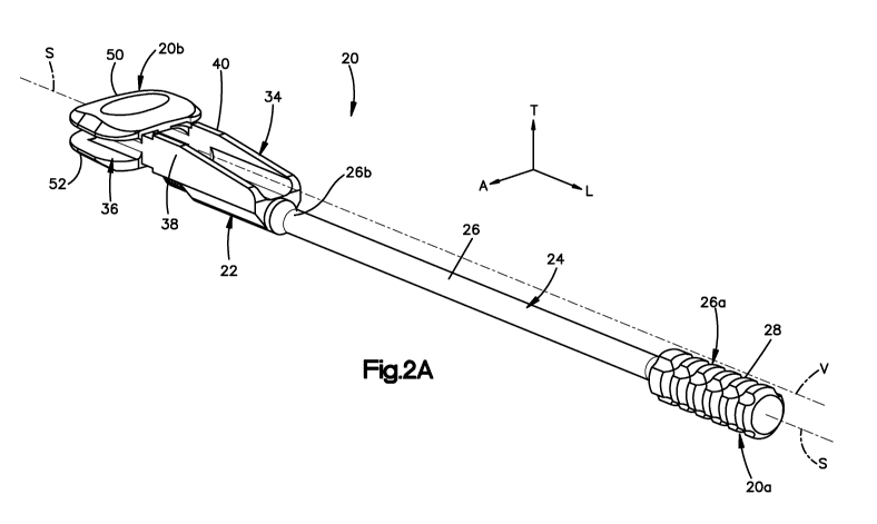
endplate configured
to face the superior vertebral body and the inferior vertebral body,
respectively. The trial
head further defines at least one visualization window extending distally
there-through
between the superior and inferior endplates.
BRIEF DESCRIPTION OF THE SEVERAL VIEWS OF THE DRAWINGS
[0008] The foregoing summary, as well as the following detailed description of
example embodiments of the invention, will be better understood when read in
conjunction with the appended drawings. For the purposes of illustrating the
trial implant
assembly of the present application, there is shown in the drawings example
embodiments. It should be understood, however, that the application is not
limited to the
precise arrangements and instrumentalities shown. In the drawings:
[0009] Fig. 1 is a perspective view of a pair of vertebral bodies separated by
an
intervertebral space;
[0010] Fig. 2A is a perspective view of a trial implant assembly including a
shaft and a trial implant in accordance with one embodiment;
[0011] Fig. 2B is a bottom plan view of the trial implant assembly illustrated
in
Fig. 2A;
[0012] Fig. 2C is a side elevation view of the trial implant assembly
illustrated
in Fig. 2A;
[0013] Fig. 2D is a sectional side elevation view of a portion of the trial
implant
assembly illustrated in Fig. 2A;
[0014] Fig. 3A is a perspective view of a distal portion of the trial implant
assembly illustrated in Figs. 2A-C;
2
CA 02758712 2011-10-13
WO 2010/120970 PCT/US2010/031148
SYNT-0902/SO 1044PCT
[0015] Fig. 3B is a distal end elevation view of the distal portion of the
trial
implant assembly illustrated in Fig. 3A;
[0016] Fig. 4A is a side elevation view of the trial implant assembly similar
to
Fig. 2C, but showing the trial implant in a translated position relative to
the shaft;
[0017] Fig. 4B is a side elevation view of the distal end of the trial implant
assembly illustrated in Fig. 2C inserted into an intervertebral disc space at
a first insertion
depth;
[0018] Fig. 4C is a side elevation view of the distal end of the trial implant
assembly illustrated in Fig. 4A inserted into an intervertebral disc space at
a second
insertion depth that is different than the first insertion depth;
[0019] Fig. 5 is a perspective view of the trial implant assembly of Fig. 2
used
in conjunction with a retainer distracter instrument and showing an anterior
side of
adjacent vertebrae;
[0020] Fig. 6 is perspective view of the trial implant assembly illustrated in
Fig.
5;
[0021] Fig. 7 is a proximal end elevation view of the trial implant assembly
illustrated in Fig. 5;
[0022] Fig. 8A is a perspective view of a distal portion of a trial implant
assembly constructed in accordance with an alternative embodiment;
[0023] Fig. 8B is a top plan view of the trial implant assembly illustrated in
Fig.
8A;
[0024] Fig. 8C is a side elevation view of the trial implant assembly
illustrated
in Fig. 8A showing an angularly offset shaft;
[0025] Fig. 8D is a distal end elevation view of the distal portion of the
trial
implant assembly illustrated in Fig. 8A;
[0026] Fig. 8E is a sectional side elevation view of the angularly offset
shaft as
illustrated in Fig. 8B constructed in accordance with one embodiment;
[0027] Fig. 8F is a sectional side elevation view of the angularly offset
shaft as
illustrated in Fig. 8B constructed in accordance with another embodiment;
[0028] Fig. 9A is a perspective view of a trial implant assembly including a
shaft and a trial implant in accordance with an alternative embodiment;
3
CA 02758712 2011-10-13
WO 2010/120970 PCT/US2010/031148
SYNT-0902/SO 1044PCT
[0029] Fig. 9B is a top plan view of the trial implant assembly illustrated in
Fig.
9A;
[0030] Fig. 9C is a side elevation view of the trial implant assembly
illustrated
in Fig. 9A;
[0031] Fig. 1 OA is a perspective view of a distal portion of the trial
implant
assembly illustrated in Figs. 9A-C;
[0032] Fig. I OB is a distal end elevation view of the distal portion of the
trial
implant assembly illustrated in Fig. 10A;
[0033] Fig. I OC is a proximal end elevation view of the distal portion of the
trial
implant assembly illustrated in Fig. I OB;
[0034] Fig. 1 IA is a perspective view of a trial implant assembly in
accordance
with an alternative embodiment;
[0035] Fig. 1 lB is a side elevation view of the trial implant assembly
illustrated
in Fig. 11A;
[0036] Fig. 11 C is a sectional side elevation view of the distal end of a
shaft and
a proximal end of a trial implant of the trial implant assembly illustrated in
Fig. 11 A;
[0037] Fig. 11D is a sectional distal end elevation view of the trial implant
assembly illustrated in Fig. 1 IA;
[0038] Fig. 12A is a perspective view of a trial implant assembly including a
shaft and a trial implant in accordance with an alternative embodiment;
[0039] Fig. 12B is a top plan view of the trial implant assembly illustrated
in
Fig. 12A;
[0040] Fig. 12C is a side elevation view of the trial implant assembly
illustrated
in Fig. 12A;
[0041] Fig. 13A is a perspective view of a distal portion of the trial implant
assembly illustrated in Figs. 12A-C;
[0042] Fig. 13B is a distal end elevation view of the distal portion of the
trial
implant assembly illustrated in Figs. 12A-C;
[0043] Fig. 13C is a sectional side elevation view of the distal end of a
shaft and
a proximal end of a trial implant of the trial implant assembly illustrated in
Figs. 13A-B;
4
CA 02758712 2011-10-13
WO 2010/120970 PCT/US2010/031148
SYNT-0902/SO 1044PCT
[0044] Fig. 13D is a distal end elevation view of the distal portion of the
trial
implant assembly similar to Fig. 13B, but also showing a pair of superior
tracks;
[0045] Fig. 14A is a perspective view of a trial implant assembly constructed
in
accordance with an alternative embodiment;
[0046] Fig. 14B is a side elevation view of the trial implant assembly
illustrated
in Fig. 14A;
[0047] Fig. 14C is a distal end elevation view of the trial implant assembly
illustrated in Fig. 14A;
[0048] Fig. 14D is a bottom plan view of a trial implant of the trial implant
assembly illustrated in Fig. 14A;
[0049] Fig. 14E is a top plan view of the trial implant illustrated in Fig.
14D;
[0050] Fig. 15A is a perspective view of a distal portion of a trial implant
assembly constructed in accordance with an alternative embodiment;
[0051] Fig. 15B is a top plan view of the trial implant assembly illustrated
in
Fig. 15A;
[0052] Fig. 15C is a side elevation view of the trial implant assembly
illustrated
in Fig. 15A;
[0053] Fig. 15D is a distal end elevation view of the trial implant assembly
illustrated in Fig. 15A;
[0054] Fig. 15E is a sectional elevation view of the trial implant assembly
illustrated in Fig. 15A;
[0055] Fig. 16A is a perspective view of a distal portion of a trial implant
assembly constructed in accordance with an alternative embodiment;
[0056] Fig. 16B is a bottom plan view of the trial implant assembly
illustrated in
Fig. 16A;
[0057] Fig. 16C is a side elevation view of the trial implant assembly
illustrated
in Fig. 16A;
[0058] Fig. 16D is a sectional elevation view of a portion of the trial
implant
assembly illustrated in Fig. 16A;
[0059] Fig. 16E is a proximal end elevation view of the trial implant assembly
illustrated in Fig. 16C;
CA 02758712 2011-10-13
WO 2010/120970 PCT/US2010/031148
SYNT-0902/SO 1044PCT
[0060] Fig. 17A is a perspective view of a trial implant assembly constructed
in
accordance with an alternative embodiment;
[0061] Fig. 17B is a sectional proximal end elevation view of the trial
implant
assembly illustrated in Fig. 17A;
[0062] Fig. 18A is a perspective view of a trial implant assembly constructed
in
accordance with an alternative embodiment; and
[0063] Fig. 18B is a proximal end elevation view of the trial implant assembly
illustrated in Fig. 18A.
DETAILED DESCRIPTION OF THE INVENTION
[0064] Referring to Fig. 1, a superior vertebral body 12a defines a superior
vertebral surface 13a of an intervertebral space 14, and an adjacent inferior
vertebral
body 12b defines an inferior vertebral surface 13b of the intervertebral space
14. Thus,
the intervertebral space 14 is disposed between the vertebral bodies 12a-b.
The vertebral
bodies 12a-b can be anatomically adjacent vertebral bodies, or can remain
after a
discectomy has been performed that removed a vertebral body from a location
between
the vertebral bodies 12a-b. As illustrated, the intervertebral space 14 is
illustrated after a
discectomy, whereby the disc material has been removed or at least partially
removed to
prepare the intervertebral space 14 to receive a disc implant that can achieve
height
restoration. Prior to inserting the permanent disc implant in the
intevertebral space, one
or more trial implants of various sizes, such as the trial implant 22 of a
trial implant
assembly 20 illustrated in Fig. 2, are inserted into the intervertebral space
14 until the
correctly sized trial implant has been determined, thereby determining the
size of the
actual disc implant to be permanently inserted. The intervertebral space 14
can be
disposed anywhere along the spine as desired.
[0065] Certain terminology is used in the following description for
convenience
only and is not limiting. The words "right", "left", "lower" and "upper"
designate
directions in the drawings to which reference is made. The words "inner" or
"distal" and
"outer" or "proximal" refer to directions toward and away from, respectively,
the
geometric center of the implant and related parts thereof. The words,
"anterior",
6
CA 02758712 2011-10-13
WO 2010/120970 PCT/US2010/031148
SYNT-0902/S01044PCT
"posterior", "superior," "inferior," "medial," "lateral," and related words
and/or phrases
are used to designate various positions and orientations in the human body to
which
reference is made and are not meant to be limiting. The terminology includes
the above-
listed words, derivatives thereof and words of similar import.
[0066] The trial implant assembly 20 is described herein as extending
horizontally along a longitudinal direction "L" and lateral direction "A", and
vertically
along a transverse direction "T". Unless otherwise specified herein, the terms
"lateral,"
"longitudinal," and "transverse" are used to describe the orthogonal
directional
components of various components. It should be appreciated that while the
longitudinal
and lateral directions are illustrated as extending along a horizontal plane,
and that the
transverse direction is illustrated as extending along a vertical plane, the
planes that
encompass the various directions may differ during use. For instance, when the
trial
implant 20 is implanted into an intervertebral space, such as the
intervertebral space 14,
the transverse direction T extends generally along the superior-inferior (or
caudal-cranial)
direction, while the plane defined by the longitudinal direction L and lateral
direction A
lie generally in the anatomical plane defined by the anterior-posterior
direction, and the
medial-lateral direction. Accordingly, the directional terms "vertical" and
"horizontal"
are used to describe the implant assembly 20 and its components as illustrated
merely for
the purposes of clarity and illustration.
[0067] Referring now also to Figs. 2A-2D, a trial implant assembly 20 is
configured to be positioned within an at least a partially cleared out disc
space, such as
the disc space 14 disposed between the superior vertebral body 12a and the
inferior
vertebral body 12b. The trial implant assembly 20 includes a trial implant 22
coupled to
a shaft 24. The shaft 24 can be formed from any desired material such as
stainless steel,
while the trial implant 22 can be formed from any desired material such as a
titanium
alloy. It should be appreciated that both the shaft 24 and the trial implant
22 can be
formed from a range of biocompatible metals or polymers, such as cobalt
chromium
molybdenum (CoCrMo), titanium and titanium alloys, stainless steel, ceramics,
or
polymers such as polyetheretherketone (PEEK), polyetherketoneketone (PEKK),
and
bioresorbable materials.
7
CA 02758712 2011-10-13
WO 2010/120970 PCT/US2010/031148
SYNT-0902/S01044PCT
[0068] The shaft 24 includes a shaft body 26 that defines a proximal end 26a,
and a distal end 26b that is separated from the proximal end 26a along a
longitudinally
extending central shaft axis S. The shaft 24 includes a handle 28 or gripping
portion at
the proximal end 26a of the shaft body 26, and a trial implant engagement
member 30 at
the distal end 26b of the shaft body 26. The handle 28 can be knurled or
otherwise
textured to facilitate an ergonomically friendly gripping surface. The shaft
body 26
defines, and thus carries, a vertebral abutment surface 27 at the distal end
26b. The
engagement member 30 is configured to be coupled to a complementary engagement
member 32 of the trial implant 22 so as to connect the shaft 24 to the trial
implant 22.
[0069] Thus, the proximal end 26a of the shaft body 26 defines a proximal end
20a of the implant assembly 20, and the trial implant 22 defines an opposed
distal end
20b of the implant assembly 20. Accordingly, a distal spatial relationship is
used herein
to refer to a longitudinal direction from the proximal end 20a toward the
distal end 20b,
and a proximal spatial relationship is used herein to refer to a longitudinal
direction from
the distal end 20b toward the proximal end 20a.
[0070] The trial implant 22 generally includes a trial base 34 coupled to the
shaft 24, a trial head 36 that is disposed distally from the trial base 34,
and a pair of
laterally spaced ribs 38 and 40 that are fixedly connected between the trial
head 36 and
the trial base 34. Thus, the trial base 34 is connected indirectly to the
trial head 36 via the
ribs 38 and 40, though it should be appreciated that the trial base 34 could
alternatively
be directly connected to the trial head 36. The trial base 34 includes a trial
base body 35
having transversely opposed upper and lower surfaces 35a and 35b, and
laterally opposed
outer surfaces 35c and 35d. As illustrated in Fig. 2D, the trial base 34
defines an
engagment member 32 that is configured to connect to the engagement member 30
of the
shaft 24.
[0071] In particular, the engagement member 30 of the shaft 24 is illustrated
as
including external threads 42 disposed in a threaded region 44 proximate to
the distal end
26b of the shaft 26. The trial implant 22 includes an aperture 46 that extends
longitudinally through the trial base body 35. The aperture 46 is sized to
receive the
threaded region 44 of the shaft 24. The engagement member 32 of the trial base
34
includes internal threads 48 disposed about the periphery of the aperture 46
that are
8
CA 02758712 2011-10-13
WO 2010/120970 PCT/US2010/031148
SYNT-0902/S01044PCT
configured to mate with the external threads 42 of the shaft 24 so as to
couple the shaft 24
to the trial implant 22.
[0072] Referring now to Figs. 2A-3B, the trial head 36 includes an upper or
superior endplate 50 that defines an upper or superior, or outer transverse,
engagement
surface 51 configured to contact the inferior endplate 13a of the superior
vertebral body
12a, and an inferior endplate 52 that defines a lower or inferior, or outer
transverse,
engagement surface 53 configured to contact the superior endplate 13b of the
inferior
vertebral body 12b. The superior endplate 50 further defines a lower or
inferior, or inner
transverse, surface 55, and the inferior endplate 52 defines an upper or
superior, or inner
transverse, surface 57. The surfaces 55 and 57 are spaced vertically along the
transverse
direction T by a gap G as illustrated, though it should be appreciated that
the endplates 50
and 52 could alternatively be connected at their inner transverse ends. Thus,
reference to
superior endplates and inferior endplates is not intended to be limited to a
pair of spaced
apart endplates unless otherwise indicated.
[0073] As described above, the trial implant assembly 20 includes a pair of
ribs
38 and 40 that are connected between the trial base 34 and the endplates 50
and 52, such
that the ribs 38 and 40 define side walls of the trial head 36. Some or all of
endplates 50
and 52, the trial base 34, and the ribs 38 and 40 can be integrally connected
or discretely
connected as desired. The ribs 38 and 40 define respective proximal ends 38a
and 40a
that are connected to the opposed lateral sides 35c and 35d of the trial base
34, and
respective distal ends 38b and 40b that are connected to the endplates 50 and
52.
[0074] The distal ends 38b and 40b are connected between the inner transverse
surfaces 55 and 57, and are laterally spaced apart so as to at least partially
define an
aperture or visualization window 60 that extends longitudinally through the
trial head 36.
The visualization window 60 is defined between the inner transverse surfaces
55 and 57
of the superior and inferior endplates 50 and 52, respectively, and the ribs
38 and 40.
Thus, the visualization window 60 is enclosed, and extends transversely
between the
inner transverse surfaces 55 and 57, and laterally between the ribs 38 and 40.
The trial
head 36 further defines first and second laterally opposed slots 39 and 41
that are
disposed on opposite sides of the window 60, and are separated from the window
by the
first and second ribs 38 and 40, respectively. The slots 39 and 41 are thus
closed on their
9
CA 02758712 2011-10-13
WO 2010/120970 PCT/US2010/031148
SYNT-0902/S01044PCT
laterally inner ends, but open at their laterally outer ends. Accordingly, the
slots 39 and
41 can be referred to as being open. The distal ends of the first and second
ribs 38 and 40
terminate proximal to the distal end of the trial head 36, or endplates 50 and
52, so as to
provide increased visualization and allow improved access to posterior
structures in the
disc space using a conventional nerve hook or probe.
[0075] In this regard, it should be appreciated that the trial implant
assembly 22
is devoid of structure that obstructs a straight visualization axis V from
extending from a
first location disposed proximal to the handle 28 to a second location that
passes through
the visualization window 60. Otherwise stated, the trial implant defines a
visualization
window such as window 60, at least a portion of which up to all of which is
visually
unobstructed. The visualization axis V can extend parallel to the central
shaft axis S as
illustrated, or can extend at an angle with respect to the central shaft axis
S.
[0076] The distal ends 38b and 40b vertically offset with respect to the
proximal
ends 38a and 40a of the ribs 38 and 40, such that the gap G between the
endplates 50 and
52 is at least partially vertically offset with respect to the aperture 46 and
the shaft 24, as
well as the upper surface 35a of the trial base 34. In accordance with the
illustrated
embodiment, the distal ends 38b and 40b are disposed above the proximal ends
38a and
40a of the ribs 38 and 40, such that the gap G between the endplates 50 and 52
is
disposed at least partially above the aperture and shaft 24, as well as the
upper surface
35a of the trial base 34. In this regard, it should be appreciated that the
inner transverse
surface 57 of the inferior endplate 52 can be disposed above or below the
upper surface
35a of the trial base 34. Accordingly, visualization is possible through the
visualization
window 60 along a distal direction from a location proximal of the trial head
36, and
further from a location proximal of the handle 28.
[0077] Referring now also to Fig. 4A, rotation of the shaft 24 relative to the
trial
implant 22 causes the threads 42 of the shaft 24 and the threads 48 of the
trial base 34 to
ride along each other, thereby causing the trial implant 22 to translate
longitudinally
relative to the shaft 24. For instance, rotation of the shaft 24 in a first
direction (e.g.,
counterclockwise) relative to the trial implant 22 causes trial implant 22 to
translate
distally in the longitudinal direction relative to the shaft 24 as indicated
by Arrow Al,
while rotation of the shaft 24 in a second opposite direction (e.g.,
clockwise) relative to
CA 02758712 2011-10-13
WO 2010/120970 PCT/US2010/031148
SYNT-0902/SO 1044PCT
the trial implant 22 causes the trial implant 22 to move proximally in the
longitudinal
direction relative to the shaft 24 as indicated by Arrow A2. Fig. 2C
illustrates the trial
head 36 in a retracted position relative to the shaft 24, while Fig. 4A
illustrates the trial
head 36 in an extended position relative to the shaft 24.
[0078] Furthermore, referring also to Figs. 4B-C, at least a portion of the
distal
end 26b of the shaft body 26 is vertically offset from the trial head. In
accordance with
the illustrated embodiment, at least a portion of the distal end 26b of the
shaft body 26 is
disposed below the outer transverse surface 53 of the inferior endplate 52.
Accordingly,
the vertebral abutment surface 27 of the shaft 24 is configured to abut one of
the
vertebrae, such as the inferior vertebra 12b, when the trial head 36 is
inserted into the
intervertebral space 14. Accordingly, when the trial head 36 is in a retracted
position as
shown in Fig. 4B, the trial head 36 is inserted into the intervertebral disc
space 14 at a
first insertion depth Dl when the abutment surface 27 abuts the inferior
vertebra 12b.
When the trial head 36 is in an extended position as shown in Fig. 4C, the
trial head 36 is
inserted into the intervertebral disc space 14 at a second insertion depth D2
when the
abutment surface 27 abuts the inferior vertebra 12b. The second insertion
depth D2 is
greater than the first insertion depth Dl .
[0079] Referring again to Fig. 4A, the shaft 24 can include a stop member 62
that is configured to abut the proximal end of the trial base 34 when the
trial implant 22 is
fully retracted. The stop member 62 thus prevents the trial implant 22 from
being
retracted to a location where the threads 42 and 48 would become disengaged.
The stop
member 62 projects radially out from the shaft body 26 so as to define a cross-
sectional
dimension greater than that of the shaft body 26, and greater than that of the
aperture 46
that receives the shaft body 26. The shaft 24 can include depth markings 66 or
other
indicia distal of the stop member 62 to indicate the position of the trial
implant 22 relative
the shaft 24.
[0080] Referring now also to Figs. 5-7, an instrument assembly 21 can include
the trial implant assembly 20 in combination with a distractor retainer
instrument 68.
The distractor retainer instrument 68 includes a superior anchor screw 70
configured to
be temporarily driven or implanted into the superior vertebral body 12a, an
inferior
anchor screw 72 configured to be temporarily driven or implanted into the
inferior
11
CA 02758712 2011-10-13
WO 2010/120970 PCT/US2010/031148
SYNT-0902/S01044PCT
vertebral body 12b. The distractor retainer instrument 68 further includes a
superior
retainer distractor tube 74 couplable to the superior anchor screw 70, and an
inferior
retainer distractor tube 76 couplable to the inferior anchor screw 72. The
distractor
retainer instrument 68 can be implemented to initially separate the superior
and inferior
vertebral bodies 12a and 12b, respectively, and retain distraction prior to
cleaning out the
disc tissue and inserting the total disc replacement implant. The anchor
screws 70 and 72
can be removed from the vertebral bodies 12a and 12b after the permanent disc
implant
has been implanted between the vertebral bodies 12a and 12b.
[0081] During operation, and with continuing reference to Figs. 1-7, at least
a
partial discectomy is performed and the intervertebral disc space 14 is
decompressed, for
instance using the distractor retainer instrument 68. A surgeon selects a
trial head 36 to
assess the size of the disc space 14 and couples the trial head 36 to the
shaft 24. In
particular, the threaded region 44 of the shaft 24 is inserted into the
aperture 46 of the
trial base 34, and the shaft 24 is rotated relative to the trial base 34 so
that the threads 42
of the shaft 24 mate with the threads 48 of the trial base 34. The shaft 24 is
continuously
rotated relative to the trial head 34 until the trial implant 22 is fully
retracted with respect
to the shaft 24. Alternatively, the trial head could already be assembled to
its own shaft
with the head fully retracted.
[0082] The surgeon can insert the trial head 36 into the intervertebral disc
space
14, for instance by tapping on the proximal end of shaft 24 with a small
mallet to advance
the trial head 36 into the intervertebral disc space 14 until the abutment
surface 27 abuts
the inferior vertebral body 12b. Should the surgeon wish to place the trial
head 36 deeper
within the disc space 14 the shaft 24 can be rotated in a counter clockwise
direction,
causing it to back out of trial base 34. Because the pitch of the thread 44 on
shaft 24 is
fixed, the surgeon can precisely control how much the shaft backs out. The
trial head 36
can then be inserted deeper into the intervertebral disc space 14 by tapping
on the
proximal end of the shaft 24 until the abutment surface 27 contacts the
vertebral body
12b again. The optional depth markings 66 can assist in determining the
desired insertion
depth of the trial head 36 in the disc space 14.
[0083] As the trial head 36 is inserted into the disc space 14, the
visualization
window 60 and the lateral slots 39 and 41 allow the surgeon to visually
determine the
12
CA 02758712 2011-10-13
WO 2010/120970 PCT/US2010/031148
SYNT-0902/S01044PCT
position of the trial head 36 without using fluoroscopy or other radio
imaging. More
particularly, the window 60 allows for visualization of the posterior
longitudinal ligament
(PLL), while the first and second lateral slots 39 and 41 allow for
visualization of the
exiting nerve roots. Accordingly, as described above with respect to the
visualization
window, at least a portion up to all of the lateral slots 39 and 41 are
visually
unobstructed. In this regard, the visualization window 60 can define a primary
visualization window, while the lateral slots 39 and 41 can define auxiliary
visualization
windows disposed adjacent the primary visualization window 60. Thus, at least
one rib,
such as ribs 38 and 40, can define at least one visualization window, such as
visualization
windows 60, 39, and 41.
[0084] Once the trial head 36 is positioned appropriately within the disc
space
14, the surgeon assesses the fit of the trial head 36 within the disc space
14. If the trial
head 36 is not properly sized for the disc space, the surgeon removes and
replaces the
trial head 36 with a differently sized trial head 36 and repeats the
evaluation process.
Once the properly size trial head 36 is in place, the trial head 36 is removed
and a
permanent total disc replacement implant having a size corresponding to the
properly
sized trial head 36 is permanently implanted in the disc space.
[0085] It should thus be appreciated that a kit can be provided that includes
a
plurality of trial implants 22, each couplable to the shaft 24, and each
having trial heads
36 of incrementally larger volumes that are sized and configured to fill and
provide the
desired spacing between the superior and inferior vertebral bodies 12a and
12b. For
instance, the trial heads 36 can define at least one or a plurality of varying
characteristics,
including a shape and/or a dimension such an outer lateral dimension, an outer
longitudinal dimension, and a height between the outer transverse engagement
surfaces
51 and 53. The trial head 36 of each trial implant 22 can be color coded, such
as
anodized or otherwise colored, such that a range of different colors is
provided to
distinguish between the range of different sizes of the corresponding trial
heads 36.
[0086] It should be appreciated that the trial implant assembly 20 has been
described in accordance with one embodiment, and that the trial implant
assembly 20
could alternatively be constructed in accordance with numerous alternative
embodiments
that provide at least one visualization window. Some of the alternative
embodiments are
13
CA 02758712 2011-10-13
WO 2010/120970 PCT/US2010/031148
SYNT-0902/S01044PCT
described below, it being appreciated that the scope of the present disclosure
is not
intended to be limited to any or all of the specific embodiments described
herein.
[0087] For instance, referring to Figs. 8A-D, a trial implant assembly 120
constructed in accordance with an alternative embodiment is illustrated
including
reference numerals corresponding to like elements of the trial implant
assembly 20
incremented by 100. Thus, the trial implant assembly 120 can be constructed
substantially as described with respect to the trial implant assembly 20
except as
otherwise noted.
[0088] As described above and illustrated with respect to Figs. 2-4C, the
shaft
24 of the trial implant assembly 20 can extend longitudinally, or
perpendicular to the
transverse direction T. As illustrated in Figs. 8A-D, it is further recognized
that the shaft
124 can extend in a direction angularly offset with respect to the
longitudinal direction L,
and thus with respect to the visualization window 160 that extends
longitudinally through
the trial head 136. In accordance with the illustrated embodiment, the shaft
124 is angled
transversely downward with respect to the longitudinal direction L so as to
define an
angle 0 with respect to the longitudinal direction L that can be any angle as
desired, such
as between 00 and 60 . Thus, the central shaft axis S can extend at a non-
perpendicular
angle with respect to the transverse axis T. As illustrated in Fig. 8E, the
trial base 134
can oriented such that the upper and lower surfaces 135a and 135b extend
parallel to the
central shaft axis S, and the aperture 146 that receives the shaft 124 extends
parallel to
the upper and lower surfaces 135a and 135b as illustrated in Fig. 2D.
Alternatively, as
illustrated in Fig. 8F, upper and lower surfaces 135a and 135b can extend
longitudinally,
and thus angularly offset with respect to the central shaft axis S, and the
aperture 146 that
receives the shaft 124 can extend through the trial base in a direction that
is angularly
offset with respect to the upper and lower surfaces 135a and 135b so as to
receive the
shaft 124 along the angularly offset shaft axis S.
[0089] Referring now to Figs. 9A-IOC, a trial implant assembly 220 constructed
in accordance with an alternative embodiment is illustrated including
reference numerals
corresponding to like elements of the trial implant assembly 20 incremented by
200.
Thus, the trial implant assembly 220 can be constructed substantially as
described with
respect to the trial implant assembly 20 except as otherwise noted. As
illustrated, the trial
14
CA 02758712 2011-10-13
WO 2010/120970 PCT/US2010/031148
SYNT-0902/S01044PCT
implant assembly 220 includes the shaft 224, and a trial implant 222
configured to be
removably coupled to the shaft 224. The trial implant 222 includes a trial
head 226
having a superior endplate 250 and an inferior endplate 252 configured to be
inserted into
an intervertebral space, a trial base 234 configured to be coupled to the
shaft 224, and
laterally spaced ribs 238 and 240 connected between the trial head 236 and the
trial base
234 as described above. Thus, the ribs 238 and 240 are spaced so as to provide
a primary
visualization window 260 and auxiliary visualization window in the form of
first and
second laterally opposed slots 239 and 241, as described above.
[0090] The trial base 234 includes a trial base body 235 that defines a pair
of
laterally or horizontally spaced engagement members 232a-b that are each
configured to
connect to a complementary engagement member 230 of the shaft 224. In
particular, the
engagement member 230 of the shaft 224 is illustrated as including external
threads 242
disposed in a threaded region 244 proximate to the distal end of the shaft
226. The
engagement members 232a-b each includes a corresponding pair of laterally
spaced
apertures 246a-b that each extends longitudinally through the trial base 234.
The
apertures 246a-b are each sized to receive the threaded region 244 of the
shaft 224. The
engagement members 232a-b of the trial base 234 includes internal threads 248
disposed
about the periphery of the apertures 246a-b that are configured to mate with
the external
threads 242 of the shaft 224 so as to couple the shaft 224 to the trial
implant 222.
[0091] The apertures 246a-b are laterally offset on opposite sides with
respect to
a lateral midpoint of the primary visualization window 260. Each aperture 246a-
b is
further vertically or transversely offset with respect to a vertical or
transverse midpoint of
the primary visualization window 260. Furthermore, one or both of the
apertures 246a-b
can extend longitudinally, or can be angled with respect to the longitudinal
direction L as
described above. The trial base 234 thus defines a pair of offset engagement
members
232a-b that are configured to be coupled to the shaft 224, such that the shaft
224 can be
coupled to the trial implant 222 at multiple locations. In particular, the
threaded region
244 of the shaft 224 can be mated with the threads 248 of either engagement
member
232a-b so as to provide relative motion between the trial implant 222 and the
shaft 224 in
the manner described above.
CA 02758712 2011-10-13
WO 2010/120970 PCT/US2010/031148
SYNT-0902/S01044PCT
[0092] Referring now to Figs. 1 IA-D, a trial implant assembly 320 constructed
in accordance with an alternative embodiment is illustrated including
reference numerals
corresponding to like elements of the trial implant assembly 20 incremented by
300.
Thus, the trial implant assembly 320 can be constructed substantially as
described with
respect to the trial implant assembly 20 except as otherwise noted. As
illustrated, the trial
implant assembly 320 includes a shaft 324, which may or may not be cannulated,
and a
trial implant 322, which may or may not be cannulated, configured to be
removably
coupled to the shaft 324. The trial implant 322 includes a trial head 336
having a
superior endplate 350 and an inferior endplate 352 configured to be inserted
into an
intervertebral space, a trial base 334 configured to be coupled to the shaft
324, and a
laterally central rib 338 connected between the trial head 336 and the trial
base 334.
[0093] The superior endplate 350 defines a lower or inferior, or inner
transverse, surface 355, and the inferior endplate 352 defines an upper or
superior, or
inner transverse, surface 357. The surfaces 355 and 357 are spaced vertically
along the
transverse direction T by a gap G as illustrated, though it should be
appreciated that the
endplates 350 and 352 could alternatively be connected at their inner
transverse ends.
The rib 338 extends distally from the trial base 334, and is connected between
the
surfaces 355 and 357 of the endplates 350 and 352, respectively. First and
second
laterally opposed visualization slots 339 and 341 extend longitudinally
through the trial
head 336 on opposed lateral sides of the rib 338. The distal end of the rib
338 can
terminate proximal to the distal end of the trial head 336, or endplates 350
and 352, so as
to provide increased visualization and allow improved access to posterior
structures in the
disc space using a conventional nerve hook or probe.
[0094] The trial base 334 defines an engagment member 332 that is configured
to connect to the engagement member 330 of the shaft 324. The trial implant
322
includes an aperture 346 that extends longitudinally through the trial base
body 335, and
can further extend longitudinally through the rib 338. The aperture 346 is
sized to
receive the threaded region 344 of the shaft 324. The engagement member 332 of
the
trial base 334 includes internal threads 348 disposed about the periphery of
the aperture
346 that are configured to mate with the external threads 342 of the shaft 324
so as to
couple the shaft 324 to the trial implant 322.
16
CA 02758712 2011-10-13
WO 2010/120970 PCT/US2010/031148
SYNT-0902/S01044PCT
[0095] The trial base 322 further includes a pair of superior tracks 373a and
375a extending up, or transversely out, from the superior or upper surface
335a of the
trial base body 335, and a pair of inferior tracks 373b and 375b extending
down, or
transversely out, from the lower or inferior surface 335b of the trial base
body 335. The
superior tracks 373a and 375a are laterally spaced from each other so as to
define a
superior longitudinally elongate guide channel 377a extending between the
superior
tracks 373a and 375a. The inferior tracks 373b and 375b are laterally spaced
from each
other so as to define an inferior longitudinally elongate guide channel 377b
extending
between the inferior tracks 373b and 375b.
[0096] The shaft 324 includes an adjustable mechanical stop 380 coupled to the
distal portion of the shaft body 326 such that the stop 380 is rotatable with
respect to the
shaft body 326 but is translatably fixed relative to the shaft 324. Thus, the
stop 380 can
rotate about the shaft body 326 but is unable to translate along the shaft
body 326. In
particular, the stop 380 includes at least one circumferential collar 381 (a
pair of
longitudinally spaced collars 381 are illustrated) that nest within a
corresponding at least
one radial groove 383 (a pair of longitudinally spaced grooves 383 are
illustrated) that
extend into the shaft body 326. Interference between the collars 381 and the
portion of
the shaft body 326 that is adjacent the grooves 383 prevent the stop 380 from
translating
along the shaft body 326. Alternatively, a snap ring can be snapped onto the
shaft 324
into a groove disposed between the collars 381, such that the interference
between the
snap ring and the collars 381 prevent translation of the stop 380 along the
shaft 324. The
collars 381, and thus the stop 380, are free to rotate about the shaft body
326 within the
radial grooves 383.
[0097] The distal surface of the distal-most collar 381 defines a stop member
362 configured to abut the proximal end of the trial base 334 when the trial
implant 322
is fully retracted on the shaft 324. The stop member 362 thus prevents the
trial implant
322 from being retracted to a location where the threads 342 and 348 would
become
disengaged. The stop member 362 projects radially out from the shaft body 326
so as to
define a cross-sectional dimension greater than that of the shaft body 326,
and greater
than that of the aperture 346 that receives the shaft body 326.
17
CA 02758712 2011-10-13
WO 2010/120970 PCT/US2010/031148
SYNT-0902/S01044PCT
[0098] The stop 380 further includes a guide body 385 extending transversely
outward and longitudinally distal from the collars 381. The guide body 385
defines a
lateral outer dimension substantially equal to or slightly less than that of
the guide
channels 377a-b. Accordingly, the guide body 385 is configured to ride within
and
translate within a select one of the guide channels 377a-b as the shaft 324 is
rotated
relative to the trial base 334, thereby causing the implant 322 to translate
relative to the
shaft 324.
[0099] The stop 380 further defines a vertebral abutment surface 327 defined
by
the distal surface of the guide body 385. When the guide body 385 is disposed
in the
superior guide channel 377a, the abutment surface 327 is configured to abut
the superior
vertebra when the trial head 336 is inserted into the intervertebral space.
When the guide
body 385 is disposed in the inferior guide channel 377b, the abutment surface
327 is
configured to abut the inferior vertebra when the trial head 336 is inserted
into the
intervertebral space. Thus, the guide channels 377a-b are configured to
maintain the
alignment of the vertebral abutment surface 327 with a select one of the
superior and
inferior vertebrae based on the anatomy of the patient. Once the trial head
336 is
positioned within the intervertebral disc space, the shaft 324 can be
uncoupled, i.e.,
unscrewed, from the trial implant 322 and the aperture 346 can serve as a
primary
visualization window into the intervertebral disc space as described above
with respect to
the primary visualization window 60. The shaft 324 can further be cannulated,
such that
the cannulation of the shaft 324 is aligned with the aperture 346.
Accordingly, the
visualization window defined by the aperture 346 can be visually accessed
through the
cannulation of the shaft 324.
[0100] Referring now to Figs. 12A-13B, a trial implant assembly 420
constructed in accordance with an alternative embodiment is illustrated
including
reference numerals corresponding to like elements of the trial implant
assembly 20
incremented by 400. Thus, the trial implant assembly 420 can be constructed
substantially as described with respect to the trial implant assembly 20
except as
otherwise noted. As illustrated, the trial implant assembly 420 includes a
shaft 424, and a
trial implant 422 configured to be removably coupled to the shaft 424. The
trial implant
422 includes a trial head 436 having a superior endplate 450 and an inferior
endplate 452
18
CA 02758712 2011-10-13
WO 2010/120970 PCT/US2010/031148
SYNT-0902/SO 1044PCT
configured to be inserted into an intervertebral space, a trial base 434
configured to be
coupled to the shaft 424, and a laterally central rib 438 connected between
the trial head
436 and the trial base 434.
[0101] The superior endplate 450 defines a lower or inferior, or inner
transverse, surface 455, and the inferior endplate 452 defines an upper or
superior, or
inner transverse, surface 457. The surfaces 455 and 457 are spaced vertically
along the
transverse direction T by a gap G as illustrated, though it should be
appreciated that the
endplates 450 and 452 could alternatively be connected at their inner
transverse ends.
The rib 438 extends distally from the trial base 434, and is connected between
the
surfaces 455 and 457 of the endplates 450 and 452, respectively. First and
second
laterally opposed visualization slots 439 and 441 extend longitudinally
through the trial
head 336 on opposed lateral sides of the rib 438. The distal end of the rib
438 can
terminate proximal to the distal end of the trial head 436, or endplates 450
and 452, so as
to provide increased visualization and allow improved access to posterior
structures in the
disc space using a conventional nerve hook or probe. Furthermore, the
laterally opposed
outer walls of the rib 438 are concave and convergingly tapered along a
direction from
the proximal end of the rib 438 toward the distal end of the rib 438, thereby
increasing
visualization of the middle of the disc space.
[0102] The trial base 434 defines an engagment member 432 that is configured
to connect to the engagement member 430 of the shaft 424. In particular, the
trial
implant 422 includes an aperture 446 that extends longitudinally through the
trial base
body 435, and further extends longitudinally through the rib 438. A portion of
substantially all of aperture 446 is sized to receive the threaded region 444
of the shaft
424. The engagement member 432 of the trial base 434 includes internal threads
448
disposed about the periphery of part or all of the aperture 446 that are
configured to mate
with the external threads 442 of the shaft 424 so as to couple the shaft 424
to the trial
implant 422.
[0103] The trial base 422 can further include a pair of superior tracks 473a
and
475a (see Fig. 13D) that extend transversely out from, and flare laterally out
from, the
superior or upper surface 435a of the trial base body 435, and a pair of
inferior tracks
473b and 475b that extend transversely out from, and flare laterally out from,
the lower
19
CA 02758712 2011-10-13
WO 2010/120970 PCT/US2010/031148
SYNT-0902/SO 1044PCT
or inferior surface 435b of the trial base body 435. Thus, the tracks 473a and
475a flare
laterally away from each other, and the tracks 473b and 475b flare laterally
away from
each other. The superior tracks 473a and 475a can be constructed substantially
as
described with respect to the inferior tracks 475b and 475b illustrated in
Figs. 12A-13C,
except that the superior tracks 473a and 475a are transversely inverted with
respect to the
inferior tracks 473b and 475b.
[0104] The shaft 424 defines a cannulation 425 that extends longitudinally
through the shaft body 426. The proximal portion of the shaft 424 includes a
mechanism
415 for connecting to tubing for siphoning out blood and/or other tissue or
debris
remaining from the discectomy as described below.
[0105] The shaft 424 further includes an adjustable mechanical stop 480
coupled to the distal portion of the shaft body 426 such that the stop 480 is
rotatable with
respect to the shaft body 426 but is translatably fixed relative to the shaft
424. Thus, the
stop 480 can rotate about the shaft body 426 but is unable to translate along
the shaft
body 426. In particular, the stop 480 includes at least one circumferential
collar 481 (a
pair of longitudinally spaced collars 481 are illustrated) that nest within a
corresponding
at least one radial groove 483 (a pair of spaced grooves 483 are illustrated)
that extend
into the shaft body 426. Interference between the collars 481 and the portion
of the shaft
body 426 that is adjacent the grooves 483 prevent the stop 480 from
translating along the
shaft body 426. Alternatively, a snap ring can be snapped onto the shaft 424
into a
groove disposed between the collars 481, such that the interference between
the snap ring
and the collars 481 prevent translation of the stop 480 along the shaft 424.
The collars
481, and thus the stop 480, are free to rotate about the shaft body 426 within
the radial
grooves 483.
[0106] The distal surface of the distal-most collar 481 defines a stop member
462 configured to abut the proximal end of the trial base 434 when the trial
implant 422
is fully retracted on the shaft 424. The stop member 462 thus prevents the
trial implant
422 from being retracted to a location where the threads 442 and 448 would
become
disengaged. The stop member 462 projects radially out from the shaft body 426
so as to
define a cross-sectional dimension greater than that of the shaft body 426,
and greater
than that of the aperture 446 that receives the shaft body 426.
CA 02758712 2011-10-13
WO 2010/120970 PCT/US2010/031148
SYNT-0902/S01044PCT
[0107] The stop 480 is forked with a first guide body 485a and a second guide
body 485b that flare laterally away from each other along a transversely
outward
direction. Each of the first and second guide bodies 485a-b are positioned
such that the
laterally inner surfaces of the guide bodies 485a-b ride along the laterally
outer surfaces
of the superior tracks 473a and 475a when the guide bodies 485a-b are aligned
with the
superior tracks, and ride along the laterally outer surfaces of the inferior
tracks 473b and
475b when the guide bodies 485a-b are aligned with the inferior tracks. In
particular,
each guide body 485a and 485b can include a pocket 487a and 487b,
respectively, that at
least partially receives the corresponding track 473 and is configured to ride
along the
track 473.
[0108] During operation, the stop 480 is rotated about the shaft 424 as
desired
so as to align the stop 480 with a select one of the superior tracks 473a and
475a, and the
inferior tracks 473b and 475b. The threads 442 of the shaft 424 are then
engaged with
the threads 448 of the trial implant 422, which causes the guide bodies 485a-b
to ride
along the selected tracks 473 and 475. Accordingly, the guide bodies 485 are
configured
to ride along the tracks 473 and 475 as the shaft 324 424 is rotated relative
to the trial
base 434, thereby causing the trial implant 422 to translate relative to the
shaft 424. The
space between the first and second guide bodies 485a-b allow the trial implant
422 to be
placed around or in close proximity to a Caspar distraction pin or various
elements of the
distractor 68 as described above with respect to Figs. 5-7.
[0109] The stop 480 further defines a vertebral abutment surface 427 defined
by
the distal surface of the guide body 485. When the guide body 485 engages the
superior
tracks 473a and 475a, the abutment surface 427 is configured to abut the
superior
vertebra when the trial head 436 is inserted into the intervertebral space.
When the guide
body 485 engages the inferior tracks 473b and 475b, the abutment surface 427
is
configured to abut the inferior vertebra when the trial head 436 is inserted
into the
intervertebral space. Thus, the tracks 473 and 475 are configured to maintain
the
alignment of the vertebral abutment surface 427 with a select one of the
superior and
inferior vertebrae based on the anatomy of the patient. Once the trial head
436 is
positioned within the intervertebral disc space, the shaft 424 can be
uncoupled, i.e.,
unscrewed, from the trial implant 422 and the aperture 446 can serve as a
primary
21
CA 02758712 2011-10-13
WO 2010/120970 PCT/US2010/031148
SYNT-0902/S01044PCT
visualization window into the intervertebral disc space as described above
with respect to
the primary visualization window 60. The cannulation 425 of the shaft 424,
which is in
alignment with the aperture 446, provides visual access to the visualization
window
defined by the aperture 446 prior to removal of the shaft 424 from the trial
implant 422.
The cannulation 425 of the shaft 424 further allows blood and other matter to
be siphoned
out from the disc space.
[0110] Once the trial head 436 is inserted into the disc space, a vacuum
source
(not shown) is connected to the connection mechanism 415 on the proximal end
of the
shaft 424 and suction is applied to remove debris, such as blood and/or tissue
debris
remaining from the discectomy from the disc space, through the through hole
425, and
out through the cannulated interior of the shaft 424. Thus, the cannulated
shaft can
provide a suction passageway for the removal of debris under an applied vacuum
pressure. The removal of tissue debris and blood increases visualization and,
further,
provides a better suited intervertebral environment for implantation of the
total disc
replacement implant.
[0111] Referring now to Figs. 14A-E, a trial implant assembly 520 constructed
in accordance with an alternative embodiment is illustrated including
reference numerals
corresponding to like elements of the trial implant assembly 20 incremented by
500.
Thus, the trial implant assembly 520 can be constructed substantially as
described with
respect to the trial implant assembly 20 except as otherwise noted. As
illustrated, the trial
implant assembly 520 includes a shaft 524, and a trial implant 522 configured
to be
removably coupled to the shaft 524. The trial implant 522 includes a trial
head 536
having a superior endplate 550 and an inferior endplate 552 configured to be
inserted into
an intervertebral space, a trial base 534 configured to be coupled to the
shaft 524, and a
laterally central rib 538 connected between the trial head 536 and the trial
base 534.
[0112] The superior endplate 550 defines a lower or inferior, or inner
transverse, surface 555, and the inferior endplate 552 defines an upper or
superior, or
inner transverse, surface 557. The surfaces 555 and 557 are spaced vertically
along the
transverse direction T by a gap G as illustrated, though it should be
appreciated that the
endplates 550 and 552 could alternatively be connected at their inner
transverse ends.
The rib 538 extends distally from the trial base 534, and is connected between
the
22
CA 02758712 2011-10-13
WO 2010/120970 PCT/US2010/031148
SYNT-0902/S01044PCT
surfaces 555 and 557 of the endplates 550 and 552, respectively. First and
second
laterally opposed visualization slots 539 and 541 extend longitudinally
through the trial
head 536 on opposed lateral sides of the rib 538. The distal end of the rib
538 can
terminate proximal to the distal end of the trial head 536, or endplates 550
and 552, so as
to provide increased visualization and allow improved access to posterior
structures in the
disc space using a conventional nerve hook or probe.
[0113] The trial base 534 further includes a pair of superior tracks 573a and
575a extending up, or transversely out, from the superior or upper surface
535a of the
trial base body 535, and a pair of inferior tracks 573b and 575b extending
down, or
transversely out, from the lower or inferior surface of the trial base body
535. The
superior tracks 573a and 575a are laterally spaced from each other so as to
define a
superior longitudinally elongate guide channel 577a extending between the
superior
tracks 573a and 575a. The inferior tracks 573b and 575b are laterally spaced
from each
other so as to define an inferior longitudinally elongate guide channel 577b
extending
between the inferior tracks 573b and 575b.
[0114] The shaft 524 includes an adjustable mechanical stop 580 coupled to the
distal portion of the shaft body 526 such that the stop 580 is rotatable with
respect to the
shaft body 526 but is translatably fixed relative to the shaft body 526. Thus,
the stop 580
can rotate about the shaft body 526 but is unable to translate along the shaft
body 526. In
particular, the stop 580 includes a pair of longitudinally spaced collars 581.
The shaft
524 includes a snap ring 597 that is disposed in a groove 583 extending into
the shaft
body 526, such that the ring 597 is disposed between the collars 581. Thus,
interference
between the ring 597 and the collars 581 prevent translation of the stop 580
along the
shaft 524. The collars 581, and thus the stop 580, are free to rotate about
the shaft body
526.
[0115] The distal surface of the distal-most collar 581 defines a stop member
562 configured to abut the proximal end of the trial base 534 when the trial
implant 522
is fully retracted on the shaft 524. The stop 580 further includes a guide
body 585
extending transversely outward and longitudinally distal from the collars 581.
The guide
body 585 defines a lateral outer dimension substantially equal to or slightly
less than that
of the guide channels 577a-b. Accordingly, the guide body 585 is configured to
ride
23
CA 02758712 2011-10-13
WO 2010/120970 PCT/US2010/031148
SYNT-0902/S01044PCT
within and translate within a select one of the guide channels 577a-b as the
shaft 524 is
rotated relative to the trial base 534, thereby causing the trial implant 522
to translate
relative to the shaft 524 in the manner described above.
[0116] The stop 580 further defines a pair vertebral abutment surfaces 527a-b
defined by the distal surface of the guide body 585. In particular, the guide
body 585
defines a pair of legs 591a-b that extend forward from the guide body 585. The
legs
59la-b are laterally separated from each other. Thus, the vertebral abutment
surfaces 527
a-b are defined by the distal surfaces of the legs 59l a-b, such that a gap
593 extends
laterally between the abutment surfaces 527a-b. The abutment surfaces 527a-b
are
configured to abut the same vertebra when the trial head 536 is inserted into
an
intervertebral space.
[0117] For instance, when the guide body 585 is disposed in the superior guide
channel 577a, the abutment surfaces 527a-b are configured to abut the superior
vertebra
when the trial head 536 is inserted into the intervertebral space. When the
guide body
585 is disposed in the inferior guide channel 577b, the abutment surfaces 527a-
b are
configured to abut the inferior vertebra when the trial head 536 is inserted
into the
intervertebral space. Thus, the guide channels 577a-b are configured to
maintain the
alignment of the vertebral abutment surface 527 with a select one of the
superior and
inferior vertebrae based on the anatomy of the patient.
[0118] Referring now to Figs. 15A-E, a trial implant assembly 620 constructed
in accordance with an alternative embodiment is illustrated including
reference numerals
corresponding to like elements of the trial implant assembly 520 incremented
by 100.
Thus, the trial implant assembly 620 can be constructed substantially as
described with
respect to the trial implant assembly 520 except as otherwise noted. As
illustrated, the
trial implant assembly 620 includes a shaft 624, and a trial implant 622
configured to be
removably coupled to the shaft 624. The trial implant 622 includes a trial
head 636
having a superior endplate 650 and an inferior endplate 652 configured to be
inserted into
an intervertebral space, a trial base 634 configured to be coupled to the
shaft 624, and a
laterally central rib 638 connected between the trial head 636 and the trial
base 634.
[0119] The stop 680 includes a guide body 685 extending transversely outward
and longitudinally distal from the collars 681. The guide body 685 defines a
lateral outer
24
CA 02758712 2011-10-13
WO 2010/120970 PCT/US2010/031148
SYNT-0902/S01044PCT
dimension substantially equal to or slightly less than that of the guide
channels 677a-b.
Accordingly, the guide body 685 is configured to ride within and translate
within a select
one of the guide channels 677a-b as the shaft 624 is rotated relative to the
trial base 634,
thereby causing the trial implant 622 to translate relative to the shaft 624
in the manner
described above.
[0120] The stop 680 further defines a pair vertebral abutment surfaces 627a-b
defined by the distal surface of the guide body 685. In particular, the guide
body 685
defines a pair of superior and inferior legs 691 a-b that extend forward from
the guide
body 685, and are transversely separated from each other Thus, the vertebral
abutment
surfaces 627a-b are defined by the distal surfaces of the legs 691 a-b, such
that the
abutment surfaces 627a-b are transversely separated. Thus, the abutment
surfaces 627a-b
are configured to abut different vertebrae when the trial head 636 is inserted
into an
intervertebral space. Specifically, the abutment surfaces 627a-b are
configured to abut
the adjacent vertebrae that define the intervertebral space into which the
trial head 636 is
inserted. Thus, the superior abutment surface 627a is configured to abut the
superior
vertebra, and the inferior abutment surface 627b is configured to abut the
inferior
vertebra. It should be appreciated that the superior and inferior legs 691 a-b
could be split
so as to each define a pair of vertebral abutment surfaces in the manner
illustrated in Figs.
14A-D.
[0121] Referring now to Figs. 16A-E, a trial implant assembly 720 constructed
in accordance with an alternative embodiment is illustrated including
reference numerals
corresponding to like elements of the trial implant assembly 520 incremented
by 200.
Thus, the trial implant assembly 720 can be constructed substantially as
described with
respect to the trial implant assembly 520 except as otherwise noted. As
illustrated, the
trial implant assembly 720 includes a shaft 724, and a trial implant 722
configured to be
removably coupled to the shaft 724. The trial implant 722 includes a trial
head 736
having a superior endplate 750 and an inferior endplate 752 configured to be
inserted into
an intervertebral space, a trial base 734 configured to be coupled to the
shaft 724, and a
laterally central rib 738 connected between the trial head 736 and the trial
base 734.
[0122] The stop 780 includes a guide body 785 extending radially outward and
longitudinally distal from the collars 781. The stop 780 defines a channel 792
extending
CA 02758712 2011-10-13
WO 2010/120970 PCT/US2010/031148
SYNT-0902/SO 1044PCT
into the radially inner end of the guide body 785. The channel 792 can extend
longitudinally through the proximal end of the guide body 785, and can
terminate prior to
the distal end of the guide body, or can extend longitudinally through the
distal end of the
guide body. The trial base 734 further includes a track 773 extending
obliquely out from
the trial base body 735. In accordance with the illustrated embodiment, the
track 773
extends out from the collars of the trial base body 735 in a direction
angularly offset with
respect to both the lateral and the transverse directions.
[0123] Thus, the track 773 is laterally offset with respect to a laterally
central
midline of the endplates 750 and 752. It should be appreciated that the track
can extend
along a direction having an upper transverse directional component, or a
downward
transverse directional component. It should be further appreciated that the
trial base can
include more than one track 773 extending out from the trial base body, each
having an
upper transverse directional component or a downward transverse directional
component.
The guide body channel 792 is configured to receive a select one of the at
least one track
773 of the trial base 734 before the shaft 724 rotatably engages the trial
base 734.
[0124] The guide body channel 792 defines a cross sectional dimension that is
substantially equal to or slightly greater than that of the track or tracks
773. Accordingly,
the track or tracks 773 are configured to ride within and translate within the
channel 792
as the shaft 724 is rotated relative to the trial base 734, thereby causing
the trial implant
722 to translate relative to the shaft 724 in the manner described above.
[0125] The stop 780 further defines a vertebral abutment surface 727 defined
by
the distal surface of the guide body 785. Thus, the vertebral abutment
surfaces 727 is
configured to abut the vertebrae that is aligned with the track 773 that is
received in the
channel 792 or otherwise engages the guide body 785. For instance, if the
guide body
785 engages a track 773 that has an upper transverse directional component,
the vertebral
abutment surface 727 can abut the superior vertebra. If the guide body 785
engages a
track 773 that has a downward transverse directional component, the vertebral
abutment
surface 727 can abut the inferior vertebra. It should be appreciated that the
stop 780
could include a pair or a plurality of guide members 785, each configured to
engage a
track 773 of the trial base 734 as desired.
26
CA 02758712 2011-10-13
WO 2010/120970 PCT/US2010/031148
SYNT-0902/S01044PCT
[0126] Referring now to Figs. 17A-B, a trial implant assembly 820 constructed
in accordance with an alternative embodiment is illustrated including
reference numerals
corresponding to like elements of the trial implant assembly 20 incremented by
800.
Thus, the trial implant assembly 820 can be constructed substantially as
described with
respect to the trial implant assembly 20 except as otherwise noted. As
illustrated, the trial
implant assembly 820 includes a shaft 824 and a trial implant 822 configured
to be
removably coupled to the shaft 824. The trial implant 822 includes a trial
head 836
having a superior endplate 850 and an inferior endplate 852 configured to be
inserted into
an intervertebral space, a trial base 834 configured to be coupled to the
shaft 824, and a
laterally central rib 838 connected between the trial base 834 and the trial
head 836. It
should be appreciated, however, that the trial base 834 could be connected
directly to the
trial head 836.
[0127] In particular, the rib 838 is connected between the distal end of the
trial
base 834 and one of the endplates 850 and 852 of the trial head 836. As
illustrated, the
rib 838 is connected to the inferior endplate 852, and is not connected
between the
endplates 850 and 852. The trial head 836 includes a pair of laterally outer
endplate
walls 888 that are connected between the endplates 850 and 852, such that an
enclosed
visualization window 860 is defined between the outer endplate walls 888 and
the inner
transverse surfaces 855 and 857 of the endplates 850 and 852. The trial base
834 is
vertically offset from the visualization window860.
[0128] Referring now to Figs. 18A-B, a trial implant assembly 920 constructed
in accordance with an alternative embodiment is illustrated including
reference numerals
corresponding to like elements of the trial implant assembly 820 incremented
by 100.
Thus, the trial implant assembly 920 can be constructed substantially as
described with
respect to the trial implant assembly 820 except as otherwise noted. As
illustrated, the
trial implant assembly 920 includes a shaft 924 and a trial implant 922
configured to be
removably coupled to the shaft 924. The trial implant 922 includes a trial
head 936
having a superior endplate 950 and an inferior endplate 952 configured to be
inserted into
an intervertebral space, a trial base 934 configured to be coupled to the
shaft 924, and a
rib 938 that is central with respect to trial base 934 connected between the
trial base 934
27
CA 02758712 2011-10-13
WO 2010/120970 PCT/US2010/031148
SYNT-0902/S01044PCT
and the trial head 936. It should be appreciated, however, that the trial base
934 could be
connected directly to the trial head 936.
[0129] In particular, the rib 938 is connected between the distal end of the
trial
base 934 and one of the laterally outer endplate walls 988 that are connected
between the
endplates 950 and 952, such that an enclosed visualization window 960 is
defined
between the outer endplate walls 988, and further defined by the inner
transverse surfaces
955 and 957 of the endplates 950 and 952. The trial base 934 is laterally
offset from the
visualization window 960.
[0130] It should be appreciated that the trial implants 822 and 922 illustrate
that
trial implants usable in connection with any of the trial implant assemblies
described
herein can be configured so as to provide a range of numerous possible
geometries and
configurations of visualization windows. The visualization windows as
described herein
can be rectangular, square, round, elliptical in shape, and can be
geometrically regular or
irregular, and may be centered with respect to the trial head or offset with
respect to the
trial head. The windows can further be open or enclosed.
[0131] While the trial implant instrument assemblies of the present invention
have been described in reference to surgical procedures for replacing a
damaged
intervertebral disc with a total disc replacement implant, it is understood
that the
teachings of the present invention are easily configurable for surgical
procedures for
fusing a damaged disc space using an interbody spacer.
[0132] It will be appreciated by those skilled in the art that changes could
be
made to the embodiments described above without departing from the broad
inventive
concept thereof. Furthermore, it should be appreciated that the structure,
features, and
methods as described above with respect to any of the embodiments described
herein can
be incorporated into any of the other embodiments described herein unless
otherwise
indicated. It is understood, therefore, that this invention is not limited to
the particular
embodiments disclosed, but it is intended to cover modifications within the
spirit and
scope of the present disclosure.
28