Note: Descriptions are shown in the official language in which they were submitted.
CA 02758936 2011-10-14
WO 2010/120898 PCT/US2010/031055
SYSTEM FOR ASSESSING THE EFFICACY OF STORED
RED BLOOD CELLS USING MICROVASCULAR NETWORKS
BACKGROUND OF THE INVENTION
1. Field of the Invention
The present invention relates to a system for the measurement of the efficacy
of stored red blood cells using microvascular devices. More particularly, the
present
invention relates to microvascular devices that simulate the capillary
networks and their
physiological function and measurement devices that measure criteria of a
sample of
previously stored blood to determine the sample's efficacy prior to
transfusion.
2. Description of Related Art
In the last few years, several clinical studies have seriously questioned the
safety
and efficacy of transfusing stored red blood cells (RBCs) in a range of
clinical situations
[Koch et al. 2008; Weinberg et al. 2008; Murphy et al. 2007, 2008; Zimrin and
Hess
2009]. During refrigerated storage, RBCs lose ATP, membrane and volume, change
shape, demonstrate a significant reduction of deformability, and, as a result,
may
become unfit for circulation [Hess and Greenwalt 2002; Zimrin and Hess 2009;
Tinmouth and Chin-Yee 2001]. If transfused, these cells may diminish local
delivery of
oxygen by retarding the flow of blood through larger vessels and by plugging
or
bypassing the capillaries of microvascular networks, and thus ultimately cause
ischemia
of tissues and critical end organs [Murthy et al. 2007; Tsai et al. 2004]. So
far,
physicians have been unable to predict how well RBCs from a particular device
of
stored blood will perfuse the microvasculature of the patient receiving
transfusion.
Human red blood cells (RBCs) are highly deformable 8 m-in-diameter
biconcave disks filled with a concentrated solution of hemoglobin and fine-
tuned by
1
CA 02758936 2011-10-14
WO 2010/120898 PCT/US2010/031055
evolution to perform their main task ¨ the transport of oxygen and carbon
dioxide. In
order to accomplish that, RBCs need to pass through the intricate networks of
microscopic blood vessels pervading every tissue and organ of the human body.
When
navigating through the microvascular networks (vessels ranging from 100 to 3
j.trn in
diameter) at physiologically high hematocrits, RBCs must undergo a wide range
of
deformations. Such deformations include folding in small capillaries and shear
deformations in large vessels of the microcirculation. The efficiency of
oxygen delivery
throughout the body is determined by the level of perfusion of the
microvascular
networks, which in turn depends on the microvascular fitness of RBCs.
A large number of experimental techniques aimed at quantifying the ability of
RBC to deform under various conditions has been developed to date, including
ektacytometry, micropipette aspiration, filtration through a polycarbonate or
nickel mesh
filter, single pore filtration, dragging by optical tweezers, and passage
through parallel
arrays of capillary-like microchannels.
Each of these methods allows for examination of the behavior of RBCs in
response to a particular mode of deformation. While providing valuable
information on
the rheological properties of RBCs at the most basic level, these measurements
are
unable to predict how well a sample of RBCs will perfuse networks of
microvessels at
physiologically high hematorcits and the clinical significance of these
measurements
remains controversial.
Accordingly, there is a need for a system to help physicians assess the
potential
efficacy and toxicity of a stored RBCs sample blood prior to transfusion by
measuring
the ability of stored RBCs perfuse artificial, microfabricated microvascular
networks that
are structured to simulate human vasculature.
SUMMARY OF THE INVENTION
2
CA 02758936 2011-10-14
WO 2010/120898 PCT/US2010/031055
The present disclosure provides for a system that evaluates the ability of
RBCs
to perfuse microvascular networks directly, in which an artificial
microvascular network
device is structured to simulate the structure of the human vasculature. The
microvascular network is structured such that the microvascular network device
includes a plurality of microchannels that are sized and structured as
capillaries of the
vasculature.
The present disclosure also provides for a system having an analysis device
and
a microvascular network that measures and quantifies (i) the overall flow rate
of the
RBCs through the network, (ii) the flow rates in microchannels) of the
network, and (iii)
the tube hematocrits in microchannels of the network to determine efficacy of
the
sample prior to transfusion. The analysis device is able to compare
measurements of
the sample of RBCs to measurements of known healthy red blood cells to
determine the
efficacy of the stored sample.
The present disclosure further provides for an artificial microvascular
network
having an array of interconnected microchannels operating simultaneously in
multi- and
single-file flow regimes with a wide range of flow rates, for any given
operational
pressure differential across the network.
The present disclosure still further provides for a system that permits RBCs
passing through the network at physiologically high hematocrit to undergo all
modes of
deformation, including but not limited to folding deformations in capillary-
sized
microchannels and shear deformations in larger channels ¨ under a variety of
different
flow conditions, in a manner similar to in vivo microcirculation.
The present disclosure provides for a system having an analysis device and a
disposable cartridge or cassette having a microvascular network device that
receives a
sample of stored blood for analysis. The analysis device is able to obtain and
compare
measurements of the stored blood to values of known (predetermined) fresh,
healthy
blood to assess the efficacy of the stored blood prior to transfusion.
3
CA 02758936 2011-10-14
WO 2010/120898 PCT/US2010/031055
A system for assessing the microvascular fitness of a sample of stored red
blood
cells. The system has a network device and at least one network unit. The
network unit
has a single inlet and a single outlet for the sample and a plurality of
microchannels.
The plurality of microchannels receives the sample from the single inlet and
drains the
sample into the single outlet. The network unit includes an aspiration
pressure means
for providing movement of liquid sample through the at least one network unit.
The
system further includes an analysis device that receives the network device
therein.
The analysis device includes a sensor for capturing measurements related to
the
sample and a processor capable of comparing the captured measurements to
corresponding measurements stored in a database of fresh and healthy red blood
cells
to determine the microvascular fitness of the stored red blood cells.
A method for assessing the microvascular fitness of a sample of stored red
blood cells includes the steps of obtaining and storing measurements from a
plurality of
samples of healthy and fresh red blood cells. The method further includes
flowing a
sample of stored red blood cells through a network device and sensing
measurements
relating to the stored red blood cells. The measurements are compared to
determine
the microvascular fitness of the stored red blood cells.
A nnicrochannel network device including at least one network unit having a
single inlet and a single outlet for the sample. The at least one network unit
also
includes a plurality of microchannels; wherein the plurality of microchannels
receive the
sample from the single inlet and drains the sample into the single outlet. An
aspiration
pressure means is provided for movement of liquid sample through the at least
one
network device. A substrate disposed beneath the at least one network unit is
also
provided. Each of the plurality of microchannels is either i) a parent
microchannel that
branches into two daughter microchannels at an angle of from approximately 200
to 80 ,
or ii) a convergence of two daughter microchannels at an angle of
approximately from
20 to 80 to the convergence channel.
4
CA 02758936 2015-04-07
In another aspect, there is provided a system comprising:
a network device comprising:
an inlet;
an outlet;
at least one network unit in fluid communication with said inlet and said
outlet
comprising a plurality of microchannels,
wherein said plurality of microchannels comprises
i) at least one parent microchannel branching into two daughter
microchannels of unequal diameter or width, at least one of said two
daughter microchannels branching at an angle from approximately 200 to
approximately 80 , measured relative to the axis of said at least one parent
channel, and
ii) at least one converged microchannel converging from two
microchannels at an angle from approximately 20 to approximately 80 ,
measured relative to the axis of said at least one converged microchannel;
and
an analysis device comprising:
one or more sensors configured to capture measurements related to a sample of
blood cells flowing from said inlet to said outlet when said system is in use;
and
a processor comprising a memory device for determining the microvascular
fitness of said sample of blood cells based on said measurements.
In another aspect, there is provided a method for assessing the microvascular
fitness of a sample of blood cells comprising:
obtaining and storing measurements from a plurality of samples of healthy
blood
cells;
flowing a sample of blood cells through a network device and sensing
measurements
related to said sample of blood cells with an analysis device; and
comparing measurements obtained from said plurality of samples of healthy
blood
cells to measurements derived from said sample of blood cells with said
analysis
device to determine the microvascular fitness of said sample of blood cells,
wherein said network device comprises:
an inlet;
4a
CA 02758936 2015-04-07
an outlet;
at least one network unit in fluid communication with said inlet and said
outlet
comprising a plurality of microchannels,
wherein said plurality of microchannels comprises
i) at least one parent microchannel branching into two daughter
microchannels of unequal diameter or width, at least one of said two
daughter microchannels branching at an angle from approximately 200 to
approximately 80 , measured relative to the axis of said at least one parent
= channel, and
ii) at least one converged microchannel converging from two
microchannels at an angle from approximately 20 to approximately 80 ,
measured relative to the axis of said at least one converged microchannel,
and
wherein said analysis device comprises:
one or more sensors configured to capture measurements related to said sample
of
blood cells flowing from said inlet to said outlet; and
a processor comprising a memory device for determining the microvascular
fitness of said sample of blood cells based on said measurements.
In another aspect, there is provided a device comprising:
at least one network unit comprising
a single inlet;
a single outlet; and
a plurality of microchannels receiving a sample from said single inlet and
drains
said sample into said single outlet; and
a substrate disposed beneath said at least one network unit,
wherein said plurality of microchannels comprises i) at least one parent
microchannel
that branches into two daughter microchannels of unequal diameter or width, at
least one of said two daughter microchannels branching at an angle from
approximately 20 to approximately 80 ,measured relative to the axis of said
at
least one parent microchannel, and ii) at least one converged microchannel
converging from of two daughter microchannels at an angle from approximately
20 to approximately 80 , measured relative to the axis of said at least one
converged microchannel.
4b
CA 02758936 2011-10-14
WO 2010/120898
PCT/US2010/031055
Other features and advantages of the invention will be apparent from the
following detailed description, and from the claims.
BRIEF DESCRIPTION OF THE DRAWINGS
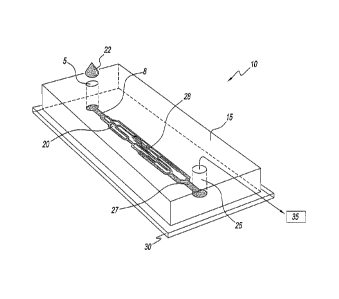
FIG. 1 illustrates a microvascular network device according to the present
invention;
FIG. 2 illustrates an exploded view of a portion of the microvascular network
device, of Fig.1, according to the present invention;
FIG. 3a and 3b illustrate a top and side view, respectively, of the
microvascular
network device according to FIG. 1 of the present invention;
FIGS. 4a and 4b illustrate a larger microvascular network device, according to
a
further embodiment of the present invention;
FIG. 5 illustrates a microvascular network device incorporated into an
analysis
device that measures the overall flow rate through the network, the
microchannel flow
rates in microchannels and hematocrits in microchannels, for a sample in the
microvascular network, according to the present invention; and
FIG. 6 illustrates a microvascular network device, including a waste reservoir
according to the present invention.
DETAILED DESCRIPTION OF THE INVENTION
Referring to the figures and, in particular, to Fig. 1, the microvascular
network
device according to the present embodiment is shown, and generally referenced
by
reference numeral 10. Microchannel network device 10 has a molded component 15
CA 02758936 2011-10-14
WO 2010/120898 PCT/US2010/031055
with a network unit 20 molded therein that is sized and structured to mimic
the internal
human vasculature. Molded component 15 rests directly on slide 30, a
substrate, that is
a coated slide to ensure closed seal with molded component 15. Microchannel
network
device 10 has an inlet port 5 and an inlet channel 8 for receipt of a blood
sample 22.
Microchannel network device 10 has an outlet port 25 and an outlet channel 27
that are
operatively associated with a vacuum source 35 to simulate the actual flow of
blood in
vivo. Network device 10 has a plurality of microchannels 50 that simulate the
capillaries
of the human vasculature.
Referring to Fig. 2, showing an enlarged view of network device 10, a
plurality of
microchannels 50, are shown. Network device 10 has a single inlet port 5 and a
single
outlet port 25 through which the entire blood sample 22 flows. Each of the
plurality of
microchannels 50 is either a parent microchannel 51 that feeds and branches
into two
daughter microchannels 55 or is a convergence channel 60 that results from the
convergence of two daughter microchannels 55. Parent channels 51 have a
greater
cross-sectional area than daughter microchannels 55 and convergence channels
60
have a greater cross-section area than daughter microchannels 55 that feed
into the
convergence channels 60.
In a preferred embodiment, network device 10 includes thirty-four 6pm-deep, 70
to 6pm-wide microchannels, bifurcating at a 45 angle, relative to the inlet
of the two
bifurcated or daughter channels 55. A different number of microchannels 50
having a
variety of dimensions could also be used. In the simplest embodiment,
microchannels
50 of the artificial microvascular network device 10 are interconnected in a
way
mimicking the overall topology of real microvasculature. A bifurcating angle
70 or
convergence angle 75 is a 45 angle, although the range for both the
bifurcation angle
70 and convergence angle could range from approximately 20 to 80 .
Bifurcating
angle 70 is measured relative to the angle at which it diverges from the axis
of the
parent channel 50. A convergence angle 75 is measured relative to the axis at
which
daughter channels 55 converges with a convergence channel 60. The 45 angle
mimics or replicates the internal human vasculature. Were a microchannel
network to
6
CA 02758936 2011-10-14
WO 2010/120898 PCT/US2010/031055
feed into daughter channels at 900 angles, feed into three daughter channels,
or be an
entirely straight channel, the actual human vasculature would not be
accurately
replicated and would not yield reliable results in subsequent analysis.
Referring to Fig. 3a, inlet port 5 and the outlet port 25, preferably, have a
teardrop shape. Inlet channel 8, replicating an arteriole, and outlet channel
27,
replicating a venule, are short in length, but are much wider than
microchannels
50. The relative size of input channel 8 and output channel 27 are
significantly
larger, and therefore will have a lower fluidic resistance than microchannels
50.
Microchannels 50 can be variable in cross section, such as rectangular or
circular or any similar shape. Referring to Figs. 3a and 3b, the length of the
microchannels 50, the region including microchannels 51, 55, and 60, is
approximately
1800pm, although the region could be larger or smaller. The length of inlet
channel 8
and outlet channel 27 is approximately 300pm, although the length could vary.
The inlet
port 5 and the outlet port 25 are tear-shaped and substantially larger than
the other
components of network device 10. The dimensions of the inlet port 5 and the
outlet port
25 are approximately 5000pm in length and 500pm in depth. Preferred samples
for
use in the network device 10 may be selected from the group consisting of:
cells,
microorganisms, and any combinations thereof suspended in an appropriate
solution.
Preferred samples are whole blood, white blood cells with or without plasma
(diluted or
undiluted), and most preferably red blood cells and platelets with or without
plasma
(diluted or undiluted).
In a further embodiment shown in Figs. 4a and 4b, network device 101 is larger
and a network unit 105 having more microchannels 501 than microchannel device
10.
However, network device 101 also has a single inlet channel 151 and a single
outlet
channel 251. Such network 101 can be used to enhance performance by having
greater sensitivity. Network device 101 is structured in the same way as
network device
10. Thus, it too replicates the human vasculature by having bifurcating
microchannels.
7
CA 02758936 2011-10-14
WO 2010/120898 PCT/US2010/031055
Other embodiments of the network may mimic the actual microvascular networks
of specific tissues and end organs (including, by not limited to, heart,
retina of the eye,
brain, kidney), the microvascular networks of said tissues and organs at
various
development stages as well as tumors. Morphometric information regarding the
geometrical dimensions of the microvessels of the microvascular networks of
these
organs and the topological information about how these microvessels connect to
form
these networks would be used in and fabricating an artificial microvascular
network with
all of the organ-specific characteristics.
There are three primary measurements that are significant to the measurement
of perfusion of blood for analysis prior to transfusion. One such measurement
is overall
flowrate Qtot. The overall flow rate through the network provides a general
assessment
of how well a sample of stored RBCs is able to perfuse the microvascular
network
device 10, 101. The overall rate of flow of blood sample through the network
is
determined by measuring the rate of flow of RBCs in the inlet channel 8 to the
outlet 27
of network device 10, for example.
The measurement of the overall rate of flow of blood sample through network
device 10, 101 provides an integrative measurement of the sample's
performance. Any
changes in the fluidic resistance of the network to the flow of blood due to a
reduction
(or an improvement) in the microvascular fitness of the sample 22 will be
reflected in
this measurement. Referring to Fig. 1, network device 10 having one inlet port
5 and
one outlet port 25, the rate of flow in inlet port 5 (arteriole) and the rate
of flow in outlet
port 25 (venule) are identical. The flow rate of blood sample in network
device 10 is
determined by measuring the average sample velocity via frame-by-frame image
analysis. A sensor is used to capture images (frames) of the channel at
precisely
known intervals. Regions within the channel walls from two sequential frames
are
cross-correlated to determine how far RBCs in a microchannel have shifted (on
average) in the time interval between the two sequential frames. The distance
that
RBCs have shifted or traveled then divided by the time interval to calculate
the average
RBC velocity in the channel.
8
CA 02758936 2011-10-14
WO 2010/120898 PCT/US2010/031055
Referring to Fig. 5, network device 10 (and 101) is preferably a disposable
element of a cartridge or cassette 90 that is inserted into an analysis device
200 that is
able conduct measurements on the blood sample that flows through plurality of
microchannels 50 of microvascular network device 10. Analysis device 200
contains a
receptacle 201 that receives network device 10 for analysis. Analysis device
200
preferably contains a sensor 205, that is able to capture frames or data
related to
sample as it flows through microchannels 50. Analysis device 200 has a memory
device 210 into which captured frames or data can be stored for later
reproduction as a
video and for analysis. Sensor 205 captures images or frames of blood along at
least
two locations along network device 10. The flow rates can be measured by
performing
frame-by-frame image analysis of the high-speed movies of the flow of blood in
the
network by sensor 205 contained within analysis device 200. Analysis device
200 also
has a processor 220 to carry out the computations related to the captured
frames or
data. Sensor is preferably one of a CCD or CMOS digital camera, a pair of
photodiodes and an ultrasonic transducer that are configured to sense the
sample as it
passes through device 10, 101.
Additionally, analysis device is 200 is able to capture and store measurement
data in a database of memory device 210 that includes measurements of a
plurality of
healthy blood samples for purposes of comparison to a stored blood sample to
determine the vascular fitness of the stored sample. The plurality of healthy
blood
samples are hundreds of fresh, healthy blood samples. The stored measurements
of
healthy samples can optionally be stored according to characteristics of the
individual
from whom the healthy sample is taken for further comparison to stored
samples.
In a specific embodiment, the image acquisition system consisted of an Olympus
BX51 microscope with an attached high-speed digital CMOS camera (Silicon Video
2112; Epix, Inc.) and a frame grabber board (PIXCI D2X; Epix, Inc.) mounted in
a
dedicated PC (Dimension XPS D300, Dell). Frame sequences were captured in
computer memory and saved on hard drive (XCAP-Lite; Epix, Inc.) for analysis
using
9
CA 02758936 2011-10-14
WO 2010/120898
PCT/US2010/031055
custom software written in MATLAB (Mathworks, Inc.) or in C++ (Microsoft
Visual C++
6.0; Microsoft, Corp.). Compatible equipment would also be used with either a
photodiode or an ultrasound device as well. The same analysis is performed
with
means other than the digital camera, for example by analyzing the signal from
a
photodiode or using ultrasound means for measuring the average velocity of the
sample
of RBCs in the microchannel.
A further measurement that is critical to the determination of efficacy of
stored
blood is the measurement of the rate of flow of blood in every microchannel 50
Qi of the
network device 10. The flow rates in individual capillary-sized microchannels
50 provide
a measure of how well stored RBCs are able to reach the smallest vessels of
the
microvasculature to complete the delivery of oxygen. The measurement of the
distribution of the rates in microvascular channels 50 of the network 10
provides a much
more detailed and a different kind of information regarding the microvascular
performance of the blood sample than the overall flow rate Qtot. A reduction
in the
capillary flow rates (with respect to a sample of fresh blood) would indicate
a poor
quality of stored blood being tested even if the overall flow rate through the
network is
approximately the same. The flow rate of blood sample 22 in microchannels 50
is
measured in the same fashion as the overall flow rate Qtot is measured.
A third measure of the fitness of stored blood is, tube hematocristl-Icti in
the
capillary microchannels of the network. Tube hematocrits provide a further
independent measure of how well stored RBCs are able to reach the
microchannels 50,
501 of microvascular devices 10, 101. When this measurement is combined with
the
measurements of capillary flow rate Qi, the oxygen carrying capacity and other
biochemical characteristics of stored red blood cells of sample 22, an
estimate of the
actual rate of oxygen delivery to tissues is provided.
The tube hematocrit in a channel in a microchannel 55 of Fig. 1, for example,
is
determined by measuring via image analysis the transmittance of blue light
(415 15nm)
passing therethrough. Because hemoglobin inside of the RBCs of sample 22
adsorbs
CA 02758936 2011-10-14
WO 2010/120898
PCT/US2010/031055
blue light very well, RBCs appear dark when illuminated with blue light and
their volume
concentration in the channel (i.e., tube hematocrit) correlates well with the
"darkness" of
the channel. Because of hemoglobin, RBCs appear dark in blue light ¨ the use
of a
narrow band-pass blue filter (415 15nm) to match hemoglobin's Soret absorption
band
facilitates the measurement of tube hematocrit in microchannels 55, for
example, of the
device 10.
Thus, Qtot, the total rate of flow through network device 10, Q,, flow in
particular
microchannels, and 1-Ict1, the tube hematocrit in each individual microchannel
of device
provide valuable information of the fitness of the RBCs in a sample 22. The
pressure differential across network 10, is kept constant during the
measurement. For
different measurements, the pressure across the network 10 could be varied
between
different measurements and during an individual measurement.
These three measurements made by using analysis device and network devices
10, 101 of the present disclosure are part of an array of parameters that
allow the
estimation of the efficacy of a stored blood sample.
In order to determine the microvascular fitness of a sample of stored blood,
the
microvascular fitness of fresh healthy blood is used as the standard for
comparison to
previously stored blood samples prior to transfusion. Thus, actual ranges of
these
three measurements will be determined experimentally by passing fresh, normal,
healthy blood through network 10 to obtain a set of pre-determined or standard
values
for healthy blood. The three measurements of healthy, fresh, normal blood of
hundreds
of individuals may be stored and used as the standard for subsequent
measurements.
Measurements of samples of stored RBCs will always be compared to this normal
standard.
Thus, to measure the ability of stored RBCs to perfuse microvascular networks
(termed "microvascular fitness" in this text), a sample of stored RBCs at
physiologically
high hematocrit is passed through microchannel network device 10 under a
constant
11
CA 02758936 2011-10-14
WO 2010/120898 PCT/US2010/031055
pressure differential from inlet port 5 to outlet port 25. The perfusion of
sample 22 is
evaluated by measuring: (i) the overall rate of flow through the network
(Qt0t) for the
constant or varying pressure difference between the inlet and the outlet,
(ii), the flow
rates (C21) in the microchannels, and (3) the tube hematocrit (Hcti )of the
microchannels.
The measurement of network perfusion for sample 22 is then compared to the
previously established standard values for fresh healthy RBCs to determine the
level of
microvascular fitness of the sample of stored RBCs relative to the normal
fresh RBCs.
Thus, the comparison provides a qualitative indication of the stored sample of
RBCs
relative to the fresh RBCs to access microvascular.
The sample RBCs 22 were preferably washed three times in phosphate buffered
saline (PBS) and passed through a leukoreduction filter to reduce the
concentration of
white blood cells (WBC) and platelets. Washed cells were diluted into GASP
buffer
(containing 9 mM Na2HPO4, 1.3 mM NaH2PO4, 140 mM NaCI, 5.5 mM glucose, and 1%
bovine serum albumin, pH 7.4, osmolarity 290 mmol/kg), or in other buffers.
The
hematocrit of sample 22 in GASP is adjusted to a specific value (often 40%),
sample
size was 20pland experiments were performed at room temperature. This is not
to
exclude the possibility of different sample sizes, different hematocrits and
running
measurements at different temperatures as well.
In addition to optional washing steps, a chemical or drug may be introduced to
observe its effects in altering deformability of RBCs in sample 22. A chemical
reaction
induced by a drug may result in subtle changes in fluidity or mechanical
properties of
sample 22, namely RBC membrane or RBC cytosol. Devices 10,101 can evaluate the
effects of these treatments on deformability and perfusability. It should be
also noted
that a blood from some individual could behave differently from the population
average
under external chemical treatment. For example, a relatively common glucose 6
phosphate dehydrogenase deficiency phenotype would be severely affected by an
oxidative stress which may be introduced by the treatment with antimalarial
drugs such
as primaquine, and may significantly change the ability of the treated red
blood cells to
perfuse the microvascular network of device.
12
CA 02758936 2011-10-14
WO 2010/120898
PCT/US2010/031055
Range for pressure differential along the network, the difference in pressure
from
the inlet to the outlet ranges from 0 mmHg to 250 mmHg (340 cmH20). The
highest
limit corresponds to the systolic blood pressure in severe hypertension (stage
4). In the
venous part of systemic circulation blood pressure is normally about 10 mmHg
(14
cmH20). The pressure difference between the arteriole (inlet) and the venule
(outlet) of
a microvascular bed is normally on the order of 30 mmHg (40 cmH20)
The overall flow ()tot and the individual flow rate Q, in each microchannel
network
50 are each measured in the devices in the dimensional units of microliters
per minute
(uL/min). A normal range for each measurement is determine by the values for
fresh
normal healthy RBCs an can be from 0 uL/min to 100uL/min. The normal range may
depend on the specific network used in the measurement.
The following chart provides the normal ranges of sample hematocrit (systemic
hematocrit) for subjects of various ages. The tube hematocrit in microchannels
50, 51,
55 and 60 of the microvascular network may be higher and lower than the value
of the
sample hematocrit.
NORMAL TUBE RANGES FOR SYSTEMIC HEMATOCRIT (Hct)
Newborns 55%-68%
One (1) week of age 47%-65%
One (1) month of age 37%-49%
Three (3) months of age 30%-36%
One (1) year of age 29%-41%
Ten (10) years of age 36%-40%
Adult males 42%-54%
Adult women 38%-46%
The microchannel network devices 10, 101 include several interconnected
microchannels 50, 501 operating in multi- or single-file flow regimes with a
wide range
of flow rates. Sample 22 having RBCs flowing through the microchannel network
devices 10, 101 at natural hematocrit would undergo all modes of deformation ¨
folding
and in shear in microchannels 50, 501 under a variety of different flow
conditions,
similar to the real microcirculation. The information provided from analysis
device 200
13
CA 02758936 2011-10-14
WO 2010/120898
PCT/US2010/031055
permits a straightforward interpretation by the physicians making the decision
regarding
transfusion and, therefore, could produce an immediate clinical value.
Microvascular network devices 10, 101 of the present application has
applicability to the study of pathological conditions. Thus, sample RBCs in
which the
red cell is more rigid because of diabetes mellitus, red cells that are
infected with
parasitic forms as occur in malaria, red cells that demonstrate genetic
abnormalities,
such as those found in thalassemia and sickle cell decease, i.e., may also be
used.
Further, cells which display the changes of metabolic or parasitic diseases
and other
pathological processes that involve the formed elements and any combinations
thereof,
may also be studied using the microvascular network devices 10, 101 of the
present
disclosure.
To manufacture network devices 10, 101, a master silicon wafer is used. The
configuration of microvascular network device10 is transferred onto a master
silicon
wafer (not shown) using a direct laser writer (Heidelberg DWL 66, Heidelberg
Instruments Mikrotechnik GmbH) and reactive ion etching (Bosch process, Unaxis
SLR
770 ICP Deep Silicon Etcher, Unaxis USA Inc). The master wafer may also be
fabricated using photolithography of SU-8 photoresist or other photosensitive
material.
Features on the silicon wafer are inversed relative to the design of network
20 of
network device 10. Recessed areas of the master wafer correspond to the
microchannels 50 of network device 10. The master wafer fabricated in this
manner
can be replica-molded many times to produce microfluidic devices in materials
such as
for example, poly (dimethyl siloxane) (PDMS, produced by either G.E. Silicones
as RTV
615 NB, or by Dow Corning as Sylgard 184).
The pattern on the master wafer is imprinted in PDMS by pouring PDMS pre-
polymer over the master wafer and allowing it to cure in an oven at the
temperature of
65 C overnight. To remove the PDMS replica from the master wafer, the replica
is cut
with a scalpel and then peeled off from the master wafer. The PDMS replica is
then
placed onto a clean surface of slide 30 with the molded features facing up to
become
14
CA 02758936 2011-10-14
WO 2010/120898
PCT/US2010/031055
molded component 15. The inlet port 5 an outlet port 25 are created by
locating the
inlet and outlet channels of the network 20 molded in the PDMS, and punching
through
upper component at these locations with a sharp, cylindrical punch (such as a
disposable biopsy punch). Outlet port 25 is connected to a waste-collecting
reservoir
with a PE tubing ¨ such that the blood sample flows from the inlet reservoir,
through the
network, and exists the device through the outlet at the top of the device. In
this
embodiment, slide 30 does not to be pre-drilled with a through hole for the
outlet.
Molded component 15 contains the actual ceiling and sidewalls of the
microchannels of the network 20. Molded component 15 is sealed to slide 30 to
form a
complete microfluidic device. To assemble the network device 10, molded
component
15 and PDMS-coated slide 30 are exposed to air plasma for 100 seconds (Plasma
Cleaner/Sterilizer, Harrick Scientific Corporation), affixed together, and
placed in an
oven at 65 C for 15 min to complete the covalent bonding of the two contact
surfaces.
Immediately following assembly, network device 10 is filled with 1% (wt/vol)
aqueous
solution of mPEG-silane (Laysan Bio, Inc.), and then washed and incubated with
GASP
buffer (1% bovine serum albumin (BSA), 9 mM Na2HPO4, 1.3 mM NaH2PO4, 140 mM
NaCl, 5.5 mM glucose, pH 7.4, 290 mmol/kg) to passivate the walls of the
channels and
prevent adhesion of blood cells to the walls.
In an alternative embodiment shown in Fig. 6, outlet port 25 is not punched
through molded component 15 as shown in Fig. 1. In contrast, molded component
15 is
sealed against slide 30 that has a 2-mm pre-drilled hole 80. In this
particular
embodiment, the distal end of output channel 28 is placed directly above hole
80,
serving as the output port and connecting the microchannel network device 10
to a
large waste-collecting reservoir 85. The pressure differential across network
device 10
in this embodiment is regulated by adjusting the relative levels of liquid in
the waste-
collecting reservoir 85 and the input reservoir of device 10. This embodiment
permits
modification to the pressure differential to be realized over network 10 so
that sample
behavior in deformation and shear can be measured over several pressure
differentials.
CA 02758936 2011-10-14
WO 2010/120898
PCT/US2010/031055
The substrate of the microvascular network device is comprised of glasses,
plastics, polymers, metals, ceramics, organic materials, inorganic materials,
and any
combinations thereof. A preferred substrate is transparent and readily uses
the
microchannel formation. The device preferably has a plurality of microchannels
each
having a diameter or width (and as well a depth) from about 1 micrometer to
about 100
micrometers.
However, neither the invention substrate nor the microchannel material is
limited
to any specific material, but may use any material that satisfies the
structural and
functional requirements of the invention. For example, any material that can
be cast into
microchannel networks may be employed. A wide spectrum of materials can be
used for
channel castings. The microchannel material is preferably not hostile to blood
cells,
especially red blood cells, and may optionally bind lubricant material that
may be useful
to facilitate cell movement. For example, PEG, mPEG-silane, and the like may
be used
to coat microchannels.
The prototype model system has applications in a variety of microvascular
network studies. This would include studies on the robustness of network
function in the
presence of elevated white cell counts or cellular aggregates. The former is a
physiological response to bacterial infection or a pathological manifestation
of
neoplastic transformation of leukocyte precursors. The latter occurs in
association with
diabetes or other hypercoagulable states and may cause or accompany vascular
occlusions that can damage heart or brain tissues. Using available pattern
generation
capabilities, a range of microvascular network designs and complexities can be
studied.
Computer simulations have shown that plasma skimming and the Fahraeus-
Lindqvist
effect might entirely account for nonlinear temporal oscillations in
microvascular blood
flow in the absence of biological regulation. This question can be directly
studied and
simulated with the device of the invention.
Some microvascular regulatory agents, such as NO, have documented effects on
red cell deformability which could effect microvascular flow dynamics and even
serve as
16
CA 02758936 2015-04-07
an independent mechanism for its regulation. The nonlinear dynamics of local
blood flow
and its dynamic regulation at the local level are also directly studied and
simulated with
the device of the invention. By modifying the device to include a drug
injection port,
more precise measurements of dose response relationships and latencies for the
effects of
such regulatory agents on RBC properties and behaviors in microvascular
networks can
be obtained. The present invention is also a useful validation tool for
earlier computer
simulations and theoretical models.
Unless otherwise defined, all technical and scientific terms used herein have
the same
meaning as commonly understood by one of ordinary skill in the art to which
this
invention belongs. Although methods and materials similar or equivalent to
those
described herein can be used in the practice or testing of the present
invention, the
preferred methods and materials are described below. In case of conflict, the
present
specification, including definitions, will control. In addition, the materials
methods, and
examples are illustrative only and not intended to be limiting of the
invention.
Although the present invention describes in detail certain embodiments, it is
understood
that variations and modifications exist known to those skilled in the art that
are within the
invention. Accordingly, the present invention is intended to encompass all
such
alternatives, modifications and variations that are within the scope of the
invention as set
forth in the following claims.
17