Note: Descriptions are shown in the official language in which they were submitted.
CA 02758977 2011-10-14
WO 2010/124234 PCT/US2010/032274
PULSED ELECTROMAGNETIC FIELD AND NEGATIVE PRESSURE THERAPY
WOUND TREATMENT METHOD AND SYSTEM
BACKGROUND OF THE INVENTION
1. Field
[0001] The present disclosure relates to a method of wound treatment.
Specifically, the
disclosure is related to a method of applying negative pressure wound
treatment and pulsed radio
frequency energy treatment to a wound of an individual, so as to enhance the
rate of wound healing.
2. Related Art
[0002] The treatment of open wounds that are too large to spontaneously close
has long been a
troublesome area of medical practice. Open wounds may heal by primary
intention, wherein the
wound edges are brought together (apposed) and held in place by mechanical
means (sutures,
staples, or adhesive strips), or by secondary intention, wherein the wound is
allowed to fill-in and
close through the physiological wound repair process. Physiological repair of
an open wound
requires proliferation of subcutaneous tissue and inward migration of
surrounding epithelial tissue.
Some wounds, however, are sufficiently large, chronic, or infected that they
are unsuitable for
closure by primary intention and unable to heal spontaneously by secondary
intention. In such
instances, a zone of stasis in which localized edema and fibrosis restricts
the flow of blood to the
epithelial and subcutaneous tissue forms in the wound bed and wound periphery.
Without sufficient
blood flow, the wound becomes senescent, arrested in a dysfunctional
disequilibrium, and/or
infected; and is accordingly unable to close spontaneously. Such wounds have
presented difficulties
to medical personnel for many years.
[0003] A problem encountered during the treatment of wounds is the selection
of an appropriate
technique for wound closure during the healing process. Primary surgical
closure employs sutures,
adhesive strips, and/or staples to force and hold the wound edges together,
allowing for rapid repair
and healing. However, such devices apply a closure force to only a very small
percentage of the
area surrounding a wound. When there is scarring, edema, fixation, or
insufficient tissue, the tension
1
CA 02758977 2011-10-14
WO 2010/124234 PCT/US2010/032274
produced by the sutures can become great causing excessive pressure to be
exerted by the sutures
upon the tissue adjacent to each suture. As a result, the adjacent tissue
often becomes ischemic
thereby rendering suturing of large wounds counterproductive. If the quantity
or size of the sutures
is increased to reduce the tension required of any single suture, the quantity
of foreign material
within the wound is concomitantly increased and the wound is more apt to
become infected.
Additionally, the size, body location or type of a particular wound may
prevent the use of sutures to
promote wound closure.
[0004] One method used for treating wounds that cannot be treated by
traditional means is
negative pressure wound therapy. Negative pressure wound therapy has been
described in U.S. Pat.
No. 4,969,880 issued to Zamierowski, as well as its continuations and
continuations-in-part, U.S.
Pat. No. 5,100,396, U.S. Pat. No. 5,261,893, and U.S. Pat. No. 5,527,293.
Further improvements
and modifications of the negative pressure wound therapy are also described in
U.S. Pat. No.
6,071,267, issued to Zamierowski; U.S. Pat. Nos. 5,636,643 and 5,645,081
issued to Argenta et al.;
and U.S. Pat. No. 6,142,982, issued to Hunt, et al. However, one problem with
negative pressure
wound therapy treatment is that not all wound types respond well to the
treatment.
[0005] Another method used for treating open wounds that cannot be treated by
traditional
means is using pulsed electromagnetic treatment devices to provide the wound
with pulsed radio
frequency energy. Methods for treating wound with pulsed radio frequency
energy have been
described in U.S. Pat. Nos. 3,043,310 and 3,181,535, issued to Milinowski;
U.S. Pat. No. 3,543,762,
issued to Kendall; U.S. Pat. No. 3,670,737, issued to Pearo; U.S. Pat. No.
5,584,863, issued to
Rauch et al.; and U.S. Pat, No. 6,353,763, issued to George et al. However, a
problem with pulsed
radio frequency energy treatment is that the rate of healing can vary and some
types of wounds may
not respond well to the treatment.
[0006] Successful wound treatment requires an understanding of wound
physiology and the
mechanism of action of wound treatment therapies. With regard to wound
physiology, it is known
that there are three distinct phases associated with the process of wound
healing. The three phases
are the inflammatory phase, the proliferative phase, and the remodeling phase.
During the
inflammatory phase, bacteria and debris are removed and macrophages release
growth factors to
2
CA 02758977 2011-10-14
WO 2010/124234 PCT/US2010/032274
stimulate angiogenesis and the production of fibroblasts. Next, in the
proliferative phase,
granulation tissue forms and epithelialization begins, which involves
migration of epithelial cells to
seal the wound; fibroblasts proliferate and synthesize collagen to fill the
wound and provide a strong
matrix on which epithelial cells grow; and contractile cells called
myofibroblasts appear in the
wound and aid in wound closure. In the remodeling phase, collagen in the scar
undergoes repeated
degradation and resynthesis, and the tensile strength of the newly formed skin
increases.
[0007] With regard to the mechanism of action of negative pressure wound
therapy treatment, it
is thought that the negative pressure wound therapy treatment promotes wound
healing by removing
excess interstitial fluid, decreasing bacterial colonization, and stimulating
granulation tissue
formation through micromechanical deformation. Therefore, it appears that
negative pressure
wound therapy treatment is effective during the inflammatory and early
proliferative phases, which
involve bacterial removal and granulation.
[0008] With regard to the mechanism of action of pulsed radio frequency energy
treatment, it is
thought that pulsed radio frequency energy treatment can stimulate growth
factor production and
induce cell proliferation in the wound bed. Studies have shown that pulsed
radio frequency energy
treatment can induce proliferation in cultured human dermal fibroblast and
epithelial cells in a dose-
and time-dependent fashion. Thus, it seems that pulsed radio frequency
treatment is effective at
propagating the proliferative and remodeling phases, which involve fibroblast
and epithelial cell
proliferation. Cytogenic evidence also suggests that pulsed radio frequency
energy treatment
modulates the inflammatory phase and stimulates angiogenesis, the stimulation
of blood flow.
[0009] It would therefore be desirable to provide a method of wound treatment
that enhances
the rate of wound healing to wounds that do not respond well to negative
pressure wound therapy
treatment alone or pulsed radio frequency energy treatment alone.
[0010] Citation of the above documents, devices and studies is not intended as
an admission that
any of the foregoing is pertinent prior art. All statements as to the contents
of these documents is
based on the information available to the applicants and does not constitute
any admission as to the
correctness of the contents of these documents.
3
CA 02758977 2011-10-14
WO 2010/124234 PCT/US2010/032274
BRIEF SUMMARY OF THE INVENTION
[0011] Disclosed herein is a method for treating a wound of an individual and
for enhancing a
rate of wound healing by applying, for a first period of time, a negative
pressure treatment to the
wound without applying a pulsed radio frequency treatment; and applying, for a
second period of
time subsequent to the first period time, a pulsed radio frequency energy
treatment to the wound
while maintaining the negative pressure treatment to enhance the rate of wound
healing. The
negative pressure treatment and the pulsed radio frequency energy treatment
are applied
concurrently for the duration of the second period of time.
[0012] The present disclosure also pertains to a method for treating a wound
of an individual
and for enhancing a rate of wound healing by applying concurrently a negative
pressure treatment
and a pulsed radio frequency energy treatment. The negative pressure treatment
and pulsed radio
frequency energy treatment of the method are maintained for a period of time
sufficient to achieve
the enhanced rate of wound healing. In one embodiment, the method of applying
concurrently the
negative pressure and pulsed radio frequency energy treatments has an enhanced
rate of wound
healing that results in at least a 90% decrease in wound volume.
BRIEF DESCRIPTION OF THE DRAWINGS
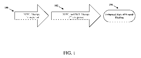
[0013] FIG. 1 is a flow chart of one embodiment of a method for combined NPWT
and PRFE
wound treatment.
[0014] FIG. 2. is a flow chart of another embodiment of a method for combined
NPWT and
PRFE wound treatment.
[0015] FIG. 3A-3D depicts a scalp avulsion wound and the effects of healing
over time with
combined PRFE and NPWT treatment. (A) depicts the wound before combined
treatment; (B)
depicts the wound at two weeks of combined treatment; (C) depicts the wound at
five weeks of
combined treatment; and (D) depicts the wound at seven weeks of combined
treatment.
[0016] FIG. 4 is a bar graph depicting the change in wound volume of the scalp
avulsion wound
over weeks of combined NPWT and PRFE treatment.
4
CA 02758977 2011-10-14
WO 2010/124234 PCT/US2010/032274
[0017] FIG. 5 is a line graph depicting the percent decrease in wound volume
of the scalp
avulsion wound over weeks of treatment with combined NPWT and PRFE treatment.
[0018] FIG. 6A-6D depicts a pilonidal wound healing over the course of time as
a result of
combined PRFE and NPWT treatment. (A) depicts the wound after 2 weeks of NPWT
treatment
alone; (B) depicts the wound after 1 week of combined treatment; (C) depicts
the wound after 2
weeks of combined treatment; and (D) depicts the wound 2 1/2 weeks after
conclusion of combined
treatment.
[0019] FIG. 7 is a bar graph depicting the change in wound volume of the
pilonidal wound over
weeks of combined NPWT and PRFE treatment.
[0020] FIG. 8 is a line graph depicting the percent decrease in wound volume
of the pilonidal
wound over weeks of combined NPWT and PRFE treatment.
[0021] FIG. 9 depicts the percent decrease in pilonidal wound area using
combined NPWT and
PRFE treatment compared to decreases in wound area using PRFE treatment alone
and NPWT
treatment alone.
[0022] FIG. 10A-IOC depicts a pressure ulcer wound and the effects of healing
over time with
combined PRFE and NPWT treatment. (A) depicts the wound after one month of
NPWT treatment;
(B) depicts the wound prior to initiation of PRFE treatment; and (C) depicts
the healed wound after
four months of combined treatment.
[0023] FIG. 11A and 11B depicts an Achilles tendon rupture and the effects of
healing with
combined PRFE and NPWT treatment, (A) depicts the wound prior to combined
treatment and (B)
depicts the healed wound after 78 days of combined treatment.
[0024] FIG. 12 is a line graph depicting the decrease in wound volume of the
Achilles tendon
rupture wound over the course of NPWT treatment and combined NPWT and PRFE
treatment.
CA 02758977 2011-10-14
WO 2010/124234 PCT/US2010/032274
DETAILED DESCRIPTION OF THE INVENTION
DEFINITIONS
[0025] As used herein, negative pressure wound therapy (hereinafter "NPWT")
refers to the
treatment of wounds and other damaged tissues through the application of
negative pressure.
[0026] As used herein, pulsed radio frequency energy treatment (hereinafter
"PRFE") refers to
the treatment of wounds and other damaged tissues through the application of
pulsed,
electromagnetic or magnetic energy fields oscillating at a radio frequency.
[0027] As used herein, the terms "% decrease" and "percent decrease" refer to
the difference in
wound volume or area before and after a given time of treatment with NPWT or
PRFE alone, or in
sequence, or combined NPWT and PRFE treatment. The difference in volume or
area is then
converted to a percentage of the original volume or area of the wound.
[0028] As used herein, the term "wound volume" refers to the dimensions of
length, width, and
depth of a wound of an individual. Measurement of wound volume requires
measurement or
approximation of wound depth, length, and width. Wound volume can be assessed
manually using
techniques such as filling the wound with saline, molding, or injecting dental
impression material or
like substance. Would volume may also be assessed digitally by using computer-
assisted calibrated
planimetry, structured lighting, and image processing.
[0029] As used herein, the term "wound area" refers to the dimensions of
length and width of a
wound of an individual. Wound area may be assessed manually by using calipers,
rulers, tracings,
and similar measurement devices. Wound area may also be assessed through use
of computerized
planimetry using digital photography and image analysis, or through ultrasound
or X-ray images.
[0030] As used herein, the term "treatment for a period" refers to applying a
selected treatment,
or combination of treatments, at least once a day for at least 70% of days in
a given period of time,
where the 70% of days is rounded down. For example, treatment for a period of
2 weeks means
treatment would be applied at least once a day for at least 9 days of the
proscribed 2 weeks. It
should be noted that the at least 70% of days may or may not be consecutive.
6
CA 02758977 2011-10-14
WO 2010/124234 PCT/US2010/032274
[0031] As used herein, the term "NPWT treatment" refers to applying negative
pressure to a
target wound site.
[0032] Typically, NPWT is applied either continuously or intermittently (for
example, cycling
on and off every few minutes) for 24-hours in a given treatment day. However,
NPWT may also be
applied for less than 24-hours a day. For example, even in instances in which
a NPWT bandage is
attached to a target wound site for an entire 24 hour period, actual negative
pressure may be applied
for only selected periods during the 24 hours. In a preferred treatment
scenario, negative pressure is
actually applied for at least 30 minutes at a time.
[0033] As used herein, the term "PRFE treatment at least once a day" refers to
applying PRFE
at least once a day for a period of time that ranges from at least 5 minutes
to 60 minutes. For
example, the length of PRFE treatment may be at least 30 minutes.
[0034] As used herein, the term "enhanced rate of wound healing" refers to a
rate of wound
healing achieved with combined NPWT and PRFE treatment that is greater than a
rate of wound
healing achieved by using only PRFE or NPWT treatment alone. Rate of wound
healing is
determined by measuring the decrease in wound volume or area over time. For
example, rate of
wound healing may be expressed as square centimeters per day or cubic
centimeters per day, or as
percentage of original area or volume per day, respectively. An enhanced rate
of wound healing
may also refer to a reduced time to wound closure, greater percentage
reduction in wound area (or
volume) in a given time period, or greater incidence of wound closure in a
given time period.
[0035] As used herein, the term "maintaining" refers to maintaining a NPWT or
PRFE
treatment according to a regimen or protocol, as prescribed by a medical
doctor. Accordingly,
maintaining treatment takes into account that the particular prescribed
regimen may include
intermittent treatments. For example, a regimen for a PRFE treatment may call
for two 30 minute
treatments, twice daily for the duration of wound treatment. Furthermore, if
the protocol calls for
two 30 minute treatments twice daily and two 30 minute treatments are given on
day one, skipped
on the second day, and resumed on the third day, then this would still be
referred to as
7
CA 02758977 2011-10-14
WO 2010/124234 PCT/US2010/032274
"maintaining" the treatment regimen or protocol as long as treatment is given
for at least 70% of
days in a given period of time, where the 70% of days is rounded down.
[0036] As used herein, the term "concurrently" refers to the application of
NPWT and PRFE
treatment on a wound at the same time, taking into account that one device may
be physically
activated before the other, and maintaining both NPWT and PRFE therapies for a
given length of
time. The term "concurrently" also takes into account that that NPWT may be
given at least 22 out
of 24 hours per day, while PRFE may be given for 30 minutes twice daily.
[0037] As used herein in, the terms "combined treatment" and "combined NPWT
and PRFE
treatment" are used interchangeably and refer to concurrently using both NPWT
and PRFE to treat a
wound.
METHODS OF COMBINED WOUND TREATMENT
[0038] The following description sets forth exemplary configurations,
parameters, and the like.
It should be recognized, however, that such description is not intended as a
limitation on the scope
of the present invention, but is instead provided as a description of
exemplary embodiments.
[0039] The following embodiments describe methods of combining NPWT treatment
with
PRFE treatment to treat an open wound that may not be closed as effectively
using standard wound
treatment therapies, advanced wound treatment therapies, NPWT treatment alone,
or PRFE
treatment alone. The combined NPWT and PRFE treatment achieves an enhanced
rate of wound
healing, compared to rates of wound healing achieved with either treatment
alone. The method
further employs prolonged, combined treatment to obtain the full benefit of
the enhanced rate of
wound healing.
[0040] The methods of combined NPWT and PRFE treatment described herein can be
applied
using any standard NPWT system that is known in the art. Briefly, NPWT systems
typically
include a vacuum pump, drainage tubing, and a dressing set. The pump may be
stationary or
portable, may rely on AC or battery power, and may allow for regulation of the
negative pressure.
8
CA 02758977 2011-10-14
WO 2010/124234 PCT/US2010/032274
[0041] Certain parameters may vary between NPWT systems, for example, the
negative
pressure may be applied in the range of -5 to -200 mmHg, -5 to -190 mmHg, -10
to -185 mmHg, -15
to -180 mmHg, -25 to -175, -35 to -170, -45 to -165 mmHg, -50 to -160 mmHg, -
60 to -150 mmHg,
-70 to -125 mmHg, -75 to -115 mmHg, -85 to -110 mmHg, -90 to -100 mmHg, -91 to
-99 mmHg, -
92 to -97 mmHg, or -93 to -95 mmHg. In one preferred embodiment, the negative
pressure is
applied at -125 mmHg.
[0042] The negative pressure may also be applied continuously or
intermittently, depending on
the type of wound. Intermittent negative pressure may refer to, for example, a
cycle of 1 minute
with negative pressure on, and 1 minute with negative pressure off, a cycle of
2 minutes with
negative pressure on, and 2 minutes with negative pressure off, a cycle of 3
minutes with negative
pressure on, and 2 minutes with negative pressure off, a cycle of 4 minutes
with negative pressure
on, and 2 minute with negative pressure off, a cycle of 5 minutes with
negative pressure on, and 2
minutes with negative pressure off, a cycle of 6 minutes with negative
pressure on, and 2 minutes
with negative pressure off, a cycle of 7 minutes with negative pressure on,
and 2 minutes with
negative pressure off, a cycle of 8 minutes with negative pressure on, and 2
minutes with negative
pressure off, a cycle of 9 minutes with negative pressure on, and 2 minutes
with negative pressure
off, or a cycle of 10 minutes with negative pressure on, and 2 minutes with
negative pressure off. In
one embodiment, intermittent negative pressure refers to a cycle of 5 minutes
with negative pressure
on, and 2 minutes with negative pressure off. Moreover, negative pressure,
whether applied
continuously or intermittently, may be administered 24-hours a day every day
for the entire period
of time of wound treatment.
[0043] NPWT may be administered 24-hours a day for the entire period of time
of wound
treatment. NPWT may also be administered for less than 24-hours a day for the
entire period of
time of wound treatment. In certain embodiments, NPWT is administered for one
20-hour period,
one 18-hour period one 16-hour period, one 12-hour period, one 10-hour period,
one 8-hour period,
two 11-hour periods, two 10-hour periods, 2 two 8-hour periods, two 6-hour
periods, two 5-hour
periods, two 4-hour periods, three 7-hour periods, three 6-hour periods, three
5-hour periods, three
4-hour periods, three 3-hour periods, four 5-hour periods, four 4-hour
periods, four 3-hour periods,
or four 2-hour periods a day for the entire period of time of wound treatment.
9
CA 02758977 2011-10-14
WO 2010/124234 PCT/US2010/032274
[0044] Additionally, the dressing sets may contain a foam, nonadherent, non-
foam, woven, or
moistened cotton gauze dressing to be placed in the wound and an adhesive film
drape for sealing
the wound. The drainage tubes may come in a variety of configurations
depending on the dressings
used or wound being treated.
[0045] Furthermore, any standard PRFE system known in the art can also be used
for the
methods described herein. Briefly, PRFE may use low-energy electromagnetic
signals as a
mitogenic stimulus for the treatment of wounds. PRFE systems may use a
nonionizing, nonthermal
radio frequency emission. The PRFE systems, for example, can have preset
waveform parameters
that can be regulated to ensure consistent dosing. The PRFE system may operate
at a frequency of
6.78 MHz, 13.56 MHz, 27.12 MHz, 40.68 MHz, 5.8 GHz, 24.125 GHz, 61.25 GHz,
122.5 GHz, or
245.0 GHz. In one embodiment, the PRFE system operates at the Federal
Communications
Commission (hereinafter "FCC") medical device frequency of 27.12 MHz, and
generates an
electromagnetic field that extends from the surface of the treatment
applicator (antenna), allowing
wounds to be treated without removal of the bandages or dressings.
[0046] The parameters of different PRFE systems may vary. For example, the
electric field (E-
field) strength, as measured 5 cm above the RF antenna, may range between
0.084 and 2,000 V/m,
0.1 and 1,900 V/m, 0.5 and 1,800 V/m, 1 and 1,700 V/m, 5 and 1,600 V/m, 10 and
1, 500 V/m, 25
and 1,300 V/m, 35 and 1,200 V/m, 45 and 1,000 V/m, 50 and 900 V/m, 75 and 800
V/m, 85 and
700 V/m, 90 and 600 V/m, 93 and 591 V/m, 95 and 500 V/m, 100 and 400 V/m, 150
and 400 V/m,
or 200 and 350 V/m. Preferably the electric field strength, as measured 5 cm
above the RF antenna
is between 50 and 900 V/m. In certain embodiments, the electric field
strength, as measured 5 cm
above the RF antenna is 591 V/m.
[0047] The H-field strength of the PRFE system may also vary between 0.02 and
10 A/m, 0.1
and 9.5 A/m, 0.5 and 9.0 A/m, 0.75 and 8.5 A/m, 1.0 and 8.0 A/m, 1.5 and 7.5
A/m, 2.0 and 7.0
A/m, 2.5 and 6.5 A/m, 3.0 and 6.0 A/m, 3.5 and 5.5 A/m, or 4.5 and 5.0 A/m.
CA 02758977 2011-10-14
WO 2010/124234 PCT/US2010/032274
[0048] The E-field strength and H-field strength of the PRFE system may also
be modulated
individually or together. Moreover, the E-field strength and H-field strength
may be optimized to
treat specific wound types and soft tissue cells.
[0049] Furthermore, the radio frequency pulses of PRFE systems may range
between 16 and
3000 microsecond pulses, 18 and 1500 microsecond pulses, 20 and 750
microsecond pulses, 22 and
500 microsecond pulses, 24 and 250 microsecond pulses, 26 and 125 microsecond
pulses, 28 and 75
microsecond pulses, 30 and 65 microsecond pulses, 30 and 55 microsecond
pulses, 30 and 45
microsecond pulses, 30and 42 microsecond pulses, or 30 and 35 microsecond
pulses. Preferably the
radio frequency pulses range between 30 and 65 microsecond pulses. More
preferably the radio
frequency pulses range between 30 and 45 microsecond pulses. In one
embodiment, the radio
frequency pulses at 42 microsecond pulses.
[0050] Additionally, the pulse frequency of PRFE systems may range between
land 1000 pulses
per second, 25 and 900 pulses per second,50 and 800 pulses per second, 100 and
700 pulses per
second, 200 and 600 pulses per second, or 300 and 500 pulses per second. In a
preferred
embodiment, the pulse frequency is 1000 pulses per second.
[0051] The pulse interval of PRFE systems may also vary between land 800
microseconds, 5
and 600 microseconds, 10 and 500 microseconds, 15 and 400 microseconds, 20 and
200
microseconds, 25 and 100 microseconds, 50 and 75 microseconds, or 55 and 65
microseconds.
[0052] Moreover, the duty cycle of PRFE systems may range between 0.4% and
10%, 0.6% and
9.5%, 0.8% and 9.0%, 1.0% and 8.5%, 1.5% and 8.0%, 2.0% and 7.5%, 2.5% and
7.0%, 3.0% and
6.5%, 3.5% and 6.0%, 4.0% and 5.5%, or 4.5% and 5.0%.
[0053] In one embodiment, the PRFE system is the Provant Therapy System,
available from
Regenesis Biomedical of Scottsdale, Arizona.
[0054] The methods of combined NPWT and PRFE treatment described herein can
utilize any
of the NPWT and PRFE treatments systems and protocols described herein or
otherwise known in
the art.
11
CA 02758977 2011-10-14
WO 2010/124234 PCT/US2010/032274
[0055] Typically, initiation of NPWT treatment involves placing a shaped wound
cover,
operably connected to a vacuum pump, substantially over a wound of an
individual. The shaped
wound cover would define a covered volume above the wound, and the covered
volume would have
a gas pressure at an initial pressure. After placing the shaped wound cover
substantially over the
wound, negative pressure would be applied by activating the vacuum pump. The
vacuum pump
would lower the gas pressure of the covered volume from the initial pressure
to a reduced pressure.
Typically, initiation of PRFE treatment involves placing over the wound a
treatment applicator that
is configured to deliver the pulsed radio frequency energy. The treatment
applicator would be
connected to a pulsed radio frequency signal generator. Once the treatment
applicator has been
placed over the wound, the generator would deliver the pulsed radio frequency
signal to the
applicator, and the applicator would deliver the pulsed radio frequency energy
to the wound. In one
exemplary method of combined treatment, the PRFE applicator may be placed
directly over a
NPWT shaped wound cover.
[0056] Referring now to the drawings, where like elements are designated by
like reference
numerals throughout, FIG. 1 and FIG. 2 depict methods of combined treatment
comprising applying
NPWT and PRFE to a wound of an individual to achieve an enhanced rate of wound
healing.
NPWT PRE-TREATMENT PRIOR TO COMBINED TREATMENT
[0057] In one embodiment, shown in FIG. 1, a method of combined NPWT and PRFE
treatment
for treating a wound of an individual involves first pre-treating the wound
with a NPWT treatment
for a first period of time 100. Preferably the NPWT treatment is given in the
absence of a PRFE
treatment during the first period of time 100. After the end of the NPWT pre-
treatment period of
time 100, the wound is treated for a second period of time with a combined
treatment of NPWT and
PRFE 102. Preferably, the combined treatment is initiated immediately after
the end of the NPWT
pre-treatment period of time 100. However, a delay between the period of pre-
treatment and
combined treatment is acceptable. The NPWT and PRFE treatments are applied
concurrently for
the duration of the second period of time 102. The combined treatment is
maintained for a second
period of time 102 that is sufficient to achieve an enhanced rate of wound
healing 104.
12
CA 02758977 2011-10-14
WO 2010/124234 PCT/US2010/032274
[0058] In certain embodiments the NPWT treatment is applied intermittently to
the wound. In
other embodiments the NPWT treatment is applied continuously.
[0059] In other embodiments, NPWT may be administered 24-hours a day for the
entire period
of time of wound treatment. In still other embodiments, NPWT may also be
administered for less
than 24-hours a day for the entire period of time of wound treatment. In
certain embodiments,
NPWT is administered for one 20-hour period, one 18-hour period one 16-hour
period, one 12-hour
period, one 10-hour period, one 8-hour period, two 11-hour periods, two 10-
hour periods, 2 two 8-
hour periods, two 6-hour periods, two 5-hour periods, two 4-hour periods,
three 7-hour periods,
three 6-hour periods, three 5-hour periods, three 4-hour periods, three 3-hour
periods, four 5-hour
periods, four 4-hour periods, four 3-hour periods, or four 2-hour periods a
day for the entire period
of time of wound treatment.
[0060] In yet another embodiment, the NPWT treatment is applied during the
interval between
the first period of time 100 and the second period of time 102. Alternatively,
in some embodiments
the NPWT treatment is discontinued at the end of the first period of time 100,
and is initiated and
maintained again throughout the second period of time 102.
[0061] In another embodiment, the PRFE treatment is applied at least once a
day, twice a day,
three times a day, four times a day, five times a day, six times a day, seven
times a day eight times a
day, nine times a day, ten times a day, or more for a period of time that
ranges from at least 5
minutes to 60 minutes, 5 minutes to 55 minutes, 5 minutes to 50 minutes, 5
minutes to 45 minutes, 5
minutes to 40 minutes, 5 minutes to 35 minutes, 5 minutes to 30 minutes, 5
minutes to 25 minutes, 5
minutes to 20 minutes, 5 minutes to 15 minutes, or 5 minutes to 10 minutes. In
certain
embodiments, the period of time is 50 minutes, 45 minutes, 40 minutes, 35
minutes, 30 minutes, 25
minutes, 20 minutes, 15 minutes, 10 minutes, or 5 minutes.
[0062] In further embodiments, the length of the first period of time 100 may
vary. For
example, the first period of time 100 may be at least one day, two days, three
days, four days, five
days, six days, one week, one and a half weeks, two weeks, two and half weeks,
three weeks, three
and a half weeks, four weeks, four and half weeks, five weeks, five and a half
weeks, six weeks, six
13
CA 02758977 2011-10-14
WO 2010/124234 PCT/US2010/032274
and a half weeks, seven weeks, seven and a half weeks, or two months. In a
certain embodiment,
the first period of time 100 is at least two weeks. In another embodiment, the
first period of time
100 is at least one week.
[0063] The length of the second period of time 102 may also vary. For example,
the second
period of time 102 may be at least one day, two days, three days, four days,
five days, six days, one
week, one and a half weeks, two weeks, two and half weeks, three weeks, three
and a half weeks,
four weeks, four and half weeks, five weeks, five and a half weeks, six weeks,
six and a half weeks,
seven weeks, seven and a half weeks, two months, two and half months, three
months, three and a
half months, four months, four and a half months, five months, five and a half
months, six months,
or longer. In a preferred embodiment, the length of the second period of time
102 is at least one
week.
[0064] The enhanced rate of wound healing 104 may result in a in wound volume
or wound area
that is, for example, at least 5%, 10%, 15%, 20%, 25%, 30%, 35%, 40%, 45%,
50%, 55%, 60%,
65%, 70%, 75%, 80%, 85%, 90%, 95%, or 99% smaller than that achieved by either
NPWT or
PRFE treatment alone. In a certain embodiment, the enhanced rate of wound
healing 104 results in
a wound volume or wound area that is at least 25% smaller than that achieved
by either NPWT or
PRFE treatment alone.
[0065] The percentage change in wound volume or wound area may be calculated
by taking the
difference in percentage between the combined NPWT and PRFE treatment over a
given period of
time and the NPWT or PRFE treatment alone over the same period of time. For
example, if the
decrease in wound volume using the combined treatment was 85% and the decrease
in wound
volume using NPWT treatment alone was 60%, then the difference in percentage
would be 25%.
[0066] In certain embodiments, the enhanced rate of wound healing 104 may
result in, for
example, at least a 10%, 15%, 20%, 25%, 30%, 35%, 40%, 45%, 50%, 55%, 60%,
65%, 70%, 75%,
80%, 85%, 90%, 95%, 99%, or 100% decrease in wound volume or wound area, over
the total
treatment period of time 100 and 102. In a preferred embodiment, the enhanced
rate of wound
14
CA 02758977 2011-10-14
WO 2010/124234 PCT/US2010/032274
healing 104 results in at least a 90% decrease in wound volume, over the total
treatment period of
time 100 and 102.
[0067] The enhanced rate of wound healing may result in a wound volume or
wound area that
decreases at a rate of at least 1%/week, 1.5%/week, 2%/week, 3%/week, 4%/week,
5%/week,
6%/week, 7%/week, 8%/week, 9%/week, 10%/week, 15%/week, 20%/week, 25%/week,
30%/week,
35% /week, 40%/week, 45%/week, 50%/week, 55%/week, 60%/week, 65%/week,
70%/week,
75%/week, 80%/week, 85%/week, 90%/week, 95%/week, or 100%/week.
[0068] The enhanced rate of wound healing may also result in a wound volume
that decreases at
a rate of at least 1 cm3/week, 5 cm3/week, 10 cm3/week, 15 cm3/week, 20
cm3/week, 25 cm3/week,
30 cm3/week, 35 cm3/week, 40 cm3/week, 42 cm3/week, 45 cm3/week, 50 cm3/week,
60 cm3/week,
70 cm3/week, 75 cm3/week, 80 cm3/week, 90 cm3/week, 95 cm3/week, or 100
cm3/week. In one
embodiment, the enhanced rate of wound healing results in a wound volume that
decreases at a rate
of at least 42 cm3/week
[0069] The enhanced rate of wound healing may further result in a wound area
that decreases at
a rate of at least 1 cm2/week, 5 cm2/week, 10 cm2/week, 15 cm2/week, 20
cm2/week, 25 cm2/week,
30 cm2/week, 35 cm2/week, 40 cm2/week, 42 cm2/week, 45 cm2/week, 50 cm2/week,
60 cm2/week,
70 cm2/week, 75 cm2/week, 80 cm2/week, 90 cm2/week, 95 cm2/week, or 100
cm2/week. In one
embodiment, the enhanced rate of wound healing results in a wound volume that
decreases at a rate
of at least 42 cm2/week.
[0070] The method of combined treatment for treating a wound described in FIG.
1 may be used
to treat various types of wounds. For example, the combined treatment may be
used to treat:
chronic wounds; large, deep, open wounds; graft and flap site wounds; full
thickness burns; partial
thickness burns; diabetic ulcers; pressure ulcers; decubitus ulcers; arterial
ulcers; avulsion injuries;
pilonidal disease; cysts; acute wounds; tendon rupture wounds; postoperative
incisions;
postoperative wounds; traumatic wounds; dermatology conditions; scleroderma;
atrophy blanche
disease; trauma; bomb blast or other military-type inflicted wounds; gunshot
wounds; bites; or
wound dehiscence. It should be understood that the method of combined NPWT and
PRFE
CA 02758977 2011-10-14
WO 2010/124234 PCT/US2010/032274
treatment may be used to treat one or more wounds of an individual. It should
be understood that
the method of combined NPWT and PRFE treatment may be used to concurrently or
sequentially
treat one or more wounds of an individual.
PROLONGED COMBINED TREATMENT
[0071] In another embodiment, depicted in FIG. 2, a method of combined
treatment 204 for
treating a wound of an individual involves applying concurrently NPWT 200 and
PRFE 202
treatments to the wound. The combined treatment 204 results in an enhanced
rate of wound
healing. The combined treatment 204 may be maintained for a period of time
sufficient to achieve
at least 100%, 99%, 98%, 95%, 90%, 85%, 80%, 75%, 70%, 65%, 60%, 55%, 50%,
45%, 40%,
35%, 30%, 25%, 20%, 15%, 13%, 10%,decrease in wound volume 206 or in wound
area.
Preferably, the combined treatment 204 is maintained for a period of time
sufficient to achieve at
least a 90% decrease in wound volume 206 or wound area.
[0072] In one embodiment resulting in at least a 90% decrease in wound volume,
the combined
treatment 204 is maintained for 3 weeks. In other embodiments, the period of
time that the
combined treatment 204 is maintained may vary, for example, it may be at least
one day, two days,
three days, four days, five days, six days, one week, one and a half weeks,
two weeks, two and half
weeks, three weeks, three and a half weeks, four weeks, four and half weeks,
five weeks, five and a
half weeks, six weeks, six and a half weeks, seven weeks, seven and a half
weeks, two months, two
and half months, three months, three and a half months, four months, four and
a half months, five
months, five and a half months, six months, or longer.
[0073] The enhanced rate of wound healing may result in a in wound volume or
wound area
that is, for example, at least 5%, 10%, 15%, 20%, 25%, 30%, 35%, 40%, 45%,
50%, 55%, 60%,
65%, 70%, 75%, 80%, 85%, 90%, 95%, or 99% smaller than that achieved by either
NPWT or
PRFE treatment alone. In a preferred embodiment, the enhanced rate of wound
healing results in a
wound volume or area that is at least 25% smaller than that achieved by either
NPWT or PRFE
treatment alone.
16
CA 02758977 2011-10-14
WO 2010/124234 PCT/US2010/032274
[0074] The enhanced rate of wound healing may also result in a wound volume or
wound area
that decreases at a rate of at least 1%/week, 1.5%/week, 2%/week, 3%/week,
4%/week, 5%/week,
6%/week, 7%/week, 8%/week, 9%/week, 10%/week, 15%/week, 20%/week, 25%/week,
30%/week,
35% /week, 40%/week, 45%/week, 50%/week, 55%/week, 60%/week, 65%/week,
70%/week,
75%/week, 80%/week, 85%/week, 90%/week, 95%/week, or 100%/week.
[0075] The enhanced rate of wound healing may further result in a wound volume
that
decreases at a rate of at least 1 cm3/week, 5 cm3/week, 10 cm3/week, 15
cm3/week, 20 cm3/week, 25
3/week, 30 cm3/week, 35 cm3/week, 40 cm3/week, 42 cm3/week, 45 cm3/week, 50
cm3
cm /week, 60
cm3/week, 70 cm3/week, 75 cm3/week, 80 cm3/week, 90 cm3/week, 95 cm3/week, or
100 cm3/week.
In one embodiment, the enhanced rate of wound healing results in a wound
volume that decreases at
a rate of at least 42 cm3/week.
[0076] The enhanced rate of wound healing may also result in a wound area that
decreases at a
rate of at least 1 cm2/week, 5 cm2/week, 10 cm2/week, 15 cm2/week, 20
cm2/week, 25 cm2/week, 30
2/week, 35 cm2/week, 40 cm2/week, 42 cm2/week, 45 cm2/week, 50 cm2/week, 60
cm2
cm /week, 70
cm2/week, 75 cm2/week, 80 cm2/week, 90 cm2/week, 95 cm2/week, or 100 cm2/week.
In one
embodiment, the enhanced rate of wound healing results in a wound volume that
decreases at a rate
of at least 42 cm2/week.
[0077] In certain embodiments, the NPWT treatment is applied intermittently to
the wound. In
other embodiments the NPWT treatment is applied continuously.
[0078] In other embodiments, NPWT is administered 24-hours a day for the
entire period of
time of wound treatment. NPWT may also be administered for less than 24-hours
a day for the
entire period of time of wound treatment. In still other embodiments, NPWT is
administered for
one 20-hour period, one 18-hour period one 16-hour period, one 12-hour period,
one 10-hour
period, one 8-hour period, two 11-hour periods, two 10-hour periods, 2 two 8-
hour periods, two 6-
hour periods, two 5-hour periods, two 4-hour periods, three 7-hour periods,
three 6-hour periods,
three 5-hour periods, three 4-hour periods, three 3-hour periods, four 5-hour
periods, four 4-hour
17
CA 02758977 2011-10-14
WO 2010/124234 PCT/US2010/032274
periods, four 3-hour periods, or four 2-hour periods a day for the entire
period of time of wound
treatment.
[0079] In a further embodiment, the PRFE treatment is applied at least once a
day, twice a day,
three times a day, four times a day, five times a day, six times a day, seven
times a day eight times a
day, nine times a day, ten times a day, or more for a period of time that
ranges from at least 5
minutes to 60 minutes, 5 minutes to 55 minutes, 5 minutes to 50 minutes, 5
minutes to 45 minutes, 5
minutes to 40 minutes, 5 minutes to 35 minutes, 5 minutes to 30 minutes, 5
minutes to 25 minutes, 5
minutes to 20 minutes, 5 minutes to 15 minutes, or 5 minutes to 10 minutes. In
certain
embodiments, the period of time is 50 minutes, 45 minutes, 40 minutes, 35
minutes, 30 minutes, 25
minutes, 20 minutes, 15 minutes, 10 minutes, or 5 minutes.
[0080] The method of combined treatment for treating a wound described in FIG.
2 may be used
to treat various types of wounds. For example, the combined treatment may be
used to treat:
chronic wounds; large, deep, open wounds; graft and flap site wounds; full
thickness burns; partial
thickness burns; diabetic ulcers; pressure ulcers; decubitus ulcers; arterial
ulcers; avulsion injuries;
pilonidal disease; cysts; acute wounds; tendon rupture wounds; postoperative
incisions;
postoperative wounds; traumatic wounds; dermatology conditions; scleroderma;
atrophy blanche
disease; trauma; bomb blast or other military-type inflicted wounds; gunshot
wounds; bites; or
wound dehiscence . It should be understood that the method of combined NPWT
and PRFE
treatment may be used to treat one or more wounds of an individual.
EXAMPLES
Example 1: Treatment and Closure of an Avulsed Scalp Wound with Significant
Bone Exposure
Using Pulsed Radio Frequency Energy Treatment and Negative Pressure Treatment
Background
[0081] Wounds involving exposed bone are categorically difficult to manage and
slow to heal.
Historically, traumatic scalp avulsions have been treated with complex musculo-
cutaneous flaps',
skin grafts, or pure secondary intention3. Successful modern treatment of
these wounds demands
18
CA 02758977 2011-10-14
WO 2010/124234 PCT/US2010/032274
aggressive, comprehensive combination therapies to expedite granulation,
contraction and
epithelialization. This example describes the use of a pulsed radio frequency
energy treatment
(PRFE)4'5 in conjunction with negative pressure wound therapy (NPWT) in the
treatment of a
serious scalp avulsion.
Case Report
[0082] A 63 year old female presented to the hospital following a roll-over
motor vehicle
accident. On examination, a full-thickness avulsion injury was identified,
with virtually complete
detachment of the skin, muscle and fascia on the crown of the head. Despite
the type and extent of
trauma, the patient had no cognitive deficits. Her co-morbidities included
diabetes, hypertension
and hyperlipidemia. The patient was indigent and illiterate. An initial
attempt at surgical closure
with approximation of the skin flap failed and the necrotic flap was
surgically debrided one week
later. The resulting wound was 18 cm x 12 cm with exposed skull (FIG. 3A).
NPWT treatment was
initiated during hospitalization using a standard protocol that included
applying continuous pressure
at -125 mmHg for the length of the treatment; and continued as an outpatient.
NPWT systems and
protocols are well known in the art. For example, the V.A.C. (KCI) system is
described in,
"V.A.C. Therapy Clinical Guidelines: A reference source for clinicians,"
Kinetics Concepts Inc.
(KCI), July, 2007.
Methods
[0083] The NPWT treatment, using the ActiV.A.C. (KCI) system, was performed
by applying
to the wound an open-celled reticulated foam dressing that sealed the wound to
maintain a vacuum.
Specifically, the open pore white polyvinyl alcohol foam (V.A.C. WhiteFoam
Dressing) dressing
was cut to fit the portions of the wound bed with exposed bone, and the black
open pore reticulated
polyurethane foam (V.A.C. GranuFoam ) was cut to fit the portions of the
wound bed without
exposed bone. The foam was placed into the wound bed and held in place with a
transparent
adhesive drape. Once the dressing was applied, an evacuation tube ran from the
wound through the
dressing, drawing excess exudates away from the wound and depositing them into
a canister
attached at the other end. The canister was attached to a vacuum pump that
provided continuous
19
CA 02758977 2011-10-14
WO 2010/124234 PCT/US2010/032274
negative pressure for the duration of the treatment. Pressure was applied at -
125 mmHg. The foam
dressings were changed every Monday, Wednesday, and Friday.
[0084] After one week of outpatient NPWT treatment, a combined treatment was
initiated by
adding PRFE treatment (Provant Therapy System, Regenesis Biomedical Inc.,
Scottsdale AZ) to
the NWPT treatment. Treatment was performed twice daily for 30 minutes. PRFE
treatment was
delivered through a solid-state 27.12 MHz fixed power output radiofrequency
generator (Provant
Therapy System, Regenesis Biomedical, Inc., Scottsdale, Arizona), which
transmits a fixed dose of
nonionizing, nonthermal radiofrequency energy, at an electric field strength
of 591 V/m, and with
42 microsecond pulses delivered at 1000 pulses per second, into the wound bed
to promote healing.
The PRFE was applied through intact NPWT foam dressings and apparatus. The
patient was treated
at home with weekly wound clinic visits.
Results
[0085] Following debridement of the necrotic skin flap, the wound volume
measured 73.48 cm3
(FIG. 3A and FIG. 4). A pre-treatment with NPWT treatment alone was conducted
for one week
(FIG. 4 and FIG. 5). After the NPWT pre-treatment, the wound volume decreased
by 13% to 63.68
cm3 (TABLE 1 and FIG. 4). After the one week of NPWT pre-treatment, the
combined treatment
was initiated by adding PRFE treatment to the NPWT treatment. One week of
combined treatment
resulted in a 71% decrease in wound volume (FIG. 4 and FIG. 5). With the
combined NPWT and
PRFFE treatment, the wound had decreased in volume by 93% after three weeks of
treatment (FIG.
4 and FIG. 5). Maintaining the combined treatment lead to closure of the wound
by week 9 of
treatment (FIG. 3D and TABLE 1). The patient tolerated the combination of NPWT
and PRFE well
and experienced no complications or adverse events.
CA 02758977 2011-10-14
WO 2010/124234 PCT/US2010/032274
TABLE 1
Week of Wound Percent Therapies Used
Treatment Volume Decrease
0 73.48 cm3 0 Initiation of NPWT alone
1 63.68 cm3 13% PRFE Added
2 21.43 cm3 71% Combined Treatment
3 19.90 cm3 73% Combined Treatment
4 4.88 cm3 93% Combined Treatment
3.44 cm3 95% Combined Treatment
6 3.17 cm3 96% Combined Treatment
7 1.96 cm3 97% Combined Treatment
8 0.90 cm3 99% Both therapies stopped
9 0 cm3 100% Discharged
Conclusions
[0086] Combined NPWT and PRFE treatment, in conjunction with a NPWT pre-
treatment, was
found to enhance the rate of wound closure in this patient with extensive,
severe scalp injuries. The
NPWT pre-treatment gave a healing rate of about 1.4 cm3/week while the
combined NPWT and
PRFE treatment lead to a healing rate of about 42 cm3/week (TABLE 1).
[0087] Despite the extensive surface of exposed bone, the wound granulated and
closed rapidly
(FIG. 3D). Additional surgical closure of wound using skin flaps was avoided.
Example 2: Treatment of Pilonidal Wound Using Pulsed Radio Frequency Energy
and Negative
Pressure Wound Therapy
Introduction
[0088] Pilonidal (herein "PN") disease occurs commonly among young men
(incidence is 1.1%
among male college students) and generates considerable morbidity and
disability, including
chronic sacral wounds, loss of productivity and lifestyle limitation6. Risk
factors include Caucasian
race, increased sweating associated with sitting and buttock friction, poor
personal hygiene, obesity
and local trauma. While generally considered an acquired disease, some
authorities assert that PN
disease is congenitah.
21
CA 02758977 2011-10-14
WO 2010/124234 PCT/US2010/032274
[0089] Among military personnel, PN has historically been a leading cause of
nontraumatic sick
days. The literature cites a recovery time approximating 100 days8' 9. 80,000
US Army soldiers
were hospitalized with pilonidal sinus disease for an average of 55 days
during World War II10
During one year of the Vietnam conflict, 2,075 US Navy sailors required 90,392
sick days for
treatment of the condition". An unfortunate and common sequel of PN surgery
has been chronic,
non-healing wounds12
[0090] In a retrospective study of 141 PN patients by C Perruchoud (2002),
treatment with
excision and open granulation led to an average length of hospital stay of 4
days, 40 post-operative
visits, 38 days lost from work, and a time to complete healing of 72 days
(10.2 weeks) 13. In another
study, negative pressure wound therapy (NPWT) treatment was added to the
treatment protocol with
a mean time to complete epithelialization of 12 weeks14. While the time to
healing is not markedly
different in these two studies, NPWT treatment has become widely used as an
adjunctive treatment
for pilonidal disease's. In vitro studies have suggested that this technology
may be beneficial in the
repair of chronic wounds'6'17
[0091] Reported herein is the case of a young man whose pilonidal cyst was
treated with
surgical debridement and primary closure, followed by dehiscence and attempted
healing by
secondary intention. The wound failed to respond to conventional treatment and
NPWT alone, but
ultimately responded briskly to a combined NPWT and PRFE treatment.
Methods
[0092] The NPWT treatment, using the ActiV.A.C. (KCI) system, was performed
by applying
to the wound the black open-celled reticulated GranuFoam dressing, covered by
the transparent
adhesive drape that sealed the wound to maintain a vacuum. Once the dressing
was applied, an
evacuation tube ran from the wound through the dressing, drawing excess
exudates away from the
wound and into a canister attached at the other end. The canister was attached
to a vacuum pump
that provided negative pressure according to standard V.A.C. (KCI) protocol
of applying
continuous pressure at -125 mmHg for the duration of the treatment. The foam
dressings were
changed every two to three days by skilled nursing personnel.
22
CA 02758977 2011-10-14
WO 2010/124234 PCT/US2010/032274
[0093] PRFE treatment was delivered through a solid-state 27.12 MHz fixed
power output
radiofrequency generator (Provant Therapy System, Regenesis Biomedical, Inc.,
Scottsdale,
Arizona), which transmits a fixed dose of nonionizing, nonthermal
radiofrequency energy, at an
electric field strength of 591 V/m, and with 42 microsecond pulses delivered
at 1000 pulses per
second, into the wound bed to promote healing. The PRFE treatment was applied
through intact
NPWT foam dressings and apparatus.
Case History and Results
[0094] A 15 year old boy presented to the Naval Hospital Bremerton outside of
Seattle,
Washington with the chief complaint of foul smelling drainage from his post-
sacral area for four
months. The patient's height and weight were 6'2" and 240 pounds. He had no
relevant medical
history and no systemic symptoms. He had extensive hair growth on his back and
buttocks. A large
sinus opening with protruding clumps of hair and draining pus was found in the
midline of the post-
sacrococcygeal area and diagnosed as a post sacral pilonidal cyst.
[0095] Gross surgical dissection was performed and a tissue mass measuring 8 x
5 x 4.5 cm was
submitted to pathology. The wound was closed primarily and drain tubes placed.
The edge of the
dermis was sutured to the post sacral fascia. The resulting wound was 1 cm
wide. Pathology
revealed a pilonidal cyst and secondary finding of adjacent atypical compound
nevus. The post-op
regimen included showers with wound cleanser twice daily and after bowel
movements. Rolled
gauze was placed in the wound bed and changed every 2 - 4 hours. The patient
was compliant with
these instructions. At one week post-op, he was afebrile with mild discomfort
at the operative site.
Fecal debris was found in the wound bed. More aggressive cleansing and
frequent dressing changes
were implemented (one roll of gauze between his gluteal clefts every two
hours).
[0096] Two weeks after the initial surgery the patient presented with a
partial dehiscence of the
wound with wound edge necrosis. The wound was cleaned and revised a second
time, and left open
to heal secondarily. The resultant wound measured 10 x 2 x 4 cm (volume 80.0
cm3). Due to the
depth and width of the wound, NPWT treatment was implemented in order to
reduce maceration
23
CA 02758977 2011-10-14
WO 2010/124234 PCT/US2010/032274
and encourage granulation. At the end of two weeks of NPWT treatment, the
wound was clean and
free of infection but no granulation tissue or reduction in dimension or
volume was noted (FIG. 6A).
[0097] Because of the failure to respond to conventional and NPWT treatment,
PRFE treatment
was added to the NPWT treatment. Pursuant to the PRFE treatment protocol, the
patient was placed
in a comfortable position, with the PRFE applicator pad placed directly
adjacent to the patient's
dressed wound on the gluteal cleft. PRFE treatment was administered twice
daily for 30 minutes
with good compliance. All PRFE treatments were performed at home without
skilled nursing
supervision. The NPWT dressings were left in place during PRFE treatments.
[0098] After one week of the combined NPWT and PRFE treatment, the wound had
decreased
in volume from by 72.5% (FIG. 7 and FIG. 8), and had very healthy granular bed
for the first time
(FIG. 6B). As shown in TABLE 2 and FIG. 7, after two weeks of the treatment
the wound volume
had decreased by 95%. At week three of the combined treatment, granulation
tissue had grown into
the NPWT foam component. Removal of the NPWT foam dressing was traumatic and
resulted in
an increase in wound volume to 6 cm3. Given that by week three of the combined
treatment the
wound had decreased over 90% in volume, the NPWT treatment was discontinued
(TABLE 2 and
FIG.7).
TABLE 2
Week of Wound Percent Therapies Used
Treatment Volume Decrease
0 80 cm3 0 Initiation of NPWT Alone
1 80 cm3 0 NPWT Alone
2 80 cm3 0 PRFE Added to NPWT
3 22 cm3 73% Combined Treatment
4 4 cm3 95% Combined Treatment
6 cm3 93% PRFE alone
7 4 cm3 95% PRFE alone
9 0 cm3 100% Wound healed
[0099] Thereafter, wound care consisted of cleansing and plain-gauze packing
every two hours
and PRFE treatment twice daily. Within two weeks, the wound was nearly closed
(FIG. 8) and
PRFE treatment was discontinued. Simple dressings were utilized until final
closure several days
later. The total treatment time with PRFE was 42 days.
24
CA 02758977 2011-10-14
WO 2010/124234 PCT/US2010/032274
[0100] The results of the combined treatment for treating the PD cyst wound
were compared to
a prospective, open-label, non-comparative case series of 26 patients treated
for venous stasis ulcers
with PRFE treatment alone28. The mean reduction in wound area over the course
of four weeks of
PRFE treatment alone was compared to the reduction in wound area using the
combined treatment
for the PD cyst case study (FIG. 9). The mean decrease in wound area for the
venous stasis ulcers
treated with PRFE plateaued at about 55%, while the combined treatment
achieved an 80% decrease
in wound area (FIG. 9). These results suggest that the combined NPWT and PRFE
treatment gives
an enhanced rate of wound healing compared to PRFE treatment alone.
[0101] The results of the combined treatment were also compared to a
multicenter, randomized
controlled trial of diabetic foot ulcer treatment with NPWT treatment alone29.
The mean reduction
in wound area at four weeks of treatment with NPWT treatment alone was
compared to the
reduction in wound volume using the combined treatment for the PD cyst case
study (FIG.9). After
four weeks of treatment the NPWT alone treatment gives a decrease in wound
area of about 60%,
while the combined treatment yielded an 80% decrease in wound area after three
weeks (FIG. 9).
These results suggest that the combined NPWT and PRFE treatment yields an
enhanced rate of
wound healing compared to NPWT treatment alone.
Discussion
[0102] Twenty years ago there was little knowledge of the cellular, molecular
and physiologic
processes involved in dermal wound healing'8. Modern techniques in cellular
and molecular
biology have revealed the role of many agents including fibroblasts,
neutrophils, macrophages,
matrix proteins, growth factors, MMPs, TIMPs, ILs, and TNFs19_21. Healing of
dermal wounds
requires coordination of these cellular and biochemical agents through the
carefully orchestrated
expression of a large set of genes and their products.
[0103] Modern wound care protocols have developed from this extensive body of
research.
Many therapeutics are available to the clinician, including topical,
pharmaceutical, biological,
antimicrobial, mechanical, and biophysical modalities. Successful healing of
complex wounds
(such as found in this individual) requires an understanding of wound
physiology and the
CA 02758977 2011-10-14
WO 2010/124234 PCT/US2010/032274
mechanism of action of the various available therapies. NPWT treatment is
thought to promote
wound healing by removing excess interstitial fluid, decreasing bacterial
colonization, and
stimulating granulation tissue formation through micromechanical deformation.
Interestingly,
NPWT treatment alone did not facilitate the closure of this dehisced wound
following surgical
revision. With the addition of PRFE treatment, rapid acceleration in healing
occurred and the
wound progressed to closure (FIG. 7 and FIG. 8).
[0104] PRFE appears to endogenously stimulate growth factor production and
incite mitosis in
the wound bed. For example in one in vitro study, George et al. treated human
and rat primary
fibroblasts and epithelial cells with PRFE for various time periods and at
various doses, with
cellular proliferation assessed quantitatively by direct counting and
spectrophotometric analysis 24
hours after treatment16. Results were compared with serum-treated controls.
The investigators
found significantly increased proliferation versus control after one 30 minute
PRFE treatment (p<
0.001). Further, their results indicated that PRFE treatment induces growth
factor production and
stimulates cell replication through a calcium-mediated intracellular pathway.
That pathway is also
known to mediate cell replication, transcription, and programmed cell death
and may be the
signaling mechanism for the proliferative effect22-24. In another in vitro
study, Gilbert et al. reported
that cell proliferation in human fibroblasts increased by up to two-fold
within 24 hours of treatment
using PRFE treatment compared with sham treated controls 17. The authors
attributed cell
proliferation to the activation by PRFE of the p44/42 mitogen-activating
protein (MAP) kinase
pathway. PRFE has been shown to induce proliferation in cultured human dermal
fibroblasts and
epithelial cells in a dose- and time-dependent fashions. The effect has also
been observed in
lymphocytes'6
[0105] Recent reports have cited the effectiveness of PRFE in the treatment of
diabetic foot
ulcers and sacral pressure ulcers26' 27. Pilonidal cyst repair often involves
wide excision and healing
by secondary intention. As in this case, disruption and complicated recovery
are not uncommon.
The results in this case suggest that PRFE treatment may help accelerate the
healing of complex
pilonidal cyst wounds.
26
CA 02758977 2011-10-14
WO 2010/124234 PCT/US2010/032274
Summary
[0106] Pilonidal cyst disease is a significant cause of morbidity among young
servicemen.
Prolonged wound healing following excision can delay redeployment and impact
personnel cost and
training efficiency. While NPWT treatment can facilitate wound healing in PN
disease, it is not
uniformly effective. PRFE treatment, when added to the stalled NPWT treatment
wound care
regimen in this case, reduced wound volume by 95% in two weeks (FIG. 6 and
TABLE 2). With
three additional weeks of primary PRFE treatment, the wound progressed to
closure. These
findings suggest that PRFE treatment may work synergistically with NPWT
treatment and may be
effective as primary treatment in the treatment of complex PN disease.
[0107] The combined NPWT and PRFE treatment was found to enhance the rate of
wound
healing compared to PRFE treatment alone or NPWT treatment alone. After two
weeks of
combined treatment percent decrease in wound area was 80%, while the percent
decrease in the
wound after three weeks of PRFE treatment alone was 55% and four weeks of NPWT
treatment
alone was 59% (FIG. 9 and TABLE 3). Thus the enhanced rate of wound healing
achieved by the
combined treatment resulted in a wound that was 25% smaller after two weeks
than a wound treated
with PRFE treatment alone for three weeks. The enhanced rate of wound healing
also resulted in a
wound that was 21% smaller after two weeks of combined treatment compared to
four weeks of
NPWT treatment alone.
TABLE 3
Week of Percent Treatment Percent Treatment
Treatment Decrease Used Decrease Used
0 0 PRFE N/A N/A
1 36% PRFE N/A N/A
2 27% PRFE N/A N/A
3 55% PRFE N/A N/A
4 55% PRFE 59% NPWT
27
CA 02758977 2011-10-14
WO 2010/124234 PCT/US2010/032274
Example 3: Treatment of Non-Healing Pressure Ulcer in a Patient with Spinal
Cord Injury Using
Pulsed Radio Frequency Energy and Negative Pressure Wound Therapy
Background
[0108] Pressure ulcers in patients with spinal cord injury may become chronic
and resistant to
treatment. Complex treatment regimens and adjuvant treatments often are
necessary, and results
may still be uncertain.
Case Report
[0109] A 60-year-old African-American man presented with a stage IV right
proximal
coccygeal ulcer that had been present for almost 12 years. The patient had a
spinal cord injury at C4
(ASIA A) with spastic quadriplegia that he sustained in a fall in 1996. He had
numerous co-morbid
conditions. He developed the ulcer shortly after his spinal cord injury, which
intermittently
improved. He had been in nursing facilities but was eventually discharged to
the care of his family.
His first primary care visit at our clinic revealed a 1.8 cm x 2.1 cm x 1.4 cm
(volume = 5.3 cm3)
stage IV pressure ulcer on the right proximal coccyx (TABLE 4). The family
reported that the ulcer
had worsened since he was last seen. Wound care was changed from daily wet-dry
dressings to
alginate dressings.
[0110] During hospitalization for elective subtotal colectomy with
endoileostomy in November
of 2007, the pressure ulcer worsened. Inpatient examination revealed an
increase in ulcer size to 2.5
x 2 x 2.5 cm (volume = 12.5 cm3) with 2.5 cm undermining (11:00-1:00 o'clock).
The ulcer, located
on the top of the coccygeal crease, was round and had a moist red wound base.
Necrotic tissue was
not present, but a small amount of serosanguinous exudate and a slightly foul
odor were noted, with
maceration around the ulcer margins. His wound care was changed to include
daily packing with
Sorbsan (uDL Laboratories, Rockford, IL) strips followed by gauze. By
December of 2007, the
wound had not improved and a negative pressure wound therapy device (NPWT),
V.A.C. (KCI
Medical, San Antonio, TX), was used.
28
CA 02758977 2011-10-14
WO 2010/124234 PCT/US2010/032274
[0111] At the initiation of NPWT in December, the ulcer measured 4 x 1.2 x 2
cm (volume =
9.6 cm) with 1.8 to 2.5 cm undermining, and after one month it had again
enlarged and measured
4.6 x 1.2 x 2.4 cm (volume = 13.2 cm3) in diameter with 1.3 to 3.8 cm
undermining, with a clean
dark red crater, foul odor, and a moderate amount of serosanginous exudate
(FIG. 10A). Silver
antimicrobial dressings were added to the protocol.
[0112] Although the wound had decreased in size (2.2 x 2 x 0.8; volume = 3.5
cm3), the width
had almost doubled with 2 cm undermining (9:00-11:0'clock). The patient and
family were eager
for resolution, as they felt progress was slow.
[0113] To optimize treatment and accelerate healing, the decision was made to
add a trial of
pulsed radio frequency energy treatment (PRFE) (Provant Therapy System,
Regenesis Biomedical,
Inc., Scottsdale, Arizona) along with NWPT (FIG. 10B). PRFE works on a
biological level to
stimulate dermal proliferation in the wound bed and to induce a cascade of
growth factors,
cytokines, and extracellular matrix proteins associated with normal wound
repair. The patient was
treated at home without nursing supervision twice daily for 30 minutes. PRFE
therapy requires no
dressing change as the energy pulses directly through dressings such as NPWT,
casts and
compression, preventing transmission of infection to the wound during
treatment.
Results
[0114] Two months of combined NPWT and PRFE treatments resulted in a 67%
reduction in
volume. After 4 months of combined treatment, full closure of the wound was
obtained (FIG. 10C).
29
CA 02758977 2011-10-14
WO 2010/124234 PCT/US2010/032274
TABLE 4
Months of Volume (cm3) Percent Change Therapies Used
Treatment
0 Occurrence of spinal cord Wound Development None
injury
0 5.3 cm3 without Initial assessment None
undermining
0 12.5 cm3 with undermining 235% increase after patient None
of 2.5 cm hospitalized for surgery
1 9.6 cm3 with undermining 23% decrease after 1 month. NPWT Initiated
of 1.8 to 2.5 cm
2 13.2 cm3 with undermining 73% increase after 1 month of NPWT
of 1.3 to 3.5 cm treatment. Continued
3.52 cm3 Progress slow and PRFE Added
(width nearly doubled) fragile eri-wound area.
7 "Dime size" 67% decrease after 2 months of NPWT Stopped
combined treatment
9 Healed Closed PRFE Stopped
Discussion
[0115] Pressure ulcers are common problems in patients with spinal cord
injuries, and many
occur quite soon after injury30. When they become chronic stage IV wounds,
treatment becomes
difficult and healing often is slow, especially if they occur in combination
with other co-morbidities
such as spinal cord injury, type II diabetes mellitus, coronary artery
disease, and anemia of chronic
disease. Complex treatment protocols involving multiple advanced wound healing
modalities are
required for good results. However, even with proper wound care, many ulcers
persist, such as in
our patient, and further diminish the patient's quality of life (QOL)
physically, psychologically,
somatically and socially. When PRFE treatment was added to the wound care
regimen, the patient's
demeanor improved from being combative to congenial.
[0116] The traditional therapy of wound bed preparation, debridement,
antimicrobial treatment,
moisture control, and appropriate dressings is effective and is recommended as
treatment for
pressure ulcers31. It is clear, however, that for chronic, difficult-to-heal
ulcers more effective
treatment is necessary. Adjuvant treatments, including topical growth factor,
NPWT, and electrical
stimulation, have been used with variable results31. An in vitro study used
PRFE to determine its
CA 02758977 2011-10-14
WO 2010/124234 PCT/US2010/032274
effect on cell proliferation of dermal fibroblast and keratinocyte epithelial
cells and found a
significant increase in cell proliferation induction or mitosis after one 30-
minute treatment4. This
study and others suggested that PRFE may aid in the healing of wounds'7'27. A
recent study has also
reported wound healing with PRFE in one patient with a long-standing,
recalcitrant stage IV sacral
ulcer27.
Conclusions
[0117] Combined NPWT and PREF therapy was used in a patient with a stage IV
ulcer that had
persisted for 12 years. The multi-modality approach resulted in a dramatic
reduction in wound size,
with near closure after 6 months of treatment, suggesting that PRFE may be of
benefit to other
patients with spinal cord injury (TABLE 4).
Example 4: Treatment of Achilles Tendon Rupture Using Pulsed Radio Frequency
Energy and
Negative Pressure Wound Therapy
Introduction
[0118] Wounds involving exposed tendon are categorically difficult to manage
and slow to heal.
Acute Achilles tendon ruptures typically affect men in the third and fourth
decades of life, most
commonly those participating in physically demanding work, sports, or
strenuous recreational
activities, and the left Achilles tendon is ruptured more frequently than the
right32. Traditional
treatment of acute Achilles tendon ruptures can be broadly classified as
operative (open or
percutaneous) or nonoperative (cast immobilization or functional bracing).
Generally, open
operative treatment has been used for athletes and young, fit patients;
percutaneous operative
treatment has been used for those who do not wish to have an open repair
(e.g., for cosmetic
reasons); and nonoperative treatment has been used for the elderly 33'34'35'36
Complications of
surgery for Achilles tendon ruptures can include infection, adhesions, and
disturbed skin
sensibility37,3a
[0119] Successful modern treatment of these types of difficult-to-heal wounds
demands
aggressive, comprehensive, combination therapies to expedite granulation,
contraction, and
31
CA 02758977 2011-10-14
WO 2010/124234 PCT/US2010/032274
epithelialization. This example describes the use of a pulsed radio frequency
energy treatment
(PRFE)4'5 in conjunction with negative pressure wound therapy (NPWT) in the
treatment of an
Achilles tendon rupture wound.
Case History and Results
[0120] An active 49-year-old male presented with an Achilles tendon rupture
wound on his
posterior foot that had persisted for about 3 weeks. The wound had become
necrotic. The
patient had seen a number of physicians, and none of them had offered him a
very good
prognosis. He was told that he may need to have surgery, he may need a brace,
and that may no
longer be able to engage in many of the physical activities to which he was
accustomed.
[0121] When the patient presented with the Achilles tendon rupture wound, he
had been on
clindamycin for 3 weeks. The area of erythema around the wound was 7 cm x 6 cm
with a
necrotic tendon and no granulation tissue.
[0122] On Day 3, the wound measured 47 mm x 45 mm x 1 mm (volume = 2115 mm3)
and
the patient was taking hydrocodone 7.5 mg for pain. After 1 week, his
antibiotic was switched
to sulfamethoxazole and trimethoprim (Bactrim DS).
[0123] On Day 12, treatment with a papain/urea enzymatic debrider (Accuzyme ,
Healthpoint Medical) was initiated, and a cadexomer matrix dressing with
iodine (Iodosorb(v,
Smith & Nephew) was applied. At this point, the wound was highly necrotic with
some eschar
over it, which was scored with a scalpel to allow the enzymatic debriding
agent to penetrate
more effectively.
[0124] On Day 17, the wound measured 50 mm x 24 mm x 1 mm (volume = 1200 mm3)
and
had a very dry, black eschar. The treatment protocol was modified to an
autolytic-enzymatic
combination debridment.
[0125] On Day 24, the wound measured 50 mm x 24 mm x 3 mm (volume = 3600 mm3)
(FIG. 11A). A sharp debridement was done, and the eschar was removed. On Day
31, another
32
CA 02758977 2011-10-14
WO 2010/124234 PCT/US2010/032274
sharp debridment was performed. The tendon was now exposed, but not removed,
and it was
protected with a gauze dressing.
[0126] On Day 32, NPWT (V.A.C. Therapy, Kinetic Concepts, Inc., San Antonio,
Texas)
was started at 125 mm Hg on Mondays, Wednesdays, and Fridays.
[0127] On Day 38, a small amount of granulation tissue was seen on the tendon;
the wound
measured 40 mm x 30 mm x 3 mm (volume = 3600 mm3).
[0128] On Day 45, the patient was switched to a different NPWT (Engenex ,
ConvaTec and
Boehringer Wound Systems, LLC, Norristown, Pennsylvania) at 75 mm Hg.
Treatment was
continued on Mondays, Wednesdays, and Fridays.
[0129] On Day 52, NPWT was continued, the tendon remained moist, granulation
buds were
seen forming toward the tendon at the base of the wound, and the wound
measured 37 mm x 27
mm x 2 mm (volume = 1998 mm3).
[0130] On Day 59, the patient was referred to an orthopedic surgeon. On Day
61, he went to
see the orthopedic surgeon and was informed that the tendon would need to be
removed and that
he would have to wear a brace for life. The wound measured 35 mm x 25 mm x 1
mm
(volume = 875 mm3) on Day 61. Patient opted to try to heal the wound without
surgical
intervention.
[0131] On Day 63, PRFE treatment (Provant Therapy System, Regenesis
Biomedical Inc.,
Scottsdale AZ) was prescribed as an alternative to excision of the exposed
tendon for reduction
of pain and edema following surgical debridement of the infected wound.
[0132] On Day 66, after 6 PRFE treatments, the tendon was covered at the very
top and the
very bottom; 10 mm of granulation tissue was observed at the base of the
wound. The wound
had decreased in total volume by 76% since the start of NPWT therapy.
[0133] On Day 73, there was more granulation tissue at the base of the wound,
and the
tendon was covered at the top and bottom. At the lowest part of the base of
the wound, 13 mm
33
CA 02758977 2011-10-14
WO 2010/124234 PCT/US2010/032274
of new granulation tissue was observed. On Day 80, granulation tissue was
covering the lowest
part of the base had increased to 15 mm. There was new epithelium at the edges
of the wound.
On Day 87, after 3 weeks of PRFE therapy, the wound measured 32 mm x 22 mm x 1
mm
(volume = 704 mm3).
[0134] On Day 123, the wound had been reduced to 3 pinpoint open areas, and on
Day 144,
the wound closed (FIG. 11B).
Conclusion
[0135] PRFE therapy, in conjunction with NPWT, was found to accelerate the
rate of wound
closure in this patient with an Achilles tendon rupture (TABLE 5 and FIG. 12).
Despite the
exposed tendon, the wound granulated and closed rapidly. Surgery was avoided,
the patient was
able to continue to engage in his active lifestyle, and he has maintained his
job as a landscape
architect.
TABLE 5
Day of Treatment Wound Volume (mm) Percent Decrease Treatment Used
3 2115 mm3 0% None
17 1200 mm3 43% None
24 3600 mm3 Increase of 67% None
32 3600 mm3 0% Initiation of NPWT
38 3600 mm3 0% NPWT Alone
45 3600 mm3 0% NPWT Alone
52 1998 mm3 44% NPWT Alone
61 875 mm3 56% NPWT Alone
63 875 mm3 0% Addition of PRFE to NPWT
66 875 mm3 0% PRFE + NPWT
73 816 mm3 1% PRFE + NPWT
80 816 mm3 0% PRFE + NPWT
87 704 mm3 14% PRFE + NPWT
96 273 mm3 61% PRFE + NPWT
103 273 mm3 0% PRFE + NPWT
111 70 mm3 74% PRFE + NPWT
115 15 mm3 78% PRFE + NPWT
123 N/A PRFE + NPWT
129 4 mm3 73% PRFE + NPWT
144 0 mm3 PRFE + NPWT
34
CA 02758977 2011-10-14
WO 2010/124234 PCT/US2010/032274
Summary
[0136] Wounds involving exposed tendon are categorically difficult to manage
and slow to heal.
While NPWT can facilitate wound healing, it is not uniformly effective. PRFE,
when added to the
stalled NPWT wound care regimen in this example, rapidly reduced wound volume
and the wounds
progressed to closure (FIG. 12). These results suggest that PRFE can work
synergistically with
NPWT and may be effective as primary therapy. Also, patient ccompliance is
generally high
because PRFE treatments are easy to administer, take only two 30-minute
periods per day, and are
painless.
Example 5: Acceleration of wound healing in the diabetic mouse model (db/db)
Using Pulse Radio
Frequency Energy Treatment and Negative Pressure Wound Therapy
Introduction
[0137] Accelerated rehabilitation of traumatic, surgical and chronic wounds
can reduce
hospitalization and forestall serious and long lasting complications such as
infection, loss of
function, loss of limb, and reduced quality of life. Novel and effective wound
care modalities are
needed which can accelerate wound repair and regeneration. Most needed are
modalities which are
non-invasive and inexpensive. One such technology is pulsed radio frequency
energy treatments
(PRFE). Another treatment modality that has been established is negative
pressure wound therapy
(NPWT).
[0138] The application of PRFE to wound repair and regeneration has been
hampered by a
limited understanding of the biophysical and biological mechanism(s) of
action. The interaction
and interplay of electromagnetic frequency, pulse width, and wave form on
various tissues and
organ systems needs to be examined using informative in vitro and in vivo
models. In a previous
study, the diabetic db/db mouse model was established to be an informative
experimental model
for determining the effects of PRFE on in vivo animal wounds.
[0139] The objective of this study is to determine the effect on wound healing
using the
diabetic db/db mouse model when both PRFE and negative pressure wound therapy
(NPWT)
CA 02758977 2011-10-14
WO 2010/124234 PCT/US2010/032274
are used to treat full thickness dermal wounds. Another objective is to
determine if PRFE and
NPWT can act additively or synergistically to increase the rate of wound
closure.
Methods and Results
[0140] The PRFE and NPWT treatments will be performed using three Provant
4201 "active
units" (Provant Therapy System), three Provant 4201 "sham units" (Provant
Therapy System),
and 8 mouse NPWT treatment chambers
[0141] The study will use diabetic db/db mice as the animal model.
[0142] There will be 5 experimental groups consisting of 10 db/db mice each.
The treatment
groups will consist of:
1. Control (sham NPWT and/or sham PRFE)
2. NPWT
3. PRFE
4. NPWT + PRFE in Parallel
5. NPWT (7 days) followed by PRFE
[0143] Treatment will be blinded. Single one square centimeter excisional
wounds will be
generated on the dorsum of each db/db mouse using standard procedures. Wounds
will be
photographed on a twice-weekly basis and the areas of each remaining open
wound will be
recorded. If there are any deaths or infections, the schedule will be slightly
adjusted.
[0144] The experiment will progress until wound closure or for a set time,
depending
information requirements.
[0145] Mice will be sacrificed for histological assessment according to the
following schedule:
1 mouse of day 7, 3 mice on day 14, 3 mice on day 21, and 3 mice when wound is
all healed.
[0146] Wound area, epithelial area, and open wound area measurements will be
plotted as a
function of time. Detailed quantitative histological analysis including
thickness of regenerating
36
CA 02758977 2011-10-14
WO 2010/124234 PCT/US2010/032274
epidermis and granulating dermis will be performed. Statistical comparisons
between groups will
be performed using ANOVA or t-tests, as applicable.
[0147] Histological samples are stained with H&E for morphological studies.
Immunohistochemical stains will use Ki-67 for general cellular proliferation
studies and CD-31 for
endothelial cell activities. Real time RT-PCR will also be performed on
samples.
37
CA 02758977 2011-10-14
WO 2010/124234 PCT/US2010/032274
References
1. Ozkan 0 et al. Rationale for reconstruction of large scalp defects using
the anterolateral thigh
flap: structural and aesthetic outcomes, J Reconstr Microsurg. 21(8):539-45
(2005).
2. Pitanen JM et al. Immediate coverage of exposed, denuded cranial bone with
split-thickness skin
grafts, Ann Plast Surg. 45(2):118-21 (2000).
3. Becker GD et al. Secondary intention healing of exposed scalp and forehead
bone after Mohs
surgery, Otolaryngol Head Neck Surg. Dec:121(6):751-4 (1999).
4. George FR et al. In vitro mechanisms of Cell Proliferation Induction: a
novel bioactive treatment
for accelerating wound healing, Wounds, 14(3):107-115 (2002).
5. Gilbert L et al. The Provant Wound Closure System Induces Activation of
p44/42 MAP Kinase in
Normal Cultured Human Fibroblasts, Ann. NY Acad Sci. 961:168-171.
6. Al-Khayat H Al-Khayat H, Risk factors for wound complication in pilonidal
sinus procedures, J
Am Coll Surg.205: 3; 439-44. Epub 2007 Jun 27.
7. da Silva JH, Pilonidal cyst: cause and treatment, Dis Colon Rectum 2000
Aug;43(8):1146-56.
8. Buie LA, Jeep disease, Southern Med J, (1944) 37:103-0.
9. Bascome J, Pilonidal sinus: experience with Karydakis flap, Br J Surg,
(1998) 85-874.
10. Abramson OJ: Outpatient management of pilonidal sinuses: excision and
primary closure, Mil
Med 1978; 143:753-757.
11. Stephens FO, Sloan DR: Conservative management of pilonidal sinus, Surg
Gynecol Obstet
1969; 129:786.
12. Allen-Mersh G: Pilonidal sinus: finding the right track for treatment, Br
J Surg,
1990;77:123132.
13. C Perruchoud, H Vuilleumier, J C Givel, Pilonidal sinus: how to choose
between excision and
open granulation versus excision and primary closure? Swiss Surg. 2002 ;8
(6):255-8 12520844.
14. Bendewald FP, Cima RR, Metcalf DR, Hassan I, Using negative pressure wound
therapy
following surgery for complex pilonidal disease: a case series, Ostomy Wound
Manage. 2007
May;53(5):40-6.
15. Lynch JB, Laing AJ, Regan PJ, Vacuum-assisted closure therapy: a new
treatment option for
recurrent pilonidal sinus disease, Report of 3 cases, Dis Colon Rectum. 2004
Jun;47(6):929-32.
38
CA 02758977 2011-10-14
WO 2010/124234 PCT/US2010/032274
16. George FR, Lukas RJ, Moffett J, Ritz MC. In-vitro mechanisms of cell
proliferation induction: a
novel bioactive treatment for accelerating wound healing, Wounds: Comp Clin
Res Pract
2002.14:10.
17. Gilbert TL, Griffin N, Moffett J, Ritz MC, George FR. The provant wound
closure system
induces activation of p44/42 MAP kinase in normal cultured human fibroblasts,
Ann N Y Acad Sci.
2002;961:168-171.
18. Waldorf H, Fewkes J, Wound healing, Adv Dermato 1995. 10:77-96.
19. Grant,G.A., A.Z.Eisen, B.L.Marmer, W.T.Roswit, and G.I.Goldberg, The
activation of human
skin fibroblast procollagenase. Sequence identification of the major
conversion products, J. Biol.
Chem. 1987; 262:5886-5889.
20. A Hascall VC and Hascall GK. 1981. Proteoglycans In Cell Biology of
Extracellular Matrix,
Hay EB, edito 10.
21. Gillitzer, R. and M.Goebeler. Chemokines in cutaneous wound healing,
J.Leukoc. Biol. 2001;
69:513-521.
22. Davis RJ. Transcriptional regulation by MAP kinases, Mol Reprod Dev.
1995;42:459-467.
23. Gilbert TL, Griffin N, Moffett J, Ritz MC, George FR. The provant wound
closure system
induces activation of p44/42 MAP kinase in normal cultured human fibroblasts,
Ann N Y Acad Sci.
2002;961:168-171.
24. Dent P, Jarvis WD, Birrer MJ, Fisher B, Schmidt-Ullrich RK, Grant S. The
roles of signaling by
the p42/p44 mitogen-activated protein (MAP) kinase pathway; a potential route
to radio- and
chemosensitization of tumor cells resulting in the induction of apoptosis and
loss of clonogenicity,
Leukemia. 1998;12:1843-1850.
25. Regenesis Biomedical. Operating Manual: Provant Wound Therapy System, 2051-
011,
Scottsdale, AZ: Regenesis Biomedical, Inc; 2007.
26. Larsen JA, Overstrert J. Pulsed Radio Frequency Energy in the Treatment of
Complex Diabetic
Foot Wounds: Two Cases, JWOCN 2008; 35(5):523-527.
27. Porreca EG, Giodano-Jablon GM. Treatment of Severe (Stage III and IV)
Chronic Pressure
Ulcers using Pulsed radio Frequency Energy in a Quadraplegic Patient. ePlasty,
The online Journal
of Plastic Surgery. 2008;8:451-454.
28. Isenberg RA. Cell Proliferation Induction (CPI ) Wound Therapy in Venous
Stasis Ulcers: A
Prospective, Multi-Center Case Series of 26 Ulcers Across Various Sites of
Care. J WOCN 2008.
35(35):564.
39
CA 02758977 2011-10-14
WO 2010/124234 PCT/US2010/032274
29. Blume PA, Walters J, Payne W et al. Comparison of Negative Pressure Wound
Therapy Using
Vacuum-Assisted Closure with Advanced Moist Wound Therapy in the Treatment of
Diabetic Foot
Ulcers: a multicenter randomized controlled trial. Diabetes Care 2008; 31:631-
636.
30. Mawson AR, Biundo JJ, Neville P, et al. Risk factors for Early-Occurring
Pressure Ulcers
Following Spinal Cord Injury. Am J Phys Med Rehabil 1988; 67:123-127.
31. Whitney J, Phillips L, Aslam R, Barbul A, Gottrup F, et al. Guidelines for
the Treatment of
Pressure Ulcers. Wound Rep Reg. 2006;14:663-679.
32. Khan RJ, Fick D, Keogh A, Crawford J, Brammar T, Parker M. Treatment of
Acute Achilles
Tendon Ruptures. A Meta-Analysis of Randomized, Controlled Trials. J Bone
Joint Surg Am.
2005;87(10):2202-2210.
33. Bossley CJ. Rupture of the Achilles Tendon. J Bone Joint Surg Am. 2000;82-
A(12):1804.
34. Maffulli N. Rupture of the Achilles Tendon. J Bone Joint Surg Am.
1999;81(7):1019-1036.
35. Raisbeck CC. Rupture of the Achilles Tendon. J Bone Joint Surg Am. 2000;82-
A(12):1804-
1805.
36. Popovic N, Lemaire R. Diagnosis and Treatment of Acute Ruptures of the
Achilles Tendon.
Current Concepts Review. Acta Orthop Belg. 1999;65(4):458-471.
37. Khan RJ, Fick D, Brammar TJ, Crawford J, Parker MJ. Interventions for
Treating Acute
Achilles Tendon Ruptures. Cochrane Database Syst Rev. 2004;(3):CD003674
38. Paavola M, Orava S, Leppilahti J, Kannus P, Jarvinen M. Chronic Achilles
Tendon Overuse
Injury: Complications After Surgical Treatment. An Analysis of 432 Consecutive
Patients. Am J
Sports Med. 2000;28(1):77-82.