Note: Descriptions are shown in the official language in which they were submitted.
CA 02759200 2011-11-22
SURGICAL FILAMENT SNARE ASSEMBLIES
BACKGROUND OF THE INVENTION
1. Cross-Reference to Related Applications
[0001] This application claims priority to U.S. Provisional Application No.
61/416,562 filed 23 November 2010 which is a provisional of U.S. Patent
Application No.
12/977,154 filed December 23, 2010, this application also claims priority to
U.S. Patent
Application Nos. 12/977,146 which was filed December 23, 2010.
2. Field of the Invention
[0002] The invention relates to filament assemblies for securing tissue to
bone and
more particularly to adjustable tensioning of tissue independent of anchor
fixation.
3. Description of the Related Art
[0003] A common injury, especially among athletes, is the complete or partial
detachment of tendons, ligaments or other soft tissues from bone. Tissue
detachment may
occur during a fall, by overexertion, or for a variety of other reasons.
Surgical intervention
is often needed, particularly when tissue is completely detached from its
associated bone.
Currently available devices for tissue attachment include screws, staples,
suture anchors
and tacks.
[0004] Arthroscopic knot tying is commonly practiced in shoulder rotator cuff
and
instability procedures. Typically, an anchor loaded with suture is attached to
bone first.
The suture is normally slidably attached to the anchor through an eyelet or
around a post,
such that a single length of suture has two free limbs. One limb of the suture
is passed
through soft tissue to be repaired such as a tendon or labrum. The two ends of
the suture
are then tied to each other, thereby capturing the soft tissue in a loop with
the anchor.
Upon tightening the loop, the soft tissue is approximated to the bone via the
anchor.
[0005] Surgeons typically tie the suture ends by first placing a surgical
sliding knot
such as the Tennessee Slider or Duncan Knot. After tightening the loop, a
number of
additional half hitches or other knots are tied. The additional knots are
needed because a
1
CA 02759200 2011-11-22
conventional sliding knot does not provide the necessary protection against
loosening or
slippage, especially when tension is placed primarily on the limbs of the
loop. Generally
accepted practice is to follow the sliding knot with at least three reversed
half hitches on
alternating posts of the suture.
[0006] Before one or more half hitches or other knots can be added to the
sliding
knot, however, there exists a potential for the sliding knot to slip, that is,
for the loop to
enlarge as the tissue places tension on the loop. This has been referred to as
"loop
security" and can reportedly occur even in the hands of very experienced
surgeons.
Sometimes, even fully-tied knots may slip. Further, the overall size of a
conventional knot
can be obstructive or intrusive, especially in tight joints, which may damage
cartilage or
other tissue by abrasion with the knot.
[0007] Suture anchor systems with sliding and locking knots for repairing torn
or
damaged tissue include U.S. Patent No. 6,767,037 by Wenstrom, Jr. Other suture
anchor
systems suited especially for meniscal repair are disclosed in U.S. Patent No.
7,390,332 by
Selvitelli et al. and are utilized in the OmniSpanTM meniscal repair system
commercially
available from DePuy Mitek Inc., 325 Paramount Drive, Raynham, Massachusetts
02767.
[0008] There are a number of suture implant systems which proclaim to be
"knotless", that is, to not require a surgeon to tie a knot during surgery.
Many such
systems control tension on tissue by the depth to which an anchor is driven
into bone. U.S.
Patent Nos. 5,782,864 and 7,381,213 by Lizardi disclose certain types of
suture anchors
which capture a fixed-length loop of suture. Adjustable loop knotless anchor
assemblies
utilizing an anchor element inserted into a sleeve are described by Thal in
U.S. Patent Nos.
5,569,306 and 6,045,574 and in U.S. Patent Application Publication No.
2009/0138042.
Other systems having clamps or other locking mechanisms include U.S. Patent
No.
5,702,397 by Goble et al. and U.S. Patent Application Publication No.
2008/0091237 by
Schwartz et al.
[0009] It is therefore desirable to have robust yet adjustable fixation of
tissue while
minimizing both the number and size of knots to be tied by a surgeon,
especially during
arthroscopic repair procedures.
2
CA 02759200 2011-11-22
SUMMARY OF THE INVENTION
[00010] An object of the present invention is to meet or exceed the tissue
tension
control and holding power of currently available suture anchor assemblies for
tissue repair
procedures while reducing the number of half hitches or other knots to be tied
by a
surgeon.
[00011] Another object of the present invention is to reduce the size of the
finished
knot for the assembly.
[00012] A still further object is to simplify the overall knot tying process
for the
surgeon while providing enhanced loop security and knot security
[00013] Yet another object of the present invention is to provide incremental
tensioning after anchor fixation.
[00014] This invention features a surgical filament snare assembly including
an
anchor capable of being fixated in bone and having a filament engagement
feature. A first
filament has a noose with first and second noose limbs connected, preferably
slidably
connected, to the filament engagement feature of the anchor. The first and
second noose
limbs emerge from the anchor as first and second free filament limbs which are
capable of
being passed through tissue to be repaired and then passable through the
noose. The
noose, such as one or more half-hitches, is capable of receiving the free
filament limbs and
strangulating them when tension is applied to at least one of the free
filament limbs and the
noose to enable incremental tensioning of the tissue after the anchor is
fixated. Preferably,
the snare assembly further includes a flexible sleeve joining at least some
portion of the
first and second free filament limbs to facilitate passing of the free
filament limbs at least
through the tissue as a single unit.
[00015] In preferred embodiments, the sleeve is formed from a braided suture.
In
certain embodiments, the first filament is a braided suture and a section of
one of the first
and second free filament limbs serves as the sleeve. In one embodiment, the
sleeve section
has fewer picks, preferably at least ten percent fewer, per unit length than
the picks per unit
length for the remainder of the first filament. In certain embodiments, the
sleeve is
positioned over the entire portion of the first and second filaments before
implantation of
the anchor in the patient, and in some embodiments the sleeve is further
positioned beyond
the filament engagement feature to cover at least some of the first and second
noose limbs.
3
CA 02759200 2011-11-22
[00016] In some embodiments, the noose is retractable toward the anchor. A
tool
with at least one projection such as a tube may be included to assist passing
the free
filament limb through the noose. In certain embodiments wherein the noose is
formed
from at least one half hitch, the assembly includes at least two tubes capable
of being
removably inserted into different loops of the half hitch to provide passages
for two ends
of free filament limbs. In some embodiments, the tubes are joined together and
have at
least one handle for manipulating the tubes. Preferably, each tube is slotted
to facilitate
removal of the free filament limbs from the tubes.
[00017] This invention may be expressed as a method of surgically repairing
tissue,
preferably utilizing a sleeve, by selecting an anchor capable of being fixated
in bone and
having a filament engagement feature. A first filament is selected having a
noose with first
and second noose limbs connected, preferably slidably connected, to the
filament
engagement feature of the anchor. The first and second noose limbs emerge from
the
anchor as first and second free filament limbs which are capable of being
passed through
tissue to be repaired and then passable through the noose. Preferably a
flexible sleeve,
joining at least some portion of the first and second free filament limbs, is
also selected to
facilitate passing of the free filament limbs at least through the tissue as a
single unit. The
anchor is fixated in bone, and at least the sleeve is passed through the
tissue to be repaired.
At least the free filament limbs, preferably with the sleeve, are passed
through the noose.
The tissue is then tensioned as desired with the noose strangulating the free
filament limbs
when tension is applied to at least one of the free filament limbs and the
noose to enable
incremental tensioning of the tissue after the anchor is fixated. The sleeve
is removed from
the patient.
[00018] This invention also features a surgical filament snare assembly having
an
anchor capable of being fixated in bone and having a filament engagement
feature, and a
first filament having a fixed-length loop, capable of being passed through
tissue and
capable of being formed into a noose, on a first portion of at least a first
limb and having a
second portion. The assembly further includes a second filament having a
collapsible loop
slidably attached to the second portion of the first filament, the collapsible
loop being
formed by a sliding knot with a tensioning limb. The tensioning limb and the
sliding knot
are capable of being passed through the noose to enable incremental tensioning
of the
4
CA 02759200 2011-11-22
tissue after the anchor is fixated in bone, with the noose strangulating the
collapsible loop
when tension is applied to at least one of the free suture limb and the noose.
At least one
of the first filament and the second filament are slidably connected to the
filament
engagement feature of the anchor.
[00019] In some embodiments, the first filament is formed as a continuous
loop, and
the collapsible loop is slidably connected to the filament engagement feature.
[00020] This invention may also be expressed as a method of surgically
repairing
tissue with a fixed-length loop by selecting an anchor capable of being
fixated in bone and
having a filament engagement feature. A first filament is selected having a
fixed-length
loop, capable of being passed through tissue to be repaired and capable of
being formed
into a noose, on a first portion of at least a first limb and having a second
portion slidably
attached to a collapsible loop of a second filament, the collapsible loop
being formed by a
sliding knot with a tensioning limb, the tensioning limb and the sliding knot
capable of
being passed through the noose. The anchor is fixated in bone, and at least a
portion of the
fixed-length loop is passed through the tissue to be repaired. A portion of
the fixed-length
loop is formed into a Lark's Head knot to serve as the noose. The tissue is
then tensioned
as desired with the noose strangulating the collapsible loop when tension is
applied to at
least one of the tensioning limb, the sliding knot and the noose to enable
incremental
tensioning of the tissue after the anchor is fixated.
[00021] This invention further features a surgical filament snare assembly
with a
bone anchor and a first filament having a noose, formed from at least one half
hitch, on a
first portion of at least a first limb and having a second portion connected
to the filament
engagement feature of the anchor. The noose is capable of receiving at least
two free
filament limbs and strangulating them when tension is applied to at least one
of the free
filament limbs and the noose. Preferably, the assembly further includes a
threader tool
having at least two projections having distal ends capable of being removably
inserted into
different loops of the half hitch. Each projection defines a channel capable
of receiving a
portion of at least one free filament limb to pass it through a loop of the
half hitch, and
each projection further defines a slot communicating with the channel to
facilitate removal
of the filament limb from the tool.
CA 02759200 2011-11-22
[00022] In certain embodiments, the projections are tubes joined together with
at
least one handle for manipulation the tube. The proximal ends of the channels
are
connected by one of an intersection and a common passage, and the tool further
includes a
stop as a proximal portion of the one of the intersection and the common
passage. In some
embodiments, the stop is movable, and may include a spring to bias the stop
toward the
intersection or common passage. In yet other embodiments, the assembly further
includes
at least two suture passers having distal ends for engaging portions of the
free filament
limbs, and the suture passers capable of pulling the free filament limbs
through the
channels when proximal-directed force is applied to proximal ends of the
suture passers.
[00023] This invention may yet also be expressed as a method of creating a
surgical
filament snare assembly by selecting a first filament having first and second
ends, and
forming at least one half hitch with a central opening in the first filament
between the first
and second ends. The first and second ends are passed through the central
opening to
define a noose with first and second noose limbs, and the half hitch is
tightened to form a
slidable knot for the noose. The first and second filament ends are passed
through a
filament engagement feature of an anchor to emerge from the anchor as first
and second
free filament limbs which are capable of being passed through tissue to be
repaired and
then passable through the noose, the noose strangulating the free filament
limbs when
tension is applied to at least one of the free filament limbs and the noose
opening.
BRIEF DESCRIPTION OF THE DRAWINGS
[00024] In what follows, preferred embodiments of the invention are explained
in
more detail with reference to the drawings, in which:
FIG. 1 is a schematic side view of a surgical filament snare assembly
according to the
present invention having an anchor and a noose;
FIG. 1A is a schematic side view of a hangman-type noose and FIG. 1B is such a
view of a
half-hitch noose to be utilized according to the present invention;
FIG. 2 is a schematic side view of the assembly of FIG. 1 removably connected
with a
cannulated driver for initially fixating the anchor with a threader loop
passed through the
noose;
6
CA 02759200 2011-11-22
FIGS. 3-10 are schematic side views illustrating a process for capturing and
tensioning
tissue to the surgical filament snare assembly according to the present
invention, with FIG.
8A providing an example of a stopper knot shown in FIGS. 8, 9 and 10;
FIGS. 11 and 12 are perspective views of an alternative half-hitch noose in
which multiple
openings are utilized to strangulate two or more free filament limbs;
FIG. 13 is a perspective view of tubes to assist threading of free filament
limbs through
noose openings of FIG. 11;
FIG. 14A is a perspective view of a double-barreled, slotted threader device;
FIG. 14B shows the device of FIG. 14A being utilized to thread a noose;
FIGS. 15-19 illustrate different snare assemblies with retractable-noose
configurations
according to the present invention;
FIG. 20 is a schematic top view of multiple filaments that are passed through
a single
noose of a snare assembly according to the present invention;
FIG. 21 is an enlarged view of one construction of the configuration shown in
FIG. 18;
FIGS. 22-27 are schematic side views of the snare assembly of FIG. 18 utilized
with
another anchor placed through tissue to be repaired;
FIG. 28 is a perspective view of a snare assembly according to the present
invention
having a cannulated suture anchor;
FIG. 29 is a schematic view of a filament having a snare formed as a half
hitch plus an
additional throw with first and second noose filament limbs extending
therefrom, and one
embodiment of a sleeve, indicated in phantom, according to the present
invention covering
some of the first and second filament limbs;
FIGS. 30 and 31 are sequential views of the filament of FIG. 29 with the free
filament
limbs being passed through the noose to form a cinch noose, with a distal
portion of a
sleeve illustrated in FIG. 31;
FIG. 32 is a schematic side view of the filament and sleeve combination of
FIG. 29
implanted in a patient with an anchor;
FIG. 33 is a sketch of calculations for the relative lengths and positions of
filament limbs
relative to a sleeve for certain embodiments according to the present
invention;
7
CA 02759200 2011-11-22
FIGS. 34-40 are schematic side views illustrating capture and tensioning of
tissue utilizing
another embodiment of sleeve and filament snare assembly according to the
present
invention;
FIG. 41 is a schematic side view of yet another sleeve and filament snare
assembly
according to the present invention;
FIGS. 42-46 are schematic side views of capture and tensioning of tissue
utilizing the
assembly of FIG. 41;
FIGS. 47-50 are schematic side views of another snare assembly according to
the present
invention utilizing a lark's head knot, with FIG. 48A depicting an alternative
fixed-length
loop;
FIG. 51 is a schematic top view of two suture passers, with the left passer
placed
diagonally over the right passer in preparation for becoming intertwined;
FIG. 52 shows the suture passers of FIG. 51 after the distal end of the left
passer has been
looped under and then over the right passer to form a simple half hitch;
FIG. 53 is a schematic top view of an improved threader tool according to the
present
invention with the suture passers of FIG. 52 held within channels by a fixed
stop;
FIG. 53A is a side cross-sectional view along lines 53A-53A of FIG. 53;
FIG. 54 is a schematic side view of an alternative threader tool with a
movable stop; and
FIGs 55-57 are schematic top views of yet other threader tools according to
the present
invention with different fixed-stop configurations.
8
CA 02759200 2011-11-22
DETAILED DESCRIPTION OF THE PRESENTLY PREFERRED
EMBODIMENTS
[00025] This invention may be accomplished by a surgical filament snare
assembly
including an anchor capable of being fixated in bone and having a filament
engagement
feature. A first filament has a noose, or a loop capable of being formed into
a noose, on a
first, proximal portion of at least a first limb and has a second portion
connected, including
slidably or fixedly connected, directly or indirectly, to the filament
engagement feature of
the anchor. The noose, such as one or more half-hitches, a Lark's Head knot,
or a
hangman-type noose, is capable of receiving at least one end of a free
filament limb or a
portion of another filament. The noose strangulates the free filament limb or
other
filament when tension is applied to the noose, to the free filament limb,
and/or to the other
filament.
[00026] In certain preferred constructions, at least a first free filament
limb, which in
some constructions is a length of the first filament and in other
constructions is a second
filament, is passed through tissue to be repaired and has at least one end
passable through
the noose to enable incremental tensioning of the tissue after the anchor is
fixated in bone.
The present application is directed to one or more improvements described
below
beginning with FIG. 29.
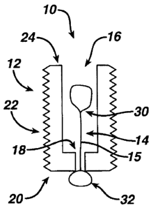
[00027] Surgical filament snare assembly 10, FIG. 1, has an anchor 12 and a
first
filament 14. In this construction, anchor 12 defines an internal passage 16
with a restricted
opening 18 at its distal end 20 which serves as a filament engagement feature.
At least one
bone-engaging feature 22, such as an external rib or helical thread, is
located on the outer
surface of anchor 12 between distal end 20 and proximal end 24.
[00028] First filament 14 has a noose 30 at its proximal end and a fixed knot
32 at
the distal end of filament post or stem 15 which interacts with restricted
opening 18 to
retain filament 14 in a fixed, permanently attached position. This arrangement
may be
referred to as the first filament 14 connected with the filament engagement
feature 18,
which includes the phrase passing through the filament engagement feature 18.
Many
conventional knots, such as a mulberry knot, can be utilized for fixed knot 32
as long as
knot 32 has sufficient bulk to prevent pull-through at clinically desired
tensions on noose
30. A number of other types of filament engagements are described below. Stem
15 is
9
CA 02759200 2011-11-22
kept as short as possible to maintain noose 30 close to anchor 12 even after
it is collapsed
as described below.
[00029] A well-known noose knot 33 is illustrated in FIG. 1A in which first
filament
14a has a hangman-type noose 30a at its proximal end and a fixed knot 32a at
the distal
end of stem 15a. Noose 30a has sliding noose knot 33 and defines an opening
34. Noose
knot 33 is tied by forming a flattened "S" or "Z" shape at the proximal end of
filament 14a
to form a large proximal loop to serve as the noose opening and a small loop
spaced from
the large loop. The doubled filament limbs are wrapped with the terminal end,
also known
as the working end. After typically four to eight wrapping turns, the terminal
end is tucked
through the small loop and trapped by pulling on whichever of the limbs of the
large loop
closes the small loop.
[00030] An alternative, simpler noose is illustrated for first filament 14b,
FIG. 1B,
having a half hitch 35, also referred to as a simple or overhand knot, tied to
form noose
30b in the middle of filament limbs 36 and 38. Multiple openings are created
by the loops
in half hitch 35 as described in more detail below, although central opening
37 is shown as
a large single opening in FIG. 1B. First filament limbs 36 and 38 are folded
around half
hitch 35 to form a double-stem arrangement, and the distal ends of first
filament limbs 36
and 38 are joined in knot 32b after being passed through a suitable filament
engagement
feature on an anchor.
[00031] Noose efficiency is defined herein as the strangulation strength per
unit
tension applied on the noose, either by pulling on the filament on which the
noose is tied or
which otherwise carries the noose, or by pulling on one or more strands or
limbs of
filaments passing through the noose. A noose with lower internal friction in
the noose
knot will tend to have a higher noose efficiency.
[00032] One instrument for inserting anchor 12 into a hole drilled in bone is
illustrated in FIG. 2. Driver 40 has a distal end 42 removably insertable into
passage 16.
Driver 40 is cannulated in this construction and has a lumen 44 with an
optional threader
filament 46 that passes through noose 30. Use of a threader filament is
optional, but may
be desirable when noose 30 is spaced only a short distance from filament
engagement
feature 18, in other words, when noose 30 is initially positioned close to or
inside of
anchor 12.
CA 02759200 2011-11-22
[00033] In one procedure according to the present invention, anchor 12 is
shown
fixated within bone B, FIG. 3, after driver 40 has been removed, in a hole 50
spaced at a
desired distance from tissue T to be repaired. Noose 30 is in an initial open
configuration.
Threader filament 46 has a sufficient length to have both a threader loop 52
on a first limb,
and a graspable portion on a second limb 54, extend proximally above skin S
while a mid-
portion of threader filament 46 is slidably associated with noose 30.
[00034] Continuing with this exemplary procedure, a second filament 60, FIG.
4, is
threaded through tissue T using a suture passing instrument, a needle, or
other tissue-
penetrating technique chosen by a surgeon. Both free filament limbs 62 and 64
are
brought together, typically above skin S, or at least outside of a joint
space, and passed
through threader loop 52, FIG. 5. Threader limb 54 is then pulled to thread
both second
filament limbs 62 and 64 through noose 30 as illustrated in FIG. 6 while noose
30 is in the
initial open configuration. Alternatively, free filament limbs 62 and 64 are
passed directly
through noose 30 without using a threader filament.
[00035] When there is high noose efficiency, a light tug is sufficient to
collapse
noose 30 on the filament limbs 62 and 64 as shown in FIG. 7 to provide initial
tensioning
on the surgical filament snare assembly 10. Generally, a higher noose
efficiency can be
utilized when one or more free filament limbs are threaded directly through
noose 30
without using a threader filament, or are threaded using a tube or threader
device such as
shown in FIGS. 13-14B below.
[00036] After initial or pre-tensioning of free filament limbs 62 and 64, FIG.
7,
tension is released on limbs 62, 64 and a slidable stopper knot 70, FIG. 8, is
tied by the
surgeon on limbs 62, 64 above skin S. An enlarged view of one construction for
stopper
knot 70a, FIG. 8A, shows a half hitch with an extra throw or turn, also known
as a double
overhand knot. A simple knot such as a single half hitch or overhand knot may
be
sufficient for some situations. Other suitable, more robust surgeon slidable
knots with
higher load capacity include the Tennessee Slider described in the
Arthroscopic Knot
Tying Manual (2005) available from DePuy Mitek, as well as the slidable,
lockable knot
by Wenstrom, Jr. in U.S. Patent No. 6,767,037. Alternatively, a mechanical
locking
mechanism may be utilized where overall profile is not critical, especially
away from a
joint or other articulating surfaces.
11
CA 02759200 2011-11-22
[00037] Stopper knot 70 is advanced, typically using a knot pusher, until it
contacts
noose 30, FIG. 9. Tension generated between tissue T and anchor 12, alone or
together
with pulling on one of the filament limbs 62 or 64, causes noose 30 to further
collapse,
FIG. 10, and strangulate the filaments. Stopper knot 70 augments the
strangulation by
transferring all tissue-generated tension on the stopper knot to the noose 30
and preventing
slippage of filament limbs 62, 64 into the noose knot. Accordingly, a self-
cinching
mechanism is created which inhibits loosening of the filaments. Tension can be
increased
incrementally in a ratchet-like effect by further advancing the stopper knot
or pulling on
one of filament limbs 62, 64.
[00038] Once satisfactory tissue tension has been achieved, one or more half
hitches may be added to stopper knot 70 to fortify the loading capacity on the
stopper knot
and reduce the risk of loosening under adverse conditions. By comparison,
conventional
sliding knots typically are reinforced by at least two or three reversed half
hitches placed
on alternating posts. Due to the self-cinching effect of the present
invention, fewer overall
hitches or other knots are needed for stopper knot 70 to meet or exceed the
load
performance relative to conventional knot systems. The present invention
thereby
accomplishes a lower overall knot profile to handle a given load. Limbs 62, 64
are
trimmed as desired. The stopper knot also minimizes fraying of the filament
ends over
time.
[00039] Preferred materials for filaments 14 and 60 include various surgical
sutures,
typically size 0 to size 5, such as OrthocordTM suture commercially available
from DePuy
Mitek, and EthibondTM suture available from Ethicon. OrthocordTM suture is
approximately fifty-five to sixty-five percent PDSTM polydioxanone, which is
bioabsorbable, and the remaining percent ultra high molecular weight
polyethylene, while
EthibondTM suture is primarily high strength polyester. The amount and type of
bioabsorbable material, if any, utilized in the first or second filament is
primarily a matter
of surgeon preference for the particular surgical procedure to be performed.
[00040] While the same type of suture, even identical suture, can be used for
both
first, noose filament 14 and second, tissue filament 60, a suture having a
lower abrasive
property at its surface may be preferred by some surgeons for second filament
60. The
lower abrasive property can be achieved by a larger diameter, a softer
composition, a softer
12
CA 02759200 2011-11-22
braid, plait or strand pattern, or a combination of such characteristics. The
term "braid" as
utilized herein includes "plait" and other multifilament patterns.
[00041] The nooses illustrated in FIGS. 1-6 above have been described as
having a
single opening through which one or more free filament limbs are threaded. A
simple half
hitch or overhand-type "pretzel"-like knot is illustrated in FIG. 11 for noose
30c having
multiple useful openings 80, 82 and 84. Side openings 80 and 84 are formed by
minor
loops 81 and 83 of the half hitch knot in first filament limbs 36c, 38c while
central opening
82 is formed by the major loop. Free filament limbs 62c and 64c are shown in
FIG. 12
extending through side opening 80 and central opening 82, respectively,
although other
combinations and permutations, such as using side openings 80 and 84, or
central opening
82 and side opening 84, are also effective. Utilizing different areas or
regions of the noose
knot significantly increases effective strangulation and gripping on the free
filament limbs.
It is expected that utilizing multiple openings in the noose knot also
minimizes any
dependence of load carrying capacity on filament compliance characteristics. A
simple,
single half hitch stopper knot 70c is also illustrated in FIG. 12.
[00042] While two or more threader filaments, or careful, potentially tedious
manipulation by a surgeon, could be utilized to achieve the configuration
shown in FIG.
12, an alternative technique which avoids inadvertent noose collapse is shown
in FIG. 13.
Tubes 90 and 92 have outer diameters suitable for sliding into the side
openings formed by
loops 81 and 83. Filament limbs 36, 38c are shown engaged with anchor 12c.
Tubes 90
and 92 define passages 94 and 96, respectively, through which free filament
limbs 62c and
64c are threaded. Tubes 90 and 92 are then disengaged from noose 30c and drawn
proximally along filament limbs 62c and 64c until they can be removed and
discarded
appropriately.
[00043] Double-barrelled threader device 100, FIG. 14A, has two threader tubes
102, 104 which are joined together with a handle 106 and provide an even
easier
technique. In one construction, device 100 is molded as a monolithic unit
using a polymer
material. Tubes 102, 104 have internal lumens 108, 110, respectively, also
referred to
herein as channels, with openings at both ends as well as slots 112, 114,
respectively,
which also run the entire length of tubes 102, 104. During use, tubes 102, 104
are placed
through loops 81d, 83d, FIG. 14B, formed from first filament limbs 36d, 38d,
and free
13
CA 02759200 2011-11-22
filament limbs 62d, 64d are inserted through lumens 108, 110. Thereafter,
limbs 62d, 64d
are simply lifted through slots 112, 114 to remove the fully-threaded
filaments from the
device 100. One or more additional such tubes can be formed and utilized as
desired.
Also, the tubes 102, 104 can be formed as "C" or "U" shapes in cross-section,
with wider
slots than illustrated.
[00044] There are a number of other configurations of snare assemblies
according to
the present invention which have one or more adjustable-length noose support
stems or
limbs that enable the noose to be retracted as desired toward an anchor. These
configurations provide an additional level of control over the final filament
positions and
tensions. Snare assembly 120, FIG. 15, has a noose 124 formed at one end of a
first
filament 122 with a stem section 126 extending into anchor 130 to pass through
ratchet-
like one-way gate or clamping mechanism 132. The remainder of filament 122
serves as a
limb 128, also referred to as a stem tail. Some examples of one-way mechanisms
are
disclosed in U.S. Patent No. 5,702,397 by Goble et al., for example, which
allow filament
movement in only one direction.
[00045] As illustrated in FIG. 15, anchor 130 is fixated in bone B. A second
filament 134 is passed through tissue T and has free limbs 136 and 138 passed
through
noose 124, initially positioned outside of a joint space surrounding tissue T.
Limb 128,
also positioned outside of the joint space, is pulled to retract noose 124
toward mechanism
132. Typically, the noose 124 is collapsed, limb 128 is trimmed, and then a
procedure
similar to that illustrated for FIGS. 7-10 above is utilized.
[00046] Snare assembly 140, FIG. 16, has first filament 140 having a noose 144
tied
with two stem limbs 146 and 148 extending into anchor 150. In this
construction, anchor
post 152 serves as a filament engagement feature to slidably attach filament
140 to anchor
150. Filament stem tail limbs 154, 156 extend out of a joint space, together
with noose
144 in an initial configuration. Second filament 160 is passed through tissue
T and then
free limbs 162, 164 are passed through noose 144 outside of the joint space.
[00047] In the procedure illustrated in FIG. 16A, limbs 154 and 156 of first
filament
142 are also passed through noose 144 and then pulled to collapse noose 144
about all four
limbs 154, 156, 162 and 164 and to retract noose 144 toward filament
engagement post
152. One or more sliding knots are tied on limb pair 154, 156 of the stem
tails to adjust the
14
CA 02759200 2011-11-22
proximity of noose 144 to the anchor 150 and then a simple knot is tied on
free limbs 162,
164 to adjust final tension on tissue T, although other combinations and
permutations can
be utilized within the scope of the present invention. Typically, the sliding
knots are
finished with one or more half hitches to "seal" or complete the fixation.
[00048] Snare assembly 170, FIG. 17, utilizes a single filament 172 both to
secure
noose 174 to anchor 180 and to tension tissue T. Stem limbs 176, 178 pass into
anchor 12
and slidably around filament engagement post 182 to emerge from anchor 180 as
tail limbs
184, 186 which are initially kept out of the joint space, along with noose
174, when anchor
180 is fixated in bone B. In some constructions, anchor post 182 is an eyelet
or a pulley
pin. Free tail limbs 184, 186 are passed through tissue T, in the same or
different places or
regions, and then through noose 174. Noose 174 is collapsed and pulled into
the joint
space by applying light tension to one, or preferably both, of the tail limbs
184, 186. A
simple stopper knot is tied between tail limbs 184, 186 and pressed against
the noose 174
while tensioning the limbs 184, 186 to place a desired amount of tension on
tissue T. The
fixation is finalized by placing one or more half hitches against the stopper
knot at noose
174.
[00049] Snare assembly 190, FIG. 18, has functional similarities to snare
assembly
120, FIG. 15, but achieves ratchet-like noose retraction without one-way gate
or clamping
mechanisms. Filament 192, FIG. 18, has a noose 194 with a stopper knot 196 at
its
terminal end to prevent pull-through and to resist fraying. A sliding knot 198
enables loop
200, having loop limbs 202 and 204, to be shortened toward anchor 205 when
post limb
206 is pulled. Loop 200 passes around anchor saddle or post 207. This and
other
adjustable loop, sliding knot configurations are described in more detail
below in relation
to FIGS. 21-27.
[00050] Snare assembly 310, FIG. 19, includes a first filament 302 with a
noose 304
and a loop 306 which is fixed in length, the overall length of filament 302
being subject to
full collapse of noose 304. A second filament 316 has a terminal end 318, a
sliding knot
322 retained at the distal end of anchor 312, a post limb 320, and an
adjustable loop 324
formed by limbs 326, 328. This configuration is described in more detail below
in relation
to FIG. 28.
CA 02759200 2011-11-22
[00051] While most of the embodiments herein have been described in relation
to
securing one or two filament limbs passed through a single place or region in
a tissue T,
this is not a limitation of the invention. Snare assembly 210, FIG. 20, has a
first filament
211 with a noose 212 through which pass free limbs 214, 216 and 218, 220 of
second and
third filaments 222 and 224, respectively. Noose 212 is engaged by stem 213
with anchor
215. Filaments 222 and 224 pass through tissue regions RI and R2,
respectively. Multiple
regions of a tissue, and potentially multiple types of sutures or other
filaments, can thereby
be secured using a single snare assembly according to the present invention.
[00052] One arrangement of the filament 192 for snare assembly 190, FIG. 18,
is
illustrated in FIG. 21 for snare assembly 190a. Noose 194a is formed merely by
creating
opening 232 in region 230 of filament 192a and passing filament 192a through
itself.
Loop 200a and sliding knot 198a are formed thereafter on post limb 206a. In
this
arrangement, any tension applied on stem 234, such as by pulling post limb
206a, not only
collapses noose 194a to strangulate objects passing through noose 194a, but
also binds the
portion of filament 192a passing through opening 232 upon itself. In other
arrangements,
a half hitch or other simple knot is tied at filament region 230, and filament
192a is then
looped through that simple knot. Stopper knot 196a such as a simple half hitch
will
prevent the terminal end from fraying or opening up, especially if a braided
filament such
as OrthocordTM suture is utilized for filament 192a.
[00053] An example of steps for manufacturing snare assembly 190, FIG. 18,
utilizing suture as filament 192 is as follows. Tie stopper knot 196 and trim
the tail of the
terminal end. Loop the suture and pass it through itself in close proximity to
the stopper
knot 196 to achieve the noose arrangement illustrated in FIG. 21, or tie a
second half hitch
in close proximity to the stopper knot and pass the suture through the half
hitch to create
the noose 194, FIG. 18. A thin mandrel or other object such as a pin may be
placed
through noose 194 to maintain patency. Sliding knot 198, such as a bunt line
half hitch
knot, is tied in close proximity to the noose 194 and the suture is placed in
sliding
engagement with feature 207 of anchor 205. Sliding knot 198 is then dressed or
finalized
as desired.
[00054] Conventionally, rotator cuff lateral row fixation involves spanning a
suture
bridge from medial anchors. Sutures are fixated with knotted or knotless
anchors at the
16
CA 02759200 2011-11-22
lateral row. Unthreaded anchors suffer more often than threaded anchors from
anchor pull
out, and suture slippage may occur at relatively low loads in many
conventional
procedures regardless of anchor type.
[00055] A presently preferred technique for rotator cuff double row repair is
illustrated in FIGS. 22-27 utilizing the snare assembly of FIG. 18. Medial row
anchor 240,
FIG. 22, is shown already embedded in bone B having cuff tissue T fixated at
the medial
row with suture 242. Preferably, a threaded anchor is utilized for anchor 240,
and may be
the same type of anchor as anchor 205. Free suture limbs 244 and 246 are
retracted out of
the joint space, shown in FIG. 22 as extending beyond skin S. Threaded anchor
205, FIG.
23 is then placed as a lateral row anchor in hole H independently of the
medial row
fixation. At this stage, collapsible loop 200 is long enough to enable sliding
knot 198 and
noose loop 194 to extend out of the joint space.
[00056] Suture limbs 244, 246 from the medial row are then passed through
noose
194, FIG. 24, preferably utilizing one of the threader devices described
above. Any
tension on suture limbs 244, 246 will collapse noose 194 around them. The size
of the
threader tube may be selected to limit the migration of noose 194 from sliding
knot 198.
Post limb 206 is then tensioned, FIG. 25, in the proximal direction indicated
by arrow 250
to retract sliding knot 198 into or in close proximity to anchor 205 and to
place initial
tension on suture bridge 258.
[00057] A simple knot such as a half hitch is then tied between suture limbs
244,
246 and pushed down against noose 194, FIG. 26, as sliding knot 260 while
limbs 244 and
246 are pulled to further tension suture bridge 258 as desired. As second or
more half
hitches 262, FIG. 27, are added after suture bridge 258 has been properly
tensioned to
permanently lock the repair and the ends of suture limbs 244 and 246 are
trimmed.
Because a single noose can handle multiple pairs of sutures as described above
in relation
to FIG. 20, additional suture bridges can be secured from multiple medial
anchors as
desired.
[00058] Adjustable suture snare assembly 310, FIG. 28, has a suture anchor 312
and
a closed, fixed-length loop 306 of a first material 302, which has a noose 304
tied at one
end. A half hitch "pretzel"-like knot 305 is shown in this construction;
another
construction having a unitary fixed loop is disclosed in U.S. Patent
Application No.
17
CA 02759200 2011-11-22
12/977,146 (Hernandez et al.), which is incorporated herein by reference. Loop
306 is
captured by, in other words, is connected to, a second filament 316 having a
terminal end
318, a post limb 320, a sliding bunt line half hitch knot 322, and an
adjustable loop 324
with loop limbs 326 and 328. Second filament 316 may be considered as part of
an
adjustable filament engagement feature of anchor 12, because filament 316
connects noose
304 to anchor 12. In one construction, suture anchor 312 is similar to the
cannulated
suture anchor disclosed by Cauldwell et al. in U.S. Patent Application
Publication No.
2008/0147063, incorporated herein by reference. In anchor systems utilized
according to
this sliding knot configuration of the present invention, however, it is not
necessary to have
a post-like suture-engaging member or other occluding element over which one
or more
sutures or suture limbs pass to serve as a restriction to proximal movement;
in many
constructions, it is sufficient to have a restricted opening 346 to prevent
withdrawal of knot
322.
[00059] Suture anchor 312 has a proximal end 330 and a distal end 332 with
opposed distal arms 334 and 336 defining cut-out 338 between them. Passage 340
is an
inner lumen which runs from proximal end 330 to distal cut-out 338. Although
knot 322 is
shown extending beyond cut-out 338 in FIG. 28 for purposes of illustration,
knot 322
preferably is seated against restricted opening 346 between arms 334 and 336,
or otherwise
maintained at the distal end 332 by a cavity or other feature, during
insertion of snare
assembly 310 into a patient to minimize interference by the knot 322 with the
bone-
engaging feature 342, or other exterior surface of anchor 312, and the bone in
which suture
anchor 312 is fixated.
[00060] One or more bone-engaging features 342, such as the helical thread
illustrated in FIG. 28 or other features such as teeth, ridges, or other
protrusions, are
formed on the exterior of anchor 312 to enhance fixation in bone. Threads such
as found
on the HealixTM anchor available from DePuy Mitek Inc. are desirable. In
another
construction, the suture anchor rotates to toggle into bone at its proximal
end to minimize
withdrawal. In a number of constructions, a hole is formed in bone prior to
anchor
insertion; in other constructions, a suture anchor is inserted directly into
bone. Further, one
or more passages or channels may be formed on the exterior of the suture
anchor, such as
channel 344 illustrated in phantom, FIG. 28, traversing bone-engaging element
342.
18
CA 02759200 2011-11-22
[00061] It is a matter of surgeon preference whether a terminal end 318 is
kept at a
length sufficient to lie against the exterior of at least one bone-engaging
feature 342 to be
trapped against bone during insertion, or is trimmed to a shorter length.
Further, a
restriction such as restricted opening may be defined at least in part by
engagement with
bone when anchor 312 is fixated in bone to prevent knot 322 from moving with
post limb
320 when tension is applied to post limb 320.
[00062] One or more such distal extensions or other protrusions may be
provided,
similar in some constructions to Cauldwell et al. cited above or to U.S.
Patent No.
7,381,213 by Lizardi, also incorporated herein by reference. In yet other
constructions, a
cylindrical or otherwise circumferential cavity, bowl or countersink feature
is provided at
the distal end of the anchor to seat the knot 322 during insertion and
fixation.
[00063] Slidable knot 322 has been described as a bunt line half hitch knot in
some
constructions, but other suitable knots will be readily apparent to those of
ordinary skill in
the suture tying art after reviewing the present invention. The term
"slidable" as used
herein is intended to include slidable, lockable knots as well as slidable
knots, such as
those described in the Arthroscopic Knot Tying Manual (2005) available from
DePuy
Mitek, as well as the slidable, lockable knot by Wenstrom, Jr. in U.S. Patent
No.
6,767,037.
[00064] Several improvements according to the present invention are
illustrated in
FIGS. 29-50. A filament 400, FIG. 29, has a noose 402 and noose limbs 404 and
406.
Noose 402 defines a central opening 408 and secondary openings 410 and 412
formed
from a half hitch plus one additional throw of limb 406 through central
opening 408. A
flexible sleeve 414 is shown in phantom encapsulating some of limbs 404 and
406 in
certain constructions, as described in more detail below.
[00065] FIGS. 30-31 illustrate the formation of a cinch noose 420, also
referred to as
an improved cinch noose construct, having an opening 422. The ends of free
filament
limbs 424 and 426 of filament 400 are passed through central opening 408, as
represented
by arrows 427 and 429 in FIG. 30, which draws noose limbs 424 and 426
therethrough.
Noose 402 is then tightened, FIG. 31, to form a slidable knot for cinch noose
420.
Alternatively, if a sleeve 414, FIG. 29, or sleeve 414a, FIG. 31, is not
utilized, or if such
sleeve is removed after being passed through tissue to be tensioned, then one
or both of
19
CA 02759200 2011-11-22
free limbs 424, 426 can be passed through one or both of openings 410, 412.
One
technique for utilizing improved cinch noose 420 is described below regarding
FIGS. 34-
40.
[00066] Filament 400 with noose 402, FIG. 29, is shown in FIG. 32 slidably
connected with anchor 430 as a snare assembly 432, after placement through
skin S into
bone B of a patient. Sleeve 414 is positioned over and encapsulates the entire
portion of
first and second free limbs 424, 426, down substantially to, but not into,
anchor 430 in this
construction.
[00067] It is a realization of the present invention that joining together at
least the
free filament limbs improves suture management and reduces the possibility of
suture
entanglement or damage by instruments, especially when passed through a
cannula. For
example, a surgeon or other user need only grasp and pass one sleeve 414
through noose
402 to thereby manipulate free filament limbs 424, 426 as a single unit.
Additional
convenience can be provided by perceptible indicators on one or more sleeves
such as
different markings, colors, diameters, braid or design patterns, or other
tactile or visual
indicia, especially if multiple tissue attachments or anchors are utilized,
such as described
above in relation to FIG. 20. Preferably, the sleeves are removed and
discarded after the
filaments have been manipulated, as described below, so the perceptible
indicators do not
need to meet long-term implantation requirements.
[00068] One technique for calculating the relative lengths of filament 501 and
sleeve
508 is illustrated in FIG. 33 for snare assembly 500 according to the present
invention. A
first factor is the distance, represented by arrow 502, between noose 504, in
a substantially
collapsed or reduced condition, and the distal end 506 of sleeve 508 over
noose limbs 503
and 505. One goal is to have distal end 506 accessible outside of a cannula
after tissue is
tensioned to enable latching or snagging of distal end 506 by a knot pusher or
grasper to
facilitate removal of sleeve 508, as described in more detail below for other
sleeves.
Typical cannula lengths for hip and shoulder surgeries are between four to six
inches, and
the cannulas are typically placed approximately one-half inch from bone. The
length of
anchor 510 is included in the calculation.
[00069] For some constructions prior to implantation in a patient, sleeve 508
is
twenty five inches in total length, with seven and one-half inches extending
from the
CA 02759200 2011-11-22
filament engagement feature of anchor 510 toward noose 504 as indicated by
arrow 512,
with seventeen and one-half inches, arrow 514, extending over and beyond free
filament
limbs 513 and 515 to proximal end 516 of sleeve 508. In one construction,
filament 501
has a total length of thirty six inches, or a folded length of eighteen
inches, with sixteen
and one-half inches, arrow 520, extending from noose 504 to anchor 510, and
one and one-
half inches, arrow 522, as free limbs 513 and 515. In another construction
wherein
filament 501 has a total length of sixty six inches and a folded length of
thirty three inches,
free filament limbs 513, 515 extend sixteen and one-half inch as represented
in phantom by
arrow 524. In either construction, marks can be placed on the filament noose
limbs 503,
505 nine inches from the center or middle, where noose 504 will be formed, to
clearly
indicate the proper positioning, arrows 502 and 512, of distal end 506 of the
sleeve 508
over filament 501 during preparation of snare assembly 500 for implantation.
[00070] A technique for utilizing the improved cinch noose 420, FIG. 31, with
a
sleeve 414a is shown in FIGS. 34-40 for another embodiment, represented by
snare
assembly 530 according to the present invention. In this construction, the
sleeve 414a,
shown with dashed lines, is slid over filament 400a and then loaded through
anchor 532 to
cover all of free limbs 424a, 426a and at least some of noose limbs 404a,
406a, preferably
covering all of noose limbs 404a, 406a as they emerge above a cannula (not
shown)
passing through skin S during initial implantation of anchor 532 in bone B,
FIG. 34 to
assist in suture management and protection.
[00071] The proximal end of sleeve 414a is passed through tissue T, FIG. 35,
and
then passed through cinch noose 420a, FIG. 36. Alternatively, sleeve 414a can
be
removed after it is passed through noose 420a so that free limbs 424a and 426a
can be
passed directly through one or more openings in noose 420a. In either scenario
for FIG.
36, the noose 420a is then dressed, that is, collapsed, FIG. 37, and then
advanced near
tissue T and tightened, FIG. 38. The sleeve 414a is then removed entirely,
FIG. 39, and
discarded according to standard procedures. The tissue repair is then finished
with one or
more half hitches 534 as desired, FIG. 40.
[00072] Materials for sleeves include braided sutures such as EthibondTM size
0
suture or OrthocordTM size 2 suture, also referred to as OrthocordTM #2
suture, which is
typically braided at sixty picks per inch. For use as a sleeve, a more relaxed
braid of
21
CA 02759200 2011-11-22
approximately thirty to forty picks per inch is preferred, more preferably
about 36 picks
per inch. If the sleeve material is formed about a core, preferably that core
is removed to
facilitate insertion of the filament limbs, which may themselves be formed of
typical suture
such as OrthocordTM #0 suture or #2 suture braided at sixty picks per inch.
[00073] In yet another sleeve embodiment according to the present invention,
one of
the free filament limbs itself serves as the sleeve. For the construction
illustrated in FIG.
41, snare assembly 540 has a filament 542 of OrthocordTM #2 suture generally
braided at
sixty picks per inch with a noose 544 and noose limbs 545 and 546 that pass
around
filament engagement feature 550 of anchor 548. Noose limbs 545 and 546 become
free
filament limbs 555 and 556, respectively, extending proximally. At point 558,
however, a
proximal section of limb 555 is braided at fewer picks per unit length,
preferably more
than ten percent fewer, more preferably at least twenty five percent fewer, to
serve as
sleeve 560 extending to its proximal end 562. The other free filament limb 556
is threaded
through sleeve 560 to emerge as proximal end 564 in this construction; in
other
constructions, the proximal end 564 lies wholly within sleeve 560.
[00074] One technique for constructing snare assembly 540 is illustrated in
FIGS.
42A-42D. Filament 542 is shown in FIG. 42A as initially manufactured with
sleeve 560
being a section of suture formed with fewer picks per inch beginning at point
558 and
extending to end 562, preferably reduced from the standard 60 picks per inch
to 36 picks
per inch in this construction. Noose 544 is then created, FIG. 42A, and then
filament ends
562, 564 are threaded through anchor 548 as shown schematically in FIG. 42C.
After a
core element within sleeve section 560 has been removed, filament end 564 is
then
threaded within sleeve 560 using a needle-type insertion device to achieve
snare assembly
540, FIG. 42D, with coaxial filament limbs in the sleeve section 560. The
length of sleeve
560 is likely to decrease as its diameter is expanded by the insertion device.
[00075] One procedure for utilizing snare assembly 540 is shown in FIGS. 43-
45.
Anchor 548 is inserted into bone B, FIG. 43, and then coaxial sleeve section
560 is passed
through tissue T, FIG. 44, and then noose 544, FIG. 45. Noose 544 is then
collapsed
toward tissue T, FIG. 46, sleeve 560 is severed from filament 542, and then
filament 542 is
tied and cut as described above for other embodiments to finish fixation of
tissue T. The
excess portion of filament 542, including coaxial sleeve section 560, is
discarded.
22
CA 02759200 2011-11-22
[00076] Another embodiment according to the present invention is illustrated
in
FIGS. 47-50. Snare assembly 570 has a fixed-length, preferably continuous loop
572 of a
first filament which a surgeon or other user utilizes to form a Lark's Head
knot, also
known as a Bale Sling Hitch, to serve as a noose 573, FIG. 48, to grip a
section of a second
filament 574 as shown in FIGS. 49-50.
[00077] Second filament 574, FIG. 47, has a collapsible loop 578 with a
sliding knot
576 such as a sliding bunt line half hitch knot, a tensioning or post limb
580, and a tag or
terminal limb 581. Collapsible loop 578 passes around filament engagement
feature 592,
also referred to as a saddle 592, of bone anchor 590. In one construction,
snare assembly
570 is manufactured in the condition shown in FIG. 47 and supplied to a user
with sliding
knot 576 already tied. To utilize snare assembly 570, a hole 594 is formed in
bone B and
the anchor 590 is inserted to the position shown in FIG. 47, and then
continuous loop 572
is passed through tissue T.
[00078] After the noose 573 is formed with a Lark's Head knot, tail 580 and
sliding
knot 576 are passed through noose 573, FIG. 49. Noose 573 is then tightened
against
sliding knot 576. A knot pusher 596, FIG. 50, assists in collapsing the loop
578 to tighten
the snare assembly 570 to apply tension to tissue T. Depending on the overall
length of
first loop 572, a portion of it may be drawn into anchor 590.
[00079] Thus, when snare assembly 570 is supplied to a surgeon or other user
with
sliding knot 576 already tied, snare assembly 570 serves another example
according to the
present invention of a pre-formed, knot-less filament system which does not
require the
user to manipulate free limbs to tie knots during an operation. Adding to the
benefits of
snare assemblies according to the present invention, including high strength
and loop
security, low knot profile, ability to tension incrementally, and easy use
with threaded
anchors, providing a loop capable of forming a Lark's Head removes altogether
the burden
of tying a knot near or within a patient.
[00080] In other words, a first filament, preferably a continuous fixed-length
suture
loop, is slidably attached to a collapsible filament loop of a second filament
having a
preformed sliding knot. In another construction shown in FIG. 48A, the fixed-
length loop
572a is formed at one end of a first filament 601, such as by pre-tying a
first bowline knot
600, and the other end of the first filament 601 is slidably attached to the
second filament
23
CA 02759200 2011-11-22
574a with another, smaller loop 603, such as formed by a second, smaller pre-
tied bowline
knot 602 through which the collapsible loop 578a passes. After the anchor is
placed in
bone, the continuous-loop end with bight 575a is passed through tissue. A
Lark's Head
knot is then created on the continuous loop 572a, which generates a very
robust noose.
[00081] One or more tools can be utilized to assist creation of the constructs
described above, especially if a half hitch is desired to be thrown on free
filament limbs
passing through different loops of a "pretzel" noose, that is, a noose with at
least one half
hitch that defines multiple loops through which the free filament limbs are
passed.
Improved threading tools and suture passers are illustrated in FIGS. 51-57 to
automatically
create a simple half hitch when two filament ends are pulled through loops of
a noose.
[00082] Suture passer 620 is shown in FIG. 51 placed diagonally over suture
passer
610. Suture passer 610 has proximal tab or handle 612, shaft 614 formed of
wire or other
flexible material, and opening 615 at distal end 616. Suture passer 620 has
proximal
handle 622, flexible shaft 624, and an opening 625 at distal end 626. Distal
end 626 is
looped under and around shaft 614 to create a simple half hitch 630, FIG. 52.
[00083] Intertwined suture passers 610 and 620 are shown held by threader tool
700
in FIG. 53. Tool 700 has projections 702 and 704 which are substantially
cylindrical tubes
in this construction, whose distal ends are similar to tubes 102 and 104 of
FIGS. 14A-14B
above. Each projection 702, 704, FIGS. 53-53A, is supported by common handle
703 and
has a longitudinal channel 706, 708, respectively, with slots 710, 712 to
facilitate
placement of filaments or passers such as suture passers 610, 620 into tool
700, and to
facilitate subsequent removal of filaments drawn into tool 700 by the passers.
Tool 700
further defines a common passage 720, formed in part by notches in the
proximal walls of
projections 702 and 704, which interconnects the proximal portions of channels
706 and
708. Half hitch 630, FIG. 52, lies within passage 720, FIG. 53, and is further
held by fixed
stop 730 with lip or overhang 732, which is an inverted "L"shape in this
construction.
Tool 700 further includes a distal finger 740 in this construction to serve as
a catch or post
for one or more filaments during the threading procedure, such as to hold a
cinch loop or
other noose in position.
[00084] In another construction shown in side view in FIG. 54, a tool 700a has
a
movable stop 730a with a strut 734a pivotally attached to handle 703a by pin
740 passing
24
CA 02759200 2011-11-22
through the lower portion of strut 734a, or other type of hinge such as a
living hinge.
Tubular projection 702a is visible in this view. Stop 730a has a lip 732a
supported by strut
734a. In one construction, a user manipulates stop 730a to hold or release
suture passers
by moving stop 730a toward or away from handle 703a as indicated by arrow 736;
stop
730a is shown in phantom in an open position after being moved away from
handle 703a.
In another construction, a spring 742, also shown in phantom, biases stop 730a
in one
direction, preferably toward handle 703a. As a user pulls suture through the
device, a
certain amount of force causes stop 730a to overcome the biasing force of
spring 742 and
move away from handle 703a to assist release of the tied suture.
[00085] Several threader tools according to the present invention having
intersecting
channels are shown in top view in FIGS. 55-57. A V-shaped tool 800, FIG. 55,
has
projections 802, 804 with intersecting channels 806 and 808, respectively, and
a distal
finger 840. A proximal trapezoidal stop 830 holds suture passers in place as
they pulled
proximally. The distal portions of projections 802, 804 become substantially
parallel to
each other to assist removal of the tied knot from tool 800.
[00086] Tool 900, FIG. 56, has straight projection 902 and curved projection
904
that define channels 906 and 908, respectively. Stop 930 forms a proximal
corner at the
intersection where sutures can be pulled proximally when force is applied at
right angles to
respective suture passers, which is expected to ease suture movement through
the channels
906, 908.
[00087] Tool 1000, FIG. 57, is a horseshoe shape to reduce forces needed to
pull
sutures through the tool 1000. Finger 1040 is positioned slightly below to
distal opening
of channels 1006, 1008 to minimize obstruction of the suture threading
process.
[00088] This invention may also be expressed as a surgical filament snare
assembly
with a bone anchor and a first filament having a noose, formed from at least
one half hitch,
on a first portion of at least a first limb and having a second portion
connected to the
filament engagement feature of the anchor. The noose is capable of receiving
at least two
free filament limbs and strangulating them when tension is applied to at least
one of the
free filament limbs and the noose. Preferably, the assembly further includes a
threader
tool having at least two projections having distal ends capable of being
removably inserted
into different loops of the half hitch. Each projection defines a channel
capable of
CA 02759200 2011-11-22
receiving a portion of at least one free filament limb to pass it through a
loop of the half
hitch, and each projection further defines a slot communicating with the
channel to
facilitate removal of the filament limb from the tool. Each slot has the same
width as its
corresponding channel in some embodiments and, in other embodiments, has a
different
width, typically a narrower width, than that of the corresponding channel.
[00089] In certain embodiments, the projections are tubes joined together with
at
least one handle for manipulation the tube. The proximal ends of the channels
are
connected by one of an intersection and a common passage, and the tool further
includes a
stop as a proximal portion of the one of the intersection and the common
passage. In some
embodiments, the stop is movable, and may include a spring to bias the stop
toward the
intersection or common passage.
[00090] In yet other embodiments, the assembly further includes at least two
suture
passers having distal ends for engaging portions of the free filament limbs,
and the suture
passers being capable of pulling the free filament limbs through the channels
when
proximal-directed force is applied to proximal ends of the suture passers.
Preferably, the
distal ends of the suture passers are intertwined in at least one half hitch
to impart at least
one half hitch to the free filament limbs when they are drawn through the
tool. Different
combinations selected from the group of an anchor, one or more filament
constructs as
described herein, a threader tool, and one or more suture passers can also be
referred to as
different kits according to the present invention.
[00091] Thus, while there have been shown, described, and pointed out
fundamental
novel features of the invention as applied to a preferred embodiment thereof,
it will be
understood that various omissions, substitutions, and changes in the form and
details of the
devices illustrated, and in their operation, may be made by those skilled in
the art without
departing from the spirit and scope of the invention. For example, it is
expressly intended
that all combinations of those elements and/or steps that perform
substantially the same
function, in substantially the same way, to achieve the same results be within
the scope of
the invention. Substitutions of elements from one described embodiment to
another are
also fully intended and contemplated. It is also to be understood that the
drawings are not
necessarily drawn to scale, but that they are merely conceptual in nature. It
is the
26
CA 02759200 2011-11-22
'intention, therefore, to be limited only as indicated by the scope of the
claims appended
hereto.
[00092] Every issued patent, pending patent application, publication, journal
article,
book or any other reference cited herein is each incorporated by reference in
their entirety.
27