Note: Descriptions are shown in the official language in which they were submitted.
CA 02759242 2011-09-19
WO 2010/106438 PCT/IB2010/000833
1
HEART VALVE PROSTHESIS WITH COLLAPSIBLE VALVE
AND METHOD OF DELIVERY THEREOF
Description
Cross-Reference to Related Application
This application claims the benefit of priority of U.S. provisional
patent application number 611210,255, entitled "MINIMALLY INVASIVE,
SUTURELESS EXPANDABLE HEART VALVE PROSTHESIS WITH A
COLLAPSIBLE VALVE," filed on March 17, 2009; U.S. provisional patent
application number 61/212,459, entitled "MINIMALLY INVASIVE,
SUTURELESS EXPANDABLE HEART VALVE PROSTHESIS WITH A
COLLAPSIBLE VALVE," filed on April 13, 2009; U.S. provisional patent
application number 61/215,944, entitled "MINIMALLY INVASIVE
SUTURELESS EXPANDABLE HEART VALVE PROSTHESIS WITH A
COLLAPSIBLE VALVE AND A METHOD OF DELIVERY," filed on
May 12, 2009; U.S. provisional patent application number 61/186,100,
entitled "A HEART VALVE PROSTHESIS WITH A COLLAPSIBLE VALVE
AND A METHOD OF DELIVERY THEREOF," filed on June 11, 2009; U.S.
provisional patent application number 61/227,193, entitled "A HEART
VALVE PROSTHESIS WITH A COLLAPSIBLE VALVE AND A METHOD
OF DELIVERY THEREOF," filed on July 21, 2009; and U.S. provisional
patent application number 61/257,979, entitled "A HEART VALVE
PROSTHESIS WITH A COLLAPSIBLE VALVE AND A METHOD OF
DELIVERY THEREOF," filed on November 4, 2009, the disclosures of
which are incorporated herein by reference in their entirety as if fully set
forth herein.
Technical Field
The present disclosure relates to minimally invasive surgical or
percutaneous replacement and/or repair of a valve, namely the mitral
CA 02759242 2011-09-19
WO 2010/106438 PCT/1B2010/000833
2
valve or the tricuspid valve. More particularly, the present disclosure
relates to a heart valve prosthesis with a collapsible valve and a method of
delivery of the prosthesis.
Background
The mitral valve and tricuspid valve are unidirectional heart
valves that separate the atria left and right respectively, from the
corresponding heart ventricles. These valves have a distinct anatomical
and physiological structure, having two (mitral) or three (tricuspid) sail-
like
leaflets connected to a subvalvular mechanism of strings (chordae
tendinae) and papillary muscles forming a part of the heart's ventricular
shape, function and size.
The heart has four chambers: the right and left atria, and the
right and left ventricles. The atria receive blood and then pump it into the
ventricles, which then pump it out into the body.
The synchronous pumping actions of the left and right sides of
the heart constitute the cardiac cycle. The cycle begins with a period of
ventricular relaxation, called ventricular diastole. The cycle ends with a
period of ventricular contraction, called ventricular systole.
The heart has four valves that ensure that blood does not flow in
the wrong direction during the cardiac cycle; that is, to ensure that the
blood does not back flow from the ventricles into the corresponding atria, or
back flow from the arteries into the corresponding ventricles. The valve
between the left atrium and the left ventricle is the mitral valve. The valve
between the right atrium and the right ventricle is the tricuspid valve. The
pulmonary valve is at the opening of the pulmonary artery. The aortic valve
is at the opening of the aorta.
The opening and closing of heart valves occur primarily as a
result of pressure differences. For example, the opening and closing of the
mitral valve occurs as a result of the pressure differences between the left
CA 02759242 2011-09-19
WO 20101106438 ".iii .uiu/uuuaaa
3
atrium and the left ventricle. During ventricular diastole, when ventricles
are relaxed, the venous return of blood from the pulmonary veins into the
left atrium causes the pressure in the atrium to exceed that in the ventricle.
As a result, the mitral valve opens, allowing blood to enter the ventricle. As
the ventricle contracts during ventricular systole, the intraventricular
pressure rises above the pressure in the atrium and pushes the mitral
valve shut.
As noted above, these valves feature a plurality of leaflets
connected to chordae tendinae and papillary muscles, which allow the
leaflets to resist the high pressure developed during contractions
(pumping) of the left and right ventricles.
In a healthy heart, the chords become taut, preventing the
leaflets from being forced into the left or right atria and everted. Prolapse
is a term used to describe the condition wherein the coaptation edges of
each leaflet initially may co-apt and close, but then the leaflets rise higher
and the edges separate and the valve leaks. This is normally prevented by
contraction of the papillary muscles and the normal length of the chords.
Contraction of the papillary muscles is simultaneous with the contraction
of the ventricle and serves to keep healthy valve leaflets tightly shut at
peak contraction pressures exerted by the ventricle.
Valve malfunction can result from the chords becoming stretched,
and in some cases tearing. When a chord tears, the result is a flailed
leaflet. Also, a normally structured valve may not function properly
because of an enlargement of the valve annulus pulling the leaflets apart.
This condition is referred to as a dilation of the annulus and generally
results from heart muscle failure. In addition, the valve may be defective at
birth or because of an acquired disease, usually infectious or
inflammatory.
Diseases of the valves can cause either narrowing (stenosis) or
dilatation (regurgitation, insufficiency) or a combination of those, of the
CA 02759242 2011-09-19
WO 2010/106438 PCT/1820101000833
4
valve. Surgical treatment for repair or replacement of the valves includes an
open-heart procedure, extracorporeal circulation and, if replaced, a
complete resection of the diseased valve.
Currently all available surgical options for valve replacement
involve open heart surgery; although minimally invasive methods for valve
replacement are more desirable, such methods are still in the
experimental stage.
Even valves which could theoretically be provided through a non-
invasive method, such as those taught by U.S. Patent No. 7,381,220,
have many drawbacks. For example, the taught valves are useful for
replacement of the existing valves; however, their installation through non-
invasive means is problematic. Furthermore, the valves themselves, even
when installed in a manner that supports existing valve tissue, must still
withstand very high pressures. Such high pressures can lead to many
different types of problems, including reflux as blood returns through heart
in a retrograde manner.
It may be desirable to provide a valve prosthesis that supports
the mitral and/or tricuspid valve without necessarily replacing it, but
instead supplements the native valve functionality by providing an
adjunctive valve prosthesis, which cooperates together with the native
valve for improved functionality. The background art also does not teach
or suggest such a valve prosthesis which may optionally be inserted
through minimally invasive surgical techniques.
Summary of Invention
In accordance with various aspects of the disclosure, a valve
prosthesis is adapted to operate in conjunction with native heart valve
leaflets. The prosthesis includes an annulus and a skirt extending from
the annulus. The skirt may be configured to be positioned through a
native heart valve annulus, and the skirt may be movable between an
CA 02759242 2011-09-19
WO 2010/106438 PCT/1B2010/000833
open configuration permitting blood flow through the skirt and a closed
configuration blocking blood flow through the skirt in cooperation with
opening and closing of the native heart valve leaflets
According to various aspects, a novel valve prosthesis, for
example, for a tricuspid valve and/or mitral valve, may be inserted through
any one or more of a minimally invasive surgical procedure, a "traditional"
operative procedure (which may for example involve open heart surgery),
or a trans-catheter procedure.
The valve prosthesis, in at least some embodiments, is a
(optionally non-stented) bioprosthesis attached by means of suture or any
other means of bonding, to an expandable, frame (platform), which may
be made from a suitable metal, including without limitation an alloy, or any
type of suitable composite material (optionally including those that include
metal). The frame can be made of self expanding alloy such as Nitinol
(nickel/titanium alloy) or made of another metal, such as a cobalt/chrome
alloy, expanded by a specialized balloon, or radial expander.
The frame engages the tissue at or near or above the top
margins of the native valve (annulus). The native valve is not removed,
and the ventricular shape and function are preserved. Therefore, the valve
prosthesis may not replace the native valve functionality but rather
supports its function.
By "native valve" or "native valve annulus" it is meant the valve or
valve annulus already present in the subject, as opposed to an artificial
valve or valve annulus.
According to some embodiments, the valve prosthesis
comprises a support structure featuring a deployable construction adapted
to be initially collapsed (crimped) in a narrow configuration suitable for
introduction through a small puncture or incision into the heart cavity such
as the left ventricle, the left atrium, the right atrium, the right ventricle
and
CA 02759242 2011-09-19
WO 2010/106438 PCT/182010/000833
6
so forth, thereby providing access to the target location. It is further
adapted to be deployed by means of removing a radial constriction such
as a sheath to allow the platform to self-expand to its deployed state in the
target location.
In some embodiments, the valve prosthesis optionally
features a flexible film made of biological tissue such as pericardia tissue
but may also optionally feature one or combination of synthetic
materials, additionally or alternatively. The prosthesis may have a
funnel like shape that is generally tubular and may have a variable
diameter that enables flow in one direction (from the atrium to the
ventricle); when the ventricle contracts, the funnel shape valve
collapses and blocks any return flow from the ventricle to the atrium.
Such retrograde flow is quite dangerous; over a prolonged period of
time, it can lead to many deleterious health effects, including on the
overall health of the heart muscle.
In an exemplary, illustrative configuration, the valve platform
of the prosthesis is anchored to the ventricle wall through extensions
that pass through the commissures of the native valve or at the plane of
the commissures and have hooks at their ends that anchor into the
ventricular wall between the chordate attachment to the ventricular wall.
Furthermore, in an illustrative example, these extensions have curved
ends that can be in any plane (but which may be at a 90 degree angle to
the plane of both extensions) that allows a wire or cable to pass through
and keep the prosthesis connected to the delivery system as long as
this wire or cable is not released. The delivery action of the prosthesis
may be reversible. That is, the device may optionally be refolded into
the catheter after having being deployed.
In an optional embodiment, these extensions should not act
on the valve in any way, including not on the valve annulus or
surrounding valve tissue, nor should these extensions apply any
CA 02759242 2011-09-19
WO 2010/106438 PCT/1B20101000833
7
pressure that may reshape the annulus or deform the leaflet
configuration.
In an exemplary embodiment, the valve prosthesis features a
"skirt" that does not restrict the motion of any of the native valve leaflets
but which is situated above such leaflets, for example in the direction of
the atrium (by "above" it is meant with regard to the direction of normal,
not retrograde, blood flow). If the leaflets prolapse into the atrium, no
blood will be able to flow into the atrium because the skirt is situated
above the native valve, thus preventing retrograde blood flow into the
atrium from the ventricle.
In an embodiment, the "skirt" is generally tubular in shape with
a diameter that may vary and which is optionally used to complete the
incompetent closure of the native valve as a whole. Thus, the skirt
specifically and the valve prosthesis generally are not intended to be
used as a replacement to the entire valve or in addition to only one
native leaflet (in contrast to the apparatus described by Macoviak et al.
in U.S. patent application publication number 2008/0065204, for
example). In an exemplary embodiment, the valve skirt may be
reinforced with at least one reinforcement along at least a portion of its
length, for example, along the entirety of its length, in order to prevent
prolapse of the skirt into the left atrium. This reinforcement is optionally
an extension from the platform.
In yet another configuration, the valve "skirt" is connected to
the extensions by a cable or wire in order to prevent the prolapsed of
the skirt into the left atrium. These connections may optionally be an
integral part of the valve platform or alternatively may be connected
separately.
In an exemplary embodiment, the closing action of the native
valve leaflets promotes the collapse of the prosthetic valve (skirt). Thus,
CA 02759242 2011-09-19
WO 2010/106438 PCT/1B2010/000833
8
during systole function, the native valve may achieve partial closure (i.e
function partially) and hence may assist the function of the valve
prosthesis.
During systole, the action of the native valve leaflets is to close
the passage between the left ventricle and the left atrium. In an
exemplary embodiment, the leaflets, while acting as such, resist part of
the pressure applied by the blood pressure in the ventricle during valve
closure as well as reducing the effective area on which the pressure is
applied to the valve prosthesis as a whole, thus reducing the total force
applied to the prosthesis for migration into the left atrium. Depending
on which valve is affected, the present invention is contemplated as a
potential treatment for all forms of valvular regurgitation, such as
tricuspid regurgitation, pulmonary regurgitation, mitral regurgitation, or
aortic regurgitation.
Brief Description of the Drawings
In the drawings:
Figure 1 shows an exemplary anatomy of a mitral valve (for
reference only);
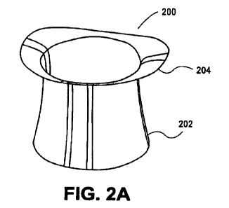
Figures 2a-2c show an exemplary valve prosthesis according
to some embodiments of the present disclosure; Figure 2a shows the
valve skirt alone, and Figures 2b and 2c show the valve skirt in place in
the heart as an example only;
Figure 3 shows an illustrative embodiment of an exemplary
valve prosthesis in accordance with various aspects of the disclosure;
Figures 4a-4c show an illustrative configuration of an
exemplary valve prosthesis according to some embodiments of the
present disclosure;
CA 02759242 2011-09-19
WO 2010/106438 PCT/1B2010/000833
9
Figure 5 shows a schematic view of the prosthetic and native
mitral valve leaflets during Diastole;
Figure 6 shows a schematic view of the prosthetic and native
mitral valve leaflets during Systole;
Figure 7 shows an illustrative embodiment of an exemplary
valve prosthesis in accordance with various aspects of the disclosure;
Figures 8A-8E show illustrative embodiments of various
exemplary valve prostheses in accordance with various aspects of the
disclosure;
Figure 9 shows an exemplary frame for a valve prosthesis in
accordance with various aspects of the disclosure;
Figure 10 shows an exemplary valve prosthesis in accordance
with various aspects of the disclosure;
Figure 11 shows an exemplary frame for a valve prosthesis in
accordance with various aspects of the disclosure;
Figure 12 shows an exemplary prosthesis in its folded state
and as it unfolds from a catheter;
Figure 13 shows an exemplary valve prosthesis in accordance
with various aspects of the disclosure;
Figures 14A and 14B show an exemplary skirt for a valve
prosthesis in accordance with various aspects of the disclosure;
Figure 15 shows an exemplary delivery system for a valve
prosthesis in accordance with various aspects of the disclosure;
Figure 16 shows a portion of an exemplary delivery system
valve prosthesis in accordance with various aspects of the disclosure;
Figure 17 shows an exemplary measuring device for use in
delivery of a valve prosthesis in accordance with various aspects of the
disclosure;
CA 02759242 2011-09-19
WO 2010/106438 PCT/1B2010/000833
Figure 18 is a flow chart of an exemplary pre-delivery method of
a valve prosthesis in accordance with various aspects of the disclosure;
and
Figure 19 is a flow chart of an exemplary delivery method of a
valve prosthesis in accordance with various aspects of the disclosure.
Detailed Description
The disclosure provides, in at least some embodiments, a valve
prosthesis and method of insertion thereof which supports the mitral
and/or tricuspid valve without replacing it. The valve prosthesis may
operate to support the native valve leaflets to provide a functioning heart
valve and to prevent retrograde motion of the blood, even if the native
valve leaflets alone are unable to completely close and/or to prevent such
retrograde motion of the blood.
Figure 1 shows an exemplary anatomy of a native mitral valve
(for reference only). As shown, a native valve 100 features an anterior
leaflet 102 and a three lobed posterior leaflet 104, which together comprise
the leaflets of native valve 100, as well as an anterior annulus 106 and a
posterior annulus 108, which together comprise the annulus of native
valve 100. Native valve 100 also features a posterolateral commissure
110 and an anteromedial commissure 112, one or both of which are
optionally used for installation of a valve prosthesis according to some
embodiments of the present disclosure.
A plurality of chordinae tendinae 116 attach the leaflets to a
lateral papillary muscle 118 or a medial papillary muscle 120. Ina healthy
heart, chordinae tendinae 116 become taut to prevent retrograde blood
flow back through the leaflets. In a non-healthy heart, for a variety of
reasons as described above, bloods flow in a retrograde manner through
the leaflets. As described in greater detail below, in at least some
CA 02759242 2011-09-19
WO 2010/106438 PCT/1B2010/000833
11
embodiments of the present disclosure, the leaflets are assisted in their
function by a valve prosthesis.
Figures 2a-2c show an exemplary valve prosthesis according to
some embodiments of the present disclosure. As shown in Figure 2a, a
valve prosthesis 200 may comprise a valve skirt 202 and a prosthetic
valve annulus 204 according to various aspects of the present disclosure.
Although not clearly shown in Figure 2a, in some aspects, the prosthetic
valve annulus 204 may have a D-shape configuration. In some aspects,
the annulus 204 may have an oval configuration.
According to various aspects, the skirt 202 may comprise a
biological tissue, such as, for example, an animal (e.g., bovine or porcine
tissue) or human pericardium. In some aspects, the skirt 202 may
comprise a synthetic material, such as, for example, polyurethane. In
various aspects, the skirt 202 may comprise a native mitral valve
processed to be biologically compatible for a particular implantation.
According to some aspects, the skirt 202 may comprise an ultra-thin sheet
of nitinol. According to various aspects of the disclosure, the skirt 202
and/or the prothetic annulus 204 may be coated with various bioactive
agents, such as anti-proliferative and/or anti-inflammmatory properties or
can have other properties such as antineoplastic, antiplatelet, anti-
coagulant, anti-fibrin, antithrombonic, antimitotic, antibiotic, antiallergic,
antioxidant as well as cystostatic agents, anti-inflammatory agents (e.g.,
steroidal anti-inflammatory agent, a nonsteroidal anti-inflammatory agent,
or a combination thereof, and anti-proliferative agents (e.g., rapamycin
and derivatives of rapamycin; everolimus and derivatives of everolimus;
taxoids including taxols, docetaxel, paclitaxel, and related derivatives of
taxoids, Biolimus A9, etc.). According to various aspects, the skirt may
have a thickness of between about 0.05 mm and about 0.4 mm.
According to some aspects, the length of valve prosthesis 200 is
at least as long as the native valve leaflets, but is not excessively long so
CA 02759242 2011-09-19
WO 2010/106438 PCT/1B2010/000833
12
as to avoid disturbing the flow through the aortic or adjacent valve. For
example, in some aspects, the length of valve prosthesis 200 is no more
than about 120% of the length of the native valve leaflets. According to
various aspects, the diameter of the bottom of valve skirt 202 is at least
about 80% of the diameter of the native valve area and no more than
about 130% of the diameter of the native valve area.
Figure 2b shows an exemplary valve prosthesis 200 in place in
a mitral valve 100 as an illustrative example only of installation. Valve
skirt 202 is shown as well, extending into a ventricle 206. Figure 2c shows
the view of Figure 2b in cross-section. Valve skirt 202 is configured and
positioned to prevent retrograde flow of blood from the ventricle 206 back
into the atrium (not shown) by assisting the function of the natural, native
leaflets of the mitral valve 100. It should be appreciated that the
exemplary valve prosthesis 200 may also be placed in a tricuspid valve in
accordance with various aspects of the disclosure.
Figure 3 shows an exemplary valve frame, or valve platform,
configured to support a valve skirt of a valve prosthesis in accordance with
various aspects of the present disclosure. Valve frame 300 may comprise
a valve annulus 306, for example, a D-shaped annulus. According to
various aspects, the semi-circular section of the D-shape may have a
length at least about 1.1 to 2 times greater than that of the straight
section.
According to various aspects, the valve frame 300 may
comprise a wire having a diameter of about 0.3 mm to about 1.0 mm,
although other diameters may be selected depending upon the material
chosen for the wire, in order to maintain a desired tensile strength of the
valve frame 300, as well as its ability to be folded and delivered through a
catheter at least in some embodiments. Any suitable material may
optionally be used for the wire as long as it retains sufficient
superelasticity and may also optionally be selected from any material
CA 02759242 2011-09-19
WO 2010/106438 PCT/1B2010/000833
13
described herein. For example, the valve frame 300 may comprise a
nickel titanium alloy (i.e., nitinol).
The valve frame 300 may include a pair of reinforcement
members 302 extending from the valve annulus 306. The reinforcement
members 302 are configured such that they extend along an interior
surface of a valve skirt (not shown) of an exemplary valve prosthesis. The
reinforcement members 302 may thus prevent the valve skirt from everting
back into the atrium after deployment. The frame 300 may also include
two or more hooks 304 extending from the valve annulus 306 and
configured to anchor the prosthesis to the ventricle wall. In summary, the
frame of valve prosthesis incorporates various anchoring members which
provide stability of the valve mechanism during cardiac function, and
prevent migration of the valve prosthesis over time relative to its originally
deployed anatomic position. For example, the anchoring members can
comprise example, hook-like members or barbs disposed at
circumferentially-distributed locations along the annulus of the frame, at
the distal ends of each reinforcement member. Additionally the anchoring
members can also comprise expandable annulus frame designs which
ensure fluid tight wall apposition along its outer periphery with the annulus
of the native valve, such as by the use of a properly sized, expandable,
nitinol frame, or in the alternative, the use of a radially-expandable,
plastically deformable, stent-like body which cooperates with the wire
frame to ensure wall apposition with the native valve annulus.
Figures 4a-4c show an illustrative configuration of an exemplary
valve prosthesis in accordance with various aspects of the disclosure. As
shown, a valve prosthesis 800 may include a valve annulus 806 with a
pair of reinforcing members 802 extending therefrom through a valve skirt
810. The valve annulus 806 may include a plurality of folded loops 308.
The folded loops 308 may enable the valve prosthesis 800, including the
valve frame, to be folded and collapsed for delivery through a catheter, as
CA 02759242 2011-09-19
WO 2010/106438 PCT/1B2010/000833
14
described in greater detail below. As shown, a pair of curved, hooked
extensions 805 extend from the valve annulus. The extensions 805 may
include hooks 804 at its ends opposite to the valve annulus 806. The
extensions may also include eyelets 807 configured to receive a delivery
cable 900 (Figure 4b) therethrough. The delivery cable 900 may pass
through the eyelets 806, circle at least partially around the base of the
skirt
810, and then down through the catheter (not shown), for example for
adjustment of the placement of the valve prosthesis 800 at the native
valve annulus, by collapsing the valve prosthesis back into the catheter for
placement in a different or adjusted location. Upon installation, once the
surgeon or doctor has positioned the valve prosthesis correctly, delivery
cable 900 may be removed, for example, by being withdrawn through the
catheter.
Figure 5 shows a schematic view of an exemplary prosthetic
valve and the native mitral valve leaflets during diastole. As shown, a
schematic valve prosthesis 1000 with a valve skirt 1002 may be installed
in a native valve 1004 having a plurality of native valve leaflets 1006. The
blood flow pressure gradient 1008 is also indicated by an arrow. Native
valve leaflets 1006 are open, and the prosthetic valve skirt 1002 is shown
as being expanded to permit blood flow.
Figure 6 shows a schematic view of the exemplary prosthetic
valve and native mitral valve leaflets during systole, when native valve
1004 should be closed. However, native valve leaflets 1006 are only
partially closed due to incomplete coaptation, resulting in valve
regurgitation. Blood flow pressure gradient 1008 has now reversed, which
could lead to retrograde blood flow, since valve leaflets 1006 are not
completely closed. However, such retrograde blood flow is prevented by
the collapse of prosthetic valve skirt 1002. The collapse of prosthetic
valve skirt 1002 is assisted by the partial closure of native valve leaflets
1006.
CA 02759242 2011-09-19
WO 2010/106438 PCT/1B2010/000833
Referring now to Figure 7, an exemplary valve frame for a valve
prosthesis in accordance with various aspects of the disclosure is
described. As shown, a valve frame 700 may include a valve annulus 706
with a pair of reinforcing members 702 extending therefrom. The
reinforcing members are configured to extend downwadly through the
interior of a valve skirt (not shown) to prevent eversion of the valve skirt
after deployment to a heart valve. The reinforcing members 702 may
include eyelets 707 at, for example, the ends of the reinforcing members
702 opposite the valve annulus 706. It should be appreciated that the
valve annulus 706 may include a plurality of folded loops (not shown) to
enable the valve prosthesis, including the valve frame 700, to be folded
and collapsed for delivery through a catheter, as described in greater
detail below.
The valve frame 700 may include a pair of hooks 704 (only one
shown in Figure 7) for anchoring the prosthesis in position relative to the
native heart valve. The hooks 704 may be slidable relative to the
reinforcing members 702 between an unexposed, delivery position and an
exposed, anchoring position.
For example, as shown in Figures 8a and 8b, each hook 704
may be slidable within a hollow reinforcing member 702. The hollow
reinforcing member 702 has an opening sized and configured to permit
passage of an anchoring portion of the hook 704 curved, while retaining a
base portion of the hook 704 that has a larger diameter than the hollow
lumen of the reinforcing member. The hook 704 may be pushed out of the
reinforcing member 702 by a pusher 709 that is an element of a delivery
system which is operable by a user.
As shown in Figures 8c and 8d, each reinforcing member 702
may comprise two reinforcing elements 702a, 702b. The hook 704 is
coupled to a sliding member 711 coupled to both reinforcing elements
702a, 702b. As shown, the hook 704 may be slidable relative to the
CA 02759242 2011-09-19
WO 2010/106438 PCT/1B2010/000833
16
reinforcing members 702 between an unexposed, delivery position and an
exposed, anchoring position. For example, as shown in Figures 8c and
8d, each hook 704 may be slidable between a pair of reinforcing members
702a, 702b. The reinforcing members 702a, 702b may include a stop
member (not shown) for preventing the hook from being slid off the
reinforcing members 702a, 702b. The hook 704 may be pushed to the
exposed, anchoring position by a pusher (not shown) that is an element of
a delivery system which is operable by a user
Referring now to Figure 9, an exemplary valve frame for a valve
prosthesis in accordance with various aspects of the disclosure is
described. As shown, a valve frame 1400 may include a valve annulus
1406 with a pair of reinforcing members 1402 extending therefrom. The
reinforcing members 1402 may be configured to extend downwadly
through the interior of a valve skirt (not shown) to prevent eversion of the
valve skirt after deployment to a heart valve. The reinforcing members
1402 may be configured such that the ends of the reinforcing members
1402 distal to the valve annulus 1406 comprise hooks 1404 for anchoring
the valve prosthesis, including the valve frame 1400, in position relative to
the native heart valve.
Figure 10 shows an illustrative configuration of an exemplary
valve prosthesis in accordance with various aspects of the disclosure. As
shown, a valve prosthesis 1500 may include a valve frame annulus 1506
comprising an expandable stent 1502. According to various aspects, the
stent may be self expanding or balloon inflated (e.g., plastically
expandable), for example, to hold the valve prosthesis 1500 in position
relative to the native heart valve. A valve skirt 1504 may extend from the
expandable stent 1502.
Referring now to Figure 11, an exemplary valve frame, or valve
platform, configured to support a valve skirt of a valve prosthesis in
accordance with various aspects of the present disclosure is described.
CA 02759242 2011-09-19
WO 2010/106438 PCT/1B2010/00UMJJ
17
Valve frame 1100 may comprise a valve annulus 1106, for example, a D-
shaped or oval annulus. According to various aspects, the valve frame
1100 may comprise a wire having a diameter of about 0.3 mm to about 1.0
mm, although other diameters may be selected depending upon the
material chosen for the wire, in order to maintain a desired tensile strength
of the valve frame 1100, as well as its ability to be folded and delivered
through a catheter at least in some embodiments. Any suitable material
may optionally be used for the wire as long as it retains sufficient super-
elasticity and may also optionally be selected from any material described
herein. For example, the valve frame 1100 may comprise a nickel
titanium alloy (i.e., nitinol).
The valve frame 1100 may include a pair of reinforcement
members 1101 extending from the valve annulus 1106. The
reinforcement members 1101 comprise a wire loop 1102 that extends
from the valve annulus 1106 along an interior surface of a valve skirt (not
shown) to a distal end of the valve skirt opposite the annulus 1106 along
the distal edge of the valve shirt and back to the valve annulus 1106 along
an interior surface of the valve skirt. The wire loop 1102 then extends
away from the valve annulus 1106 along an interior surface of the valve
skirt in a direction toward the distal end of the valve skirt, along the
distal
edge of the valve skirt, and back to the valve annulus 1106 along an
interior surface of the valve skirt to complete the loop. The reinforcement
members 1101 may thus prevent the valve skirt from everting back into
the atrium after deployment.
According to various aspects, the reinforcement members of the
disclosure may be secured, for example, by suturing, to the valve skirt at
any or all locations coextensive between the reinforcement member and
the valve skirt.
As shown, a pair of curved, hooked extensions 1103 extend
from the valve annulus 1106. The extensions 1103 may include hooks
CA 02759242 2011-09-19
WO 2010/106438 PCT/1B2010/000833
18
1104 at their ends opposite to the valve annulus 1106. The extensions
1103 may also include eyelets (unnumbered) configured to receive a
delivery cable (not shown) therethrough. Alternatively or additionally, the
reinforcement members 1101 may include eyelets configured to receive a
delivery cable.
Figures 12a-12d show the prosthesis in its folded state and as it
unfolds from a catheter. As shown in Figure 12a, a valve prosthesis 1200
(shown as the frame only for the purpose of description and without any
intention of being limiting) is shown completely folded into a catheter 1202
(it is possible that valve prosthesis 1200 could be so completely collapsed
that no portion is visible; however, for a clearer illustration, a part of
valve
prosthesis 1200 is shown slightly protruding from catheter 1202).
In Figure 12b, valve prosthesis 1200 starts to emerge from
catheter 1202; in Figure 12c, valve prosthesis 1200 continues to emerge
from catheter 1202.
Figure 12d shows valve prosthesis 1200 completely emerged
from catheter 1202 and ready for installation on the native valve annulus
Referring now to Figures 13a-13d, an illustrative configuration of
an exemplary valve prosthesis in accordance with various aspects of the
disclosure is depicted. As shown, a valve prosthesis 1300 may include a
valve annulus 1306 such as, for example, a D-shaped annulus. The valve
annulus 1306 may include a plurality of folded loops 1308. The folded
loops 308 may enable the valve prosthesis 800, including the valve frame,
to be folded and collapsed for delivery through a catheter.
A first pair of reinforcing members 1302 may extend from the
annulus 1306 through an interior of a valve skirt 1310 (Figure 13d).
According to some aspects, the reinforcing members 1302 may extend
from each end of the substantially straight portion of the D-shaped
annulus 1306. The extensions may also include eyelets 1307 configured
to receive a delivery cable (not shown) therethrough. In some aspects, a
CA 02759242 2011-09-19
WO 2010/106438 PCT/1B2010/000833
19
pair of hooks 1304 extend from the valve annulus 106 proximate the
reinforcing members 1302. According to various aspects, a third hook
1314 may be provided at a region of the curved portion of the D-shaped
annulus 1306 that is furthest from the straight portion of the annulus 1306
or at the approximate midpoint of the curved portion. The hooks 1304,
1314 may be configured to anchor the valve prosthesis 1300 in position at
the native heart valve. The extensions 805 may include hooks 804 at its
ends opposite to the valve annulus 806.
A second pair of reinforcing members 1322 may extend from
the valve annulus 1306 along the inner surface of the valve skirt 1310
(Figure 13d). According to some aspects, the second pair of reinforcing
members 1322 may extend from regions of the curved portion of the D-
shaped annulus 1306 in opposition to the first pair of reinforcing members
1312.
Referring now to Figure 13d, the valve skirt 1310 may comprise
a first skirt portion 1320 and a second skirt portion 1330. When the valve
skirt 1310 is urged to a closed position coaptation by the normal pressure
gradient between the ventricle and atrium, the second pair of reinforcing
members 1322 cause the second skirt member 1330 to close around the
second pair of reinforcing members 1322, thus giving the appearance
from a top view of the valve prosthesis (Figure 13d) that the valve skirt
1310 has four leaflets instead of two valve skirt portions.
Figures 14a-14d illustrate an exemplary valve skirt 1310 of a
valve prosthesis in accordance with various aspects of the disclosure.
Figures 14a and 14d illustrate the valve skirt 1310 in a relaxed yet
substantially closed configuration, while Figures 14b and 14c illustrate the
valve skirt 1310 in an expanded ex vivo configuration. As shown, the
valve skirt 1310 includes a first skirt portion 1320 and a second skirt
portion 1330. As shown in Figure 14a and 14d, the region 1340 of the
valve skirt 1310 where the first and second skirt portions 1320, 1330 meet
CA 02759242 2011-09-19
WO 2010/106438 PCT/1B2010/000833
in a relaxed yet substantially closed configuration along a curved segment
to form a D-shape similar to that of the valve annulus 1306. Further, the
D-shaped annulus 1306 and D-shaped closure region 1340 are similar to
those of the native heart valve.
Referring now to Figure 15, an exemplary valve prosthesis in
accordance with various aspects of the disclosure is described. As
shown, the exemplary prosthesis 1700 can be configured from two wires
1701, 1702 twisted and wound together. As illustrated, the first wire 1701
may define a portion of the valve annulus 1706 and at least one folded
loop 1708 as well as one or more hooks (1314) at the apex of the curved
part of the D-shape. The second wire 1702 may define a further portion of
the valve annulus 1706, one or more hooks 1704, and one or more
reinforcing members 1702.
Figures 16 and 17 show portion of an exemplary delivery
system for delivering and deploying a valve prosthesis in accordance with
various aspects of the disclosure. Figure 16 illustrates a delivery system
1600 including an outer sheath 2100, two inner sheaths 2200, and two
cables or rods 2300. The inner sheaths 2200 may be disposed in the
outer sheath 2100 and may be exposed, for example, by pulling the outer
sheath 2100 in a proximal direction relative to the inner sheaths 2200.
Similarly, one cable or rod 2300 may be disposed in each of the inner
sheaths 2200. The cable or rod 2300 may be exposed, for example, by
pulling the inner sheath 2200 in a proximal direction relative to the cable or
rod 2300. According to various aspects, the cable or rods 2300 may be
coupled to one or more reinforcing members, hooks, and/or extensions of
a valve prosthesis, for example, by passing through eyelets provided on
the one or more reinforcing members, hooks, and/or extensions of the
valve prosthesis. The cables or rods 2300 can operate as pushers for
moving hooks from a withdrawn position to an anchoring position in
accordance with various aspects of the disclosure.
CA 02759242 2011-09-19
WO 2010/106438 PCT/182010/000N33
21
Referring now to Figure 17, any of the aforementioned hooks
used for anchoring the valve prosthesis to tissue can be folded for delivery
into a tubular sheath 2400. The various hooks can be pulled into the
sheath 2400 by passing a wire or cable 2410 through an eyelet 2420 of
the hook 2430 and pulling the hook 2430 into the sheath 2400 with the
wire or cable 2410. The sheath 2400 can be retracted to deploy the hook
2430 upon delivery.
Figure 18 illustrates an exemplary tool, for example, measuring
frame 1800, for use with an exemplary method for delivering a valve
prosthesis in accordance with various aspects of the disclosure. The
measuring frame 1800 includes a single leg 1810 extending from an
annulus 1820. The annulus 1820 may include markings (not shown) to
help size the native valve annulus as described below. Use of the tool is
described in connection with the method illustrated in Figure 19 below.
Referring now to Figure 19, an exemplary pre-delivery
procedure is described with respect to the provided flow chart. The pre-
delivery process begins at step 1900 where a sheath containing the
measuring frame 1800 is inserted into the left atrium from the left ventricle.
The process continues to step 1910 where the measuring frame 1800 is
advanced from the sheath. Then, in step 1920, the measuring frame 1800
is deployed such that the leg 1810 is at one commisure of a heart valve.
The process proceeds to step 1930.
In step 1930, the user observes which one of various markers,
for example, radiopaque markers, on the annulus 1820 aligns with the
second commisure of the heart valve. Next, in step 1940, the user notes
the size of the annulus relative to the measuring frame 1800. The process
concludes in step 1950 where the measuring frame 1800 is retracted into
the sheath and the correct size and configuration for a valve prosthesis is
selected.
CA 02759242 2011-09-19
WO 2010/106438 PCT/1B2010/000833
22
Figure 20 is a flow chart showing an exemplary method for
delivering a valve prosthesis in accordance with various aspects of the
disclosure. The method begins at step 2000 where a delivery system is
inserted into the left atrium from the left ventricle. The process proceeds
to step 2010 where the outer sheath 2100 is pulled proximally until a valve
annulus is fully deployed. The process then goes to step 2020.
In step 2020, the delivery system is rotated until a first leg of the
valve prosthesis is positioned opposite to one commisure of the heart
valve. The process continues to step 2030 where the inner sheath 2200
associated with the first leg is retracted until the first leg is positioned
at
the commisure. The process then proceeds to step 2040 where the inner
sheath 2200 associated with the second leg is retracted until the second
leg is positioned at the second commisure. The process continues to step
2050.
Next in step 2050, the entire delivery system 1600 is retracted
proximally until the valve annulus is positioned at the native valve annulus.
Then, in step 2060, the hooks are activated either by being pushed into an
anchoring position or by retraction of a tubular sheath enclosed the hooks.
The process continues to step 2070 where the device is tested for leakage
by observing the flow across the valve using such means as ultrasound.
For example, various pre-treatment and post-treatment diagnostic
techniques are available for assessing valvular sufficiency and/or leakage,
such as transthoracic, echo-Doppler based echocardiography (TTE), and
transesophageal, echo-Doppler based echocardiography (TEE); cardiac
catherization with radiopaque dye; stress tests; and other known
techniques. The process then concludes at step 2080 where the cables
2300 are withdrawn to release the reinforcing members.
It would be appreciated by persons skilled in the art that
radiopaque markers can be incorporated into the valve prosthesis such as
by the use of radiopaque material, for example, tantalum, platinum, and/or
CA 02759242 2011-09-19
WO 2010/106438 PCT/1B2010/000833
23
gold, which may be physically secured to the valve frame such as by
collars crimped or welded on the frame at various locations along the
annulus and/or the skirt and/or at the distal ends of the reinforcement
members. Alternatively, radiopaque markers can be practice by use of
gold thread woven into desired locations of the valve skirt.
It will be apparent to those skilled in the art that various
modifications and variations can be made to the heart valve prosthesis
and method of delivery of the present disclosure without departing from
the scope of the invention. Other embodiments of the invention will be
apparent to those skilled in the art from consideration of the specification
and practice of the invention disclosed herein. It is intended that the
specification and examples be considered as exemplary only.