Note: Descriptions are shown in the official language in which they were submitted.
CA 02759538 2016-08-25
WO 2010/123874 CA 02759538
PCT/US2010/031719
ANTIBODIES SPECIFIC TO CADHERIN-17
FIELD OF THE INVENTION
[001] The present invention relates generally to the fields of immunology
and molecular
biology.
[002] More specifically, provided herein are antibodies and other
therapeutic proteins
directed against cell adhesion molecule Cadherin-17, nucleic acids encoding
such antibodies
and therapeutic proteins, methods for preparing inventive monoclonal
antibodies and other
therapeutic proteins, and methods for the treatment of diseases, such as
cancers mediated by
Cadherin-17 expression/activity and/or associated with abnormal
expression/activity of
ligands therefore.
BACKGROUND OF THE INVENTION
[003] Cadherins are calcium dependent cell adhesion molecules. They
preferentially
interact with themselves in a homophilic manner in connecting cells; cadherins
may thus
contribute to the sorting of heterogeneous cell types. The cadherin molecule
Cadherin-17 is
also known as liver-intestine cadherin or intestinal peptide-associated
transporter HPT-1.
Cadherin-17 may have a role in the morphological organization of liver and
intestine. It is
also involved in intestinal peptide transport. The Cadherin-17 structure is
characterized as
having an extracellular domain with 7 cadherin domains, a single hydrophobic
transmembrane domain and a short C-terminal cytoplasmic tail. Only one human
Cadherin-
17 isoform is known, Genbank Accession No. NM_004063. Cadherin-17 has the
accession
number Q12864 in the SWISS-PROT and trEMBL databases (held by the Swiss
Institute of
Bioinformatics (SIB) and the European Bioinformatics Institute (EBI) which are
available at
www.expasy.com). The mouse Cadherin-17 orthologue (Q9R100) shows 76% identity
to the
human Cadherin-17.
[004] According to SWISS-PROT, Cadherin-17 is expressed in the
gastrointestinal tract
and pancreatic duct. It is not detected in kidney, lung, liver, brain, adrenal
gland or skin.
Cadherin-17 expression has been reported in gastric cancer (see, for example,
Ito et al.,
Virchows Arch. 2005 Oct;447(4):717-22; Su et al., Mod Pathol. 2008
Nov;21(11):1379-86;
Ko et al., Biochem Biophys Res Commun. 2004 Jun 25;319(2):562-8; and Dong et
al., Dig
1
CA 02759538 2011-10-20
WO 2010/123874
PCT/US2010/031719
Dis Sci. 2007 Feb;52(2):536-42), pancreatic cancer and colorectal cancer (Su
et al., Mod
Pathol. 2008 Nov;21(11):1379-86) and hepatocellular carcinoma (Wong et al.,
Biochem
Biophys Res Commun. 2003 Nov 21;311(3):618-24). International Patent
Application
W02008/026008 discloses Cadherin-17 as a marker for colorectal cancer and as a
biological
target for therapeutic antibodies and other pharmaceutical agents.
SUMMARY OF THE INVENTION
[005] The present invention provides antibodies directed against Cadherin-
17, nucleic
acids encoding such antibodies and therapeutic proteins, methods for preparing
anti-
Cadherin-17 monoclonal antibodies and other therapeutic proteins, and methods
for the
treatment of diseases, such as Cadherin-17 mediated disorders, e.g., human
cancers, including
colorectal cancer.
[006] Thus, the present invention provides isolated monoclonal antibodies,
in particular
murine, chimeric, humanized, and fully-human monoclonal antibodies, that bind
to Cadherin-
17 and that exhibit one or more desirable functional property. Such properties
include, for
example, high affinity specific binding to human Cadherin-17. Also provided
are methods
for treating a variety of Cadherin-17-mediated diseases using the antibodies,
proteins, and
compositions of the present invention.
[007] The present invention provides an isolated monoclonal antibody, or an
antigen-
binding portion thereof, an antibody fragment, or an antibody mimetic which
binds an
epitope on human Cadherin-17 recognized by an antibody comprising a heavy
chain variable
region comprising an amino acid sequence set forth in a SEQ ID NO: selected
from the group
consisting of 38, 35, 36, 37, 39, 40, 41, 42, 43, 44, 45 and 46 and a light
chain variable region
comprising an amino acid sequence set forth in a SEQ ID NO: selected from the
group
consisting of 49, 47, 48, 50, 51, 52, 53, 54, 55, 56, 57 and 58. In some
embodiments the
isolated antibody is a full-length antibody of an IgGl, IgG2, IgG3, or IgG4
isotype.
[008] In some embodiments, the antibody of the present invention is
selected from the
group consisting of: a whole antibody, an antibody fragment, a humanized
antibody, a single
chain antibody, an immunoconjugate, a defucosylated antibody, and a bispecific
antibody.
The antibody fragment may be selected from the group consisting of: a UniBody,
a domain
antibody, and a Nanobody. In some embodiments, the immunoconjugates of the
invention
comprise a therapeutic agent. In another aspect of the invention, the
therapeutic agent is a
cytotoxin or a radioactive isotope.
2
CA 02759538 2011-10-20
WO 2010/123874
PCT/US2010/031719
[009] In some embodiments, the antibody of the present invention is
selected from the
group consisting of: an Affibody, a DARPin, an Anticalin, an Avimer, a
Versabody, and a
Duocalin.
[010] In alternative embodiments, compositions of the present invention
comprise an
isolated antibody or antigen-binding portion and a pharmaceutically acceptable
carrier.
[011] In other aspects, the antibody of the present invention is a
composition comprising
the isolated antibody or antigen-binding portion according to the invention
and a
pharmaceutically acceptable carrier.
[012] In some embodiments, the invention comprises an isolated nucleic acid
molecule
encoding the heavy or light chain of the isolated antibody or antigen-binding
portion which
binds an epitope on human Cadherin-17. Other aspects of the invention comprise
expression
vectors comprising such nucleic acid molecules, and host cells comprising such
expression
vectors.
[013] In some embodiments, the present invention provides a method for
preparing an
anti-Cadherin-17 antibody, said method comprising the steps of: obtaining a
host cell that
contains one or more nucleic acid molecules encoding the antibody of the
invention; growing
the host cell in a host cell culture; providing host cell culture conditions
wherein the one or
more nucleic acid molecules are expressed; and recovering the antibody from
the host cell or
from the host cell culture.
[014] In other embodiments, the invention is directed to methods for
treating or
preventing a disease associated with target cells expressing Cadherin-17, said
method
comprising the step of administering to a subject an anti-Cadherin-17
antibody, or antigen-
binding portion thereof, in an amount effective to treat or prevent the
disease. In some
aspects, the disease treated or prevented by the antibodies or antigen-binding
portion thereof
of the invention, is human cancer. In some embodiments, the disease treated or
prevented by
the antibodies of the present invention is colorectal cancer.
[015] In other embodiments, the invention is directed to an anti-Cadherin-17
antibody, or
antigen-binding portion thereof, for use in treating or preventing a disease
associated with
target cells expressing Cadherin-17. In some aspects, the disease treated or
prevented by the
antibodies or antigen-binding portion thereof of the invention, is human
cancer. In some
embodiments, the disease treated or prevented by the antibodies of the present
invention is
colorectal cancer.
[016] In other embodiments, the invention is directed to the use of an anti-
Cadherin-17
antibody, or antigen-binding portion thereof, for the manufacture of a
medicament for use in
3
CA 02759538 2011-10-20
WO 2010/123874
PCT/US2010/031719
treating or preventing a disease associated with target cells expressing
Cadherin-17. In some
aspects, the disease treated or prevented by the medicament of the invention,
is human
cancer. In some embodiments, the disease treated or prevented by the
medicament of the
present invention is colorectal cancer.
[017] In other embodiments, the present invention is an isolated monoclonal
antibody or
an antigen binding portion thereof, an antibody fragment, or an antibody
mimetic which
binds an epitope on human Cadherin-17 recognized by an antibody comprising a
heavy chain
variable region and a light chain variable region selected from the group
consisting of the
heavy chain variable region amino acid sequence set forth in SEQ ID NO:35 and
the light
chain variable region amino acid sequence set forth in SEQ ID NO:47; the heavy
chain
variable region amino acid sequence set forth in SEQ ID NO:36 and the light
chain variable
region amino acid sequence set forth in SEQ ID NO:48; the heavy chain variable
region
amino acid sequence set forth in SEQ ID NO:37 and the light chain variable
region amino
acid sequence set forth in SEQ ID NO:48; the heavy chain variable region amino
acid
sequence set forth in SEQ ID NO:38 and the light chain variable region amino
acid sequence
set forth in SEQ ID NO:49; the heavy chain variable region amino acid sequence
set forth in
SEQ ID NO:39 and the light chain variable region amino acid sequence set forth
in SEQ ID
NO:50; the heavy chain variable region amino acid sequence set forth in SEQ ID
NO:40 and
the light chain variable region amino acid sequence set forth in SEQ ID NO:51;
the heavy
chain variable region amino acid sequence set forth in SEQ ID NO:40 and the
light chain
variable region amino acid sequence set forth in SEQ ID NO:54; the heavy chain
variable
region amino acid sequence set forth in SEQ ID NO:40 and the light chain
variable region
amino acid sequence set forth in SEQ ID NO:55; the heavy chain variable region
amino acid
sequence set forth in SEQ ID NO:41 and the light chain variable region amino
acid sequence
set forth in SEQ ID NO:52; the heavy chain variable region amino acid sequence
set forth in
SEQ ID NO:42 and the light chain variable region amino acid sequence set forth
in SEQ ID
NO:53; the heavy chain variable region amino acid sequence set forth in SEQ ID
NO:43 and
the light chain variable region amino acid sequence set forth in SEQ ID NO:56;
the heavy
chain variable region amino acid sequence set forth in SEQ ID NO:44 and the
light chain
variable region amino acid sequence set forth in SEQ ID NO:55; the heavy chain
variable
region amino acid sequence set forth in SEQ ID NO:45 and the light chain
variable region
amino acid sequence set forth in SEQ ID NO:57; and the heavy chain variable
region amino
acid sequence set forth in SEQ ID NO:46 and the light chain variable region
amino acid
sequence set forth in SEQ ID NO:58. In further aspects, the antibody is
selected from the
4
CA 02759538 2011-10-20
WO 2010/123874
PCT/US2010/031719
group consisting of: a whole antibody, an antibody fragment, a humanized
antibody, a single
chain antibody, an immunoconjugate, a defucosylated antibody, and a bispecific
antibody. In
further aspects of the invention, the antibody fragment is selected from the
group consisting
of: a UniBody, a domain antibody, and a Nanobody. In some embodiments, the
antibody
mimetic is selected from the group consisting of: an Affibody, a DARPin, an
Anticalin, an
Avimer, a Versabody, and a Duocalin. In further embodiments, the composition
comprises
the isolated antibody or antigen binding portion thereof and a
pharmaceutically acceptable
carrier.
[018] In some embodiments, the present invention is an isolated nucleic
acid molecule
encoding the heavy or light chain of the isolated antibody or antigen binding
portion thereof
of antibody of the invention, and in further aspects may include an expression
vector
comprising such nucleic acids, and host cells comprising such expression
vectors.
[019] Another embodiment of the present invention is a hybridoma expressing
the
antibody or antigen binding portion thereof of any one of antibodies of the
invention.
[020] Other aspects of the invention are directed to methods of making the
antibodies of
the invention, comprising the steps of:
immunizing an animal with a Cadherin-17 peptide;
recovering mRNA from the B cells of said animal;
converting said mRNA to cDNA;
expressing said cDNA in phages such that anti-Cadherin-17 antibodies encoded
by
said cDNA are presented on the surface of said phages;
selecting phages that present anti-Cadherin-17 antibodies;
recovering nucleic acid molecules from said selected phages that encode said
anti-
Cadherin-17 immunoglobulins;
expressing said recovered nucleic acid molecules in a host cell; and
recovering antibodies from said host cell that bind Cadherin-17.
[021] In some aspects of the invention, the isolated monoclonal antibody,
or an antigen
binding portion thereof, binds an epitope on the Cadherin-17 polypeptide
having an amino
acid sequence of SEQ ID NOS: 136 or 137 recognized by an antibody comprising a
heavy
chain variable region comprising an amino acid sequence set forth in a SEQ ID
NO: selected
from the group consisting of 38, 35, 36, 37, 39, 40, 41, 42, 43, 44, 45, or 46
and a light chain
variable region comprising an amino acid sequence set forth in a SEQ ID NO:
selected from
the group consisiting of 49, 47, 48, 50, 51, 52, 53, 54, 55, 56, 57, or 58.
CA 02759538 2016-08-25
WO 2010/123874 CA 02759538
PCT/US2010/031719
[022] Other features and advantages of the instant invention will be
apparent from the
following detailed description and examples which should not be construed as
limiting.
BRIEF DESCRIPTION OF THE DRAWINGS
[023] Figure 1 shows the nucleotide sequence (SEQ ID NO:59) and amino acid
sequence
(SEQ ID NO:35) of the heavy chain variable region of the PTA001_Al monoclonal
antibody.
The CDR1 (SEQ ID NO: l), CDR2 (SEQ ID NO:5) and CDR3 (SEQ ID NO:14) regions
are
delineated.
[024] Figure 2 shows the nucleotide sequence (SEQ ID NO:60) and amino acid
sequence
(SEQ ID NO:36) of the heavy chain variable region of the PTA001_A2 monoclonal
antibody.
The CDR1 (SEQ ID NO:2), CDR2 (SEQ ID NO:6) and CDR3 (SEQ ID NO:15) regions are
delineated.
[025] Figure 3 shows the nucleotide sequence (SEQ ID NO:61) and amino acid
sequence
(SEQ ID NO:37) of the heavy chain variable region of the PTA00 I_A3 monoclonal
antibody.
The CDR1 (SEQ ID NO:3), CDR2 (SEQ ID NO:7) and CDR3 (SEQ ID NO:16) regions are
del ineated.
[026] Figure 4 shows the nucleotide sequence (SEQ ID NO:62) and amino acid
sequence
(SEQ ID NO:38) of the heavy chain variable region of the PTA001_A4 monoclonal
antibody.
The CDR1 (SEQ ID NO:4), CDR2 (SEQ ID NO:8) and CDR3 (SEQ ID NO:17) regions are
delineated.
[027] Figure 5 shows the nucleotide sequence (SEQ ID NO:63) and amino acid
sequence
(SEQ ID NO:39) of the heavy chain variable region of the PTA001_A5 monoclonal
antibody.
The CDR1 (SEQ ID NO:3), CDR2 (SEQ ID NO:7) and CDR3 (SEQ ID NO:18) regions are
delineated.
[028] Figure 6 shows the nucleotide sequence (SEQ ID NO:64) and amino acid
sequence
(SEQ ID NO:40) of the heavy chain variable region of the PTA001_A6, PTA001_A9
and
PTA001 A10 monoclonal antibodies. The CDR1 (SEQ ID NO:3), CDR2 (SEQ ID NO:7)
and CDR3 (SEQ ID NO:16) regions are delineated.
[029] Figure 7 shows the nucleotide sequence (SEQ ID NO:65) and amino acid
sequence
(SEQ ID NO:41) of the heavy chain variable region of the PTA001_A7 monoclonal
antibody.
The CDR1 (SEQ ID NO:3), CDR2 (SEQ ID NO:9) and CDR3 (SEQ ID NO:19) regions are
delineated.
6
CA 02759538 2011-10-20
WO 2010/123874
PCT/US2010/031719
[030] Figure 8 shows the nucleotide sequence (SEQ ID NO:66) and amino acid
sequence
(SEQ ID NO:42) of the heavy chain variable region of the PTA001_A8 monoclonal
antibody.
The CDR1 (SEQ ID NO:3), CDR2 (SEQ ID NO:7) and CDR3 (SEQ ID NO:16) regions are
delineated.
[031] Figure 9 shows the nucleotide sequence (SEQ ID NO:67) and amino acid
sequence
(SEQ ID NO:43) of the heavy chain variable region of the PTA001_A11 monoclonal
antibody. The CDR1 (SEQ ID NO:3), CDR2 (SEQ ID NO:10) and CDR3 (SEQ ID NO:20)
regions are delineated.
[032] Figure 10 shows the nucleotide sequence (SEQ ID NO:68) and amino acid
sequence (SEQ ID NO:44) of the heavy chain variable region of the PTA001_Al2
monoclonal antibody. The CDR1 (SEQ ID NO:3), CDR2 (SEQ ID NO:11) and CDR3 (SEQ
ID NO:21) regions are delineated.
[033] Figure 11 shows the nucleotide sequence (SEQ ID NO:69) and amino acid
sequence (SEQ ID NO:45) of the heavy chain variable region of the PTA001_A13
monoclonal antibody. The CDR1 (SEQ ID NO:3), CDR2 (SEQ ID NO:12) and CDR3 (SEQ
ID NO:18) regions are delineated.
[034] Figure 12 shows the nucleotide sequence (SEQ ID NO:70) and amino acid
sequence (SEQ ID NO:46) of the heavy chain variable region of the PTA001_A14
monoclonal antibody. The CDR1 (SEQ ID NO:2), CDR2 (SEQ ID NO:13) and CDR3 (SEQ
ID NO:15) regions are delineated.
[035] Figure 13 shows the nucleotide sequence (SEQ ID NO:71) and amino acid
sequence (SEQ ID NO:47) of the light chain variable region of the PTA001_A1
monoclonal
antibody. The CDR1 (SEQ ID NO:22), CDR2 (SEQ ID NO:28) and CDR3 (SEQ ID
NO:32) regions are delineated.
[036] Figure 14 shows the nucleotide sequence (SEQ ID NO:72) and amino acid
sequence (SEQ ID NO:48) of the light chain variable region of the PTA001_A2
and
PTA001_A3 monoclonal antibodies. The CDR1 (SEQ ID NO:23), CDR2 (SEQ ID NO:29)
and CDR3 (SEQ ID NO:33) regions are delineated.
[037] Figure 15 shows the nucleotide sequence (SEQ ID NO:73) and amino acid
sequence (SEQ ID NO:49) of the light chain variable region of the PTA001_A4
monoclonal
antibody. The CDR1 (SEQ ID NO:24), CDR2 (SEQ ID NO:30) and CDR3 (SEQ ID
NO:34) regions are delineated.
[038] Figure 16 shows the nucleotide sequence (SEQ ID NO:74) and amino acid
sequence (SEQ ID NO:50) of the light chain variable region of the PTA001_A5
monoclonal
7
CA 02759538 2011-10-20
WO 2010/123874
PCT/US2010/031719
antibody. The CDR1 (SEQ ID NO:25), CDR2 (SEQ ID NO:31) and CDR3 (SEQ ID
NO:33) regions are delineated.
[039] Figure 17 shows the nucleotide sequence (SEQ ID NO:75) and amino acid
sequence (SEQ ID NO:51) of the light chain variable region of the PTA001_A6
monoclonal
antibody. The CDR1 (SEQ ID NO:23), CDR2 (SEQ ID NO:29) and CDR3 (SEQ ID
NO:33) regions are delineated.
[040] Figure 18 shows the nucleotide sequence (SEQ ID NO:76) and amino acid
sequence (SEQ ID NO:52) of the light chain variable region of the PTA001_A7
monoclonal
antibody. The CDR1 (SEQ ID NO:26), CDR2 (SEQ ID NO:29) and CDR3 (SEQ ID
NO:33) regions are delineated.
[041] Figure 19 shows the nucleotide sequence (SEQ ID NO:77) and amino acid
sequence (SEQ ID NO:53) of the light chain variable region of the PTA001_A8
monoclonal
antibody. The CDR1 (SEQ ID NO:25), CDR2 (SEQ ID NO:31) and CDR3 (SEQ ID
NO:33) regions are delineated.
[042] Figure 20 shows the nucleotide sequence (SEQ ID NO:78) and amino acid
sequence (SEQ ID NO:54) of the light chain variable region of the PTA001_A9
monoclonal
antibody. The CDR1 (SEQ ID NO:23), CDR2 (SEQ ID NO:29) and CDR3 (SEQ ID
NO:33) regions are delineated.
[043] Figure 21 shows the nucleotide sequence (SEQ ID NOs:79 and 81) and
amino acid
sequence (SEQ ID NO:55) of the light chain variable region of the PTA001_A10
and
PTA001_Al2 monoclonal antibodies. The CDR1 (SEQ ID NO:25), CDR2 (SEQ ID NO:29)
and CDR3 (SEQ ID NO:33) regions are delineated.
[044] Figure 22 shows the nucleotide sequence (SEQ ID NO:80) and amino acid
sequence (SEQ ID NO:56) of the light chain variable region of the PTA001_A11
monoclonal
antibody. The CDR1 (SEQ ID NO:27), CDR2 (SEQ ID NO:29) and CDR3 (SEQ ID
NO:33) regions are delineated.
[045] Figure 23 shows the nucleotide sequence (SEQ ID NO:82) and amino acid
sequence (SEQ ID NO:57) of the light chain variable region of the PTA001_A13
monoclonal
antibody. The CDR1 (SEQ ID NO:25), CDR2 (SEQ ID NO:31) and CDR3 (SEQ ID
NO:33) regions are delineated.
[046] Figure 24 shows the nucleotide sequence (SEQ ID NO:83) and amino acid
sequence (SEQ ID NO:58) of the light chain variable region of the PTA001_A14
monoclonal
antibody. The CDR1 (SEQ ID NO:23), CDR2 (SEQ ID NO:29) and CDR3 (SEQ ID
NO:33) regions are delineated.
8
CA 02759538 2011-10-20
WO 2010/123874
PCT/US2010/031719
[047] Figure 25 shows the alignment of the nucleotide sequences of the
heavy chain
CDR1 region of PTA001_Al (SEQ ID NO:84) with nucleotides 240-269 of the mouse
germline VH 7-39 nucleotide sequence (SEQ ID NO:125) and the alignments of the
nucleotide sequences of the heavy chain CDR1 regions of PTA001_A2 and
PTA001_A14
(SEQ ID NO:85); PTA001_A3, PTA001_A5, PTA001_A6, PTA001_A7, PTA001_A8,
PTA001_A9, PTA001_A10, PTA001_A11, PTA001_Al2 and PTA001_A13 (SEQ ID
NO:86); and PTA001_A4 (SEQ ID NO:87) with nucleotides 67-96 of the mouse
germline VH
II gene H17 nucleotide sequence (SEQ ID NO:126).
[048] Figure 26 shows the alignments of the nucleotide sequences of the
heavy chain
CDR2 regions of PTA001_A2 (SEQ ID NO:89); PTA001_A3, PTA001_A5, PTA001_A6,
PTA001_A8, PTA001_A9 and PTA001_A10 (SEQ ID NO:90); PTA001_A4 (SEQ ID
NO:91); PTA001_A7 (SEQ ID NO:92); PTA001_A1 1 (SEQ ID NO:93); PTA001_Al2 (SEQ
ID NO:94); PTA001_A13 (SEQ ID NO:95); and PTA001_A14 (SEQ ID NO:96) with
nucleotides 1096-1146 of the mouse germline VH II region VH105 nucleotide
sequence (SEQ
ID NO:127).
[049] Figure 27 shows the alignment of the nucleotide sequence of the light
chain CDR1
region of PTA001_Al (SEQ ID NO:105) with nucleotides 1738-1785 of the mouse
germline
VK 1-110 nucleotide sequence (SEQ ID NO:128), the alignment of the nucleotide
sequence of
the light chain CDR1 region of PTA001_A4 (SEQ ID NO:107) with nucleotides 510-
560 of
the mouse germline VK 8-30 nucleotide sequence (SEQ ID NO: 130) and the
alignments of
the nucleotide sequences of the light chain CDR1 regions of PTA001_A2,
PTA001_A3,
PTA001_A6, PTA001_A9 and PTA001_A14 (SEQ ID NO:106); PTA001_A5 and
PTA001_A13 (SEQ ID NO:108); PTA001_A7 (SEQ ID NO:109); PTA001_A8 (SEQ ID
NO:110); PTA001_A10 (SEQ ID NO:111); PTA001_A1 1 (SEQ ID NO:112); and
PTA001_Al2 (SEQ ID NO:113) with nucleotides 1807-1854 of the mouse germline VK
24-
140 nucleotide sequence (SEQ ID NO:133).
[050] Figure 28 shows the alignment of the nucleotide sequence of the light
chain CDR2
region of PTA001_A4 (SEQ ID NO:116) with nucleotides 606-626 of the mouse
germline
VK 8-30 nucleotide sequence (SEQ ID NO:131) and the alignments of the
nucleotide
sequences of the light chain CDR2 regions of PTA001_A2, PTA001_A3, PTA001_A6,
PTA001_A9 and PTA001_A14 (SEQ ID NO:115); PTA001_A5 and PTA001_A13 (SEQ ID
NO:117); PTA001_A7, PTA001_A10, PTA001_A11 and PTA001_Al2 (SEQ ID NO:118);
and PTA001 A8 (SEQ ID NO:119) with nucleotides 1900-1920 of the mouse germline
VK
24-140 nucleotide sequence (SEQ ID NO:134).
9
CA 02759538 2016-08-25
WO 201 0/1 23874 CA 02759538
PCT/US2010/031719
[051] Figure 29 shows the alignment of the nucleotide sequence of the light
chain CDR3
region of PTA001_Al (SEQ ID NO:120) with nucleotides 1948-1971 of the mouse
germline
VK 1 - 1 1 0 nucleotide sequence (SEQ ID NO:129), the alignment of the
nucleotide sequence of
the light chain CDR3 region of PTA001_A4 (SEQ ID NO:122) with nucleotides 723-
749 of
the mouse germline VK 8-30 nucleotide sequence (SEQ ID NO:132).
[052] Figure 29 also show the alignments of the nucleotide sequences of the
light chain
CDR3 regions of PTA001_A2, PTA001_A3, PTA001_A6, PTA001_A9, PTA001_A10,
PTA001 All, PTA001 Al2 and PTA001_A14 (SEQ ID NO:121); PTA001 A5,
PTA001 A8 and PTA001_A13 (SEQ ID NO:123); and PTA001_A7 (SEQ ID NO:124) with
nucleotides 2017-2043 of the mouse germline VK 24-140 nucleotide sequence (SEQ
ID
NO:135).
[053] Figure 30 shows results of FACS analysis on PTA001_A4 in LoVo cells.
[054] Figure 31 shows results of FACS analysis on PTA001_A4 in LoVo and LS174T
cells.
[055] Figure 32A shows surface binding of PTA001_A4/ secondary antibody FITC
conjugate complex to LoVo cells after 60 minutes of incubation.
[056] Figure 32B shows internalization of PTA001_A4/ secondary antibody F1TC
conjugate complex after 120 minutes of incubation with LoVo cells.
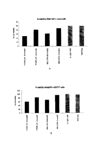
[057] Figure 33A shows results of internalisation of PTA001_A4 by MabZAP assay
in
LoVo colon cancer cells.
[058] Figure 33B shows results of internalisation of PTA001_A4 by MabZAP assay
in
LoVo colon cancer cells.
[059] Figure 33C shows results of internalisation of PTA001_A4 by MabZAP assay
in
LS174T colon cancer cells.
[060] Figure 33D shows results of internalisation of PTA001_A4 by MabZAP assay
in
LS174T colon cancer cells.
[061] Figure 34A shows results of internalisation of PTA001_A4 by MabZAP assay
in
LoVo colon cancer cells.
[062] Figure 34B shows results of internalisation of PTA001_A4 by MabZAP assay
in
LS174T colon cancer cells.
DETAILED DESCRIPTION OF THE INVENTION
[063] The present invention relates to isolated antibodies, including, but
not limited to
monoclonal antibodies, for example, which bind specifically to Cadherin-17
with high
CA 02759538 2016-08-25
WO 2010/123874 CA 02759538
PCT/US2010/031719
affinity. In certain embodiments, the antibodies of the invention comprise
particular
structural features such as CDR regions comprising particular amino acid
sequences. The
invention provides isolated antibodies, defucosylated antibodies,
immunoconjugates,
bispecific molecules, affibodies, domain antibodies, nanobodies, and
unibodies, methods of
making said molecules, and pharmaceutical compositions comprising said
molecules and a
pharmaceutical carrier. The invention also relates to methods of using the
molecules, such as
to detect Cadherin-17, as well as to treat diseases associated with expression
of Cadherin-17,
such as Cadherin-17 expressed on tumors, including those tumors of colorectal
cancer.
[064] In order that the present invention may be more readily understood,
certain terms
are first defined. Additional definitions are set forth throughout the
detailed description.
[065] The terms "Cadherin-17", "Liver-intestine cadherin", "LI-cadherin",
"Intestinal
peptide-associated transported HPT-1" and "CDH17" are used interchangeably.
Cadherin-17
has also been identified as OGTA001 in International Patent Application
W02008/026008.
Human antibodies of this disclosure may, in certain cases, cross-react with
Cadherin-17 from
species other than human. In certain embodiments, the antibodies may be
completely
specific for one or more human Cadherin-17 and may not exhibit species or
other types of
non-human cross-reactivity. The complete amino acid sequence of an exemplary
human
Cadherin-17 has Genbank accession number NM 004063.
[066] The term "immune response" refers to the action of, for example,
lymphocytes,
antigen presenting cells, phagocytic cells, granulocytes, and soluble
macromolecules
produced by the above cells or the liver (including antibodies, cytokines, and
complement)
that results in selective damage to, destruction of, or elimination from the
human body of
invading pathogens, cells or tissues infected with pathogens, cancerous cells,
or, in cases of
autoimmunity or pathological inflammation, normal human cells or tissues.
[067] A "signal transduction pathway" refers to the biochemical
relationship between
various of signal transduction molecules that play a role in the transmission
of a signal from
one portion of a cell to another portion of a cell. As used herein, the phrase
"cell surface
receptor" includes, for example, molecules and complexes of molecules capable
of receiving
a signal and the transmission of such a signal across the plasma membrane of a
cell. An
example of a -cell surface receptor" of the present invention is the Cadherin-
17 receptor.
[068] The term "antibody" as referred to herein includes whole antibodies
and any
antigen binding fragment (i.e., "antigen-binding portion") or single chains
thereof. An
"antibody" refers to a glycoprotein which may comprise at least two heavy (H)
chains and
11
CA 02759538 2016-08-25
WO 2010/123874 CA 02759538
PCT/US2010/031719
two light (L) chains inter-connected by disulfide bonds, or an antigen binding
portion thereof.
Each heavy chain is comprised of a heavy chain variable region (abbreviated
herein as VH)
and a heavy chain constant region. The heavy chain constant region is
comprised of three
domains, CHI, CH2 and CH3. Each light chain is comprised of a light chain
variable region
(abbreviated herein as VL or VK) and a light chain constant region. The light
chain constant
region is comprised of one domain, CL. The VH and VL / VK regions can be
further
subdivided into regions of hypervariability, termed complementarity
determining regions
(CDR), interspersed with regions that are more conserved, termed framework
regions (FR).
Each VH and VL / VK is composed of three CDRs and four FRs, arranged from
amino-
terminus to carboxy-terminus in the following order: FR1, CDR1, FR2, CDR2,
FR3, CDR3,
FR4. The variable regions of the heavy and light chains contain a binding
domain that
interacts with an antigen. The constant regions of the antibodies may mediate
the binding of
the immunoglobulin to host tissues or factors, including various cells of the
immune system
(e.g., effector cells) and the first component (Clq) of the classical
complement system.
1069] The definition of "antibody" includes, but is not limited to, full
length antibodies,
antibody fragments, single chain antibodies, bispecific antibodies,
minibodies, domain
antibodies, synthetic antibodies (sometimes referred to herein as -antibody
mimetics"),
chimeric antibodies, humanized antibodies, antibody fusions (sometimes
referred to as
"antibody conjugates"), and fragments of each, respectively.
[070] In one embodiment, the antibody is an antibody fragment. Specific
antibody
fragments include, but are not limited to, (i) the Fab fragment consisting of
VL, VH, CL and
CHI domains, (ii) the Fd fragment consisting of the VH and CHI domains, (iii)
the Fv
fragment consisting of the VL and VH domains of a single antibody, (iv) the
dAb fragment,
which consists of a single variable domain, (v) isolated CDR regions, (vi)
F(ab')2 fragments,
a bivalent fragment comprising two linked Fab fragments (vii) single chain Fv
molecules
(scFv), wherein a VH domain and a VL domain are linked by a peptide linker
which allows
the two domains to associate to form an antigen binding site, (viii)
bispecific single chain Fv
dimers, and (ix) "diabodies" or -triabodies", multivalent or multispecific
fragments
constructed by gene fusion. The antibody fragments may be modified. For
example, the
molecules may be stabilized by the incorporation of disulfide bridges linking
the VH and VL
domains. Examples of antibody formats and architectures are described in
Holliger &
Hudson, 2006, Nature Biotechnology 23(9): l 1 2 6 - 1 1 3 6 , and Carter 2006,
Nature Reviews
Immunology 6:343-357.
12
CA 02759538 2016-08-25
WO 2010/123874 CA 02759538
PCT/US2010/031719
[071] In one embodiment, an antibody disclosed herein may be a multispecific
antibody,
and notably a bispecific antibody, also sometimes referred to as "diabodies".
These are
antibodies that bind to two (or more) different antigens. Diabodies can be
manufactured in a
variety of ways known in the art, e.g., prepared chemically or from hybrid
hybridomas. In
one embodiment, the antibody is a minibody. Minibodies are minimized antibody-
like
proteins comprising a scFv joined to a CH3 domain. In some cases, the scFv can
be joined to
the Fc region, and may include some or all of the hinge region. For a
description of
multispecific antibodies see Holliger & Hudson, 2006, Nature Biotechnology
23(9):1126-
1 136.
[072] By "CDR" as used herein is meant a Complementarity Determining Region of
an
antibody variable domain. Systematic identification of residues included in
the CDRs have
been developed by Kabat (Kabat et al., 1991, Sequences of Proteins of
Immunological
Interest, 5th Ed., United States Public Health Service, National Institutes of
Health, Bethesda)
and alternately by Chothia (Chothia & Lesk, 1987, J. Mol. Biol. 196: 901-917;
Chothia et al.,
1989, Nature 342: 877-883; AI-Lazikani et al., 1997, J. Mol. Biol. 273: 927-
948). For the
purposes of the present invention, CDRs are defined as a slightly smaller set
of residues than
the CDRs defined by Chothia. VL CDRs are herein defined to include residues at
positions
27-32 (CDR1), 50-56 (CDR2), and 91-97 (CDR3), wherein the numbering is
according to
Chothia. Because the VL CDRs as defined by Chothia and Kabat are identical,
the
numbering of these VL CDR positions is also according to Kabat. VH CDRs are
herein
defined to include residues at positions 27-33 (CDR1), 52-56 (CDR2), and 95-
102 (CDR3),
wherein the numbering is according to Chothia. These VH CDR positions
correspond to
Kabat positions 27-35 (CDR1), 52-56 (CDR2), and 95-102 (CDR3).
1073] As will be appreciated by those in the art, the CDRs disclosed herein
may also include
variants. for example when backmutating the CDRs disclosed herein into
different framework
regions. Generally, the amino acid identity between individual variant CDRs
are at least 80%
to the sequences depicted herein, and more typically with preferably
increasing identities of
at least 85%, 90%, 91%, 92%, 93%, 94%, 95%, 96%, 97%, 98%, 99%, and almost
100%. In
a similar manner, "percent (%) nucleic acid sequence identity- with respect to
the nucleic
acid sequence of the binding proteins identified herein is defined as the
percentage of
nucleotide residues in a candidate sequence that are identical with the
nucleotide residues in
the coding sequence of the antigen binding protein. A specific method utilizes
the BLASTN
module of WU-BLAST-2 set to the default paratneters, with overlap span and
overlap
fraction set to 1 and 0.125, respectively.
13
CA 02759538 2011-10-20
WO 2010/123874
PCT/US2010/031719
[074] Generally, the nucleic acid sequence identity between the nucleotide
sequences
encoding individual variant CDRs and the nucleotide sequences depicted herein
are at least
80%, and more typically with preferably increasing identities of at least 80%,
81%, 82%,
83%, 84%, 85%, 86%, 87%, 88%, 89%, 90%, 91%, 92%, 93%, 94%, 95%, 96%, 97%,
98%,
or 99%, and almost 100%.
[075] Thus, a "variant CDR" is one with the specified homology, similarity, or
identity to
the parent CDR of the invention, and shares biological function, including,
but not limited to,
at least 80%, 81%, 82%, 83%, 84%, 85%, 86%, 87%, 88%, 89%, 90%, 91%, 92%, 93%,
94%, 95%, 96%, 97%, 98%, or 99% of the specificity and/or activity of the
parent CDR.
[076] While the site or region for introducing an amino acid sequence
variation is
predetermined, the mutation per se need not be predetermined. For example, in
order to
optimize the performance of a mutation at a given site, random mutagenesis may
be
conducted at the target codon or region and the expressed antigen binding
protein CDR
variants screened for the optimal combination of desired activity. Techniques
for making
substitution mutations at predetermined sites in DNA having a known sequence
are well
known, for example, M13 primer mutagenesis and PCR mutagenesis. Screening of
the
mutants is done using assays of antigen binding protein activities as
described herein.
[077] Amino acid substitutions are typically of single residues; insertions
usually will be on
the order of from about one (1) to about twenty (20) amino acid residues,
although
considerably larger insertions may be tolerated. Deletions range from about
one (1) to about
twenty (20) amino acid residues, although in some cases deletions may be much
larger.
[078] Substitutions, deletions, insertions or any combination thereof may be
used to arrive
at a final derivative or variant. Generally these changes are done on a few
amino acids to
minimize the alteration of the molecule, particularly the immunogenicity and
specificity of
the antigen binding protein. However, larger changes may be tolerated in
certain
circumstances.
[079] By "Fab" or "Fab region" as used herein is meant the polypeptide that
comprises the
VH, CHL VL, and CL immunoglobulin domains. Fab may refer to this region in
isolation, or
this region in the context of a full length antibody, antibody fragment or Fab
fusion protein,
or any other antibody embodiments as outlined herein.
[080] By "Fv" or "Fy fragment" or "Fy region" as used herein is meant a
polypeptide that
comprises the VL and VH domains of a single antibody.
[081] By "framework" as used herein is meant the region of an antibody
variable domain
exclusive of those regions defined as CDRs. Each antibody variable domain
framework can
14
CA 02759538 2011-10-20
WO 2010/123874
PCT/US2010/031719
be further subdivided into the contiguous regions separated by the CDRs (FR1,
FR2, FR3 and
FR4).
[082] The term "antigen-binding portion" of an antibody (or simply
"antibody portion"),
as used herein, refers to one or more fragments of an antibody that retain the
ability to
specifically bind to an antigen (e.g., Cadherin-17). It has been shown that
the antigen-
binding function of an antibody can be performed by fragments of a full-length
antibody.
Examples of binding fragments encompassed within the term "antigen-binding
portion" of an
antibody include (i) a Fab fragment, a monovalent fragment consisting of the
VL / VK, VH, CL
and CH1 domains; (ii) a F(ab')2 fragment, a bivalent fragment comprising two
Fab fragments
linked by a disulfide bridge at the hinge region; (iii) a Fab' fragment, which
is essentially an
Fab with part of the hinge region (see, FUNDAMENTAL IMMUNOLOGY (Paul ed.,
3rd ed. 1993); (iv) a Fd fragment consisting of the VH and CH 1 domains;
(v) a Fy
fragment consisting of the VL and VH domains of a single arm of an antibody;
(vi) a dAb
fragment (Ward et al., (1989) Nature 341:544-546), which consists of a VH
domain; (vii) an
isolated complementarity determining region (CDR); and (viii) a nanobody, a
heavy chain
variable region containing a single variable domain and two constant domains.
Furthermore,
although the two domains of the Fy fragment, VL / VK and VH, are coded for by
separate
genes, they can be joined, using recombinant methods, by a synthetic linker
that enables them
to be made as a single protein chain in which the VL / VK and VH regions pair
to form
monovalent molecules (known as single chain Fy (scFv); see e.g., Bird et al.
(1988) Science
242:423-426; and Huston et al. (1988) Proc. Natl. Acad. Sci. USA 85:5879-
5883). Such
single chain antibodies are also intended to be encompassed within the term
"antigen-binding
portion" of an antibody. These antibody fragments are obtained using
conventional
techniques known to those with skill in the art, and the fragments are
screened for utility in
the same manner as are intact antibodies.
[083] An "isolated antibody" as used herein, is intended to refer to an
antibody that is
substantially free of other antibodies having different antigenic
specificities (e.g., an isolated
antibody that specifically binds Cadherin-17 is substantially free of
antibodies that
specifically bind antigens other than Cadherin-17). An isolated antibody that
specifically
binds Cadherin-17 may, however, have cross-reactivity to other antigens, such
as Cadherin-
17 molecules from other species. Moreover, and/or alternatively an isolated
antibody may be
substantially free of other cellular material and/or chemicals in a form not
normally found in
nature.
CA 02759538 2011-10-20
WO 2010/123874
PCT/US2010/031719
[084] In some embodiments, the antibodies of the invention are recombinant
proteins,
isolated proteins or substantially pure proteins. An "isolated" protein is
unaccompanied by at
least some of the material with which it is normally associated in its natural
state, for example
constituting at least about 5%, or at least about 50% by weight of the total
protein in a given
sample. It is understood that the isolated protein may constitute from 5 to
99.9% by weight of
the total protein content depending on the circumstances. For example, the
protein may be
made at a significantly higher concentration through the use of an inducible
promoter or high
expression promoter, such that the protein is made at increased concentration
levels. In the
case of recombinant proteins, the definition includes the production of an
antibody in a wide
variety of organisms and/or host cells that are known in the art in which it
is not naturally
produced.
[085] The terms "monoclonal antibody" or "monoclonal antibody composition"
as used
herein refer to a preparation of antibody molecules of single molecular
composition. A
monoclonal antibody composition displays a single binding specificity and
affinity for a
particular epitope.
[086] As used herein, "isotype" refers to the antibody class (e.g., IgM or
IgG1) that is
encoded by the heavy chain constant region genes.
[087] The phrases "an antibody recognizing an antigen" and "an antibody
specific for an
antigen" are used interchangeably herein with the term "an antibody which
binds specifically
to an antigen".
[088] The term "antibody derivatives" refers to any modified form of the
antibody, e.g., a
conjugate of the antibody and another agent or antibody. For example,
antibodies of the
present invention may be conjugated to a toxin, a label, etc. The antibodies
of the present
invention may be nonhuman, chimeric, humanized, or fully human. For a
description of the
concepts of chimeric and humanized antibodies see Clark et al., 2000 and
references cited
therein (Clark, 2000, Immunol Today 21:397-402). Chimeric antibodies comprise
the
variable region of a nonhuman antibody, for example VH and VL domains of mouse
or rat
origin, operably linked to the constant region of a human antibody (see for
example U.S.
Patent No. 4,816,567). In a preferred embodiment, the antibodies of the
present invention are
humanized. By "humanized" antibody as used herein is meant an antibody
comprising a
human framework region (FR) and one or more complementarity determining
regions
(CDR's) from a non-human (usually mouse or rat) antibody. The non-human
antibody
providing the CDR's is called the "donor" and the human immunoglobulin
providing the
framework is called the "acceptor". Humanization relies principally on the
grafting of donor
16
CA 02759538 2011-10-20
WO 2010/123874
PCT/US2010/031719
CDRs onto acceptor (human) VL and VH frameworks (US Patent No, 5,225,539).
This
strategy is referred to as "CDR grafting". "Backmutation" of selected acceptor
framework
residues to the corresponding donor residues is often required to regain
affinity that is lost in
the initial grafted construct (US 5,530,101; US 5,585,089; US 5,693,761; US
5,693,762; US
6,180,370; US 5,859,205; US 5,821,337; US 6,054,297; US 6,407,213). The
humanized
antibody optimally also will comprise at least a portion of an immunoglobulin
constant
region, typically that of a human immunoglobulin, and thus will typically
comprise a human
Fc region. Methods for humanizing non-human antibodies are well known in the
art, and can
be essentially performed following the method of Winter and co-workers (Jones
et al., 1986,
Nature 321:522-525; Riechmann et cd.,1988, Nature 332:323-329; Verhoeyen et
al., 1988,
Science, 239:1534-1536). Additional examples of humanized murine monoclonal
antibodies
are also known in the art, for example antibodies binding human protein C
(O'Connor et al.,
1998, Protein Eng11:321-8), interleukin 2 receptor (Queen et al., 1989, Proc
Natl Acad Sci,
USA 86:10029-33), and human epidermal growth factor receptor 2 (Carter et al.,
1992, Proc
Natl Acad Sci USA 89:4285-9). In an alternate embodiment, the antibodies of
the present
invention may be fully human, that is the sequences of the antibodies are
completely or
substantially human. A number of methods are known in the art for generating
fully human
antibodies, including the use of transgenic mice (Bruggemann et al., 1997,
Curr Opin
Biotechnol 8:455-458) or human antibody libraries coupled with selection
methods (Griffiths
et al., 1998, Curr Opin Biotechnol 9:102-108).
[089] The term "humanized antibody" is intended to refer to antibodies in
which CDR
sequences derived from the germline of another mammalian species, such as a
mouse, have
been grafted onto human framework sequences. Additional framework region
modifications
may be made within the human framework sequences.
[090] The term "chimeric antibody" is intended to refer to antibodies in
which the
variable region sequences are derived from one species and the constant region
sequences are
derived from another species, such as an antibody in which the variable region
sequences are
derived from a mouse antibody and the constant region sequences are derived
from a human
antibody.
[091] The term "specifically binds" (or "immunospecifically binds") is not
intended to
indicate that an antibody binds exclusively to its intended target. Rather, an
antibody
"specifically binds" if its affinity for its intended target is about 5-fold
greater when
compared to its affinity for a non-target molecule. Suitably there is no
significant cross-
reaction or cross-binding with undesired substances, especially naturally
occurring proteins
17
CA 02759538 2011-10-20
WO 2010/123874
PCT/US2010/031719
or tissues of a healthy person or animal. The affinity of the antibody will,
for example, be at
least about 5 fold, such as 10 fold, such as 25-fold, especially 50-fold, and
particularly 100-
fold or more, greater for a target molecule than its affinity for a non-target
molecule. In some
embodiments, specific binding between an antibody or other binding agent and
an antigen
means a binding affinity of at least 106 M-1. Antibodies may, for example,
bind with affinities
of at least about 107 M-1, such as between about 108 M-1to about 109 M-1,
about 109 M-1 to
about 1010 M-1, or about 1010 M-1to about 1011 M-1. Antibodies may, for
example, bind with
an EC50 of 50 nM or less, 10 nM or less, 1 nM or less, 100 pM or less, or more
preferably 10
pM or less.
[092] The term "does not substantially bind" to a protein or cells, as used
herein, means
does not bind or does not bind with a high affinity to the protein or cells,
i.e. binds to the
protein or cells with a KD of 1 x 10⁻⁶ M or more, more preferably 1 x 10⁻⁵ M
or more, more
preferably 1 x 10⁻⁴ M or more, more preferably 1 x 10⁻³ M or more, even more
preferably 1 x
10-2 M or more.
[093] The term "EC50" as used herein, is intended to refer to the potency
of a compound
by quantifying the concentration that leads to 50% maximal response/effect.
EC50 may be
determined by Scratchard or FACS.
[094] The term "Kassoc" or "Ka," as used herein, is intended to refer to
the association rate
of a particular antibody-antigen interaction, whereas the term "Ichs" or "Kd,"
as used herein,
is intended to refer to the dissociation rate of a particular antibody-antigen
interaction. The
term "Kip," as used herein, is intended to refer to the dissociation constant,
which is obtained
from the ratio of Ka to Ka (i.e,. Ka/Ka) and is expressed as a molar
concentration (M). KD
values for antibodies can be determined using methods well established in the
art. A
preferred method for determining the KD of an antibody is by using surface
plasmon
resonance, preferably using a biosensor system such as a Biacore system.
[095] As used herein, the term "high affinity" for an IgG antibody refers
to an antibody
having a KD of 1 x 10⁻⁷ M or less, more preferably 5 x 10⁻⁸ M or less, even
more preferably
lx1 0-8 M or less, even more preferably 5 x 10⁻⁹ M or less and even more
preferably 1 x 10⁻⁹
M or less for a target antigen. However, "high affinity" binding can vary for
other antibody
isotypes. For example, "high affinity" binding for an IgM isotype refers to an
antibody
having a KD of 10-6 M or less, more preferably 10-7 M or less, even more
preferably 10-8 M or
less.
[096] The term "epitope" or "antigenic determinant" refers to a site on an
antigen to
which an immunoglobulin or antibody specifically binds. Epitopes can be formed
both from
18
CA 02759538 2011-10-20
WO 2010/123874
PCT/US2010/031719
contiguous amino acids or noncontiguous amino acids juxtaposed by tertiary
folding of a
protein. Epitopes formed from contiguous amino acids are typically retained on
exposure to
denaturing solvents, whereas epitopes formed by tertiary folding are typically
lost on
treatment with denaturing solvents. An epitope typically includes at least 3,
4, 5, 6, 7, 8, 9,
10, 11, 12, 13, 14 or 15 amino acids in a unique spatial conformation. Methods
of
determining spatial conformation of epitopes include techniques in the art and
those
described herein, for example, x-ray crystallography and 2-dimensional nuclear
magnetic
resonance (see, e.g., Epitope Mapping Protocols in Methods in Molecular
Biology, Vol. 66,
G. E. Morris, Ed. (1996)).
[097] Accordingly, also encompassed by the present invention are antibodies
that bind to
(i.e., recognize) the same epitope as the antibodies described herein (i.e.,
PTA001_A1,
PTA001 A2, PTA001_A3, PTA001 A4, PTA001_A5, PTA001 A6, PTA001_A7,
PTA001 A8, PTA001_A9, PTA001 A10, PTA001 All, PTA001 Al2, PTA001 Al3 and
PTA001 A14). Antibodies that bind to the same epitope can be identified by
their ability to
cross-compete with (i.e., competitively inhibit binding of) a reference
antibody to a target
antigen in a statistically significant manner. Competitive inhibition can
occur, for example, if
the antibodies bind to identical or structurally similar epitopes (e.g.,
overlapping epitopes), or
spatially proximal epitopes which, when bound, causes steric hindrance between
the
antibodies.
[098] Competitive inhibition can be determined using routine assays in
which the
immunoglobulin under test inhibits specific binding of a reference antibody to
a common
antigen. Numerous types of competitive binding assays are known, for example:
solid phase
direct or indirect radioimmunoassay (RIA), solid phase direct or indirect
enzyme
immunoassay (EIA), sandwich competition assay (see Stahli et al., Methods in
Enzymology
9:242 (1983)); solid phase direct biotin-avidin EIA (see Kirkland et al., J.
Immunol. 137:3614
(1986)); solid phase direct labeled assay, solid phase direct labeled sandwich
assay (see
Harlow and Lane, Antibodies: A Laboratory Manual, Cold Spring Harbor Press
(1988));
solid phase direct label RIA using 1-125 label (see Morel et al., Mol.
Immunol. 25(1):7
(1988)); solid phase direct biotin-avidin EIA (Cheung et al., Virology 176:546
(1990)); and
direct labeled RIA. (Moldenhauer et al., Scand. J. Immunol. 32:77 (1990)).
Typically, such
an assay involves the use of purified antigen bound to a solid surface or
cells bearing either of
these, an unlabeled test immunoglobulin and a labeled reference
immunoglobulin.
Competitive inhibition is measured by determining the amount of label bound to
the solid
surface or cells in the presence of the test immunoglobulin. Usually the test
immunoglobulin
19
CA 02759538 2011-10-20
WO 2010/123874
PCT/US2010/031719
is present in excess. Usually, when a competing antibody is present in excess,
it will inhibit
specific binding of a reference antibody to a common antigen by at least 50-
55%, 55-60%,
60-65%, 65-70% 70-75% or more.
[099] Other techniques include, for example, epitope mapping methods, such
as x-ray
analyses of crystals of antigen:antibody complexes which provides atomic
resolution of the
epitope. Other methods monitor the binding of the antibody to antigen
fragments or mutated
variations of the antigen where loss of binding due to a modification of an
amino acid residue
within the antigen sequence is often considered an indication of an epitope
component. In
addition, computational combinatorial methods for epitope mapping can also be
used. These
methods rely on the ability of the antibody of interest to affinity isolate
specific short peptides
from combinatorial phage display peptide libraries. The peptides are then
regarded as leads
for the definition of the epitope corresponding to the antibody used to screen
the peptide
library. For epitope mapping, computational algorithms have also been
developed which
have been shown to map conformational discontinuous epitopes.
[0100] As used herein, the term "subject" includes any human or nonhuman
animal. The
term "nonhuman animal" includes all vertebrates, e.g., mammals and non-
mammals, such as
nonhuman primates, sheep, dogs, cats, horses, cows, chickens, amphibians,
reptiles, etc.
[0101] Various aspects of the invention are described in further detail in the
following
subsections.
Anti-Cadherin-17 Antibodies
[0102] The antibodies of the invention are characterized by particular
functional features
or properties of the antibodies. For example, the antibodies bind specifically
to human
Cadherin-17. Preferably, an antibody of the invention binds to Cadherin-17
with high
affinity, for example with a KD of 8 x 10⁻⁷ M or less, even more typically 1 x
10-8 M or less.
The anti-Cadherin-17 antibodies of the invention preferably exhibit one or
more of the
following characteristics:
binds to human Cadherin-17 with a EC50 of 50 nM or less, 10 nM or less, 1 nM
or
less, 100 pM or less, or more preferably 10 pM or less;
binds to human cells expressing Cadherin-17.
[0103] In one embodiment, the antibodies preferably bind to an antigenic
epitope present
in Cadherin-17, which epitope is not present in other proteins. The antibodies
typically bind
Cadherin-17 but does not bind to other proteins, or binds to proteins with a
low affinity, such
as a KD of 1 x 10⁻⁶ M or more, more preferably 1 x 10⁻⁵ M or more, more
preferably 1 x 10⁻⁴
M or more, more preferably 1 x 10⁻³ M or more, even more preferably 1 x 10⁻² M
or more.
CA 02759538 2011-10-20
WO 2010/123874
PCT/US2010/031719
Preferably, the antibodies do not bind to related proteins, for example, the
antibodies do not
substantially bind to other cell adhesion molecules. In one embodiment, the
antibody may be
internalized into a cell expressing Cadherin-17. Standard assays to evaluate
antibody
internalization are known in the art, including, for example, a HumZap
internalization assay.
[0104] Standard assays to evaluate the binding ability of the antibodies
toward Cadherin-
17 are known in the art, including for example, ELISAs, Western blots, RIAs,
and flow
cytometry analysis. Suitable assays are described in detail in the Examples.
The binding
kinetics (e.g., binding affinity) of the antibodies also can be assessed by
standard assays
known in the art, such as by Biacore system analysis. To assess binding to
Raji or Daudi B
cell tumor cells, Raji (ATCC Deposit No. CCL-86) or Daudi (ATCC Deposit No.
CCL-213)
cells can be obtained from publicly available sources, such as the American
Type Culture
Collection, and used in standard assays, such as flow cytometric analysis.
Monoclonal Antibodies Of The Invention
[0105] Preferred antibodies of the invention are the monoclonal antibodies
PTA001_A1,
PTA001 A2, PTA001_A3, PTA001_A4, PTA001_A5, PTA001_A6, PTA001_A7,
PTA001 A8, PTA001_A9, PTA001_A10, PTA001_A11, PTA001_Al2, PTA001_A13 and
PTA001 A14, isolated and structurally characterized as described in Examples 1-
6. The VH
amino acid sequences of PTA001_Al, PTA001_A2, PTA001_A3, PTA001_A4,
PTA001 A5, PTA001_A6, PTA001_A7, PTA001_A8, PTA001_A9, PTA001_A10,
PTA001 All, PTA001 Al2, PTA001 Al3 and PTA001 Al4 are shown in SEQ ID
NOs:35-46. The VK amino acid sequences of PTA001_Al, PTA001_A2, PTA001_A3,
PTA001 A4, PTA001_A5, PTA001_A6, PTA001_A7, PTA001_A8, PTA001_A9,
PTA001 A10, PTA001 All, PTA001 Al2, PTA001 Al3 and PTA001 Al4 are shown in
SEQ ID NOs:47-58.
[0106] Given that each of these antibodies can bind to Cadherin-17, the VH and
VK
sequences can be "mixed and matched" to create other anti-Cadherin-17 binding
molecules of
the invention. Cadherin-17 binding of such "mixed and matched" antibodies can
be tested
using the binding assays described above and in the Examples (e.g., ELISAs).
Preferably,
when VH and VK chains are mixed and matched, a VH sequence from a particular
VH/VK
pairing is replaced with a structurally similar VH sequence. Likewise,
preferably a VK
sequence from a particular VHNK pairing is replaced with a structurally
similar VK sequence.
[0107] Accordingly, in one aspect, the invention provides an antibody,
comprising:
a heavy chain variable region comprising an amino acid sequence set forth in a
SEQ ID NO:
selected from the group consisting of 38, 35, 36, 37, 39, 40, 41, 42, 43, 44,
45 and 46 and a
21
CA 02759538 2011-10-20
WO 2010/123874
PCT/US2010/031719
light chain variable region comprising an amino acid sequence set forth in a
SEQ ID NO:
selected from the group consisting of 49, 47, 48, 50, 51, 52, 53, 54, 55, 56,
57 and 58;
wherein the antibody specifically binds Cadherin-17, preferably human Cadherin-
17.
[0108] Preferred heavy and light chain combinations include:
a heavy chain variable region comprising the amino acid sequence of SEQ ID
NO:35 and a
light chain variable region comprising the amino acid sequence of SEQ ID
NO:47; or
a heavy chain variable region comprising the amino acid sequence of SEQ ID
NO:36; and a
light chain variable region comprising the amino acid sequence of SEQ ID
NO:48, or
a heavy chain variable region comprising the amino acid sequence of SEQ ID
NO:37; and a
light chain variable region comprising the amino acid sequence of SEQ ID
NO:48, or
a heavy chain variable region comprising the amino acid sequence of SEQ ID
NO:38; and a
light chain variable region comprising the amino acid sequence of SEQ ID
NO:49, or
a heavy chain variable region comprising the amino acid sequence of SEQ ID
NO:39; and a
light chain variable region comprising the amino acid sequence of SEQ ID
NO:50, or
a heavy chain variable region comprising the amino acid sequence of SEQ ID
NO:40; and a
light chain variable region comprising the amino acid sequence of SEQ ID
NO:51, or
a heavy chain variable region comprising the amino acid sequence of SEQ ID
NO:41; and a
light chain variable region comprising the amino acid sequence of SEQ ID
NO:52, or
a heavy chain variable region comprising the amino acid sequence of SEQ ID
NO:42; and a
light chain variable region comprising the amino acid sequence of SEQ ID
NO:53, or
a heavy chain variable region comprising the amino acid sequence of SEQ ID
NO:40; and a
light chain variable region comprising the amino acid sequence of SEQ ID
NO:54, or
a heavy chain variable region comprising the amino acid sequence of SEQ ID
NO:40; and a
light chain variable region comprising the amino acid sequence of SEQ ID
NO:55, or
a heavy chain variable region comprising the amino acid sequence of SEQ ID
NO:43; and a
light chain variable region comprising the amino acid sequence of SEQ ID
NO:56, or
a heavy chain variable region comprising the amino acid sequence of SEQ ID
NO:44; and a
light chain variable region comprising the amino acid sequence of SEQ ID
NO:55, or
a heavy chain variable region comprising the amino acid sequence of SEQ ID
NO:45; and a
light chain variable region comprising the amino acid sequence of SEQ ID
NO:57, or
a heavy chain variable region comprising the amino acid sequence of SEQ ID
NO:46; and a
light chain variable region comprising the amino acid sequence of SEQ ID
NO:58.
[0109] In another aspect, the invention provides antibodies that comprise the
heavy chain
and light chain CDR1s, CDR2s and CDR3s of PTA001_A 1, PTA001_A2, PTA001_A3,
22
CA 02759538 2011-10-20
WO 2010/123874
PCT/US2010/031719
PTA001_A4, PTA001_A5, PTA001_A6, PTA001_A7, PTA001_A8, PTA001_A9,
PTA001_A10, PTA001_A11, PTA001_Al2, PTA001_A13 and PTA001_A14, or
combinations thereof The amino acid sequences of the VH CDR1s of PTA001_Al,
PTA001_A2, PTA001_A3, PTA001_A4, PTA001_A5, PTA001_A6, PTA001_A7,
PTA001_A8, PTA001_A9, PTA001_A10, PTA001_A11, PTA001_Al2, PTA001_A13 and
PTA001_A14 are shown in SEQ ID NOs: 1-4. The amino acid sequences of the VH
CDR2s
of PTA001 Al, PTA001 A2, PTA001_A3, PTA001_A4, PTA001 A5, PTA001 A6,
PTA001_A7, PTA001_A8, PTA001_A9, PTA001_A10, PTA001_All, PTA001_Al2,
PTA001_A13 and PTA001_A14 are shown in SEQ ID NOs: 5-13. The amino acid
sequences of the VH CDR3s of PTA001_Al, PTA001_A2, PTA001_A3, PTA001_A4,
PTA001_A5, PTA001_A6, PTA001_A7, PTA001_A8, PTA001_A9, PTA001_A10,
PTA001_A11, PTA001_Al2, PTA001_A13 and PTA001_A14 are shown in SEQ ID
NOs:14-21. The amino acid sequences of the VK CDR1s of PTA001_Al, PTA001_A2,
PTA001_A3, PTA001_A4, PTA001_A5, PTA001_A6, PTA001_A7, PTA001_A8,
PTA001_A9, PTA001_A10, PTA001_A11, PTA001_Al2, PTA001_A13 and PTA001_A14
are shown in SEQ ID NOs:22-27. The amino acid sequences of the VK CDR2s of
PTA001_Al, PTA001_A2, PTA001_A3, PTA001_A4, PTA001_A5, PTA001_A6,
PTA001_A7, PTA001_A8, PTA001_A9, PTA001_A10, PTA001_All, PTA001_Al2,
PTA001_A13 and PTA001_A14 are shown in SEQ ID NOs:28-31. The amino acid
sequences of the VK CDR3s of PTA001_Al, PTA001_A2, PTA001_A3, PTA001_A4,
PTA001_A5, PTA001_A6, PTA001_A7, PTA001_A8, PTA001_A9, PTA001_A10,
PTA001_A11, PTA001_Al2, PTA001_A13 and PTA001_A14 are shown in SEQ ID
NOs:32-34. The CDR regions are delineated using the Kabat system (Kabat, E.
A., et al.
(1991) Sequences of Proteins of Immunological Interest, Fifth Edition, U.S.
Department of
Health and Human Services, NIH Publication No. 91-3242).
[0110] Given that each of these antibodies can bind to Cadherin-17 and that
antigen-
binding specificity is provided primarily by the CDR1, CDR2, and CDR3 regions,
the VH
CDR1, CDR2, and CDR3 sequences and VK CDR1, CDR2, and CDR3 sequences can be
"mixed and matched" (i.e., CDRs from different antibodies can be mixed and
matched,
although each antibody generally contains a VH CDR1, CDR2, and CDR3 and a VK
CDR1,
CDR2, and CDR3) to create other anti-Cadherin-17 binding molecules of the
invention.
Accordingly, the invention specifically includes every possible combination of
CDRs of the
heavy and light chains.
23
CA 02759538 2011-10-20
WO 2010/123874
PCT/US2010/031719
[0111] Cadherin-17 binding of such "mixed and matched" antibodies can be
tested using the
binding assays described above and in the Examples (e.g., ELISAs, Biacore
analysis).
Preferably, when VH CDR sequences are mixed and matched, the CDR1, CDR2 and/or
CDR3 sequence from a particular VH sequence is replaced with a structurally
similar CDR
sequence(s). Likewise, when VK CDR sequences are mixed and matched, the CDR1,
CDR2
and/or CDR3 sequence from a particular VK sequence preferably is replaced with
a
structurally similar CDR sequence(s). It will be readily apparent to the
ordinarily skilled
artisan that novel VH and VK sequences can be created by substituting one or
more VH and/or
VL / VK CDR region sequences with structurally similar sequences from the CDR
sequences
disclosed herein for monoclonal antibodies PTA001_A1, PTA001_A2, PTA001_A3,
PTA001 A4, PTA001_A5, PTA001 A6, PTA001_A7, PTA001 A8, PTA001_A9,
PTA001 A10, PTA001 All, PTA001 Al2, PTA001 Al3 and PTA001 A14.
[0112] Accordingly, in another aspect, the invention provides an isolated
monoclonal
antibody, or antigen binding portion thereof comprising:
a heavy chain variable region CDR1 comprising an amino acid sequence selected
from the
group consisting of SEQ ID NOs:1-4;
a heavy chain variable region CDR2 comprising an amino acid sequence selected
from the
group consisting of SEQ ID NOs:5-13;
a heavy chain variable region CDR3 comprising an amino acid sequence selected
from the
group consisting of SEQ ID NOs:14-21;
a light chain variable region CDR1 comprising an amino acid sequence selected
from the
group consisting of SEQ ID NOs:22-27;
a light chain variable region CDR2 comprising an amino acid sequence selected
from the
group consisting of SEQ ID NOs:28-31; and
a light chain variable region CDR3 comprising an amino acid sequence selected
from the
group consisting of SEQ ID NOs:32-34;
with all possible combinations being possible, wherein the antibody
specifically binds
Cadherin-17, preferably human Cadherin-17
[0113] In a preferred embodiment, the antibody comprises:
a heavy chain variable region CDR1 comprising SEQ ID NO:1;
a heavy chain variable region CDR2 comprising SEQ ID NO:5;
a heavy chain variable region CDR3 comprising SEQ ID NO:14;
a light chain variable region CDR1 comprising SEQ ID NO:22;
24
CA 02759538 2011-10-20
WO 2010/123874
PCT/US2010/031719
a light chain variable region CDR2 comprising SEQ ID NO:28; and
a light chain variable region CDR3 comprising SEQ ID NO:32.
[0114] In another preferred embodiment, the antibody comprises:
a heavy chain variable region CDR1 comprising SEQ ID NO:2;
a heavy chain variable region CDR2 comprising SEQ ID NO:6;
a heavy chain variable region CDR3 comprising SEQ ID NO:15;
a light chain variable region CDR1 comprising SEQ ID NO:23;
a light chain variable region CDR2 comprising SEQ ID NO:29; and
a light chain variable region CDR3 comprising SEQ ID NO:33.
[0115] In another preferred embodiment, the antibody comprises:
a heavy chain variable region CDR1 comprising SEQ ID NO:3;
a heavy chain variable region CDR2 comprising SEQ ID NO:7;
a heavy chain variable region CDR3 comprising SEQ ID NO:16;
a light chain variable region CDR1 comprising SEQ ID NO:23;
a light chain variable region CDR2 comprising SEQ ID NO:29; and
a light chain variable region CDR3 comprising SEQ ID NO:33.
[0116] In another preferred embodiment, the antibody comprises:
a heavy chain variable region CDR1 comprising SEQ ID NO:4;
a heavy chain variable region CDR2 comprising SEQ ID NO:8;
a heavy chain variable region CDR3 comprising SEQ ID NO:17;
a light chain variable region CDR1 comprising SEQ ID NO:24;
a light chain variable region CDR2 comprising SEQ ID NO:30; and
a light chain variable region CDR3 comprising SEQ ID NO:34.
[0117] In another preferred embodiment, the antibody comprises:
a heavy chain variable region CDR1 comprising SEQ ID NO:3;
a heavy chain variable region CDR2 comprising SEQ ID NO:7;
a heavy chain variable region CDR3 comprising SEQ ID NO:18;
a light chain variable region CDR1 comprising SEQ ID NO:25;
a light chain variable region CDR2 comprising SEQ ID NO:31; and
a light chain variable region CDR3 comprising SEQ ID NO:33.
[0118] In another preferred embodiment, the antibody comprises:
a heavy chain variable region CDR1 comprising SEQ ID NO:3;
a heavy chain variable region CDR2 comprising SEQ ID NO:9;
a heavy chain variable region CDR3 comprising SEQ ID NO:19;
CA 02759538 2011-10-20
WO 2010/123874
PCT/US2010/031719
a light chain variable region CDR1 comprising SEQ ID NO:26;
a light chain variable region CDR2 comprising SEQ ID NO:29; and
a light chain variable region CDR3 comprising SEQ ID NO:33.
[0119] In another preferred embodiment, the antibody comprises:
a heavy chain variable region CDR1 comprising SEQ ID NO:3;
a heavy chain variable region CDR2 comprising SEQ ID NO:7;
a heavy chain variable region CDR3 comprising SEQ ID NO:16;
a light chain variable region CDR1 comprising SEQ ID NO:25;
a light chain variable region CDR2 comprising SEQ ID NO:31; and
a light chain variable region CDR3 comprising SEQ ID NO:33.
[0120] In another preferred embodiment, the antibody comprises:
a heavy chain variable region CDR1 comprising SEQ ID NO:3;
a heavy chain variable region CDR2 comprising SEQ ID NO:7;
a heavy chain variable region CDR3 comprising SEQ ID NO:16;
a light chain variable region CDR1 comprising SEQ ID NO:25;
a light chain variable region CDR2 comprising SEQ ID NO:29; and
a light chain variable region CDR3 comprising SEQ ID NO:33.
[0121] In another preferred embodiment, the antibody comprises:
a heavy chain variable region CDR1 comprising SEQ ID NO:3;
a heavy chain variable region CDR2 comprising SEQ ID NO:10;
a heavy chain variable region CDR3 comprising SEQ ID NO:20;
a light chain variable region CDR1 comprising SEQ ID NO:27;
a light chain variable region CDR2 comprising SEQ ID NO:29; and
a light chain variable region CDR3 comprising SEQ ID NO:33.
[0122] In another preferred embodiment, the antibody comprises:
a heavy chain variable region CDR1 comprising SEQ ID NO:3;
a heavy chain variable region CDR2 comprising SEQ ID NO:11;
a heavy chain variable region CDR3 comprising SEQ ID NO:21;
a light chain variable region CDR1 comprising SEQ ID NO:25;
a light chain variable region CDR2 comprising SEQ ID NO:29; and
a light chain variable region CDR3 comprising SEQ ID NO:33.
[0123] In another preferred embodiment, the antibody comprises:
a heavy chain variable region CDR1 comprising SEQ ID NO:3;
a heavy chain variable region CDR2 comprising SEQ ID NO:12;
26
CA 02759538 2011-10-20
WO 2010/123874
PCT/US2010/031719
a heavy chain variable region CDR3 comprising SEQ ID NO:18;
a light chain variable region CDR1 comprising SEQ ID NO:25;
a light chain variable region CDR2 comprising SEQ ID NO:31; and
a light chain variable region CDR3 comprising SEQ ID NO:33.
[0124] In another preferred embodiment, the antibody comprises:
a heavy chain variable region CDR1 comprising SEQ ID NO:2;
a heavy chain variable region CDR2 comprising SEQ ID NO:13;
a heavy chain variable region CDR3 comprising SEQ ID NO:15;
a light chain variable region CDR1 comprising SEQ ID NO:23;
a light chain variable region CDR2 comprising SEQ ID NO:29; and
a light chain variable region CDR3 comprising SEQ ID NO:33.
[0125] It is well known in the art that the CDR3 domain, independently from
the CDR1
and/or CDR2 domain(s), alone can determine the binding specificity of an
antibody for a
cognate antigen and that multiple antibodies can predictably be generated
having the same
binding specificity based on a common CDR3 sequence. See, for example, Klimka
et al.,
British J. of Cancer 83(2):252-260 (2000) (describing the production of a
humanized anti-
CD30 antibody using only the heavy chain variable domain CDR3 of murine anti-
CD30
antibody Ki-4); Beiboer et al., J. Mol. Biol. 296:833-849 (2000) (describing
recombinant
epithelial glycoprotein-2 (EGP-2) antibodies using only the heavy chain CDR3
sequence of
the parental murine MOC-31 anti-EGP-2 antibody); Rader et al., Proc. Natl.
Acad. Sci.
U.S.A. 95:8910-8915 (1998) (describing a panel of humanized anti-integrin
ct,133 antibodies
using a heavy and light chain variable CDR3 domain of a murine anti-integrin
ct,133 antibody
LM609 wherein each member antibody comprises a distinct sequence outside the
CDR3
domain and capable of binding the same epitope as the parent murine antibody
with affinities
as high or higher than the parent murine antibody); Barbas et al., J. Am.
Chem. Soc.
116:2161-2162 (1994) (disclosing that the CDR3 domain provides the most
significant
contribution to antigen binding); Barbas et al., Proc. Natl. Acad. Sci. U.S.A.
92:2529-2533
(1995) (describing the grafting of heavy chain CDR3 sequences of three Fabs
(SI-1, SI-40,
and SI-32) against human placental DNA onto the heavy chain of an anti-tetanus
toxoid Fab
thereby replacing the existing heavy chain CDR3 and demonstrating that the
CDR3 domain
alone conferred binding specificity); and Ditzel et al., J. Immunol. 157:739-
749 (1996)
(describing grafting studies wherein transfer of only the heavy chain CDR3 of
a parent
polyspecific Fab LNA3 to a heavy chain of a monospecific IgG tetanus toxoid-
binding Fab
27
CA 02759538 2016-08-25
WO 2010/123874 CA 02759538
PCT/US2010/031719
p313 antibody was sufficient to retain binding specificity of the parent Fab).
[0126] Accordingly, the present invention provides monoclonal antibodies
comprising one
or more heavy and/or light chain CDR3 domains from an antibody derived from a
human or
non-human animal, wherein the monoclonal antibody is capable of specifically
binding to
Cadherin-17. Within certain aspects, the present invention provides monoclonal
antibodies
comprising one or more heavy and/or light chain CDR3 domain from a non-human
antibody,
such as a mouse or rat antibody, wherein the monoclonal antibody is capable of
specifically
binding to Cadherin-17. Within some embodiments, such inventive antibodies
comprising
one or more heavy and/or light chain CDR3 domain from a non-human antibody (a)
are
capable of competing for binding with; (b) retain the functional
characteristics; (c) bind to the
same epitope; and/or (d) have a similar binding affinity as the corresponding
parental non-
human antibody.
[0127] Within other aspects, the present invention provides monoclonal
antibodies
comprising one or more heavy and/or light chain CDR3 domains from a human
antibody,
such as, for example, a human antibody obtained from a non-human animal,
wherein the
human antibody is capable of specifically binding to Cadherin-17. Within other
aspects, the
present invention provides monoclonal antibodies comprising one or more heavy
and/or light
chain CDR3 domain from a first human antibody, such as, for example, a human
antibody
obtained from a non-human animal, wherein the first human antibody is capable
of
specifically binding to Cadherin-17 and wherein the CDR3 domain from the first
human
antibody replaces a CDR3 domain in a human antibody that is lacking binding
specificity for
Cadherin-17 to generate a second human antibody that is capable of
specifically binding to
Cadherin-17. Within some embodiments, such inventive antibodies comprising one
or more
heavy and/or light chain CDR3 domain from the first human antibody (a) are
capable of
competing for binding with; (b) retain the functional characteristics; (c)
bind to the same
epitope; and/or (d) have a similar binding affinity as the corresponding
parental first human
antibody.
Antibodies Having Particular Germline Sequences
[0128] In certain embodiments, an antibody of the invention comprises a
heavy chain
variable region from a particular germline heavy chain immunoglobulin gene
and/or a light
chain variable region from a particular germline light chain immunoglobulin
gene.
[0129] For example, in a preferred embodiment, the invention provides an
isolated
monoclonal antibody, or an antigen-binding portion thereof, comprising a heavy
chain
28
CA 02759538 2011-10-20
WO 2010/123874
PCT/US2010/031719
variable region that is the product of or derived from a murineVH 7-39 gene, a
murine VH II
region VH105 gene or a murine VH II gene H17, wherein the antibody
specifically binds
Cadherin-17. In yet another preferred embodiment, the invention provides an
isolated
monoclonal antibody, or an antigen-binding portion thereof, comprising a light
chain variable
region that is the product of or derived from a murine VK 1-110 gene, a murine
VK 8-30 gene
or a murine VK 24-140 gene, wherein the antibody specifically binds Cadherin-
17.
[0130] In yet another preferred embodiment, the invention provides an isolated
monoclonal antibody, or antigen-binding portion thereof, wherein the antibody:
comprises a heavy chain variable region that is the product of or derived from
a murine VH 7-
39 gene (which gene includes the nucleotide sequence set forth in SEQ ID NO:
125);
comprises a light chain variable region that is the product of or derived from
a murine VK 1-
110 gene (which gene includes the nucleotide sequences set forth in SEQ ID
NOs: 128 and
129); andspecifically binds to Cadherin-17, preferably human Cadherin-17.
Examples of an
antibody having VH and VK of VH 7-39 and VK 1-110, respectively, is PTA001_A1.
[0131] In yet another preferred embodiment, the invention provides an isolated
monoclonal antibody, or antigen-binding portion thereof, wherein the antibody:
comprises a heavy chain variable region that is the product of or derived from
a murine VH II
gene H17 or a murine VH II region VH105 gene (which genes include the
nucleotide
sequences set forth in SEQ ID NO: 126 and 127 respectively);
comprises a light chain variable region that is the product of or derived from
a murine VK 8-
30 gene (which gene includes the nucleotide sequences set forth in SEQ ID NOs:
130, 131
and 132); and
specifically binds to Cadherin-17, preferably human Cadherin-17.
[0132] Examples of an antibody having VH of VH II gene H17 or VH II region
VH105 and VK
of VK 8-30 is PTA001_A4.
[0133] In yet another preferred embodiment, the invention provides an isolated
monoclonal antibody, or antigen-binding portion thereof, wherein the antibody:
comprises a heavy chain variable region that is the product of or derived from
a murine VH II
gene H17 or a murine VH II region VH105 gene (which genes include the
nucleotide
sequences set forth in SEQ ID NO: 126 and 127 respectively);
comprises a light chain variable region that is the product of or derived from
a murine VK 24-
140 gene (which gene includes the nucleotide sequences set forth in SEQ ID
NOs: 133, 134
and 135); and
specifically binds to Cadherin-17, preferably human Cadherin-17. Examples of
antibodies
29
CA 02759538 2011-10-20
WO 2010/123874
PCT/US2010/031719
having VH of VH II gene H17 or VH II region VH105 and VK of VK 24-140 are
PTA001_A2,
PTA001 A3, PTA001_A5, PTA001 A6, PTA001_A7, PTA001 A8, PTA001_A9,
PTA001 A10, PTA001 All, PTA001 Al2, PTA001 Al3 and PTA001 A14.
[0134] As used herein, an antibody comprises heavy or light chain variable
regions that is
"the product of" or "derived from" a particular germline sequence if the
variable regions of
the antibody are obtained from a system that uses murine germline
immunoglobulin genes.
Such systems include screening a murine immunoglobulin gene library displayed
on phage
with the antigen of interest. An antibody that is "the product of" or "derived
from" a murine
germline immunoglobulin sequence can be identified as such by comparing the
nucleotide or
amino acid sequence of the antibody to the nucleotide or amino acid sequences
of murine
germline immunoglobulins and selecting the murine germline immunoglobulin
sequence that
is closest in sequence (i.e., greatest % identity) to the sequence of the
antibody. An antibody
that is "the product of" or "derived from" a particular murine germline
immunoglobulin
sequence may contain amino acid differences as compared to the germline
sequence, due to,
for example, naturally-occurring somatic mutations or intentional introduction
of site-directed
mutation. However, a selected antibody typically is at least 90% identical in
amino acids
sequence to an amino acid sequence encoded by a murine germline immunoglobulin
gene
and contains amino acid residues that identify the antibody as being murine
when compared
to the germline immunoglobulin amino acid sequences of other species (e.g.,
human germline
sequences). In certain cases, an antibody may be at least 95%, or even at
least 96%, 97%,
98%, or 99% identical in amino acid sequence to the amino acid sequence
encoded by the
germline immunoglobulin gene. Typically, an antibody derived from a particular
murine
germline sequence will display no more than 10 amino acid differences from the
amino acid
sequence encoded by the murine germline immunoglobulin gene. In certain cases,
the
antibody may display no more than 5, or even no more than 4, 3, 2, or 1 amino
acid
difference from the amino acid sequence encoded by the germline immunoglobulin
gene.
Homologous Antibodies
[0135] In yet another embodiment, an antibody of the invention comprises heavy
and light
chain variable regions comprising amino acid sequences that are homologous to
the amino
acid sequences of the preferred antibodies described herein, and wherein the
antibodies retain
the desired functional properties of the anti-Cadherin-17 antibodies of the
invention.
For example, the invention provides an isolated monoclonal antibody, or
antigen binding
portion thereof, comprising a heavy chain variable region and a light chain
variable region,
wherein:
CA 02759538 2011-10-20
WO 2010/123874
PCT/US2010/031719
the heavy chain variable region comprises an amino acid sequence that is at
least
80% identical to an amino acid sequence selected from the group consisting of
SEQ ID
NOs:35-46;
the light chain variable region comprises an amino acid sequence that is at
least 80%
identical to an amino acid sequence selected from the group consisting of SEQ
ID NOs:47-
58; and
the antibody binds to human Cadherin-17. Such antibodies may bind to human
Cadherin-17 with an EC50 of 50 nM or less, 10 nM or less, 1 nM or less, 100 pM
or less, or
more preferably 10 pM or less.
[0136] The antibody may also bind to CHO cells transfected with human Cadherin-
17.
[0137] In various embodiments, the antibody can be, for example, a human
antibody, a
humanized antibody or a chimeric antibody.
[0138] In other embodiments, the VH and/or VK amino acid sequences may be 85%,
90%,
95%, 96%, 97%, 98% or 99% homologous to the sequences set forth above. An
antibody
having VH and VK regions having high (i.e., 80% or greater) identical to the
VH and VK
regions of the sequences set forth above, can be obtained by mutagenesis
(e.g., site-directed
or PCR-mediated mutagenesis) of nucleic acid molecules encoding SEQ ID NOs:59-
83
followed by testing of the encoded altered antibody for retained function
using the functional
assays described herein.
[0139] The percent identity between the two sequences is a function of the
number of
identical positions shared by the sequences (i.e., % homology = # of identical
positions/total
# of positions x 100), taking into account the number of gaps, and the length
of each gap,
which need to be introduced for optimal alignment of the two sequences. The
comparison of
sequences and determination of percent identity between two sequences can be
accomplished
using a mathematical algorithm, as described in the non-limiting examples
below.
[0140] The percent identity between two amino acid sequences can be determined
using
the algorithm of E. Meyers and W. Miller (Comput. AppL Biosci., 4:11-17
(1988)) which has
been incorporated into the ALIGN program (version 2.0), using a PAM120 weight
residue
table, a gap length penalty of 12 and a gap penalty of 4. In addition, the
percent identity
between two amino acid sequences can be determined using the Needleman and
Wunsch (J.
Mol. Biol. 48:444-453 (1970)) algorithm which has been incorporated into the
GAP program
in the GCG software package (available at http://www.gcg.com), using either a
Blossum 62
matrix or a PAM250 matrix, and a gap weight of 16, 14, 12, 10, 8, 6, or 4 and
a length weight
of 1, 2, 3, 4, 5, or 6.
31
CA 02759538 2011-10-20
WO 2010/123874
PCT/US2010/031719
[0141] Additionally or alternatively, the protein sequences of the present
invention can
further be used as a "query sequence" to perform a search against public
databases to, for
example, identify related sequences. Such searches can be performed using the
XBLAST
program (version 2.0) of Altschul, et al. (1990) J. Mol. Biol. 215:403-10.
BLAST protein
searches can be performed with the XBLAST program, score = 50, wordlength = 3
to obtain
amino acid sequences homologous to the antibody molecules of the invention. To
obtain
gapped alignments for comparison purposes, Gapped BLAST can be utilized as
described in
Altschul et al., (1997) Nucleic Acids Res. 25(17):3389-3402. When utilizing
BLAST and
Gapped BLAST programs, the default parameters of the respective programs
(e.g., XBLAST
and NBLAST) can be used. See www.ncbi.nlm.nih.gov.
Antibodies with Conservative Modifications
[0142] In certain embodiments, an antibody of the invention comprises a heavy
chain
variable region comprising CDR1, CDR2 and CDR3 sequences and a light chain
variable
region comprising CDR1, CDR2 and CDR3 sequences, wherein one or more of these
CDR
sequences comprise specified amino acid sequences based on the preferred
antibodies
described herein (e.g., PTA001_Al, PTA001_A2, PTA001_A3, PTA001_A4, PTA001_A5,
PTA001 A6, PTA001_A7, PTA001 A8, PTA001_A9, PTA001 A10, PTA001 All,
PTA001 Al2, PTA001 Al3 or PTA001 A14), or conservative modifications thereof,
and
wherein the antibodies retain the desired functional properties of the anti-
Cadherin-17
antibodies of the invention. Accordingly, the invention provides an isolated
monoclonal
antibody, or antigen binding portion thereof, comprising a heavy chain
variable region
comprising CDR1, CDR2, and CDR3 sequences and a light chain variable region
comprising
CDR1, CDR2, and CDR3 sequences, wherein:
the heavy chain variable region CDR3 sequence comprises an amino acid sequence
selected
from the group consisting of amino acid sequences of SEQ ID NOs:14-21, and
conservative
modifications thereof;
the light chain variable region CDR3 sequence comprises an amino acid sequence
selected
from the group consisting of amino acid sequence of SEQ ID NOs:32-34, and
conservative
modifications thereof; and
the antibody binds to human Cadherin-17. Such antibodies may bind to human
Cadherin-17
with an EC50 of 50 nM or less, 10 nM or less, 1 nM or less, 100 pM or less, or
more
preferably 10 pM or less.
[0143] The antibody may also bind to CHO cells transfected with human Cadherin-
17.
32
CA 02759538 2011-10-20
WO 2010/123874
PCT/US2010/031719
[0144] In a preferred embodiment, the heavy chain variable region CDR2
sequence
comprises an amino acid sequence selected from the group consisting of amino
acid
sequences of SEQ ID NOs:5-13, and conservative modifications thereof; and the
light chain
variable region CDR2 sequence comprises an amino acid sequence selected from
the group
consisting of amino acid sequences of SEQ ID NOs:28-31, and conservative
modifications
thereof In another preferred embodiment, the heavy chain variable region CDR1
sequence
comprises an amino acid sequence selected from the group consisting of amino
acid
sequences of SEQ ID NOs:1-4, and conservative modifications thereof; and the
light chain
variable region CDR1 sequence comprises an amino acid sequence selected from
the group
consisting of amino acid sequences of SEQ ID NOs:22-27, and conservative
modifications
thereof
[0145] In various embodiments, the antibody can be, for example, human
antibodies,
humanized antibodies or chimeric antibodies.
[0146] As used herein, the term "conservative sequence modifications" is
intended to refer
to amino acid modifications that do not significantly affect or alter the
binding characteristics
of the antibody containing the amino acid sequence. Such conservative
modifications include
amino acid substitutions, additions and deletions. Modifications can be
introduced into an
antibody of the invention by standard techniques known in the art, such as
site-directed
mutagenesis and PCR-mediated mutagenesis. Conservative amino acid
substitutions are ones
in which the amino acid residue is replaced with an amino acid residue having
a similar side
chain. Families of amino acid residues having similar side chains have been
defined in the
art. These families include amino acids with basic side chains (e.g., lysine,
arginine,
histidine), acidic side chains (e.g., aspartic acid, glutamic acid), uncharged
polar side chains
(e.g., glycine, asparagine, glutamine, serine, threonine, tyrosine, cysteine,
tryptophan),
nonpolar side chains (e.g., alanine, valine, leucine, isoleucine, proline,
phenylalanine,
methionine), beta-branched side chains (e.g., threonine, valine, isoleucine)
and aromatic side
chains (e.g., tyrosine, phenylalanine, tryptophan, histidine). Thus, one or
more amino acid
residues within the CDR regions of an antibody of the invention can be
replaced with other
amino acid residues from the same side chain family and the altered antibody
can be tested
for retained function using the functional assays described herein.
[0147] The heavy chain CDR1 sequence of SEQ ID NO:1-4 may comprise one or more
conservative sequence modification, such as one, two, three, four, five or
more amino acid
substitutions, additions or deletions; the light chain CDR1 sequence of SEQ ID
NO:22-27
may comprise one or more conservative sequence modification, such as one, two,
three, four,
33
CA 02759538 2011-10-20
WO 2010/123874
PCT/US2010/031719
five or more amino acid substitutions, additions or deletions; the heavy chain
CDR2 sequence
shown in SEQ ID NO:5-13 may comprise one or more conservative sequence
modification,
such as one, two, three, four, five or more amino acid substitutions,
additions or deletions; the
light chain CDR2 sequence shown in SEQ ID NO:28-31 may comprise one or more
conservative sequence modification, such as one, two, three, four, five or
more amino acid
substitutions, additions or deletions; the heavy chain CDR3 sequence shown in
SEQ ID
NO:14-21 may comprise one or more conservative sequence modification, such as
one, two,
three, four, five or more amino acid substitutions, additions or deletions;
and/or the light
chain CDR3 sequence shown in SEQ ID NO:32-34 may comprise one or more
conservative
sequence modification, such as one, two, three, four, five or more amino acid
substitutions,
additions or deletions.
Antibodies that Bind to the Same Epitope as Anti-Cadherin-17 Antibodies of the
Invention
[0148] In another embodiment, the invention provides antibodies that bind to
the same
epitope on human Cadherin-17 as any of the Cadherin-17 monoclonal antibodies
of the
invention (i.e., antibodies that have the ability to cross-compete for binding
to Cadherin-17
with any of the monoclonal antibodies of the invention). In preferred
embodiments, the
reference antibody for cross-competition studies can be the monoclonal
antibody
PTA001 Al (haying VH and VK sequences as shown in SEQ ID NOs:35 and 47,
respectively), the monoclonal antibody PTA001_A2 (haying VH and VK sequences
as shown
in SEQ ID NOs:36 and 48 respectively), the monoclonal antibody PTA001_A3
(haying VH
and VK sequences as shown in SEQ ID NOs:37 and 48 respectively), the
monoclonal
antibody PTA001_A4 (haying VH and VK sequences as shown in SEQ ID NOs:38 and
49
respectively), the monoclonal antibody PTA001_A5 (haying VH and VK sequences
as shown
in SEQ ID NOs:39 and 50 respectively), the monoclonal antibody PTA001_A6
(haying VH
and VK sequences as shown in SEQ ID NOs:40 and 51 respectively), the
monoclonal
antibody PTA001_A7 (haying VH and VK sequences as shown in SEQ ID NOs:41 and
52
respectively), the monoclonal antibody PTA001_A8 (haying VH and VK sequences
as shown
in SEQ ID NOs:42 and 53 respectively), the monoclonal antibody PTA001_A9
(haying VH
and VK sequences as shown in SEQ ID NOs:40 and 54 respectively), the
monoclonal
antibody PTA001_A10 (haying VH and VK sequences as shown in SEQ ID NOs:40 and
55
respectively), the monoclonal antibody PTA001_A11 (haying VH and VK sequences
as
shown in SEQ ID NOs:43 and 56 respectively), the monoclonal antibody
PTA001_Al2
(haying VH and VK sequences as shown in SEQ ID NOs:44 and 55 respectively),
the
monoclonal antibody PTA001_A13 (haying VH and VK sequences as shown in SEQ ID
34
CA 02759538 2011-10-20
WO 2010/123874
PCT/US2010/031719
NOs:45 and 57 respectively) or the monoclonal antibody PTA001_A14 (having VH
and VK
sequences as shown in SEQ ID NOs:46 and 58 respectively). Such cross-competing
antibodies can be identified based on their ability to cross-compete with
PTA001_A1,
PTA001 A2, PTA001_A3, PTA001_A4, PTA001_A5, PTA001_A6, PTA001_A7,
PTA001 A8, PTA001_A9, PTA001_A10, PTA001_A11, PTA001_Al2, PTA001_A13 or
PTA001 Al4 in standard Cadherin-17 binding assays. For example, BIAcore
analysis,
ELISA assays or flow cytometry may be used to demonstrate cross-competition
with the
antibodies of the current invention. The ability of a test antibody to inhibit
the binding of, for
example, PTA001_Al, PTA001_A2, PTA001_A3, PTA001_A4, PTA001_A5, PTA001_A6,
PTA001 A7, PTA001_A8, PTA001_A9, PTA001_A10, PTA001_All, PTA001_Al2,
PTA001 Al3 or PTA001 A14, to human Cadherin-17 demonstrates that the test
antibody
can compete with PTA001_A1, PTA001_A2, PTA001_A3, PTA001_A4, PTA001_A5,
PTA001 A6, PTA001_A7, PTA001_A8, PTA001_A9, PTA001_A10, PTA001_A11,
PTA001 Al2, PTA001 Al3 or PTA001 Al4 for binding to human Cadherin-17 and thus
binds to the same epitope on human Cadherin-17 as PTA001_A 1, PTA001_A2,
PTA001_A3,
PTA001 A4, PTA001_A5, PTA001_A6, PTA001_A7, PTA001_A8, PTA001_A9,
PTA001 A10, PTA001 All, PTA001 Al2, PTA001 Al3 or PTA001 A14.
Engineered and Modified Antibodies
[0149] An antibody of the invention further can be prepared using an antibody
having one
or more of the VH and/or VL sequences disclosed herein which can be used as
starting
material to engineer a modified antibody, which modified antibody may have
altered
properties as compared to the starting antibody. An antibody can be engineered
by
modifying one or more amino acids within one or both variable regions (i.e.,
VH and/or VO,
for example within one or more CDR regions and/or within one or more framework
regions.
Additionally or alternatively, an antibody can be engineered by modifying
residues within the
constant region(s), for example to alter the effector function(s) of the
antibody.
[0150] In certain embodiments, CDR grafting can be used to engineer variable
regions of
antibodies. Antibodies interact with target antigens predominantly through
amino acid
residues that are located in the six heavy and light chain complementarity
determining
regions (CDRs). For this reason, the amino acid sequences within CDRs are more
diverse
between individual antibodies than sequences outside of CDRs. Because CDR
sequences are
responsible for most antibody-antigen interactions, it is possible to express
recombinant
antibodies that mimic the properties of specific naturally occurring
antibodies by constructing
expression vectors that include CDR sequences from the specific naturally
occurring
CA 02759538 2016-08-25
WO 2010/123874 CA 02759538
PCT/US2010/031719
antibody grafted onto framework sequences from a different antibody with
different
properties (see, e.g., Riechmann, L. et al. (1998) Nature 332:323-327; Jones,
P. et al. (1986)
Nature 321:522-525; Queen, C. et al. (1989) Proc. Natl. Acad. See. U.S.A.
86:10029-10033;
U.S. Patent No. 5,225,539 to Winter, and U.S. Patent Nos. 5,530,101;
5,585,089; 5,693,762
and 6,180,370 to Queen et al.).
[0151] Accordingly, another embodiment of the invention pertains to an
isolated
monoclonal antibody, or antigen binding portion thereof, comprising a heavy
chain variable
region comprising CDR1, CDR2, and CDR3 sequences comprising an amino acid
sequence
selected from the group consisting of SEQ ID NOs:1-4, SEQ ID NOs:5-13, and SEQ
ID
NOs:14-21, respectively, and a light chain variable region comprising CDR1,
CDR2, and
CDR3 sequences comprising an amino acid sequence selected from the group
consisting of
SEQ ID NOs:22-27, SEQ ID NOs:28-31, and SEQ ID NOs:32-34, respectively. Thus,
such
antibodies contain the VH and VK CDR sequences of monoclonal antibodies
PTA001_A1,
PTA001_A2, PTA001_A3, PTA001_A4, PTA001 A5, PTA001_A6, PTA001_A7,
PTA001_A8, PTA001_A9, PTA001_A10, PTA001_A11, PTA001_Al2, PTA001_A13 or
PTA001 Al4 yet may contain different framework sequences from these
antibodies.
[0152] Such framework sequences can be obtained from public DNA databases
or
published references that include germline antibody gene sequences. For
example, germline
DNA sequences for murine heavy and light chain variable region genes can be
found in the
IMGT (international ImMunoGeneTics) murine germline sequence database
(available on the
Internet at imgt.cines.fr/), as well as in Kabat, E. A., et al. (1991)
Sequences of Proteins of
Immunological Interest, Fifth Edition, U.S. Department of Health and Human
Services, NIH
Publication No. 91-3242. As another example, the germline DNA sequences for
murine
heavy and light chain variable region genes can be found in the Genbank
database.
[0153] Antibody protein sequences are compared against a compiled protein
sequence
database using one of the sequence similarity searching methods called the
Gapped BLAST
(Altschul et al. (1997) Nucleic Acids Research 25:3389-3402), which is well
known to those
skilled in the art. BLAST is a heuristic algorithm in that a statistically
significant alignment
between the antibody sequence and the database sequence is likely to contain
high-scoring
segment pairs (HSP) of aligned words. Segment pairs whose scores cannot be
improved by
extension or trimming is called a hit. Briefly, the nucleotide sequences in
the database are
translated and the region between and including FR1 through FR3 framework
region is
retained. The database sequences have an average length of 98 residues.
Duplicate sequences
36
CA 02759538 2011-10-20
WO 2010/123874
PCT/US2010/031719
which are exact matches over the entire length of the protein are removed. A
BLAST search
for proteins using the program blastp with default, standard parameters except
the low
complexity filter, which is turned off, and the substitution matrix of
BLOSUM62, filters for
top 5 hits yielding sequence matches. The nucleotide sequences are translated
in all six
frames and the frame with no stop codons in the matching segment of the
database sequence
is considered the potential hit. This is in turn confirmed using the BLAST
program tblastx,
which translates the antibody sequence in all six frames and compares those
translations to
the nucleotide sequences in the database dynamically translated in all six
frames.
[0154] The identities are exact amino acid matches between the antibody
sequence and the
protein database over the entire length of the sequence. The positives
(identities +
substitution match) are not identical but amino acid substitutions guided by
the BLOSUM62
substitution matrix. If the antibody sequence matches two of the database
sequences with
same identity, the hit with most positives would be decided to be the matching
sequence hit.
[0155] Preferred framework sequences for use in the antibodies of the
invention are those
that are structurally similar to the framework sequences used by selected
antibodies of the
invention, e.g., similar to the VH 7-39 framework sequence, the VH II gene H17
framework
sequence, the VH II region VH105 framework sequence, the VK 1-110 framework
sequence,
the VK 8-30 framework sequence and/or the VK 24-140 framework sequences used
by
preferred monoclonal antibodies of the invention. The VH CDR1, CDR2, and CDR3
sequences, and the VK CDR1, CDR2, and CDR3 sequences, can be grafted onto
framework
regions that have the identical sequence as that found in the germline
immunoglobulin gene
from which the framework sequence derive, or the CDR sequences can be grafted
onto
framework regions that contain one or more mutations as compared to the
germline
sequences. For example, it has been found that in certain instances it is
beneficial to mutate
residues within the framework regions to maintain or enhance the antigen
binding ability of
the antibody (see e.g., U.S. Patent Nos. 5,530,101; 5,585,089; 5,693,762 and
6,180,370 to
Queen et al.).
[0156] Another type of variable region modification is to mutate amino acid
residues
within the VH and/or VK CDR1, CDR2 and/or CDR3 regions to thereby improve one
or more
binding properties (e.g., affinity) of the antibody of interest. Site-directed
mutagenesis or
PCR-mediated mutagenesis can be performed to introduce the mutation(s) and the
effect on
antibody binding, or other functional property of interest, can be evaluated
in in vitro or in
vivo assays as described herein and provided in the Examples. In some
embodiments,
conservative modifications (as discussed above) are introduced. Alternatively,
non-
37
CA 02759538 2011-10-20
WO 2010/123874
PCT/US2010/031719
conservative modifications can be made. The mutations may be amino acid
substitutions,
additions or deletions, but are preferably substitutions. Moreover, typically
no more than
one, two, three, four or five residues within a CDR region are altered,
although as will be
appreciated by those in the art, variants in other areas (framework regions
for example) can
be greater.
[0157] Accordingly, in another embodiment, the instant disclosure provides
isolated anti-
Cadherin-17 monoclonal antibodies, or antigen binding portions thereof,
comprising a heavy
chain variable region comprising: (a) a VH CDR1 region comprising an amino
acid sequence
selected from the group consisting of SEQ ID NOs:1-4, or an amino acid
sequence haying
one, two, three, four or five amino acid substitutions, deletions or additions
as compared to
SEQ ID NOs:1-4; (b) a VH CDR2 region comprising an amino acid sequence
selected from
the group consisting of SEQ ID NOs:5-13, or an amino acid sequence haying one,
two, three,
four or five amino acid substitutions, deletions or additions as compared to
SEQ ID NOs:5-
13; (c) a VH CDR3 region comprising an amino acid sequence selected from the
group
consisting of SEQ ID NOs:14-21, or an amino acid sequence haying one, two,
three, four or
five amino acid substitutions, deletions or additions as compared to SEQ ID
NOs:14-21; (d) a
VK CDR1 region comprising an amino acid sequence selected from the group
consisting of
SEQ ID NOs:22-27, or an amino acid sequence haying one, two, three, four or
five amino
acid substitutions, deletions or additions as compared to SEQ ID NOs:22-27;
(e) a VK CDR2
region comprising an amino acid sequence selected from the group consisting of
SEQ ID
NOs:28-31, or an amino acid sequence haying one, two, three, four or five
amino acid
substitutions, deletions or additions as compared to SEQ ID NOs:28-31; and (f)
a VK CDR3
region comprising an amino acid sequence selected from the group consisting of
SEQ ID
NOs:32-34, or an amino acid sequence haying one, two, three, four or five
amino acid
substitutions, deletions or additions as compared to SEQ ID NOs:32-34.
[0158] Engineered antibodies of the invention include those in which
modifications have
been made to framework residues within VH and/or VK, e.g. to improve the
properties of the
antibody. Typically such framework modifications are made to decrease the
immunogenicity
of the antibody. For example, one approach is to "backmutate" one or more
framework
residues to the corresponding germline sequence. More specifically, an
antibody that has
undergone somatic mutation may contain framework residues that differ from the
germline
sequence from which the antibody is derived. Such residues can be identified
by comparing
the antibody framework sequences to the germline sequences from which the
antibody is
derived.
38
CA 02759538 2016-08-25
WO 2010/123874 CA 02759538
PCT/US2010/031719
101591 Another type of framework modification involves mutating one or more
residues
within the framework region, or even within one or more CDR regions, to remove
T cell
epitopes to thereby reduce the potential immunogenicity of the antibody. This
approach is
also referred to as "deimmunization- and is described in further detail in
U.S. Patent
Publication No. 2003/0153043 by Carr et al
[01601 In addition or alternative to modifications made within the
framework or CDR
regions, antibodies of the invention may be engineered to include
modifications within the Fc
region, typically to alter one or more functional properties of the antibody,
such as serum
half-life, complement fixation, Fc receptor binding, and/or antigen-dependent
cellular
cytotoxicity. Furthermore, an antibody of the invention may be chemically
modified (e.g.,
one or more chemical moieties can be attached to the antibody) or be modified
to alter its
glycosylation, again to alter one or more functional properties of the
antibody. Each of these
embodiments is described in further detail below. The numbering of residues in
the Fc region
is that of the EU index of Kabat.
[0161] In one embodiment, the hinge region of CH1 is modified such that the
number of
cysteine residues in the hinge region is altered, e.g., increased or
decreased. This approach is
described further in U.S. Patent No. 5,677,425 by Bodmer et al. The number of
cysteine
residues in the hinge region of CH1 is altered to, for example, facilitate
assembly of the light
and heavy chains or to increase or decrease the stability of the antibody.
[0162] In another embodiment, the Fc hinge region of an antibody is mutated
to decrease
the biological half life of the antibody. More specifically, one or more amino
acid mutations
are introduced into the CH2-CH3 domain interface region of the Fc-hinge
fragment such that
the antibody has impaired Staphylococcal protein A (SpA) binding relative to
native Fc-hinge
domain SpA binding. This approach is described in further detail in U.S.
Patent No.
6,165,745 by Ward et al.
[0163] In another embodiment, the antibody is modified to increase its
biological half life.
Various approaches are possible. For example, one or more of the following
mutations can
be introduced: T252L, T254S, T256F, as described in U.S. Patent No. 6,277,375
to Ward.
Alternatively, to increase the biological half life, the antibody can be
altered within the CHI
or CL region to contain a salvage receptor binding epitope taken from two
loops of a CH2
domain of an Fc region of an IgG, as described in U.S. Patent Nos. 5,869,046
and 6,121,022
by Presta et al.
[0164] In another embodiment, the antibody is produced as a UniBody as
described in
W02007/059782.
39
CA 02759538 2011-10-20
WO 2010/123874
PCT/US2010/031719
[0165] In yet other embodiments, the Fc region is altered by replacing at
least one amino
acid residue with a different amino acid residue to alter the effector
function(s) of the
antibody. For example, one or more amino acids selected from amino acid
residues 234, 235,
236, 237, 297, 318, 320 and 322 can be replaced with a different amino acid
residue such that
the antibody has an altered affinity for an effector ligand but retains the
antigen-binding
ability of the parent antibody. The effector ligand to which affinity is
altered can be, for
example, an Fc receptor or the Cl component of complement. This approach is
described in
further detail in U.S. Patent Nos. 5,624,821 and 5,648,260, both by Winter et
al.
[0166] In another example, one or more amino acids selected from amino acid
residues
329, 331 and 322 can be replaced with a different amino acid residue such that
the antibody
has altered Clq binding and/or reduced or abolished complement dependent
cytotoxicity
(CDC). This approach is described in further detail in U.S. Patent No.
6,194,551 by Idusogie
et al.
[0167] In another example, one or more amino acid residues within amino acid
positions
231 and 239 are altered to thereby alter the ability of the antibody to fix
complement. This
approach is described further in PCT Publication WO 94/29351 by Bodmer et al.
[0168] In yet another example, the Fc region is modified to increase the
ability of the
antibody to mediate antibody dependent cellular cytotoxicity (ADCC) and/or to
increase the
affinity of the antibody for an Fcy receptor by modifying one or more amino
acids at the
following positions: 238, 239, 248, 249, 252, 254, 255, 256, 258, 265, 267,
268, 269, 270,
272, 276, 278, 280, 283, 285, 286, 289, 290, 292, 293, 294, 295, 296, 298,
301, 303, 305,
307, 309, 312, 315, 320, 322, 324, 326, 327, 329, 330, 331, 333, 334, 335,
337, 338, 340,
360, 373, 376, 378, 382, 388, 389, 398, 414, 416, 419, 430, 434, 435, 437, 438
or 439. This
approach is described further in PCT Publication WO 00/42072 by Presta.
Moreover, the
binding sites on human IgG1 for Fc7R1, Fc7RII, Fc7RIII and FcRn have been
mapped and
variants with improved binding have been described (see Shields, R.L. et al.
(2001) J. Biol.
Chem. 276:6591-6604). Specific mutations at positions 256, 290, 298, 333, 334
and 339
were shown to improve binding to Fc7RIII. Additionally, the following
combination mutants
were shown to improve Fc7RIII binding: T256A/S298A, 5298A/E333A, 5298A/K224A
and
5298A/E333A/K334A. Further ADCC variants are described for example in
W02006/019447.
[0169] In yet another example, the Fc region is modified to increase the
half-life of the
antibody, generally by increasing binding to the FcRn receptor, as described
for example in
PCT/U52008/088053, US 7,371,826, US 7,670,600 and WO 97/34631.
CA 02759538 2011-10-20
WO 2010/123874
PCT/US2010/031719
[0170] In still another embodiment, the glycosylation of an antibody is
modified. For
example, an aglycoslated antibody can be made (i.e., the antibody lacks
glycosylation).
Glycosylation can be altered to, for example, increase the affinity of the
antibody for antigen.
Such carbohydrate modifications can be accomplished by, for example, altering
one or more
sites of glycosylation within the antibody sequence. For example, one or more
amino acid
substitutions can be made that result in elimination of one or more variable
region framework
glycosylation sites to thereby eliminate glycosylation at that site. Such
aglycosylation may
increase the affinity of the antibody for antigen. Such an approach is
described in further
detail in U.S. Patent Nos. 5,714,350 and 6,350,861 by Co et al., and can be
accomplished by
removing the asparagine at position 297.
[0171] Additionally or alternatively, an antibody can be made that has an
altered type of
glycosylation, such as a hypofucosylated antibody haying reduced amounts of
fucosyl
residues or an antibody haying increased bisecting GlcNac structures. This is
sometimes
referred to in the art as a "engineered glycoform". Such altered glycosylation
patterns have
been demonstrated to increase the ADCC ability of antibodies. Such
carbohydrate
modifications can generally be accomplished in two ways; for example, in some
embodiments, the antibody is expressed in a host cell with altered
glycosylation machinery.
Cells with altered glycosylation machinery have been described in the art and
can be used as
host cells in which to express recombinant antibodies of the invention to
thereby produce an
antibody with altered glycosylation. Reference is made to the POTELLIGENTO
technology.
For example, the cell lines Ms704, Ms705, and Ms709 lack the
fucosyltransferase gene,
FUT8 (alpha (1,6) fucosyltransferase), such that antibodies expressed in the
Ms704, Ms705,
and Ms709 cell lines lack fucose on their carbohydrates. The Ms704, Ms705, and
Ms709
FUT8-/- cell lines were created by the targeted disruption of the FUT8 gene in
CHO/DG44
cells using two replacement vectors (see U.S. Patent Publication No.
2004/0110704 by
Yamane et al., US Patent No. 7,517,670 and Yamane-Ohnuki et al. (2004)
Biotechnol Bioeng
87:614-22). As another example, EP 1,176,195 by Hanai et al. describes a cell
line with a
functionally disrupted FUT8 gene, which encodes a fucosyl transferase, such
that antibodies
expressed in such a cell line exhibit hypofucosylation by reducing or
eliminating the alpha
1,6 bond-related enzyme. Hanai et al. also describe cell lines which have a
low enzyme
activity for adding fucose to the N-acetylglucosamine that binds to the Fc
region of the
antibody or does not have the enzyme activity, for example the rat myeloma
cell line YB2/0
(ATCC CRL 1662). PCT Publication WO 03/035835 by Presta describes a variant
CHO cell
line, Lec13 cells, with reduced ability to attach fucose to Asn(297)-linked
carbohydrates, also
41
CA 02759538 2011-10-20
WO 2010/123874
PCT/US2010/031719
resulting in hypofucosylation of antibodies expressed in that host cell (see
also Shields, R.L.
et al. (2002) J. Biol. Chem. 277:26733-26740). PCT Publication WO 99/54342 by
Umana et
al. describes cell lines engineered to express glycoprotein-modifying glycosyl
transferases
(e.g., beta(1,4)-N-acetylglucosaminyltransferase III (GnTIII)) such that
antibodies expressed
in the engineered cell lines exhibit increased bisecting GlcNac structures
which results in
increased ADCC activity of the antibodies (see also Umana et al. (1999) Nat.
Biotech.
17:176-180). Alternatively, the fucose residues of the antibody may be cleaved
off using a
fucosidase enzyme. For example, the fucosidase alpha-L-fucosidase removes
fucosyl
residues from antibodies (Tarentino, A.L. et al. (1975) Biochem. 14:5516-23).
[0172] Alternatively, engineered glycoforms, particularly afucosylation, can
be done using
small molecule inhibitors of glycosylation pathway enzymes. See for example
Rothman et
al., Mol. Immunol. 26(12):113-1123 (1989); Elbein, FASEB J. 5:3055 (1991);
PCT/US2009/042610 and US Patent No. 7,700,321.
[0173] Another modification of the antibodies herein that is contemplated by
the invention
is pegylation. An antibody can be pegylated to, for example, increase the
biological (e.g.,
serum) half life of the antibody. To pegylate an antibody, the antibody, or
fragment thereof,
typically is reacted with polyethylene glycol (PEG), such as a reactive ester
or aldehyde
derivative of PEG, under conditions in which one or more PEG groups become
attached to
the antibody or antibody fragment. Preferably, the pegylation is carried out
via an acylation
reaction or an alkylation reaction with a reactive PEG molecule (or an
analogous reactive
water-soluble polymer). As used herein, the term "polyethylene glycol" is
intended to
encompass any of the forms of PEG that have been used to derivatize other
proteins, such as
mono (CI-CIO) alkoxy- or aryloxy-polyethylene glycol or polyethylene glycol-
maleimide.
In certain embodiments, the antibody to be pegylated is an aglycosylated
antibody. Methods
for pegylating proteins are known in the art and can be applied to the
antibodies of the
invention. See for example, EP 0 154 316 by Nishimura et al. and EP 0 401 384
by Ishikawa
et al.
[0174] In additional embodiments, for example in the use of the antibodies of
the invention
for diagnostic or detection purposes, the antibodies may comprise a label. By
"labeled"
herein is meant that a compound has at least one element, isotope or chemical
compound
attached to enable the detection of the compound. In general, labels fall into
three classes: a)
isotopic labels, which may be radioactive or heavy isotopes; b) magnetic,
electrical, thermal;
and c) colored or luminescent dyes; although labels include enzymes and
particles such as
magnetic particles as well. Preferred labels include, but are not limited to,
fluorescent
42
CA 02759538 2016-08-25
WO 2010/123874 CA 02759538
PCT/US2010/031719
lanthanide complexes (including those of Europium and Terbium), and
fluorescent labels
including, but not limited to, quantum dots, fluorescein, rhodamine,
tetramethylrhodamine,
eosin, erythrosin, coumarin, methyl-coumarins, pyrene, Malacite green,
stilbene, Lucifer
Yellow, Cascade Blue, Texas Red, the Alexa dyes, the Cy dyes, and others
described in the
6th Edition of the Molecular Probes Handbook by Richard P. Haugland.
Antibody Physical Properties
[0175] The antibodies of the present invention may be further characterized
by the various
physical properties of the anti-Cadherin-17 antibodies. Various assays may be
used to detect
and/or differentiate different classes of antibodies based on these physical
properties.
[0176] In some embodiments, antibodies of the present invention may contain
one or more
glycosylation sites in either the light or heavy chain variable region. The
presence of one or
more glycosylation sites in the variable region may result in increased
immunogenicity of the
antibody or an alteration of the pK of the antibody due to altered antigen
binding (Marshall et
al (1972) Annu Rev Biochem 41:673-702; Gala FA and Morrison SL (2004)J Immunol
172:5489-94; Wallick et al (1988)J Exp Med 168:1099-109; Spiro RG (2002)
Glycobiology
12:43R-56R; Parekh et al (1985) Nature 316:452-7; Mimura et al. (2000) Mol
Immunol
37:697-706). Glycosylation has been known to occur at motifs containing an N-X-
S/T
sequence. Variable region glycosylation may be tested using a Glycoblot assay,
which
cleaves the antibody to produce a Fab, and then tests for glycosylation using
an assay that
measures periodate oxidation and Schiff base formation. Alternatively,
variable region
glycosylation may be tested using Dionex light chromatography (Dionex-LC),
which cleaves
saccharides from a Fab into monosaccharides and analyzes the individual
saccharide content.
In some instances, it is preferred to have an anti-Cadherin-17 antibody that
does not contain
variable region glycosylation. This can be achieved either by selecting
antibodies that do not
contain the glycosylation motif in the variable region or by mutating residues
within the
glycosylation motif using standard techniques well known in the art.
[0177] In a preferred embodiment, the antibodies of the present invention
do not contain
asparagine isomerism sites. A deamidation or isoaspartic acid effect may occur
on N-G or D-
G sequences, respectively. The deamidation or isoaspartic acid effect results
in the creation
of isoaspartic acid which decreases the stability of an antibody by creating a
kinked structure
off a side chain carboxy terminus rather than the main chain. The creation of
isoaspartic acid
can be measured using an iso-quant assay, which uses a reverse-phase HPLC to
test for
isoaspartic acid.
43
CA 02759538 2011-10-20
WO 2010/123874
PCT/US2010/031719
[0178] Each antibody will have a unique isoelectric point (pI), but generally
antibodies
will fall in the pH range of between 6 and 9.5. The pI for an IgG1 antibody
typically falls
within the pH range of 7-9.5 and the pI for an IgG4 antibody typically falls
within the pH
range of 6-8. Antibodies may have a pI that is outside this range. Although
the effects are
generally unknown, there is speculation that antibodies with a pI outside the
normal range
may have some unfolding and instability under in vivo conditions. The
isoelectric point may
be tested using a capillary isoelectric focusing assay, which creates a pH
gradient and may
utilize laser focusing for increased accuracy (Janini et al (2002)
Electrophoresis 23:1605-11;
Ma et al. (2001) Chromatographia 53:S75-89; Hunt et al (1998) J Chromatogr A
800:355-
67). In some instances, it is preferred to have an anti-Cadherin-17 antibody
that contains a pI
value that falls in the normal range. This can be achieved either by selecting
antibodies with
a pI in the normal range, or by mutating charged surface residues using
standard techniques
well known in the art.
[0179] Each antibody will have a melting temperature that is indicative of
thermal stability
(Krishnamurthy R and Manning MC (2002) Curr Pharm Biotechnol 3:361-71). A
higher
thermal stability indicates greater overall antibody stability in vivo. The
melting point of an
antibody may be measured using techniques such as differential scanning
calorimetry (Chen
et al (2003) Pharm Res 20:1952-60; Ghirlando et al (1999) Immunol Lett 68:47-
52). Tmi
indicates the temperature of the initial unfolding of the antibody. Tm2
indicates the
temperature of complete unfolding of the antibody. Generally, it is preferred
that the Tmi of
an antibody of the present invention is greater than 60 C, preferably greater
than 65 C, even
more preferably greater than 70 C. Alternatively, the thermal stability of an
antibody may be
measure using circular dichroism (Murray et al. (2002) J. Chromatogr Sci
40:343-9).
[0180] In a preferred embodiment, antibodies are selected that do not rapidly
degrade.
Fragmentation of an anti-Cadherin-17 antibody may be measured using capillary
electrophoresis (CE) and MALDI-MS, as is well understood in the art (Alexander
AJ and
Hughes DE (1995) Anal Chem 67:3626-32).
[0181] In another preferred embodiment, antibodies are selected that have
minimal
aggregation effects. Aggregation may lead to triggering of an unwanted immune
response
and/or altered or unfavorable pharmacokinetic properties. Generally,
antibodies are
acceptable with aggregation of 25% or less, preferably 20% or less, even more
preferably
15% or less, even more preferably 10% or less and even more preferably 5% or
less.
Aggregation may be measured by several techniques well known in the art,
including size-
44
CA 02759538 2011-10-20
WO 2010/123874
PCT/US2010/031719
exclusion column (SEC) high performance liquid chromatography (HPLC), and
light
scattering to identify monomers, dimers, trimers or multimers.
Methods of Engineering Antibodies
[0182] As discussed above, the anti-Cadherin-17 antibodies having VH and VK
sequences
disclosed herein can be used to create new anti-Cadherin-17 antibodies by
modifying the VH
and/or VK sequences, or the constant region(s) attached thereto. Thus, in
another aspect of
the invention, the structural features of an anti-Cadherin-17 antibody of the
invention, e.g.
PTA001 Al, PTA001_A2, PTA001 A3, PTA001_A4, PTA001 A5, PTA001_A6,
PTA001 A7, PTA001_A8, PTA001 A9, PTA001 A10, PTA001 All, PTA001 Al2,
PTA001 Al3 or PTA001 A14, are used to create structurally related anti-
Cadherin-17
antibodies that retain at least one functional property of the antibodies of
the invention, such
as binding to human Cadherin-17. For example, one or more CDR regions of
PTA001_Al,
PTA001 A2, PTA001_A3, PTA001 A4, PTA001_A5, PTA001 A6, PTA001_A7,
PTA001 A8, PTA001_A9, PTA001 A10, PTA001 All, PTA001 Al2, PTA001 Al3 or
PTA001 A14, or mutations thereof, can be combined recombinantly with known
framework
regions and/or other CDRs to create additional, recombinantly-engineered, anti-
Cadherin-17
antibodies of the invention, as discussed above. Other types of modifications
include those
described in the previous section. The starting material for the engineering
method is one or
more of the VH and/or VK sequences provided herein, or one or more CDR regions
thereof
To create the engineered antibody, it is not necessary to actually prepare
(i.e., express as a
protein) an antibody having one or more of the VH and/or VK sequences provided
herein, or
one or more CDR regions thereof Rather, the information contained in the
sequence(s) is
used as the starting material to create a "second generation" sequence(s)
derived from the
original sequence(s) and then the "second generation" sequence(s) is prepared
and expressed
as a protein.
[0183] Accordingly, in another embodiment, the invention provides a method for
preparing an anti-Cadherin-17 antibody comprising:
providing: (i) a heavy chain variable region antibody sequence comprising a
CDR1 sequence
selected from the group consisting of SEQ ID NOs:1-4, a CDR2 sequence selected
from the
group consisting of SEQ ID NOs:5-13, and/or a CDR3 sequence selected from the
group
consisting of SEQ ID NOs:14-21; and/or (ii) a light chain variable region
antibody sequence
comprising a CDR1 sequence selected from the group consisting of SEQ ID NOs:22-
27, a
CDR2 sequence selected from the group consisting of SEQ ID NOs:28-31, and/or a
CDR3
sequence selected from the group consisting of SEQ ID NOs:32-34;
CA 02759538 2011-10-20
WO 2010/123874
PCT/US2010/031719
altering at least one amino acid residue within the heavy chain variable
region antibody
sequence and/or the light chain variable region antibody sequence to create at
least one
altered antibody sequence; and
expressing the altered antibody sequence as a protein.
[0184] Standard molecular biology techniques can be used to prepare and
express the
altered antibody sequence.
[0185] Preferably, the antibody encoded by the altered antibody sequence(s) is
one that
retains one, some or all of the functional properties of the anti-Cadherin-17
antibodies
described herein, which functional properties include, but are not limited to:
binds to human Cadherin-17 with a KD of lx1 0-7 M or less;
binds to human CHO cells transfected with Cadherin-17.
[0186] The functional properties of the altered antibodies can be assessed
using standard
assays available in the art and/or described herein, such as those set forth
in the Examples
(e.g., flow cytometry, binding assays).
[0187] In certain embodiments of the methods of engineering antibodies of the
invention,
mutations can be introduced randomly or selectively along all or part of an
anti-Cadherin-17
antibody coding sequence and the resulting modified anti-Cadherin-17
antibodies can be
screened for binding activity and/or other functional properties as described
herein.
Mutational methods have been described in the art. For example, PCT
Publication WO
02/092780 by Short describes methods for creating and screening antibody
mutations using
saturation mutagenesis, synthetic ligation assembly, or a combination thereof
Alternatively,
PCT Publication WO 03/074679 by Lazar et al. describes methods of using
computational
screening methods to optimize physiochemical properties of antibodies.
Nucleic Acid Molecules Encoding Antibodies of the Invention
[0188] Another aspect of the invention pertains to nucleic acid molecules that
encode the
antibodies of the invention. The nucleic acids may be present in whole cells,
in a cell lysate,
or in a partially purified or substantially pure form. A nucleic acid is
"isolated" or "rendered
substantially pure" when purified away from other cellular components or other
contaminants, e.g., other cellular nucleic acids or proteins, by standard
techniques, including
alkaline/SDS treatment, CsC1 banding, column chromatography, agarose gel
electrophoresis
and others well known in the art. See, F. Ausubel, et al., ed. (1987) Current
Protocols in
Molecular Biology, Greene Publishing and Wiley Interscience, New York. A
nucleic acid of
the invention can be, for example, DNA or RNA and may or may not contain
intronic
sequences. In a preferred embodiment, the nucleic acid is a cDNA molecule.
46
CA 02759538 2011-10-20
WO 2010/123874
PCT/US2010/031719
[0189] Nucleic acids of the invention can be obtained using standard molecular
biology
techniques. For antibodies expressed by hybridomas, cDNAs encoding the light
and heavy
chains of the antibody made by the hybridoma can be obtained by standard PCR
amplification or cDNA cloning techniques. For antibodies obtained from an
immunoglobulin
gene library (e.g., using phage display techniques), nucleic acids encoding
the antibody can
be recovered from the library.
[0190] Preferred nucleic acids molecules of the invention are those encoding
the VH and
VK sequences of the PTA001_A1, PTA001_A2, PTA001_A3, PTA001_A4, PTA001_A5,
PTA001_A6, PTA001_A7, PTA001_A8, PTA001_A9, PTA001_A10, PTA001_A11,
PTA001_Al2, PTA001_A13 or PTA001_A14 monoclonal antibodies. DNA sequences
encoding the VH sequences of PTA001_Al, PTA001_A2, PTA001_A3, PTA001_A4,
PTA001_A5, PTA001_A6, PTA001_A7, PTA001_A8, PTA001_A9, PTA001_A10,
PTA001_A11, PTA001_Al2, PTA001_A13 and PTA001_A14 are shown in SEQ ID NOs:
59-70. DNA sequences encoding the VK sequences of PTA001_Al, PTA001_A2,
PTA001_A3, PTA001_A4, PTA001_A5, PTA001_A6, PTA001_A7, PTA001_A8,
PTA001_A9, PTA001_A10, PTA001_A11, PTA001_Al2, PTA001_A13 and PTA001_A14
are shown in SEQ ID NOs: 71-83.
[0191] Other preferred nucleic acids of the invention are nucleic acids having
at least 80%
sequence identity, such as at least 85%, at least 90%, at least 95%, at least
98% or at least
99% sequence identity, with one of the sequences shown in SEQ ID NOs: 59-83,
which
nucleic acids encode an antibody of the invention, or an antigen-binding
portion thereof
[0192] The percent identity between two nucleic acid sequences is the number
of positions
in the sequence in which the nucleotide is identical, taking into account the
number of gaps
and the length of each gap, which need to be introduced for optimal alignment
of the two
sequences. The comparison of sequences and determination of percent identity
between two
sequences can be accomplished using a mathematical algorithm, such as the
algorithm of
Meyers and Miller or the XBLAST program of Altschul described above.
[0193] Still further, preferred nucleic acids of the invention comprise one or
more CDR-
encoding portions of the nucleic acid sequences shown in SEQ ID NOs:59-83. In
this
embodiment, the nucleic acid may encode the heavy chain CDR1, CDR2 and/or CDR3
sequence of PTA001_Al, PTA001_A2, PTA001_A3, PTA001_A4, PTA001_A5,
PTA001_A6, PTA001_A7, PTA001_A8, PTA001_A9, PTA001_A10, PTA001_A11,
PTA001_Al2, PTA001_A13 or PTA001_A14 or the light chain CDR1, CDR2 and/or CDR3
sequence of PTA001_Al, PTA001_A2, PTA001_A3, PTA001_A4, PTA001_A5,
47
CA 02759538 2011-10-20
WO 2010/123874
PCT/US2010/031719
PTA001 A6, PTA001_A7, PTA001 A8, PTA001_A9, PTA001 A10, PTA001 All,
PTA001 Al2, PTA001 Al3 or PTA001 A14.
[0194] Nucleic acids which have at least 80%, such as at least 85%, at least
90%, at least
95%, at least 98% or at least 99% sequence identity, with such a CDR-encoding
portion of
SEQ ID NO:59-83 (VH and VK seqs) are also preferred nucleic acids of the
invention. Such
nucleic acids may differ from the corresponding portion of SEQ ID NO:59-83 in
a non-CDR
coding region and/or in a CDR-coding region. Where the difference is in a CDR-
coding
region, the nucleic acid CDR region encoded by the nucleic acid typically
comprises one or
more conservative sequence modifications as defined herein compared to the
corresponding
CDR sequence of PTA001_Al, PTA001_A2, PTA001_A3, PTA001_A4, PTA001_A5,
PTA001 A6, PTA001_A7, PTA001 A8, PTA001_A9, PTA001 A10, PTA001 All,
PTA001 Al2, PTA001 Al3 or PTA001 A14.
[0195] Once DNA fragments encoding VH and VK segments are obtained, these DNA
fragments can be further manipulated by standard recombinant DNA techniques,
for example
to convert the variable region genes to full-length antibody chain genes, to
Fab fragment
genes or to a scFv gene. In these manipulations, a VK- or VH-encoding DNA
fragment is
operatively linked to another DNA fragment encoding another protein, such as
an antibody
constant region or a flexible linker. The term "operatively linked", as used
in this context, is
intended to mean that the two DNA fragments are joined such that the amino
acid sequences
encoded by the two DNA fragments remain in-frame.
[0196] The isolated DNA encoding the VH region can be converted to a full-
length heavy
chain gene by operatively linking the VH-encoding DNA to another DNA molecule
encoding
heavy chain constant regions (CH1, CH2 and CH3). The sequences of murine heavy
chain
constant region genes are known in the art (see e.g., Kabat, E. A., et al.
(1991) Sequences of
Proteins of Immunological Interest, Fifth Edition, U.S. Department of Health
and Human
Services, NIH Publication No. 91-3242) and DNA fragments encompassing these
regions can
be obtained by standard PCR amplification. The heavy chain constant region can
be an IgGl,
IgG2, IgG3, IgG4, IgA, IgE, IgM or IgD constant region, but most preferably is
an IgG1 or
IgG4 constant region. For a Fab fragment heavy chain gene, the VH-encoding DNA
can be
operatively linked to another DNA molecule encoding only the heavy chain CH1
constant
region.
[0197] The isolated DNA encoding the VL/VK region can be converted to a full-
length
light chain gene (as well as a Fab light chain gene) by operatively linking
the VL-encoding
DNA to another DNA molecule encoding the light chain constant region, CL. The
sequences
48
CA 02759538 2011-10-20
WO 2010/123874
PCT/US2010/031719
of murine light chain constant region genes are known in the art (see e.g.,
Kabat, E. A., et al.
(1991) Sequences of Proteins of Immunological Interest, Fifth Edition, U.S.
Department of
Health and Human Services, NIH Publication No. 91-3242) and DNA fragments
encompassing these regions can be obtained by standard PCR amplification. In
preferred
embodiments, the light chain constant region can be a kappa or lambda constant
region.
[0198] To create a scFy gene, the VH- and VL / VK-encoding DNA fragments are
operatively linked to another fragment encoding a flexible linker, e.g.,
encoding the amino
acid sequence (G1y4-Ser)3, such that the VH and VL / VK sequences can be
expressed as a
contiguous single-chain protein, with the VL / VK and VH regions joined by the
flexible linker
(see e.g., Bird et al. (1988) Science 242:423-426; Huston et al. (1988) Proc.
Natl. Acad. Sci.
USA 85:5879-5883; McCafferty et al., (1990) Nature 348:552-554).
Production of Monoclonal Antibodies
[0199] According to the invention Cadherin-17 or a fragment or derivative
thereof may be
used as an immunogen to generate antibodies which immunospecifically bind such
an
immunogen. Such immunogens can be isolated by any convenient means. One
skilled in the
art will recognize that many procedures are available for the production of
antibodies, for
example, as described in Antibodies, A Laboratory Manual, Ed Harlow and David
Lane,
Cold Spring Harbor Laboratory (1988), Cold Spring Harbor, N.Y. One skilled in
the art will
also appreciate that binding fragments or Fab fragments which mimic antibodies
can also be
prepared from genetic information by various procedures (Antibody Engineering:
A Practical
Approach (Borrebaeck, C., ed.), 1995, Oxford University Press, Oxford; J.
Immunol. 149,
3914-3920 (1992)).
[0200] In one embodiment of the invention, antibodies to a specific domain of
Cadherin-
17 are produced. In a specific embodiment, hydrophilic fragments of Cadherin-
17 are used
as immunogens for antibody production.
[0201] In the production of antibodies, screening for the desired antibody can
be
accomplished by techniques known in the art, e.g. ELISA (enzyme-linked
immunosorbent
assay). For example, to select antibodies which recognize a specific domain of
Cadherin-17,
one may assay generated hybridomas for a product which binds to a Cadherin-17
fragment
containing such domain. For selection of an antibody that specifically binds a
first Cadherin-
17 homolog but which does not specifically bind to (or binds less avidly to) a
second
Cadherin-17 homolog, one can select on the basis of positive binding to the
first Cadherin-17
homolog and a lack of binding to (or reduced binding to) the second Cadherin-
17 homolog.
Similarly, for selection of an antibody that specifically binds Cadherin-17
but which does not
49
CA 02759538 2011-10-20
WO 2010/123874
PCT/US2010/031719
specifically bind to (or binds less avidly to) a different isoform of the same
protein (such as a
different glycoform having the same core peptide as Cadherin-17), one can
select on the basis
of positive binding to Cadherin-17 and a lack of binding to (or reduced
binding to) the
different isoform (e.g. a different glycoform). Thus, the present invention
provides an
antibody (such as a monoclonal antibody) that binds with greater affinity (for
example at least
2-fold, such as at least 5-fold, particularly at least 10-fold greater
affinity) to Cadherin-17
than to a different isoform or isoforms (e.g. glycoforms) of Cadherin-17.
[0202] Polyclonal antibodies which may be used in the methods of the invention
are
heterogeneous populations of antibody molecules derived from the sera of
immunized
animals. Unfractionated immune serum can also be used. Various procedures
known in the
art may be used for the production of polyclonal antibodies to Cadherin-17, a
fragment of
Cadherin-17, a Cadherin-17-related polypeptide, or a fragment of a Cadherin-17-
related
polypeptide. For example, one way is to purify polypeptides of interest or to
synthesize the
polypeptides of interest using, e.g. solid phase peptide synthesis methods
well known in the
art. See, e.g. Guide to Protein Purification, Murray P. Deutcher, ed., Meth.
Enzymol. Vol 182
(1990); Solid Phase Peptide Synthesis, Greg B. Fields ed., Meth. Enzymol. Vol
289 (1997);
Kiso et al., Chem. Pharm. Bull. (Tokyo) 38: 1192-99, 1990; Mostafavi et al.,
Biomed. Pept.
Proteins Nucleic Acids 1: 255-60, 1995; Fujiwara et al., Chem. Pharm. Bull.
(Tokyo) 44:
1326-31, 1996. The selected polypeptides may then be used to immunize by
injection various
host animals, including but not limited to rabbits, mice, rats, etc., to
generate polyclonal or
monoclonal antibodies. Various adjuvants (i.e. immunostimulants) may be used
to enhance
the immunological response, depending on the host species, including, but not
limited to,
complete or incomplete Freund's adjuvant, a mineral gel such as aluminum
hydroxide,
surface active substance such as lysolecithin, pluronic polyol, a polyanion, a
peptide, an oil
emulsion, keyhole limpet hemocyanin, dinitrophenol, and an adjuvant such as
BCG (bacille
Calmette-Guerin) or corynebacterium parvum. Additional adjuvants are also well
known in
the art.
[0203] For preparation of monoclonal antibodies (mAbs) directed toward
Cadherin-17, any
technique which provides for the production of antibody molecules by
continuous cell lines
in culture may be used. For example, the hybridoma technique originally
developed by
Kohler and Milstein (1975, Nature 256:495-497), as well as the trioma
technique, the human
B-cell hybridoma technique (Kozbor et al., 1983, Immunology Today 4:72), and
the
EBV-hybridoma technique to produce human monoclonal antibodies (Cole et al.,
1985, in
Monoclonal Antibodies and Cancer Therapy, Alan R. Liss, Inc., pp. 77-96). Such
antibodies
CA 02759538 2016-08-25
WO 2010/123874 CA 02759538
PCT/US2010/031719
may be of any immunoglobulin class including IgG, IgM, IgE, IgA, IgD and any
subclass
thereof. The hybridoma producing the monoclonal antibodies may be cultivated
in vitro or in
vivo. In an additional embodiment of the invention, monoclonal antibodies can
be produced
in germ-free animals utilizing known technology (PCT/US90/02545).
[0204] The preferred animal system for preparing hybridomas is the murine
system.
Hybridonta production in the mouse is a very well-established procedure.
Immunization
protocols and techniques for isolation of immunized splenocytes for fusion are
known in the
art. Fusion partners (e.g., murine myeloma cells) and fusion procedures are
also known.
[0205] The monoclonal antibodies include but are not limited to human
monoclonal
antibodies and chimeric monoclonal antibodies (e.g. human-mouse chimeras).
[0206] Chimeric or humanized antibodies of the present invention can be
prepared based
on the sequence of a non-human monoclonal antibody prepared as described
above. DNA
encoding the heavy and light chain immunoglobulins can be obtained from the
non-human
hybridoma of interest and engineered to contain non-murine (e.g., human)
immunoglobulin
sequences using standard molecular biology techniques. For example, to create
a chimeric
antibody, murine variable regions can be linked to human constant regions
using methods
known in the art (see e.g., U.S. Patent No. 4,816,567 to Cabilly et al.). To
create a
humanized antibody, murine CDR regions can be inserted into a human framework
using
methods known in the art (see e.g., U.S. Patent No. 5,225,539 to Winter, and
U.S. Patent
Nos. 5,530,101; 5,585,089; 5,693,762 and 6,180,370 to Queen et al.).
102071 Completely human antibodies can be produced using transgenic or
transchromosomic mice which are incapable of expressing endogenous
immunoglobulin
heavy and light chain genes, but which can express human heavy and light chain
genes. The
transgenic mice are immunized in the normal fashion with a selected antigen,
e.g. all or a
portion of Cadherin-17. Monoclonal antibodies directed against the antigen can
be obtained
using conventional hybridoma technology. The human immunoglobulin transgenes
harbored
by the transgenic mice rearrange during B cell differentiation, and
subsequently undergo
class switching and somatic mutation. Thus, using such a technique, it is
possible to produce
therapeutically useful IgG, IgA, IgM and IgE antibodies. These transgenic and
transchromosomic mice include mice of the HuMAb Mouse R (Medarex R, Inc.) and
KM
Mouse R strains. The HuMAb Mouse R strain (Medarex R, Inc.) is described in
Lonberg and
Huszar (1995, Int. Rev. Immunol. 13:65-93). For a detailed discussion of this
technology for
producing human antibodies and human monoclonal antibodies and protocols for
producing
51
CA 02759538 2011-10-20
WO 2010/123874
PCT/US2010/031719
such antibodies, see, e.g. U.S. Patent 5,625,126; U.S. Patent 5,633,425; U.S.
Patent
5,569,825; U.S. Patent 5,661,016; and U.S. Patent 5,545,806. The KM mouse
strain refers
to a mouse that carries a human heavy chain transgene and a human light chain
transchromosome and is described in detail in PCT Publication WO 02/43478 to
Ishida et al.
[0208] Still further, alternative transgenic animal systems expressing human
immunoglobulin genes are available in the art and can be used to raise anti-
Cadherin-17
antibodies of the invention. For example, an alternative transgenic system
referred to as the
Xenomouse (Amgen, Inc.) can be used; such mice are described in, for example,
U.S. Patent
Nos. 5,939,598; 6,075,181; 6,114,598; 6,150,584 and 6,162,963 to Kucherlapati
et al.
[0209] Completely human antibodies which recognize a selected epitope can be
generated
using a technique referred to as "guided selection". In this approach a
selected non-human
monoclonal antibody, e.g. a mouse antibody, is used to guide the selection of
a completely
human antibody recognizing the same epitope. (Jespers et al. (1994)
Bio/technology
12:899-903).
[0210] Moreover, alternative transchromosomic animal systems expressing human
immunoglobulin genes are available in the art and can be used to raise anti-
Cadherin-17
antibodies. For example, mice carrying both a human heavy chain
transchromosome and a
human light chain tranchromosome, referred to as "TC mice" can be used; such
mice are
described in Tomizuka et al. (2000) Proc. Natl. Acad. Sci. USA 97:722-727.
Furthermore,
cows carrying human heavy and light chain transchromosomes have been described
in the art
(Kuroiwa et al. (2002) Nature Biotechnology 20:889-894) and PCT application
No.
W02002/092812 and can be used to raise anti-Cadherin-17 antibodies.
[0211] Human monoclonal antibodies of the invention can also be prepared
using SCID
mice into which human immune cells have been reconstituted such that a human
antibody
response can be generated upon immunization. Such mice are described in, for
example,
U.S. Patent Nos. 5,476,996 and 5,698,767 to Wilson et al.
[0212] The antibodies of the present invention can be generated by the use of
phage
display technology to produce and screen libraries of polypeptides for binding
to a selected
target. See, e.g. Cwirla et al., Proc. Natl. Acad. Sci. USA 87, 6378-82, 1990;
Devlin et al.,
Science 249, 404-6, 1990, Scott and Smith, Science 249, 386-88, 1990; and
Ladner et al.,
U.S. Patent No. 5,571,698. A basic concept of phage display methods is the
establishment of
a physical association between DNA encoding a polypeptide to be screened and
the
polypeptide. This physical association is provided by the phage particle,
which displays a
polypeptide as part of a capsid enclosing the phage genome which encodes the
polypeptide.
52
CA 02759538 2016-08-25
WO 2010/123874 CA 02759538
PCT/US2010/031719
The establishment of a physical association between polypeptides and their
genetic material
allows simultaneous mass screening of very large numbers of phage bearing
different
polypeptides. Phage displaying a polypeptide with affinity to a target bind to
the target and
these phage are enriched by affinity screening to the target. The identity of
polypeptides
displayed from these phage can be determined from their respective genomes.
Using these
methods a polypeptide identified as having a binding affinity for a desired
target can then be
synthesized in bulk by conventional means. See, e.g. U.S. Patent No.
6,057,098. In
particular, such phage can be utilized to display antigen binding domains
expressed from a
repertoire or combinatorial antibody library (e.g. human or murine). Phage
expressing an
antigen binding domain that binds the antigen of interest can be selected or
identified with
antigen, e.g. using labeled antigen or antigen bound or captured to a solid
surface or bead.
Phage used in these methods are typically filamentous phage including fd and
M13 binding
domains expressed from phage with Fab, Fv or disulfide stabilized Fv antibody
domains
recombinantly fused to either the phage gene III or gene VIII protein. Phage
display methods
that can be used to make the antibodies of the present invention include those
disclosed in
Brinkman et al., J. Immunol. Methods 182:41-50 (1995); Ames et al., J.
Immunol. Methods
184:177-186 (1995); Kettleborough et al., Eur. J. Immunol. 24:952-958 (1994);
Persic et al.,
Gene 187 9-18 (1997); Burton et al., Advances in Immunology 57:191-280 (1994);
PCT
Application No. PCT/GB91/01134; PCT Publications WO 90/02809; WO 91/10737; WO
92/01047; WO 92/18619; WO 93/11236; WO 95/15982; WO 95/20401; and U.S. Patent
Nos.
5,698,426; 5,223,409; 5,403,484; 5,580,717; 5,427,908; 5,750,753; 5,821,047;
5,571,698;
5,427,908; 5,516,637; 5,780,225; 5,658,727; 5,733,743 and 5,969,108.
102131 As described in the above references, after phage selection, the
antibody coding
regions from the phage can be isolated and used to generate whole antibodies,
including
human antibodies, or any other desired antigen binding fragment, and expressed
in any
desired host, including mammalian cells, insect cells, plant cells, yeast, and
bacteria, e.g. as
described in detail below. For example, techniques to recombinantly produce
Fab, Fab' and
F(ab')2 fragments can also be employed using methods known in the art such as
those
disclosed in PCT publication WO 92/22324; Mullinax et al., BioTechniques
12(6):864-869
(1992); and Sawai et al., AJRI 34:26-34 (1995); and Better et al., Science
240:1041-1043
(1988).
53
CA 02759538 2011-10-20
WO 2010/123874
PCT/US2010/031719
[0214] Examples of techniques which can be used to produce single-chain Fvs
and
antibodies include those described in U.S. Patents 4,946,778 and 5,258,498;
Huston et al.,
Methods in Enzymology 203:46-88 (1991); Shu et al., PNAS 90:7995-7999 (1993);
and
Sken-a et al., Science 240:1038-1040 (1988).
[0215] The invention provides functionally active fragments, derivatives or
analogs of the
anti-Cadherin-17 immunoglobulin molecules. Functionally active means that the
fragment,
derivative or analog is able to elicit anti-anti-idiotype antibodies (i.e.,
tertiary antibodies) that
recognize the same antigen that is recognized by the antibody from which the
fragment,
derivative or analog is derived. Specifically, in a particular embodiment the
antigenicity of
the idiotype of the immunoglobulin molecule may be enhanced by deletion of
framework and
CDR sequences that are C-terminal to the CDR sequence that specifically
recognizes the
antigen. To determine which CDR sequences bind the antigen, synthetic peptides
containing
the CDR sequences can be used in binding assays with the antigen by any
binding assay
method known in the art.
[0216] The present invention provides antibody fragments such as, but not
limited to,
F(ab')2 fragments and Fab fragments. Antibody fragments which recognize
specific epitopes
may be generated by known techniques. F(ab')2 fragments consist of the
variable region, the
light chain constant region and the CH1 domain of the heavy chain and are
generated by
pepsin digestion of the antibody molecule. Fab fragments are generated by
reducing the
disulfide bridges of the F(ab')2 fragments. The invention also provides heavy
chain and light
chain dimers of the antibodies of the invention, or any minimal fragment
thereof such as Fvs
or single chain antibodies (SCAs) (e.g. as described in U.S. Patent 4,946,778;
Bird, 1988,
Science 242:423-42; Huston et al., 1988, Proc. Natl. Acad. Sci. USA 85:5879-
5883; and
Ward et al., 1989, Nature 334:544-54), or any other molecule with the same
specificity as the
antibody of the invention. Single chain antibodies are formed by linking the
heavy and light
chain fragments of the Fy region via an amino acid bridge, resulting in a
single chain
polypeptide. Techniques for the assembly of functional Fy fragments in E. coli
may be used
(Skerra et al., 1988, Science 242:1038-1041).
[0217] In other embodiments, the invention provides fusion proteins of the
immunoglobulins of the invention (or functionally active fragments thereof),
for example in
which the immunoglobulin is fused via a covalent bond (e.g. a peptide bond),
at either the
N-terminus or the C-terminus to an amino acid sequence of another protein (or
portion
thereof, preferably at least 10, 20 or 50 amino acid portion of the protein)
that is not the
immunoglobulin. Preferably the immunoglobulin, or fragment thereof, is
covalently linked to
54
CA 02759538 2011-10-20
WO 2010/123874
PCT/US2010/031719
the other protein at the N-terminus of the constant domain. As stated above,
such fusion
proteins may facilitate purification, increase half-life in vivo, and enhance
the delivery of an
antigen across an epithelial barrier to the immune system.
[0218] The immunoglobulins of the invention include analogs and derivatives
that are
modified, i.e., by the covalent attachment of any type of molecule as long as
such covalent
attachment does not impair immunospecific binding. For example, but not by way
of
limitation, the derivatives and analogs of the immunoglobulins include those
that have been
further modified, e.g. by glycosylation, acetylation, pegylation,
phosphylation, amidation,
derivatization by known protecting/blocking groups, proteolytic cleavage,
linkage to a
cellular ligand or other protein, etc. Any of numerous chemical modifications
may be carried
out by known techniques, including, but not limited to specific chemical
cleavage,
acetylation, formylation, etc. Additionally, the analog or derivative may
contain one or more
non-classical amino acids.
Immunization of Mice
[0219] Mice can be immunized with a purified or enriched preparation of
Cadherin-17
antigen and/or recombinant Cadherin-17, or cells expressing Cadherin-17.
Preferably, the
mice will be 6-16 weeks of age upon the first infusion. For example, a
purified or
recombinant preparation (100 p.g) of Cadherin-17 antigen can be used to
immunize the mice
intraperitoneally.
[0220] Cumulative experience with various antigens has shown that the mice
respond
when immunized intraperitoneally (IP) with antigen in complete Freund's
adjuvant. However,
adjuvants other than Freund's are also found to be effective. In addition,
whole cells in the
absence of adjuvant are found to be highly immunogenic. The immune response
can be
monitored over the course of the immunization protocol with plasma samples
being obtained
by retroorbital bleeds. The plasma can be screened by ELISA (as described
below) to test for
satisfactory titres. Mice can be boosted intravenously with antigen on 3
consecutive days with
sacrifice and removal of the spleen taking place 5 days later. In one
embodiment, A/J mouse
strains (Jackson Laboratories, Bar Harbor, Me.) may be used.
Generation of Transfectomas Producing Monoclonal Antibodies
[0221] Antibodies of the invention can be produced in a host cell transfectoma
using, for
example, a combination of recombinant DNA techniques and gene transfection
methods as is
well known in the art (e.g., Morrison, S. (1985) Science 229:1202).
[0222] For example, to express the antibodies, or antibody fragments thereof,
DNAs
encoding partial or full-length light and heavy chains, can be obtained by
standard molecular
CA 02759538 2011-10-20
WO 2010/123874
PCT/US2010/031719
biology techniques (e.g., PCR amplification or cDNA cloning using a hybridoma
that
expresses the antibody of interest) and the DNAs can be inserted into
expression vectors such
that the genes are operatively linked to transcriptional and translational
control sequences. In
this context, the term "operatively linked" is intended to mean that an
antibody gene is ligated
into a vector such that transcriptional and translational control sequences
within the vector
serve their intended function of regulating the transcription and translation
of the antibody
gene. The expression vector and expression control sequences are chosen to be
compatible
with the expression host cell used.
[0223] The host cell may be co-transfected with two expression vectors of the
invention,
the first vector encoding a heavy chain derived polypeptide and the second
vector encoding a
light chain derived polypeptide. The two vectors may contain identical
selectable markers
which enable equal expression of heavy and light chain polypeptides.
Alternatively, a single
vector may be used which encodes both heavy and light chain polypeptides. In
such
situations, the light chain should be placed before the heavy chain to avoid
an excess of toxic
free heavy chain (Proudfoot, 1986, Nature 322:52; Kohler, 1980, Proc. Natl.
Acad. Sci. USA
77:2197). The coding sequences for the heavy and light chains may comprise
cDNA or
genomic DNA.
[0224] The antibody genes are inserted into the expression vector by standard
methods
(e.g., ligation of complementary restriction sites on the antibody gene
fragment and vector, or
blunt end ligation if no restriction sites are present). The light and heavy
chain variable
regions of the antibodies described herein can be used to create full-length
antibody genes of
any antibody isotype by inserting them into expression vectors already
encoding heavy chain
constant and light chain constant regions of the desired isotype such that the
VH segment is
operatively linked to the CH segment(s) within the vector and the VK segment
is operatively
linked to the CL segment within the vector. Additionally or alternatively, the
recombinant
expression vector can encode a signal peptide that facilitates secretion of
the antibody chain
from a host cell. The antibody chain gene can be cloned into the vector such
that the signal
peptide is linked in-frame to the amino terminus of the antibody chain gene.
The signal
peptide can be an immunoglobulin signal peptide or a heterologous signal
peptide (i.e., a
signal peptide from a non-immunoglobulin protein).
[0225] In addition to the antibody chain genes, the recombinant expression
vectors of the
invention carry regulatory sequences that control the expression of the
antibody chain genes
in a host cell. The term "regulatory sequence" is intended to include
promoters, enhancers
and other expression control elements (e.g., polyadenylation signals) that
control the
56
CA 02759538 2011-10-20
WO 2010/123874
PCT/US2010/031719
transcription or translation of the antibody chain genes. Such regulatory
sequences are
described, for example, in Goeddel (Gene Expression Technology. Methods in
Enzymology
185, Academic Press, San Diego, CA (1990)). It will be appreciated by those
skilled in the
art that the design of the expression vector, including the selection of
regulatory sequences,
may depend on such factors as the choice of the host cell to be transformed,
the level of
expression of protein desired, etc. Preferred regulatory sequences for
mammalian host cell
expression include viral elements that direct high levels of protein
expression in mammalian
cells, such as promoters and/or enhancers derived from cytomegalovirus (CMV),
Simian
Virus 40 (5V40), adenovirus, (e.g., the adenovirus major late promoter (AdMLP)
and
polyoma. Alternatively, nonviral regulatory sequences may be used, such as the
ubiquitin
promoter or 13-globin promoter. Still further, regulatory elements composed of
sequences
from different sources, such as the SRa promoter system, which contains
sequences from the
5V40 early promoter and the long terminal repeat of human T cell leukemia
virus type 1
(Takebe, Y. et al. (1988) Mol. Cell. Biol. 8:466-472).
[0226] In addition to the antibody chain genes and regulatory sequences, the
recombinant
expression vectors of the invention may carry additional sequences, such as
sequences that
regulate replication of the vector in host cells (e.g., origins of
replication) and selectable
marker genes. The selectable marker gene facilitates selection of host cells
into which the
vector has been introduced (see, e.g., U.S. Pat. Nos. 4,399,216, 4,634,665 and
5,179,017, all
by Axel et al.). For example, typically the selectable marker gene confers
resistance to drugs,
such as G418, hygromycin or methotrexate, on a host cell into which the vector
has been
introduced. Preferred selectable marker genes include the dihydrofolate
reductase (DHFR)
gene (for use in dhfr- host cells with methotrexate selection/amplification)
and the neo gene
(for G418 selection).
[0227] For expression of the light and heavy chains, the expression vector(s)
encoding the
heavy and light chains is transfected into a host cell by standard techniques.
The various
forms of the term "transfection" are intended to encompass a wide variety of
techniques
commonly used for the introduction of exogenous DNA into a prokaryotic or
eukaryotic host
cell, e.g., electroporation, calcium-phosphate precipitation, DEAE-dextran
transfection and
the like. Although it is theoretically possible to express the antibodies of
the invention in
either prokaryotic or eukaryotic host cells, expression of antibodies in
eukaryotic cells, and
most preferably mammalian host cells, is the most preferred because such
eukaryotic cells,
and in particular mammalian cells, are more likely than prokaryotic cells to
assemble and
57
CA 02759538 2011-10-20
WO 2010/123874
PCT/US2010/031719
secrete a properly folded and immunologically active antibody. Prokaryotic
expression of
antibody genes has been reported to be ineffective for production of high
yields of active
antibody (Boss, M. A. and Wood, C. R. (1985) Immunology Today 6:12-13).
[0228] Preferred mammalian host cells for expressing the recombinant
antibodies of the
invention include Chinese hamster ovary cells (CHO), in conjunction with a
vector such as
the major intermediate early gene promoter element from human cytomegalovirus
(Foecking
et al., 1986, Gene 45:101; Cockett et al., 1990, Bio/Technology 8:2), dhfr-CHO
cells,
described in Urlaub and Chasin, (1980) Proc. Natl. Acad. Sci. USA 77:4216-
4220, used with
a DHFR selectable marker, e.g., as described in R. J. Kaufman and P. A. Sharp
(1982) J. Mol.
Biol. 159:601-621), NSO myeloma cells, COS cells and SP2 cells. In particular,
for use with
NSO myeloma cells, another preferred expression system is the GS gene
expression system
disclosed in WO 87/04462 (to Wilson), WO 89/01036 (to Bebbington) and EP
338,841 (to
Bebbington).
[0229] A variety of host-expression vector systems may be utilized to express
an antibody
molecule of the invention. Such host-expression systems represent vehicles by
which the
coding sequences of interest may be produced and subsequently purified, but
also represent
cells which may, when transformed or transfected with the appropriate
nucleotide coding
sequences, express the antibody molecule of the invention in situ. These
include but are not
limited to microorganisms such as bacteria (e.g. E. coli, B. subtilis)
transformed with
recombinant bacteriophage DNA, plasmid DNA or cosmid DNA expression vectors
containing antibody coding sequences; yeast (e.g. Saccharomyces, Pichia)
transformed with
recombinant yeast expression vectors containing antibody coding sequences;
insect cell
systems infected with recombinant virus expression vectors (e.g. baculovirus)
containing the
antibody coding sequences; plant cell systems infected with recombinant virus
expression
vectors (e.g. cauliflower mosaic virus, CaMV; tobacco mosaic virus, TMV) or
transformed
with recombinant plasmid expression vectors (e.g. Ti plasmid) containing
antibody coding
sequences; or mammalian cell systems (e.g. COS, CHO, BHK, 293, 3T3 cells)
harboring
recombinant expression constructs containing promoters derived from the genome
of
mammalian cells (e.g. metallothionein promoter) or from mammalian viruses
(e.g. the
adenovirus late promoter; the vaccinia virus 7.5K promoter).
[0230] In bacterial systems, a number of expression vectors may be
advantageously
selected depending upon the use intended for the antibody molecule being
expressed. For
example, when a large quantity of such a protein is to be produced, for the
generation of
pharmaceutical compositions comprising an antibody molecule, vectors which
direct the
58
CA 02759538 2011-10-20
WO 2010/123874
PCT/US2010/031719
expression of high levels of fusion protein products that are readily purified
may be desirable.
Such vectors include, but are not limited, to the E. coli expression vector
pUR278 (Ruther et
al., 1983, EMBO J. 2:1791), in which the antibody coding sequence may be
ligated
individually into the vector in frame with the lac Z coding region so that a
fusion protein is
produced; pIN vectors (Inouye & Inouye, 1985, Nucleic Acids Res. 13:3101-3109;
Van
Heeke & Schuster, 1989, J. Biol. Chem. 24:5503-5509); and the like. pGEX
vectors may
also be used to express foreign polypeptides as fusion proteins with
glutathione S-transferase
(GST). In general, such fusion proteins are soluble and can easily be purified
from lysed
cells by adsorption and binding to a matrix glutathione-agarose beads followed
by elution in
the presence of free glutathione. The pGEX vectors are designed to include
thrombin or
factor Xa protease cleavage sites so that the cloned target gene product can
be released from
the GST moiety.
[0231] In an insect system, Autographa californica nuclear polyhedrosis virus
(AcNPV) is
used as a vector to express foreign genes. The virus grows in Spodoptera
frugiperda cells.
The antibody coding sequence may be cloned individually into non-essential
regions (for
example the polyhedrin gene) of the virus and placed under control of an AcNPV
promoter
(for example the polyhedrin promoter). In mammalian host cells, a number of
viral-based
expression systems (e.g. an adenovirus expression system) may be utilized.
[0232] As discussed above, a host cell strain may be chosen which modulates
the
expression of the inserted sequences, or modifies and processes the gene
product in the
specific fashion desired. Such modifications (e.g. glycosylation) and
processing (e.g.
cleavage) of protein products may be important for the function of the
protein.
[0233] For long-term, high-yield production of recombinant antibodies, stable
expression
is preferred. For example, cell lines that stably express an antibody of
interest can be
produced by transfecting the cells with an expression vector comprising the
nucleotide
sequence of the antibody and the nucleotide sequence of a selectable (e.g.
neomycin or
hygromycin), and selecting for expression of the selectable marker. Such
engineered cell
lines may be particularly useful in screening and evaluation of compounds that
interact
directly or indirectly with the antibody molecule.
[0234] The expression levels of the antibody molecule can be increased by
vector
amplification (for a review, see Bebbington and Hentschel, The use of vectors
based on gene
amplification for the expression of cloned genes in mammalian cells in DNA
cloning, Vol.3.
(Academic Press, New York, 1987)). When a marker in the vector system
expressing
antibody is amplifiable, increase in the level of inhibitor present in culture
of host cell will
59
CA 02759538 2011-10-20
WO 2010/123874
PCT/US2010/031719
increase the number of copies of the marker gene. Since the amplified region
is associated
with the antibody gene, production of the antibody will also increase (Crouse
et al., 1983,
Mol. Cell. Biol. 3:257).
[0235] When recombinant expression vectors encoding antibody genes are
introduced into
mammalian host cells, the antibodies are produced by culturing the host cells
for a period of
time sufficient to allow for expression of the antibody in the host cells or,
more preferably,
secretion of the antibody into the culture medium in which the host cells are
grown. Once the
antibody molecule of the invention has been recombinantly expressed, it may be
purified by
any method known in the art for purification of an antibody molecule, for
example, by
chromatography (e.g. ion exchange chromatography, affinity chromatography such
as with
protein A or specific antigen, and sizing column chromatography),
centrifugation, differential
solubility, or by any other standard technique for the purification of
proteins.
[0236] Alternatively, any fusion protein may be readily purified by utilizing
an antibody
specific for the fusion protein being expressed. For example, a system
described by
Janknecht et al. allows for the ready purification of non-denatured fusion
proteins expressed
in human cell lines (Janknecht et al., 1991, Proc. Natl. Acad. Sci. USA
88:8972-897). In this
system, the gene of interest is subcloned into a vaccinia recombination
plasmid such that the
open reading frame of the gene is translationally fused to an amino-terminal
tag consisting of
six histidine residues. The tag serves as a matrix binding domain for the
fusion protein.
Extracts from cells infected with recombinant vaccinia virus are loaded onto
Ni2+ nitriloacetic
acid-agarose columns and histidine-tagged proteins are selectively eluted with
imidazole-containing buffers.
Characterization of Antibody Binding to Antigen
[0237] The antibodies that are generated by these methods may then be selected
by first
screening for affinity and specificity with the purified polypeptide of
interest and, if required,
comparing the results to the affinity and specificity of the antibodies with
polypeptides that
are desired to be excluded from binding. The antibodies can be tested for
binding to
Cadherin-17 by, for example, standard ELISA. The screening procedure can
involve
immobilization of the purified polypeptides in separate wells of microtiter
plates. The
solution containing a potential antibody or groups of antibodies is then
placed into the
respective microtiter wells and incubated for about 30 min to 2 h. The
microtiter wells are
then washed and a labeled secondary antibody (for example, an anti-mouse
antibody
conjugated to alkaline phosphatase if the raised antibodies are mouse
antibodies) is added to
CA 02759538 2011-10-20
WO 2010/123874
PCT/US2010/031719
the wells and incubated for about 30 min and then washed. Substrate is added
to the wells and
a color reaction will appear where antibody to the immobilized polypeptide(s)
is present.
[0238] The antibodies so identified may then be further analyzed for affinity
and
specificity in the assay design selected. In the development of immunoassays
for a target
protein, the purified target protein acts as a standard with which to judge
the sensitivity and
specificity of the immunoassay using the antibodies that have been selected.
Because the
binding affinity of various antibodies may differ; certain antibody pairs
(e.g. in sandwich
assays) may interfere with one another sterically, etc., assay performance of
an antibody may
be a more important measure than absolute affinity and specificity of an
antibody.
[0239] Those skilled in the art will recognize that many approaches can be
taken in
producing antibodies or binding fragments and screening and selecting for
affinity and
specificity for the various polypeptides, but these approaches do not change
the scope of the
invention.
[0240] To determine if the selected anti-Cadherin-17 monoclonal antibodies
bind to unique
epitopes, each antibody can be biotinylated using commercially available
reagents (Pierce,
Rockford, IL). Competition studies using unlabeled monoclonal antibodies and
biotinylated
monoclonal antibodies can be performed using Cadherin-17 coated-ELISA plates.
Biotinylated mAb binding can be detected with a strep-avidin-alkaline
phosphatase probe.
[0241] To determine the isotype of purified antibodies, isotype ELISAs can be
performed
using reagents specific for antibodies of a particular isotype.
[0242] Anti-Cadherin-17 antibodies can be further tested for reactivity with
Cadherin-17
antigen by Western blotting. Briefly, Cadherin-17 can be prepared and
subjected to sodium
dodecyl sulfate polyacrylamide gel electrophoresis. After electrophoresis, the
separated
antigens are transferred to nitrocellulose membranes, blocked with 10% fetal
calf serum, and
probed with the monoclonal antibodies to be tested.
[0243] The binding specificity of an antibody of the invention may also be
determined by
monitoring binding of the antibody to cells expressing Cadherin-17, for
example by flow
cytometry. Typically, a cell line, such as a CHO cell line, may be transfected
with an
expression vector encoding Cadherin-17. The transfected protein may comprise a
tag, such
as a myc-tag, preferably at the N-terminus, for detection using an antibody to
the tag.
Binding of an antibody of the invention to Cadherin-17 may be determined by
incubating the
transfected cells with the antibody, and detecting bound antibody. Binding of
an antibody to
the tag on the transfected protein may be used as a positive control.
61
CA 02759538 2011-10-20
WO 2010/123874
PCT/US2010/031719
[0244] The specificity of an antibody of the invention for Cadherin-17 may be
further
studied by determining whether or not the antibody binds to other proteins,
such as another
member of the Cadherin family using the same methods by which binding to
Cadherin-17 is
determined.
Immunoconjugates
[0245] In another aspect, the present invention features an anti-Cadherin-17
antibody, or a
fragment thereof, conjugated to a therapeutic moiety, such as a cytotoxin, a
drug (e.g., an
immunosuppressant) or a radiotoxin. Such conjugates are referred to herein as
"immunoconjugates". Immunoconjugates that include one or more cytotoxins are
referred to
as "immunotoxins." A cytotoxin or cytotoxic agent includes any agent that is
detrimental to
(e.g., kills) cells. Examples include taxol, cytochalasin B, gramicidin D,
ethidium bromide,
emetine, mitomycin, etoposide, tenoposide, vincristine, vinblastine,
colchicin, doxorubicin,
daunorubicin, dihydroxy anthracin dione, mitoxantrone, mithramycin,
actinomycin D, 1-
dehydrotestosterone, glucocorticoids, procaine, tetracaine, lidocaine,
propranolol, and
puromycin and analogs or homologs thereof Therapeutic agents also include, for
example,
antimetabolites (e.g., methotrexate, 6-mercaptopurine, 6-thioguanine,
cytarabine, 5-
fluorouracil decarbazine), alkylating agents (e.g., mechlorethamine, thioepa
chlorambucil,
melphalan, carmustine (BSNU) and lomustine (CCNU), cyclothosphamide, busulfan,
dibromomannitol, streptozotocin, mitomycin C, and cis-dichlorodiamine platinum
(II) (DDP)
cisplatin), anthracyclines (e.g., daunorubicin (formerly daunomycin) and
doxorubicin),
antibiotics (e.g., dactinomycin (formerly actinomycin), bleomycin,
mithramycin, and
anthramycin (AMC)), and anti-mitotic agents (e.g., vincristine and
vinblastine).
[0246] Other preferred examples of therapeutic cytotoxins that can be
conjugated to an
antibody of the invention include duocarmycins, calicheamicins, maytansines
and auristatins,
and derivatives thereof An example of a calicheamicin antibody conjugate is
commercially
available (Mylotarg0; American Home Products).
[0247] Cytotoxins can be conjugated to antibodies of the invention using
linker
technology available in the art. Examples of linker types that have been used
to conjugate a
cytotoxin to an antibody include, but are not limited to, hydrazones,
thioethers, esters,
disulfides and peptide-containing linkers. A linker can be chosen that is, for
example,
susceptible to cleavage by low pH within the lysosomal compartment or
susceptible to
cleavage by proteases, such as proteases preferentially expressed in tumor
tissue such as
cathepsins (e.g., cathepsins B, C, D).
62
CA 02759538 2016-08-25
WO 2010/123874 CA 02759538
PCT/US2010/031719
[0248] Examples of cytotoxins are described, for example, in U.S. Patent
Nos. 6,989,452,
7,087,600, and 7,129,261, and in PCT Application Nos. PCT/US2002/17210,
PCT/US2005/017804, PCT/US2006/37793, PCT/US2006/060050, PCT/US2006/060711,
W02006/110476, and in U.S. Patent Application No. 60/891,028. For further
discussion of
types of cytotoxins, linkers and methods for conjugating therapeutic agents to
antibodies, see
also Saito, G. et al. (2003) Adv. Drug Deliv. Rev. 55:199-215; Trail, P.A. et
al. (2003)
Cancer Immunol. Immunother. 52:328-337; Payne, G. (2003) Cancer Cell 3:207-
212; Allen,
T.M. (2002) Nat. Rev. Cancer 2:750-763; Pastan, T. and Kreitman, R. J. (2002)
Curr. Opin.
Investig. Drugs 3:1089-1091; Senter, P.D. and Springer, C.J. (2001) Adv. Drug
Deliv. Rev.
53:247-264.
[0249] Antibodies of the present invention also can be conjugated to a
radioactive isotope
to generate cytotoxic radiopharmaceuticals, also referred to as
radioimmunoconjugates.
Examples of radioactive isotopes that can be conjugated to antibodies for use
diagnostically
or therapeutically include, but are not limited to, iodine131, indium 111,
yttrium90 and
lutetium177. Method for preparing radioimmunoconjugates are established in the
art.
Examples of radioimmunoconjugates are commercially available, including
Zevalin (IDEC
Pharmaceuticals) and Bexxar (Corixa Pharmaceuticals), and similar methods can
be used to
prepare radioimmunoconjugates using the antibodies of the invention.
102501 The antibody conjugates of the invention can be used to modify a
given biological
response, and the drug moiety is not to be construed as limited to classical
chemical
therapeutic agents. For example, the drug moiety may be a protein or
polypeptide possessing
a desired biological activity. Such proteins may include, for example, an
enzymatically
active toxin, or active fragment thereof, such as abrin, ricin A, pseudomonas
exotoxin, or
diphtheria toxin; a protein such as tumor necrosis factor or interferon-y; or,
biological
response modifiers such as, for example, lymphokines, interleukin-1 ('IL-1"),
interleukin-2
("IL-2"), interleukin-6 ("IL-6"), granulocyte macrophage colony stimulating
factor ("GM-
CSF"), granulocyte colony stimulating factor ("G-CSF"), or other growth
factors.
[02511 Techniques for conjugating such therapeutic moiety to antibodies are
well known,
see, e.g., Arnon et al., "Monoclonal Antibodies For Immunotargeting Of Drugs
In Cancer
Therapy," in Monoclonal Antibodies And Cancer Therapy, Reisfeld et al. (eds.),
pp. 243-56
(Alan R. Liss, Inc. 1985); Hellstrom et al., "Antibodies For Drug Delivery,"
in Controlled
Drug Delivery (2nd Ed.), Robinson et al. (eds.), pp. 623-53 (Marcel Dekker,
Inc. 1987);
Thorpe, "Antibody Carriers Of Cytotoxic Agents In Cancer Therapy: A Review,"
in
63
CA 02759538 2011-10-20
WO 2010/123874
PCT/US2010/031719
Monoclonal Antibodies '84: Biological And Clinical Applications, Pinchera et
al. (eds.), pp.
475-506 (1985); "Analysis, Results, And Future Prospective Of The Therapeutic
Use Of
Radiolabeled Antibody In Cancer Therapy," in Monoclonal Antibodies For Cancer
Detection
And Therapy, Baldwin et al. (eds.), pp. 303-16 (Academic Press 1985), and
Thorpe et al.,
Immunol. Rev., 62:119-58 (1982).
Bispecific Molecules
[0252] In another aspect, the present invention features bispecific molecules
comprising an
anti-Cadherin-17 antibody, or a fragment thereof, of the invention. An
antibody of the
invention, or antigen-binding portions thereof, can be derivatized or linked
to another
functional molecule, e.g., another peptide or protein (e.g., another antibody
or ligand for a
receptor) to generate a bispecific molecule that binds to at least two
different binding sites or
target molecules. The antibody of the invention may in fact be derivatized or
linked to more
than one other functional molecule to generate multispecific molecules that
bind to more than
two different binding sites and/or target molecules; such multispecific
molecules are also
intended to be encompassed by the term "bispecific molecule" as used herein.
To create a
bispecific molecule of the invention, an antibody of the invention can be
functionally linked
(e.g., by chemical coupling, genetic fusion, noncovalent association or
otherwise) to one or
more other binding molecules, such as another antibody, antibody fragment,
peptide or
binding mimetic, such that a bispecific molecule results.
[0253] Accordingly, the present invention includes bispecific molecules
comprising at
least one first binding specificity for Cadherin-17 and a second binding
specificity for a
second target epitope. In a particular embodiment of the invention, the second
target epitope
is an Fc receptor, e.g., human FcyRI (CD64) or a human Fca receptor (CD89).
Therefore, the
invention includes bispecific molecules capable of binding both to FcyR or
FcaR expressing
effector cells (e.g., monocytes, macrophages or polymorphonuclear cells
(PMNs)), and to
target cells expressing Cadherin-17. These bispecific molecules target
Cadherin-17
expressing cells to effector cell and trigger Fc receptor-mediated effector
cell activities, such
as phagocytosis of Cadherin-17 expressing cells, antibody dependent cell-
mediated
cytotoxicity (ADCC), cytokine release, or generation of superoxide anion.
[0254] In an embodiment of the invention in which the bispecific molecule is
multispecific, the molecule can further include a third binding specificity,
in addition to an
anti-Fc binding specificity and an anti-Cadherin-17 binding specificity. In
one embodiment,
the third binding specificity is an anti-enhancement factor (EF) portion,
e.g., a molecule
64
CA 02759538 2016-08-25
WO 2010/123874 CA 02759538
PCT/US2010/031719
which binds to a surface protein involved in cytotoxic activity and thereby
increases the
immune response against the target cell. The -anti-enhancement factor portion-
can be an
antibody, functional antibody fragment or a ligand that binds to a given
molecule, e.g., an
antigen or a receptor, and thereby results in an enhancement of the effect of
the binding
determinants for the Fc receptor or target cell antigen. The -anti-enhancement
factor portion"
can bind an Fc receptor or a target cell antigen. Alternatively, the anti-
enhancement factor
portion can bind to an entity that is different from the entity to which the
first and second
binding specificities bind. For example, the anti-enhancement factor portion
can bind a
cytotoxic T-cell (e.g. via CD2, CD3, CD8, CD28, CD4, CD40, ICAM-1 or other
immune cell
that results in an increased immune response against the target cell).
[0255] In one embodiment, the bispecific molecules of the invention
comprise as a binding
specificity at least one antibody, or an antibody fragment thereof, including,
e.g., an Fab,
Fab', F(ab')2, Fv, Fd, dAb or a single chain Fv. The antibody may also be a
light chain or
heavy chain dimer, or any minimal fragment thereof such as a Fv or a single
chain construct
as described in U.S. Patent No. 4,946,778 to Ladner et al..
[0256] In one embodiment, the binding specificity for an Fcy receptor is
provided by a
monoclonal antibody, the binding of which is not blocked by human
immunoglobulin G
(IgG). As used herein, the term "IgG receptor" refers to any of the eight y-
chain genes
located on chromosome 1. These genes encode a total of twelve transmembrane or
soluble
receptor isoforms which are grouped into three Fcy receptor classes: FcyRI
(CD64), Fey
RII(CD32), and FcyRIII (CD16). In one preferred embodiment, the Fcy receptor
is a human
high affinity FcyRI. The human FcyRI is a 72 kDa molecule, which shows high
affinity for
monomeric IgG (108 - 109 M-1).
[0257] The production and characterization of certain preferred anti-Fcy
monoclonal
antibodies are described in PCT Publication WO 88/00052 and in U.S. Patent No.
4,954,617
to Fanger et al.. These antibodies bind to an epitope of FcyRI, FcyRII or
FcyRIII at a site
which is distinct from the Fcy binding site of the receptor and, thus, their
binding is not
blocked substantially by physiological levels of IgG. Specific anti-FcyRI
antibodies useful in
this invention are mAb 22, mAb 32, mAb 44, mAb 62 and mAb 197. The hybridoma
producing mAb 32 is available from the American Type Culture Collection, ATCC
Accession No. HB9469. In other embodiments, the anti-Fcy receptor antibody is
a
humanized form of monoclonal antibody 22
CA 02759538 2011-10-20
WO 2010/123874
PCT/US2010/031719
(H22). The production and characterization of the H22 antibody is described in
Graziano,
R.F. et al. (1995) J. Immunol 155 (10): 4996-5002 and PCT Publication WO
94/10332 to
Tempest et al.. The H22 antibody producing cell line was deposited at the
American Type
Culture Collection under the designation HA022CL1 and has the accession no.
CRL 11177.
[0258] In still other preferred embodiments, the binding specificity for an Fc
receptor is
provided by an antibody that binds to a human IgA receptor, e.g., an Fc-alpha
receptor (Feat
RI (CD89)), the binding of which is preferably not blocked by human
immunoglobulin A
(IgA). The term "IgA receptor" is intended to include the gene product of one
a-gene (Feat
RI) located on chromosome 19. This gene is known to encode several
alternatively spliced
transmembrane isoforms of 55 to 110 kDa. FectRI (CD89) is constitutively
expressed on
monocytes/macrophages, eosinophilic and neutrophilie granulocytes, but not on
non-effector
cell populations. FectRI has medium affinity ( 5 x 10⁷ M-1) for both IgAl and
IgA2, which
is increased upon exposure to cytokines such as G-CSF or GM-CSF (Morton, H.C.
et al.
(1996) Critical Reviews in Immunology 16:423-440). Four FeaRI-specific
monoclonal
antibodies, identified as A3, A59, A62 and A77, which bind FectRI outside the
IgA ligand
binding domain, have been described (Monteiro, R.C. et al. (1992) J. Immunol.
148:1764).
[0259] FectRI and FcyRI are preferred trigger receptors for use in the
bispecific molecules
of the invention because they are (1) expressed primarily on immune effector
cells, e.g.,
monocytes, PMNs, macrophages and dendritic cells; (2) expressed at high levels
(e.g., 5,000-
100,000 per cell); (3) mediators of cytotoxic activities (e.g., ADCC,
phagocytosis); and (4)
mediate enhanced antigen presentation of antigens, including self-antigens,
targeted to them.
[0260] Antibodies which can be employed in the bispecific molecules of the
invention are
murine, human, chimeric and humanized monoclonal antibodies.
[0261] The bispecific molecules of the present invention can be prepared by
conjugating
the constituent binding specificities, e.g., the anti-FcR and anti-Cadherin-17
binding
specificities, using methods known in the art. For example, each binding
specificity of the
bispecific molecule can be generated separately and then conjugated to one
another. When
the binding specificities are proteins or peptides, a variety of coupling or
cross-linking agents
can be used for covalent conjugation. Examples of cross-linking agents include
protein A,
carbodiimide, N-succinimidyl-S-acetyl-thioacetate (SATA), 5,5'-dithiobis(2-
nitrobenzoic
acid) (DTNB), o-phenylenedimaleimide (oPDM), N-suceinimidy1-3-(2-
pyridyldithio)propionate (SPDP), and sulfosuccinimidyl 4-(N-maleimidomethyl)
cyclohaxane-l-carboxylate (sulfo-SMCC) (see e.g., Karpoysky et al. (1984) J.
Exp. Med.
66
CA 02759538 2016-08-25
WO 2010/123874 CA 02759538
PCT/US2010/031719
160:1686; Liu, MA et al. (1985) Proc. Natl. Acad. Sci. USA 82:8648). Other
methods
include those described in Paulus (1985) Behring Ins. Mitt. No. 78, 118-132;
Brennan et al.
(1985) Science 229:81-83, and Glennie et al. (1987)J. Immunol. 139: 2367-
2375). Preferred
conjugating agents are SATA and sulfo-SMCC, both available from Pierce
Chemical Co.
(Rockford, IL).
[0262] When the binding specificities are antibodies, they can be
conjugated via sulfhydryl
bonding of the C-terminus hinge regions of the two heavy chains. In a
particularly preferred
embodiment, the hinge region is modified to contain an odd number of
sulfhydryl residues,
preferably one, prior to conjugation.
[0263] Alternatively, both binding specificities can be encoded in the same
vector and
expressed and assembled in the same host cell. This method is particularly
useful where the
bispecific molecule is a mAb x mAb, mAb x Fab, Fab x F(abi), or ligand x Fab
fusion
protein. A bispecific molecule of the invention can be a single chain molecule
comprising
one single chain antibody and a binding determinant, or a single chain
bispecific molecule
comprising two binding determinants. Bispecific molecules may comprise at
least two single
chain molecules. Methods for preparing bispecific molecules are described for
example in
U.S. Patent Numbers 5,260,203; 5,455,030; 4,881,175; 5,132,405; 5,091,513;
5,476,786;
5,013,653; 5,258,498; and 5,482,858.
[0264] Binding of the bispecific molecules to their specific targets can be
confirmed by,
for example, enzyme-linked immunosorbent assay (ELISA), radioimmunoassay
(RIA),
FACS analysis, bioassay (e.g., growth inhibition), or Western Blot assay. Each
of these
assays generally detects the presence of protein-antibody complexes of
particular interest by
employing a labeled reagent (e.g., an antibody) specific for the complex of
interest. For
example, the FcR-antibody complexes can be detected using e.g., an enzyme-
linked antibody
or antibody fragment which recognizes and specifically binds to the antibody-
FcR
complexes. Alternatively, the complexes can be detected using any of a variety
of other
immunoassays. For example, the antibody can be radioactively labeled and used
in a
radioimmunoassay (RIA) (see, for example, Weintraub, B., Principles of
Radioimmunoassays, Seventh Training Course on Radioligand Assay Techniques,
The
Endocrine Society, March, 1986). The radioactive isotope can be detected by
such means as
the use of a y-counter or a scintillation counter or by autoradiography.
67
CA 02759538 2016-08-25
WO 2010/123874 CA 02759538
PCT/US2010/031719
Antibody Fragments and Antibody Mimetics
[0265] The instant invention is not limited to traditional antibodies and
may be practiced
through the use of antibody fragments and antibody mimetics. As detailed
below, a wide
variety of antibody fragment and antibody mimetic technologies have now been
developed
and are widely known in the art. While a number of these technologies, such as
domain
antibodies, Nanobodies, and UniBodies make use of fragments of, or other
modifications to,
traditional antibody structures, there are also alternative technologies, such
as Affibodies,
DARPins, Anticalins, Avimers, and Versabodies that employ binding structures
that, while
they mimic traditional antibody binding, are generated from and function via
distinct
mechanisms.
[0266] Domain Antibodies (dAbs) are the smallest functional binding units
of antibodies,
corresponding to the variable regions of either the heavy (VH) or light (VT)
chains of human
antibodies. Domain Antibodies have a molecular weight of approximately 13 kDa.
Domantis
has developed a series of large and highly functional libraries of fully human
VH and VL
dAbs (more than ten billion different sequences in each library), and uses
these libraries to
select dAbs that are specific to therapeutic targets. In contrast to many
conventional
antibodies, Domain Antibodies are well expressed in bacterial, yeast, and
mammalian cell
systems. Further details of domain antibodies and methods of production
thereof may be
obtained by reference to US Patent Nos 6,291,158; 6,582,915; 6,593,081;
6,172,197;
6,696,245; US Serial No. 2004/0110941; European patent application No. 1433846
and
European Patents 0368684 & 0616640; W005/035572, W004/101790, W004/081026,
W004/058821, W004/003019 and W003/002609.
[0267] Nanobodies are antibody-derived therapeutic proteins that contain
the unique
structural and functional properties of naturally-occurring heavy-chain
antibodies. These
heavy-chain antibodies contain a single variable domain (VHH) and two constant
domains
(CH2 and CH3). Importantly, the cloned and isolated VHH domain is a perfectly
stable
polypeptide harboring the full antigen-binding capacity of the original heavy-
chain antibody.
Nanobodies have a high homology with the VH domains of human antibodies and
can be
further humanized without any loss of activity. Importantly, Nanobodies have a
low
immunogenic potential, which has been confirmed in primate studies with
Nanobody lead
compounds.
[0268] Nanobodies combine the advantages of conventional antibodies with
important
features of small molecule drugs. Like conventional antibodies, Nanobodies
show high target
68
CA 02759538 2016-08-25
WO 2010/123874 CA 02759538
PCT/US2010/031719
specificity, high affinity for their target and low inherent toxicity.
However, like small
molecule drugs they can inhibit enzymes and readily access receptor clefts.
Furthermore,
Nanobodies are extremely stable, can be administered by means other than
injection (see e.g.
WO 04/041867) and are easy to manufacture. Other advantages of Nanobodies
include
recognizing uncommon or hidden epitopes as a result of their small size,
binding into cavities
or active sites of protein targets with high affinity and selectivity due to
their unique 3-
dimensional, drug format flexibility, tailoring of half-life and ease and
speed of drug
discovery.
[0269] Nanobodies are encoded by single genes and are efficiently produced
in almost all
prokaryotic and eukaryotic hosts e.g. E. coli (see e.g. US 6,765,087), molds
(for example
Aspergillus or Trichoderma) and yeast (for example Saccharomyces,
Kluyveromyces,
Hansenula or Pichia) (see e.g. US 6,838,254). The production process is
scalable and multi-
kilogram quantities of Nanobodies have been produced. Because Nanobodies
exhibit a
superior stability compared with conventional antibodies, they can be
formulated as a long
shelf-life, ready-to-use solution.
[0270] The Nanoclone method (see e.g. WO 06/079372) is a proprietary method
for
generating Nanobodies against a desired target, based on automated high-
throughout
selection of B-cells and could be used in the context of the instant
invention.
[0271] UniBodies are another antibody fragment technology; however this one
is based
upon the removal of the hinge region of IgG4 antibodies. The deletion of the
hinge region
results in a molecule that is essentially half the size of traditional IgG4
antibodies and has a
univalent binding region rather than the bivalent binding region of IgG4
antibodies. It is also
well known that IgG4 antibodies are inert and thus do not interact with the
immune system,
which may be advantageous for the treatment of diseases where an immune
response is not
desired, and this advantage is passed onto UniBodies. For example, UniBodies
may function
to inhibit or silence, but not kill, the cells to which they are bound.
Additionally, UniBody
binding to cancer cells do not stimulate them to proliferate. Furthermore,
because UniBodies
are about half the size of traditional IgG4 antibodies, they may show better
distribution over
larger solid tumors with potentially advantageous efficacy. UniBodies are
cleared from the
body at a similar rate to whole IgG4 antibodies and are able to bind with a
similar affinity for
their antigens as whole antibodies. Further details of UniBodies may be
obtained by
69
CA 02759538 2016-08-25
WO 2010/123874 CA 02759538
PCT/US2010/031719
reference to patent application W02007/059782.
[0272] Affibody molecules represent a new class of affinity proteins based
on a 58-amino
acid residue protein domain, derived from one of the IgG-binding domains of
staphylococcal
protein A. This three helix bundle domain has been used as a scaffold for the
construction of
combinatorial phagemid libraries, from which Affibody variants that target the
desired
molecules can be selected using phage display technology (Nord K, Gunneriusson
E,
Ringdahl J, Stahl S, Uhlen M, Nygren PA, Binding proteins selected from
combinatorial
libraries of an a-helical bacterial receptor domain, Nat Biotechnol
1997;15:772-7. Ronmark
J, Gronlund H, Uhlen M, Nygren PA, Human immunoglobul in A (IgA)-specific
ligands from
combinatorial engineering of protein A, Eur J Biochem 2002;269:2647-55.). The
simple,
robust structure of Affibody molecules in combination with their low molecular
weight (6
kDa), make them suitable for a wide variety of applications, for instance, as
detection
reagents (Ronmark J, Hansson M, Nguyen T, et al, Construction and
characterization of
affibody-Fc chimeras produced in Escherichia coli, J Immunol Methods
2002;261:199-211)
and to inhibit receptor interactions (Sandstorm K, Xu Z, Forsberg G, Nygren
PA, Inhibition
of the CD28-CD80 co-stimulation signal by a CD28-binding Affibody ligand
developed by
combinatorial protein engineering, Protein Eng 2003;16:691-7). Further details
of Affibodies
and methods of production thereof may be obtained by reference to US Patent No
5831012.
[0273] Labelled Affibodies may also be useful in imaging applications for
determining
abundance of Isoforms.
[0274] DARPins (Designed Ankyrin Repeat Proteins) are one example of an
antibody
mimetic DRP (Designed Repeat Protein) technology that has been developed to
exploit the
binding abilities of non-antibody polypeptides. Repeat proteins such as
ankyrin or leucine-
rich repeat proteins, are ubiquitous binding molecules, which occur, unlike
antibodies, intra-
and extracellularly. Their unique modular architecture features repeating
structural units
(repeats), which stack together to form elongated repeat domains displaying
variable and
modular target-binding surfaces. Based on this modularity, combinatorial
libraries of
polypeptides with highly diversified binding specificities can be generated.
This strategy
includes the consensus design of self-compatible repeats displaying variable
surface residues
and their random assembly into repeat domains.
[0275] DARPins can be produced in bacterial expression systems at very high
yields and
they belong to the most stable proteins known. Highly specific, high-affinity
DARPins to a
CA 02759538 2016-08-25
WO 2010/123874 CA 02759538
PCT/US2010/031719
broad range of target proteins, including human receptors, cytokines, kinases,
human
proteases, viruses and membrane proteins, have been selected. DARPins having
affinities in
the single-digit nanomolar to picomolar range can be obtained.
[0276] DARPins have been used in a wide range of applications, including
ELISA,
sandwich ELISA, flow cytometric analysis (FACS), immunohistochemistry (IHC),
chip
applications, affinity purification or Western blotting. DARPins also proved
to be highly
active in the intracellular compartment for example as intracellular marker
proteins fused to
green fluorescent protein (GFP). DARPins were further used to inhibit viral
entry with IC50
in the pM range. DARPins are not only ideal to block protein-protein
interactions, but also to
inhibit enzymes. Proteases, kinases and transporters have been successfully
inhibited, most
often an allosteric inhibition mode. Very fast and specific enrichments on the
tumor and very
favorable tumor to blood ratios make DARPins well suited for in vivo
diagnostics or
therapeutic approaches.
[0277] Additional information regarding DARPins and other DRP technologies
can be
found in US Patent Application Publication No. 2004/0132028, and International
Patent
Application Publication No. WO 02/20565.
[0278] Anticalins are an additional antibody mimetic technology, however in
this case the
binding specificity is derived from lipocalins, a family of low molecular
weight proteins that
are naturally and abundantly expressed in human tissues and body fluids.
Lipocalins have
evolved to perform a range of functions in vivo associated with the
physiological transport
and storage of chemically sensitive or insoluble compounds. Lipocalins have a
robust
intrinsic structure comprising a highly conserved 13-barrel which supports
four loops at one
terminus of the protein. These loops form the entrance to a binding pocket and
conformational differences in this part of the molecule account for the
variation in binding
specificity between individual lipocalins.
[0279] While the overall structure of hypervariable loops supported by a
conserved 13-sheet
framework is reminiscent of immunoglobulins, lipocalins differ considerably
from antibodies
in terms of size, being composed of a single polypeptide chain of 1 60-1 80
amino acids which
is marginally larger than a single immunoglobul in domain.
[0280] Lipocalins are cloned and their loops are subjected to engineering
in order to create
Antical ins. Libraries of structurally diverse Antical ins have been generated
and Anticalin
display allows the selection and screening of binding function, followed by
the expression
and production of soluble protein for further analysis in prokaryotic or
eukaryotic systems.
71
CA 02759538 2016-08-25
WO 2010/123874 CA 02759538
PCT/US2010/031719
Studies have successfully demonstrated that Anticalins can be developed that
are specific for
virtually any human target protein can be isolated and binding affinities in
the nanomolar or
higher range can be obtained.
[0281] Anticalins can also be formatted as dual targeting proteins, so-
called Duocalins. A
Duocalin binds two separate therapeutic targets in one easily produced
monomeric protein
using standard manufacturing processes while retaining target specificity and
affinity
regardless of the structural orientation of its two binding domains.
[0282] Modulation of multiple targets through a single molecule is
particularly
advantageous in diseases known to involve more than a single causative factor.
Moreover,
bi- or multivalent binding formats such as Duocalins have significant
potential in targeting
cell surface molecules in disease, mediating agonistic effects on signal
transduction pathways
or inducing enhanced internalization effects via binding and clustering of
cell surface
receptors. Furthermore, the high intrinsic stability of Duocalins is
comparable to monomeric
Anticalins, offering flexible formulation and delivery potential for
Duocalins.
[0283] Additional information regarding Anticalins can be found in US
Patent No.
7,250,297 and International Patent Application Publication No. WO 99/16873.
[0284] Another antibody mimetic technology useful in the context of the
instant invention
are Avimers. Avimers are evolved from a large family of human extracellular
receptor
domains by in vitro exon shuffling and phage display, generating multidomain
proteins with
binding and inhibitory properties. Linking multiple independent binding
domains has been
shown to create avidity and results in improved affinity and specificity
compared with
conventional single-epitope binding proteins. Other potential advantages
include simple and
efficient production of multitarget-specific molecules in Escherichia coli,
improved
thermostability and resistance to proteases. Avimers with sub-nanomolar
affinities have been
obtained against a variety of targets.
[0285] Additional information regarding Avimers can be found in US Patent
Application
Publication Nos. 2006/0286603, 2006/0234299, 2006/0223114, 2006/0177831,
2006/0008844, 2005/0221384, 2005/0164301, 2005/0089932, 2005/0053973,
2005/0048512,
2004/0175756.
[0286] Versabodies are another antibody mimetic technology that could be
used in the
context of the instant invention. Versabodies are small proteins of 3-5 kDa
with >15%
cysteines, which form a high disulfide density scaffold, replacing the
hydrophobic core that
typical proteins have. The replacement of a large number of hydrophobic amino
acids,
72
CA 02759538 2016-08-25
WO 2010/123874 CA 02759538
PCT/US2010/031719
comprising the hydrophobic core, with a small number of disulfides results in
a protein that is
smaller, more hydrophilic (less aggregation and non-specific binding), more
resistant to
proteases and heat, and has a lower density of T-cell epitopes, because the
residues that
contribute most to MHC presentation are hydrophobic. All four of these
properties are well-
known to affect immunogenicity, and together they are expected to cause a
large decrease in
immunogenicity.
[0287] The inspiration for Versabodies comes from the natural injectable
biopharmaceuticals produced by leeches, snakes, spiders, scorpions, snails,
and anemones,
which are known to exhibit unexpectedly low immunogenicity. Starting with
selected natural
protein families, by design and by screening the size, hydrophobicity,
proteolytic antigen
processing, and epitope density are minimized to levels far below the average
for natural
injectable proteins.
[0288] Given the structure of Versabodies, these antibody mimetics offer a
versatile format
that includes multi-valency, multi-specificity, a diversity of half-life
mechanisms, tissue
targeting modules and the absence of the antibody Fc region. Furthertnore,
Versabodies are
manufactured in E. coli at high yields, and because of their hydrophilicity
and small size,
Versabodies are highly soluble and can be formulated to high concentrations.
Versabodies are
exceptionally heat stable (they can be boiled) and offer extended shelf-life.
[0289] Additional information regarding Versabodies can be found in US
Patent
Application Publication No. 2007/0191272.
[0290] The detailed description of antibody fragment and antibody mimetic
technologies
provided above is not intended to be a comprehensive list of all technologies
that could be
used in the context of the instant specification. For example, and also not by
way of
limitation, a variety of additional technologies including alternative
polypeptide-based
technologies, such as fusions of complimentary determining regions as outlined
in Qui el al.,
Nature Biotechnology, 25(8) 921-929 (2007), as well as nucleic acid-based
technologies,
such as the RNA aptamer technologies described in US Patent Nos. 5,789,157,
5,864,026,
5,712,375, 5,763,566, 6,013,443, 6,376,474, 6,613,526, 6,114,120, 6,261,774,
and 6,387,620,
could be used in the context of the instant invention.
Pharmaceutical Compositions
[0291] In another aspect, the present invention provides a composition,
e.g., a
pharmaceutical composition, containing one or a combination of monoclonal
antibodies, or
73
CA 02759538 2011-10-20
WO 2010/123874
PCT/US2010/031719
antigen-binding portion(s) thereof, of the present invention, formulated
together with a
pharmaceutically acceptable carrier. Such compositions may include one or a
combination of
(e.g., two or more different) antibodies, or immunoconjugates or bispecific
molecules of the
invention. For example, a pharmaceutical composition of the invention can
comprise a
combination of antibodies (or immunoconjugates or bispecifics) that bind to
different
epitopes on the target antigen or that have complementary activities.
[0292] Pharmaceutical compositions of the invention also can be administered
in
combination therapy, i.e., combined with other agents. For example, the
combination therapy
can include an anti-Cadherin-17 antibody of the present invention combined
with at least one
other anti-tumor agent, or an anti-inflammatory or immunosuppressant agent.
Examples of
therapeutic agents that can be used in combination therapy are described in
greater detail
below in the section on uses of the antibodies of the invention.
[0293] As used herein, "pharmaceutically acceptable carrier" includes any and
all solvents,
dispersion media, coatings, antibacterial and antifungal agents, isotonic and
absorption
delaying agents, and the like that are physiologically compatible. Preferably,
the carrier is
suitable for intravenous, intramuscular, subcutaneous, parenteral, spinal or
epidermal
administration (e.g., by injection or infusion). Depending on the route of
administration, the
active compound, i.e., antibody, immunoconjugate, or bispecific molecule, may
be coated in
a material to protect the compound from the action of acids and other natural
conditions that
may inactivate the compound.
[0294] The pharmaceutical compounds of the invention may include one or more
pharmaceutically acceptable salts. A "pharmaceutically acceptable salt" refers
to a salt that
retains the desired biological activity of the parent compound and does not
impart any
undesired toxicological effects (see e.g., Berge, S.M., et al. (1977) J.
Pharm. Sci. 66:1-19).
Examples of such salts include acid addition salts and base addition salts.
Acid addition salts
include those derived from nontoxic inorganic acids, such as hydrochloric,
nitric, phosphoric,
sulfuric, hydrobromic, hydroiodic, phosphorous and the like, as well as from
nontoxic
organic acids such as aliphatic mono- and dicarboxylic acids, phenyl-
substituted alkanoic
acids, hydroxy alkanoic acids, aromatic acids, aliphatic and aromatic sulfonic
acids and the
like. Base addition salts include those derived from alkaline earth metals,
such as sodium,
potassium, magnesium, calcium and the like, as well as from nontoxic organic
amines, such
as N,N'-dibenzylethylenediamine, N-methylglucamine, chloroprocaine, choline,
diethanolamine, ethylenediamine, procaine and the like.
74
CA 02759538 2011-10-20
WO 2010/123874
PCT/US2010/031719
[0295] A pharmaceutical composition of the invention also may include a
pharmaceutically acceptable anti-oxidant. Examples of pharmaceutically
acceptable
antioxidants include: (1) water soluble antioxidants, such as ascorbic acid,
cysteine
hydrochloride, sodium bisulfate, sodium metabisulfite, sodium sulfite and the
like; (2) oil-
soluble antioxidants, such as ascorbyl palmitate, butylated hydroxyanisole
(BHA), butylated
hydroxytoluene (BHT), lecithin, propyl gallate, alpha-tocopherol, and the
like; and (3) metal
chelating agents, such as citric acid, ethylenediamine tetraacetic acid
(EDTA), sorbitol,
tartaric acid, phosphoric acid, and the like.
[0296] Examples of suitable aqueous and nonaqueous carriers that may be
employed in the
pharmaceutical compositions of the invention include water, ethanol, polyols
(such as
glycerol, propylene glycol, polyethylene glycol, and the like), and suitable
mixtures thereof,
vegetable oils, such as olive oil, and injectable organic esters, such as
ethyl oleate. Proper
fluidity can be maintained, for example, by the use of coating materials, such
as lecithin, by
the maintenance of the required particle size in the case of dispersions, and
by the use of
surfactants.
[0297] These compositions may also contain adjuvants such as preservatives,
wetting
agents, emulsifying agents and dispersing agents. Prevention of presence of
microorganisms
may be ensured both by sterilization procedures, supra, and by the inclusion
of various
antibacterial and antifungal agents, for example, paraben, chlorobutanol,
phenol sorbic acid,
and the like. It may also be desirable to include isotonic agents, such as
sugars, sodium
chloride, and the like into the compositions. In addition, prolonged
absorption of the
injectable pharmaceutical form may be brought about by the inclusion of agents
which delay
absorption such as aluminum monostearate and gelatin.
[0298] Pharmaceutically acceptable carriers include sterile aqueous solutions
or
dispersions and sterile powders for the extemporaneous preparation of sterile
injectable
solutions or dispersion. The use of such media and agents for pharmaceutically
active
substances is known in the art. Except insofar as any conventional media or
agent is
incompatible with the active compound, use thereof in the pharmaceutical
compositions of
the invention is contemplated. Supplementary active compounds can also be
incorporated
into the compositions.
[0299] Therapeutic compositions typically must be sterile and stable under the
conditions
of manufacture and storage. The composition can be formulated as a solution,
microemulsion, liposome, or other ordered structure suitable to high drug
concentration. The
carrier can be a solvent or dispersion medium containing, for example, water,
ethanol, polyol
CA 02759538 2011-10-20
WO 2010/123874
PCT/US2010/031719
(for example, glycerol, propylene glycol, and liquid polyethylene glycol, and
the like), and
suitable mixtures thereof The proper fluidity can be maintained, for example,
by the use of a
coating such as lecithin, by the maintenance of the required particle size in
the case of
dispersion and by the use of surfactants. In many cases, it will be preferable
to include
isotonic agents, for example, sugars, polyalcohols such as mannitol, sorbitol,
or sodium
chloride in the composition. Prolonged absorption of the injectable
compositions can be
brought about by including in the composition an agent that delays absorption,
for example,
monostearate salts and gelatin.
[0300] Sterile injectable solutions can be prepared by incorporating the
active compound
in the required amount in an appropriate solvent with one or a combination of
ingredients
enumerated above, as required, followed by sterilization microfiltration.
Generally,
dispersions are prepared by incorporating the active compound into a sterile
vehicle that
contains a basic dispersion medium and the required other ingredients from
those enumerated
above. In the case of sterile powders for the preparation of sterile
injectable solutions, the
preferred methods of preparation are vacuum drying and freeze-drying
(lyophilization) that
yield a powder of the active ingredient plus any additional desired ingredient
from a
previously sterile-filtered solution thereof
[0301] The amount of active ingredient which can be combined with a carrier
material to
produce a single dosage form will vary depending upon the subject being
treated, and the
particular mode of administration. The amount of active ingredient which can
be combined
with a carrier material to produce a single dosage form will generally be that
amount of the
composition which produces a therapeutic effect. Generally, out of one hundred
per cent, this
amount will range from about 0.01 per cent to about ninety-nine percent of
active ingredient,
preferably from about 0.1 per cent to about 70 per cent, most preferably from
about 1 per cent
to about 30 per cent of active ingredient in combination with a
pharmaceutically acceptable
carrier.
[0302] Dosage regimens are adjusted to provide the optimum desired response
(e.g., a
therapeutic response). For example, a single bolus may be administered,
several divided
doses may be administered over time or the dose may be proportionally reduced
or increased
as indicated by the exigencies of the therapeutic situation. It is especially
advantageous to
formulate parenteral compositions in dosage unit form for ease of
administration and
uniformity of dosage. Dosage unit form as used herein refers to physically
discrete units
suited as unitary dosages for the subjects to be treated; each unit contains a
predetermined
quantity of active compound calculated to produce the desired therapeutic
effect in
76
CA 02759538 2011-10-20
WO 2010/123874
PCT/US2010/031719
association with the required pharmaceutical carrier. The specification for
the dosage unit
forms of the invention are dictated by and directly dependent on (a) the
unique characteristics
of the active compound and the particular therapeutic effect to be achieved,
and (b) the
limitations inherent in the art of compounding such an active compound for the
treatment of
sensitivity in individuals.
[0303] For administration of the antibody, the dosage ranges from about 0.0001
to 100
mg/kg, and more usually 0.01 to 5 mg/kg, of the host body weight. For example
dosages can
be 0.3 mg/kg body weight, 1 mg/kg body weight, 3 mg/kg body weight, 5 mg/kg
body weight
or 10 mg/kg body weight or within the range of 1-10 mg/kg. An exemplary
treatment regime
entails administration once per week, once every two weeks, once every three
weeks, once
every four weeks, once a month, once every 3 months or once every three to 6
months.
Preferred dosage regimens for an anti-Cadherin-17 antibody of the invention
include 1 mg/kg
body weight or 3 mg/kg body weight via intravenous administration, with the
antibody being
given using one of the following dosing schedules: (i) every four weeks for
six dosages, then
every three months; (ii) every three weeks; (iii) 3 mg/kg body weight once
followed by 1
mg/kg body weight every three weeks.
[0304] In some methods, two or more monoclonal antibodies with different
binding
specificities are administered simultaneously, in which case the dosage of
each antibody
administered falls within the ranges indicated. Antibody is usually
administered on multiple
occasions. Intervals between single dosages can be, for example, weekly,
monthly, every
three months or yearly. Intervals can also be irregular as indicated by
measuring blood levels
of antibody to the target antigen in the patient. In some methods, dosage is
adjusted to
achieve a plasma antibody concentration of about 1-1000 mg /ml and in some
methods about
25-300 mg /ml.
[0305] Alternatively, antibody can be administered as a sustained release
formulation, in
which case less frequent administration is required. Dosage and frequency vary
depending
on the half-life of the antibody in the patient. In general, human antibodies
show the longest
half life, followed by humanized antibodies, chimeric antibodies, and nonhuman
antibodies.
The dosage and frequency of administration can vary depending on whether the
treatment is
prophylactic or therapeutic. In prophylactic applications, a relatively low
dosage is
administered at relatively infrequent intervals over a long period of time.
Some patients
continue to receive treatment for the rest of their lives. In therapeutic
applications, a
relatively high dosage at relatively short intervals is sometimes required
until progression of
the disease is reduced or terminated, and preferably until the patient shows
partial or
77
CA 02759538 2011-10-20
WO 2010/123874
PCT/US2010/031719
complete amelioration of symptoms of disease. Thereafter, the patient can be
administered a
prophylactic regime.
[0306] Actual dosage levels of the active ingredients in the pharmaceutical
compositions
of the present invention may be varied so as to obtain an amount of the active
ingredient
which is effective to achieve the desired therapeutic response for a
particular patient,
composition, and mode of administration, without being toxic to the patient.
The selected
dosage level will depend upon a variety of pharmacokinetic factors including
the activity of
the particular compositions of the present invention employed, or the ester,
salt or amide
thereof, the route of administration, the time of administration, the rate of
excretion of the
particular compound being employed, the duration of the treatment, other
drugs, compounds
and/or materials used in combination with the particular compositions
employed, the age,
sex, weight, condition, general health and prior medical history of the
patient being treated,
and like factors well known in the medical arts.
[0307] A "therapeutically effective dosage" of an anti-Cadherin-17 antibody of
the
invention preferably results in a decrease in severity of disease symptoms, an
increase in
frequency and duration of disease symptom-free periods, or a prevention of
impairment or
disability due to the disease affliction. For example, for the treatment of
Cadherin-17+
tumors, a "therapeutically effective dosage" preferably inhibits cell growth
or tumor growth
by at least about 20%, more preferably by at least about 40%, even more
preferably by at
least about 60%, and still more preferably by at least about 80% relative to
untreated subjects.
The ability of a compound to inhibit tumor growth can be evaluated in an
animal model
system predictive of efficacy in human tumors. Alternatively, this property of
a composition
can be evaluated by examining the ability of the compound to inhibit cell
growth, such
inhibition can be measured in vitro by assays known to the skilled
practitioner. A
therapeutically effective amount of a therapeutic compound can decrease tumor
size, or
otherwise ameliorate symptoms in a subject. One of ordinary skill in the art
would be able to
determine such amounts based on such factors as the subject's size, the
severity of the
subject's symptoms, and the particular composition or route of administration
selected.
[0308] A composition of the present invention can be administered via one or
more routes
of administration using one or more of a variety of methods known in the art.
As will be
appreciated by the skilled artisan, the route and/or mode of administration
will vary
depending upon the desired results. Preferred routes of administration for
antibodies of the
invention include intravenous, intramuscular, intradermal, intraperitoneal,
subcutaneous,
spinal or other parenteral routes of administration, for example by injection
or infusion. The
78
CA 02759538 2016-08-25
WO 2010/123874 CA 02759538
PCT/US2010/031719
phrase -parenteral administration" as used herein means modes of
administration other than
enteral and topical administration, usually by injection, and includes,
without limitation,
intravenous, intramuscular, intraarterial, intrathecal, intracapsular,
intraorbital, intracardiac,
intradermal, intraperitoneal, transtracheal, subcutaneous, subcuticular,
intraarticular,
subcapsular, subarachnoid, intraspinal, epidural and intrasternal injection
and infusion.
[0309] Alternatively, an antibody of the invention can be administered via
a non-parenteral
route, such as a topical, epidermal or mucosal route of administration, for
example,
intranasally, orally, vaginally, rectally, sublingually or topically.
[0310] The active compounds can be prepared with carriers that will protect
the compound
against rapid release, such as a controlled release formulation, including
implants,
transdermal patches, and microencapsulated delivery systems. Biodegradable,
biocompatible
polymers can be used, such as ethylene vinyl acetate, polyanhydrides,
polyglycolic acid,
collagen, polyorthoesters, and polylactic acid. Many methods for the
preparation of such
formulations are patented or generally known to those skilled in the art. See,
e.g., Sustained
and Controlled Release Drug Delivery Systems, J.R. Robinson, ed., Marcel
Dekker, Inc.,
New York, 1978.
[0311] Therapeutic compositions can be administered with medical devices
known in the
art. For example, in a preferred embodiment, a therapeutic composition of the
invention can
be administered with a needleless hypodermic injection device, such as the
devices disclosed
in U.S. Patent Nos. 5,399,163; 5,383,851; 5,312,335; 5,064,413; 4,941,880;
4,790,824; or
4,596,556. Examples of well-known implants and modules useful in the present
invention
include: U.S. Patent No. 4,487,603, which discloses an implantable micro-
infusion pump for
dispensing medication at a controlled rate; U.S. Patent No. 4,486,194, which
discloses a
therapeutic device for administering medicants through the skin; U.S. Patent
No. 4,447,233,
which discloses a medication infusion pump for delivering medication at a
precise infusion
rate; U.S. Patent No. 4,447,224, which discloses a variable flow implantable
infusion
apparatus for continuous drug delivery; U.S. Patent No. 4,439,196, which
discloses an
osmotic drug delivery system having multi-chamber compartments; and U.S.
Patent
No. 4,475,196, which discloses an osmotic drug delivery system. Many other
such implants,
delivery systems, and modules are known to those skilled in the art.
[0312] In certain embodiments, the monoclonal antibodies of the invention
can be
formulated to ensure proper distribution in vivo. For example, the blood-brain
barrier (BBB)
excludes many highly hydrophilic compounds. To ensure that the therapeutic
compounds of
79
CA 02759538 2011-10-20
WO 2010/123874
PCT/US2010/031719
the invention cross the BBB (if desired), they can be formulated, for example,
in liposomes.
For methods of manufacturing liposomes, see, e.g., U.S. Patents 4,522,811;
5,374,548; and
5,399,331. The liposomes may comprise one or more moieties which are
selectively
transported into specific cells or organs, thus enhance targeted drug delivery
(see, e.g., V.V.
Ranade (1989) J. Clin. Pharmacol. 29:685). Exemplary targeting moieties
include folate or
biotin (see, e.g.,U U.S. Patent 5,416,016 to Low et al.); mannosides (Umezawa
et al., (1988)
Biochem. Biophys. Res. Commun. 153:1038); antibodies (P.G. Bloeman et al.
(1995) FEBS
Lett. 357:140; M. Owais et al. (1995) Antimicrob. Agents Chemother. 39:180);
surfactant
protein A receptor (Briscoe et al. (1995) Am. J. Physiol. 1233:134); p120
(Schreier et cd.
(1994) J. Biol. Chem. 269:9090); see also K. Keinanen; M.L. Laukkanen (1994)
FEBS Lett.
346:123; J.J. Killion; I.J. Fidler (1994) Immunomethods 4:273.
Uses and Methods
[0313] The antibodies, antibody compositions and methods of the present
invention have
numerous in vitro and in vivo diagnostic and therapeutic utilities involving
the diagnosis and
treatment of Cadherin-17 mediated disorders.
[0314] In some embodiments, these molecules can be administered to cells in
culture, in
vitro or ex vivo, or to human subjects, e.g., in vivo, to treat, prevent and
to diagnose a variety
of disorders. As used herein, the term "subject" is intended to include human
and non-human
animals. Non-human animals include all vertebrates, e.g., mammals and non-
mammals, such
as non-human primates, sheep, dogs, cats, cows, horses, chickens, amphibians,
and reptiles.
Preferred subjects include human patients having disorders mediated by
Cadherin-17 activity.
The methods are particularly suitable for treating human patients having a
disorder associated
with aberrant Cadherin-17 expression. When antibodies to Cadherin-17 are
administered
together with another agent, the two can be administered in either order or
simultaneously.
[0315] Given the specific binding of the antibodies of the invention for
Cadherin-17, the
antibodies of the invention can be used to specifically detect Cadherin-17
expression on the
surface of cells and, moreover, can be used to purify Cadherin-17 via
immunoaffinity
purification.
[0316] Furthermore, given the expression of Cadherin-17 on tumor cells, the
antibodies,
antibody compositions and methods of the present invention can be used to
treat a subject
with a tumorigenic disorder, e.g., a disorder characterized by the presence of
tumor cells
expressing Cadherin-17 including, for example, colorectal cancer. Cadherin-17
has been
demonstrated to be internalised on antibody binding as illustrated in Example
10 below, thus
CA 02759538 2011-10-20
WO 2010/123874
PCT/US2010/031719
enabling the antibodies of the invention to be used in any payload mechanism
of action e.g.
an ADC approach, radio immuno conjugate, or ADEPT approach.
[0317] In one embodiment, the antibodies (e.g., monoclonal antibodies,
multispecific and
bispecific molecules and compositions) of the invention can be used to detect
levels of
Cadherin-17, or levels of cells which contain Cadherin-17 on their membrane
surface, which
levels can then be linked to certain disease symptoms. Alternatively, the
antibodies can be
used to inhibit or block Cadherin-17 function which, in turn, can be linked to
the prevention
or amelioration of certain disease symptoms, thereby implicating Cadherin-17
as a mediator
of the disease. This can be achieved by contacting a sample and a control
sample with the
anti-Cadherin-17 antibody under conditions that allow for the formation of a
complex
between the antibody and Cadherin-17. Any complexes formed between the
antibody and
Cadherin-17 are detected and compared in the sample and the control.
[0318] In another embodiment, the antibodies (e.g., monoclonal antibodies,
multispecific
and bispecific molecules and compositions) of the invention can be initially
tested for binding
activity associated with therapeutic or diagnostic use in vitro. For example,
compositions of
the invention can be tested using the flow cytometric assays described in the
Examples
below.
[0319] The antibodies (e.g., monoclonal antibodies, multispecific and
bispecific
molecules, immunoconjugates and compositions) of the invention have additional
utility in
therapy and diagnosis of Cadherin-17 related diseases. For example, the
monoclonal
antibodies, the multispecific or bispecific molecules and the immunoconjugates
can be used
to elicit in vivo or in vitro one or more of the following biological
activities: to inhibit the
growth of and/or kill a cell expressing Cadherin-17; to mediate phagocytosis
or ADCC of a
cell expressing Cadherin-17 in the presence of human effector cells, or to
block Cadherin-17
ligand binding to Cadherin-17.
[0320] In a particular embodiment, the antibodies (e.g., monoclonal
antibodies,
multispecific and bispecific molecules and compositions) are used in vivo to
treat, prevent or
diagnose a variety of Cadherin-17-related diseases. Examples of Cadherin-17-
related
diseases include, among others, human cancer tissues representing colorectal
cancer.
[0321] Suitable routes of administering the antibody compositions (e.g.,
monoclonal
antibodies, multispecific and bispecific molecules and immunoconjugates) of
the invention in
vivo and in vitro are well known in the art and can be selected by those of
ordinary skill. For
example, the antibody compositions can be administered by injection (e.g.,
intravenous or
81
CA 02759538 2011-10-20
WO 2010/123874
PCT/US2010/031719
subcutaneous). Suitable dosages of the molecules used will depend on the age
and weight of
the subject and the concentration and/or formulation of the antibody
composition.
[0322] As previously described, anti-Cadherin-17 antibodies of the invention
can be co-
administered with one or other more therapeutic agents, e.g., a cytotoxic
agent, a radiotoxic
agent or an immunosuppressive agent. The antibody can be linked to the agent
(as an
immunocomplex) or can be administered separate from the agent. In the latter
case (separate
administration), the antibody can be administered before, after or
concurrently with the agent
or can be co-administered with other known therapies, e.g., an anti-cancer
therapy, e.g.,
radiation. Such therapeutic agents include, among others, anti-neoplastic
agents such as
doxorubicin (adriamycin), cisplatin bleomycin sulfate, carmustine,
chlorambucil, and
cyclophosphamide hydroxyurea which, by themselves, are only effective at
levels which are
toxic or subtoxic to a patient. Cisplatin is intravenously administered as a
100 mg/kg dose
once every four weeks and adriamycin is intravenously administered as a 60-75
mg/ml dose
once every 21 days. Other agents suitable for co-administration with the
antibodies of the
invention include other agents used for the treatment of cancers, e.g.
pancreatic or colorectal
cancer, such as Avastin , 5FU and gemcitabine. Co-administration of the anti-
Cadherin-17
antibodies, or antigen binding fragments thereof, of the present invention
with
chemotherapeutic agents provides two anti-cancer agents which operate via
different
mechanisms which yield a cytotoxic effect to human tumor cells. Such co-
administration can
solve problems due to development of resistance to drugs or a change in the
antigenicity of
the tumor cells which would render them unreactive with the antibody.
[0323] Target-specific effector cells, e.g., effector cells linked to
compositions (e.g.,
monoclonal antibodies, multispecific and bispecific molecules) of the
invention can also be
used as therapeutic agents. Effector cells for targeting can be human
leukocytes such as
macrophages, neutrophils or monocytes. Other cells include eosinophils,
natural killer cells
and other IgG- or IgA-receptor bearing cells. If desired, effector cells can
be obtained from
the subject to be treated. The target-specific effector cells can be
administered as a
suspension of cells in a physiologically acceptable solution. The number of
cells
administered can be in the order of 108-109 but will vary depending on the
therapeutic
purpose. In general, the amount will be sufficient to obtain localization at
the target cell, e.g.,
a tumor cell expressing Cadherin-17, and to affect cell killing by, e.g.,
phagocytosis. Routes
of administration can also vary.
[0324] Therapy with target-specific effector cells can be performed in
conjunction with
other techniques for removal of targeted cells. For example, anti-tumor
therapy using the
82
CA 02759538 2011-10-20
WO 2010/123874
PCT/US2010/031719
compositions (e.g., monoclonal antibodies, multispecific and bispecific
molecules) of the
invention and/or effector cells armed with these compositions can be used in
conjunction
with chemotherapy. Additionally, combination immunotherapy may be used to
direct two
distinct cytotoxic effector populations toward tumor cell rejection. For
example, anti-
Cadherin-17 antibodies linked to anti-Fc-gamma RI or anti-CD3 may be used in
conjunction
with IgG- or IgA-receptor specific binding agents.
[0325] Bispecific and multispecific molecules of the invention can also be
used to
modulate Fc7R or Fc7R levels on effector cells, such as by capping and
elimination of
receptors on the cell surface. Mixtures of anti-Fc receptors can also be used
for this purpose.
[0326] The compositions (e.g., monoclonal antibodies, multispecific and
bispecific
molecules and immunoconjugates) of the invention which have complement binding
sites,
such as portions from IgGl, -2, or -3 or IgM which bind complement, can also
be used in the
presence of complement. In one embodiment, ex vivo treatment of a population
of cells
comprising target cells with a binding agent of the invention and appropriate
effector cells
can be supplemented by the addition of complement or serum containing
complement.
Phagocytosis of target cells coated with a binding agent of the invention can
be improved by
binding of complement proteins. In another embodiment target cells coated with
the
compositions (e.g., monoclonal antibodies, multispecific and bispecific
molecules) of the
invention can also be lysed by complement. In yet another embodiment, the
compositions of
the invention do not activate complement.
[0327] The compositions (e.g., monoclonal antibodies, multispecific and
bispecific
molecules and immunoconjugates) of the invention can also be administered
together with
complement. In certain embodiments, the instant disclosure provides
compositions
comprising antibodies, multispecific or bispecific molecules and serum or
complement.
These compositions can be advantageous when the complement is located in close
proximity
to the antibodies, multispecific or bispecific molecules. Alternatively, the
antibodies,
multispecific or bispecific molecules of the invention and the complement or
serum can be
administered separately.
[0328] Also within the scope of the present invention are kits comprising the
antibody
compositions of the invention (e.g., monoclonal antibodies, bispecific or
multispecific
molecules, or immunoconjugates) and instructions for use. The kit can further
contain one
more more additional reagents, such as an immunosuppressive reagent, a
cytotoxic agent or a
radiotoxic agent, or one or more additional antibodies of the invention (e.g.,
an antibody
83
CA 02759538 2011-10-20
WO 2010/123874
PCT/US2010/031719
having a complementary activity which binds to an epitope in the Cadherin-17
antigen
distinct from the first antibody).
[0329] Accordingly, patients treated with antibody compositions of the
invention can be
additionally administered (prior to, simultaneously with, or following
administration of an
antibody of the invention) with another therapeutic agent, such as a cytotoxic
or radiotoxic
agent, which enhances or augments the therapeutic effect of the antibodies.
[0330] In other embodiments, the subject can be additionally treated with an
agent that
modulates, e.g., enhances or inhibits, the expression or activity of Fc7 or
Fc7 receptors by, for
example, treating the subject with a cytokine. Preferred cytokines for
administration during
treatment with the multispecific molecule include of granulocyte colony-
stimulating factor
(G-CSF), granulocyte- macrophage colony-stimulating factor (GM-CSF),
interferon- 7 (IFN-
7), and tumor necrosis factor (TNF).
[0331] The compositions (e.g., antibodies, multispecific and bispecific
molecules) of the
invention can also be used to target cells expressing Fc7R or Cadherin-17, for
example for
labeling such cells. For such use, the binding agent can be linked to a
molecule that can be
detected. Thus, the invention provides methods for localizing ex vivo or in
vitro cells
expressing Fc receptors, such as Fc7R, or Cadherin-17. The detectable label
can be, e.g., a
radioisotope, a fluorescent compound, an enzyme, or an enzyme co-factor.
[0332] In a particular embodiment, the invention provides methods for
detecting the
presence of Cadherin-17 antigen in a sample, or measuring the amount of
Cadherin-17
antigen, comprising contacting the sample, and a control sample, with a
monoclonal
antibody, or an antigen binding portion thereof, which specifically binds to
Cadherin-17,
under conditions that allow for formation of a complex between the antibody or
portion
thereof and Cadherin-17. The formation of a complex is then detected, wherein
a difference
complex formation between the sample compared to the control sample is
indicative the
presence of Cadherin-17 antigen in the sample.
[0333] In other embodiments, the invention provides methods for treating a
Cadherin-17
mediated disorder in a subject, e.g., human cancers, including colorectal
cancer.
[0334] In yet another embodiment, immunoconjugates of the invention can be
used to
target compounds (e.g., therapeutic agents, labels, cytotoxins, radiotoxins
immunosuppressants, etc.) to cells which have Cadherin-17 cell surface
receptors by linking
such compounds to the antibody. For example, an anti-Cadherin-17 antibody can
be
conjugated to any of the toxin compounds described in US Patent Nos. 6,281,354
and
6,548,530, US patent publication Nos. 2003/0050331, 2003/0064984,
2003/0073852, and
84
CA 02759538 2016-08-25
WO 2010/123874 CA 02759538
PCT/US2010/031719
2004/0087497, or published in WO 03/022806. Thus, the invention also provides
methods
for localizing ex vivo or in vivo cells expressing Cadherin-17 (e.g., with a
detectable label,
such as a radioisotope, a fluorescent compound, an enzyme, or an enzyme co-
factor).
Alternatively, the immunoconjugates can be used to kill cells which have
Cadherin-17 cell
surface receptors by targeting cytotoxins or radiotoxins to Cadherin-17.
[0335] The present invention is further illustrated by the following
examples which should
not be construed as further limiting.
[0336] The discussion of the references herein is intended to merely
summarize the
assertions made by their authors and no admission is made that any reference
constitutes prior
art and Applicants' reserve the right to challenge the accuracy and pertinence
of the cited
references.
[0337] The foregoing invention has been described in some detail by way of
illustration
and example for purposes of clarity of understanding. The scope of the claims
should not be
limited by the preferred embodiment and examples, but should be given the
broadest
interpretation consistent with the description as a whole.
EXAMPLES
Example 1: Construction of a Phage-Display Library
[0338] A recombinant protein composed of domains 1-2 of the extracellular
domain of
Cadherin-17 (SEQ ID NO:136) was generated in bacteria by standard recombinant
methods
and used as antigen for immunization (see below). A recombinant protein
composed of the
full length extracellular domain of Cadherin-17 (SEQ ID NO:137) was also
eurkaryotically
synthesized by standard recombinant methods and used for screening.
Immunization and mRNA isolation
[0339] A phage display library for identification of Cadherin-17-binding
molecules was
constructed as follows. A/J mice (Jackson Laboratories, Bar Harbor, Me.) were
immunized
intraperitoneally with recombinant Cadherin-17 antigen (domains 1-2 of the
extracellular
domain), using 100 ug protein in Freund's complete adjuvant, on day 0, and
with 100 ug
antigen on day 28. Test bleeds of mice were obtained through puncture of the
retro-orbital
CA 02759538 2011-10-20
WO 2010/123874
PCT/US2010/031719
sinus. If, by testing the titers, they were deemed high by ELISA using
biotinylated Cadherin-
17 antigen immobilized via neutravidin (Reacti-Bind(TM) NeutrAvidin(TM)-Coated
Polystyrene Plates, Pierce, Rockford, Ill.), the mice were boosted with 100
lag of protein on
day 70, 71 and 72, with subsequent sacrifice and splenectomy on day 77. If
titers of antibody
were not deemed satisfactory, mice were boosted with 100 lag antigen on day 56
and a test
bleed taken on day 63. If satisfactory titers were obtained, the animals were
boosted with 100
lag of antigen on day 98, 99, and 100 and the spleens harvested on day 105.
[0340] The spleens were harvested in a laminar flow hood and transferred to a
petri dish,
trimming off and discarding fat and connective tissue. The spleens were
macerated quickly
with the plunger from a sterile 5 cc syringe in the presence of 1.0 ml of
solution D (25.0 g
guanidine thiocyanate (Boehringer Mannheim, Indianapolis, Ind.), 29.3 ml
sterile water, 1.76
ml 0.75 M sodium citrate pH 7.0, 2.64 ml 10% sarkosyl (Fisher Scientific,
Pittsburgh, Pa.),
0.36 ml 2-mercaptoethanol (Fisher Scientific, Pittsburgh, Pa.)). This spleen
suspension was
pulled through an 18 gauge needle until all cells were lysed and the viscous
solution was
transferred to a microcentrifuge tube. The petri dish was washed with 100 !al
of solution D to
recover any remaining spleen. This suspension was then pulled through a 22
gauge needle an
additional 5-10 times.
[0341] The sample was divided evenly between two microcentrifuge tubes and the
following added, in order, with mixing by inversion after each addition: 50
!al 2 M sodium
acetate pH 4.0, 0.5 ml water-saturated phenol (Fisher Scientific, Pittsburgh,
Pa.), 100 !al
chloroform/isoamyl alcohol 49:1 (Fisher Scientific, Pittsburgh, Pa.). The
solution was
vortexed for 10 seconds and incubated on ice for 15 min. Following
centrifugation at 14 krpm
for 20 min at 2-8 C., the aqueous phase was transferred to a fresh tube. An
equal volume of
water saturated phenol:chloroform:isoamyl alcohol (50:49:1) was added, and the
tube
vortexed for ten seconds. After a 15 min incubation on ice, the sample was
centrifuged for 20
min at 2-8 C., and the aqueous phase transferred to a fresh tube and
precipitated with an
equal volume of isopropanol at -20 C. for a minimum of 30 min. Following
centrifugation at
14 krpm for 20 min at 4 C., the supernatant was aspirated away, the tubes
briefly spun and
all traces of liquid removed from the RNA pellet.
[0342] The RNA pellets were each dissolved in 300 !al of solution D, combined,
and
precipitated with an equal volume of isopropanol at -20 C. for a minimum of
30 min. The
sample was centrifuged 14 krpm for 20 min at 4 C., the supernatant aspirated
as before, and
the sample rinsed with 100 !al of ice-cold 70% ethanol. The sample was again
centrifuged 14
krpm for 20 min at 4 C., the 70% ethanol solution aspirated, and the RNA
pellet dried in
86
CA 02759538 2011-10-20
WO 2010/123874
PCT/US2010/031719
vacuo. The pellet was resuspended in 100 !al of sterile diethyl pyrocarbonate-
treated water.
The concentration was determined by A260 using an absorbance of 1.0 for a
concentration of
40 [ig/m1. The RNAs were stored at -80 C.
Preparation of Complementary DNA (cDNA)
[0343] The total RNA purified from mouse spleens as described above was used
directly
as template for cDNA preparation. RNA (50 [ig) was diluted to 100 [IL with
sterile water,
and 10 [IL of 130 ng/ILEL oligo dT12 (synthesized on Applied Biosystems Model
392 DNA
synthesizer) was added. The sample was heated for 10 min at 70 C., then
cooled on ice.
Forty [IL 5* first strand buffer was added (Gibco/BRL, Gaithersburg, Md.),
along with 20 [IL
0.1 M dithiothreitol (Gibco/BRL, Gaithersburg, Md.), 10 [IL 20 mM
deoxynucleoside
triphosphates (dNTP's, Boehringer Mannheim, Indianapolis, Ind.), and 10 [IL
water on ice.
The sample was then incubated at 37 C. for 2 min. Ten [IL reverse
transcriptase
(Superscript(TM) II, Gibco/BRL, Gaithersburg, Md.) was added and incubation
was
continued at 37 C. for 1 hr. The cDNA products were used directly for
polymerase chain
reaction (PCR).
Amplification of Antibody Genes by PCR
[0344] To amplify substantially all of the H and L chain genes using PCR,
primers were
chosen that corresponded to substantially all published sequences. Because the
nucleotide
sequences of the amino termini of H and L contain considerable diversity, 33
oligonucleotides were synthesized to serve as 5' primers for the H chains, and
29
oligonucleotides were synthesized to serve as 5' primers for the kappa L
chains as described
in U.S. patent application Ser. No. 08/835,159, filed Apr. 4, 1997. The
constant region
nucleotide sequences for each chain required only one 3' primer for the H
chains and one 3'
primer for the kappa L chains.
[0345] A 50 [IL reaction was performed for each primer pair with 50 !Imo' of
5' primer, 50
!Imo' of 3' primer, 0.25 [IL Taq DNA Polymerase (5 units/ L, Boehringer
Mannheim,
Indianapolis, Ind.), 3 [IL cDNA (prepared as described), 5 [IL 2 mM dNTP's, 5
[IL 10*Taq
DNA polymerase buffer with MgC12 (Boehringer Mannheim, Indianapolis, Ind.),
and H20 to
50 L. Amplification was done using a GeneAmp(R) 9600 thermal cycler (Perkin
Elmer,
Foster City, Calif) with the following thermocycle program: 94 C. for 1 min;
30 cycles of
94 C. for 20 sec, 55 C. for 30 sec, and 72 C. for 30 sec; 72 C. for 6 min;
4 C.
[0346] The dsDNA products of the PCR process were then subjected to asymmetric
PCR
using only a 3' primer to generate substantially only the anti-sense strand of
the target genes.
A 100 [IL reaction was done for each dsDNA product with 200 !Imo' of 3'
primer, 2 itit of ds-
87
CA 02759538 2011-10-20
WO 2010/123874
PCT/US2010/031719
DNA product, 0.5 L Taq DNA Polymerase, 10 L 2 mM dNTP's, 10 L 10*Taq DNA
polymerase buffer with MgC12 (Boehringer Mannheim, Indianapolis, Ind.), and
H20 to 100
L. The same PCR program as that described above was used to amplify the single-
stranded
(ss)-DNA.
Purification of Single-Stranded DNA by High Performance Liquid Chromatography
and
Kinasing Single-Stranded DNA
[0347] The H chain ss-PCR products and the L chain single-stranded PCR
products were
ethanol precipitated by adding 2.5 volumes ethanol and 0.2 volumes 7.5 M
ammonium
acetate and incubating at -20 C. for at least 30 min. The DNA was pelleted by
centrifuging
in an Eppendorf centrifuge at 14 krpm for 10 min at 2-8 C. The supernatant
was carefully
aspirated, and the tubes were briefly spun a 2nd time. The last drop of
supernatant was
removed with a pipette. The DNA was dried in vacuo for 10 min on medium heat.
The H
chain products were pooled in 210 L water and the L chain products were
pooled separately
in 210 L water. The single-stranded DNA was purified by high performance
liquid
chromatography (HPLC) using a Hewlett Packard 1090 HPLC and a Gen-Pak(TM) FAX
anion exchange column (Millipore Corp., Milford, Mass.). The gradient used to
purify the
single-stranded DNA is shown in Table 1, and the oven temperature was 60 C.
Absorbance
was monitored at 260 nm. The single-stranded DNA eluted from the HPLC was
collected in
0.5 min fractions. Fractions containing single-stranded DNA were ethanol
precipitated,
pelleted and dried as described above. The dried DNA pellets were pooled in
200 L sterile
water.
Table 1 - HPLC gradient for purification of ss-DNA
Time (min) %A %B %C Flow (ml/min)
0 70 30 0 0.75
2 40 60 0 0.75
17 15 85 0 0.75
18 0 100 0 0.75
23 0 100 0 0.75
24 0 0 100 0.75
28 0 0 100 0.75
29 0 100 0 0.75
34 0 100 0 0.75
35 70 30 0 0.75
88
CA 02759538 2011-10-20
WO 2010/123874
PCT/US2010/031719
Buffer A is 25 mM Tris, 1 mM EDTA, pH 8.0
Buffer B is 25 mM Tris, 1 mM EDTA, 1 M NaC1, pH 8.0
Buffer C is 40 mm phosphoric acid
[0348] The single-stranded DNA was 5'-phosphorylated in preparation for
mutagenesis.
Twenty-four [IL 10* kinase buffer (United States Biochemical, Cleveland,
Ohio), 10.4 [IL 10
mM adenosine-5'-triphosphate (Boehringer Mannheim, Indianapolis, Ind.), and 2
[IL
polynucleotide kinase (30 units/ L, United States Biochemical, Cleveland,
Ohio) was added
to each sample, and the tubes were incubated at 37 C. for 1 hr. The reactions
were stopped
by incubating the tubes at 70 C. for 10 min. The DNA was purified with one
extraction of
Tris equilibrated phenol (pH>8.0, United States Biochemical, Cleveland,
Ohio):chloroform:isoamyl alcohol (50:49:1) and one extraction with
chloroform:isoamyl
alcohol (49:1). After the extractions, the DNA was ethanol precipitated and
pelleted as
described above. The DNA pellets were dried, then dissolved in 50 [IL sterile
water. The
concentration was determined by measuring the absorbance of an aliquot of the
DNA at 260
nm using 33 pg/m1 for an absorbance of 1Ø Samples were stored at -20 C.
Preparation of Uracil Templates Used in Generation of Spleen Antibody Phage
Libraries
[0349] One ml of E. coli CJ236 (BioRAD, Hercules, Calif) overnight culture was
added to
50 ml 2*YT in a 250 ml baffled shake flask. The culture was grown at 37 C. to
0D600=0.6,
inoculated with 10 [1.1 of a 1/100 dilution of BS45 vector phage stock
(described in U.S.
patent application Ser. No. 08/835,159, filed Apr. 4, 1997) and growth
continued for 6 hr.
Approximately 40 ml of the culture was centrifuged at 12 krpm for 15 minutes
at 4 C. The
supernatant (30 ml) was transferred to a fresh centrifuge tube and incubated
at room
temperature for 15 minutes after the addition of 15 [1.1 of 10 mg/ml RNaseA
(Boehringer
Mannheim, Indianapolis, Ind.). The phage were precipitated by the addition of
7.5 ml of 20%
polyethylene glycol 8000 (Fisher Scientific, Pittsburgh, Pa.)/3.5M ammonium
acetate (Sigma
Chemical Co., St. Louis, Mo.) and incubation on ice for 30 min. The sample was
centrifuged
at 12 krpm for 15 min at 2-8 C. The supernatant was carefully discarded, and
the tube briefly
spun to remove all traces of supernatant. The pellet was resuspended in 400
[1.1 of high salt
buffer (300 mM NaC1, 100 mM Tris pH 8.0, 1 mM EDTA), and transferred to a 1.5
ml tube.
[0350] The phage stock was extracted repeatedly with an equal volume of
equilibrated
phenol:chloroform:isoamyl alcohol (50:49:1) until no trace of a white
interface was visible,
and then extracted with an equal volume of chloroform:isoamyl alcohol (49:1).
The DNA
was precipitated with 2.5 volumes of ethanol and 1/5 volume 7.5 M ammonium
acetate and
89
CA 02759538 2011-10-20
WO 2010/123874
PCT/US2010/031719
incubated 30 min at -20 C. The DNA was centrifuged at 14 krpm for 10 min at 4
C., the
pellet washed once with cold 70% ethanol, and dried in vacuo. The uracil
template DNA was
dissolved in 30 pi sterile water and the concentration determined by A260
using an
absorbance of 1.0 for a concentration of 40 p.g/ml. The template was diluted
to 250 ng/p.L
with sterile water, aliquoted, and stored at -20 C.
Mutagenesis of Uracil Template with ss-DNA and Electroporation into E. coli to
Generate
Antibody Phage Libraries
[0351] Antibody phage display libraries were generated by simultaneously
introducing
single-stranded heavy and light chain genes onto a phage display vector uracil
template. A
typical mutagenesis was performed on a 2 p.g scale by mixing the following in
a 0.2 ml PCR
reaction tube: 8 p.1 of (250 ng/p.L) uracil template, 8 p.L of 10* annealing
buffer (200 mM
Tris pH 7.0, 20 mM MgC12, 500 mM NaC1), 3.33 p.1 of kinased single-stranded
heavy chain
insert (100 ng/p.L), 3.1 p.1 of kinased single-stranded light chain insert
(100 ng/p.L), and
sterile water to 80 p.l. DNA was annealed in a GeneAmp(R) 9600 thermal cycler
using the
following thermal profile: 20 sec at 94 C., 85 C. for 60 sec, 85 C. to 55
C. ramp over 30
min, hold at 55 C. for 15 min. The DNA was transferred to ice after the
program finished.
The extension/ligation was carried out by adding 8 p.1 of 10* synthesis buffer
(5 mM each
dNTP, 10 mM ATP, 100 mM Tris pH 7.4, 50 mM MgC12, 20 mM DTT), 8 p.L T4 DNA
ligase (1 U/p.L, Boehringer Mannheim, Indianapolis, Ind.), 8 p.L diluted T7
DNA polymerase
(1 U/p.L, New England BioLabs, Beverly, Mass.) and incubating at 37 C. for 30
min. The
reaction was stopped with 300 p.L of mutagenesis stop buffer (10 mM Tris pH
8.0, 10 mM
EDTA). The mutagenesis DNA was extracted once with equilibrated phenol
(pH>8):chloroform:isoamyl alcohol (50:49:1), once with chloroform:isoamyl
alcohol (49:1),
and the DNA was ethanol precipitated at -20 C. for at least 30 min. The DNA
was pelleted
and the supernatant carefully removed as described above. The sample was
briefly spun again
and all traces of ethanol removed with a pipetman. The pellet was dried in
vacuo. The DNA
was resuspended in 4 p.L of sterile water.
[0352] One microliter of mutagenesis DNA (500 ng) was transferred into 40 p.1
electrocompetent E. coliDH12S (Gibco/BRL, Gaithersburg, Md.) using
electroporation. The
transformed cells were mixed with approximately 1.0 ml of overnight XL-1 cells
which were
diluted with 2*YT broth to 60% the original volume. This mixture was then
transferred to a
15-ml sterile culture tube and 9 ml of top agar added for plating on a 150-mm
LB agar plate.
Plates were incubated for 4 hrs at 37 C. and then transferred to 20 C.
overnight. First round
antibody phage were made by eluting phage off these plates in 10 ml of 2*YT,
spinning out
CA 02759538 2011-10-20
WO 2010/123874
PCT/US2010/031719
debris, and taking the supernatant. These samples are the antibody phage
display libraries
used for selecting antibodies against Cadherin-17. Efficiency of the
electroporations was
measured by plating 10 ul of a 10-4 dilution of suspended cells on LB agar
plates, follow by
overnight incubation of plates at 37 C. The efficiency was calculated by
multiplying the
number of plaques on the 10-4 dilution plate by 106. Library electroporation
efficiencies are
typically greater than 1*107 phage under these conditions.
Transformation of E. coli by Electroporation
[0353] Electrocompetent E. coli cells were thawed on ice. DNA was mixed with
40 L of
these cells by gently pipetting the cells up and down 2-3 times, being careful
not to introduce
an air bubble. The cells were transferred to a Gene Pulser cuvette (0.2 cm
gap, BioRAD,
Hercules, Calif) that had been cooled on ice, again being careful not to
introduce an air
bubble in the transfer. The cuvette was placed in the E. coli Pulser (BioRAD,
Hercules,
Calif) and electroporated with the voltage set at 1.88 kV according to the
manufacturer's
recommendation. The transformed sample was immediately resuspended in 1 ml of
2*YT
broth or 1 ml of a mixture of 400 ul 2*YT/600 ul overnight XL-1 cells and
processed as
procedures dictated.
Plating M13 Phage or Cells Transformed with Antibody Phage-Display Vector
Mutagenesis
Reaction
[0354] Phage samples were added to 200 uL of an overnight culture of E. coli
XL1-Blue
when plating on 100 mm LB agar plates or to 600 uL of overnight cells when
plating on 150
mm plates in sterile 15 ml culture tubes. After adding LB top agar (3 ml for
100 mm plates or
9 ml for 150 mm plates, top agar stored at 55 C. (see, Appendix Al, Sambrook
et al.,
supra.), the mixture was evenly distributed on an LB agar plate that had been
pre-warmed (37
C.-55 C.) to remove any excess moisture on the agar surface. The plates were
cooled at
room temperature until the top agar solidified. The plates were inverted and
incubated at 37
C. as indicated.
Preparation of Biotinylated Cadherin-17 and Biotinylated Antibodies
[0355] Concentrated recombinant Cadherin-17 antigen (full length
extracellular domain)
was extensively dialyzed into BBS (20 mM borate, 150 mM NaC1, 0.1% NaN3, pH
8.0).
After dialysis, 1 mg of Cadherin-17 (1 mg/ml in BBS) was reacted with a 15
fold molar
excess of biotin-XX-NHS ester (Molecular Probes, Eugene, Oreg., stock solution
at 40 mM
in DMSO). The reaction was incubated at room temperature for 90 min and then
quenched
with taurine (Sigma Chemical Co., St. Louis, Mo.) at a final concentration of
20 mM. The
biotinylated reaction mixture was then dialyzed against BBS at 2-8 C. After
dialysis,
91
CA 02759538 2011-10-20
WO 2010/123874
PCT/US2010/031719
biotinylated Cadherin-17 was diluted in panning buffer (40 mM Tris, 150 mM
NaC1, 20
mg/ml BSA, 0.1% Tween 20, pH 7.5), aliquoted, and stored at -80 C. until
needed.
[0356] Antibodies were reacted with 3-(N-maleimidylpropionyl)biocytin
(Molecular
Probes, Eugene, Oreg.) using a free cysteine located at the carboxy terminus
of the heavy
chain. Antibodies were reduced by adding DTT to a final concentration of 1 mM
for 30 min
at room temperature. Reduced antibody was passed through a Sephadex G50
desalting
column equilibrated in 50 mM potassium phosphate, 10 mM boric acid, 150 mM
NaC1, pH
7Ø 3-(N-maleimidylpropiony1)-biocytin was added to a final concentration of
1 mM and the
reaction allowed to proceed at room temperature for 60 min. Samples were then
dialyzed
extensively against BBS and stored at 2-8 C.
Preparation of Avidin Magnetic Latex
[0357] The magnetic latex (Estapor, 10% solids, Bangs Laboratories, Fishers,
Ind.) was
thoroughly resuspended and 2 ml aliquoted into a 15 ml conical tube. The
magnetic latex was
suspended in 12 ml distilled water and separated from the solution for 10 min
using a magnet
(PerSeptive Biosystems, Framingham, Mass.). While maintaining the separation
of the
magnetic latex with the magnet, the liquid was carefully removed using a 10 ml
sterile
pipette. This washing process was repeated an additional three times. After
the final wash, the
latex was resuspended in 2 ml of distilled water. In a separate 50 ml conical
tube, 10 mg of
avidin-HS (NeutrAvidin, Pierce, Rockford, Ill.) was dissolved in 18 ml of 40
mM Tris, 0.15
M sodium chloride, pH 7.5 (TBS). While vortexing, the 2 ml of washed magnetic
latex was
added to the diluted avidin-HS and the mixture mixed an additional 30 seconds.
This mixture
was incubated at 45 C. for 2 hr, shaking every 30 minutes. The avidin
magnetic latex was
separated from the solution using a magnet and washed three times with 20 ml
BBS as
described above. After the final wash, the latex was resuspended in 10 ml BBS
and stored at
4 C.
[0358] Immediately prior to use, the avidin magnetic latex was equilibrated in
panning
buffer (40 mM Tris, 150 mM NaC1, 20 mg/ml BSA, 0.1% Tween 20, pH 7.5). The
avidin
magnetic latex needed for a panning experiment (200 ul/sample) was added to a
sterile 15 ml
centrifuge tube and brought to 10 ml with panning buffer. The tube was placed
on the magnet
for 10 min to separate the latex. The solution was carefully removed with a 10
ml sterile
pipette as described above. The magnetic latex was resuspended in 10 ml of
panning buffer to
begin the second wash. The magnetic latex was washed a total of 3 times with
panning
buffer. After the final wash, the latex was resuspended in panning buffer to
the starting
volume.
92
CA 02759538 2011-10-20
WO 2010/123874
PCT/US2010/031719
Example 2: Selection of Recombinant Polyclonal Antibodies to Cadherin-17
Antigen
[0359] Binding reagents that specifically bind to Cadherin-17 were selected
from the
phage display libraries created from hyperimmunized mice as described in
Example 1.
Panning
[0360] First round antibody phage were prepared as described in Example 1
using BS45
uracil template. Electroporations of mutagenesis DNA were performed yielding
phage
samples derived from different immunized mice. To create more diversity in the
recombinant
polyclonal library, each phage sample was panned separately.
[0361] Before the first round of functional panning with biotinylated Cadherin-
17 antigen,
antibody phage libraries were selected for phage displaying both heavy and
light chains on
their surface by panning with 7F11-magnetic latex (as described in Examples 21
and 22 of
U.S. patent application Ser. No. 08/835,159, filed Apr. 4, 1997). Functional
panning of these
enriched libraries was performed in principle as described in Example 16 of
U.S. patent
application Ser. No. 08/835,159. Specifically, 10 [IL of 1*10-6 M biotinylated
Cadherin-17
antigen was added to the phage samples (approximately 1*10-8 M Cadherin-17
final
concentration), and the mixture allowed to come to equilibrium overnight at 2-
8 C.
[0362] After reaching equilibrium, samples were panned with avidin magnetic
latex to
capture antibody phage bound to Cadherin-17. Equilibrated avidin magnetic
latex (Example
1), 200 [IL latex per sample, was incubated with the phage for 10 min at room
temperature.
After 10 min, approximately 9 ml of panning buffer was added to each phage
sample, and the
magnetic latex separated from the solution using a magnet. After a ten minute
separation,
unbound phage was carefully removed using a 10 ml sterile pipette. The
magnetic latex was
then resuspended in 10 ml of panning buffer to begin the second wash. The
latex was washed
a total of three times as described above. For each wash, the tubes were in
contact with the
magnet for 10 min to separate unbound phage from the magnetic latex. After the
third wash,
the magnetic latex was resuspended in 1 ml of panning buffer and transferred
to a 1.5 mL
tube. The entire volume of magnetic latex for each sample was then collected
and
resuspended in 200 !al 2*YT and plated on 150 mm LB plates as described in
Example 1 to
amplify bound phage. Plates were incubated at 37 C. for 4 hr, then overnight
at 20 C.
[0363] The 150 mm plates used to amplify bound phage were used to generate the
next
round of antibody phage. After the overnight incubation, second round antibody
phage were
eluted from the 150 mm plates by pipetting 10 mL of 2*YT media onto the lawn
and gently
shaking the plate at room temperature for 20 min. The phage samples were then
transferred to
93
CA 02759538 2011-10-20
WO 2010/123874
PCT/US2010/031719
15 ml disposable sterile centrifuge tubes with a plug seal cap, and the debris
from the LB
plate pelleted by centrifuging the tubes for 15 min at 3500 rpm. The
supernatant containing
the second round antibody phage was then transferred to a new tube.
[0364] A second round of functional panning was set up by diluting 100 ILIL of
each phage
stock into 900 ILIL of panning buffer in 15 ml disposable sterile centrifuge
tubes. Biotinylated
Cadherin-17 antigen was then added to each sample as described for the first
round of
panning, and the phage samples incubated for 1 hr at room temperature. The
phage samples
were then panned with avidin magnetic latex as described above. The progress
of panning
was monitored at this point by plating aliquots of each latex sample on 100 mm
LB agar
plates to determine the percentage of kappa positives. The majority of latex
from each
panning (99%) was plated on 150 mm LB agar plates to amplify the phage bound
to the latex.
The 100 mm LB agar plates were incubated at 37 C. for 6-7 hr, after which the
plates were
transferred to room temperature and nitrocellulose filters (pore size 0.45 mm,
BA85 Protran,
Schleicher and Schuell, Keene, N.H.) were overlaid onto the plaques.
[0365] Plates with nitrocellulose filters were incubated overnight at room
temperature and
then developed with a goat anti-mouse kappa alkaline phosphatase conjugate to
determine the
percentage of kappa positives as described below. Phage samples with lower
percentages
(<70%) of kappa positives in the population were subjected to a round of
panning with 7F11-
magnetic latex before performing a third functional round of panning overnight
at 2-8 C.
using biotinylated Cadherin-17 antigen at approximately 2*10-9 M. This round
of panning
was also monitored for kappa positives. Individual phage samples that had
kappa positive
percentages greater than 80% were pooled and subjected to a final round of
panning
overnight at 2-8 C. at 5*10-9 M Cadherin-17. Antibody genes contained within
the eluted
phage from this fourth round of functional panning were subcloned into the
expression
vector, pBRncoH3.
[0366] The subcloning process was done generally as described in Example 18 of
U.S.
patent application Ser. No. 08/835,159. After subcloning, the expression
vector was
electroporated into DH1OB cells and the mixture grown overnight in 2*YT
containing 1%
glycerol and 10 u.g/m1 tetracycline. After a second round of growth and
selection in
tetracycline, aliquots of cells were frozen at -80 C. as the source for
Cadherin-17 polyclonal
antibody production. Monoclonal antibodies were selected from these polyclonal
mixtures by
plating a sample of the mixture on LB agar plates containing 10 u.g/m1
tetracycline and
screening for antibodies that recognized Cadherin-17.
Expression and Purification of Recombinant Antibodies Against Cadherin-17
94
CA 02759538 2011-10-20
WO 2010/123874
PCT/US2010/031719
[0367] A shake flask inoculum was generated overnight from a -70 C. cell bank
in an
Innova 4330 incubator shaker (New Brunswick Scientific, Edison, N.J.) set at
37 C., 300
rpm. The inoculum was used to seed a 20 L fermentor (Applikon, Foster City,
Calif)
containing defined culture medium (Pack et al. (1993) Bio/Technology 11: 1271-
1277)
supplemented with 3 g/L L-leucine, 3 g/L L-isoleucine, 12 g/L casein digest
(Difco, Detroit,
Mich.), 12.5 g/L glycerol and 10 pg/m1 tetracycline. The temperature, pH and
dissolved
oxygen in the fermentor were controlled at 26 C., 6.0-6.8 and 25% saturation,
respectively.
Foam was controlled by addition of polypropylene glycol (Dow, Midland, Mich.).
Glycerol
was added to the fermentor in a fed-batch mode. Fab expression was induced by
addition of
L(+)-arabinose (Sigma, St. Louis, Mo.) to 2 g/L during the late logarithmic
growth phase.
Cell density was measured by optical density at 600 nm in an UV-1201
spectrophotometer
(Shimadzu, Columbia, Md.). Following run termination and adjustment of pH to
6.0, the
culture was passed twice through an M-210B-EH Microfluidizer (Microfluidics,
Newton,
Mass.) at 17,000 psi. The high pressure homogenization of the cells released
the Fab into the
culture supernatant.
[0368] The first step in purification was expanded bed immobilized metal
affinity
chromatography (EB-IMAC). Streamline(TM) chelating resin (Pharmacia,
Piscataway, N.J.)
was charged with 0.1 M NiC12 and was then expanded and equilibrated in 50 mM
acetate,
200 mM NaC1, 10 mM imidazole, 0.01% NaN3, pH 6.0 buffer flowing in the upward
direction. A stock solution was used to bring the culture homogenate to 10 mM
imidazole,
following which it was diluted two-fold or higher in equilibration buffer to
reduce the wet
solids content to less than 5% by weight. It was then loaded onto the
Streamline column
flowing in the upward direction at a superficial velocity of 300 cm/hr. The
cell debris passed
through unhindered, but the Fab was captured by means of the high affinity
interaction
between nickel and the hexahistidine tag on the Fab heavy chain. After
washing, the
expanded bed was converted to a packed bed and the Fab was eluted with 20 mM
borate, 150
mM NaC1, 200 mM imidazole, 0.01% NaN3, pH 8.0 buffer flowing in the downward
direction.
[0369] The second step in the purification used ion-exchange chromatography
(IEC). Q
Sepharose FastFlow resin (Pharmacia, Piscataway, N.J.) was equilibrated in 20
mM borate,
37.5 mM NaC1, 0.01% NaN3, pH 8Ø The Fab elution pool from the EB-IMAC step
was
diluted four-fold in 20 mM borate, 0.01% NaN3, pH 8.0 and loaded onto the IEC
column.
After washing, the Fab was eluted with a 37.5-200 mM NaC1 salt gradient. The
elution
fractions were evaluated for purity using an Xcell II(TM) SDS-PAGE system
(Novex, San
CA 02759538 2011-10-20
WO 2010/123874
PCT/US2010/031719
Diego, Calif) prior to pooling. Finally, the Fab pool was concentrated and
diafiltered into 20
mM borate, 150 mM NaCl, 0.01% NaN3, pH 8.0 buffer for storage. This was
achieved in a
Sartocon Slice(TM) system fitted with a 10,000 MWCO cassette (Sartorius,
Bohemia, N.Y.).
The final purification yields were typically 50%. The concentration of the
purified Fab was
measured by UV absorbance at 280 nm, assuming an absorbance of 1.6 for a 1
mg/ml
solution.
Example 3: Selection of Antibodies to Cadherin-17 Antigen From Tumor Membrane
Preparations
[0370] Antibodies selected in Example 2 were further screened against tumor
membrane
preparations to isolate antibodies that preferentially bind to Cadherin-17 on
cancer cells and
not to normal intestinal epithelia.
[0371] Biotinylated plasma membrane preparations from paired colorectal cancer
and
normal adjacent tissue samples were used to pan phage samples with avidin
magnetic latex to
capture antibody phage bound to Cadherin-17 as described in Example 2.
Antibodies were
selected from these polyclonal mixtures by screening for antibodies that
preferentially bind to
Cadherin-17 on the colorectal cancer cells and not to the normal intestinal
epithelia. These
antibodies were then isolated as described in Example 4 and analyzed for
binding to
Cadherin-17.
Example 4: Selection of Monoclonal Antibodies to Cadherin-17 from the
Recombinant
Polyclonal Antibody Mixtures
[0372] Monoclonal antibodies against Cadherin-17 were isolated from clones
containing
the recombinant polyclonal mixtures (Example 3) by plating a diluted sample of
the mixture
on LB agar plates containing 10 u.g/m1 tetracycline. Individual colonies were
then tested for
the ability to produce antibody that recognized recombinant Cadherin-17 using
surface
plasmon resonance (BIACORE) (BIACORE, Uppsala, Sweden). Small scale production
of
these monoclonal antibodies was accomplished using a Ni-chelate batch-binding
method (see
below). Antibodies isolated from this method were diluted 1:3 in HBS-EP (0.01
M HEPES,
pH 7.4, 0.15 M NaCl, 3 mM EDTA, 0.005% polysorbate 20 (v/v)), captured with a
goat anti-
mouse kappa antibody (Southern Biotechnology Associates, Inc, Birmingham,
Ala.) coupled
to a BIACORE CMS sensor chip, and tested for the ability to bind recombinant
Cadherin-17.
Minipreparation of Monoclonal Antibodies by Ni-Chelate Batch-Binding Method
96
CA 02759538 2011-10-20
WO 2010/123874
PCT/US2010/031719
[0373] Individual colonies were isolated from the recombinant polyclonal
mixtures
(Example 3) and used to inoculate 3 ml cultures of 2*YT medium containing 1%
glycerol
supplemented with 10 ug/m1 tetracycline. These cultures were grown in an
Innova 4330
incubator shaker (New Brunswick Scientific, Edison, N.J.) set at 37 C., 300
rpm. The next
morning 0.5 ml of each culture was used to inoculate shake flasks containing
50 ml of
defined medium, (Pack et al. (1993) Bio/Technology 11: 1271-1277) supplemented
with 3
g/L L-leucine, 3 g/L L-isoleucine, 12 g/L casein digest (Difco, Detroit,
Mich.), 12.5 g/L
glycerol and 10 ug/m1 tetracycline. These cultures were shaken at 300 rpm, 37
C. until an
optical density of 4 was reached at 600 nm. Fab expression was then induced by
adding L(+)-
arabinose (Sigma, St. Louis, Mo.) to 2 g/L and shifting the temperature to 23
C. with
overnight shaking. The next day the following was added to the 50 ml cultures:
0.55 ml of 1
M imidazole, 5 ml B-PER (Pierce, Rockford, Ill.) and 2 ml Ni-chelating resin
(Chelating
Sepharose FastFlow(TM) resin Pharmacia, Piscataway, N.J.). The mixture was
shaken at 300
rpm, 23 C. for 1 hour after which time shaking was stopped and the resin
allowed to settle to
the bottom of the flasks for 15 minutes.
[0374] The supernatant was then poured off and the resin resuspended in 40 ml
of BBS (20
mM borate, 150 mM NaCl, 0.1% NaN3, pH 8.0) containing 10 mM imidazole. This
suspension was transferred to a 50 ml conical tube and the resin washed a
total of 3 times
with BBS containing 10 mM imidazole. Washing was accomplished by low speed
centrifugation (1100 rpm for 1 minute), removal of supernatant and,
resuspension of the resin
in BBS containing 10 mM imidazole. After the supernatant of the final wash was
poured off,
0.5 ml of 1 M imidazole was added to each tube, vortex briefly, and
transferred to a sterile
microcentrifuge tube. The samples were then centrifuged at 14 krpm for 1
minutes and the
supernatant transferred to a new microcentrifuge tube. Antibodies contained in
the
supernatant were then analyzed for binding to Cadherin-17 using a BIACORE
(BIACORE,
Uppsala, Sweden).
Example 5: Specificity of Monoclonal Antibodies to Cadherin-17 Determined by
Flow
Cytometry Analysis
[0375] The specificity of antibodies against Cadherin-17 selected in Example 4
was tested
by flow cytometry. To test the ability of the antibodies to bind to cell
surface Cadherin-17
protein, the antibodies were incubated with Cadherin-17-expressing cells: LoVo
and
L5174T, human colorectal cancer lines. Cells were washed and resuspended in
PBS. Four
microliters of the suspensions were applied to wells of an eight well
microscope slide and
97
CA 02759538 2011-10-20
WO 2010/123874
PCT/US2010/031719
allowed to air dry. The slides were lightly heated to fix the smears to the
slide and covered
with 0.1 mg/ml of antibody diluted in PBS containing 1% BSA. The smears were
incubated
with antibody for 1 h at 37 C. in a moist chamber. After washing the slides
three times by
soaking in PBS for 5 min each, the smears were covered with fluorescein
isothiocyanate-
conjugated rabbit anti-mouse IgG (H&L) F(ab')2 (Zymed Laboratories, Inc.,
South San
Francisco, Calif.) diluted 1:80 in PBS, 1% BSA, 0.05% Evans Blue (Sigma). The
slides were
incubated for 1 h at 37 C. in a moist chamber then washed as described above.
After a final
wash in deionized water, the slides were allowed to air dry in the dark.
Coverslips were
mounted using a 90% glycerol mounting medium containing 10 mg/ml p-
phenylenediamine,
pH 8Ø
[0376] The results of the flow cytometry analysis demonstrated that 14
monoclonal
antibodies designated PTA001_Al, PTA001_A2, PTA001_A3, PTA001_A4, PTA001_A5,
PTA001 A6, PTA001_A7, PTA001_A8, PTA001_A9, PTA001_A10, PTA001_A11,
PTA001 Al2, PTA001 Al3 and PTA001 Al4 bind effectively to cell-surface human
Cadherin-17.
Example 6: Structural Characterization of Monoclonal Antibodies to Cadherin-17
[0377] The cDNA sequences encoding the heavy and light chain variable regions
of the
PTA001 Al, PTA001_A2, PTA001_A3, PTA001_A4, PTA001_A5, PTA001_A6,
PTA001 A7, PTA001_A8, PTA001_A9, PTA001_A10, PTA001_All, PTA001_Al2,
PTA001 Al3 and PTA001 Al4 monoclonal antibodies were obtained using standard
PCR
techniques and were sequenced using standard DNA sequencing techniques.
[0378] The antibody sequences may be mutagenized to revert back to germline
residues at
one or more residues.
[0379] The nucleotide and amino acid sequences of the heavy chain variable
region of
PTA001 Al are shown in Figure 1 and in SEQ ID NO:59 and 35, respectively.
[0380] The nucleotide and amino acid sequences of the light chain variable
region of
PTA001 Al are shown in Figure 13 and in SEQ ID NO:71 and 47, respectively.
[0381] Comparison of the PTA001_A1 heavy chain immunoglobulin sequence to the
known murine germline immunoglobulin heavy chain sequences demonstrated that
the
PTA001 Al heavy chain utilizes a VH segment from murine germline VH 7-39.
Further
analysis of the PTA001_A1 VH sequence using the Kabat system of CDR region
determination led to the delineation of the heavy chain CDR1, CDR2 and CDR3
regions as
shown in Figure 1, and in SEQ ID NOs:1, 5 and 14, respectively. The alignment
of the
PTA001 Al CDR1 VH sequence to the germline VH 7-39 sequence is shown in Figure
25.
98
CA 02759538 2011-10-20
WO 2010/123874
PCT/US2010/031719
[0382] Comparison of the PTA001_A1 light chain immunoglobulin sequence to the
known
murine germline immunoglobulin light chain sequences demonstrated that the
PTA001_Al
light chain utilizes a VK segment from murine germline VK 1-110. Further
analysis of the
PTA001 Al VK sequence using the Kabat system of CDR region determination led
to the
delineation of the light chain CDR1, CDR2 and CDR3 regions as shown in Figure
13 and in
SEQ ID NOs:22, 28 and 32, respectively. The alignments of the PTA001_A1 CDR1
and
CDR3 VK sequences to the germline VK 1-110 sequence are shown in Figures 27
and 29
respectively.
[0383] The nucleotide and amino acid sequences of the heavy chain variable
region of
PTA001 A2 are shown in Figure 2 and in SEQ ID NO:60 and 36, respectively.
[0384] The nucleotide and amino acid sequences of the light chain variable
region of
PTA001 A2 are shown in Figure 14 and in SEQ ID NO:72 and 48, respectively.
[0385] Comparison of the PTA001_A2 heavy chain immunoglobulin sequence to the
known murine germline immunoglobulin heavy chain sequences demonstrated that
the
PTA001 A2 heavy chain utilizes a VH segment from murine germline VH region
VH105 and
VH II gene H17. Further analysis of the PTA001_A2 VH sequence using the Kabat
system of
CDR region determination led to the delineation of the heavy chain CDR1, CDR2
and CDR3
regions as shown in Figure 2 and in SEQ ID NOs:2, 6 and 15, respectively. The
alignment of
the PTA001 A2 CDR1 VH sequence to the germline VH II gene H17 sequence is
shown in
Figure 25 and the alignment of the PTA001_A2 CDR2 VH sequence to the germline
VH II
region VH105 is shown in Figure 26.
[0386] Comparison of the PTA001_A2 light chain immunoglobulin sequence to the
known
murine germline immunoglobulin light chain sequences demonstrated that the
PTA001_A2
light chain utilizes a VK segment from murine germline VK 24-140. Further
analysis of the
PTA001 A2 VK sequence using the Kabat system of CDR region determination led
to the
delineation of the light chain CDR1, CDR2 and CDR3 regions as shown in Figure
14, and in
SEQ ID NOs:23, 29 and 33, respectively. The alignments of the PTA001_A2 CDR1,
CDR2
and CDR3 VK sequences to the germline VK 24-140 sequence are shown in Figures
27, 28
and 29 respectively.
[0387] The nucleotide and amino acid sequences of the heavy chain variable
region of
PTA001 A3 are shown in Figure 3 and in SEQ ID NO:61 and 37, respectively.
[0388] The nucleotide and amino acid sequences of the light chain variable
region of
PTA001 A3 are shown in Figure 14 and in SEQ ID NO:72 and 48, respectively.
99
CA 02759538 2011-10-20
WO 2010/123874
PCT/US2010/031719
[0389] Comparison of the PTA001_A3 heavy chain immunoglobulin sequence to the
known murine germline immunoglobulin heavy chain sequences demonstrated that
the
PTA001 A3 heavy chain utilizes a VH segment from murine germline VH II region
VH105
and VH II gene H17. Further analysis of the PTA001_A3 VH sequence using the
Kabat
system of CDR region determination led to the delineation of the heavy chain
CDR1, CDR2
and CDR3 regions as shown in Figure 3, and in SEQ ID NOs: 3, 7 and 16,
respectively. The
alignment of the PTA001_A3 CDR1 VH sequence to the germline VH II gene H17
sequence
is shown in Figure 25 and the alignment of the PTA001_A3 CDR2 VH sequence to
the
germline VH II region VH105 is shown in Figure 26.
[0390] Comparison of the PTA001_A3 light chain immunoglobulin sequence to the
known
murine germline immunoglobulin light chain sequences demonstrated that the
PTA001_A3
light chain utilizes a VK segment from murine germline VK 24-140. Further
analysis of the
PTA001 A3 VK sequence using the Kabat system of CDR region determination led
to the
delineation of the light chain CDR1, CDR2 and CDR3 regions as shown in Figure
14 and in
SEQ ID NOs:23, 29, and 33, respectively. The alignments of the PTA001_A3 CDR1,
CDR2
and CDR3 VK sequences to the germline VK 24-140 sequence are shown in Figures
27, 28
and 29 respectively.
[0391] The nucleotide and amino acid sequences of the heavy chain variable
region of
PTA001 A4 are shown in Figure 4 and in SEQ ID NO:62 and 38, respectively.
[0392] The nucleotide and amino acid sequences of the light chain variable
region of
PTA001 A4 are shown in Figure 15 and in SEQ ID NO:73 and 49, respectively.
[0393] Comparison of the PTA001_A4 heavy chain immunoglobulin sequence to the
known murine germline immunoglobulin heavy chain sequences demonstrated that
the
PTA001 A4 heavy chain utilizes a VH segment from murine germline VH II region
VH105
and VH II gene H17. Further analysis of the PTA001_A4 VH sequence using the
Kabat
system of CDR region determination led to the delineation of the heavy chain
CDR1, CDR2
and CDR3 regions as shown in Figure 4 and in SEQ ID NOs: 4, 8 and 17,
respectively. The
alignment of the PTA001_A4 CDR1 VH sequence to the germline VH II gene H17
sequence
is shown in Figure 25 and the alignment of the PTA001_A4 CDR2 VH sequence to
the
germline VH II region VH105 is shown in Figure 26.
[0394] Comparison of the PTA001_A4 light chain immunoglobulin sequence to the
known
murine germline immunoglobulin light chain sequences demonstrated that the
PTA001_A4
light chain utilizes a VK segment from murine germline VK 8-30. Further
analysis of the
PTA001 A4 VK sequence using the Kabat system of CDR region determination led
to the
100
CA 02759538 2011-10-20
WO 2010/123874
PCT/US2010/031719
delineation of the light chain CDR1, CDR2 and CDR3 regions as shown in Figure
15 and in
SEQ ID NOs:24, 30, and 34, respectively. The alignments of the PTA001_A4 CDR1,
CDR2
and CDR3 VK sequences to the germline VK 8-30 sequence are shown in Figures
27, 28 and
29 respectively.
[0395] The nucleotide and amino acid sequences of the heavy chain variable
region of
PTA001 A5 are shown in Figure 5 and in SEQ ID NO:63 and 39, respectively.
[0396] The nucleotide and amino acid sequences of the light chain variable
region of
PTA001 A5 are shown in Figure 16 and in SEQ ID NO:74 and 50, respectively.
[0397] Comparison of the PTA001_A5 heavy chain immunoglobulin sequence to the
known murine germline immunoglobulin heavy chain sequences demonstrated that
the
PTA001 A5 heavy chain utilizes a VH segment from murine germline VH II region
VH105
and VH II gene H17. Further analysis of the PTA001_A5 VH sequence using the
Kabat
system of CDR region determination led to the delineation of the heavy chain
CDR1, CDR2
and CDR3 regions as shown in Figure 5, and in SEQ ID NOs: 3, 7 and 18,
respectively. The
alignment of the PTA001_A5 CDR1 VH sequence to the germline VH II gene H17
sequence
is shown in Figure 25 and the alignment of the PTA001_A5 CDR2 VH sequence to
the
germline VH II region VH105 is shown in Figure 26.
[0398] Comparison of the PTA001_A5 light chain immunoglobulin sequence to the
known
murine germline immunoglobulin light chain sequences demonstrated that the
PTA001_A5
light chain utilizes a VK segment from murine germline VK 24-140. Further
analysis of the
PTA001 A5 VK sequence using the Kabat system of CDR region determination led
to the
delineation of the light chain CDR1, CDR2 and CDR3 regions as shown in Figure
16, and in
SEQ ID NOs:25, 31, and 33, respectively. The alignments of the PTA001_A5 CDR1,
CDR2
and CDR3 VK sequences to the germline VK 24-140 sequence are shown in Figures
27, 28
and 29 respectively.
[0399] The nucleotide and amino acid sequences of the heavy chain variable
region of
PTA001 A6 are shown in Figure 6 and in SEQ ID NO:64 and 40, respectively.
[0400] The nucleotide and amino acid sequences of the light chain variable
region of
PTA001 A6 are shown in Figure 17 and in SEQ ID NO:75 and 51, respectively.
[0401] Comparison of the PTA001_A6 heavy chain immunoglobulin sequence to the
known murine germline immunoglobulin heavy chain sequences demonstrated that
the
PTA001 A6 heavy chain utilizes a VH segment from murine germline VH II region
VH105
and VH II gene H17. Further analysis of the PTA001_A6 VH sequence using the
Kabat
system of CDR region determination led to the delineation of the heavy chain
CDR1, CDR2
101
CA 02759538 2011-10-20
WO 2010/123874
PCT/US2010/031719
and CDR3 regions as shown in Figure 6 and in SEQ ID NOs: 3, 7 and 16,
respectively. The
alignment of the PTA001_A6 CDR1 VH sequence to the germline VH II gene H17
sequence
is shown in Figure 25 and the alignment of the PTA001_A6 CDR2 VH sequence to
the
germline VH II region VH105 is shown in Figure 26.
[0402] Comparison of the PTA001_A6 light chain immunoglobulin sequence to the
known
murine germline immunoglobulin light chain sequences demonstrated that the
PTA001_A6
light chain utilizes a VK segment from murine germline VK 24-140. Further
analysis of the
PTA001 A6 VK sequence using the Kabat system of CDR region determination led
to the
delineation of the light chain CDR1, CDR2 and CDR3 regions as shown in Figure
17 and in
SEQ ID NOs:23, 29, and 33, respectively. The alignments of the PTA001_A6 CDR1,
CDR2
and CDR3 VK sequences to the germline VK 24-140 sequence are shown in Figures
27, 28
and 29 respectively.
[0403] The nucleotide and amino acid sequences of the heavy chain variable
region of
PTA001 A7 are shown in Figure 7 and in SEQ ID NO:65 and 41, respectively.
[0404] The nucleotide and amino acid sequences of the light chain variable
region of
PTA001 A7 are shown in Figure 18 and in SEQ ID NO:76 and 52, respectively.
[0405] Comparison of the PTA001_A7 heavy chain immunoglobulin sequence to the
known murine germline immunoglobulin heavy chain sequences demonstrated that
the
PTA001 A7 heavy chain utilizes a VH segment from murine germline VH II region
VH105
and VH II gene H17. Further analysis of the PTA001_A7 VH sequence using the
Kabat
system of CDR region determination led to the delineation of the heavy chain
CDR1, CDR2
and CDR3 regions as shown in Figure 7 and in SEQ ID NOs: 3, 9 and 19,
respectively. The
alignment of the PTA001_A7 CDR1 VH sequence to the germline VH II gene H17
sequence
is shown in Figure 25 and the alignment of the PTA001_A7 CDR2 VH sequence to
the
germline VH II region VH105 is shown in Figure 26.
[0406] Comparison of the PTA001_A7 light chain immunoglobulin sequence to the
known
murine germline immunoglobulin light chain sequences demonstrated that the
PTA001_A7
light chain utilizes a VK segment from murine germline VK 24-140. Further
analysis of the
PTA001 A7 VK sequence using the Kabat system of CDR region determination led
to the
delineation of the light chain CDR1, CDR2 and CDR3 regions as shown in Figure
18 and in
SEQ ID NOs:26, 29, and 33, respectively. The alignments of the PTA001_A7 CDR1,
CDR2
and CDR3 VK sequences to the germline VK 24-140 sequence are shown in Figures
27, 28
and 29 respectively.
102
CA 02759538 2011-10-20
WO 2010/123874
PCT/US2010/031719
[0407] The nucleotide and amino acid sequences of the heavy chain variable
region of
PTA001 A8 are shown in Figure 8 and in SEQ ID NO:66 and 42, respectively.
[0408] The nucleotide and amino acid sequences of the light chain variable
region of
PTA001 A8 are shown in Figure 19 and in SEQ ID NO:77 and 53, respectively.
[0409] Comparison of the PTA001_A8 heavy chain immunoglobulin sequence to the
known murine germline immunoglobulin heavy chain sequences demonstrated that
the
PTA001 A8 heavy chain utilizes a VH segment from murine germline VH II region
VH105
and VH II gene H17. Further analysis of the PTA001_A8 VH sequence using the
Kabat
system of CDR region determination led to the delineation of the heavy chain
CDR1, CDR2
and CDR3 regions as shown in Figure 8 and in SEQ ID NOs: 3, 7 and 16,
respectively. The
alignment of the PTA001_A8 CDR1 VH sequence to the germline VH II gene H17
sequence
is shown in Figure 25 and the alignment of the PTA001_A8 CDR2 VH sequence to
the
germline VH II region VH105 is shown in Figure 26.
[0410] Comparison of the PTA001_A8 light chain immunoglobulin sequence to the
known
murine germline immunoglobulin light chain sequences demonstrated that the
PTA001_A8
light chain utilizes a VK segment from murine germline VK 24-140. Further
analysis of the
PTA001 A8 VK sequence using the Kabat system of CDR region determination led
to the
delineation of the light chain CDR1, CDR2 and CDR3 regions as shown in Figure
19 and in
SEQ ID NOs:25, 31, and 33, respectively. The alignments of the PTA001_A8 CDR1,
CDR2
and CDR3 Vk sequences to the germline VK 24-140 sequence are shown in Figures
27, 28
and 29 respectively.
[0411] The nucleotide and amino acid sequences of the heavy chain variable
region of
PTA001 A9 are shown in Figure 6 and in SEQ ID NO:64 and 40, respectively.
[0412] The nucleotide and amino acid sequences of the light chain variable
region of
PTA001 A9 are shown in Figure 20 and in SEQ ID NO:78 and 54, respectively.
[0413] Comparison of the PTA001_A9 heavy chain immunoglobulin sequence to the
known murine germline immunoglobulin heavy chain sequences demonstrated that
the
PTA001 A9 heavy chain utilizes a VH segment from murine germline VH II region
VH105
and VH II gene H17. Further analysis of the PTA001_A9 VH sequence using the
Kabat
system of CDR region determination led to the delineation of the heavy chain
CDR1, CDR2
and CDR3 regions as shown in Figure 6 and in SEQ ID NOs: 3, 7 and 16,
respectively. The
alignment of the PTA001_A9 CDR1 VH sequence to the germline VH II gene H17
sequence
is shown in Figure 25 and the alignment of the PTA001_A9 CDR2 VH sequence to
the
germline VH II region VH105 is shown in Figure 26.
103
CA 02759538 2011-10-20
WO 2010/123874
PCT/US2010/031719
[0414] Comparison of the PTA001_A9 light chain immunoglobulin sequence to the
known
murine germline immunoglobulin light chain sequences demonstrated that the
PTA001_A9
light chain utilizes a VK segment from murine germline VK 24-140. Further
analysis of the
PTA001 A9 VK sequence using the Kabat system of CDR region determination led
to the
delineation of the light chain CDR1, CDR2 and CDR3 regions as shown in Figure
20 and in
SEQ ID NOs:23, 29, and 33, respectively. The alignments of the PTA001_A9 CDR1,
CDR2
and CDR3 VK sequences to the germline VK 24-140 sequence are shown in Figures
27, 28
and 29 respectively.
[0415] The nucleotide and amino acid sequences of the heavy chain variable
region of
PTA001 A10 are shown in Figure 6 and in SEQ ID NO:64 and 40, respectively.
[0416] The nucleotide and amino acid sequences of the light chain variable
region of
PTA001 A10 are shown in Figure 21 and in SEQ ID NO:79 and 55, respectively.
[0417] Comparison of the PTA001_A10 heavy chain immunoglobulin sequence to the
known murine germline immunoglobulin heavy chain sequences demonstrated that
the
PTA001 A10 heavy chain utilizes a VH segment from murine germline VH II region
VH105
and VH II gene H17. Further analysis of the PTA001_A10 VH sequence using the
Kabat
system of CDR region determination led to the delineation of the heavy chain
CDR1, CDR2
and CDR3 regions as shown in Figure 6 and in SEQ ID NOs: 3, 7 and 16,
respectively. The
alignment of the PTA001_A10 CDR1 VH sequence to the germline VH II gene H17
sequence
is shown in Figure 25 and the alignment of the PTA001_A10 CDR2 VH sequence to
the
germline VH II region VH105 is shown in Figure 26.
[0418] Comparison of the PTA001_A10 light chain immunoglobulin sequence to the
known murine germline immunoglobulin light chain sequences demonstrated that
the
PTA001 A10 light chain utilizes a VK segment from murine germline VK 24-140.
Further
analysis of the PTA001_A10 VK sequence using the Kabat system of CDR region
determination led to the delineation of the light chain CDR1, CDR2 and CDR3
regions as
shown in Figure 21 and in SEQ ID NOs:25, 29, and 33, respectively. The
alignments of the
PTA001 A10 CDR1, CDR2 and CDR3 VK sequences to the germline VK 24-140 sequence
are shown in Figures 27, 28 and 29 respectively.
[0419] The nucleotide and amino acid sequences of the heavy chain variable
region of
PTA001 All are shown in Figure 9 and in SEQ ID NO:67 and 43, respectively.
[0420] The nucleotide and amino acid sequences of the light chain variable
region of
PTA001 All are shown in Figure 22 and in SEQ ID NO:80 and 56, respectively.
104
CA 02759538 2011-10-20
WO 2010/123874
PCT/US2010/031719
[0421] Comparison of the PTA001_A11 heavy chain immunoglobulin sequence to the
known murine germline immunoglobulin heavy chain sequences demonstrated that
the
PTA001 All heavy chain utilizes a VH segment from murine germline VH II region
VH105
and VH II gene H17. Further analysis of the PTA001_A11 VH sequence using the
Kabat
system of CDR region determination led to the delineation of the heavy chain
CDR1, CDR2
and CDR3 regions as shown in Figure 9 and in SEQ ID NOs: 3, 10 and 20,
respectively. The
alignment of the PTA001_A11 CDR1 VH sequence to the germline VH II gene H17
sequence
is shown in Figure 25 and the alignment of the PTA001_A11 CDR2 VH sequence to
the
germline VH II region VH105 is shown in Figure 26.
[0422] Comparison of the PTA001_A11 light chain immunoglobulin sequence to the
known murine germline immunoglobulin light chain sequences demonstrated that
the
PTA001 All light chain utilizes a VK segment from murine germline VK 24-140.
Further
analysis of the PTA001_A11 VK sequence using the Kabat system of CDR region
determination led to the delineation of the light chain CDR1, CDR2 and CDR3
regions as
shown in Figure 22 and in SEQ ID NOs:27, 29, and 33, respectively. The
alignments of the
PTA001 All CDR1, CDR2 and CDR3 VK sequences to the germline VK 24-140 sequence
are shown in Figures 27, 28 and 29 respectively.
[0423] The nucleotide and amino acid sequences of the heavy chain variable
region of
PTA001 Al2 are shown in Figure 10 and in SEQ ID NO:68 and 44, respectively.
[0424] The nucleotide and amino acid sequences of the light chain variable
region of
PTA001 Al2 are shown in Figure 21 and in SEQ ID NO:81 and 55, respectively.
[0425] Comparison of the PTA001_Al2 heavy chain immunoglobulin sequence to the
known murine germline immunoglobulin heavy chain sequences demonstrated that
the
PTA001 Al2 heavy chain utilizes a VH segment from murine germline VH II region
VH105
and VH II gene H17. Further analysis of the PTA001_Al2 VH sequence using the
Kabat
system of CDR region determination led to the delineation of the heavy chain
CDR1, CDR2
and CDR3 regions as shown in Figure 10 and in SEQ ID NOs: 3, 11 and 21,
respectively.
The alignment of the PTA001_Al2 CDR1 VH sequence to the germline VH II gene
H17
sequence is shown in Figure 25 and the alignment of the PTA001_Al2 CDR2 VH
sequence to
the germline VH II region VH105 is shown in Figure 26.
[0426] Comparison of the PTA001_Al2 light chain immunoglobulin sequence to the
known murine germline immunoglobulin light chain sequences demonstrated that
the
PTA001 Al2 light chain utilizes a VK segment from murine germline VK 24-140.
Further
analysis of the PTA001_Al2 VK sequence using the Kabat system of CDR region
105
CA 02759538 2011-10-20
WO 2010/123874
PCT/US2010/031719
determination led to the delineation of the light chain CDR1, CDR2 and CDR3
regions as
shown in Figure 21 and in SEQ ID NOs:25, 29, and 33, respectively. The
alignments of the
PTA001 Al2 CDR1, CDR2 and CDR3 VK sequences to the germline VK 24-140 sequence
are shown in Figures 27, 28 and 29 respectively.
[0427] The nucleotide and amino acid sequences of the heavy chain variable
region of
PTA001 Al3 are shown in Figure 11 and in SEQ ID NO:69 and 45, respectively.
[0428] The nucleotide and amino acid sequences of the light chain variable
region of
PTA001 Al3 are shown in Figure 23 and in SEQ ID NO:82 and 57, respectively.
[0429] Comparison of the PTA001_A13 heavy chain immunoglobulin sequence to the
known murine germline immunoglobulin heavy chain sequences demonstrated that
the
PTA001 Al3 heavy chain utilizes a VH segment from murine germline VH II region
VH105
and VH II gene H17. Further analysis of the PTA001_A13 VH sequence using the
Kabat
system of CDR region determination led to the delineation of the heavy chain
CDR1, CDR2
and CDR3 regions as shown in Figure 11 and in SEQ ID NOs: 3, 12 and 18,
respectively.
The alignment of the PTA001_A13 CDR1 VH sequence to the germline VH II gene
H17
sequence is shown in Figure 25 and the alignment of the PTA001_A13 CDR2 VH
sequence to
the germline VH II region VH105 is shown in Figure 26.
[0430] Comparison of the PTA001_A13 light chain immunoglobulin sequence to the
known murine germline immunoglobulin light chain sequences demonstrated that
the
PTA001 Al3 light chain utilizes a VK segment from murine germline VK 24-140.
Further
analysis of the PTA001_A13 VK sequence using the Kabat system of CDR region
determination led to the delineation of the light chain CDR1, CDR2 and CDR3
regions as
shown in Figure 23 and in SEQ ID NOs:25, 31, and 33, respectively. The
alignments of the
PTA001 Al3 CDR1, CDR2 and CDR3 VK sequences to the germline VK 24-140 sequence
are shown in Figures 27, 28 and 29 respectively.
[0431] The nucleotide and amino acid sequences of the heavy chain variable
region of
PTA001 Al4 are shown in Figure 12 and in SEQ ID NO:70 and 46, respectively.
[0432] The nucleotide and amino acid sequences of the light chain variable
region of
PTA001 Al4 are shown in Figure 24 and in SEQ ID NO:83 and 58, respectively.
[0433] Comparison of the PTA001_A14 heavy chain immunoglobulin sequence to the
known murine germline immunoglobulin heavy chain sequences demonstrated that
the
PTA001 Al4 heavy chain utilizes a VH segment from murine germline VH II region
VH105
and VH II gene H17. Further analysis of the PTA001_A14 VH sequence using the
Kabat
system of CDR region determination led to the delineation of the heavy chain
CDR1, CDR2
106
CA 02759538 2011-10-20
WO 2010/123874
PCT/US2010/031719
and CDR3 regions as shown in Figure 12 and in SEQ ID NOs: 2, 13 and 15,
respectively.
The alignment of the PTA001_A14 CDR1 VH sequence to the germline VH II gene
H17
sequence is shown in Figure 25 and the alignment of the PTA001_A14 CDR2 VH
sequence to
the germline VH II region VH105 is shown in Figure 26.
[0434] Comparison of the PTA001_A14 light chain immunoglobulin sequence to the
known murine germline immunoglobulin light chain sequences demonstrated that
the
PTA001 Al4 light chain utilizes a VK segment from murine germline VK 24-140.
Further
analysis of the PTA001_A14 VK sequence using the Kabat system of CDR region
determination led to the delineation of the light chain CDR1, CDR2 and CDR3
regions as
shown in Figure 24 and in SEQ ID NOs:23, 29, and 33, respectively. The
alignments of the
PTA001 Al4 CDR1, CDR2 and CDR3 VK sequences to the germline VK 24-140 sequence
are shown in Figures 27, 28 and 29 respectively.
Example 7: Immunohistochemistry on FFPE sections using anti-Cadherin-17
antibodies
[0435] Immunohistochemistry was performed on FFPE sections of colorectal tumor
and
normal adjacent tissue using the anti-Cadherin-17 antibodies PTA001_A3,
PTA001_A4,
PTA001_A6, PTA001 A8 and PTA001_A9.
[0436] EX-De-Wax was from BioGenex, CA, USA. Tissue sections and arrays were
from
Biomax, MD, USA.
[0437] Slides were heated for 2 h at 60 C in 50 ml Falcons in a water bath
with no buffer.
Each Falcon had one slide or two slides back-to back with long gel loading tip
between them
to prevent slides from sticking to each other. Slides were deparaffinised in
EZ-DeWax for 5
min in black slide rack, then rinsed well with the same DeWax solution using 1
ml pipette,
then washed with water from the wash bottle. Slides were placed in a coplin
jar filled with
water until the pressure cooker was ready; the water was changed a couple of
times.
[0438] Water was exchanged for antigen retrieval solution = 1 x citrate
buffer, pH 6
(DAKO). Antigen was retrieved by the pressure cooker method. The slides in the
plastic
coplin jar in antigen retrieval solution were placed into a pressure cooker
which was then
heated up to position 6 (the highest setting). 15-20 min into the incubation,
the temperature
was reduced to position 3 and left at that (when the temperature inside the
pressure cooker
was 117 C) for another 20-25 minutes. Then the hob was switched off and the
cooker was
placed onto the cold hob and the pressure was released by carefully moving the
handle into
the position between "open" and "closed". The whole system was left to release
the pressure
and to cool down for another 20 minutes. The lid was opened and samples taken
out to rest
107
CA 02759538 2011-10-20
WO 2010/123874
PCT/US2010/031719
on the bench. The slides were washed lx5min with PBS-3T (0.5 L PBS + 3 drops
of Tween-
20) and placed in PBS.
[0439] After antigen retrieval, slides were mounted in the Shandon
Coverplate system.
Trapping of air bubbles between the slide and plastic coverplate was prevented
by placing the
coverplate into the coplin jar filled with PBS and gently sliding the slide
with tissue sections
into the coverplate. The slide was pulled out of the coplin jar while holding
it tightly together
with the coverplate. The assembled slide was placed into the rack, letting PBS
trapped in the
funnel and between the slide and coverplate to run through. Slides were washed
with 2x2 ml
(or 4x1 ml) PBS-3T, 1x2 ml PBS, waiting until all PBS had gone through the
slide and
virtually no PBS was left in the funnel.
[0440] Endogenous peroxide blockade was performed using 1-4 drops of
peroxide
solution per slide; the incubation time was 5 minutes. The slides were rinsed
with water and
then once with 2 ml PBS-3T and once with 2 ml PBS; it was important to wait
until virtually
no liquid was left in the funnel before adding a new portion of wash buffer.
[0441] The primary antibody was diluted with an Antibody diluent reagent
(DAKO).
Optimal dilution was determined to be 1:400. Up to 200 n1 of diluted primary
antibody was
applied to each slide and incubated for 45 minutes at room temperature. Slides
were washed
with 2x2 ml (or 4x1 ml) PBS-3T and then 1x2 ml PBS.
[0442] The goat anti-mouse kappa HRP secondary (1 mg/ml, cat.1050-05,
Southern
Biotech) was applied 2x2 drops per slide and incubated for 35 min at room
temperature. The
slides were washed as above.
[0443] The DAB substrate was made up in dilution buffer; 2 ml containing 2
drops of
substrate was enough for 10 slides. The DAB reagent was applied to the slides
by applying a
few drops at a time and left for 10 min. The slides were washed 1x2 ml (or 2x1
ml) with
PBS-3T and 1x2 ml (or 2x1 ml) with PBS.
[0444] Hematoxylin (DAKO) was applied; 1 ml was enough for 10 slides and
slides were
incubated for 1 min at room temperature. The funnels of the Shandon Coverplate
system
were filled with 2 ml of water and let to run through. When slides were clear
of the excess of
hematoxylin, the system was disassembled, tissue sections and/or arrays were
washed with
water from the wash bottle and placed into black slide rack. Tissues were
dehydrated by
incubating in EZ-DeWax for 5 min and then in 95% ethanol for 2-5 min.
[0445] Slides were left to dry on the bench at room temperature and then
mounted in
mounting media and covered with coverslip.
108
CA 02759538 2011-10-20
WO 2010/123874
PCT/US2010/031719
[0446] Immunohistochemical analysis on antibodies PTA001_A3, PTA001_A4,
PTA001 A6, PTA001 A8 and PTA001 A9 revealed specific membrane staining of
tumor
cells in colorectal cancer and no appreciable staining of normal adjacent
tissue in all cases.
Antibody PTA001_A4, in particular, showed clear specific membrane staining of
tumor cells.
Example 8: Immunohistochemistry on frozen sections using anti-Cadherin-17
antibodies
[0447] Immunohistochemistry was performed on frozen paired tumor and normal
adjacent
tissues using the anti-Cadherin-17 antibodies PTA001_A4, PTA001_A6, PTA001_A8
and
PTA001 A9.
[0448] Tissue sections were from BioChain Institute Inc., CA, USA.
[0449] Frozen sections were washed with PBS twice for 3 minutes each and were
then
placed in PBS.
[0450] Endogenous peroxide blockade was performed using Peroxidase Blocker
(S2001,
DAKO). 1-4 drops of peroxidase blocker was added to each slide and incubated
for 5
minutes. The slides were rinsed three times with 3 ml PBS.
[0451] The primary antibody was diluted with an Antibody diluent reagent
(DAKO). 150
1.11 of diluted primary antibody was applied to each slide and incubated for
45 minutes at room
temperature. Slides were washed with twice for 3 minutes with PBS-3T (500 ml
PBS + 3
drops of Tween-20) and then once for 3 minutes with PBS.
[0452] The goat anti-mouse kappa HRP secondary was applied at 1:1000 (1 mg/ml,
cat.1050-05, Southern Biotech) and incubated for 35 min at room temperature.
The slides
were washed as above.
[0453] The DAB substrate was made up in dilution buffer; 2 ml containing 2
drops of
substrate was enough for 10 slides. The DAB reagent was applied to the slides
by applying a
few drops at a time and incubated for 10 min. The slides were washed once for
3 minutes
with PBS-3T and twice for 3 minutes with water.
[0454] Hematoxylin (DAKO) was applied; 1 ml was enough for 10 slides and
slides were
incubated for 1 min at room temperature.
[0455] Slides were left to dry on the bench at room temperature and then
mounted in
water-based mounting media from Vector and covered with coverslip.
[0456] Immunohistochemical analysis on antibodies PTA001_A4, PTA001_A6,
PTA001 A8 and PTA001 A9 on three colorectal cancer samples along with the
paired
normal adjacent tissue samples revealed strong specific membrane staining of
tumor cells in
109
CA 02759538 2016-08-25
WO 2010/123874 CA 02759538
PCT/US2010/031719
colorectal cancer and some weak staining of normal adjacent tissue. Antibody
PTA001_A4,
in particular, showed clear specific membrane staining of tumor cells.
Example 9: Western Blotting using anti-Cadherin-17 antibodies
[0457] Western blotting was performed using anti-Cadherin-17 antibodies
PTA001_A3,
PTA001_A4, PTA001_A6, PTA001 A8 and PTA001 A9 to detect Cadherin-17 in a panel
of
genetically characterized colorectal cancer cell lines representing
combinations of three
critical mutation phenotypes (p53, APC and RER+/).
[0458] Snap frozen cell pellets were lysed in modified RIPA buffer (50 mM
Tris-HC1, pH
7.4, 1% NP-40, 0.25% Na-deoxycholate, 150 mM NaC1, 1 mM EDTA) containing
protease
inhibitors (Roche) and cleared by spinning down at 18,000 g for 15 min at 4 C.
The 4x LDS
sample loading buffer (Invitrogen Inc.) was added to the cleared lysate to a
final
concentration of lx; sample was heated for 10 min at 70 C and kept at -80 C
afterwards.
Before loading on a gel samples were re-heated.
[0459] Proteins from 10 lig (PC/JW) and 20 jig (other cell lines) of lysate
per lane were
separated by mini-gel electrophoresis on NuPAGE Novex precast mini-gel
(Invitrogen, UK).
Gels were blotted onto nitrocellulose membrane with iBlot Dry Blotting System
(Invitrogen,
UK).
[0460] Membrane was incubated with animal-free blocker (Vector) and probed
with anti-
Cadherin-17 antibody in animal-free blocker at 1:500 dilution, at 4 C, for 14-
18 h, rotating.
The secondary was anti-mouse DyLight 488 conjugate (Pierce).
[0461] A clear signal was detected at a size corresponding to the Cadherin-
17 protein (92
kDa) in 11 of the 14 chosen colorectal cell lines for all 5 Cadherin-17
antibodies
(PTA001_A3, PTA001_A4, PTA001_A6, PTA001_A8 and PTA001_A9). Cell lines were
carefully selected for different genetic backgrounds relevant to the
initiation and progression
of CRC (RER+ = replication error positive tumor phenotype; RER- = replication
error
deficient tumor phenotype; p53 wild type or mutant and APC wild type or mutant
genotype).
Table 2 below shows the phenotype of the colorectal tumor derived cell lines.
Table 2 ¨ Phenotype of colorectal tumor derived cell lines
110
CA 02759538 2011-10-20
WO 2010/123874
PCT/US2010/031719
Cell line Characteristics
LS411 Colorectal carcinoma, Tumor stage: Dukes' type B; Tumorigenic in
nude
mice.
LoVo Colorectal adenocarcinoma, Dukes' type C, grade IV derived from
metastatic site; Tumorigenic in nude mice.
Vaco 5 Colorectal carcinoma.
DLD-1 Colorectal adenocarcinoma, Dukes' type C; Tumorigenic in nude mice.
LS 174T Colorectal adenocarcinoma, Dukes' type B, Tumorigenic in nude mice.
HCT 116 Colorectal carcinoma, Tumorigenic in nude mice.
PC/JW Adenocarcinoma. Tumorigenic in ethylic nude mice.
C99 Colorectal adenocarcinoma.
C84 Colorectal adenocarcinoma.
HT29 Colorectal adenocarcinoma, Tumorigenic in nude mice.
LS513 Colorectal carcinoma, Dukes' type C; Tumorigenic in nude mice.
NCI-H716 Colorectal adenocarcinoma; Tumorigenic in nude mice.
Caco2 Colorectal adenocarcinoma
Colo205 Colorectal adenocarcinoma
Example 10: Internalization of anti-Cadherin-17 antibodies
[0462] PTA001 A4 was shown to be internalized by LoVo cells upon binding to
the cells
using a Immunofluorescence microscopy assay. The Immunofluorescence microscopy
assay
showed internalization of the anti-Cadherin-17 monoclonal antibodies through
binding of an
anti-human IgG secondary antibody conjugated to Fluorescein isothiocyanate
(GamK-FITC).
First, PTA001_A4 were bound to the surface of the LoVo cells. Then, the
secondary
antibody conjugated to Fluorescein isothiocyanate were bound to the primary
antibodies.
Next, the PTA001_A4/secondary antibody FITC conjugate complex was internalized
by the
cells.
[0463] The Immunofluorescence microscopy assay was conducted as follows. LoVo
cell
were incubated at 37 C for 12 hours for cells to adhere to each other. PTA001
A4 and
secondary antibody conjugated to Fluorescein isothiocyanate were serially
diluted, washed
with FACS buffer (PBS, 2% FBS) and then added to the culture media. The media
was then
washed again with FACS buffer (PBS, 2% FBS) and incubated at 37%, after which
200 ul
2% PFA was added. Coverslips were mounted using a 9 ul ageous mountaing media
and the
cells were then visualized at regular time intervals using Leica fluorescent
microscope.
Figure 32A and Figure 32B shows surface binding of PTA001_A4/ secondary
antibody FITC
conjugate complex to LoVo cells after 60 minutes of incubation and
internalization of
PTA001 A4/ secondary antibody FITC conjugate complex after 120 minutes.
111
CA 02759538 2016-08-25
WO 2010/123874 CA 02759538
PCT/US2010/031719
[0464] The monoclonal antibody, PTA001_A4, was shown to be internalized by
LS147T and
LoVo cells upon binding to the cells using a MabZap assay. The MabZAP assay
showed
internalization of the anti-CDH17 monoclonal antibodies through binding of an
anti-human
IgG secondary antibody conjugated to the toxin saporin. (Advanced Targeting
System, San
Diego, CA, IT-22-100). First, PTA001_A4 was bound to the surface of the LS147T
and
LoVo cells. Then, the MabZAP antibodies were bound to the primary antibodies.
Next, the
MabZAP complex was internalized by the cells. The entrance of Saporin into the
cells
resulted in protein synthesis inhibition and eventual cell death.
[0465] The MabZAP assay was conducted as follows. Each of the cells was seeded
at a
density of 5x10³ cells per well. The anti-CDH17 monoclonal antibodies or an
isotype control
human IgG were serially diluted then added to the cells. The MabZAP was then
added at a
concentration of 50 ug/m1 and the plates allowed to incubate for 48 and 72
hours. Cell
viability in the plates was detected by CellTiter-Glo Luminescent Cell
Viability Assay kit
(Promega, G7571) and the plates were read at 490nM by a Luminomitor (Tuner
BioSystems,
Sunnyvale, CA). The data was analyzed by Prism (Graphpad). Cell death was
proportional to
the concentration of PTA001_A4 and monoclonal antibody. Figures 34A and 34B
show that
the anti-CDH17 monoclonal antibodies were efficiently internalized by LS174T
and LoVo
cells respectively as compared to the anti-human IgG isotype control antibody.
SEQUENCE LISTING
SEQ ID SEQUENCE SEQUENCE
NO DESCRIPTION
1 VH CDR1 amino acid GFTFSNYGMS
PTA 001_A 1
2 VH CDR 1 amino acid GYTESDHATH
PTA001_A2. PTA001_A 14
3 VH CDR1 amino acid GYTFTDHAIH
PTA00 I_A.3, PTA00 1_A5,
PTA001_A6. PTA001_A7,
PTA001_A8, PTA001 A9,
PTA001_A 10, PTA001_A I I,
PTA00I_Al2, PTA001_A13
4 VH CDR 1 amino acid GY1LTDHTIH
PTA001_A4
VH CDR2 amino acid AINRDGGTTYYTDNVKG
PTA001_A 1
6 VH CDR2 amino acid YIYPRHGTTNYNENFKG
PTA001_A2
7 VH CDR2 amino acid YIYPEHGTIKYNEKEKG
PTA00 I_A3, PTA001_A5,
112
CA 02759538 2011-10-20
WO 2010/123874
PCT/US2010/031719
PTA001_A6, PTA001_A8,
PTA001_A9, PTA001_A10
8 VH CDR2 amino acid YIYPRDGITGYNEKFKG
PTA001_A4
9 VH CDR2 amino acid YIYPRDGFTKYNEKFKG
PTA001_A7
VH CDR2 amino acid YIYPEHGSITYNEKFKG
PTA001_All
11 VH CDR2 amino acid YIYPRDDFAKVNEKFKG
PTA001_Al2
12 VH CDR2 amino acid YIYPEHGTITYNEKFKG
PTA001_A13
13 VH CDR2 amino acid YIFPRDAFSLNNEKFKG
PTA001_A14
14 VH CDR3 amino acid FLLWDGWYFDV
PTA001_A1
VH CDR3 amino acid RNYFYVMDY
PTA001_A2, PTA001_A14
16 VH CDR3 amino acid TNYFYVMEY
PTA001_A3, PTA001_A6,
PTA001_A8, PTA001_A9,
PTA001_A10
17 VH CDR3 amino acid GYSYRNYAYYYDY
PTA001_A4
18 VH CDR3 amino acid RNYLYIMDY
PTA001_A5, PTA001_A13
19 VH CDR3 amino acid TNYFYTMDY
PTA001_A7
VH CDR3 amino acid RNYLYVMDY
PTA001_All
21 VH CDR3 amino acid TNYLYIMDY
PTA001_Al2
22 VK CDR1 amino acid RSSQSLLHSNGNTYLH
PTA001_A1
23 VK CDR1 amino acid TSSKSLLRSNGNTYLY
PTA001_A2, PTA001_A3,
PTA001_A6, PTA001_A9,
PTA001_A14
24 VK CDR1 amino acid KSSQSLLHSSNQKNYLA
PTA001_A4
VK CDR1 amino acid RSSKSLLRSNGNTYLY
PTA001_A5, PTA001_A8,
PTA001_A10, PTA001_Al2,
PTA001_A13
26 VK CDR1 amino acid RSSKSLLRTNGNTYLH
PTA001_A7
27 VK CDR1 amino acid RSTKSLLRSNGNTYLY
PTA001_All
28 VK CDR2 amino acid KVSNRFS
PTA001_A1
29 VK CDR2 amino acid RMSNLAS
PTA001_A2, PTA001_A3,
PTA001_A6, PTA001_A7,
PTA001_A9, PTA001_A10,
PTA001_A11, PTA001_Al2,
PTA001_A14
VK CDR2 amino acid WASTRES
PTA001_A4
31 VK CDR2 amino acid RLSNLAS
113
CA 02759538 2011-10-20
WO 2010/123874
PCT/US2010/031719
PTA001_A5, PTA001_A8,
PTA001_A13
32 VK CDR3 amino acid SQSTHVLT
PTA001_A1
33 VK CDR3 amino acid MQHLEYPFT
PTA001_A2, PTA001_A3,
PTA001_A5, PTA001_A6,
PTA001_A7, PTA001_A8,
PTA001_A9, PTA001_A10,
PTA001_A11, PTA001_Al2,
PTA001_A13, PTA001_A14
34 VK CDR3 amino acid QQYYSYPWT
PTA001_A4
35 VH amino acid PTA001_Al
LGKPWRYPREVHGENKVKQSTIALALLPLLETPVAKAEVQLLETGGGVVKPG
GSLKLSCAASGETESNYGMSWVRQTPEKRLEWVAAINRDGGTTYYTDNVKG
RETISRDNAKNSLYLQMSSLRSEDTALYYCARQELLWDGWYEDVWGAGTTV
TVSSAKTTPPSVYPLAPGSAAQTNSMVTLGCLVKGYFPEPVTVTWNSGSLSSG
VHTEPAVLQSDLYTLSSSVTVPSSTWPSETVTCNVAHPASSTKVDKKIVPRDC
36 VH amino acid PTA001_A2
LGKPWRYPREVHGENKVKQSTIALALLPLLETPVAKAQVQLQQSDAELVKPG
ASVKISCKVSGYTF SDHAIHWMSQRPGQGLKWIGYIYPRHGTTNYNENFKGK
ATLTADTSSSTAYMQLNSLTSEDSAVYFCARMRNYFYVMDYWGQGTSVTVS
SAKTTPPSVYPLAPGSAAQTNSMVTLGCLVKGYFPEPVTVTWNSGSLSSGVH
TEPAVLQSDLYTLSSSVTVPSSTWPSETVTCNVAHPASSTKVDKKIVPRDC
37 VH amino acid PTA001_A3
LGKPWRYPREVHGENKVKQSTIALALLPLLETPVAKAQVLLQQSDAELVKPG
ASVKISCKASGYTETDHAIHWVKQRPEQGLEWIGYIYPEHGTIKYNEKEKGKA
TLTADKSSSTAYMQLNSLTSEDSAVYFCSRLTNYFYVMEYWGQGTSVTVSSA
KTTPPSVYPLAPGSAAQTNSMVTLGCLVKGYFPEPVTVTWNSGSLSSGVHTFP
AVLQSDLYTLSSSVTVPSSTWPSETVTCNVAHPASSTKVDKKIVPRDC
38 VH amino acid PTA001_A4
LGKPWRYPREVHGENKVKQSTIALALLPLLETPVAKAEVQLQQSVAELVKPG
ASVKMSCKVSGYTLTDHTIHWMKQRPEQGLEWIGYIYPRDGITGYNEKEKGK
ATLTADTSSSTAYMQLNSLTSEDSAVYFCARWGYSYRNYAYYYDYWGQGT
TLTVSSAKTTPPSVYPLAPGSAAQTNSMVTLGCLVKGYEPEPVTVTWNSGSLS
SGVHTEPAVLQSDLYTLSSSVTVPSSTWPSETVTCNVAHPASSTKVDKKIVPR
DC
39 VH amino acid PTA001_A5
LGKPWRYPREVHGENKVKQSTIALALLPLLETPVAKAQVQLQQSDADLVKPG
ASVKISCKASGYTETDHAIHWVKQRPEQGLEWIGYIYPEHGTIKYNEKEKGKA
TLTADKSSSTAYMQLNSLTSEDSAVYFCARLRNYLYIMDYWGQGT SVTVSSA
KTTPPSVYPLAPGSAAQTNSMVTLGCLVKGYFPEPVTVTWNSGSLSSGVHTFP
AVLQSDLYTLSSSVTVPSSTWPSETVTCNVAHPASSTKVDKKIVPRDC
40 VH amino acid PTA001_A6,
LGKPWRYPREVHGENKVKQSTIALALLPLLETPVAKAQVQLQQSDAELVKPG
PTA001_A9, PTA001_A10
ASVKISCKASGYTETDHAIHWVKQRPEQGLEWIGYIYPEHGTIKYNEKEKGKA
TLTADKSSSTAYMQLNSLTSEDSAVYFCSRLTNYFYVMEYWGQGTSVTVSSA
KTTPPSVYPLAPGSAAQTNSMVTLGCLVKGYFPEPVTVTWNSGSLSSGVHTFP
AVLQSDLYTLSSSVTVPSSTWPSETVTCNVAHPASSTKVDKKIVPRDC
41 VH amino acid PTA001_A7
LGKPWRYPREVHGENKVKQSTIALALLPLLETPVAKAQVQLQQSDAELVKPG
ASVKISCKVSGYTETDHAIHWMKQRPEQGLEWIGYIYPRDGETKYNEKEKGK
ATLTADTSSSTAYMQLNSLTSEDSTVYFCARMTNYFYTMDYWGQGTSVTVS
SAKTTPPSVYPLAPGSAAQTNSMVTLGCLVKGYFPEPVTVTWNSGSLSSGVH
TEPAVLQSDLYTLSSSVTVPSSTWPSETVTCNVAHPASSTKVDKKIVPRDC
42 VH amino acid PTA001_A8
LGKPWRYPREVHGENKVKQSTIALALLPLLETPVAKAQVQLQQSDADLVKPG
ASVKISCKASGYTETDHAIHWVKQRPEQGLEWIGYIYPEHGTIKYNEKEKGKA
TLTADKSSSTAYMQLNSLTSEDSAVYFCSRLTNYFYVMEYWGQGTSVTVSSA
KTTPPSVYPLAPGSAAQTNSMVTLGCLVKGYFPEPVTVTWNSGSLSSGVHTFP
AVLQSDLYTLSSSVTVPSSTWPSETVTCNVAHPASSTKVDKKIVPRDC
43 VH amino acid PTA001_All
LGKPWRYPREVHGENKVKQSTIALALLPLLETPVAKAQVQLQQSDAELVKPG
ASVKISCKASGYTETDHAIHWVKQRPEQGLEWIGYIYPEHGSITYNEKEKGKA
TLTADKSSSTVYMHLNSLTSEDSAVYFCARLRNYLYVMDYWGQGTSVTVSS
AKTTPPSVYPLAPGSAAQTNSMVTLGCLVKGYFPEPVTVTWNSGSLSSGVHT
EPAVLQSDLYTLSSSVTVPSSTWPSETVTCNVAHPASSTKVDKKIVPRDC
44 VH amino acid PTA001_Al2
LGKPWRYPREVHGENKVKQSTIALALLPLLETPVAKAQVQLQQSEAELVKPG
ASVKLSCKASGYTETDHAIHWMKQRPEQGLEWIGYIYPRDDEAKVNEKEKG
KATLTADTSSSTAYMQLNSLTSEDSAVYECARMTNYLYIMDYWGQGTSVTV
SSAKTTPPSVYPLAPGSAAQTNSMVTLGCLVKGYFPEPVTVTWNSGSLSSGV
HTEPAVLQSDLYTLSSSVTVPSSTWPSETVTCNVAHPASSTKVDKKIVPRDC
45 VH amino acid PTA001_A13
LGKPWRYPREVHGENKVKQSTIALALLPLLETPVAKAQVQLQQSDAELVKPG
ASVKISCKASGYTETDHAIHWVKQRPEQGLEWIGYIYPEHGTITYNEKEKGKA
TLTADKSSSTVYMHLNSLTSEDSAVYFCARLRNYLYIMDYWGQGT SVTVSSA
KTTPPSVYPLAPGSAAQTNSMVTLGCLVKGYFPEPVTVTWNSGSLSSGVHTFP
AVLQSDLYTLSSSVTVPSSTWPSETVTCNVAHPASSTKVDKKIVPRDC
46 VH amino acid PTA001_A14
LGKPWRYPREVHGENKVKQSTIALALLPLLETPVAKAQVQLQQSDAALVKPG
ASVKISCKVSGYTESDHAIHWMKQRPEQGLEWIGYIEPRDAFSLNNEKEKGK
114
CA 02759538 2011-10-20
WO 2010/123874
PCT/US2010/031719
ATLSADTSSSTAYMELTSLTFEDSAVYFCARMRNYFYVMDYWGQGTSVTVS
SAKTTPPSVYTLAPGSAAQTNSMVTLGCLVKGYFPEPVTVTWNSGSLSSGVH
TFPAVLQSDLYTLSSSVTVP SSTWPSETVTCNVAHPASSTKVDKKIVPRDC
47 VK amino acid PTA001Al
RILPDAFYRNSLLFLHTRFFGWSETMKYLLPTAAAGLLLLAAQPAMADVVLT
_
QTPLSLPVTLGDQASISCRSSQSLLHSNGNTYLHWYLLKPGQSPKWYKVSN
RF SGVPDRF SGSGSGTDFTLKITRVEAEDLGVYFCSQSTHVLTFGAGTKLELK
RADAAPTVSIFPPSSEQLTSGGASVVCFLNNFYPKDINVKWKIDGSERQNGVL
NSWTDQDSKDSTYSMSSTLTLTKDEYERHNSYTCEATHKT STSPIVKSFNRNE
SYPYDVPDYAS
48 VK amino
acid PTA001A2, RILPDAFYRNSLLFLHTRFFGWSETMKYLLPTAAAGLLLLAAQPAMADIVMT
PTA001A3 _
QAAPSVPVTPGESVSISCTSSKSLLRSNGNTYLYWFLQRPGQSPQLLIYRMSNL
_
ASGVPDRFSGSGSGTAFTLRISRVEAEDVGVYYCMQHLEYPFTFGSGTKLEIK
RADAAPTVSIFPPSSEQLTSGGASVVCFLNNFYPKDINVKWKIDGSERQNGVL
NSWTDQDSKDSTYSMSSTLTLTKDEYERHNSYTCEATHKT STSPIVKSFNRNE
SYPYDVPDYAS
49 VK amino acid PTA001A4
RILPDAFYRNSLLFLHTRFFGWSETMKYLLPTAAAGLLLLAAQPAMADIVMS
_
QSP SSLAVSVGEKVTMSCKSSQSLLHSSNQKNYLAWYQQKPGQSPKVLIYWA
STRESGVPDRFTGSGSGTDFTLTITSVKSEDLAVYYCQQYYSYPWTFGGGTRL
EIKRADAAPTVSIFPP SSEQLTSGGASVVCFLNNFYPKDINVKWKIDGSERQNG
VLNSWTDQDSKDSTYSMSSTLTLTKDEYERHNSYTCEATHKTSTSPIVKSFNR
NESYPYDVPDYAS
50 VK amino acid PTA001A5
RILPDAFYRNSLLFLHTRFFGWSETMKYLLPTAAAGLLLLAAQPAMADIVMT
_
QAAPSVPVTPGESVSISCRSSKSLLRSNGNTYLYWFLQRPGQSPQLLIYRLSNL
ASGVPDRFSGSGSGTAFTLRISRVEAEDVGVYYCMQHLEYPFTFGSGTKLEIK
RADAAPTVSIFPPSSEQLTSGGASVVCFLNNFYPKDINVKWKIDGSERQNGVL
NSWTDQDSKDSTYSMSSTLTLTKDEYERHNSYTCEATHKT STSPIVKSFNRNE
SYPYDVPDYAS
51 VK amino acid PTA001A6
RILPDAFYRNSLLFLHTRFFGWSETIKYLLPTAAAGLLLLAAQPAMADIVMTQ
_
AAPSVPVTPGESVSISCTSSKSLLRSNGNTYLYWFLQRPGQSPQLLIYRMSNLA
SGVPDRFSGSGSGTAFTLRISRVEAEDVGVYYCMQHLEYPFTFGSGTKLEIKR
ADAAPTVSILPPSSEQLTSGGASVVCFLNISFYPKDINVKWKIDGSERQNGVLN
SWTDQDSKDSTYSMSSTLTLTKDEYERHNSYTCEATHKTSTSPIVKSFNRNES
YPYDVPDYAS
52 VK amino acid PTA001A7
RILPDAFYRNSLLFLHTRFFGWSETMKYLLPTAAAGLLLLAAQPAMADIVMT
_
QAAPSVPVTPGESVSISCRSSKSLLRTNGNTYLHWFLQRPGQSPQLLIYRMSNL
ASGVPDRFSGSGSGTVFTLRISRVEAEDVGVYYCMQHLEYPFTFGSGTKLEIK
RADAAPTVSIFPPSSEQLTSGGASVVCFLNNFYPKDINVKWKIDGSERQNGVL
NSWTDQDSKDSTYSMSSTLTLTKDEYERHNSYTCEATHKT STSPIVKSFNRNE
SYPYDVPDYAS
53 VK amino acid PTA001A8
RILPYAFYRNSLLFLHTRFFGWSETMKYLLPTAAAGLLLLAAQPAMADIVMT
_
QAAPSVPVTPGESVSISCRSSKSLLRSNGNTYLYWFLQRPGQSPQLLIYRLSNL
ASGVPDRFSGSGSGTAFTLRISRVEAEDVGVYYCMQHLEYPFTFGSGTKLEIK
RADAAPTVSIFPPSSEQLTSGGASVVCFLNNFYPKDINVKWKIDGSERQNGVL
NSWTDQDSKDSTYSMSSTLTLTKDEYERHNSYTCEATHKT STSPIVKSFNRNE
SYPYDVPDYAS
54 VK amino acid PTA001A9
RILPDAFYRNSLLFLHTRFFGWSETMKYLLPTAAAGLLLLAAQPAMADIVMT
_
QAAPSVPVTPGESVSISCTSSKSLLRSNGNTYLYWFLQRPGQSPQLLIYRMSNL
ASGVPDRFSGSGSGTAFTLRISRVEAEDVGVYYCMQHLEYPFTFGSGTKLEIK
RADAAPTVSISPPSSEQLTSGGASVVCFLNNFYPKDINVKWKIDGSERQNGVL
NSWTDQDSKDSTYSMSSTLTLTKDEYERHNSYTCEATHKT STSPIVKSFNRNE
SYPYDVPDYAS
55 VK amino
acid PTA00 l_A10, RILPDAFYRNSLLFLHTRFFGWSETMKYLLPTAAAGLLLLAAQPAMADIVMT
PTA001Al2
QAAPSVPVTPGESVSISCRSSKSLLRSNGNTYLYWFLQRPGQSPQLLIYRMSNL
_
ASGVPDRFSGSGSGTAFTLRISRVEAEDVGVYYCMQHLEYPFTFGGGTKLEIK
RADAAPTVSIFPPSSEQLTSGGASVVCFLNNFYPKDINVKWKIDGSERQNGVL
NSWTDQDSKDSTYSMSSTLTLTKDEYERHNSYTCEATHKT STSPIVKSFNRNE
SYPYDVPDYAS
56 VK amino
acid PTA001All RILPDAFYRNSLLFLHTRFFGWSETMKYLLPTAAAGLLLLAAQPAMADIVMT
_
QAAPSVSVTPGESVSISCRSTKSLLRSNGNTYLYWFLQRPGQSPQLLIVRMSNL
ASGVPDRFSGSGSGTAFTLRISRVEAEDVGVYYCMQHLEYPFTFGGGTKLEIK
RADAAPTVSIFPPSSEQLTSGGASVVCFLNNFYPKDINVKWKIDGSERQNGVL
NSWTDQDSKDSTYSMSSTLTLTKDEYERHNSYTCEATHKT STSPIVKSFNRNE
SYPYDVPDYAS
57 VK amino
acid PTA001A13 RILPDAFYRNSLLFLHTRFFGWSETMKYLLPTAAAGLLLLAAQPAMADIVMT
_
QAAPSVPVTPGESVSISCRSSKSLLRSNGNTYLYWFLQRPGQSPQLLIYRLSNL
ASGVPDRFSGSGSGTAFTLRISRVEAEDVGVYYCMQHLEYPFTFGSGTKLEIK
RADAAPTVSIFPQYSEQLTTGGASVVCFLNNFYPKDINVKWKIDGSERQNGVL
NSWTDQDSKDSTYSMSSTLTLTKDEYERHNSYTCEATHKT STSPIVKSFNRNE
SYPYDVPDYAS
58 VK amino
acid PTA001A14 RILPDAFYRNSLLFLHTRFFGWSETMKYLLPTAAAGLLLLAAQPAMADIVMT
_
QAAPSVPVTPGESVSISCTSSKSLLRSNGNTYLYWFLQRPGQSPQLLIYRMSNL
ASGVPDRFSGSGSGTAFTLRISRVEAEDVGVYYCMQHLEYPFTFGSGTNLEIK
RADAAPTVSIFTTSREQLTSGGASVVCFLNNFYPKDINVK
59 VH n.t. PTA001A1
TGACTGGGAAAACCCTGGCGTTACCCACGCTTTGTACATGGAGAAAATAA
_
AGTGAAACAAAGCACTATTGCACTGGCACTCTTACCGCTCTTATTTACCCC
115
CA 02759538 2011-10-20
WO 2010/123874
PCT/US2010/031719
TGTGGCAAAAGCCGAAGTGCAGCTGTTGGAGACTGGGGGAGGCGTAGTG
AAGCCCGGAGGGTCCCTTAAACTCTCCTGTGCAGCCTCTGGATTCACTTTC
AGTAACTATGGCATGTCTTGGGTTCGCCAGACTCCGGAGAAGAGGCTGGA
GTGGGTCGCAGCCATTAATCGTGATGGTGGTACCACCTACTATACAGACA
ATGTGAAGGGCCGATTCACCATCTCCAGAGACAATGCCAAGAACAGCCTG
TACCTGCAAATGAGCAGTCTGAGGTCTGAGGACACAGCCTTGTATTACTG
TGCAAGACAGTTCCTTCTCTGGGACGGCTGGTACTTCGATGTCTGGGGCGC
AGGGACCACGGTCACCGTCTCCTCAGCCAAAACGACACCCCCATCTGTCT
ATCCACTGGCCCCTGGATCTGCTGCCCAAACTAACTCCATGGTGACCCTGG
GATGCCTGGTCAAGGGCTATTTCCCTGAGCCAGTGACAGTGACCTGGAAC
TCTGGATCCCTGTCCAGCGGTGTGCACACCTTCCCAGCTGTCCTGCAGTCT
GACCTCTACACTCTGAGCAGCTCAGTGACTGTCCCCTCCAGCACCTGGCCC
AGCGAGACCGTCACCTGCAACGTTGCCCACCCGGCCAGCAGCACCAAGGT
GGACAAGAAAATTGTGCCCAGGGATTGT
60 VH n.t. PTA001A2
TGACTGGGAAAACCCTGGCGTTACCCACGCTTTGTACATGGAGAAAATAA
_
AGTGAAACAAAGCACTATTGCACTGGCACTCTTACCGCTCTTATTTACCCC
TGTGGCAAAAGCCCAGGTTCAGCTGCAACAGTCTGACGCTGAGTTGGTGA
AACCTGGAGCTTCAGTGAAGATATCCTGCAAGGTTTCTGGCTACACCTTCA
GTGACCATGCTATTCACTGGATGAGTCAGAGACCTGGACAGGGCCTGAAA
TGGATTGGATATATTTATCCTAGACATGGGACTACTAACTACAATGAGAA
CTTCAAGGGCAAGGCCACACTGACTGCAGACACATCCTCCAGCACAGCCT
ACATGCAGCTCAACAGCCTGACATCTGAAGATTCTGCCGTCTATTTCTGTG
CAAGAATGAGAAACTACTTCTATGTTATGGACTACTGGGGTCAAGGAACC
TCAGTCACCGTCTCCTCAGCCAAAACGACACCCCCATCTGTCTATCCACTG
GCCCCTGGATCTGCTGCCCAAACTAACTCCATGGTGACCCTGGGATGCCTG
GTCAAGGGCTATTTCCCTGAGCCAGTGACAGTGACCTGGAACTCTGGATC
CCTGTCCAGCGGTGTGCACACCTTCCCAGCTGTCCTGCAGTCTGACCTCTA
CACTCTGAGCAGCTCAGTGACTGTCCCCTCCAGCACCTGGCCCAGCGAGA
CCGTCACCTGCAACGTTGCCCACCCGGCCAGCAGCACCAAGGTGGACAAG
AAAATTGTGCCCAGGGATTGT
61 VH n.t. PTA001A3
TGACTGGGAAAACCCTGGCGTTACCCACGCTTTGTACATGGAGAAAATAA
_
AGTGAAACAAAGCACTATTGCACTGGCACTCTTACCGCTCTTATTTACCCC
TGTGGCAAAAGCCCAGGTTCTGCTGCAACAGTCTGACGCTGAGTTGGTGA
AACCTGGGGCTTCAGTGAAGATATCCTGCAAGGCTTCTGGCTACACCTTCA
CTGACCATGCTATTCACTGGGTGAAGCAGAGGCCTGAACAGGGCCTGGAA
TGGATTGGATATATTTATCCTGAACATGGAACTATTAAGTATAATGAGAA
GTTCAAGGGCAAGGCCACATTGACTGCAGATAAATCCTCCAGCACTGCCT
ATATGCAGCTCAACAGCCTGACATCTGAGGATTCAGCAGTGTATTTCTGTT
CAAGACTCACTAACTACTTCTATGTTATGGAGTATTGGGGTCAAGGAACCT
CAGTCACCGTCTCCTCAGCCAAAACGACACCCCCATCTGTCTATCCACTGG
CCCCTGGATCTGCTGCCCAAACTAACTCCATGGTGACCCTGGGATGCCTGG
TCAAGGGCTATTTCCCTGAGCCAGTGACAGTGACCTGGAACTCTGGATCC
CTGTCCAGCGGTGTGCACACCTTCCCAGCTGTCCTGCAGTCTGACCTCTAC
ACTCTGAGCAGCTCAGTGACTGTCCCCTCCAGCACCTGGCCCAGCGAGAC
CGTCACCTGCAACGTTGCCCACCCGGCCAGCAGCACCAAGGTGGACAAGA
AAATTGTGCCCAGGGATTGT
62 VH n.t. PTA001A4
TGACTGGGAAAACCCTGGCGTTACCCACGCTTTGTACATGGAGAAAATAA
_
AGTGAAACAAAGCACTATTGCACTGGCACTCTTACCGCTCTTATTTACCCC
TGTGGCAAAAGCCGAGGTTCAGCTGCAGCAGTCTGTCGCTGAGTTGGTGA
AACCTGGAGCTTCAGTGAAGATGTCATGCAAGGTTTCTGGCTACACCCTC
ACTGACCATACTATTCACTGGATGAAGCAGAGGCCTGAACAGGGCCTGGA
ATGGATTGGATATATTTACCCTAGAGATGGAATAACTGGGTACAATGAGA
AGTTCAAGGGCAAGGCCACACTGACTGCAGACACTTCTTCCAGCACAGCC
TACATGCAGCTCAACAGCCTGACATCTGAGGATTCTGCAGTCTATTTCTGT
GCCAGATGGGGCTATAGTTACAGGAATTACGCGTACTACTATGACTACTG
GGGCCAAGGCACCACTCTCACAGTCTCCTCAGCCAAAACGACACCCCCAT
CTGTCTATCCACTGGCCCCTGGATCTGCTGCCCAAACTAACTCCATGGTGA
CCCTGGGATGCCTGGTCAAGGGCTATTTCCCTGAGCCAGTGACAGTGACC
TGGAACTCTGGATCCCTGTCCAGCGGTGTGCACACCTTCCCAGCTGTCCTG
CAGTCTGACCTCTACACTCTGAGCAGCTCAGTGACTGTCCCCTCCAGCACC
TGGCCCAGCGAGACCGTCACCTGCAACGTTGCCCACCCGGCCAGCAGCAC
CAAGGTGGACAAGAAAATTGTGCCCAGGGATTGT
63 VH n.t. PTA001A5
TGACTGGGAAAACCCTGGCGTTACCCACGCTTTGTACATGGAGAAAATAA
_
AGTGAAACAAAGCACTATTGCACTGGCACTCTTACCGCTCTTATTTACCCC
TGTGGCAAAAGCCCAGGTTCAGCTGCAACAGTCTGACGCTGACTTGGTGA
AACCTGGGGCTTCAGTGAAGATATCCTGCAAGGCTTCTGGCTACACCTTCA
CTGACCATGCTATTCACTGGGTGAAACAGAGGCCTGAACAGGGCCTGGAA
TGGATTGGATATATTTATCCTGAACATGGAACTATTAAGTATAATGAGAA
GTTCAAGGGCAAGGCCACATTGACTGCAGATAAATCCTCCAGCACTGCCT
ATATGCAGCTCAACAGCCTGACATCTGAGGATTCAGCAGTGTATTTCTGTG
CAAGACTCAGGAACTATTTGTATATTATGGACTACTGGGGTCAAGGAACC
TCAGTCACCGTCTCCTCAGCCAAAACGACACCCCCATCTGTCTATCCACTG
GCCCCTGGATCTGCTGCCCAAACTAACTCCATGGTGACCCTGGGATGCCTG
GTCAAGGGCTATTTCCCTGAGCCAGTGACAGTGACCTGGAACTCTGGATC
CCTGTCCAGCGGTGTGCACACCTTCCCAGCTGTCCTGCAGTCTGACCTCTA
116
CA 02759538 2011-10-20
WO 2010/123874
PCT/US2010/031719
CACTCTGAGCAGCTCAGTGACTGTCCCCTCCAGCACCTGGCCCAGCGAGA
CCGTCACCTGCAACGTTGCCCACCCGGCCAGCAGCACCAAGGTGGACAAG
AAAATTGTGCCCAGGGATTGT
64 VH n.t. PTA001_A6,
TGACTGGGAAAACCCTGGCGTTACCCACGCTTTGTACATGGAGAAAATAA
PTA001A9, _
PTA001A10 AGTGAAACAAAGCACTATTGCACTGGCACTCTTACCGCTCTTATTTACCCC
_
TGTGGCAAAAGCCCAGGTTCAGCTGCAACAGTCTGACGCTGAGTTGGTGA
AACCTGGGGCTTCAGTGAAGATATCCTGCAAGGCTTCTGGCTACACCTTCA
CTGACCATGCTATTCACTGGGTGAAGCAGAGGCCTGAACAGGGCCTGGAA
TGGATTGGATATATTTATCCTGAACATGGAACTATTAAGTATAATGAGAA
GTTCAAGGGCAAGGCCACATTGACTGCAGATAAATCCTCCAGCACTGCCT
ATATGCAGCTCAACAGCCTGACATCTGAGGATTCAGCAGTGTATTTCTGTT
CAAGACTCACTAACTACTTCTATGTTATGGAGTATTGGGGTCAAGGAACCT
CAGTCACCGTCTCCTCAGCCAAAACGACACCCCCATCTGTCTATCCACTGG
CCCCTGGATCTGCTGCCCAAACTAACTCCATGGTGACCCTGGGATGCCTGG
TCAAGGGCTATTTCCCTGAGCCAGTGACAGTGACCTGGAACTCTGGATCC
CTGTCCAGCGGTGTGCACACCTTCCCAGCTGTCCTGCAGTCTGACCTCTAC
ACTCTGAGCAGCTCAGTGACTGTCCCCTCCAGCACCTGGCCCAGCGAGAC
CGTCACCTGCAACGTTGCCCACCCGGCCAGCAGCACCAAGGTGGACAAGA
AAATTGTGCCCAGGGATTGT
65 VH n.t. PTA001A7
TGACTGGGAAAACCCTGGCGTTACCCACGCTTTGTACATGGAGAAAATAA
_
AGTGAAACAAAGCACTATTGCACTGGCACTCTTACCGCTCTTATTTACCCC
TGTGGCAAAAGCCCAGGTTCAGCTGCAACAGTCTGACGCTGAGTTGGTGA
AACCTGGAGCCTCAGTGAAGATATCCTGCAAGGTTTCTGGCTACACCTTCA
CTGACCATGCTATTCACTGGATGAAACAGAGGCCTGAACAGGGCCTGGAA
TGGATTGGATATATTTATCCTAGAGATGGTTTTACTAAGTACAATGAGAAG
TTCAAGGGCAAGGCCACACTGACTGCAGACACATCCTCCAGCACAGCCTA
CATGCAGCTCAACAGCCTGACATCTGAGGATTCTACAGTCTATTTCTGTGC
AAGAATGACTAACTACTTCTATACTATGGACTACTGGGGTCAAGGAACCT
CAGTCACCGTCTCCTCAGCCAAAACGACACCCCCATCTGTCTATCCACTGG
CCCCTGGATCTGCTGCCCAAACTAACTCCATGGTGACCCTGGGATGCCTGG
TCAAGGGCTATTTCCCTGAGCCAGTGACAGTGACCTGGAACTCTGGATCC
CTGTCCAGCGGTGTGCACACCTTCCCAGCTGTCCTGCAGTCTGACCTCTAC
ACTCTGAGCAGCTCAGTGACTGTCCCCTCCAGCACCTGGCCCAGCGAGAC
CGTCACCTGCAACGTTGCCCACCCGGCCAGCAGCACCAAGGTGGACAAGA
AAATTGTGCCCAGGGATTGT
66 VH n.t. PTA001A8
TGACTGGGAAAACCCTGGCGTTACCCACGCTTTGTACATGGAGAAAATAA
_
AGTGAAACAAAGCACTATTGCACTGGCACTCTTACCGCTCTTATTTACCCC
TGTGGCAAAAGCCCAGGTTCAGCTGCAACAGTCTGACGCTGACTTGGTGA
AACCTGGGGCTTCAGTGAAGATATCCTGCAAGGCTTCTGGCTACACCTTCA
CTGACCATGCTATTCACTGGGTGAAACAGAGGCCTGAACAGGGCCTGGAA
TGGATTGGATATATTTATCCTGAACATGGAACTATTAAGTATAATGAGAA
GTTCAAGGGCAAGGCCACATTGACTGCAGATAAATCCTCCAGCACTGCCT
ATATGCAGCTCAACAGCCTGACATCTGAGGATTCAGCAGTGTATTTCTGTT
CAAGACTCACTAACTACTTCTATGTTATGGAGTATTGGGGTCAAGGAACCT
CAGTCACCGTCTCCTCAGCCAAAACGACACCCCCATCTGTCTATCCACTGG
CCCCTGGATCTGCTGCCCAAACTAACTCCATGGTGACCCTGGGATGCCTGG
TCAAGGGCTATTTCCCTGAGCCAGTGACAGTGACCTGGAACTCTGGATCC
CTGTCCAGCGGTGTGCACACCTTCCCAGCTGTCCTGCAGTCTGACCTCTAC
ACTCTGAGCAGCTCAGTGACTGTCCCCTCCAGCACCTGGCCCAGCGAGAC
CGTCACCTGCAACGTTGCCCACCCGGCCAGCAGCACCAAGGTGGACAAGA
AAATTGTGCCCAGGGATTGT
67 VH n.t. PTA001A11
TGACTGGGAAAACCCTGGCGTTACCCACGCTTTGTACATGGAGAAAATAA
_
AGTGAAACAAAGCACTATTGCACTGGCACTCTTACCGCTCTTATTTACCCC
TGTGGCAAAAGCCCAGGTTCAGCTGCAACAGTCTGACGCTGAGTTGGTGA
AACCTGGGGCTTCAGTGAAGATATCCTGCAAGGCTTCTGGCTACACCTTCA
CTGACCATGCTATTCACTGGGTGAAGCAGAGGCCTGAACAGGGCCTGGAA
TGGATTGGATATATTTATCCTGAACATGGTAGTATTACGTATAATGAGAAG
TTCAAGGGCAAGGCCACATTGACTGCAGATAAATCCTCCAGTACTGTCTA
TATGCACCTCAATAGCCTGACATCTGAGGATTCAGCAGTGTATTTCTGTGC
AAGACTCAGGAACTACTTGTATGTTATGGACTACTGGGGTCAAGGAACCT
CAGTCACCGTCTCCTCAGCCAAAACGACACCCCCATCTGTCTATCCACTGG
CCCCTGGATCTGCTGCCCAAACTAACTCCATGGTGACCCTGGGATGCCTGG
TCAAGGGCTATTTCCCTGAGCCAGTGACAGTGACCTGGAACTCTGGATCC
CTGTCCAGCGGTGTGCACACCTTCCCAGCTGTCCTGCAGTCTGACCTCTAC
ACTCTGAGCAGCTCAGTGACTGTCCCCTCCAGCACCTGGCCCAGCGAGAC
CGTCACCTGCAACGTTGCCCACCCGGCCAGCAGCACCAAGGTGGACAAGA
AAATTGTGCCCAGGGATTGT
68 VH n.t. PTA001Al2
TGACTGGGAAAACCCTGGCGTTACCCACGCTTTGTACATGGAGAAAATAA
_
AGTGAAACAAAGCACTATTGCACTGGCACTCTTACCGCTCTTATTTACCCC
TGTGGCAAAAGCCCAGGTTCAGCTGCAACAGTCTGAGGCTGAGCTTGTGA
AGCCTGGGGCTTCAGTGAAGCTGTCCTGCAAGGCTTCTGGCTACACCTTCA
CTGACCATGCTATTCACTGGATGAAACAGAGGCCTGAACAGGGCCTGGAA
TGGATTGGATATATCTACCCCAGAGATGATTTTGCTAAGGTGAATGAGAA
GTTCAAGGGCAAGGCCACACTGACAGCAGACACATCCTCCAGCACAGCCT
ACATGCAGCTCAACAGCCTGACATCTGAGGATTCTGCAGTCTATTTCTGTG
117
CA 02759538 2011-10-20
WO 2010/123874
PCT/US2010/031719
CAAGAATGACTAACTACCTCTATATTATGGACTACTGGGGTCAAGGAACC
TCAGTCACCGTCTCCTCAGCCAAAACGACACCCCCATCTGTCTATCCACTG
GCCCCTGGATCTGCTGCCCAAACTAACTCCATGGTGACCCTGGGATGCCTG
GTCAAGGGCTATTTCCCTGAGCCAGTGACAGTGACCTGGAACTCTGGATC
CCTGTCCAGCGGTGTGCACACCTTCCCAGCTGTCCTGCAGTCTGACCTCTA
CACTCTGAGCAGCTCAGTGACTGTCCCCTCCAGCACCTGGCCCAGCGAGA
CCGTCACCTGCAACGTTGCCCACCCGGCCAGCAGCACCAAGGTGGACAAG
AAAATTGTGCCCAGGGATTGT
69 VH n.t. PTA001A13 TGACTGGGAAAACCCTGGCGTTACCCACGCTTTGTACATGGAGAAAATAA
_
AGTGAAACAAAGCACTATTGCACTGGCACTCTTACCGCTCTTATTTACCCC
TGTGGCAAAAGCCCAGGTTCAGCTGCAACAGTCTGACGCTGAGTTGGTGA
AACCTGGGGCTTCAGTGAAGATATCCTGCAAGGCTTCTGGCTACACCTTCA
CTGACCATGCTATTCACTGGGTGAAGCAGAGGCCTGAACAGGGCCTGGAA
TGGATTGGATATATTTATCCTGAACATGGTACTATTACGTATAATGAGAAG
TTCAAGGGCAAGGCCACATTGACTGCAGATAAATCCTCCAGTACTGTCTA
TATGCACCTCAATAGCCTGACATCTGAGGATTCAGCAGTGTATTTCTGTGC
AAGACTCAGGAACTATTTGTATATTATGGACTACTGGGGTCAAGGAACCT
CAGTCACCGTCTCCTCAGCCAAAACGACACCCCCATCTGTCTATCCACTGG
CCCCCGGATCTGCTGCCCAAACTAACTCCATGGTGACCCTGGGATGCCTG
GTCAAGGGCTATTTCCCTGAGCCAGTGACAGTGACCTGGAACTCTGGATC
CCTGTCCAGCGGTGTGCACACCTTCCCAGCTGTCCTGCAGTCTGACCTCTA
CACTCTGAGCAGCTCAGTGACTGTCCCCTCCAGCACCTGGCCCAGCGAGA
CCGTCACCTGCAACGTTGCCCACCCGGCCAGCAGCACCAAGGTGGACAAG
AAAATTGTGCCCAGGGATTGT
70 VH n.t. PTA001A14 TGACTGGGAAAACCCTGGCGTTACCCACGCTTTGTACATGGAGAAAATAA
_
AGTGAAACAAAGCACTATTGCACTGGCACTCTTACCGCTCTTATTTACCCC
TGTGGCAAAAGCCCAGGTTCAGCTGCAACAGTCTGACGCCGCGTTGGTGA
AACCTGGAGCTTCAGTGAAGATATCGTGCAAGGTTTCTGGCTACACCTTCA
GTGACCATGCTATTCACTGGATGAAGCAGAGGCCTGAACAGGGCCTGGAA
TGGATTGGATATATTTTTCCTAGAGATGCTTTTAGTTTGAACAATGAGAAG
TTCAAGGGCAAGGCCACACTGAGTGCAGACACATCCTCCAGCACAGCCTA
CATGGAGCTCACCAGCCTGACATTTGAGGATTCTGCAGTCTATTTCTGTGC
AAGAATGAGAAACTACTTCTATGTTATGGACTACTGGGGTCAAGGAACCT
CAGTCACCGTCTCCTCAGCCAAAACGACACCCCCATCTGTCTATACACTGG
CCCCTGGATCTGCTGCCCAAACTAACTCCATGGTGACCCTGGGATGCCTGG
TCAAGGGCTATTTCCCTGAGCCAGTGACAGTGACCTGGAACTCTGGATCC
CTGTCCAGCGGTGTGCACACCTTCCCAGCTGTCCTGCAGTCTGACCTCTAC
ACTCTGAGCAGCTCAGTGACTGTCCCCTCCAGCACCTGGCCCAGCGAGAC
CGTCACCTGCAACGTTGCCCACCCGGCCAGCAGCACCAAGGTGGACAAGA
AAATTGTGCCCAGGGATTGT
71 VK n.t. PTA001A1 TAAGATTAGCGGATCCTACCTGACGCTTTTTATCGCAACTCTCTACTGTTT
_
CTCCATACCCGTTTTTTTGGATGGAGTGAAACGATGAAATACCTATTGCCT
ACGGCAGCCGCTGGATTGTTATTACTCGCTGCCCAACCAGCCATGGCCGA
TGTTGTGCTGACCCAGACTCCACTCTCCCTGCCTGTCACTCTTGGAGATCA
AGCCTCCATCTCTTGCAGATCTAGTCAGAGCCTTTTACACAGTAATGGAAA
CACCTATTTACATTGGTACCTGCTGAAGCCAGGCCAGTCTCCAAAGCTCCT
GATCTACAAAGTTTCCAACCGATTTTCTGGGGTCCCAGACAGGTTCAGTGG
CAGTGGATCAGGGACAGATTTCACACTCAAGATCACCAGAGTGGAGGCTG
AGGATCTGGGAGTTTATTTCTGCTCTCAAAGTACACATGTGCTCACGTTCG
GTGCTGGGACCAAGCTGGAGCTGAAACGGGCTGATGCTGCACCAACTGTA
TCCATCTTCCCACCATCCAGTGAGCAGTTAACATCTGGAGGTGCCTCAGTC
GTGTGCTTCTTGAACAACTTCTACCCCAAAGACATCAATGTCAAGTGGAA
GATTGATGGCAGTGAACGACAAAATGGCGTCCTGAACAGTTGGACTGATC
AGGACAGCAAAGACAGCACCTACAGCATGAGCAGCACCCTCACGTTGACC
AAGGACGAGTATGAACGACATAACAGCTATACCTGTGAGGCCACTCACAA
GACATCAACTTCACCCATTGTCAAGAGCTTCAACAGGAATGAGTCTTATCC
ATATGATGTGCCAGATTATGCGAGCTAA
72 VK n.t. PTA001A2, TAAGATTAGCGGATCCTACCTGACGCTTTTTATCGCAACTCTCTACTGTTT
PTA001A3 _ CTCCATACCCGTTTTTTTGGATGGAGTGAAACGATGAAATACCTATTGCCT
_
ACGGCAGCCGCTGGATTGTTATTACTCGCTGCCCAACCAGCCATGGCCGA
TATTGTGATGACCCAGGCTGCACCCTCTGTACCTGTCACTCCTGGAGAGTC
AGTATCCATCTCCTGCACGTCTAGTAAGAGTCTCCTGCGTAGTAATGGCAA
CACTTACTTGTATTGGTTCCTGCAGAGGCCAGGCCAGTCTCCTCAGCTCCT
GATATATCGGATGTCCAACCTTGCCTCGGGAGTCCCAGACAGGTTCAGTG
GCAGTGGGTCAGGAACTGCTTTCACACTGAGAATCAGTAGAGTGGAGGCT
GAGGATGTGGGTGTTTATTACTGTATGCAACATCTAGAATATCCTTTCACG
TTCGGCTCGGGGACAAAGTTGGAAATAAAACGGGCTGATGCTGCACCAAC
TGTATCCATCTTCCCACCATCCAGTGAGCAGTTAACATCTGGAGGTGCCTC
AGTCGTGTGCTTCTTGAACAACTTCTACCCCAAAGACATCAATGTCAAGTG
GAAGATTGATGGCAGTGAACGACAAAATGGCGTCCTGAACAGTTGGACTG
ATCAGGACAGCAAAGACAGCACCTACAGCATGAGCAGCACCCTCACGTTG
ACCAAGGACGAGTATGAACGACATAACAGCTATACCTGTGAGGCCACTCA
CAAGACATCAACTTCACCCATTGTCAAGAGCTTCAACAGGAATGAGTCTT
ATCCATATGATGTGCCAGATTATGCGAGCTAA
73 VK n.t. PTA001_A4 TAAGATTAGCGGATCCTACCTGACGCTTTTTATCGCAACTCTCTACTGTTT
118
CA 02759538 2011-10-20
WO 2010/123874
PCT/US2010/031719
CTCCATACCCGTTTTTTTGGATGGAGTGAAACGATGAAATACCTATTGCCT
ACGGCAGCCGCTGGATTGTTATTACTCGCTGCCCAACCAGCCATGGCCGA
CATCGTTATGTCTCAGTCTCCATCCTCCCTAGCTGTGTCAGTTGGAGAGAA
GGTTACTATGAGCTGCAAGTCCAGCCAGAGCCTTTTACATAGTAGCAATC
AAAAGAACTACTTGGCCTGGTACCAGCAGAAACCAGGGCAGTCTCCTAAA
GTGCTGATTTACTGGGCATCCACTAGAGAATCTGGGGTCCCTGATCGCTTC
ACAGGCAGTGGATCTGGGACAGATTTCACTCTCACCATCACCAGTGTGAA
GTCTGAAGACCTGGCAGTTTATTACTGTCAGCAATATTATAGCTATCCGTG
GACGTTCGGTGGCGGCACCAGGCTGGAAATCAAACGGGCTGATGCTGCAC
CAACTGTATCCATCTTCCCACCATCCAGTGAGCAGTTAACATCTGGAGGTG
CCTCAGTCGTGTGCTTCTTGAACAACTTCTACCCCAAAGACATCAATGTCA
AGTGGAAGATTGATGGCAGTGAACGACAAAATGGCGTCCTGAACAGTTGG
ACTGATCAGGACAGCAAAGACAGCACCTACAGCATGAGCAGCACCCTCAC
GTTGACCAAGGACGAGTATGAACGACATAACAGCTATACCTGTGAGGCCA
CTCACAAGACATCAACTTCACCCATTGTCAAGAGCTTCAACAGGAATGAG
TCTTATCCATATGATGTGCCAGATTATGCGAGCTAA
74 VK n.t. PTA001A5 TAAGATTAGCGGATCCTACCTGACGCTTTTTATCGCAACTCTCTACTGTTT
_
CTCCATACCCGTTTTTTTGGATGGAGTGAAACGATGAAATACCTATTGCCT
ACGGCAGCCGCTGGATTGTTATTACTCGCTGCCCAACCAGCCATGGCCGA
TATTGTGATGACCCAGGCTGCACCCTCTGTACCTGTCACTCCTGGAGAGTC
AGTATCCATCTCCTGCAGGTCTAGTAAGAGTCTCCTGCGCAGTAATGGCA
ACACTTACTTGTATTGGTTCCTGCAGAGGCCAGGCCAGTCTCCTCAGCTCC
TGATATATCGGCTGTCCAACCTTGCCTCAGGAGTCCCAGACAGGTTCAGTG
GCAGTGGGTCTGGAACTGCTTTCACACTGAGAATCAGTAGAGTGGAGGCT
GAGGATGTGGGTGTTTATTACTGTATGCAACATCTAGAATATCCTTTCACA
TTCGGCTCGGGGACAAAGTTGGAAATAAAACGGGCTGATGCTGCACCAAC
TGTATCCATCTTCCCACCATCCAGTGAGCAGTTAACATCTGGAGGTGCCTC
AGTCGTGTGCTTCTTGAACAACTTCTACCCCAAAGACATCAATGTCAAGTG
GAAGATTGATGGCAGTGAACGACAAAATGGCGTCCTGAACAGTTGGACTG
ATCAGGACAGCAAAGACAGCACCTACAGCATGAGCAGCACCCTCACGTTG
ACCAAGGACGAGTATGAACGACATAACAGCTATACCTGTGAGGCCACTCA
CAAGACATCAACTTCACCCATTGTCAAGAGCTTCAACAGGAATGAGTCTT
ATCCATATGATGTGCCAGATTATGCGAGCTAA
75 VK n.t. PTA001A6 TAAGATTAGCGGATCCTACCTGACGCTTTTTATCGCAACTCTCTACTGTTT
_
CTCCATACCCGTTTTTTTGGATGGAGTGAAACGATAAAATACCTATTGCCT
ACGGCAGCCGCTGGATTGTTATTACTCGCTGCCCAACCAGCCATGGCCGA
TATTGTGATGACCCAGGCTGCACCCTCTGTACCTGTCACTCCTGGAGAGTC
AGTATCCATCTCCTGCACGTCTAGTAAGAGTCTCCTGCGTAGTAATGGCAA
CACTTACTTGTATTGGTTCCTGCAGAGGCCAGGCCAGTCTCCTCAGCTCCT
GATATATCGGATGTCCAACCTTGCCTCGGGAGTCCCAGACAGGTTCAGTG
GCAGTGGGTCAGGAACTGCTTTCACACTGAGAATCAGTAGAGTGGAGGCT
GAGGATGTGGGTGTTTATTACTGTATGCAACATCTAGAATATCCTTTCACG
TTCGGCTCGGGGACAAAGTTGGAAATAAAACGGGCTGATGCTGCACCAAC
TGTATCCATCCTCCCACCATCCAGTGAGCAGTTAACATCTGGAGGTGCCTC
AGTCGTGTGCTTCTTGAACAACTTCTACCCCAAAGACATCAATGTCAAGTG
GAAGATTGATGGCAGTGAACGACAAAATGGCGTCCTGAACAGTTGGACTG
ATCAGGACAGCAAAGACAGCACCTACAGCATGAGCAGCACCCTCACGTTG
ACCAAGGACGAGTATGAACGACATAACAGCTATACCTGTGAGGCCACTCA
CAAGACATCAACTTCACCCATTGTCAAGAGCTTCAACAGGAATGAGTCTT
ATCCATATGATGTGCCAGATTATGCGAGCTAA
76 VK n.t. PTA001A7 TAAGATTAGCGGATCCTACCTGACGCTTTTTATCGCAACTCTCTACTGTTT
_
CTCCATACCCGTTTTTTTGGATGGAGTGAAACGATGAAATACCTATTGCCT
ACGGCAGCCGCTGGATTGTTATTACTCGCTGCCCAACCAGCCATGGCCGA
TATTGTGATGACCCAGGCTGCACCCTCTGTACCTGTCACTCCTGGAGAGTC
AGTTTCCATCTCCTGCAGGTCTTCTAAGAGTCTCCTGCGTACTAATGGCAA
CACTTACTTGCATTGGTTCCTGCAGAGGCCAGGCCAGTCTCCTCAGCTCCT
GATATATCGGATGTCCAACCTTGCCTCAGGAGTCCCAGACAGGTTCAGTG
GCAGTGGGTCAGGAACTGTTTTCACACTGAGAATCAGTAGAGTGGAGGCT
GAGGATGTGGGTGTTTATTACTGTATGCAACATCTAGAATATCCATTCACG
TTCGGCTCGGGGACAAAGTTGGAAATAAAAAGGGCTGATGCTGCACCAAC
TGTATCCATCTTCCCACCATCCAGTGAGCAGTTAACATCTGGAGGTGCCTC
AGTCGTGTGCTTCTTGAACAACTTCTACCCCAAAGACATCAATGTCAAGTG
GAAGATTGATGGCAGTGAACGACAAAATGGCGTCCTGAACAGTTGGACTG
ATCAGGACAGCAAAGACAGCACCTACAGCATGAGCAGCACCCTCACGTTG
ACCAAGGACGAGTATGAACGACATAACAGCTATACCTGTGAGGCCACTCA
CAAGACATCAACTTCACCCATTGTCAAGAGCTTCAACAGGAATGAGTCTT
ATCCATATGATGTGCCAGATTATGCGAGCTAA
77 VK n.t. PTA001A8
TAAGATTAGCGGATCCTACCTTACGCTTTTTATCGCAACTCTCTACTGTTTC
_
TCCATACCCGTTTTTTTGGATGGAGTGAAACGATGAAATACCTATTGCCTA
CGGCAGCCGCTGGATTGTTATTACTCGCTGCCCAACCAGCCATGGCCGAT
ATTGTGATGACCCAGGCTGCACCCTCTGTACCTGTCACTCCTGGAGAATCA
GTATCCATCTCCTGCAGGTCTAGTAAGAGTCTCCTGCGTAGTAATGGCAAC
ACTTACTTGTATTGGTTCCTGCAGAGGCCAGGCCAGTCTCCTCAGCTCCTG
ATATATCGGCTGTCTAACCTTGCCTCAGGAGTCCCAGACAGGTTCAGTGGC
AGTGGGTCAGGAACTGCTTTCACACTGAGAATCAGTAGAGTGGAGGCTGA
119
CA 02759538 2011-10-20
WO 2010/123874
PCT/US2010/031719
GGATGTGGGTGTTTATTACTGTATGCAACATCTAGAATATCCTTTCACATT
CGGCTCGGGGACAAAGTTGGAAATAAAACGGGCTGATGCTGCACCAACTG
TATCCATCTTCCCACCATCCAGTGAGCAGTTAACATCTGGAGGTGCCTCAG
TCGTGTGCTTCTTGAACAACTTCTACCCCAAAGACATCAATGTCAAGTGGA
AGATTGATGGCAGTGAACGACAAAATGGCGTCCTGAACAGTTGGACTGAT
CAGGACAGCAAAGACAGCACCTACAGCATGAGCAGCACCCTCACGTTGAC
CAAGGACGAGTATGAACGACATAACAGCTATACCTGTGAGGCCACTCACA
AGACATCAACTTCACCCATTGTCAAGAGCTTCAACAGGAATGAGTCTTAT
CCATATGATGTGCCAGATTATGCGAGCTAA
78 VK n.t. PTA001A9 TAAGATTAGCGGATCCTACCTGACGCTTTTTATCGCAACTCTCTACTGTTT
_
CTCCATACCCGTTTTTTTGGATGGAGTGAAACGATGAAATACCTATTGCCT
ACGGCAGCCGCTGGATTGTTATTACTCGCTGCCCAACCAGCCATGGCCGA
TATTGTGATGACCCAGGCTGCACCCTCTGTACCTGTCACTCCTGGAGAGTC
AGTATCCATCTCCTGCACGTCTAGTAAGAGTCTCCTGCGTAGTAATGGCAA
CACTTACTTGTATTGGTTCCTGCAGAGGCCAGGCCAGTCTCCTCAGCTCCT
GATATATCGGATGTCCAACCTTGCCTCGGGAGTCCCAGACAGGTTCAGTG
GCAGTGGGTCAGGAACTGCTTTCACACTGAGAATCAGTAGAGTGGAGGCT
GAGGATGTGGGTGTTTATTACTGTATGCAACATCTAGAATATCCTTTCACG
TTCGGCTCGGGGACAAAGTTGGAAATAAAACGGGCTGATGCTGCACCAAC
TGTATCCATCTCCCCACCATCCAGTGAGCAGTTAACATCTGGAGGTGCCTC
AGTCGTGTGCTTCTTGAACAACTTCTACCCCAAAGACATCAATGTCAAGTG
GAAGATTGATGGCAGTGAACGACAAAATGGCGTCCTGAACAGTTGGACTG
ATCAGGACAGCAAAGACAGCACCTACAGCATGAGCAGCACCCTCACGTTG
ACCAAGGACGAGTATGAACGACATAACAGCTATACCTGTGAGGCCACTCA
CAAGACATCAACTTCACCCATTGTCAAGAGCTTCAACAGGAATGAGTCTT
ATCCATATGATGTGCCAGATTATGCGAGCTAA
79 VK n.t. PTA001A10 TAAGATTAGCGGATCCTACCTGACGCTTTTTATCGCAACTCTCTACTGTTT
_
CTCCATACCCGTTTTTTTGGATGGAGTGAAACGATGAAATACCTATTGCCT
ACGGCAGCCGCTGGATTGTTATTACTCGCTGCCCAACCAGCCATGGCCGA
TATTGTGATGACCCAGGCTGCACCCTCTGTACCTGTCACTCCTGGAGAGTC
AGTATCCATCTCCTGCAGGTCCAGTAAGAGTCTCCTGCGTAGTAATGGCA
ACACTTACTTGTATTGGTTCCTGCAGAGGCCAGGCCAGTCTCCTCAGCTCC
TCATATATCGGATGTCCAACCTTGCCTCAGGAGTCCCAGACAGGTTCAGTG
GCAGTGGGTCAGGAACTGCCTTCACACTGAGAATCAGTAGAGTGGAGGCT
GAGGATGTGGGTGTTTATTACTGTATGCAACATCTAGAATATCCTTTCACG
TTCGGAGGGGGGACCAAGCTGGAAATAAAACGGGCTGATGCTGCACCAA
CTGTATCCATCTTCCCACCATCCAGTGAGCAGTTAACATCTGGAGGTGCCT
CAGTCGTGTGCTTCTTGAACAACTTCTACCCCAAAGACATCAATGTCAAGT
GGAAGATTGATGGCAGTGAACGACAAAATGGCGTCCTGAACAGTTGGACT
GATCAGGACAGCAAAGACAGCACCTACAGCATGAGCAGCACCCTCACGTT
GACCAAGGACGAGTATGAACGACATAACAGCTATACCTGTGAGGCCACTC
ACAAGACATCAACTTCACCCATTGTCAAGAGCTTCAACAGGAATGAGTCT
TATCCATATGATGTGCCAGATTATGCGAGCTAA
80 VK n.t. PTA001A11
TAAGATTAGCGGATCCTACCTGACGCTTTTTATCGCAACTCTCTTCTGTTTC
_
TCCATACCCGTTTTTTTGGATGGAGTGAAACGATGAAATACCTATTGCCTA
CGGCAGCCGCTGGATTGTTATTACTCGCTGCCCAACCAGCCATGGCCGAT
ATTGTGATGACCCAGGCTGCACCCTCTGTATCTGTCACTCCTGGAGAGTCA
GTATCCATCTCCTGCAGGTCTACTAAGAGTCTCCTGCGTAGTAATGGCAAC
ACTTACTTGTATTGGTTCCTCCAGAGGCCAGGCCAGTCTCCTCAGCTCCTG
ATATATCGGATGTCCAACCTTGCCTCAGGAGTCCCAGACAGGTTCAGTGG
CAGTGGGTCAGGAACTGCTTTCACACTGAGAATCAGTAGAGTGGAGGCTG
AGGATGTGGGTGTTTATTACTGTATGCAACATCTAGAATATCCTTTCACGT
TCGGAGGGGGGACCAAGCTGGAAATAAAACGGGCTGATGCTGCACCAAC
TGTATCCATCTTCCCACCATCCAGTGAGCAGTTAACATCTGGAGGTGCCTC
AGTCGTGTGCTTCTTGAACAACTTCTACCCCAAAGACATCAATGTCAAGTG
GAAGATTGATGGCAGTGAACGACAAAATGGCGTCCTGAACAGTTGGACTG
ATCAGGACAGCAAAGACAGCACCTACAGCATGAGCAGCACCCTCACGTTG
ACCAAGGACGAGTATGAACGACATAACAGCTATACCTGTGAGGCCACTCA
CAAGACATCAACTTCACCCATTGTCAAGAGCTTCAACAGGAATGAGTCTT
ATCCATATGATGTGCCAGATTATGCGAGCTAA
81 VK n.t. PTA001Al2 TAAGATTAGCGGATCCTACCTGACGCTTTTTATCGCAACTCTCTACTGTTT
_
CTCCATACCCGTTTTTTTGGATGGAGTGAAACGATGAAATACCTATTGCCT
ACGGCAGCCGCTGGATTGTTATTACTCGCTGCCCAACCAGCCATGGCCGA
TATTGTGATGACCCAGGCTGCACCCTCTGTACCTGTCACTCCTGGAGAGTC
AGTATCCATCTCCTGCAGGTCTAGTAAGAGTCTCCTACGTAGTAATGGCAA
CACTTACTTGTATTGGTTCCTGCAGAGGCCAGGCCAGTCTCCTCAGCTCCT
GATATATCGGATGTCCAACCTTGCCTCAGGAGTCCCAGACAGGTTCAGTG
GCAGTGGGTCAGGAACTGCCTTCACACTGAGAATCAGTAGAGTGGAGGCT
GAGGATGTGGGTGTTTATTACTGTATGCAACATCTAGAATATCCTTTCACG
TTCGGAGGGGGGACCAAGCTGGAAATAAAACGGGCTGATGCTGCACCAA
CTGTATCCATCTTCCCACCATCCAGTGAGCAGTTAACATCTGGAGGTGCCT
CAGTCGTGTGCTTCTTGAACAACTTCTACCCCAAAGACATCAATGTCAAGT
GGAAGATTGATGGCAGTGAACGACAAAATGGCGTCCTGAACAGTTGGACT
GATCAGGACAGCAAAGACAGCACCTACAGCATGAGCAGCACCCTCACGTT
GACCAAGGACGAGTATGAACGACATAACAGCTATACCTGTGAGGCCACTC
120
CA 02759538 2011-10-20
WO 2010/123874
PCT/US2010/031719
ACAAGACATCAACTTCACCCATTGTCAAGAGCTTCAACAGGAATGAGTCT
TATCCATATGATGTGCCAGATTATGCGAGCTAA
82 VK n.t. PTA001A13 TAAGATTAGCGGATCCTACCTGACGCTTTTTATCGCAACTCTCTACTGTTT
_
CTCCATACCCGTTTTTTTGGATGGAGTGAAACGATGAAATACCTATTGCCT
ACGGCAGCCGCTGGATTGTTATTACTCGCTGCCCAACCAGCCATGGCCGA
TATTGTGATGACCCAGGCTGCACCCTCTGTACCTGTCACTCCTGGAGAGTC
AGTATCCATCTCCTGCAGGTCTAGTAAGAGTCTCCTGCGCAGTAATGGCA
ACACTTACTTGTATTGGTTCCTGCAGAGGCCAGGCCAGTCTCCTCAGCTCC
TGATATATCGGCTGTCCAACCTTGCCTCAGGAGTCCCAGACAGGTTCAGTG
GCAGTGGGTCAGGAACTGCTTTCACACTGAGAATCAGTAGAGTGGAGGCT
GAGGATGTGGGTGTTTATTACTGTATGCAACATCTAGAATATCCTTTCACA
TTCGGCTCGGGGACAAAGTTGGAAATAAAACGGGCTGATGCTGCACCAAC
TGTATCCATCTTCCCACAATACAGTGAGCAGTTAACAACTGGAGGTGCCTC
AGTCGTGTGCTTCTTGAACAACTTCTACCCCAAAGACATCAATGTCAAGTG
GAAGATTGATGGCAGTGAACGACAAAATGGCGTCCTGAACAGTTGGACTG
ATCAGGACAGCAAAGACAGCACCTACAGCATGAGCAGCACCCTCACGTTG
ACCAAGGACGAGTATGAACGACATAACAGCTATACCTGTGAGGCCACTCA
CAAGACATCAACTTCACCCATTGTCAAGAGCTTCAACAGGAATGAGTCTT
ATCCATATGATGTGCCAGATTATGCGAGCTAA
83 VK n.t. PTA001A14
CGGATCCTACCTGACGCTTTTTATCGCAACTCTCTACTGTTTCTCCATACCC
_
GTTTTTTTGGATGGAGTGAAACGATGAAATACCTATTGCCTACGGCAGCC
GCTGGATTGTTATTACTCGCTGCCCAACCAGCCATGGCCGATATTGTGATG
ACCCAGGCTGCACCCTCTGTACCTGTCACTCCTGGAGAGTCAGTATCCATC
TCCTGCACGTCTAGTAAGAGTCTCCTGCGTAGTAATGGCAACACTTACTTG
TATTGGTTCCTGCAGAGGCCAGGCCAGTCTCCTCAGCTCCTGATATATCGG
ATGTCCAACCTTGCCTCGGGAGTCCCAGACAGGTTCAGTGGCAGTGGGTC
AGGAACTGCTTTCACACTGAGAATCAGTAGAGTGGAGGCTGAGGATGTGG
GTGTTTATTACTGTATGCAACATCTAGAATATCCTTTCACGTTCGGCTCGG
GGACAAATTTGGAAATAAAACGGGCTGATGCTGCACCAACTGTATCCATC
TTCACAACATCCAGAGAGCAGTTAACATCTGGAGGTGCCTCAGTCGTGTG
CTTCTTGAACAACTTCTACCCCAAAGACATCAATGTCAAG
84 VH CDR1 n.t. PTA001_A1 GGATTCACTTTCAGTAACTATGGCATGTCT
85 VH CDR1 n.t. PTA001_A2, GGCTACACCTTCAGTGACCATGCTATTCAC
PTA001_A14
86 VH CDR1 n.t. PTA001_A3, GGCTACACCTTCACTGACCATGCTATTCAC
PTA001_A5, PTA001_A6,
PTA001_A7, PTA001_A8,
PTA001_A9, PTA001_A10,
PTA001_A1 1, PTA001_Al2,
PTA001_A13
87 VH CDR1 n.t. PTA001_A4 GGCTACACCCTCACTGACCATACTATTCAC
88 VH CDR2 n.t. PTA001_A1
GCCATTAATCGTGATGGTGGTACCACCTACTATACAGACAATGTGAAGGG
C
89 VH CDR2 n.t. PTA001_A2
TATATTTATCCTAGACATGGGACTACTAACTACAATGAGAACTTCAAGGG
C
90 VH CDR2 n.t. PTA001_A3,
TATATTTATCCTGAACATGGAACTATTAAGTATAATGAGAAGTTCAAGGG
PTA001_A5, PTA001_A6, c
PTA001_A8, PTA001_A9,
PTA001_A1 0
91 VH CDR2 n.t. PTA001_A4
TATATTTACCCTAGAGATGGAATAACTGGGTACAATGAGAAGTTCAAGGG
C
92 VH CDR2 n.t. PTA001_A7
TATATTTATCCTAGAGATGGTTTTACTAAGTACAATGAGAAGTTCAAGGGC
93 VH CDR2 n.t. PTA001_A11
TATATTTATCCTGAACATGGTAGTATTACGTATAATGAGAAGTTCAAGGGC
94 VH CDR2 n.t. PTA001 Al2
TATATCTACCCCAGAGATGATTTTGCTAAGGTGAATGAGAAGTTCAAGGG
C
95 VH CDR2 n.t. PTA001_A13
TATATTTATCCTGAACATGGTACTATTACGTATAATGAGAAGTTCAAGGGC
96 VH CDR2 n.t. PTA001_A14
TATATTTTTCCTAGAGATGCTTTTAGTTTGAACAATGAGAAGTTCAAGGGC
97 VH CDR3 n.t. PTA001_A1 TTCCTTCTCTGGGACGGCTGGTACTTCGATGTC
98 VH CDR3 n.t. PTA001_A2, AGAAACTACTTCTATGTTATGGACTAC
PTA001_A14
99 VH CDR3 n.t. PTA001_A3, ACTAACTACTTCTATGTTATGGAGTAT
PTA001_A6, PTA001_A8,
PTA001_A9, PTA001_A10
100 VH CDR3 n.t. PTA001_A4 GGCTATAGTTACAGGAATTACGCGTACTACTATGACTAC
101 VH CDR3 n.t. PTA001_A5, AGGAACTATTTGTATATTATGGACTAC
PTA001_A13
121
CA 02759538 2011-10-20
WO 2010/123874
PCT/US2010/031719
102 VH CDR3 n.t. PTA001_A7 ACTAACTACTTCTATACTATGGACTAC
103 VH CDR3 n.t. PTA001_A11 AGGAACTACTTGTATGTTATGGACTAC
104 VH CDR3 n.t. PTA001_Al2 ACTAACTACCTCTATATTATGGACTAC
105 VK CDR1 n.t. PTA001_A1
AGATCTAGTCAGAGCCTTTTACACAGTAATGGAAACACCTATTTACAT
106 VK CDR1 n.t. PTA001_A2,
ACGTCTAGTAAGAGTCTCCTGCGTAGTAATGGCAACACTTACTTGTAT
PTA001_A3, PTA001_A6,
PTA001_A9, PTA001_A14
107 VK CDR1 n.t. PTA001_A4
AAGTCCAGCCAGAGCCTTTTACATAGTAGCAATCAAAAGAACTACTTGGC
108 VK CDR1 n.t. PTA001_A5,
AGGTCTAGTAAGAGTCTCCTGCGCAGTAATGGCAACACTTACTTGTAT
PTA001_A13
109 VK CDR1 n.t. PTA001_A7
AGGTCTTCTAAGAGTCTCCTGCGTACTAATGGCAACACTTACTTGCAT
110 VK CDR1 n.t. PTA001_A8
AGGTCTAGTAAGAGTCTCCTGCGTAGTAATGGCAACACTTACTTGTAT
111 VK CDR1 n.t. PTA001_A10
AGGTCCAGTAAGAGTCTCCTGCGTAGTAATGGCAACACTTACTTGTAT
112 VK CDR1 n.t. PTA001_A11
AGGTCTACTAAGAGTCTCCTGCGTAGTAATGGCAACACTTACTTGTAT
113 VK CDR1 n.t. PTA001_Al2
AGGTCTAGTAAGAGTCTCCTACGTAGTAATGGCAACACTTACTTGTAT
114 VK CDR2 n.t. PTA001_A1 AAAGTTTCCAACCGATTTTCT
115 VK CDR2 n.t. PTA001_A2, CGGATGTCCAACCTTGCCTCG
PTA001_A3, PTA001_A6,
PTA001_A9, PTA001_A14
116 VK CDR2 n.t. PTA001_A4 TGGGCATCCACTAGAGAATCT
117 VK CDR2 n.t. PTA001_A5, CGGCTGTCCAACCTTGCCTCA
PTA001_A13
118 VK CDR2 n.t. PTA001_A7, CGGATGTCCAACCTTGCCTCA
PTA001_A10, PTA001_A11,
PTA001_Al2
119 VK CDR2 n.t. PTA001_A8 CGGCTGTCTAACCTTGCCTCA
120 VK CDR3 n.t. PTA001_A1 TCTCAAAGTACACATGTGCTCACG
121 VK CDR3 n.t. PTA001_A2, ATGCAACATCTAGAATATCCTTTCACG
PTA001_A3, PTA001_A6,
PTA001_A9, PTA001_A10,
PTA001_A11, PTA001_Al2,
PTA001_A14
122 VK CDR3 n.t. PTA001_A4 CAGCAATATTATAGCTATCCGTGGACG
123 VK CDR3 n.t. PTA001 A5
_ , ATGCAACATCTAGAATATCCTTTCACA
PTA001_A8, PTA001_A13
124 VK CDR3 n.t. PTA001_A7 ATGCAACATCTAGAATATCCATTCACG
125 VH7-39 (Genbank GGATTCACTTTCAGTAGCTATGGCATGTCT
AJ851868.3) n.t. 240-269
126 VHII gene H17 (GenBank GGCTACACCTTCACTGACCATACTATTCAC
X02466.1) n.t. 67-96
127 VHII region VH105 (Genbank
TATATTTATCCTAGAGATGGTAGTACTAAGTACAATGAGAAGTTCAAGGG
J00507) n.t.1096-1146
128 VK1-110 (GenBank AGATCTAGTCAGAGCCTTGTACACAGTAATGGAAACACCTATTTACAT
AY591695.1)n.t. 1738-1785
129 VK1-110 (GenBank TCTCAAAGTACACATGTTCCTCCC
AY591695.1)n.t. 1948-1971
130 VK8-30 (GenBank AAGTCCAGTCAGAGCCTTTTATATAGTAGCAATCAAAAGAACTACTTGGC
AJ235948.1) n.t. 510-560
131 VK8-30 (GenBank TGGGCATCCACTAGGGAATCT
AJ235948.1) n.t. 606-626
132 VK8-30 (GenBank CAGCAATATTATAGCTATCCTCCCACA
AJ235948.1) n.t. 723-749
133 VK24-140 (GenBank AGGTCTAGTAAGAGTCTCCTGCATAGTAATGGCAACACTTACTTGTAT
AY591710.1)n.t. 1807-1854
134 VK24-140 (GenBank CGGATGTCCAACCTTGCCTCA
AY591710.1)n.t. 1900-1920
122
CA 02759538 2011-10-20
WO 2010/123874
PCT/US2010/031719
135 VK24-140 (GenBank ATGCAACATCTAGAATATCCTTTCACA
AY591710.1)n.t. 2017-2043
136 Cadherin-17 ECD domains 1-
QEGKFSGPLKPMTFSIYEGQEPSQIIFQFKANPPAVTFELTGETDNIFVIEREGLL
2
YYNRALDRETRSTHNLQVAALDANGIIVEGPVPITIKVKDINDNRPTFLQSKYE
GSVRQNSRPGKPFLYVNATDLDDPATPNGQLYYQIVIQLPMINNVMYFQINN
KTGAISLTREGSQELNPAKNPSYNLVISVKDMGGQSENSFSDTTSVDIIVTENI
WKAPKP
137 Cadherin-17 ECD
QEGKFSGPLKPMTFSIYEGQEPSQIIFQFKANPPAVTFELTGETDNIFVIEREGLL
YYNRALDRETRSTHNLQVAALDANGIIVEGPVPITIKVKDINDNRPTFLQSKYE
GSVRQNSRPGKPFLYVNATDLDDPATPNGQLYYQIVIQLPMINNVMYFQINN
KTGAISLTREGSQELNPAKNPSYNLVISVKDMGGQSENSFSDTTSVDIIVTENI
WKAPKPVEMVENSTDPHPIKITQVRWNDPGAQYSLVDKEKLPRFPFSIDQEG
DIYVTQPLDREEKDAYVFYAVAKDEYGKPLSYPLEIHVKVKDINDNPPTCPSP
VTVFEVQENERLGNSIGTLTAHDRDEENTANSFLNYRIVEQTPKLPMDGLFLI
QTYAGMLQLAKQSLKKQDTPQYNLTIEVSDKDFKTLCFVQINVIDINDQIPIFE
KSDYGNLTLAEDTNIGSTILTIQATDADEPFTGSSKILYHIIKGDSEGRLGVDTD
PHTNTGYVIIKKPLDFETAAVSNIVFKAENPEPLVFGVKYNASSFAKFTLIVTD
VNEAPQFSQHVFQAKVSEDVAIGTKVGNVTAKDPEGLDISYSLRGDTRGWLK
IDHVTGEIFSVAPLDREAGSPYRVQVVATEVGGSSLSSVSEFHLILMDVNDNPP
RLAKDYTGLFFCHPLSAPGSLIFEATDDDQHLFRGPHFTFSLGSGSLQNDWEV
SKINGTHARLSTRHTDFEEREYVVLIRINDGGRPPLEGIVSLPVTFCSCVEGSCF
RPAGHQTGIPTVGM
123