Note: Descriptions are shown in the official language in which they were submitted.
1
ANTI-HUMAN ROR1 ANTIBODIES
CROSS-REFERENCE TO RELATED APPLICATIONS
[0001] This patent application claims the benefit of U.S. Provisional
Patent Application
No. 61/172,099 filed April 23, 2009
[0001A] This invention was made with U.S. Government support under project
numbers AIABC010647 and
ZIABC010648 by the National Institutes of Health, National Cancer Institute.
The U.S. Government has certain rights in the
invention.
[00021 A computer-readable
nucleotide/amino acid sequence listing is submitted concurrently herewith and
identified as
follows: One 8,967 Byte ASCII (Text) file named "706266ST25.TXT," created on
March 26,
2010.
BACKGROUND OF THE INVENTION
100031 Antibody therapies and diagnostics have been developed for use in
treating a wide
range of conditions including autoimmune diseases or disorders, infectious
diseases, and
cancers. Such therapies are useful but also can be associated with undesirable
immunogenicity and can damage healthy cells and tissues.
[0004] B-cell chronic lymphocytic leukemia (B-CLL) and and mantle cell
lymphoma
(MCI.) are two incurable forms of B-cell lymphoma with a combined incidence of
new cases
that exceeds 18,000 patients per year in the United States alone. Antibody
therapies that have
been developed for B cell lymphomas, which include rituximab, a chimeric
monoclonal
antibody (mAb), and alemtuzumab, a humanized mAb. However, the target antigens
for both
of these drugs (CD20 and CD52, respectively) are expressed not only in
malignant B cells but
also in normal B cells, and CD52 is ubiquitously expressed on a variety of
normal cells of the
immune system. Therefore, immunosuppression can be a concern with these
antibody
therapies. Currently in the United States and Europe, there is no commercial
therapeutic
antibody that specifically recognizes an antigen present on malignant B cells,
but not on
normal B cells.
[0005] There is a desire for additional therapeutic and diagnostic
antibodies having good
efficacy and that exhibit minimal binding and/or damage to non-diseased cells.
CA 2759733 2018-08-03
CA 02759733 2011-10-21
WO 2010/124188
PCT/US2010/032208
2
BRIEF SUMMARY OF THE INVENTION
[0006] The invention provides an isolated antibody with specificity for the
extracellular
domain of receptor tyrosine kinase-like orphan receptor 1 (ROR 1), which is
selectively
expressed on the surface of malignant cells, including B-cell tumors and other
cancers.
[0007] In particular, the invention provides an isolated antibody having
specificity for
human ROR1 and having (a) a heavy chain with at least 90% identity to a
sequence selected
from the group consisting of SEQ ID NO: 1, (b) a fight chain with at least 90%
identity to a
sequence selected from the group consisting of SEQ ID NO: 2; or (c) both a
heavy chain of
(a) and a light chain of (b).
[0008] The invention also provides an isolated antibody having specificity
for human
ROR1 and having at least one CDR that includes a sequence selected from the
group
consisting of SEQ ID NO:4, SEQ ID NO:6, SEQ ID NO:8, SEQ ID NO: 11, SEQ ID NO:
13,
and SEQ ID NO: 15. In other embodiments, the isolated antibody can include one
or more
variants of the foregoing CDRs which have 1, 2 or 3 amino acid substitutions,
insertions, or
deletions.
[0009] The invention further provides a pharmaceutical composition
comprising an
antibody of the invention and a pharmaceutically acceptable carrier.
[0010] In addition, the invention provides a method of treating a disease
or condition
associated with elevated expression of ROR1 (e.g., a B-cell lymphoma, renal
cell carcinoma,
colon cancer, or breast cancer) by administering a therapeutically effective
amount of an
isolated antibody of the invention or a pharmaceutical composition thereof to
a subject in
need thereof.
[0011] The antibodies and compositions of the invention can also be used in
diagnostic
methods to detect cells with altered levels of ROR1, e.g., in a sample or in a
subject.
BRIEF DESCRIPTION OF THE DRAWING(S)
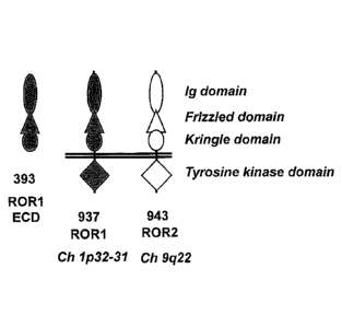
[0012] Figure 1 is a schematic that depicts the Ig-, Frizzled-, and Kringle-
like domains of
ROR1, receptor tyrosine kinase-like orphan receptor 2 (ROR2), and an ROR1-
derived
extracellular domain fragment (ROR1-ECD), as well as the transmembrane and
intracellular
tyrosine kinase domains of ROR1 and ROR2.
[0013] Figure 2A is a schematic that depicts ROR-ECD.
CA 02759733 2016-07-14
WO 2010/124188
PCT/US2010/032208
3
[0014] Figure 2B is a schematic that depicts ROR1-ECD fused to a human Fe
domain
(Fc-ROR1).
[0015] Figure 3 is a graph depicting the results of a BIACORE"binding
analysis using the
indicated concentrations of mouse monoclonal antibody (mAb) 2A2 in terms of
response
(RU) versus time (seconds).
[0016] Figure 4 is a pair of histogram panels depicting the results of
fluorescence
activated cell sorting (FACS) analysis evaluating mAB 2A2 binding to
peripheral blood
mononuclear cells (PBMC) taken from B-CLL patients (first panel) and cultured
JeKo-1 cells
(second panel) in terms of the-number of events versus fluorescence intensity
for (A) lug/mL
(-6.5 nM) mAb 2A2, (B) 0.1 g/mL (-650 pM) mAb 2A2, (C) 0.01 1.tg/mL (-65 pM)
mAb
2A2, and (D) 0.001 g/mL (-6.5pM) mAb 2A2, with 1 .tg/mL polyclonal mouse IgG
(solid
black histogram) and goat anti-mouse IgG polyelonal antibody conjugated to
fluorescein
isothiocyanate (FITC) (broken line histogram) as controls.
[0017] Figure 5 is a list of the amino acid sequences corresponding to the
mAb 2A2
variable region heavy chain (VH) (SEQ ID NO: 1), light chain (VL) (SEQ ID NO:
2), VH
framework regions FR1-FR4 (SEQ ID NOs: 3, 5, 7, and 9), VII complementarity
determining
regions CDRI-3 (SEQ ID NOs: 4, 6, and 8), VL FR1-FR4 (SEQ ID NOs: 10, 12, 14,
and 16),
and VL CDR1-CDR3 (SEQ ID NOs: 11, 13, and 15) regions.
[0018] Figure 6 is a list of DNA coding sequences corresponding to the
"original" VII
(SEQ ID NO: 17) and VL (SEQ ID NO: 19) cDNAs isolated from mAb 2A2 and their
respective VH (SEQ ID NO: 18) and VL (SEQ ID NO: 20) coding sequences
optimized for
expression in a mammalian system.
[0019] Figure 7A is a schematic that depicts mAb 2A2, including its
constant and
variable regions (no fill), and a chimeric antibody (ch2A2), that included
human constant
regions (dark fill) and mouse mAb 2A2 variable region (no fill).
[0020] Figure 7B is a graph that depicts the results of ELISA studies
comparing mAb
2A2 and ch2A2 binding to ROR1 LCD (as well as binding to ROR2) as a function
of
antibody concentration.
[0021] Figure 8 is a pair of histogram panels depicting the results of FACS
analysis
evaluating mAb 2A2 (first panel) and ch2A2 (second panel) binding to JeKo-1.
cells.
[0022] Figure 9 is a graph that depicts the results of a flow eytometry
assay comparing
dsEv 2A2-PE38 immunotoxin and mAb 2A2 with respect to the ability to bind to
JeKo-1
CA 02759733 2011-10-21
WO 2010/124188
PCT/US2010/032208
4
cells in terms of fluorescence intensity of labeling antibody as a function of
dsFy 2A2-PE38
and mAb 2A2 concentration.
[0023] Figure 10 is a graph that depicts the results of a cytotoxity assay
in terms of
cytotoxity % (of cells) as a function of dsFy 2A2-PE38 concentration.
[0024] Figure 11 is a dot plot depicting the results of apoptosis analysis
in terms of
annexin V and propidium iodide signal for 2 1.tM dsFy 2A2-PE38 as applied to
JeKo-1 cells.
[0025] Figure 12 is a schematic depiction of ELISA of mAb 2A2 binding to
various
hROR-1 extracellular domain constructs, with murine ROR1 as a control.
DETAILED DESCRIPTION OF THE INVENTION
[0026] Receptor tyrosine kinase-like orphan receptor 1 (ROR1) is a
conserved embryonic
protein whose expression becomes progressively reduced during embryonic
development in
mammals. The intact protein, including its extracellular domain, does not
appear to be
significantly expressed in normal, adult mammalian tissues. In particular,
studies have not
identified significant expression of ROR1 on the cell surface of normal adult
human tissues,
including normal B cells. Baskar et al., Clin. Cancer Res., 14: 396-404
(2008),
DaneshManesh et al., bit. I Cancer, 123: 1190-1195 (2008), and Fukuda et al.,
Proc. Nat'l.
Acad. Sci. USA, 105: 3047-3052 (2008). However, ROR1 is expressed on the cell
surface of
malignant B-cells, including B-cell chronic lymphocytic leukemia (B-CLL) and
mantle cell
lymphoma (MCL). It has also been reported that ROR1 is expressed in certain
other cancer
cell lines including Burkett's lymphoma, renal cell carcinoma, colon cancer,
and breast
cancer. U.S. Patent Application Publ. 2007/0207510. Therefore, ROR1 can be
considered a
selective marker for these cancers. The invention provides an antibody to this
selective
marker.
[0027] hi particular, the invention provides an antibody having specificity
for ROR1,
comprising (a) a heavy chain having at least 90% identity to SEQ ID NO: 1; (b)
a light chain
variable domain having at least 90% sequence identity to SEQ ID NO: 2; or (c)
both a heavy
chain of (a) and alight chain of (b). In a preferred embodiment, thc antibody
comprises both
a heavy chain of (a) and a light chain of (b).
[0028] The antibody can be an isolated antibody having specificity for
human RORI,
wherein the antibody comprises a heavy chain having at least 90% identity to a
sequence
such as SEQ ID NO: 1. In other embodiments, the percentage identity can be at
least 91%, at
CA 02759733 2011-10-21
WO 2010/124188 PCT/US2010/032208
least 92%, at least 93%, at least 94%, at least 95%, at least 96%, at least
97%, at least 98%, or
at least 99%, or even 100%. In preferred embodiments, the heavy chain has at
least 95%
identity to SEQ ID NO: I. In more preferred embodiments, the heavy chain has
100%
identity to SEQ ID NO: I.
[0029] The antibody can be an isolated antibody having specificity for
human ROR1,
wherein the antibody comprises a light chain having at least 90% identity to a
sequence such
as SEQ ID NO: 2. In other embodiments, the percentage identity can be at least
91%, at least
92%, at least 93%, at least 94%, at least 95%, at least 96%, at least 97%, at
least 98%, or at
least 99%, or even 100%. In preferred embodiments, the light chain has at
least 95% identity
to SEQ ID NO: 2. In more preferred embodiments, the light chain has 100%
identity to SEQ
ID NO: 2.
100301 In some embodiments, the antibody can comprise any heavy chain as
described
above, in combination with any suitable light chain, such as those described
above.
Likewise, the antibody can comprise any of the light chains as described above
in
combination with any suitable heavy chain, such as those described above. For
example, in
preferred embodiments, the antibody comprises a heavy chain having at least
90% identity to
SEQ ID NO: 1 and a light chain having at least 90% identity to SEQ ID NO: 2.
In a preferred
embodiment, the antibody comprises the heavy chain of SEQ ID NO: 1 and the
light chain of
SEQ ID NO: 2.
[0031] Percent (%) identity of peptide sequences can be calculated, for
example, as 100 x
[(identical positions)imin(TGA, TGB)], where TGA and TGB are the sum of the
number of
residues and internal gap positions in peptide sequences A and 13 in the
alignment that
minimizes TGA and TGB. See, e.g., Russell et al., J. Mol Biol., 244: 332-350
(1994).
[0032] The antibody of the invention can be any antibody including a full
length antibody
or an antibody fragment. The antibody can be polyclonal, monoclonal,
recombinant,
chimeric, Or humanized. Furthermore, the antibody can be of any isotype
including without
limitation IgA, IgD, IgE, IgG, or IgM. Thus, for example, the antibody can be
any IgA such
as IgAl or IgA2, or any IgG such as IgGl, IgG2, IgG3, IgG4, or synthetic IgG.
The antibody
can also be any antibody fragment having specificity for the extracellular
domain of human
ROR1, such as F(ab)2, Fv, scFv, IgGAC1-I2, F(ab')2, scFv2CH3, F(ab), VL, VH,
seFv4,
scFv3, scFv2, dsFv, Fv, scFv-Fe, (seFv)2, a diabody, and a bivalent antibody.
The antibody
CA 02759733 2011-10-21
WO 2010/124188 PCT/US2010/032208
6
can be any modified or synthetic antibody, including, but not limited to, non-
depleting IgG
antibodies, T-bodies, or other Fe or Fab variants of antibodies.
[0033] In addition to a heavy chain as described above, the antibody of the
invention can
further comprise a light chain selected from a Fab library using sequential
naive chain
shuffling. Likewise, in addition to a light chain as described above, the
antibody of the
invention can further comprise a heavy chain selected from a Fab library using
sequential
naive chain shuffling.
100341 In some embodiments, the invention provides an isolated antibody,
having
specificity for human ROR1, comprising at least one CDR having a sequence
selected from
the group consisting of SEQ ID NO:4, SEQ 1D NO:6, SEQ ID NO:8, SEQ ID NO: 11,
SEQ
ID NO: 13, and SEQ ID NO: 15. The invention also provides an isolated antibody
with
specificity for ROR1 comprising at least one or more variants of the foregoing
CDR
sequences, which include 1, 2, or 3 substitutions, insertions, deletions, or
combinations
thereof in a sequence selected from the group consisting of SEQ ID NO:4, SEQ
ID NO:6,
SEQ ID NO:8, SEQ ID NO: 11, SEQ ID NO: 13, and SEQ ID NO: 15. For example, a
recombinant chimeric or humanized antibody (or fragment thereof) can include
one, two,
three, four, five, or all six of the foregoing CDR sequences.
[0035] In some embodiments, the invention provides an antibody with avidity
for ROR1
of about 10 tiM or less, 5 RM or less, 2 uM or less, 1 uM or less, 500 nM or
less, 400 nM or
less, 300 nM or less, or 200 nM or less. The invention also provides an
antibody with avidity
for ROR1 of about 100 riM or less, about 75 nM or less, about 50 nM or less,
about 25 nM or
less, about 10 nM or less, or about 5 nM or less. The invention further
provides an antibody
with avidity for ROR1 of about 1 nM or less, about 800 pM or less, about 700
pM or less,
about 600 pM or less, about 500 pM or less, about 400 pM or less, about 300 pM
or less,
about 200 pM or less, or about 100 pM or less. Avidity can be measured using
art-known
techniques, such as ELISA or BIACORE.
[0036] The antibody of the invention can be produced by any suitable
technique, for
example, using any suitable eukaryotic or non-eukaryotic expression system. In
certain
embodiments, the antibody is produced using a mammalian expression system. hi
some
embodiments, the heavy chain can be encoded by a DNA sequence such as SEQ ID
NO: 17
or SEQ ID NO: 18, while the light chain can be encoded by a DNA sequence such
as SEQ ID
NO: 19 or SEQ ID NO: 20.
CA 02759733 2011-10-21
WO 2010/124188
PCT/US2010/032208
7
[0037] The antibody of the invention can be produced using a suitable non-
eukaryotic
expression system such as a bacterial expression system. Bacterial expression
systems can be
used to produce fragments such as a F(ab)2, Fv, scFv, IgGACH2, F(ab')2,
scFv2CH3, F(ab),
VL, VH, scFv4, scFv3, scFv2, dsFv, Fv, sav-Fc, (scFv)2, and diabodies.
Techniques for
altering DNA coding sequences to produce such fragments are known in the art.
[0038] The antibody of the invention can be conjugated to a synthetic
molecule using any
type of suitable conjugation. Recombinant engineering and incorporated
selenocysteine (e.g.,
as described in International Application Publication WO/2008/122039) can be
used to
conjugate a synthetic molecule. Other methods of conjugation can include
covalent coupling
to native or engineered lysine side-chain amines or cysteine side-chain
thiols. See, e.g., Wu
et al., Nat. Biotechnol., 23: 1137-1146 (2005). The synthetic molecule can be
any molecule
such as one targeting a tumor. Of course, it will be understood that the
synthetic molecule
also can be a protein or an antibody.
100391 Synthetic molecules include therapeutic agents such as cytotoxic,
cytostatic, or
antiangiogenic agents and radioisotopes. A cytotoxic agent can be a plant,
fungal, or
bacterial molecule (e.g., a protein toxin). A therapeutic agent can be a
maytansinoid (e.g.,
maytansinol or DM1 maytansinoid), a taxane, or a calicheamicin. Therapeutic
agents include
vincristine and prednisone. A therapeutic agent can be an antimetabolite
(e.g., an antifolate
such as methotrexate, a fluoropyrimidine such as 5-fluorouracil, cytosine
arabinoside, or an
analogue of purine or adenosine); an intercalating agent (for example, an
anthracycline such
as doxorubicin, daunomycin, cpirubicin, idarubicin, mitomycin-C, dactinomycin,
or
mithramycin); a platinum derivative (e.g., cisplatin or carboplatin); an
alkylating agent (e.g.,
nitrogen mustard, melphalan, chlorambucil, busulphan, cyclophospharnide,
ifosfamide
nitrosoureas or thiotepa); an antimitotic agent (e.g., a vinca alkaloid like
vincristine or taxoid
such as paclitaxel or docetaxel); a topoisomerase inhibitor (for example,
etoposide and
teniposide, amsacrine, topotecan); a cell cycle inhibitor (for example, a
flavopyridol); or a
microbtubule agent (e.g., an epothilone, discodennolide analog, or
eleutherobin analog). A
therapeutic agent can be a proteosorrie inhibitor or a topoisomerase inhibitor
such as
bortezomib, arnsacrine, etoposide, etoposide phosphate, teniposide, or
doxonibicin.
Therapeutic radioisotopes include yttrium (90Y), lutetium (177Lu), actinium
(225Ac),
praseodymium, astatine (211
At) rhenium (1a6Re), bismuth (212Bi or 213Bi), and rhodium
CA 02759733 2011-10-21
WO 2010/124188
PCT/US2010/032208
8
(188Rh). Antiangiogenic agents include linomide, bevacuzimab, angiostatin, and
razoxane.
The synthetic molecule can be another antibody such as rituximab or
bevacuzimab.
[00401 A synthetic molecule can also be a label. Labels can be useful in
diagnostic
applications and can include, for example, contrast agents. A contrast agent
can be a
radioisotope label such as iodine (131I or 1251), indium (1111n), technetium
(99Tc), phosphorus
(32P), carbon (14C), tritium (3H), other radioisotope (e.g., a radioactive
ion) or a therapeutic
radioisotope listed above. Additionally, contrast agents can include
radiopaque materials,
magnetic resonance imaging (MR1) agents, ultrasound imaging agents, and any
other contrast
agents suitable for detection by a device that images an animal body. A
synthetic molecule
can also be a fluorescent label, a biologically active enzyme label, a
luminescent label, or a
chromophore label.
100411 In some embodiments, the antibody can also have specificity for one
or more
antigens in addition to ROR1 For example, the antibody of the invention can be
engineered
(e.g., as a bivalent diabody or a conjugated Fab dinaer or trimer) to have
specificity for ROR1
and another tumor antigen, e.g., an antigen associated with B-CLL, MCL,
Burkett's
lymphoma, renal cell carcinoma, colon cancer (e.g., colon adenocarcinoma), or
breast cancer
(e.g., breast adenocarcinoma). The antibody can be engineered to have
specificity for ROR1
and an antigen that promotes activation or targeting of cytotoxic effector
cells.
1100421 The invention further provides eukaryotic or non-cukaryotic cells
that have been
recombinantly engineered to produce an antibody of the invention. The
eukaryotic or non-
eukaryotic cells can be used as an expression system to produce the antibody
of the invention.
In another embodiment, the invention provides ROR1 targeted immune cells that
are
engineered to recombinantly express an ROR1 specific antibody of the
invention. For
example, the invention provides a T-cell engineered to express an antibody of
the invention
(e.g., an scFv, scFv-Fc, (scFv)2), which is linked to a synthetic molecule
with the following
domains: a spacer or hinge region (e.g., a CD28, CD28 or IgG hinge), a
transmembrane
region (e.g., a transmembrane canonical domain), and an intracellular T-cell
receptor (TCR)
signaling domain, thereby forming a T-body (or chimeric antigen receptor
(CAR)).
Intracellular TCR signaling domains that can be included in a T-body (or CAR)
include, but
are not limited to, CD3c, FcR-1, and Syk-PTK signaling domains as well as the
CD28, 4-
IBB, and CD1 34 co-signaling domains. Methods for constructing T-cells
expressing a T-
CA 02759733 2011-10-21
WO 2010/124188 PCT/US2010/032208
9
body (or CAR) are known in the art. See, e.g., Marcu-Malina et al., Expert
Opinion on
Biological Therapy., Vol. 9, No. 5, posted online April 16 (2009).
[0043] The invention provides a method of inhibiting cells that express
ROR1 (ROR1
cells) by contacting the cells with an antibody of the invention. The antibody
can be a naked
(unconjugated) antibody or an antibody conjugated to a synthetic molecule,
e.g., a cytotoxic,
cytostatic, or antiangiogenic agent or a radioisotope. The method can be used
to inhibit
ROR1 cells in vitro or in a subject (i.e., in vivo). The contacted ROR1 cells
be in, for
example, a cell culture or animal model of a disorder associated with elevated
levels of
ROR1. The method is useful, for example, to measure and/or rank (relative to
another
antibody) the antibody's inhibitory activity for a specific ROR1 cell type.
Inhibiting ROR1
cells can include blocking or reducing the activity or growth of ROR1 cells.
Inhibiting can
also include the killing of ROR1 cells. While the method is not bound by or
limited to any
mechanism of action, inhibitory activity can be mediated by blocking ROR1-
mediated
signaling or by blocking the signaling of an ROR1 associated receptor.
Inhibitory activity
can also be mediated by recruitment of immune system effectors that attack
ROR1 cells, e.g.,
by activating constitutents of the antibody-dependent cell-mediated
cytotoxicity (ADCC) or
complement systems.
[0044] The invention also provides a method of treating a subject that has,
is suspected to
have, or is at risk for a disorder associated with elevated levels of ROR1.
Generally, the
method includes administering a therapeutically effective amount of an
isolated antibody of
the invention to the subject. The antibody can be any anti-ROR1 antibody of
the invention as
described above. Thus, the antibody can be chimeric, humanized, synthetic,
F(ab)2, Fv,
scFv, IgGACH2, F(ab')2, scFv2CH3, F(ab), VL, VH, scFv4, scFv3, scFv2, dsFv,
Fv, or
(scFv)2. In some embodiments, the method includes administering an IgG, an
scFv, a dsFv, a
F(a1:02, a diabody, or a bivalent antibody. The admistered antibody can be
conjugated to a
synthetic molecule described above, e.g., a cytotoxic, cytostatic, or
antiangiogenic agent or a
therapeutic radioisotope. An exemplary cytotoxic agent is Pseudomonas exotoxin
A (PE38).
Disorders that can be treated include, for example, B-CLL and MCL. Other
disorders
associated with elevated ROR1 that can be treated include Burkett's lymphoma,
renal cell
carcinoma, colon cancer (e.g., colon adenocarcinoma), and breast cancer (e.g.,
breast
adenocarcinoma).
CA 02759733 2011-10-21
WO 2010/124188
PCT/US2010/032208
[0045] The invention also provides a method of treating a subject that has,
is suspected to
have, or is at risk for a disorder associated vvith elevated levels of ROR1 by
adoptive transfer
of the genetically engineered T-cells described herein, which express an
antibody of the
invention as a T-body (or CAR) that selectively binds RORI . Recombinant
technology can
be used to introduce T-body (or CAR) encoding genetic material into any
suitable T-cells,
e.g., central memory T-cells from the subject to be treated. The T-cells
carrying the genetic
material can be expanded (e.g., in the presence of cytokines). The genetically
engineered T-
cells are transferred, typically by infusion, to the patient. The transferred
T-cells of the
invention can then mount an immune response against ROR1 expressing cells in
the subject.
The adoptive transfer method can be used, for example, to treat subjects that
have or are
suspected to have B-CLL, MCL, Burkett's lymphoma, renal cell carcinoma, colon
cancer
(e.g., colon adenocareinoma), or breast cancer (e.g., breast adenocarcinoma).
[0046] In some embodiments, the foregoing methods of treatment can further
include co-
administering a second therapeutic agent for the disorder associated with
elevated RORI
For example, when the disorder to be treated involves an ROR1-expressing
cancer, the
method can further include co-administration of a cytotoxic, cystostatie, or
antiangiogenic
agent suitable for treating the cancer. If the cancer is a B-cell lymphoma,
the method can
further include, for example, co-administration of ritaximab, alemtuzumab, or
a CHOP
chemotherapeutic regimen.
100471 The terms "treat," "treating," "treatment," and "therapeutically
effective" used
herein do not necessarily imply 100% or complete treatment. Rather, there are
varying
degrees of treatment of which one of ordinary skill in the art recognizes as
having a potential
benefit or therapeutic effect. In this respect, the inventive method can
provide any amount of
any level of treatment. Furthermore, the treatment provided by the inventive
method can
include the treatment of one Or more conditions or symptoms of the disease
being treated.
[0048] In another embodiment, the invention provides method of detecting in
a test
sample an altered level of ROR1 (e.g., cell surface ROR1), for example,
relative to a control.
Generally, the method includes contacting an antibody of the invention to the
test sample and
deteimining the amount of antibody that selectively binds to material (e.g.,
cells) in the
sample to thereby determine the level of ROR1 in the test sample. A test
sample can be from
a cell culture or from a test subject, e.g., a plasma or a tissue sample from
a subject that has,
is suspected to have, or is at risk for a disease or condition associated with
elevated ROR1 in
CA 02759733 2011-10-21
WO 2010/124188
PCT/US2010/032208
11
a subject. A control level desirably corresponds to the ROR1 level detected
using the same
antibody in a corresponding sample(s) fiuna one or more control cultures or
subjects.
Methods of using the antibody of the invention to determine ROR1 levels can
include any
immunoassay such as immuno- (Western) blotting, enzyme-linked immunosorbent
assay
(ELISA), and flow cytometry, e.g., fluorescence-activated cell sorting (FACS)
analysis.
[0049] The method of detection can be used to screen for the presence of a
disorder
associated with elevated ROR1. The method includes obtaining a sample from a
test subject
in need of screening, e.g., a subject that has, is suspected to have, or is at
risk for a disorder
associated with elevated ROR1. The level of ROR1 (e.g., the amount or
concentration) in the
sample is measured using an antibody of the invention, and the level in the
sample is
compared to a control level of ROR1. The control level represents, for
example, the mean
level (e.g., the amount or concentration) in sample(s) from one or,
preferably, multiple
control group subjects that do not have a disorder associated with elevated
RORI
Alternatively, the control level can correspond to the level or mean level of
ROR1 in one or
more samples taken from the test subject at one or more prior times, when the
test subject did
not have or did not exhibit, a condition associated with elevated ROR1. A
significantly
higher level of ROR1 in the test sample relative to the control level is
indicative of a disorder
associated with elevated ROR1 in the subject.
[0050] In subjects such as humans, where cell surface ROR1 expression is
largely
restricted to embryonic development, a control level of ROR1 can be zero or
none. Thus, in
some embodiments of the method of the detection provided by the invention, any
significant
and detectable amount of ROR1 in a test sample can be indicative of a disorder
associated
with elevated ROR1 in the subject.
[0051] Additionally, the method of detection can be used to monitor the
progress of a
disorder associated with elevated ROR1. The method includes obtaining a sample
from a
subject in need of screening, e.g., a subject having been diagnosed or
suspected to have a
disorder associated with elevated ROR1. The level of ROR1 in the sample is
measured using
an antibody of the invention, and the level in the sample is compared to a
control level
corresponding to the level or mean level of ROR1 in one or more samples taken
from the test
subject at one or more prior times. Levels of ROR1 that are significantly
elevated or
decreased relative to control indicate that the subject's disorder is
deteriorating or improving,
respectively.
CA 02759733 2011-10-21
WO 2010/124188 PCT/US2010/032208
12
[0052] The foregoing method of detection can be used to screen for the
presence or to
monitor the progress of disorders including, for example, B-CLL, MCL,
Burkett's lymphoma,
renal cell carcinoma, colon cancer (e.g., colon adenocarcinonia), and breast
cancer (e.g.,
breast adenocarcinoma).
[0053] The invention provides a method for screening a subject for an
altered level of
ROR1. Generally, the method includes administering to the subject an antibody
of the
invention that is conjugated to a label (e.g., a contrast agent), imaging the
subject in a manner
suitable for detecting the label, and determining whether a region in the
subject has an altered
density or concentration of label as compared to the background level of label
in proximal
tissue. Alternatively, the method includes determining whether there is an
altered density or
concentration of label in a region as compared to the density or concentration
of label
previously detected in the same region of the subject. Methods of imaging a
subject can
include x-ray imaging, x-ray computed tomography (CT) imaging (e.g., CT
angiography
(CTA) imaging), magnetic resonance (MR) imaging, magnetic resonance
angiography
(MRA), nuclear medicine, ultrasound (US) imaging, optical imaging,
elastography, infrared
imaging, microwave imaging, and the like, as appropriate for detecting the
label conjugated
to the antibody. In a preferred embodiment, the subject has, is suspected to
have, or is at risk
for an RORI -expressing tumor, such as B-CLL, MCL, Burkett's lymphoma, renal
cell
carcinoma, tumor of the colon (e.g., colon adenocarcinoma), or breast tumor
(e.g., breast
adenocarcinoma), and the method is used to screen for or detect the presence
of the tumor. In
another embodiment, the method can be used to monitor the size or density of
an ROR1-
expressing tumor over time, e.g., during a course of treatment.
[0054] The invention also provides a pharmaceutical composition comprising
an antibody
as described above and a pharmaceutically acceptable carrier. Pharmaceutical
compositions
can be prepared from any of the antibodies described herein. An exemplary
composition
includes a chimeric antibody having SEQ ID NO: 1 (heavy chain) and/or SEQ ID
NO: 2
(light chain). Another exemplary composition comprises a humanized antibody
having one,
two, three, four, five, or six CDRs selected from the group consisting of SEQ
ID NO:4, SEQ
ID NO:6, SEQ to NO:8, SEQ ID NO: 11, SEQ ID NO: 13, and SEQ ID NO: 15. Still
another exemplary pharmaceutical composition includes a dsFv fragment, which
comprises
the sequence of SEQ ID NO: 1 with a glycine to cysteine substitution at
position 44 (heavy
CA 02759733 2011-10-21
WO 2010/124188 PCT/US2010/032208
13
chain) and/or the sequence of SEQ ID NO: 2 with a glycine to cysteine
substitution at
position 100 (light chain).
[0055] The composition of the invention comprises a carrier for the
antibody, desirably a
pharmaceutically acceptable carrier. The pharmaceutically acceptable carrier
can be any
suitable pharmaceutically acceptable carrier. The term "pharmaceutically
acceptable carrier"
as used herein means one or more compatible solid or liquid fillers, diluents,
other excipients,
or encapsulating substances which are suitable for administration into a human
or veterinary
patient (e.g., a physiologically acceptable carrier or a pharmacologically
acceptable carrier).
The term "carrier" denotes an organic or inorganic ingredient, natural or
synthetic, with
which the active ingredient is combined to facilitate the application. The
pharmaceutically
acceptable carrier can be co-mingled with one or more of the active
components, e.g., a
hybrid molecule, and with each other, when more than one pharmaceutically
acceptable
carrier is present in the composition in a manner so as not to substantially
impair the desired
pharmaceutical efficacy. "Pharmaceutically acceptable" materials typically are
capable of
administration to a patient without the production of significant undesirable
physiological
effects such as nausea, dizziness, rash, or gastric upset. It is, for example,
desirable for a
composition comprising a pharmaceutically acceptable carrier not to be
immunogenic when
administered to a human patient for therapeutic purposes.
[0056] The pharmaceutical composition can contain suitable buffering
agents, including,
for example, acetic acid in a salt, citric acid in a salt, boric acid in a
salt, and phosphoric acid
in a salt. The pharmaceutical compositions also optionally can contain
suitable preservatives,
such as benzalkonium chloride, chlorobutanol, parabens, and thimerosal.
[0057] The pharmaceutical composition can be presented in unit dosage form
and can be
prepared by any suitable method, many of which are well known in the art of
pharmacy.
Such methods include the step of bringing the antibody of the invention into
association with
a carrier that constitutes one or more accessory ingredients. In general, the
composition is
prepared by uniformly and intimately bringing the active agent into
association with a liquid
carrier, a finely divided solid carrier, or both, and then, if necessary,
shaping the product.
[0058] A composition suitable for parenteral administration conveniently
comprises a
sterile aqueous preparation of the inventive composition, which preferably is
isotonic with
the blood of the recipient. This aqueous preparation can be formulated
according to known
methods using suitable dispersing or wetting agents and suspending agents. The
sterile
CA 02759733 2011-10-21
WO 2010/124188
PCT/US2010/032208
14
injectable preparation also can be a sterile injectable solution or suspension
in a non-toxic
parenterally-acceptable diluent or solvent, for example, as a solution in 1,3-
butane diol.
Among the acceptable vehicles and solvents that can be employed are water,
Ringer's
solution, and isotonic sodium chloride solution. In addition, sterile, fixed
oils are
conventionally employed as a solvent or suspending medium. For this purpose
any bland
fixed oil can be employed, such as synthetic mono-or di-glycerides. In
addition, fatty acids
such as oleic acid can be used in the preparation of injectables. Carrier
formulations suitable
for oral, subcutaneous, intravenous, intramuscular, etc. administrations can
be found in
Remington 's Pharmaceutical Sciences, Mack Publishing Co., Easton, PA.
[0059] The delivery systems useful in the context of the invention include
time-released,
delayed release, and sustained release delivery systems such that the delivery
of the inventive
composition occurs prior to, and with sufficient time to cause, sensitization
of the site to be
treated. The inventive composition can be used in conjunction with other
therapeutic agents
or therapies. Such systems can avoid repeated administrations of the inventive
composition,
thereby increasing convenience to the subject and the physician, and may be
particularly
suitable for certain compositions of the invention.
[0060] Many types of release delivery systems are available and known to
those of
ordinary skill in the art. Suitable release delivery systems include polymer
base systems such
as poly(lactide-glycolide), copolyoxalates, polycaprolactones,
polyesteramides,
polyorthoesters, polyhydroxybutyric acid, and polyanhydrides. Micro capsules
of the
foregoing polymers containing drugs are described in, for example, U.S. Patent
5,075,109.
Delivery systems also include non-polymer systems that are lipids including
sterols such as
cholesterol, cholesterol esters, and fatty acids or neutral fats such as mono-
di-and tri-
glycerides; hydrogel release systems; sylastic systems; peptide based systems;
wax coatings;
compressed tablets using conventional binders and excipients; partially fused
implants; and
the like. Specific examples include, but are not limited to: (a) erosional
systems in which the
active composition is contained in a form within a matrix such as those
described in U.S.
Patents 4,452,775, 4,667,014, 4,748,034, and 5,239,660 and (b) diffusional
systems in which
an active component permeates at a controlled rate from a polymer such as
described in U.S.
Patents 3,832,253 and 3,854,480. In addition, pump-based hardware delivery
systems can be
used, some of which are adapted for implantation.
CA 02759733 2011-10-21
WO 2010/124188
PCT/US2010/032208
[0061] The term "subject" is used herein, for example, in connection with
therapeutic and
diagnostic methods, to refer to human or animal subjects. Animal subjects
include, but are
not limited to, animal models, such as, mammalian models of conditions or
disorders
associated with elevated ROR1 expression such as B-CLL, MCL, Burkett's
lymphoma, renal
cell carcinoma, colon cancer, (e.g., colon adenocarcinoma), and breast cancer
(e.g., breast
adenocarcinoma).
[0062] The invention also provides kits suitable for carrying out the
methods of the
invention. Typically, a kit comprises two or more components required for
performing a
therapeutic or detection method of the invention. Kit components include, but
are not limited
to, one or more antibody of the invention, appropriate reagents, and/or
equipment.
[00631 A kit can comprise an antibody of the invention and an immunoassay
buffer
suitable for detecting ROR1 (e.g. by ELISA or FACS). The kit may also contain
one or more
microtiter plates, standards, assay diluents, wash buffers, adhesive plate
covers, and/or
instructions for carrying out a method of the invention using the kit. The kit
can include an
antibody of the invention bound to a substrate (e.g., a multi-well plate or a
chip), which is
suitably packaged and useful to detect ROR1. in some embodiments, the kit
includes an
antibody of the invention that is conjugated to a label, such as, a
fluorescent label, a
biologically active enzyme label, a luminescent label, or a chromophore label.
The kit can
further include reagents for visualizing the conjugated antibody, e.g., a
substrate for the
enzyme. In some embodiments, the kit includes an antibody of the invention
that is
conjugated to a contrast agent and, optionally, one or more reagents or pieces
of equipment
useful for imaging the antibody in a subject.
[0064] Generally the antibody of the invention in a kit is suitably
packaged, e.g., in a vial,
pouch, ampoule, and/or any container appropriate for a therapeutic or
detection method. Kit
components can be provided as concentrates (including lyophilized
compositions), which
may be further diluted prior to use or they can be provided at the
concentration of use. When
the antibody of the invention for use in vivo, single dosages may be provided
in sterilized
containers having the desired amount and concentration of agents.
[0065] The following examples further illustrate the invention but, of
course, should not
be construed as in any way limiting its scope.
CA 02759733 2016-07-14
WO 2010/124188 " PCT/US2010/032208
16
EXAMPLE
[0066] This example demonstrates the preparation of monoclonal antibodies
with
specificity for ROR I .
[0067] Mice were immunized with a fusion protein consisting of the human Fe
domain
and extracellular domain of human ROR1 (Fe-ROR1). As depicted in Figure 1, the
extracellular domain of RORI includes an immunoglobulin (Ig) domain, a
Frizzled domain,
and a Kringlc domain, which span about 393 amino acids of the amino-terminal
portion of
ROR1. A nucleic acid sequence encoding the ROR1 extracellular domain was
recombinantly
linked to a nucleic acid sequence encoding human Fcl to form a recombinant
construct that
then was expressed in HEIC 293 cells. The produced Fc-ROR1 fusion protein
(depicted in
Figure 2B) was harvested and purified by ion exchange chromatography and gel
filtration or
by Protein A affinity chromatography. The results of a Coomassie-stained
protein gel
analysis of purified Fc-ROR1 under non-reducing and reducing conditions are
shown in
Figure 2C.
[0068] Mice were immunized with the Fe-ROR1, and antibody-producing cells
were
immortalized to produce hybridomas. Supernatants from twenty hybridomas were
screened
for specific ROR1 binding. An EL1SA plate was coated with human ROR1-ECD (50
ng per
well overnight at 4 C), then blocked with 3% BSA-PBS (room temperature for 2
hours), and
subsequently contacted with dilutions of affinity purified of mouse mAb (room
temperature
for 2 hours). After washing (with PBS-Tweenm20), horseradish peroxidase (HRP)-
conjugated
goat anti-mouse (1:2000 dilution) was added (1 hour at room temperature) and
washed. The
bound antibody was detected using the peroxidase substrate ABTS (2,2'-azino-
bis(3-
ethylbenzthiazoline-6-sulfonic acid). Four monoclonal antibodies (mAb 2A2,
2D11, 1A1,
and 1A7) were identified by ELISA as specifically binding ROR1.
[0069] The four specific mAbs were further characterized as IgG using goat
anti-human
IgG polyclonal antibodies (pAbs) conjugated to HRP. Flow cytornetry was used
to evaluate
the ability of each of the four antibodies to bind to cells expressing ROR1.
The mAb 2A2
antibody was also further characterized by surface plasmon resonance (BIACORE,
GE
Healthcare, Piscataway, NJ). The results of the foregoing experiments and also
the antibody
expression yield for two hybridornas are summarized in Table 1.
CA 02759733 2016-07-14
WO 2010/124188
PCT/US2010/032208
17
Table 1
mAb Isotype ELISA Flow Cytometry BIACORE mAb
Expression
2A2 mouse IgGlic -H-+ -1-1- 10 mg/L
2D11 mouse IgGlx ++ ++ not determined 23 mg/L
1A1 mouse IgGU< ++ not determined
not determined
1A7 mouse IgG1K ++ i not determined
not determined
[0070] The foregoing
results demonstrate the isolation of four mAbs with specificity for
the extracellular domain of human ROR1.
EXAMPLE 2
[0071] This example
further demonstrates the desirable binding properties of mAb 2A2
for ROR1.
[0072] Hybridoma 2A2 of Example 1 was grown in CELLINE Disposable
Bioreactors
(BD 13iosciences, San Jose, CA) using animal component-free BD Cell MAb
Medium.
Expressed mAb 2A2 was purified by Protein G affinity chromatography.
[00731 The avidity of mAb
2A2 for ROR1-ECD was determined using surface plasmon
resonance in a BIACORE X100 instrument (GE Healthcare, Piscataway, NJ). Human
ROR1-ECD was coupled to a BIACORE CM5 chip at 1600 resonance units. MAb 2A2
was
diluted in BBS-EP buffer (0.01 M HEPES, 0.15 M NaC1, 3 mM EDTA, and 0.005%
surfactant P20, pH 7.4; GE Healthcare) to various concentrations (from about
200 nM to
about 12.5 nM) and injected sequentially. As depicted by the extended, flat
tail in Figure 3,
antibody binding was very stable at all concentrations tested. Association (k-
on) and
dissociation (k-off) rate constants were calculated based on a 1:1 Langmuir
binding model
using BIAEVALUATION software (GE Healthcare). The equilibrium dissociation
constant
(Kd --= k-off/k-on) was calculated, and the avidity of mAb 2A2 was determined
to be 100 pM.
[0074] Fluorescence-
activated cell sorting (FACS) analysis was performed to test the
ability of mAb 2A2 to bind to peripheral blood mononuclear cells (PBMC) from
normal
patients, to PBMC from a B-CLL patient, and to the human ROR1-expressing MCL
cell line
JeKo-1. Antibody binding was detected using goat anti-mouse IgG polyclonal
antibody
conjugated to fluorescein isothiocyanate (FITC). Results for antibody
concentrations of from
about 0.001 ug/ML to about 1 j.tglmL (about 6.5 pM to about 6.5 nM), secondary
antibody
CA 02759733 2011-10-21
WO 2010/124188
PCT/US2010/032208
18
alone, and polyclonal mouse IgG control are depicted in the histograms of
Figure 4. Control
mouse IgG (black-filled histogram) did not show a significant shift relative
to secondary
antibody alone (broken line histogram). mAb 2A2 did exhibit significant
binding to JeKo-1
(Figure 4, second panel) and to primary B-CLL cells present in the PBMC sample
from a
representative patient (Figure 4, first panel). Binding to each of these cell
populations was
observed using mAb 2A2 concentrations as low as 1 ng/mL and 10 ng/mL,
respectively (see
Figure 4).
100751 Similar FACS analyses of mAb 2A2 binding to normal B cells from
healthy
donors were negative.
[0076] The foregoing results demonstrate that mAb 2A2 has good avidity for
its antigen
and can be used to specifically distinguish (i) tumor cells obtained from
lymphoma patients
from (ii) normal B-cells taken from healthy subjects.
EXAMPLE 3
[0077] This example demonstrates the identification of mAb 2A2 variable
domain coding
and amino acid sequences.
[0078] The variable domain encoding sequences of heavy (VH) and light (VL)
chain of
mAb 2A2 were RT-PCR amplified from hybridorna 2A2 total RNA using published
primer
sequences (Morrison, S.L., in Curent Protocols in Immunology, Suppl. 47, pp.
2.12,1-
2.12.17, John Wiley and Sons, 2002). The cDNAs were cloned and analyzed by DNA
sequencing. VH and VL chain sequences were independently confiimed by liquid
chromatography-tandem mass spectrometry (LC-MS/MS) analysis of the tryptic
digests of
the heavy and light chain polypeptides, which were resolved by sodium dodecyl
sulfate
polyacrylamide gel electrophorcsis (SDS-PAGE). The amino acid sequences and
cDNA
sequences of the VH and VL chains of mAb 2A2 are depicted in Figures 5 and 6
("original
sequences"), respectively.
EXAMPLE 4
[0079] This example demonstrates the construction and characterization of a
chimeric
human-mouse antibody with desirable binding properties for ROR1.
[0080] Codon optimization of mAb 2A2 VH and VL DNA sequences (SEQ ID NO: 17
and SEQ ID NO: 19, respectively) produced the sequences of SEQ ID NO: 18 and
SEQ ID
NO: 20, respectively, which are depicted in Figure 6. Optimized sequences were
used to
CA 02759733 2011-10-21
WO 2010/124188 PCT/US2010/032208
19
generate a chimeric antibody vector, generally according to the method
described in
Morrison, S.L., in Curent Protocols in Immunology, Coligan et al. Eds., Suppl.
47, pp.
2.12.1-2.12.17, John Wiley and Sons, 2002. The optimized VH and VL sequences
as well as
human CicL segments were cloned into the pIgG mammalian expression vector
which is
described in Rader et al., FASEB J., 16:2000-2002 (2002). The resulting
plasmids were
transfected and expressed in HEK 293F cells to produce chimeric mouse/human
2A2 IgG1
(ch2A2) as described in Hofer et al., J. Immunol. Methods, 318: 75-87 (2007).
The chimeric
ch2A2 antibody is schematically depicted in Figure 7A with mouse and human
antibody
domains shown by light and dark fills, respectively.
[0081] Ch2A2 and mouse mAb 2A2 were evaluated by ELISA for their ability to
bind to
ROR1-ECD and a commercial ROR2-Fc (R&D Systems, Minneapolis, MN). ELISA was
performed as described in Example 1, with chimeric ch2A2 being detected using
HRP-
conjugated goat anti-human kappa antibody. ELISA results are depicted in
Figure 7B. While
neither ch2A2 nor mAb 2A2 bound to the ROR2-Fc control, ch2A2 retained
virtually all of
the mAb 2A2 binding affinity for the extraceullar domain of ROR1 (see Figure
7B).
[0082] Ch2A2 was also compared to mAb 2A2 by FACS for its ability to bind
to the
human ROR1-expressing MCL cell line JeKo-1 and to PBMC from B-CLL patients. A
FACS analysis was carried out as described in Example 2 above, except that mAb
2A2 (1
[tg/m1) and ch2A2 (1 g/m1) were detected using allophycoeyanin (APC)
conjugated goat
anti-mouse IgG (Fe) and APC conjugated goat anti-human IgG (Fe), respectively.
The
results of the FACS analysis for JeKo-1 cells are depicted in Figure 8.
Control mouse IgG
did not show a significant shift relative to secondary antibody alone (black-
filled histogram).
The results indicate that mAb 2A2 and ch2A2 at 1 [tg/m1 similarly bind to JeKo-
1 (see
Figure 8).
[0083] Similar results were obtained by FACS analysis comparing mAb 2A2 and
ch2A2
binding to PBMC from B-CLL patients.
[0084] The foregoing data demonstrate the generation of chimeric
mouse/human
antibody of the invention with conserved specificity and affinity for the
extracellular domain
of ROR1, including native RORI expressed on the cell surface of malignant B-
cells.
CA 02759733 2016-07-14
WO 2010/124188 PCT/US2010/032208
= 20
EXAMPLE 5
[0085] This example demonstrates the construction and characterization of a
disulfide
stabilized fragment (dsFv) of mAb 2A2 fused to an immunotoxin.
[0086] A dsFv fragment of mAb 2A2 (dsFv 2A2) was generated and fused to a
38-kDa
fragment of Pseudomonas exotoxin A (PE38) generally according to methods
described in
Pastan et al., Methods MoL Biol., 248: 503-518 (2004). The original VH and VL
coding
sequences of mAb 2A2 (see Figure 6) were altered to introduce a glycine to
eysteine
substitution at positions 44 and 100 of SEQ ID NO: 1 and SEQ ID NO: 2,
respectively
(substituted residues are underlined in Figure 5). The altered VH coding
sequence was
subcloned in-frame with a PE38 coding sequence in a pRB98 vector carrying a
chloramphenicol resistance gene (the vector is described in Kreitman et al.,
in Drug
Targeting, Francis et al., Eds., Vol. 25, pp. 215-226, Humana Press Inc,
Totowa, N.J., 2000).
Altered VH and VT, chains were separately expressed in E. coli, and the
resulting proteins
were harvested and solubilized. The VH and VL were refolded together to form
&Ey 2A2-
PE38 fusion immunotoxin, which was purified by Q-Sepharoserion-exchange
chromatography as described in Pastan et al., supra, 2004.
[0087] Recombinant dsFv 2A2-PE38 immunotoxin was evaluated by flow
cytometry and
compared to mouse mAb 2A2 for its ability to bind to the human ROR1-expressing
mantle
cell lymphoma cell line JeKo-1. JeKo-1 cell binding by mAb 2A2 was detected
using a goat
anti-mouse IgG polyclonal antibody (pAb) conjugated to APC (Jackson
ImmunoResearch
Laboratories, West Grove, PA) at 1:300 dilution. JeKo-1 cell binding of dsFv
2A2-PE38 was
detected using rabbit anti-Pseuciomonas exotoxin A pAb (1:100 dilution) (Sigma-
Aldrich, St.
Louis, MO) as a secondary antibody and goat anti-rabbit IgG pAb conjugated to
Cy5 (1:300
dilution) (Jackson ImmunoResearch Laboratories) as a tertiary antibody. Flow
eytometry
results are depicted in Figure 9A. The results demonstrate that, despite the
inherent
monovalency of the recombinant dsFv 2A2-PE38 immunotoxin, its binding to
native cell
surface ROR1 was detectable at concentrations of less than 1 ug/mL (Figure 9).
[0088] An analysis of dsFv 2A2-PE38 immunotoxin binding to PBMC from B-CLL
patients showed similar results. Additonally, ELISA experiments demonstrated
that dsFv
2A2-PE38 immunotoxin retains binding specificity for the extracellular domain
of human
ROR1.
CA 02759733 2011-10-21
WO 2010/124188 PCT/US2010/032208
21
[0089] The foregoing example provides a recombinant imrnunotoxin conjugated
antibody
of the invention, which is based on mAb 2A2 and which has conserved binding
specificity for
ROR1, including native ROR1 expressed on the cell surface of malignant B-
cells.
EXAMPLE 6
[0090] This example demonstrates dsFy 2A2-PE38 mediated cytotoxicity of
ROR1
expressing cells.
[0091] JeKo-1 cells were cultured in Roswell Park Memorial Institute (RPMI)
1640
medium supplemented with 10% fetal calf serum and incubated for 51 hours at 37
C in a 96-
well tissue culture plate with various doses (0-100 ug/mi,) of the dsFy 2A2-
PE38
imniunotoxin prepared in Example 5. The cells were subsequently analyzed by
flow
cytometry using annexin V and propidium iodide to stain apoptotic and dead
cells,
respectively. The percentage of cells that were positive for both annexin V
and propidium
iodide is shown as cytotoxicity on the y-axis, as a function of the
concentration of dsFy 2A2-
PE38, of Figure 10. Figure 11 further demonstrates that the cytotoxicity of
dsPv 2A2-PE38
(2 uM) included not only cell death (necrosis) as evidenced by propidium
iodide staining, but
also extensive apoptosis, as evidenced by annexin V staining.
[0092] The data confirmed the ability of dsFy 2A2-PE38 to effect dose-
dependent killing
of JeKo-1 cells at microgram concentrations or less.
EXAMPLE 7
[0093] This example demonstrates epitope mapping of the interaction between
mAb 2A2
and ROR1 using ELISA.
[0094] Recombinant proteins containing one, two, or three of the three
extracellular
domains of human ROR1 (di -Ig, d2-Frizzled, and d3-Kringle) as shown in Figure
12 were
coated on an ELISA plate. Each of the proteins was genetically fused to a
human Fe region;
a protein containing the three extracellular domains without the Fe region was
also prepared.
Cross-reactivity to mouse ROR1 was determined by coating recombinant Pc-mouse
ROR1
protein on the ELISA plate. Binding of mouse mAb 2A2 to the different ROR1
proteins was
detected using HRP-conjugated donkey anti-mouse IgG polyclonal antibodies. The
reactivity
was scored "++" for strong binding, "1" for moderate binding, and "'-" for no
detectable
binding. As shown in Figure 12, strong binding was detected for Fc-hROR1,
which
contained all three extracellular domains, as well as hROR1ECD, which
contained the
CA 02759733 2016-07-14
WO 2010/124188 PCT/US2010/032208
22
=
extracellular domains without an Fe region, and Fc-hROR1(d1d2), which
contained the Ig
and Frizzled domains. Moderate binding was detected for Fc-hROR(d1), which
contained
the Ig domain only. No binding was detected for Pc-hROR1(d2d3), Fe-hROR1(d2),
or Fe-
hROR(d3).
[0095] These results show that the binding epitope for the 2A2-ROR1
interaction is likely
in the Ig domain, but is assisted either directly or indirectly by the
Frizzled domain of ROR1.
[0096] The foregoing results demonstrate the dose-dependent killing of ROR1
expressing
cells by an immunotoxin-conjugated antibody of the invention.
[0097] [BLANK]
[0098] The use of the terms "a" and "an" and "the" and similar referents in
the context of
describing the invention (especially in the context of the following claims)
are to be
construed to cover both the singular and the plural, unless otherwise
indicated herein or
clearly contradicted by context. The terms "comprising," "having,"
"including," and
"containing" are to be construed as open-ended terms (i.e., meaning
"including, but not
limited to,") unless otherwise noted. Recitation of ranges of values herein
are merely
intended to serve as a shorthand method of referring individually to each
separate value
falling within the range, unless otherwise indicated herein, and each separate
value is
incorporated into the specification as if it were individually recited herein.
All methods
described herein can be performed in any suitable order unless otherwise
indicated herein or
otherwise clearly contradicted by context. The use of any and all examples, or
exemplary
language (e.g., "such as") provided herein, is intended merely to better
illuminate the
invention and does not pose a limitation on the scope of the invention unless
otherwise
claimed_ No language in the specification should be construed as indicating
any non-claimed
element as essential to the practice of the invention.
[0099] Preferred embodiments of this invention are described herein,
including the best
mode known to the inventors for carrying out the invention. Variations of
those preferred
embodiments may become apparent to those of ordinary skill in the art upon
reading the
foregoing description. The inventors expect skilled artisans to employ such
variations as
appropriate, and the inventors intend for the invention to be practiced
otherwise than as
CA 02759733 2011-10-21
WO 2010/124188
PCT/US2010/032208
23
specifically described herein. Accordingly, this invention includes all
modifications and
equivalents of the subject matter recited in the claims appended hereto as
permitted by
applicable law. Moreover, any combination of the above-described elements in
all possible
variations thereof is encompassed by the invention unless otherwise indicated
herein or
otherwise clearly contradicted by context.