Note: Descriptions are shown in the official language in which they were submitted.
CA 02760175 2016-02-18
APPARATUSES AND COMPOSITIONS FOR
CRYOPRES ERVATI ON OF CELLULAR MONOLAYERS
REFERENCE TO RELATED APPLICATIONS
[0001] This application claims priority to and the benefit of U.S. Provisional
Application No. 61/173,888, filed April 29, 2009.
BACKGROUND OF THE INVENTION
[0002] Cryopreservation is a process by which samples such as biological
materials
are frozen under controlled conditions and stored at low temperatures.
Cryopreservation is
frequently used to store cell cultures, for example, which must be maintained
over time in
order to ensure a ready supply of cells for re-growth and experimentation.
Cells for such
purposes are routinely frozen in suspension in industrial cryovials. Freezing
methods have
been developed to minimize the impact of osmotic shock and intracellular ice
crystal
formation, two factors that contribute to cell death during the freezing
process and frozen
storage.
[0003] Under current methods, however, a significant number of cells are still
lost
to cell death during the freeze-thaw process. Cell loss can be substantial in
homogeneous cell
suspensions, and cell loss increases as the system undergoing preservation
becomes more
complex (e.g., tissues and organs). Moreover, current methods are insufficient
for effective
large-scale cryopreservation of cell samples and tissues in a multi-vessel
format, for example
as adherent cells in a multiwell format. Unacceptably high well-to-well
variability as well as
unsatisfactory overall post-thaw viability currently render large-scale
processes for bulk
freezing of cells in multi-well plates commercially non-viable.
SUMMARY OF THE INVENTION
[0004] The present invention is based on the discovery that specially
configured
vessels, when combined with an optimized preservation media, can significantly
reduce the
well-to-well variability, and improve the integrity, viability,
- 1 -
8103717 1
CA 02760175 2011-10-26
WO 2010/127158
PCT/US2010/033032
recovery and shelf-life of cryopreserved cells, including confluent cell
monolayers.
As described and contemplated herein, this discovery enables for the first
time, a
consistently available supply of reliable cryopreserved cells for a wide
variety of
relevant applications such as but not limited to disease diagnosis, toxicity
screening
and small molecule/pharmaceutical analysis. Set forth herein are exemplary
embodiments which illustrate how to make, use and test the invention as well
as
teachings relating to the same which describe the present invention in a
manner
understood by the skilled artisan and which fully enable practice of the
present
invention by the skilled artisan.
[0005] The present invention relates to apparatuses, kits, and compositions
for the freezing and cryopreservation of cultured cells and tissues. In one
aspect, the
invention provides apparatuses for the large-scale cryopreservation of cell
cultures
in biocompatible vessels, such as multiwell tissue culture plates, and an ice
nucleating device which facilitates consistent well to well ice nucleation, a
step
necessary for the uniform survival of cells during the cryopreservation
process.
[0006] The ice nucleating device is a mechanical (i.e., non-chemical)
device which provides an initiation point for ice nucleation. The mechanical
ice
nucleating device can be located, for example, on the vessel cover, on the
vessel
itself, or on a vessel insert, such that the mechanical ice nucleating device
becomes
submerged in, or comes into contact with, the cryopreservation medium
containing
the biologic sample.
[0007] In some embodiments, the apparatuses of the invention can include
an insulating material to facilitate effective cooling and warming. The
insulating
material is a mechanical component, which provides a means of thermal
insulation
to the exterior (e.g., periphery) or interior of the vessel. The insulating
device can be
located, for example, on the exterior space of a vessel such that the cooling
and
warming rates of the insulated portion of the vessel are similar to the other
sections
of the vessel. The insulating material can be disposed on the vessel, integral
with the
vessel, or detachable from the vessel. In some embodiments, the insulating
material
is located adjacent to, but not in contact with, the vessel.
[0008] The apparatuses of the invention can be used with a nutrient-rich
biopreservation medium that is configured to optimally maintain cellular
osmotic
- 2 -
CA 02760175 2011-10-26
WO 2010/127158
PCT/US2010/033032
and ionic balances, control free radical accumulation, and reduce the stress
responses under non-normothermic conditions. The preferred biopreservation
medium for optimal storage and post-thaw recovery is CRYOSTORTm (BioLife
Solutions, Inc., Bothell, WA), but the apparatuses can be used with any
suitable
biopreservation medium.
[0009] In some embodiments, the combination of the insulating material,
mechanical ice nucleating device and biopreservation (e.g., a freezing) medium
allows for uniform ice nucleation and thawing, and improved post-thaw
viability.
Thus, the invention enables cells and tissues to be cryopreserved in ready-to-
use
configurations for high throughput analysis for screening or diagnostic
purposes.
Moreover, the present invention enables cell cultures to be frozen and stored
for
extended lengths of time via an easy-to-use method. The invention is
particularly
useful for the cryopreservation of fully intact, viable cell monolayers in
ready-to-use
formats for high throughput screening.
[0010] In another aspect, the invention provides an apparatus for
cryopreserving cells. The apparatus includes a vessel comprising a
biocompatible
substrate. The vessel has an interior and an exterior. The apparatus can
include a
mechanical ice nucleating device which is disposed in or on the vessel
interior and
initiates ice crystal formation. In one preferred embodiment, the apparatus is
sterile.
In some embodiments, the exterior of the vessel includes an insulating
material
which contacts at least a portion of the vessel's exterior. In other
embodiments, the
insulating material can be included on or within the vessel.
[0011] In another preferred embodiment, the vessel is a multiwell cell or
tissue culture plate (e.g., 6-well, 12-well, 96 well, 384-well, 1536-well). It
will be
appreciated that any multiwell formats can be used with the present invention.
[0012] In a preferred embodiment, the mechanical ice nucleating device
includes one or more structural elements (e.g., a three dimensional
protrusion) which
occupy a portion of the vessel interior, such as but not limited to a
protrusion which
projects from a surface of the vessel interior, from a surface of a vessel
cover, or
from a surface of a vessel insert. In some embodiments, the mechanical ice
nucleating device includes at least one physical anomaly on the interior
surface of
the vessel, such as but not limited to a score, a scratch, an etching, a nick,
or other
- 3 -
CA 02760175 2011-10-26
WO 2010/127158
PCT/US2010/033032
physical irregularity on a surface of the substrate. In some embodiments, the
mechanical ice nucleating device is a plastic protrusion, or another three
dimensional element.
[0013] The mechanical ice nucleating device can be integral with the
vessel. The protrusion can be of any suitable shape (e.g., spike-like, needle-
like,
sphere-like, pyramid-like, or cone-like) or construction (e.g., hollow, solid,
semi-
permeable). In some embodiments, the mechanical ice nucleating agent is a
removeable mesh or mesh-like insert. The mechanical ice nucleating device can
also be or include a non-smooth coating on a surface of the vessel interior.
In
further embodiments, the vessel includes a separable cover, and the cover can
include an ice nucleating device having a structural element which protrudes
from a
surface of the cover into the vessel interior. The ice nucleating device can
be
detachably connected to the cover, to the vessel, to a vessel insert.
Alternatively, the
ice nucleating device can be integral with the cover, the vessel, or the
insert. In
further embodiments, the ice nucleating device can be present on a vessel
(e.g, a
well) insert which vessel insert can be seperable from the vessel.
[0014] In one embodiment, the apparatus can include a cryopreservation
medium, CRYOSTORTm, or a functional equivalent.
[0015] In another aspect, the invention provides for an apparatus for
cryopreserving a cell monolayer. The apparatus comprises a vessel, preferably
a
multiwell plate (e.g, a cell culture or tissue culture plate), and a
mechanical ice
nucleating device associated with at least one well of the multiwell tissue
culture
plate. The mechanical ice nucleating device can be integral with at least one
well, or
detachably associated with at least one well of the multiwell tissue culture
plate. In
some embodiments, the apparatus includes an insulating material which contacts
at
least a portion of the vessel. In some embodiments, the vessel is sterile.
[0016] In another aspect, the invention provides a vessel where an
insulating material is disposed on all or a portion of the vessel's exterior
to aid in
cooling and warming of the vessel. In some embodiments, the insulating
material
can be disposed on the exterior or the vessel, the interior of the vessel, or
both the
exterior and interior of the vessel. The insulating material can be any type
of
material but in the preferred form the insulation would be the same material
as that
- 4 -
CA 02760175 2011-10-26
WO 2010/127158
PCT/US2010/033032
used to make the vessel and will aid in minimizing the variations in the
cooling and
warming rates from well to well in a multiwell vessel. Types of insulation are
well
known in the art and include but are not limited to caulks, foams, sprays, or
strips of
thermally insolative materials. In the preferred embodiment, the vessel is a
multiwell plate and the insulating material is applied in the space between
the
exterior wells (i.e., the wells on the perimeter of the plate) and the outside
edge of
the plate. It is understood that any or all of the wells in a multiwell plate
can be
insulated with an insulation device as described herein. In some embodiments,
the
insulating material will be part of the interior of the vessel, for example,
occupying
some portion of a well or wells. In some embodiments, the insulation device
can be
an integral part of the vessel or the insulation device can be detachable.
[0017] In a further aspect, the invention provides an apparatus which
includes a sterile vessel for holding cells or tissue, and a mechanical ice
nucleating
device disposed in said vessel. The apparatus can optionally include an
insulating
material disposed on all or a portion of the vessel.
[0018] In yet another aspect, the invention provides kits for cryopreserving
cells. The kits can include any apparatus described herein and a
biopreservation
medium, such as the CRYOSTORTm cryopreservation medium, or a functional
equivalent.
[0019] In another aspect, the invention provides compositions. The
compositions include a sterile vessel for holding cells. The vessel has an
interior
and an exterior. The composition also includes a mechanical ice nucleating
device,
a biopreservation medium (e.g., a cryopreservation medium or hypothermic
preservation medium), and cells disposed in or in contact with the
biopreservation
medium within the interior of the vessel. The ice nucleating device can be a
mechanical ice nucleating device, such as those mentioned above, which is
disposed
on or in the vessel interior. In some embodiments, the vessel is insulated
with an
insulating material. This and other aspects and embodiments of the present
invention are suitable for the preservation of cells, whether progenitor,
primary,
immortalized, or other, as well as tissues.
[0020] In
another aspect, the invention provides an apparatus for
cryopreserving cells. The
apparatus can include a vessel comprising a
- 5 -
CA 02760175 2011-10-26
WO 2010/127158
PCT/US2010/033032
biocompatible substrate, wherein the vessel further comprises an interior and
an
exterior; an insulating material that is added to the free space surrounding
the
exterior or interior wells and/or occupying the interior space of at least one
well of a
multiwell vessel to aid in consistent cooling and warming of all wells; and a
mechanical ice nucleating device disposed in or on the vessel interior for
initiating
ice crystal formation. In some embodiments, the apparatus is sterile. In some
embodiments, the insulating material is comprised of the same material as the
vessel, and in some embodiments the insulating material is comprised of a
different
material as the vessel. The insulating material can be, for example, any
caulk, foam,
spray, or sheet, which will provide an insulating effect. In some embodiments,
the
insulating material occupies a portion of the vessel exterior or any free
space
surrounding any well of a multi-well tissue culture plate. The insulating
material
optionally can occupy any or all of the vessels, so as to fill the air space
above the
top level of the cells and cryoprotectant media and the lower or bottom
surface of
the lid or cover. In some embodiments, the insulating material occupies both
the
exterior and interior spaces of the vessel. The insulating material can be
attached to
the vessel or vessel lid directly, and the insulating material can be
detachable from
the vessel or vessel lid.
[0021] In
another aspect, the invention provides an apparatus for
cryopreserving a cell monolayer. The apparatus can include a multiwell cell
culture
plate having a plurality of wells and, the multiwell cell culture plate
forming at least
one free space which not occupied by a well; an insulating material integral
with or
disposed in at least a portion of the at least one free space surrounding at
least one
well; and an ice nucleating device integral with at least one well of the
multiwell cell
culture plate.
[0022] In
another aspect, the invention provides an apparatus for
cryopreserving a cell monolayer. The apparatus can include a multiwell cell
culture
plate forming a plurality of wells, wherein each well has an interior space
for
containing fluid; a removable lid for covering the multiwell cell culture
plate; an
insulating material detachably associated with the lid, wherein the insulating
material is configured to occupy the interior space of at least one well above
the
- 6 -
CA 02760175 2011-10-26
WO 2010/127158
PCT/US2010/033032
fluid; and an ice nucleating device integral with at least one well of the
multiwell
cell culture plate.
[0023] In
another aspect, the invention provides an apparatus for
cryopreserving a cell monolayer. The apparatus can include a multiwell cell
culture
plate; an insulating material integral with the exterior area of the vessel,
or interior
or exterior space surrounding at least one well; and an ice nucleating device
detachably associated with at least one well of the multiwell cell culture
plate.
[0024] In
another aspect, the invention provides an apparatus for
cryopreserving a cell. The apparatus can include a sterile vessel for holding
cells or
tissue, the vessel having an exterior surface; an insulating material integral
with the
exterior of the vessel; and a mechanical ice nucleating device disposed in
said
vessel. In some embodiments, the cells are in suspension.
[0025] In
another aspect, the invention provides a composition. The
composition can include a sterile vessel for holding cells, wherein the vessel
further
comprises an interior and an exterior; an insulating material; an ice
nucleating
device; a biopreservation medium; and cells disposed in or in contact with the
biopreservation medium within the interior of the vessel. In some embodiments,
the
ice nucleating device comprises a mechanical ice nucleating device disposed on
or
in the vessel interior for initiating ice crystal formation. In some
embodiments, the
cells comprise primary cells, immortalized cells, or tissue. In some
embodiments,
the cells are monolayers or cells in suspension.
[0026] This Summary is provided merely to introduce certain concepts and
not to identify any key or essential features of the claimed subject matter.
BRIEF DESCRIPTION OF THE DRAWINGS
[0027] The aspects, embodiments, and features of the invention can be
better understood with reference to the drawings described below. The drawings
are
not necessarily to scale, emphasis instead generally being placed upon
illustrating
the principles of the invention. The drawings are provided to highlight
specific
embodiments of the invention and are not intended to limit the invention, the
scope
of which is limited only by the claims. In the drawings, like numerals are
used to
indicate like parts throughout the various views.
- 7 -
CA 02760175 2011-10-26
WO 2010/127158
PCT/US2010/033032
[0028] Figures 1A-D show cross-sectional views of ice nucleating devices,
in accordance with an illustrative embodiment of the invention.
[0029] Figures 1E-G show a multiwell plate insert having a plurality of ice
nucleating devices, in accordance with an illustrative embodiment of the
invention.
[0030] Figures 1H and 11 show a multiwell plate insert for receiving a
plurality of ice nucleating devices, in accordance with an illustrative
embodiment of
the invention.
[0031] Figures 1J and 1K show an ice nucleating device having a single
cone-shaped point, in accordance with an illustrative embodiment of the
invention.
[0032] Figures 1L-1P show an ice nucleating device having a plurality of
cone-shaped points, in accordance with an illustrative embodiment of the
invention.
[0033] Figures 1Q and 1R show an ice nucleating device having a plurality
of pyramid-shaped points, in accordance with an illustrative embodiment of the
invention.
[0034] Figures 2A-B show cross-sectional views of an insulated vessel, in
accordance with an illustrative embodiment of the invention.
[0035] Figure 3 is a graph showing the differences in cooling rates of
interior and exterior wells of a 96-well tissue culture plate with and without
the
addition of an insulating material, in accordance with an illustrative
embodiment of
the invention.
[0036] Figure 4 is a diagram showing the relative fluorescence of Normal
Human Dermal Fibroblast (NHDF) cells in each well of a 96-well tissue culture
plate following freezing in either media with serum and 5% DMSO, media with
serum and 10% DMSO, CRYOSTORTm CS5 (5% DMSO), or CRYOSTORTm CS10
(10% DMSO), in accordance with an illustrative embodiment of the invention.
Three different freezing methods were investigated; (1) plates submerged in an
alcohol bath in a styrofoam cooler in a -80 C freezer, (2) -20 C freezer then
directly
into a -80 C freezer, and (3) an automated controlled rate freezing device set
to -
1 C/minute. No nucleating device was used.
[0037] Figure 5 is a graph showing relative percent viability of NHDF cells
following freezing in each of the conditions tested in Figure 4.
- 8 -
CA 02760175 2011-10-26
WO 2010/127158
PCT/US2010/033032
[0038] Figure 6 is a diagram showing the relative fluorescence of Normal
Human Dermal Fibroblast (NHDF) cells in each well of a 96-well tissue culture
plate following freezing using CRYOSTORTm CS5 with and without a nucleating
device, in accordance with an illustrative embodiment of the invention.
[0039] Figure 7 is a graph showing relative fluorescence of NHDF cells
following freezing using CRYOSTORTm CS5 with and without a nucleating device,
in accordance with an illustrative embodiment of the invention.
[0040] Figure 8 is a diagram showing the relative fluorescence of Chinese
Hamster Ovary (CHO) cells in each well of a 96-well tissue culture plate
following
freezing using CRYOSTORTm CS5 with and without a nucleating device, in
accordance with an illustrative embodiment of the invention.
[0041] Figure 9 is a graph showing relative fluorescence of CHO cells
following freezing using CRYOSTORTm CS5 with and without a nucleating device,
in accordance with an illustrative embodiment of the invention.
[0042] Figures 10A-C show perspective views of ice nucleating devices, in
accordance with an illustrative embodiment of the invention.
[0043] Figure 11 is a diagram showing the relative fluorescence of Normal
Human Dermal Fibroblast (NHDF) cells in each well of a 96-well tissue culture
plate following freezing using the alcohol bath in a -80 C freezing method
using
CRYOSTORTm CS5 or culture media with serum and 5% DMSO with and without a
nucleating device, in accordance with an illustrative embodiment of the
invention.
[0044] Figure 12 is a graph showing relative percent viability of NHDF
cells following freezing in each of the conditions tested in Figure 11.
[0045] Figure 13 is a diagram showing the relative fluorescence of Normal
Human Dermal Fibroblast (NHDF) cells in each well of a 96-well tissue culture
plate following freezing using the -20 C to directly in a -80 C freezing
method using
CRYOSTORTm CS5 or culture media with serum and 5% DMSO with and without a
nucleating device, in accordance with an illustrative embodiment of the
invention.
[0046] Figure 14 is a graph showing relative percent viability of NHDF
cells following freezing in each of the conditions tested in Figure 13.
[0047] Figure 15 is a diagram showing the relative fluorescence of Normal
Human Dermal Fibroblast (NHDF) cells in each well of a 96-well tissue culture
- 9 -
CA 02760175 2011-10-26
WO 2010/127158
PCT/US2010/033032
plate following freezing using the controlled rate freezer (-1 C/minute)
freezing
method using CRYOSTORTm CS5 or culture media with serum and 5% DMSO with
and without a nucleating device, in accordance with an illustrative embodiment
of
the invention.
[0048] Figure 16 is a graph showing relative percent viability of NHDF
cells following freezing in each of the conditions tested in Figure 15.
[0049] Figure 17 is a diagram showing the relative fluorescence of Normal
Human Dermal Fibroblast (NHDF) cells in each well of a 96-well tissue culture
plate following freezing using the -20 C to directly in a -80 C freezing
method using
CRYOSTORTm CS5 with and without a nucleating device (single cone, low-density
array, and high-density array spike devices ¨ Figures 10A-C ¨ were used), and
with
and without an insulating device in accordance with an illustrative embodiment
of
the invention.
[0050] Figure 18 is a diagram showing the relative fluorescence of Normal
Human Dermal Fibroblast (NHDF) cells in each well of a 96-well tissue culture
plate following freezing using the controlled rate freezer (-1 C/minute)
freezing
method using CRYOSTORTm CS5 or culture media with serum and 5% DMSO with
and without a nucleating device (single cone, low-density array, and high-
density
array spike devices ¨ Figures 10A-C ¨ were used), and with and without an
insulating device in accordance with an illustrative embodiment of the
invention.
DETAILED DESCRIPTION OF THE INVENTION
[0051] These and other aspects, embodiments, and features of the invention
are also described in the following sections of the application, which are
provided to
highlight specific embodiments of the invention and are not intended to limit
the
invention, the scope of which is limited only by the claims.
[0052] The present invention provides apparatuses, kits and compositions
for the freezing, thawing and use of cultured cells (e.g., cell monolayers,
suspended
cells) in multiwell vessel formats. In addition, the present invention is
suitable for
the preservation of cells, whether progenitor, primary, immortalized, or
other, as
well as tissues. In particular, the present invention overcomes the
limitations met by
- 10-
CA 02760175 2011-10-26
WO 2010/127158
PCT/US2010/033032
previous inventions and meets the needs of providing a method, composition,
and
apparatus to produce uniformly frozen adherent cell monolayers in multiwell
tissue
culture plates that upon thawing yields acceptably uniform cell viability and
functional performance levels in each of the wells of a tissue culture plate.
Importantly, these criteria are uniform for each well of a multiwell plate
following
preservation allowing for accurate and immediate testing of the entire plate.
The
well-to-well uniformity and improved viability and function of cell culture
monolayers allows, for example, pharmaceutical companies and toxicology
testing
laboratories to utilize the plated cells for high throughput screening of
absorption,
distribution, metabolism, excretion, and toxicology (ADME/T) of drug compounds
in an in vitro model. Without the present invention, uniform cell density,
viability,
and functional performance among each of the wells could not be accomplished
following cryopreservation and therefore this concept could not be practiced.
The
present invention will significantly reduce time and labor costs associated
with high
throughput screening of plated cells.
[0053] For the present invention, cell cultures are plated, for example, on
multiwell tissue culture plates under standard culture conditions to obtain an
adherent cell monolayer. Once the desired cell density level is attained, the
cell
culture medium is removed and replaced with chilled (preferably between 2 and
8 C) CRYOSTORTm cryopreservation medium containing 5 or 10% DMSO
(BioLife Solutions, Inc., Bothell, WA). While CRYOSTORTm is the most optimal
and preferred cryopreservation media, alternative formulations could be used.
Furthermore, the present invention is not limited to CRYOSTORTm with 5 or 10%
DMSO as other CRYOSTORTm formulations with varying levels of DMSO can be
applied. The current invention is also not limited to the use of DMSO as the
cryoprotectant. The volume of the cryopreservation medium added should be at
least enough to entirely cover the bottom of the desired well.
[0054] Designed to prepare and preserve cells in ultra low temperature
environments (for example, about -80 C to -196 C), CRYOSTORTm provides a non-
toxic, protective environment for cells and tissues during the freezing,
storage, and
thawing process. CRYOSTORTm, a member of BioLife's HYPOTHERMOSOL
platform, is uniquely formulated to address the molecular-biological aspects
of cells
-11-
CA 02760175 2011-10-26
WO 2010/127158
PCT/US2010/033032
during the cryopreservation process thereby directly reducing the level of
cryopreservation-induced cell death and improving post-thaw cell viability and
function. Through modulating the cellular biochemical response to the
cryopreservation process, CRYOSTORTm provides for enhanced cell viability and
functionality while eliminating the need to include serum, proteins or high
levels of
cytotoxic agents. CRYOSTORTm has been shown to significantly improve cell
viability and function following cryopreservation in comparison to traditional
culture media + serum + DMSO approaches. In addition to improving overall cell
survival and function, CRYOSTORTm also provides the advantage of being a
completely defined serum- and protein-free cryopreservation medium.
[0055] In one embodiment, the cryopreservation medium comprises an
ingredient selected from the group consisting of: an aqueous solution of
electrolytes
containing potassium ions at a concentration range of from about 35 to about
45
mM, sodium ions at a concentration range of from about 80 to about 120 mM,
magnesium ions at a concentration range of from about 2 to about 10 mM,
chloride
ions at a concentration range of from about 15 to about 20 mM, and calcium
ions at
a concentration range of from about 0.01 to about 0.1 mM; an impermeant anion;
mannitol; a macromolecular oncotic agent; at least one simple sugar; a
substrate for
the regeneration of ATP; a biological pH buffer effective under physiological
hypothermic conditions, and combinations thereof The cryopreservation medium
additionally comprises a cryoprotectant. In some embodiments, the
cryoprotectant
is DMSO, and the DMSO is present at between about 0 % to about 20 %, such as,
for example, 1%, 2%, 3%, 4%, 5%, 7%, 10%, 15%, or 20%. The cryopreservation
medium can optionally comprise glutathione, a vitamin E derivative, an
antioxidant,
a caspase inhibitor, or combinations thereof
[0056] It is understood that, when referenced throughout, CRYOSTORTm
is identified and referenced as an exemplary cryopreservation solution,
respectively,
and that the present invention contemplates CRYOSTORTm as preferred
embodiments of cryopreservation solutions, respectively, suitable for use with
the
tissues, cells, materials and methods set forth herein. It is further
understood that the
present invention also contemplates functional equivalents of CRYOSTORTm; all
that is required is that a cryopreservation solution meet the functional
requirements
- 12 -
CA 02760175 2011-10-26
WO 2010/127158
PCT/US2010/033032
set forth herein and perform in a comparable manner when used in accordance
with
the present teachings.
[0057] In one embodiment, the mechanical ice nucleating device is a
needle-like protrusion that extends into the liquid medium of each well to be
nucleated. The ice nucleating device can be attached to or integral with a
vessel lid
as shown in Figures lA and 1B or a removable vessel insert as shown in Figures
1C-
G and Figure 10 A-C. In another embodiment, the ice nucleating device can be a
part of the inner wall of the vessel. In yet another embodiment, the ice
nucleating
device can be placed directly in the fluid or in the well. One of skill in the
art will
appreciate that multiple and alternative ice nucleating devices can be used in
a single
vessel.
[0058] The apparatus can be sterile, and in preferred embodiments the
apparatus is sterilized. The vessel (e.g., a well) can be made of plastic such
as the
plastic that comprises a multiwell tissue culture plate. The vessel can
provide a
substrate for the attachment and growth of cell cultures. In preferred
embodiments,
the growth and attachment of the cell cultures is in the form of a cellular
monolayer.
The fluid added to the vessel can be any fluid for the purpose of propagating,
maintaining, or preserving the cell culture or cellular monolayer. In various
embodiments, the fluid is CRYOSTORTm a cryogenic compatible, serum-free,
protein-free nutrient matrix solution.
[0059] In some embodiments, the ice nucleating device is a physically
pointed projection having a rough (i.e., non-smooth surface). The ice
nucleation
device can be composed of any suitable material that promotes ice nucleation.
In
preferred embodiments, the ice nucleation device is made of the same material
that
the vessel (e.g., multiwell plate) is made of
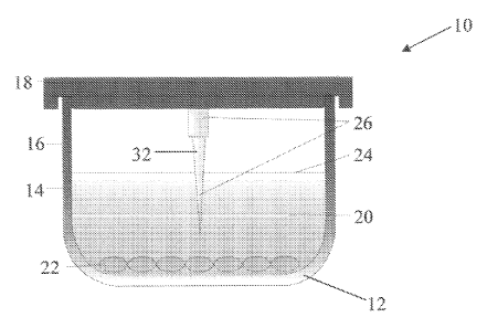
[0060] Referring to Figures 1A-D, cross-sectional schematic views of an
apparatus 10 are shown, in accordance with an illustrative embodiment of the
invention. The apparatus 10 includes a vessel 12 having an inner wall 14 and
an
outer wall 16. The vessel 12 can be, for example, a multiwell tissue culture
plate
having a plurality of wells. The apparatus 10 can include a removable lid 18
which
covers the vessel 12. Vessel 12 is intended to contain a fluid overlay 20,
such as
cryopreservation media or growth media, that covers a cellular monolayer 22.
The
- 13 -
CA 02760175 2011-10-26
WO 2010/127158
PCT/US2010/033032
apparatus 10 further includes an ice nucleating device 26 which can be
attached to or
integral with the vessel lid 18, as shown in Figures lA and 1B. The ice
nucleating
device 26 or the lid 18 are configured such that all or a portion of the ice
nucleating
device 26 comes into contact with the fluid overlay 20 in the vessel 12 when
the lid
18 is placed on the vessel 12.
[0061] Referring to Figures 1C and 1D, in some embodiments the ice
nucleating device 26 is attached to or integral with a removable insert 28
which is
separable from the vessel 12 and the lid 18. The insert 28 can be configured
to
releasably engage the top 30 of the vessel 12 and/or can be configured to
engage the
inner wall 14 or outer wall 16 of the vessel 12. Vessel inserts are well known
and a
person of skill in the art will appreciate that many different insert
configurations can
be used in accordance with the invention. The ice nucleating device 26 or the
insert
28 are configured such that all or a portion of the ice nucleating device 26
comes
into contact with the fluid overlay 20 in the vessel 12 when the insert is
placed in or
on the vessel.
[0062] Referring to Figure 1A, the ice nucleating device 26 can comprise a
primary needle-like or spike-shaped protrusion 32. Referring to Figures 1B and
1C,
the ice nucleating device 26 can include one or more secondary protrusions 34
that
project from the primary protrusion 32. The secondary protrusions 34 provide
additional nucleation sites for ice crystal formation.
[0063] Referring to Figure 1D, the ice nucleating device 26 can comprise a
stem 36 that supports a surface 38 which has one or more secondary protrusions
34.
In some embodiments, only secondary protrusions 34 come into contact with the
fluid overlay 20 in the vessel 12. The surface 38 can be any suitable shape
and can
be, for example, substantially disc-shaped and sized smaller than the vessel
opening
to provide many ice nucleation points across the entire vessel (e.g., a well).
[0064] In some embodiments, the ice nucleation device described herein is
attached to or integral with an interior surface of the vessel, such as an
inner wall or
bottom.
[0065] In various embodiments, more than one ice nucleation device is
disposed in or on the vessel.
- 14 -
CA 02760175 2011-10-26
WO 2010/127158
PCT/US2010/033032
[0066] Regardless of whether the ice nucleating device is located on the
lid, on a removable insert, or on an interior surface of the vessel sidewall,
the
apparatus is configured such that one or more primary or secondary protrusions
come into contact with the fluid overlay covering the cellular monolayer. In
preferred embodiments, the protrusions of the ice nucleation device do not
come in
contact with the cell monolayer.
[0067] The invention is particularly useful for high throughput screening of
multiwell plates. Thus, in preferred embodiments, the apparatus includes a
tissue
culture plate which comprises more than one vessel (i.e., well), such as 4-
well, 6-
well, 8-well, 12-well, 96-well, 384-well, or 1536-well plates. A person of
skill in
the art will appreciate that the invention can be used in connection with a
multiwell
plate having any number of wells.
[0068] Figures 1E-G show an ice nucleating device configured as a
removable insert for a multiwell plate, in accordance with an illustrative
embodiment of the invention. Referring to Figure 1E, a top view of an ice
nucleating device insert 100 is shown. Ice nucleating device insert 100
includes a
base 102 that supports a plurality of protrusions 106. Protrusions 106 can be
integral with base 102 or protrusions 106 can separate from base 102 and
configured
for insertion into the base 102. In some embodiments, protrusions 106 can be
partitioned into two or more zones 104, 104', 104". For example, when an
insert
includes two or more different types of protrusions, the different types of
protrusions
can be segregated into different zones. In some embodiments, when multiple
types
of protrusions are used, they can be arranged randomly or in repeating
patterns. In
addition, one or more protrusions can be omitted such that when the insert is
placed
on the multiwell plate, the corresponding wells have no protrusion.
[0069] Figure 1F shows a side view of an ice nucleating device insert 100,
and Figure G shows a perspective view of an ice nucleating device insert 100,
in
accordance with an illustrative embodiment of the invention.
[0070] Figures 1H and 11 show an ice nucleating device base 108
configured as a removable insert for a multiwell plate, in accordance with an
illustrative embodiment of the invention. Referring to Figure 1H, the base 108
contains a plurality of through-holes 110 for receiving protrusions. The
insert base
- 15 -
CA 02760175 2011-10-26
WO 2010/127158
PCT/US2010/033032
108 shown in Figures 1H and 11 is dimensioned such that it can be removeably
inserted into a 96 well plate. As will be appreciated, the embodiment shown is
for
illustrative purposes only, and the dimensions of the insert readily can be
configured
for use with multiwell plates of any size. Referring to Figure 11, a cross-
section
through plane A-A in Figure 1H is shown. In this embodiment, insert base 108
includes through-holes 110 for receiving protrusions. Through-holes 110 have a
first, narrower diameter through which the protrusion fits, and a second wider
diameter for engaging a base on each protrusion. Thus, in this embodiment,
protrusions are inserted from the opposite side of the insert from which they
project.
In some embodiments, recesses are used rather than through-holes for receiving
protrusion bases.
[0071] In some embodiments, the insert is made of the same material as the
multiwell plate, for example a plastic such as polystyrene, polycarbonate, or
acrylic.
The thickness of the insert base will vary depending on the construction
material. In
some embodiments, the thickness of the insert is, for example, about 0.1 mm to
about 10 mm, more preferably about 1 mm to about 3 mm, and more preferably
still
about 2 mm. It will be readily appreciated that the foregoing dimensions are
illustrative only and that any suitable dimensions and configurations can be
used
without departing from the scope of the invention.
[0072] In addition, protrusions can be made of any suitable material, and
can be made out of the same material as the base for ease of manufacture. In a
preferred embodiment, the protrusions are made of a plastic such as
polystyrene.
[0073] Figure 1J, shows a side-view of an ice nucleating protrusion 120
having a single cone-shaped point 122, in accordance with an illustrative
embodiment of the invention. Protrusion 120 has a first end forming a cone-
shaped
point 122, a second end forming a base 126, and a stem 124 connecting the
first end
and the second end. Stem 124 is long enough to contact fluid in the well of
the
vessel (e.g., a well of a multiwell plate). As will be appreciated, the length
and
width of stem 124 can vary without departing from the scope and spirit of the
invention. In a preferred embodiment for use with 96 well plates, stem 124 has
a
length of about 5 to about 15 mm, and more preferably about 10 mm. In some
embodiments, stem 124 is substantially columnar in shape and has a diameter of
- 16-
CA 02760175 2011-10-26
WO 2010/127158
PCT/US2010/033032
about 1 mm to about 2 mm, and more preferably about 1.5 mm, however any
suitable shape can be used. In some embodiments, the cone-shaped point 122 can
have a height of about 0.25 mm to about 1 mm, and more preferably about 0.67
mm,
and the cone can have a sharp point. Figure 1K shows a top view of protrusion
120.
It will be readily appreciated that the foregoing dimensions are illustrative
only and
that any suitable dimensions and configurations can be used without departing
from
the scope of the invention.
[0074] In various embodiments, ice nucleating protrusions can have a base
for engaging or securing the protrusion in a base insert. Referring again to
Figure
1J, protrusion 120 has a base 126 that is wider than stem 124. In a preferred
embodiment, base 126 has a diameter of about 3 mm to about 7 mm, and more
preferably about 4.5 mm. Thus, stem 124 passes through the first, smaller
diameter
of through-hole 110 in insert base 108 (Figure 11) and base 126 is received by
the
second, large diameter of through-hole 110. Protrusion base 126 can be secured
in
through-holes 110 or recesses by press fit, snap fit, adhesive, welding (e.g,.
sonic
welding), or any other suitable fastening mechanisms. In some embodiments,
there
is no widened base 126 and the second end of the protrusion is substantially
the
same diameter as the stem 124. It will be readily appreciated that the
foregoing
dimensions are illustrative only and that any suitable dimensions and
configurations
can be used without departing from the scope of the invention.
[0075] Figures 1L to 1P show an ice nucleating protrusion 128 having a
plurality of cone-shaped points 130, in accordance with an illustrative
embodiment
of the invention. Referring to Figure 1, which shows a side view of protrusion
128,
the plurality of cone-shaped points 130 can have a height of about 0.1 mm to
about 1
mm, and more preferably about 0.25 mm. Any number of cone-shaped points can
be included, such as for example, between about 2 points and about 50 points,
and
more preferably between about 5 points and about 15 points, and more
preferably
still about 7 points to 9 points. In some embodiments, one or more cones have
sharp
points. In Figures 1M-P, an 8 point embodiment is shown with a cone-shaped
point
in the center encircled by 7 cone-shaped points. As shown in Figure 10, the
cone-
shaped points 130 are spaced about 0.48 mm from peak to peak and at an arc of
about 51.43 degrees from peak to peak relative to the center cone-shaped
point. It
- 17-
CA 02760175 2011-10-26
WO 2010/127158
PCT/US2010/033032
will be readily appreciated that the foregoing dimensions are illustrative
only and
that any suitable dimensions and configurations can be used without departing
from
the scope of the invention.
[0076] Figure 1M shows a perspective view of protrusion 128, and Figure
10 shows a close-up perspective view of the first end of protrusion 128.
Figure 1N
shows a top view of protrusion 128.
[0077] Figures 1Q and 1R show a protrusion 134 having a plurality of
pyramid-shaped points 136. The plurality of pyramid-shaped points 136 can have
a
height of about 0.1 mm to about 1 mm, and more preferably about 0.25 mm. Any
number of pyramid-shaped points 136 can be included, such as for example,
between about 2 points and about 50 points, and more preferably between about
20
points and about 40 points. In some embodiments, the pyramids have sharp
points.
Referring to Figure 1R, in some embodiments the pyramid-shaped points 136 are
spaced about 0.24 mm from peak to peak and about 0.24 mm from trough to
trough.
It will be readily appreciated that the foregoing dimensions are illustrative
only and
that any suitable dimensions and configurations can be used without departing
from
the scope of the invention.
[0078] As will be appreciated, any suitable shape can be used for the point
or points of a protrusion. Where multiple points are used the points can be
the same
shape or different shapes, and the points can be evenly spaced or randomly
spaced
and arranged randomly or in a pattern.
[0079] In
some embodiments, the base or cover has no through-holes or
recesses and the ice nucleating protrusions are joined directly to the base or
cover.
In some embodiments, the base or cover and the ice nucleating protrusions are
manufactured (e.g., molded or machined) as a single integral unit.
[0080] In preferred embodiments, the vessel is a multiwell plate 50 which
has been insulated to promote even cooling between exterior (i.e., outer) and
interior
(i.e., inner) wells during cryopreservation. Referring to Figure 2A,
insulating
material 52 is applied to the free space 54 between the exterior wells 56 and
the
outer wall 58 of the multiwell plate 50 such that a portion of the free space
52 in the
periphery of the multiwell plate 50 is filled with insulating material 52. In
some
embodiments, the insulting material 52 fills substantially all of the free
space 54
- 18-
CA 02760175 2011-10-26
WO 2010/127158
PCT/US2010/033032
between the exterior wells 56 and the outer wall 58 of the multiwell plate 50.
Insulating material can also be applied to some or all of the free space
between two
or more exterior wells 56. Insulating material can also be applied to some or
all of
the free space 64 between two or more interior wells 60 of the multiwell
plate. In
some embodiments, insulating material 52 is applied to the underside of a
multiwell
plate 50 where the free space 52 is accessible.
[0081] In some embodiments, the insulating material is applied such that it
fills some or all of the free space surrounding one or more wells of a
multiwell plate.
In another embodiment, the insulating material is part of, or occupies part
of, each
well of the multiwell plate. In yet another embodiment, the insulating
material
surrounds the exterior or interior wells, and/or occupies the interior of at
least one
well or vessel.
[0082] Referring to Figure 2B, the insulating material 62 can be attached to
or integral with the lid 18 of the vessel, and the insulating material can be
configured
to occupy some or all of the vessel's interior above the fluid line 24 of the
fluid
overlay 20.
[0083] In a preferred embodiment, the insulation material is the same
material utilized in the tissue culture plate such as, for example, acrylic,
polycarbonate and polystyrene. Using the same material is advantageous for
ease of
manufacturing. In some embodiments, the insulating material can consist of a
specific insulating material such as acrylic caulk, weather stripping, hot
glue, and
other forms of insulating material including but not limited to caulk, foams,
sprays,
or sheets. The insulating material can be attached to or integral with the
vessel, or
the insulating material can be detachable from the vessel.
[0084] The present invention overcomes the previous limitations by
providing an apparatus, method, and composition for the production of frozen
ready
to use cell cultures for diagnostic assays, comprising the steps of providing
cells, and
a substrate selected from the group consisting of glass and plastic; placing
the cells
on the substrate under conditions such that the cells are attached to the
substrate to
produce a cell monolayer; and freezing the cell monolayer under conditions
such
that the cell monolayer remains intact and attached to the substrate and is
viable
upon thawing. In the preferred embodiment, the substrate is the plastic
comprising
- 19-
CA 02760175 2011-10-26
WO 2010/127158
PCT/US2010/033032
the well of a multiwell plate. In still further embodiments, the substrate is
glass.
However, it is not intended that the present invention be limited to any
particular
substrate. Furthermore, while attached cell monolayers are preferred, the
invention
is not limited to cell monolayers. The invention can also be used to
cryopreserve
other complex cellular structures, such as tissues and organs.
[0085] One embodiment of the present invention provides a container
system to promote and initiate the nucleation of ice. In order to successfully
freeze
biological materials in a reproducible manner, it is common practice to cool
the
materials to a temperature below the melting point thereof, then after a short
period
of thermal equilibration, to nucleate ice in the supercooled material. In the
present
embodiment, the container is a multiwell tissue culture plate where a
disposable and
removable insert having a needle-like protrusion would be suspended in the
media
within each of the culture wells; in some embodiments, the well is a vial
(cryovial);
the insert can contain a single sterile protrusion or many protrusions having
one or
more nucleating sites whereby the liquid media comes in contact with the ice
nucleating device. In preferred embodiments, the ice nucleating device is part
of a
container which contains a disposable and removable insert; the insert can
comprise
one or more ice-nucleating structures (i.e., protrusions) extending from the
lid of the
tissue culture plate into the media surrounding the cell culture. The ice
nucleating
protrusions are preferentially made from plastic, however it is not intended
that the
present invention be limited to any particular material. In further
embodiments, the
ice nucleating device would comprise one or more ice-nucleating protrusions
extending from the sides or bottom of the wells of the tissue culture plate
into the
media surrounding the cell culture.
[0086] Once thawed, the removable insert containing the ice nucleating
device can be removed. Another embodiment of the present invention provides a
container with a media composition for effective cryopreservation of cells and
tissues. The preservation media is a nutrient solution which can be protein-
free and
sera-free and can be adapted for cellular and tissue cryopreservation. The
cryogenic
preservation solution is preferentially CRYOSTORTm (BioLife Solutions, Inc.,
Bothell, WA). While CRYOSTORTm is the preferred embodiment combined with
DMSO as an optimal cryoprotective agent, other cryoprotective agents can be
used
-20-
CA 02760175 2011-10-26
WO 2010/127158
PCT/US2010/033032
comprising of one or more selected from the group consisting of sucrose,
trehalose,
lactose, glucose, DMSO, propylene glycol, ethylene glycol, a dextran,
glycerol,
hydroxyethyl starch, polyvinyl pyrrolidine, formamide, 1-2-propanediol,
ethanol,
methanol, and polyethylene glycol.
[0087] The present invention also provides methods for the production of
attached, frozen, ready-to-use cell monolayers comprising the steps of: 1)
providing
cells and a multiwell tissue culture plate, which can optionally include the
aforementioned insulating material surrounding the exterior of any wells
and/or
occupying some portion of the interior of any wells; 2) placing the cells on
the
selected multiwell tissue culture plate under conditions such that the cells
are
attached to the substrate to produce a cell monolayer; 3) the cell culture
media is
replaced with a protein-free and serum-free cryopreservation medium under
sterile
conditions, the preferred biopreservation media being CRYOSTORTm; 4) the
aforementioned ice nucleating device present as incorporated in any of the
aforementioned descriptions; 5) the entire container is then placed in a
vacuum
sealed air-tight package; 6) the sealed plate is then placed and enclosed in a
Styrofoam container, which provides a reasonably consistent and reproducible
rate
of cooling. It is not intended that the container be vacuum sealed. It is also
not
intended that the container be limited to Styrofoam , however, as any
container
providing a controlled rate of temperature reduction can be utilized. In some
embodiments, the Styrofoam container can include isopropyl alcohol which the
plates are bathed in while cooling, and the isopropyl bath can be pre-chilled
to about
0 to -10 C before adding the multiwell tissue culture plate. In preferred
methods,
the multiwell tissue culture plates are incubated at about 4 C for about 10
minutes,
before the plates are transferred to -80 C for storage.
[0088] Once in the container, the entire apparatus is placed directly into a
freezer preferably set at a temperature of -80 C; the temperature of the
sample is
then reduced at a rate near 1-2 C/minute although variations of the cooling
rate can
be used; when the preservation media temperature reaches a temperature within
the
preferred range of -5 to -10 C, uniform ice-nucleation occurs; the temperature
of the
culture then continues to cool to the designated temperature of the freezer;
the
preferred end temperature is -80 C, but it is not intended that the present
method be
-21 -
CA 02760175 2011-10-26
WO 2010/127158
PCT/US2010/033032
limited to this temperature; once frozen, the cell cultures can be stored
indefinitely,
although the preferred storage time would be 1 day to 1 year. When needed for
use,
the cell cultures are removed from the freezer and preferably thawed by
immersing
the entire package in a liquid bath with a temperature of 37 C; however in
some
embodiments, the invention can be thawed in an apparatus without liquid. In
certain
embodiments, the apparatus is a cell culture incubator with a temperature of
37 C;
while the preferred temperature is 37 C, the invention is not limited to an
exact
temperature of 37 C; once thawed, the ice nucleating device is removed; the
insulating material can be removed if possible but removal is not required;
the
cryopreservation medium is removed and replenished with cell culture growth
media; whereby under the combined conditions results in a cell monolayer that
remains attached to the substrate with minimal loss of viability and function
when
compared to the starting material.
[0089] In another embodiment, the sealed plates can be transferred to a -
20 C freezer following the 10 minute incubation at about 4 C. The sealed
plates are
then incubated for about 15 minutes at about -20 C and then transferred
directly into
a -80 C freezer. In this embodiment of the freezing method, no styrofoam
cooler/alcohol bath is used. The sealed plates remain in the -80C freezer for
storage.
In yet another embodiment, the sealed plates can be transferred to an
automated
controlled rate cooling device. In this method, the plates can be transferred
following the 10 minute incubation at about 4 C to a pre-cooled chamber at
about
4 C. Alternatively, the plates can be transferred directly to the chamber
without
prior incubation at about 4 C. Once the plates are placed into the chamber, a
preset
cooling rate can be run to freeze the plates. Once the temperature reaches
about -
80 C in the chamber, the plate can be transferred to a -80 C freezer for
storage.
[0090] For the present invention, the preferred method incorporates the
combination of the CRYOSTORTm cryopreservation media, the ice nucleating
device, and optionally the insulating material. This unique combination which
is
unlike the methods currently available provides the cell monolayer with a
serum-
free, protein free solution optimized for storage of cells at sub-zero
(frozen)
temperatures and a means of controlling and promoting uniform nucleation of
ice
near the melting point of the fluid; the combined method creates an optimal
- 22 -
CA 02760175 2011-10-26
WO 2010/127158
PCT/US2010/033032
circumstance that allows for cryopreservation and exceeds in the
cryopreservation
process by allowing uniform cell density and viability from well to well of a
multiwell plate following the cryopreservation process, and improved overall
post-
thaw viability and function. Levels of post-thaw viability and function are
dependent
upon the freezing method applied and the cell type used. Once the combined
cryopreservation medium and ice-nucleating device are added to the cell
monolayer
following the preferred method, the apparatus would be vacuum sealed using
standard technique to provide optimal freezing and storage conditions. The
sealed
apparatus can then be placed into a container such that the apparatus does not
come
in direct contact with the freezing element; the apparatus can be completely
enclosed
within the container; the container can provide some insulation such that the
temperature of the fluid in the apparatus is reduced at a controlled rate. An
example
of such could be a container made of Styrofoam foam. The container with the
apparatus can then be placed into a freezer or freezing device; the preferred
freezing
device reaches an end temperature between -70 C to -90 C; while preferred, the
present method is not limited to this temperature range. Under the present
conditions, ice-nucleation within the apparatus typically occurs when the
media
temperature within the wells reaches -5 to -10 C and ice-nucleation from well
to
well over the entire multiwell plate will be uniform. Once frozen, the cell
monolayer can be maintained in such a state until required for use. In the
preferred
embodiment, the cryopreserved monolayer could be stored for 1 day to 1 year.
[0091] Upon use, the cryopreserved apparatus can be removed from the
freezer or freezing device and submerged in a liquid bath; in the preferred
method
the bath would be water maintained at a temperature near 37 C; the temperature
is
not limited to 37 C, but to achieve optimal post-thaw viability and function
the
temperature should be kept between 25-40 C. While this is the preferred
method,
additional methods can be applied such that the apparatus is placed in a dry
environment like an incubator or heating block. Optimal thawing rates are best
achieved if the entire outer surface area of the apparatus can be exposed to
the
thawing mechanism. In the preferred embodiment, the thawed multiwell apparatus
would be removed from the sealed container, the mechanical ice nucleating
device
removed, and the cryopreservation media removed and replaced with standard
- 23 -
CA 02760175 2011-10-26
WO 2010/127158
PCT/US2010/033032
culture medium. These steps can be performed under sterile or non-sterile
conditions. The thawed monolayers can be used for testing and evaluation at
any
time thereafter.
[0092] The present invention overcomes previous limitations in the field
by providing an apparatus, related method, and composition that results in
uniform
freezing of the cell monolayers consistently across the entire multiwell
tissue culture
plate and potentially providing improved post-thaw cell viability and
function,
which in some cell types may be comparable to that of non-frozen monolayers.
Specifically, the present invention improves upon previous inventions by
including
both an insulating material to aid consistent well to well cooling and warming
and a
mechanical device to control ice-nucleation during the freezing process.
Furthermore, the present invention includes the use of a unique protein-free
and
serum-free preservation medium designed specifically for maintenance,
protection,
and storage of cells held in a frozen state. Additionally, the combination of
the
insulating material and mechanical ice nucleating device along with the
CRYOSTORTm cryopreservation media provides for an optimal preservation
environment and homogeneous ice-nucleation resulting in improved viability and
function of the cell monolayer. Finally, the present invention overcomes the
limitations of previous inventions by providing specific and simplified method
for
the freezing, storage and thawing of cell monolayers for ready-to-use formats.
[0093] The following examples are provided for illustration, not limitation.
Example 1: Cooling profile of interior and exterior wells of a 96-well
plates and
the effects of an insulating material.
[0094] In this example, 96-well tissue culture plates were used to
investigate the differences in cooling rate between interior and exterior
wells and the
efficacy of including an insulating material. For this example, the insulating
material
was applied to the outer underside edge of the exterior wells as demonstrated
in
Figure 2A. To insulate plates, standard clear acrylic latex caulk plus
silicone was
applied to the underside exterior wells of the 96-well tissue culture plates
previously
described. Caulk was applied into the outside gap found between the exterior
wells
and the outer plate edge of the tissue culture plates (see Figure 2A). Caulk
was
- 24 -
CA 02760175 2011-10-26
WO 2010/127158
PCT/US2010/033032
applied with a standard caulk gun. The caulk was added to the exterior well
gaps
until any obvious air space was filled. The excess caulk was wiped away and
leveled
off so that the caulk insulating layer was flush with the plate well bottoms.
Plates
were then left overnight so that the caulk could set. Once prepared, plates
were
tested as described to determine insulation efficacy.
[0095] Plates without an insulating material were tested to compare the
results and efficacy of the insulating material. After the insulating material
was
added, 80 ill of culture media was added to each well. A thermocouple was
attached
to the inside of a centrally located interior well and another thermocouple
was
attached to the inside of an exterior well. The tips of the thermocouples were
immersed in the liquid culture media but did not touch the well surface. The
lid was
placed on the plates and the plates were then placed into a -80 C freezer.
Temperature readings were collected over a time period of 0-30 minutes.
[0096] As shown in Figure 3, the inclusion of an insulating material
effectively reduces the variability in cooling rates observed between the
interior and
exterior culture wells of the 96-well plate. Without the inclusion of an
insulating
material, exterior wells cooled at a much faster rate as compared to interior
wells.
Using the cooling process, exterior wells not having an insulating material
reached a
temperature of -8.5 C after 20 minutes, while the interior well had only
reached -
3.8 C. This range in temperature differences could significantly affect the
ice-
nucleation events from well to well. Exterior wells typically have ice
nucleation
events at an earlier time point compared to interior wells. The significant
differences
in temperature from well to well relate directly to differences observed in
post-thaw
cell recovery and viability. When exterior wells were surrounded with the
addition
of an insulating material, cooling rates much more closely resembled the
cooling
rate of the interior wells. After 20 minutes, the temperature of the exterior
well with
an insulating material was at -4.3 C, while the temperature of the interior
well was
at -3.8 C. The results of this series of experiments demonstrate the
feasibility and
efficacy of using an insulating material to aid in improving the consistency
of well
to well cooling and eventual ice-nucleation.
- 25 -
CA 02760175 2011-10-26
WO 2010/127158
PCT/US2010/033032
Example 2: Freezing of NHDF cell monolayers in 96-well plate testing different
cryopreservation media and freezing methods
[0097] In this example, different cryopreservation media were investigated
as models to cryopreserve NHDF, normal human dermal fibroblast, cell
monolayers
in multiwell plates following three separate freezing methods. NHDF cells were
cultured and subsequently plated at an equal number of cells/well in a 96-well
culture plate (BD Falcon). The cultures were left undisturbed for one day to
achieve
confluent attached cell monolayers. Prior to preparing the cultures for
preservation,
an initial assessment of the metabolic activity was performed to determine
cell
viability prior to freezing. alamarBlue (TREK Diagnostic Systems) was
utilized to
assess cell viability.
[0098] alamarBlue is soluble, stable in culture medium and is non-toxic.
The continuous monitoring of cells in culture is therefore permitted.
Specifically,
alamarBlue does not alter the viability of cells cultured for various times
as
compared to assessment by trypan blue exclusion. Because alamarBlue is non-
toxic, the cells under study can be returned to culture or used for other
purposes
including histological studies. Proliferation measurements with alamarBlue
can be
made by using either spectrophotometry or fluorometry to monitor the
absorption of
alamarBlue supplemented cell culture media at two wavelengths.
[0099] To perform the assay, alamarBlue was used according to
manufacturer instructions. Briefly, cell culture media was removed from the
wells
and alamarBlue was added (100 [d/well) to each well and incubated at 37 C for
1
hour. Following the incubation, the plates were analyzed with a fluorescent
microplate reader (Tecan; Infinite 200 model) with an excitation at 530-560 nm
and
emission at 590 nm. The MagellanTM software (Tecan, Switzerland) is used in
combination with Infinite 200 fluorescent microplate reader for fluorescent
data
acquisition. Relative fluorescent units for pre-freeze cell monolayers were
set to
100% (Control) and the experimental conditions are compared to the pre-freeze
control.
[0100] In order to assess the data and efficacy of each experiment, the raw
fluorescent values were collected via a fluorescent plate reader. The raw
fluorescent
values or relative fluorescent units were collected for each well of the 96-
well plate.
-26-
CA 02760175 2011-10-26
WO 2010/127158
PCT/US2010/033032
For each study/experiment, the relative fluorescent units were used to
determine
relative cell viability (per manufacturer's (TREK Diagnostic Systems') product
materials). For the current studies, the relative fluorescent units collected
for the
various experimental conditions tested were compared to non-frozen (37 C
control)
plated cells. Typically, an average relative fluorescence of at least 6 wells
of a 96-
well plate was determined (more depending on the condition tested). The
average
numbers of the experimental conditions were then compared to the control non-
frozen average and a percentage was determined. The variability observed
between
each of the tested wells for each experimental was expressed as either the
percent
error or standard deviation (performed with Excel software).
[0101] Following the pre-freeze viability assessment, the alamarBlue was
removed and the various cryopreservation media were added. Four different
cryopreservation media conditions were tested: NHDF complete cell culture
media +
5% DMSO (Media 5%), NHDF complete cell culture media + 10% DMSO (Media
10%), CRYOSTORTm +5% DMSO (C55), and CRYOSTORTm + 10% DMSO
(CS10). The 96-well plate was divided into 4 quadrants consisting of 24 wells
in
each. 80p1/well of the respective cryopreservation media was added to each 24-
well
quadrant. No ice nucleating device was used. The 96-well plate was then placed
into a Ziploc bag and sealed. The sealed plate was then placed at 2-8 C for
10
minutes prior to freezing. After 10 minutes, plates were subject to freezing
via three
separate methods: (1) Alcohol bath method - The sealed plate was then placed
into a
Styrofoam box and the entire container was then put into a -80 C freezer. The
wells
were then visualized for seeding events (ice nucleation). With this method,
seeding
events were noted as early as 20 minutes while the final seeding event
occurred
around 60 minutes post storage; (2) -80 C freezer method - The sealed plate
was
transferred to a -20 C freezer and stored for 15 minutes and then transferred
directly
into a -80 C freezer and freezing continued. With this method, seeding events
were
noted as early as 10 minutes while the final seeding event occurred around 40
minutes post storage; (3) Controlled rate freezer method - The sealed plate
was
transferred to an automated control rate freezing device (Cryomed) with the
chamber
temperature set to 4 C. Once the plate was placed into the chamber, the
temperature
of the chamber was reduced 1 C/minute to a final temperature of -80 C. When
the
-27 -
CA 02760175 2011-10-26
WO 2010/127158
PCT/US2010/033032
program was completed, the plate was transferred to the -80 C freezer for
storage.
With this method, no seeding events were visualized since the plate was inside
the
chamber. The plates were kept at -80 C for 24 hours. Plates were then removed
and
submerged completely in a 37 C water bath. Within 5 minutes, all of the wells
had
thawed. The cryopreservation media was removed from the plate, replenished
with
100[d/well of fresh culture media, and the entire plate was placed in a 37 C
incubator to recover. Cell viability was assessed 24 hours post thaw as
described
previously for the pre-freeze controls.
[0102] Figure 4 is an image obtained from the MagellanTM software used in
combination with the fluorescent microplate reader. The image portrays the
relative
fluorescent intensity based on NHDF cell density and metabolic activity for
each
well as a color for all wells of the test plate. Color associated with
fluorescent
intensity can also be correlated to viability as depicted on Figure 5.
Relative
fluorescent units for pre-freeze cell monolayers were set to 100% (Control)
and the
experimental conditions are compared to the pre-freeze control. Using this
format, a
well depicted as red has a high fluorescent intensity and a high relative
viability,
while a well depicted as blue has a very low associated relative viability.
The overall
scale is determined by the well having the highest fluorescent intensity
(darker red
color) and the well with the lowest overall fluorescent intensity (darker blue
color).
Wells having like colors have similar fluorescent intensities and similar
relative
viability.
[0103] As depicted in Figure 4, the results of this experiment indicate that
with each freezing method tested, the CRYOSTORTm solutions result in the
highest
recovery of NHDF cells compared to the traditional media + DMSO solutions. Of
the CRYOSTORTm solutions, the CS5 results in the best recovery and these
general
recovery trends are consistent with results obtained with NHDF cells
cryopreserved
in traditional cryovial formats (suspended cells). Additionally, results
indicate that
the alcohol bath method provides the most optimal freezing method while the
controlled rate freezer method may be the least optimal with the NHDF cell
model.
A high level of well to well variability is evident with each of the solutions
in each
of the freezing methods.
-28-
CA 02760175 2011-10-26
WO 2010/127158
PCT/US2010/033032
[0104] As shown in Figure 5, the results of this experiment demonstrate
that cell monolayers cryopreserved in the CRYOSTORTm media performed
significantly better than the monolayers cryopreserved with the traditional
media
and DMSO (CS10 and CS5 compared to Media 10% and Media 5%). Cell
monolayers cryopreserved in the CS5 performed the best with 70%, 40%, and 20%
viability post-thaw respectively for each of the freezing methods compared to
the
control, while cell monolayers cryopreserved in media and 5% DMSO resulted in
less than 10% viability post-thaw in each of the freezing methods. A high
level of
variability in well-to-well viability was experienced in each of the
conditions using
CRYOSTORTm due to the level of random ice nucleation noted during the freezing
process. Essentially no variability is noticed with the media and DMSO
conditions,
but this is because no cells were recovered from any of the freezing methods.
CRYOSTORTm media showed the highest post-thaw viability for cryopreserving
cell monolayers when compared to standard cryopreservation media.
Example 3: Freezing of NHDF cell monolayers in 96-well plate testing efficacy
and variability of CRYOSTORTm CS5 cryopreservation media combined with or
without an ice nucleating device
[0105] In this example, an ice nucleating device as described in Figure 1
was developed and utilized to determine device utility and efficacy compared
to
having no ice nucleating device. For the provided examples demonstrated in
Figures
6-9, the ice nucleating device is essentially as described in Figure 1. The
ice
nucleating device was prepared from the same material as the 96-well tissue
culture
plates (BD Biosciences, Billerica, MA). Each device was cut from the culture
plate
material using a razor blade to a length of about 8cm and a width of about lcm
(the
exact width and length varied slightly for each device). The devices were
rectangular
in shape and did not have a single pointed end. The edges were rough,
resembling
multiple ice nucleating points. Once the individual ice nucleating devices
were
prepared, a soldering device was used to melt one end of the device and
allowing it
to be attached to the lid of a tissue culture plate. Once cooled, the device
was firmly
attached to the lid and protruded from the underside of the lid as depicted in
Figures
lA and 1B. In all, 48 ice nucleating devices were prepared and attached
(1/well)
-29-
CA 02760175 2011-10-26
WO 2010/127158
PCT/US2010/033032
making up half of an entire 96-well tissue culture plate. Each device was
confirmed
to penetrate the liquid fill level of the tissue culture well without touching
the
bottom surface of the well.
[0106] For this study, NHDF cell monolayers were utilized, and cell
monolayers were prepared and formed as described in previous examples. NHDF
cell monolayers were prepared for cryopreservation essentially as described in
Example 2. Briefly, CRYOSTOR CS5 was added to all wells except for the 4 wells
in the center of the plate. These wells received standard cell culture media
with 5%
DMSO as the cryopreservation solution. Following the addition of the chilled
cryopreservation solution, a prototype ice nucleating device lid, described
above,
containing ice nucleating spikes protruding from the underside of the lid was
placed
on the plate. The lid contained a single ice nucleating device for each well.
Half of
the plate lid was designed to contain an ice nucleating device while the other
half did
not, which allowed for intra-experimental comparison. Each ice nucleating
device
was attached directly to the plate lid surface and an ice nucleating device
extended
into the center of each cell culture well when the lid was placed on the
tissue culture
plate. The ice nucleating devices were long enough to penetrate the liquid
medium
but did not touch the well surface or the cell monolayer.
[0107] After the lid containing the ice nucleating device was added to the
plates, the plate was placed into a freezer-safe plastic bag and vacuum
sealed. Plates
were then subjected to a controlled freezing rate. Plates were first stored at
2-8 C for
10 minutes and then placed into a Styrofoam foam cooling chamber at -80 C. The
Styrofoam foam cooling chamber contained enough isopropyl alcohol such that
the
plate when placed into the chamber would be completely covered. The Styrofoam
foam cooling chamber was previously chilled such that the bath temperature was
around 0 C when plates were placed into the chamber. A 1-2 C per minute
cooling
rate was achieved. Plates remained in the Styrofoam foam cooling chamber for 3
hours. After 3 hours, plates were removed and stored at -80 C for 24 hours,
which
was sufficient time to ensure that the media in each well froze solid. Plates
were
thawed using a 37 C water bath as described previously in Example 2. Once
thawed,
plates were removed from the freezer-safe plastic bag. The ice nucleating
device was
removed from the wells along with the cryopreservation solution, and fresh
cell
- 30 -
CA 02760175 2011-10-26
WO 2010/127158
PCT/US2010/033032
culture media was added. The plates were then evaluated as in previous
experiments.
Data shown are representative of multiple experiments. The plates were
evaluated
using a plate reader as described in Example 2.
[0108] Figure 6 is an image obtained from the MagellanTM software used in
combination with the fluorescent microplate reader. The image portrays the
relative
fluorescent intensity based on NHDF cell density and metabolic activity for
each
well as a color for all wells of the test plate. Color associated with
fluorescent
intensity can also be correlated to viability as depicted on Figure 8.
Relative
fluorescent units for pre-freeze cell monolayers were set to 100% (Control)
and the
experimental conditions are compared to the pre-freeze control. Using this
format, a
well depicted as red has a high fluorescent intensity and a high relative
viability,
while a well depicted as blue has a very low associated relative viability.
The overall
scale is determined by the well having the highest fluorescent intensity
(darker red
color) and the well with the lowest overall fluorescent intensity (darker blue
color).
Wells having like colors have similar fluorescent intensities and similar
relative
viability.
[0109] As shown in Figure 6, the addition of the ice nucleating device
effectively reduces well to well variability compared to wells not having an
ice
nucleating device. The wells having the ice nucleating device have a high
number of
wells with a very similar color range, while the wells without an ice
nucleating
device have a much wider range of color from one well to another. The large
variation in color seen from well to well is directly correlated to the
increased
variability in cell density and viability from well to well observed without
the
presence of an ice nucleating device. It should be noted that fluorescent
intensity
was very low in all wells having media and 5% DMSO as the cryoprotective
agent.
When the relative fluorescent intensity for each of the wells is plotted on a
graph as
shown in Figure 7, the drastic decrease in well to well variability using an
ice
nucleating device is easily observed from the lower standard deviation. In
Figure 7,
the average relative fluorescent units for the respective wells is shown. To
demonstrate the range in well to well variability, the standard deviation for
wells
having an ice nucleating device and those without an ice nucleating device is
depicted. The standard deviation is significantly less for the sample wells
containing
-31-
CA 02760175 2011-10-26
WO 2010/127158
PCT/US2010/033032
the ice nucleating device as compared to the standard deviation of the sample
wells
without an ice nucleating device. In addition, the relative fluorescent units
obtained
from non-frozen control samples is averaged and shown in Figure 7. Notably,
the
standard deviation of the wells with the ice nucleation device is comparable
to that
of the non-frozen control. Furthermore, the overall average relative
fluorescent
intensity for wells having an ice nucleating device is higher than that of
those
without an ice nucleating device. When compared to the non-preserved control,
this
correlates to an average of 60% viability for wells with an ice nucleating
device and
40% for wells without an ice nucleating device following 1 day of recovery
post-
thaw. It should be noted that the overall viability obtained is cell type
dependent.
Well to well consistency is of significant importance to the utility of frozen
cell
monolayers for high throughput analysis.
Example 4: Freezing of CHO cell monolayers in 96-well plate testing efficacy
and variability of CRYOSTORTm CS5 cryopreservation media combined with or
without an ice nucleating device (Alcohol bath freezing method and ice
nucleation
device)
[0110] In this example, an ice nucleating device as described in Example 3
was utilized to determine device utility and efficacy compared to having no
ice
nucleating device. For this study, Chinese Hamster Ovary (CHO) cell monolayers
were utilized, and prepared and formed as described in Example 2. The plates
were
frozen and thawed as described in Example 2. Figures shown are representative
of
multiple experiments. The plates were evaluated using a plate reader as
described in
Example 2.
[0111] Figure 8 is an image obtained from the MagellanTM software used in
combination with the fluorescent microplate reader as described in Example 3.
The
image portrays the relative fluorescent intensity based on CHO cell density
and
metabolic activity for each well as a color for all wells of the test plate.
Color
associated with fluorescent intensity can also be correlated to viability as
depicted
on Figure 7.
[0112] As shown in Figure 9, the addition of the ice nucleating device
effectively reduces well to well variability compared to wells not having an
ice
- 32 -
CA 02760175 2011-10-26
WO 2010/127158
PCT/US2010/033032
nucleating device. The wells having the ice nucleating device have a high
number of
wells with a very similar color range, while the wells without an ice
nucleating
device have a much wider range of color from one well to another. The large
variation in color seen in wells without an ice nucleating device is directly
correlated
to the increased variability in cell density and viability from well to well.
When the
relative fluorescent intensity for each of the wells is plotted on a graph as
shown in
Figure 9, the large decrease in well to well variability using an ice
nucleating device
is easily observed from the lower standard deviation. In Figure 9, the average
relative fluorescent units for the respective wells is shown. To demonstrate
the range
in well to well variability, the standard deviation for wells having an ice
nucleating
device and those without an ice nucleating device is depicted. The standard
deviation is significantly less for the sample wells containing the ice
nucleating
device as compared to the standard deviation of the sample wells without an
ice
nucleating device. In addition, the relative fluorescent units obtained from
non-
frozen control samples is averaged and shown in Figure 9. It is important to
notice
that the standard deviation of the wells with the ice nucleation device is
comparable
to that of the non-frozen control. Furthermore, the overall average relative
fluorescent intensity for wells having an ice nucleating device is higher than
that of
those without an ice nucleating device. When compared to the non-preserved
control, this correlates to an average of 110% viability for wells with an ice
nucleating device and 100% for wells without an ice nucleating device
following 1
day of recovery post-thaw. The results of this example are consistent with the
results
described in Example 3. The addition of an ice nucleating device can
significantly
reduce the well to well variability compared to the cell recovery in wells
without an
ice nucleating device.
Example 5: Freezing of NHDF cell monolayers in 96-well plate testing efficacy
and
variability of CRYOSTORTm CS5 cryopreservation media or media and DMSO
combined with or without an ice nucleating device
[0113] In this example, three ice nucleating device inserts as described in
Figure 10 were designed and developed and utilized to determine device utility
and
efficacy compared to having no ice nucleating device. For the provided
examples
- 33 -
CA 02760175 2011-10-26
WO 2010/127158
PCT/US2010/033032
demonstrated in Figures 11-16, the ice nucleating device is essentially as
described
in Figures 1E-1R and 10. The ice nucleating devices were prepared from the
same
polystyrene material as the 96-well tissue culture plates. Each insert device
was
prepared from a mold design. The devices are rectangular in shape and were
manufactured having a specific ice nucleating spike design as described in
Figure
10A-C; spike design A ¨ single cone spike, spike design B ¨ low density array,
spike design C ¨ high density array. Ice nucleating spikes for each of the
designs
were manufactured so that each of the specific protrusions had equal length
and
width. In all, 48 ice nucleating devices were prepared making up half of an
entire
96-well tissue culture plate for each design, while the other half did not
have any
spikes and acted as an internal control (no ice nucleating device) for each
experiment. Each device was confirmed to penetrate the liquid fill level of
the tissue
culture well without touching the bottom surface of the well. Each of the
insert
devices were manufactured so that they could be used in typical standard 96-
well
plate formats. The devices depicted in Figure 10 represent potential devices
that
could be manufactured in mass quantities.
[0114] For this study, NHDF cell monolayers were utilized, and cell
monolayers were prepared and formed as described in previous examples. NHDF
cell monolayers were prepared for cryopreservation essentially as described in
Example 2. Briefly, CRYOSTOR CS5 was added to all wells of the bottom half of
the plate, while media + 5% DMSO was added to all wells in the upper half of
the
plate. Following the addition of the chilled cryopreservation solution, the
manufactured ice nucleating device inserts, described above, were placed on
the
plates. The left half of the ice nucleating insert contained an ice nucleating
devices
while the right half did not, which allowed for intra-experimental comparison.
Once
the insert was placed onto the plate, the plate lid was placed on the top. The
plates
were then sealed and stored for 10 minutes at 2-8 C as described in Example 3.
[0115] Plates were then frozen using each of the three freezing methods
described in Example 2. Figures 11-12 were obtained using the alcohol bath
freezing
method, Figures 13-14 were obtained using the -80 C freezing method, while
Figures 15-16 were obtained using the controlled rate freezing method. Plates
were
thawed using a 37 C water bath as described previously in Example 2. Once
thawed,
- 34 -
CA 02760175 2011-10-26
WO 2010/127158
PCT/US2010/033032
plates were removed from the freezer-safe plastic bag. The ice nucleating
device was
removed from the wells along with the cryopreservation solution, and fresh
cell
culture media was added. The plates were then evaluated as in previous
experiments.
Data shown are representative of multiple experiments. The plates were
evaluated
using a plate reader as described in Example 2.
[0116] Figure 11 is an image obtained from the MagellanTM software used
in combination with the fluorescent microplate reader as described in Example
2.
The image portrays the relative fluorescent intensity based on NHDF cell
density
and metabolic activity for each well as a color for all wells of the test
plate. Color
associated with fluorescent intensity can also be correlated to viability as
depicted
on Figure 12.
[0117] As shown in Figure 11, the addition of the ice nucleating device
using the alcohol bath freezing method effectively reduces well to well
variability
compared to wells not having an ice nucleating device. The wells having the
ice
nucleating device have a high number of wells with a very similar color range,
while
the wells without an ice nucleating device have a much wider range of color
from
one well to another. This is consistent for each of the device designs tested.
The
large variation in color seen in wells without an ice nucleating device is
directly
correlated to the increased variability in cell density and viability from
well to well.
Little or no recovery is noted in any of the wells having media and DMSO with
any
of the device designs tested. When the relative fluorescent intensity for each
of the
wells is plotted on a graph as shown in Figure 12, the large decrease in well
to well
variability using an ice nucleating device is easily observed from the lower
standard
deviation for each of the spike designs tested. In Figure 12, the average
relative
fluorescent units for the respective well conditions is shown. To demonstrate
the
range in well to well variability, the standard deviation for wells having an
ice
nucleating device and those without an ice nucleating device is depicted. The
standard deviation is significantly less for the sample wells containing the
ice
nucleating devices as compared to the standard deviation of the sample wells
without an ice nucleating device. In addition, the relative fluorescent units
obtained
from non-frozen control samples is averaged and shown in Figure 12. It is
important
to notice that the standard deviation of the wells with the ice nucleation
device is
- 35 -
CA 02760175 2011-10-26
WO 2010/127158
PCT/US2010/033032
comparable to that of the non-frozen control. The results of this example are
consistent with the results described in Example 3. The addition of an ice
nucleating
device can significantly reduce the well to well variability compared to the
cell
recovery in wells without an ice nucleating device.
[0118] Figure 13 is an image obtained from the MagellanTM software used
in combination with the fluorescent microplate reader as described in Example
2.
The image portrays the relative fluorescent intensity based on NHDF cell
density
and metabolic activity for each well as a color for all wells of the test
plate. Color
associated with fluorescent intensity can also be correlated to viability as
depicted
on Figure 14.
[0119] As shown in Figure 13, the addition of the ice nucleating device
using the -80 C freezing method effectively reduces well to well variability
compared to wells not having an ice nucleating device. The wells having the
ice
nucleating device have a high number of wells with a very similar color range,
while
the wells without an ice nucleating device have a much wider range of color
from
one well to another. This is consistent for each of the device designs tested,
although
it should be noted that the low density array spike device looks to be less
effective
compared to the other two device designs (single cone spike, high density
array).
The large variation in color seen in wells without an ice nucleating device is
directly
correlated to the increased variability in cell density and viability from
well to well.
Little or no recovery is noted in any of the wells having media and DMSO with
any
of the device designs tested. When the relative fluorescent intensity for each
of the
wells is plotted on a graph as shown in Figure 14, the large decrease in well
to well
variability using an ice nucleating device is easily observed from the lower
standard
deviation for each of the spike designs tested. In Figure 14, the average
relative
fluorescent units for the respective well conditions is shown. To demonstrate
the
range in well to well variability, the standard deviation for wells having an
ice
nucleating device and those without an ice nucleating device is depicted. The
standard deviation is significantly less for the sample wells containing the
ice
nucleating devices as compared to the standard deviation of the sample wells
without an ice nucleating device. In addition, the relative fluorescent units
obtained
from non-frozen control samples is averaged and shown in Figure 14. It is
important
- 36 -
CA 02760175 2011-10-26
WO 2010/127158
PCT/US2010/033032
to notice that the standard deviation of the wells with the ice nucleation
device is
comparable to that of the non-frozen control. The results of this example are
consistent with the results described in Example 3. The addition of an ice
nucleating
device can significantly reduce the well to well variability compared to the
cell
recovery in wells without an ice nucleating device.
[0120] Figure 15 is an image obtained from the MagellanTM software used
in combination with the fluorescent microplate reader as described in Example
2.
The image portrays the relative fluorescent intensity based on NHDF cell
density
and metabolic activity for each well as a color for all wells of the test
plate. Color
associated with fluorescent intensity can also be correlated to viability as
depicted
on Figure 16.
[0121] As shown in Figure 15, the addition of the ice nucleating device
using the controlled rate freezing method effectively reduces well to well
variability
compared to wells not having an ice nucleating device. The wells having the
ice
nucleating device have a high number of wells with a very similar color range,
while
the wells without an ice nucleating device have a much wider range of color
from
one well to another. This is consistent for each of the device designs tested.
The
large variation in color seen in wells without an ice nucleating device is
directly
correlated to the increased variability in cell density and viability from
well to well.
Little or no recovery is noted in any of the wells having media and DMSO with
any
of the device designs tested. When the relative fluorescent intensity for each
of the
wells is plotted on a graph as shown in Figure 16, the large decrease in well
to well
variability using an ice nucleating device is easily observed from the lower
standard
deviation for each of the spike designs tested. In Figure 16, the average
relative
fluorescent units for the respective well conditions is shown. To demonstrate
the
range in well to well variability, the standard deviation for wells having an
ice
nucleating device and those without an ice nucleating device is depicted. The
standard deviation is significantly less for the sample wells containing the
ice
nucleating devices as compared to the standard deviation of the sample wells
without an ice nucleating device. In addition, the relative fluorescent units
obtained
from non-frozen control samples is averaged and shown in Figure 16. It is
important
to notice that the standard deviation of the wells with the ice nucleation
device is
-37-
CA 02760175 2011-10-26
WO 2010/127158
PCT/US2010/033032
comparable to that of the non-frozen control. The results of this example are
consistent with the results described in Example 3. The addition of an ice
nucleating
device can significantly reduce the well to well variability compared to the
cell
recovery in wells without an ice nucleating device.
Example 6: Freezing of NHDF cell monolayers in 96-well plate testing efficacy
and
variability of CRYOSTORTm CS5 cryopreservation media combined with or without
an ice nucleating device and with or without an insulating device
[0122] An insulating device as described in Figure 2A was inserted around
the outer edge of a 96-well plate to and utilized to determine device utility
and
efficacy compared to having no insulating device. For the provided examples
demonstrated in Figures 17-18, the ice nucleating device is essentially as
described
in Figure 10. The insulating devices were prepared by applying a standard
insulating
caulk (acrylic latex caulk plus silicone) to the bottom outside edge of a 96-
well
plate. The insulating device was level with the bottom portion of the plate
consistent
with the bottom of each of the wells. The device was added to half of each
plate,
while the right half served as an internal experimental control. The
insulating device
and plate were maintained overnight at ambient temperature prior to use.
[0123] For this study, NHDF cell monolayers were utilized, and cell
monolayers were prepared and formed as described in previous examples. NHDF
cell monolayers were prepared for cryopreservation essentially as described in
Example 2. Briefly, CRYOSTOR CS5 was added to all wells of the plate.
Following
the addition of the chilled cryopreservation solution, the manufactured ice
nucleating
device inserts, described in Figure 10, were placed on the plates. The left
half of the
ice nucleating insert contained an ice nucleating devices while the right half
did not,
which allowed for intra-experimental comparison. Once the insert was placed
onto
the plate, the plate lid was placed on the top. The plates were then sealed
and stored
for 10 minutes at 2-8 C as described in Example 3.
[0124] Plates were then frozen using the freezing methods described in
Example 2. Figure 17 was obtained using the -80 C freezing method, while
Figures
18 were obtained using the controlled rate freezing method. Plates were thawed
using a 37 C water bath as described previously in Example 2. Once thawed,
plates
- 38 -
CA 02760175 2011-10-26
WO 2010/127158
PCT/US2010/033032
were removed from the freezer-safe plastic bag. The ice nucleating device was
removed from the wells along with the cryopreservation solution, and fresh
cell
culture media was added. The plates were then evaluated as in previous
experiments.
Data shown are representative of multiple experiments. The plates were
evaluated
using a plate reader as described in Example 2.
[0125] Figure 17 is an image obtained from the MagellanTM software used
in combination with the fluorescent microplate reader as described in Example
2.
The image portrays the relative fluorescent intensity based on NHDF cell
density
and metabolic activity for each well as a color for all wells of the test
plate.
[0126] As shown in Figure 17 and 18, the addition of the insulating device
around the outer plate wells of the 96-well plate reduces well to well
variability
compared to wells not having an insulating device. The improvement is
noticeable
only in the wells having the ice nucleating device present as well. The outer
wells
having the insulating device and ice nucleating device have a greater number
of
wells with a very similar color range compared to the wells only having the
ice
nucleating device. The slightly higher variation in color seen in wells
without an
insulating device is directly correlated to the increased variability in cell
density and
viability from well to well. The addition of an insulating device can aid to
reduce the
well to well variability compared to the cell recovery in wells without an
insulating
device.
Example 7: Freezing of human hepatocyte cell monolayers in 96-well plate in
CRYOSTORTm CS10 cryopreservation media with and without an ice nucleating
device
[0127] Human hepatocyte cell monolayers are frozen with and without a
single cone ice nucleating device, low density array, or high density array as
described in Figures 10A-C to assess device utility and efficacy. Human
hepatocyte
cell monolayers are prepared and formed as per the manufacturer's instructions
in
each well of a 96 well plate, and the cell monolayers are prepared for
cryopreservation essentially as described in Example 2. Briefly, CRYOSTORTm
CS10 is added to all wells of the plate. Following addition of the chilled
cryopreservation solution, an ice nucleating device insert, described above,
is placed
- 39 -
CA 02760175 2011-10-26
WO 2010/127158
PCT/US2010/033032
on the plates. One half of the ice nucleating device insert contains ice
nucleating
devices while the other half has no devices. This allows for intra-
experimental
comparison. The ice nucleating device insert is placed on the plate and the
plate lid
is placed on the top. The plates are then sealed and stored for 10 minutes at
2-8 C
as described in Example 3.
[0128] Plates then are frozen using the -80 C freezing methods and
controlled rate freezing methods described in Example 2. Subsequently, plates
are
thawed using a 37 C water bath as described previously in Example 2. Once
thawed, plates are removed from the freezer-safe plastic bag. The ice
nucleating
device is removed from the wells along with the cryopreservation solution, and
fresh
hepatocyte cell culture media is added. The plates are then evaluated for
hepatocyte
recovery and viability for initial feasibility and overall well to well
variability.
[0129] In addition, hepatocyte cell function is evaluated to assess and
compare the efficacy of nucleating device as compared to wells where no
nucleating
device is used. The optimal freezing method determined from the initial
hepatocyte
study above will be used. Plates are prepared and frozen as described
previously.
Three plates from at least 3 different lots of hepatocytes are tested. Once
thawed,
cell viability and cell function are evaluated. Hepatocyte function is
assessed by
albumin secretion, cytochrome P450 analysis, and/or urea synthesis (2 of the 3
assays will be used). Overall function and viability is compared to non-frozen
control cultures. Overall well to well variability is assessed as a final
measure of the
device efficacy.
[0130] The use of headings and sections in the application is not meant to
limit the invention; each section can apply to any aspect, embodiment, or
feature of
the invention.
[0131] Throughout the application, where compositions are described as
having, including, or comprising specific components, or where processes are
described as having, including or comprising specific process steps, it is
contemplated that compositions of the present teachings also consist
essentially of,
or consist of, the recited components, and that the processes of the present
teachings
also consist essentially of, or consist of, the recited process steps.
-40-
CA 02760175 2016-02-18
[0132] In the application, where an element or component is said to be
included in
and/or selected from a list of recited elements or components, it should be
understood that
the element or component can be any one of the recited elements or components
and can be
selected from a group consisting of two or more of the recited elements or
components.
Further, it should be understood that elements and/or features of a
composition, an
apparatus, or a method described herein can be combined in a variety of ways
without
departing from the spirit and scope of the present teachings, whether explicit
or implicit
herein.
[0133] The use of the terms "include," "includes," "including,"
"have," "has," or
"having" should be generally understood as open-ended and non-limiting unless
specifically
stated otherwise.
[0134] The use of the singular herein includes the plural (and vice
versa) unless
specifically stated otherwise. Moreover, the singular forms "a," "an," and
"the" include
plural forms unless the context clearly dictates otherwise. In addition, where
the use of the
term "about" is before a quantitative value, the present teachings also
include the specific
quantitative value itself, unless specifically stated otherwise. Further, the
use of "or" means
"and/or" unless stated otherwise.
[0135] It should be understood that the order of steps or order for
performing
certain actions is immaterial so long as the present teachings remain
operable. Moreover,
two or more steps or actions may be conducted simultaneously.
[0136] Where a range or list of values is provided, each intervening value
between
the upper and lower limits of that range or list of values is individually
contemplated and is
encompassed within the invention as if each value were specifically enumerated
herein. In
addition, smaller ranges between and including the upper and lower limits of a
given range
are contemplated and encompassed within the invention. The listing of
exemplary values or
ranges is not a disclaimer of other values or ranges between and including the
upper and
lower limits of a given range.
[0137] The invention can be embodied in other specific forms. The present
embodiments are therefore to be considered illustrative and not restrictive.
Also, the scope
- 41 -
8103840.1
CA 02760175 2016-02-18
of the claims should not be limited by those embodiments, but should be given
the
broadest interpretation consistent with the description as a whole.
[0138] What is claimed is:
- 42 -
8103840.1