Note: Descriptions are shown in the official language in which they were submitted.
CA 02760783 2011-11-02
WO 2010/128090 1 PCT/EP2010/056127
A SYSTEM, METHOD, AND LUMINESCENT MARKER FOR IMPROVED DIFFUSE
LUMINESCENT IMAGING OR TOMOGRAPHY IN SCATTERING MEDIA
Field of the Invention
This invention pertains in general to the field of photoluminescence imaging
or
photoluminescence tomography of absorbing and scattering media, as well as
photoluminescent
markers for such photoluminescence imaging of scattering media or for such
photoluminescence
tomography of scattering media.
Background of the Invention
An example of a scattering medium which is of interest for photoluminescence
imaging
(in short luminescence imaging) or photoluminescence tomography (in short
luminescence
tomography) is biological tissue. Tissue optics is a field devoted to study
the interaction of light
with such tissue. Over the last decades, the field has grown rapidly. With
increasing knowledge of
the light tissue interaction, the interest in applying tissue optics as a
diagnostic tool is also
emerging, reaping the fruits from the fundamental research.
An area in tissue optics, which the present disclosure is partly dealing with,
is
photoluminescence imaging including photoluminescence tomography, which are
non-invasive
approaches for in-vivo imaging of humans or animals. These imaging approaches
are
2 0 luminescence-based and require an external source of light for
excitation of luminescent
biological markers.
Photoluminescence is a process in which a substance absorbs photons and then
re-
radiates photons. A specific form of luminescence is fluorescence, where
typically emitted
photons are of lower energy than those used for illumination. Thus, in
fluorescence, the
fluorescent wavelength is Stokes shifted to a longer wavelength with reference
to the wavelength
of the illuminating light.
Fluorescent imaging is known and can, for example, be used to study biological
responses from drugs in small animals over a period of time, without the need
to sacrifice them.
Shimomura, Chalfie and Tsien were rewarded with the Nobel prize in 2008 for
3 0 discovering and developing the green fluorescent protein, which has
become a very important
fluorescent marker.
However, hitherto, fluorescence molecular imaging and tomography systems for
diffuse
luminescent imaging or diffuse luminescent tomography in absorbing and
scattering media suffer
from a number of drawbacks. They have for instance a low resolution or
contrast, which makes
diagnostic tasks based on the imaging results difficult. Hence, there is a
need for such systems
CA 02760783 2011-11-02
WO 2010/128090 2 PCT/EP2010/056127
having improved image quality, e.g. by improved contrast and/or resolution of
the two-
dimensional or three-dimensional images provided.
Further, these systems are sensitive to ever-present endogenous tissue
autofluorescence, deteriorating measurement results. Since the fluorescence
signal from the
fluorescent biological markers and the background autofluorescence often
overlaps, separating
them is difficult and often not reliably possible,
The autofluorescence conceals the fluorescence signal when using Stokes-
shifted
fluorophores, effectively limiting the signal-to-background sensitivity.
Thus, there is a need for an improved diffuse luminescent imaging or
luminescent
tomography system, method or luminescent markers for luminescent imaging or
luminescent
tomography which in particular allow for increased effectiveness by improved
contrast and/or
improved imaging resolution.
Summary of the Invention
Accordingly, embodiments of the present invention preferably seek to mitigate,
alleviate
or eliminate one or more deficiencies, disadvantages or issues in the art,
such as the above-
identified, singly or in any combination by providing a system, a method, and
uses according to
the appended patent claims,
In this present disclosure, it is shown that by replacing the traditional
Stokes-shifted
2 0 fluorophores with a new type of luminescent markers, namely non-linear
markers, the above
objects and improvements are achieved.
According to a first aspect of the invention, a method of imaging a region in
a scattering
medium by diffuse luminescence molecular imaging is provided. The region
comprises at least
one luminescent marker arranged in the scattering medium at a marker position,
where the
luminescent marker is a non-linear luminescent marker. The method comprises
exciting the
luminescent marker by excitation light emitted by one or more light sources
into an excitation
volume from at least one light source position, detecting luminescence from
the luminescent
marker due to the excitation light by a detector at a luminescent light
detection position, providing
movement between the light source position and the marker position, and
imaging the
3 0 luminescent marker based on a non-linear dependence of the detected
luminescence on the
excitation light intensity and the light source position in relation to the
marker position.
According to a second aspect of the invention, a system for diffuse
luminescence
molecular imaging of a region of interest in a scattering medium is provided.
The system
comprises a luminescent marker for use in the luminescent molecular imaging of
the scattering
medium, where the luminescent marker is a non-linear luminescent marker
arranged in the
CA 02760783 2011-11-02
WO 2010/128090 3 PCT/EP2010/056127
scattering medium. The system comprises one or more light sources positioned
by at least one
light source position for exciting the luminescent marker by excitation light
emitted by the one or
more light sources into an excitation volume. The system comprises a detector
at a luminescent
light detection position detecting luminescence from the luminescent marker
due to the excitation
light, wherein the luminescent molecular imaging comprises imaging the
luminescent marker
based on a non-linear dependence of the detected luminescence on the
excitation light intensity
and the light source position in relation to the marker position.
In embodiments the luminescent marker is comprised in a group of non-linear
luminescent markers configured to upconvert incoming light of an illumination
wavelength, such
that luminescence occurs at a luminescence wavelength that is shorter than
said illumination
wavelength when said luminescent marker is illuminated with said incoming
light.
The luminescent marker is in certain embodiments a biological luminescent
marker.
According to another aspect of the invention, a use of a system of the second
aspect of
the invention is provided for luminescence imaging or tomography of tablets.
According to another aspect of the invention, a use of a system of the second
aspect of
the invention is provided for in-vivo or in-vitro luminescence imaging or
tomography of a small
animal.
According to another aspect of the invention, a use of a system of the second
aspect of
the invention is provided for functional diagnostics, such as cancer
diagnostics, by said
2 0 luminescence imaging or tomography.
In an embodiment, the non-linear markers are attached to an imaging contrast
agent for
another imaging modality. For instance a non-linear marker is attached to a
contrast agent for
imaging with a conventional imaging modality, such as Magnetic Resonance
Imaging (MRI), X-
Ray, etc. In a specific embodiment, a non-linear marker is attached to an
organic gadolinium
complex or gadolinium compound, which has paramagnetic properties.
Further embodiments of the invention are defined in the dependent claims,
wherein
features for the second and subsequent aspects of the invention are as for the
first aspect mutatis
mutandis.
Some embodiments provide for increased resolution in diffuse luminescence
molecular
imaging and in fluorescence molecular tomography.
Some embodiments provide for determination of distribution of ingredients in
tablets.
For instance, a non-linear luminescent marker or fluorophore may be attached
to an active
ingredient in a tablet. The spatial distribution of the active ingredient may
thus advantageously be
determined.
CA 02760783 2011-11-02
WO 2010/128090 4 PCT/EP2010/056127
Some embodiments provide for enhanced contrast in medical magnetic resonance
imaging, when non-linear markers are used as an MRI contrast agent. At the
same time,
luminescence imaging or tomography may be made, providing for functional
diagnostic
information combined with high resolution NARI of one and the same region of
interest and in-vivo.
It should be emphasized that the term "comprises/comprising" when used in this
specification is taken to specify the presence of stated features, integers,
steps or components
but does not preclude the presence or addition of one or more other features,
integers, steps,
components or groups thereof.
Brief Description of the Drawings
These and other aspects, features and advantages of which embodiments of the
invention are capable of will be apparent and elucidated from the following
description of
embodiments of the present invention, reference being made to the accompanying
drawings, in
which
Fig. 1 is a graph showing a typical signal with an autofluorescence
background;
Fig. 1A is a Jablonski diagram;
Fig. 1B is a graph showing fluorescence spectra from some tissue fluorophores;
Figs 2 a)-c) are schematic illustrations of a) radiative and nonradiative
energy transfer;
b) Resonant and nonresonant energy transfer; and c) Comparison of ETU (left)
and ESA (right)
upconversion;
Figs. 3 a) and b) are schematic illustrations of a) single excitation
fluorescence, and b)
multiple excitation in upconversion fluorescence;
Fig. 4A is a schematic illustration of an upconversion processes in the
Yb3+¨Tm3+ ion
pair of a upconversion nanocrystal;
Figure 4B is a graph showing the emission spectrum for the upconversion
nanocrystals
of Fig. 4A;
Figs. 5 a), b) and c) are schematic illustrations of planar imaging
implementations,
namely (a) a setup used for fluorophore imaging (epi-fluorescence); (b) a
setup to be used for
fluorophore reconstruction in transillumination; and (c) another setup for
fluorescence diffuse
3 0 optical tomography.
Figs. 6 a) to d) are images and graphs showing various fluorescence intensity
distributions;
Figs. 7 a) to c) are schematic illustrations of the difference between
fluorescence
imaging with linear and non-linear fluorophores;
CA 02760783 2011-11-02
WO 2010/128090 5 PCT/EP2010/056127
Fig. 8 is a schematic illustration of excitation and emission light
propagation in a
scattering medium;
Fig. 9 shows a comparison of tomographical reconstructions between a linear
and a
non-linear fluorophore;
Figs. 10A and 10B show sensitivity profiles for fluorophores having linear
(10A) and
quadratic (10B) power dependence; and
Fig. 11 is a schematic illustration of the fluorescence tomography problem.
Fig. 12 is a graph showing the normalized singular-value distribution of a
weight matrix
W, for single-beam excitation and combined single-beam excitation and dual-
beam excitation.
Figs. 13A and 13B are three-dimensional reconstructions of upconverting
nanoparticles,
using (10A) only single-beam images, and using (10B) both single-beam and dual-
beam images.
Figs. 14A to 14F shows cross-sectional slices of the reconstructed relative
nanoparticles distribution for reconstructions using (14A - 14C) only single-
beam images, and
using (14D - 14F) both single-beam and dual-beam images.
Figs. 15A to 15F shows cross-sectional slices of the reconstructed relative
Rhodamine
6G distribution for reconstructions using (15A - 15C) only single-beam images,
and using (15D -
15F) both single-beam and dual-beam images.
Figs. 16A to 16C shows fluorescence images for a) linear conventional
fluorescence
dye and b) upconverting nanoparticles, and c) cross-sections of the images in
a) and b).
Description of embodiments
Some embodiments of this disclosure pertain to an area within the
aforementioned
tissue optics dealing with diffuse luminescence imaging and tomography. For
most visible
wavelengths, light does not penetrate more than a few millimeters into tissue.
But in the
diagnostic window (wavelength 600 to 1600 nm), the light penetration is
sufficient to allow
imaging through up to several centimeters. This opens up the possibility of
imaging fluorescent
contrast agents deep in tissue. Fluorescent imaging of diffusely scattered
light has a notable
importance in biomedical applications.
Fluorescence tomography is based on three-dimensional reconstructions of
contrast
3 0 agent distributions inside humans or animal. The three-dimensional
reconstructions are based on
fluorescence imaging techniques.
As mentioned above, the area of fluorescence imaging and tomography of
diffusely
scattered light has long been adversely affected by the ever-present
endogenous tissue
autofluorescence, and suffered from poor contrast and resolution. The
autofluorescence conceals
CA 02760783 2011-11-02
WO 2010/128090 6 PCT/EP2010/056127
the signal from the contrast agents when using Stokes-shifted fluorophores,
effectively limiting the
signal-to-back-ground sensitivity.
Experiments on tissue phantoms, with realistic optical properties, were
performed, and
it was shown that it is possible to detect an auto-fluorescence-free signal.
Also, using the
nanocrystals for three-dimensional tomographic reconstruction is disclosed.
Hence, non-linear markers, such as upconverting nanocrystals, are shown being
important biological markers for tissue imaging purposes.
Several applications within biomedical imaging of the fluorescence imaging or
tomography are described below. This is a specific case for scattering media.
Other applications are provided in non-biological areas. Examples for such
areas are
luminescent imaging or tomography for material testing, including quality
control of tablets, filters
for liquids or gases through which flows a medium with non-linear markers,
etc.
In the context of the present application and embodiment of the invention,
fluorescence
imaging represents all types of imaging of luminescence. Also, any imaging or
tomography
discussed is in highly scattering media, traditionally providing poor
resolution due to the diffuse
character of the light detected, Embodiments of the present invention
advantageously improve
contrast and resolution of such luminescent imaging, including in luminescent
tomography.
Specific embodiments of the invention will now be described with reference to
the
accompanying drawings. This invention may, however, be embodied in many
different forms and
should not be construed as limited to the embodiments set forth herein;
rather, these
embodiments are provided so that this disclosure will be thorough and
complete, and will fully
convey the scope of the invention to those skilled in the art. The terminology
used in the detailed
description of the embodiments illustrated in the accompanying drawings is not
intended to be
limiting of the invention. In the drawings, like numbers refer to like
elements.
Below, an overview of the fundamentals of fluorescence imaging and tissue
optics are
given, followed by a description of non-linear markers, such as upconverting
nanocrystals, and
fluorescence optical tomography using upconverting nanocrystals. Moreover,
results from
experiments and simulations are disclosed. In the text below fluorescence
imaging represents all
types of imaging of luminescence. Also, any imaging or tomography discussed is
in highly
scattering media, providing poor resolution due to the diffuse character of
the light detected.
Fluorescence contrast
The process of light emission from a fluorescing molecule (fluorophore) can be
described in a Jablonski diagram, see Fig. 1A. Figure 1A shows a Jablonski
diagram showing the
various decay paths from an excited state of a molecule. In the lower part of
the figure, a
CA 02760783 2011-11-02
WO 2010/128090 7 PCT/EP2010/056127
fluorescence spectrum from haematoporphyrin in ethanol is shown. The
abbreviations are: Sn:
singlet states; Tn: triplet states; Abs: absorption; Sc: scattering; IC:
internal conversion; F:
fluorescence; IX: intersystem crossing; P: phosphorescence; A: transfer to
other molecules. Also
the approximate time-scale for some processes is shown down right in Fig. 1A,
as lifetimes (LT),
also denoted T.
If an incoming photon has an energy that corresponds to the gap between two
energy
bands in the molecule, it can be absorbed. The photon energy will thereby be
used for excitation
of the molecule to the higher energy band. Excited states are unstable and the
molecule will
return to the ground state. The deexcitation may follow a number of different
pathways, as
illustrated in Figure 1A. The labelled levels are electronic levels,
corresponding to the energy
levels of atoms. SO, Si, etc. are singlet states for which the sum of the
electron spin quantum
numbers is zero, while TO, Ti, etc. are triplet states for which the spin of
one electron has
changed sign. For large molecules the intervals between the levels are very
small and the states
overlap due to molecular interactions. When a photon is absorbed by a molecule
it will not
necessarily excite the molecule to the lowest vibrational level in the excited
electronic level, but
more likely to a higher vibrational state. This is a result of the Franck-
Condon principle stating that
during the rapid (10-15 s) absorption process, the atoms do not change their
location in the
vibrational motion. When a molecule is excited to a high energy level, a rapid
relaxation to the
lowest rotational-vibrational state of Si will follow. The short time scale
(1O-12s) of this relaxation
is due to the high density of rotational vibrational levels. From Si the
molecules can proceed to
the state SO through radiationless kinetic interactions. This is called
internal conversion (IC).
Alternatively, the de-excitation may result in the emission of a photon and
this process
is called fluorescence. Since the transition may be terminated in any of the
rotational-vibrational
states of SO, the energy of the different photons will not have a distinct
value, but rather a broad
distribution. Thus, a fluorescence spectrum from a molecule will be broad,
most often without any
significant structures. The form of the spectrum will reflect the probability
of transitions to the
lower levels (SO). In the lower part of Fig. 1A the fluorescence spectrum of
haematoporphyrin,
which is a tumour marker, or photosensitizer, and will be discussed later on,
is shown. Once the
pathway absorption-IC-fluorescence is completed, the molecule is back in its
original state and
3 0 configuration. Hence, the fluorescence process is non-destructive and
reversible, which is an
advantage in, for instance, medical diagnostics.
Although spin forbidden, a transition to the triplet system may occur. Also in
the triplet
system a rapid internal conversion to the lowest excited state will occur.
Since a transition to SO is
spin forbidden, this will proceed at a much lower rate (t 10-6-1 s) than the
transition Si SO. This
3 5 process is called phosphorescence and is less often observed at room
temperature.
CA 02760783 2011-11-02
WO 2010/128090 8 PCT/EP2010/056127
Several other paths are possible for the excited molecule, such as energy
transfer to
other molecules, electron transfer, excimer formation and excitation to
repulsive states leading to
molecular dissociation. These processes are indicated with an A in Fig. 1A.
Many fluorescent molecules have one important feature in common, that is the
unbroken chain of conjugated double bonds, i.e. every second bond is a double
bond. The
structure of haematoporphyrin is an example for this (not shown). This is a
fluorescent molecule
used for fluorescence diagnostics and photodynamic therapy of tumours.
With the knowledge of the fluorescence properties of important tissue
fluorophores, a
fluorescence recording of an unknown sample will yield the relative
contribution of each
fluorophore. If the fluorescence characteristics are the same as for the
isolated fluorophores, the
concentration of the fluorophores can be estimated. This is, however, not
always the case.
Rather, the fluorescence properties are dependent on environmental factors
such as polarity and
pH.
Another important aspect of fluorescence is the rapid relaxation in the
excited as well as
in the ground state. The molecule looses some of its excitation energy by
relaxation. Also,
redistribution of solvent dipoles around the fluorophore and specific
interactions, such as
hydrogen bonding, contribute to this relaxation procedure. Thus, the energy of
the fluorescence
photons is lower than that of the excitation, or in other words, the
fluorescence wavelength is
longer than the excitation wavelength. This is called Stokes shift and is
different for different
molecular environments. Hence, a general knowledge of the molecular
environment is required
for an adequate fluorescence diagnosis.
Fluorescence imaging
In contrast to point monitoring devices, Fluorescence imaging systems can
detect a
fluorescence signal in large number of points. Thus, a two-dimensional image
of an area of
interest is created. Atypical system comprises a camera together with a
tunable filter, see Figure
5a. A similar setup in transillumination is schematically illustrated in Fig.
5b. With a tunable filter
the wanted detection wavelengths can easily be selected and a spectral
resolution of
approximately 20 nm wide may be achieved.
Fluorescence imaging with non-linear fluorophores
A particularly interesting subsection of fluorescence imaging is that of using
non-linear
fluorophores of the present embodiments. In the context of the present
application, a "non-linear
marker" is a luminescent marker, wherein a luminescence (L) of the marker is
not linearly
dependent on the luminous flow of excitation light (E). Non-linear markers
thus have a
CA 02760783 2011-11-02
WO 2010/128090 9 PCT/EP2010/056127
luminescence according to: L=k*EAx , wherein x>1, and wherein k is a positive
constant. The
non-linear markers may also have a luminescence according to the following
relationships:
L=k*EAx + b, L=k(E)*EAx + b, L=k(E)*EAx + b(E), or L=k*EAx + b(E), where k and
b are material
constants that are either constant or depending on the local field of
excitation light (E), i.e. for k(E)
and b(E). In comparison to conventional luminescence imaging, non-linear
markers (or
fluorophores) may thus require more than one photons for excitation. This
drastically decreases
the excitation volume and provides a more localized excitation point. In this
manner, contrast and
resolution of luminescent imaging is improved, as is demonstrated below. In
more detail, contrast
and resolution of diffuse light in luminescent imaging of absorbing and
scattering media is
improved. Embodiments of the present invention take advantage of this effect.
To illustrate the difference between fluorescence imaging with linear and non-
linear
fluorophores, reference is made to figure 7a-c. Fig. 7a illustrates a linear
fluorescence image in
gray-scale. Each pixel (705) corresponds to one excitation point (704) in a
grid pattern (701). Fig.
7b illustrates an image obtained with a two-photon, non-linear fluorophore,
i.e. non-linear
luminescent marker (702). In Fig. 7c the fluorophore (702) is shown in red
(larger circle) (703),
and the black dots (704) indicate the points of excitation in the grid pattern
(701). The circle (703)
corresponds to the projected image of the marker (702) on the grid pattern
(701). The excitation
points (704) corresponds to the positions of the light source, i.e. laser
(503), when scanning the
luminescent marker (702). It can clearly be seen that using the non-linear
fluorophore increases
2 0 contrast and resolution of the fluorescent image. This is further
supported by Fig. 9 and Figs.
10A,B described below. In particular, when the light source is in the position
marked as 706 in
Fig. 7c, close to the marker (702) or corresponding projected image (703) of
the marker (702) on
the grid pattern (701), the excitation volume is sufficiently small and
localized to the light source
position (706) for the non-linear marker, such that no luminescence is
detected in the
corresponding pixel (708) in Fig. 7b. For the linear fluorescence image in
Fig. 7a, the
corresponding pixel (707) receives luminescence due to the increased
excitation volume in the
scattering media. The two-photon non-linear dependence provides the narrow
photon-density of
the excitation volume, Thus, imaging the marker (702) based on the non-linear
dependence of
the detected luminescence on the excitation light intensity, the resolution
may be increased.
3 0 By making not an image of the fluorescence distribution itself, but
rather of the
florescence intensity for different excitation locations, images like in fig.
16(a) and (b) can be
obtained. Since the excitation volume is smaller for the non-linear
fluorophores, it will yield a
smaller part of the fluorescent marker to shine and thus increase the
resolution compared to
conventional linear fluorophores.
CA 02760783 2011-11-02
WO 2010/128090 10 PCT/EP2010/056127
Non-linear fluorophores require in general higher excitation intensities
compared to
linear fluorophores and some non-linear fluorophores even require coherent
excitation. In
scattering media, high intensities are difficult to achieve, since light
cannot be focused, but rather
spreads in every direction. This makes some non-linear fluorophores more
suitable for
fluorescence imaging in scattering media as compared to others. The
fluorophores need to have
an exceptionally high yield, and they may not require coherent excitation. Up-
converting
nanoparticles are one such non-linear fluorophore with high yield and non-
coherent excitation.
Applications of fluorescence imaging
Fluorescence tomography
A planar image of the fluorescence emitted from the surface of an object
contains
information about several aspects. The spectroscopic features yield the type
of fluorophore, and
the intensity is related to the concentration of the fluorophore.
This holds for fluorophores situated, or excited, on the object surface.
Considering
deeply situated fluorophores, the complexity increases manifold. This is due
to the fact that the
spectroscopic features as well as intensity are connected and affected by the
optical properties of
the object bulk tissue, i.e. surrounding tissue. Several factors must be
considered, i.e.
Excitation light absorption and scattering. The fluorophore must be excited in
order to
emit light hence the excitation light must reach the fluorophore location.
D Excitation light source position. A source positioned close to a fluorophore
will excite
the fluorophore more than compared to a source positioned far from the source,
given the same
excitation light.
Fluorophore position and size. Here the fluorophore is treated as an internal
structured, i.e. a well-defined region containing a homogeneous distribution
of fluorescent marker.
Dependent on the size and position the emitted
fluorescence will have different appearance on the boundary.
Emission light absorption and scattering. Emission is attenuated when it
propagates
through the tissue. Usually the optical properties for the emission are not
the same as for the
excitation light.
U Emission light collection position. The collected intensity is dependent on
where (on
the boundary) it is detected. This is due to the inequality of the propagation
path from the
emission site (fluorophore position) and the collection site (boundary).
Due to the fact that these factors are connected in ever changing ways the
need for
tools to interpret the collected signals is inevitable. The fundamental goal
in using optical
tomographic techniques for fluorescence imaging of deeply situated fluorescent
markers is then
CA 02760783 2011-11-02
WO 2010/128090 11 PCT/EP2010/056127
To quantify and localize a fluorophore within an absorbing and scattering
object.
The term "quantify" means that the true concentration of a fluorophore is
sought
whereas the term "localize" means that the concentration in every three-
dimensional voxel of the
object is sought. The two terms also leads to the possibility to form a three-
dimensional image,
based on the fluorophore contrast, of the interior of the object hence
motivating the use of the
name tomography.
Applications of fluorescence tomography
Small animal imaging
Today, only lndocyanine green (ICG) has been granted FDA approval to be used
on
human patients for medical diagnostics but for small animal imaging the
possible fluorophores are
numerous. This is a result of the accelerated research within probe
development over the past
years triggered by the use of different microscopic techniques utilizing
fluorescence for imaging
biomedical phenomena in cells.
The fluorophores can be categorized into active probes and activateable
probes.
The active probes are non-specific fluorophores that are attached to an
affinity ligand
specific for the target. These ligands can be antibodies, peptides and labeled
small molecules.
The active probe emits fluorescence upon excitation even if it is not attached
to the target ligand.
This results in background fluorescence which is non-specific, i.e. no
information about the target
to be imaged.
The activateable probes are more specific since these only emit fluorescence
when
"switched on". The fluorophores are arranged in close proximity to a quencher
alternatively
several fluorophores are placed together to self-quench each other. This
arrangement is possible
due to an enzyme-specific peptide sequence. In the presence of an enzyme the
peptide
2 5 sequence can be cleaved thus the fluorophores are free to emit light,
no quenching. The use of
activateable probes has been demonstrated for identification of proteases in
vivo. The
activateable probes are sometimes referred to as smart probes or optical
beacons since they only
are able to emit light upon excitation when the target molecule is present.
Fluorescent probes are
targeting a specific molecule or a specific biological event thus the function
is imaged. This is in
3 0 contrast to other non-targeting fluorescent dyes, e.g. ICG, which are
used to visualize
vascularization and permeability. Another way of increasing the contrast is to
use probes that are
genetically encoded. A transgene (reporter gene) is inserted in the cell. The
transgene encodes
for a fluorescent protein (FP) which upon transcription will be produced
intrinsically inside the
animal. The probes can be detected using optical techniques and this modality
is called indirect
35 fluorescence imaging since the fluorescence emitted visualizes the
presence of gene regulation
CA 02760783 2011-11-02
WO 2010/128090 12 PCT/EP2010/056127
or gene expression. Cells can be transfected with a reporter gene and cell
tracking can be
imaged. Fusing the FP to a gene of interest makes it possible to image almost
any protein in vivo.
The FPs in indirect fluorescence imaging provides interesting imaging
capabilities e.g. protein-
protein interactions due to the fact that the protein of interest might be
unaffected while the FP
emits fluorescence.
There exist several types of fluorescent proteins but the main family is based
on green
fluorescent proteins (GFP). The probe development is pushing forward to
develop GFP emitting
and absorbing in the NIR region. Today no N IR FPs is present but yellow and
red fluorescent
proteins have been reported (YFP and RFP). The contrast is dependent on the
fluorophore
concentration and the fluorophore position. The contrast is also controlled by
so called active
probes. If the fluorophore is not active no fluorescence will be emitted. An
ever present problem
using fluorescence diagnostics in biological media is autofluorescence and the
background
fluorescence.
Autofluorescence is the fluorescence emitted by endogenous chromophores while
the
background fluorescence is fluorescence originating from fluorescent probes
outside the region-
of-interest. Ways of theoretically subtract the autofluorescence and the
background fluorescence
has been reported. The presence of non-specific fluorescence effectively
reduces the contrast.
Clinical cancer diagnostics
2 0 The main application so far is breast cancer diagnostics using ICG or
derivatives of the
same. Fluorescent proteins is evidently not an alternative for human
applications hence
fluorophore imaging will be achieved by functionalizing non-specific molecular
probes.
Non-linear fluorophore tomography
Due to the quadratic dependence of the emitted fluorescence in e.g. up-
converting
nanocrystals, the fluorescence tomography is improved.
Fig. 9 depicts the differences between using a linear fluorophore and a
quadratic
fluorophore.
Fig. 8 is a schematic illustration of excitation (801) and emission (802)
light propagation
3 0 in a scattering medium.
Fig. 11 is a schematic description of the fluorescence tomography problem. An
excitation source emitting excitation light (803) is translated on the
boundary whereas the emitted
fluorescence (804) is static in position while the emission intensity is
changing dependent on the
source location.
CA 02760783 2011-11-02
WO 2010/128090 13 PCT/EP2010/056127
Figures 10A and 10B show sensitivity profiles for fluorophores having linear
(10A) and
quadratic (10B) power dependence. The source is on the left in the figures,
and the distance to
the detector is L. The calculations were performed using the analytic
expression for the Green's
function for an infinite homogenous medium. As seen in the figures 10A and
10B, the quadratic
sensitivity profile is very sharp in the vicinity of the light source, around
a symmetrical revolution
around the x-axis. This implies that it is possible to extract information
with higher sensitivity
(resolutions) in one plane.
In other embodiments of non-linear fluorophores of higher order, e.g. cubic
fluorophores, the contrast enhancement is even further improved (not shown).
Tissue optics and autofluorescence of tissue
Within the field of tissue optics, light interaction with tissue is studied.
Optically,
biological tissues are inhomogeneous and absorptive media, with a slightly
higher refractive index
than water. When light interacts with tissue, multiple scattering and
absorption events are
5 expected to occur, where the possibilities for these events are highly
wavelength dependent.
Since tissue has a high concentration of water, it is an advantage to use
light from a wavelength
region where the absorption from water is low, this will enforce an ultimate
limit on the usable
wavelengths. However, in transdermal non-invasive applications, as in certain
embodiments, light
needs to penetrate the skin which will put further constraints on the usable
wavelengths.
2 0 The skin can be seen as a layered structure, with the stratum corneum
on top, followed
by the epidermis and the dermis below. The stratum corneum and epidermis are
very effective in
attenuating light, mainly due to high absorption for wavelengths < 300 nm from
aromatic amino
acids, nucleic acids and urocanic acid. For longer wavelengths, 350-1200 nm,
melanin in the
epidermis is the major absorber. As light enters the dermis, scattering begins
to dominate over
2 5 absorption. The dermis can thus be described as a turbid tissue matrix.
For tissue types below
the dermis, scattering usually dominates over absorption. In a crude
approximation, the scattering
can be modeled using Rayleigh scattering. This implies that light at shorter
wavelengths will be
much more scattered than light at longer wavelengths.
Considering both the scattering and the absorption in tissue, the transdermal
diagnostic
3 0 window resides in the longer wavelength regions and can be considered
to range from 600 nm to
1600 nm.
Tissue contains several endogenous fluorophores which have a strong
fluorescence
with small Stokes shift when excited by A < 600 nm. For longer wavelengths in
the diagnostics
window, the endogenous autofluorescence from tissue is in general much weaker.
However, in
3 5 many imaging and tomography applications, the signal itself is also
weak, thus still limited by the
CA 02760783 2011-11-02
WO 2010/128090 14 PCT/EP2010/056127
background autofluorescence which causes artifacts. A typical signal
(continuous line) with an
autofluorescence background spectrum (dashed line) is shown in Fig. 1.
The aforementioned autofluorescence, or the tissues own endogenous
fluorescence, is
caused by several different fluorophores. Some of the common tissue
fluorophores are collagen
and elastin present in connective fibres, tryptophan present in most proteins
and flavins and
nicotinamid adenine dinucleotide (NADH) active in the digestion of cells, see
Fig. 1B showing
spectra of collagen (101), Elastin (102), NADH (103), and caroten (104).
The spectra are also influenced by the optical properties of the tissue.
Strong
absorbers, such as haemoglobin, can absorb fluorescence light at certain
wavelengths and thus
change the appearance of the fluorescence spectrum, creating false dips and
peaks.
Haemoglobin may also decrease the overall intensity of the fluorescence
spectrum, without
changing its shape, by absorbing the excitation light.
Exogeneous fluorophores
Some examples for exogenous fluorophores are fluorescent proteines (FP), N IR-
dyes
(ND), Quantum dots (QD), or Photosensitizers (PS).
Quantum dots are a linear fluorophore that emits a signal that is more Stokes
shifted
than the tissue autofluorescence. Quantum dots are fluorophores that absorb
mainly in the
ultraviolet (UV) region. Since using illuminating light at short wavelengths
is not ideal for
transdermal measurements and UV light is subject to shallow transdermal
penetration depths and
risks for DNA damage in the illuminated tissue, QD are not suitable for many
applications.
Furthermore, quantum dots are often fabricated of materials that are highly
toxic for organisms.
Moreover, studies have shown that quantum dots tend to react when exposed to
biological
environments and can be very harmful.
Non-linear fluorophores
Examples for non-linear fluorophores are nanoparticles (NP), described in more
detail
below.
Upconversion
Upconversion is a non-linear process that occurs when two or more photons are
absorbed and a photon of higher energy, than those of the incoming photons, is
released.
The process is for instance observed in materials containing a meta-stable
state that
can trap one electron for a long time, increasing the interaction-probability
with another arriving
photon.
CA 02760783 2011-11-02
WO 2010/128090 15 PCT/EP2010/056127
In some embodiments, luminescent markers in form of solids doped with
different rare
earth ions are used to obtain upconversion.
Solid state upconverting materials are for instance fabricated by doping the
materials
with rare earth ions. The rare earths fills their outer electron shells before
their inner shells, giving
them sharp atomic-like spectral lines, even when bound in solid materials.
Upconversion can happen due to numerous processes, which impact the
upconversion
process differently depending on the ion pairs and the excitation intensities.
Some upconversion processes are illustrated in Figs 2 a)-c).
Some of the processes involve energy transfer between ions. This energy
diffusion, can
be radiative or non-radiative, resonant or non-resonant, see Fig. 2a and Fig.
2b. In the radiative
case, a photon is released from the sensitizer and absorbed by the activator,
while in the non-
radiative case, the excitation energy will jump from one ion to the other via
an electrostatic
interaction. The two cases can be experimentally distinguished. The radiative
transfer is
dependent on the shape of the sample and also affects the emission spectrum as
well as the
lifetime of the activator. When the transition is non-resonant, it has to be
phonon-assisted. The
non-resonant transitions are encountered for higher energy differences between
rare-earth ions
compared to other solid materials, especially in the non-radiative case.
Furthermore, Energy Transfer Upconversion (ETU) and Excited-State Absorption
(ESA)
processes are illustrated in Fig. 2c on the left respectively on the right of
the Figure. Excited state
absorptions happen when an ion, being in an excited state, absorbs one more
photon. The
probability for this process is usually small, and can only be observed under
coherent pumping.
Energy Transfer Upconversion is a process involving energy transfer between
ions. Here, an
activator in an excited state is considered. Energy can then be transferred
non-radiatively from a
sensitizer. This is possible because only energy differences are significant
in preserving the
energy.
Figs. 3 a) and b) are schematic illustrations of fluorescence and multiple
excitation in
upconversion luminescence, respectively. In Fig. 3 a) the emission wavelength
(EM) is longer
than the excitation wavelength (EX). Fig. 3 b) shows multiple excitation
occurring in step 1 (EX1)
and step 2 (EX2), where the emission wavelength (EM) is shorter than the
excitation
wavelengths.
Nanosized Upconverting Crystals
Upconverting nanocrystals are herein disclosed as fluorophores in biomedical
imaging
applications due to their unique property to efficiently emit anti-Stokes
shifted light upon near-
CA 02760783 2011-11-02
WO 2010/128090 16 PCT/EP2010/056127
infrared (N IR) excitation. This provides for detecting a fluorescent signal
in a region where no
autofluorescence is present.
Nanosized upconverting particles are for instance lanthanide doped oxides
(Y203),
which are easy to fabricate.
Other nanosized upconverting particles are for instance fluorides, which have
higher
efficiencies than Y203. The higher efficiencies can be explained by the low
phonon energies in
fluorides, which lower the probability for non-radiative decay.
Further nanosized upconverting particles are for instance made of sodium
yttrium
tetrafluoride (NaYF4), co-doped with either Yb3+/Er3+ or Yb3+/Tm3+.
NaYF4 can crystallize in two phases, cubic or hexagonal, called a-NaYF4 and 13-
NaYF4, respectively. The upconverted luminescence from the 13-phase material
is approximately
one order of magnitude higher compared to the upconverted luminescence from
the a-phase.
Currently, it is also possible to fabricate nanosized particles in either the
cubic or hexagonal
phase.
Disregarding the efficiency differences, the particles also show other size-
dependent
properties. For example, the ratio between the different emission lines is
different for
nanoparticles and bulk material.
Because of their unique optical properties, upconverting nanoparticles are
suitable as
biological markers for different bioimaging applications. There are cheap
laser diodes at the
excitation wavelength of 980 nm, which is a very suitable wavelength for
bioimaging applications
since the light penetrates relatively deep in tissue, which lowers the risk of
photodamage.
With upconverting nanocrystals, luminescent imaging does not suffer from any
autofluorescence. Luminescent imaging is provided with better contrast, e.g.
compared to
biological markers of Stokes-shifted fluorophores.
In addition, the non-linear fluorophores, such as the upconverting
nanoparticles may
also be biofunctionalized, giving them for example tumor seeking abilities.
The non-linear fluorophores may be water soluble, allowing for easy
administration in
certain applications, such as in solutions for intravenous, peroral, or
enteral administration.
A way to provide upconverting nanoparticles as water soluble, is to coat the
particles
with a structure that is polar. Coatings may for instance be made of polymers
or silica. Both
synthetic polymers, for example, Polyethylene glycol (PEG), and natural
polymers may be used
for the coating. These polymers are stable in biological environments and do
not interfere with the
optical properties of the nanocrystals in any significant negative way.
Coating the particles with silica usually gives a very robust coating, which
is in particular
advantageous in biological environments.
CA 02760783 2011-11-02
WO 2010/128090 17 PCT/EP2010/056127
Water soluble upconverting nanoparticles may be provided without coatings.
Hydroxyl
groups may be attached to the surfaces of the upconverting nanoparticles,
either by chemical
bonds or physical absorption. Hydroxyl groups are by definition formed by
covalent binding, and
the final structure has polar properties.
In addition, a stable protective coating may be applied to the nanoparticles
for making
them advantageously suitable for use in biological environments.
Functionalization
Functionalization of the upconverting nanoparticles may be made in similar
ways than
functionalizing quantum dots, such as described in X. Gao et. al., In vivo
cancer targeting and
imaging with semiconductor quantum dots, Nature Biotechnology, 22, 8:969-976,
2004, which is
incorporated herein in its entirety for all purposes. In Gao et. al. methods
are described that are
applicable on upconverting rare-earth doped nanoparticles.
The upconverting nanoparticles used in an embodiment in this disclosure were
NaYF4-
crystals prepared according to the method described in G. Yi et. al.,
Synthesis, characterization,
and biological application of size-controlled nanocrystalline NaYF4:Yb,Er
infrared-to-visible up-
conversion phosphors. Nano Letters, 4, 11:2191-2196, 2004, doped with a
combination of Yb3
and Tm3+. The energy diagrams for the two ions are shown in Fig. 4A. Fig. 4A
is a schematic
illustration of upconversion processes in the Yb3+/Tm3+ ion pair. Nonradiative
upconverting
processes are illustrated with dashed arrows and non-radiative decays are
omitted for clarity. Fig.
4B is a graph showing the emission spectrum for these upconverting
nanoparticles. The blue
emission line at 477 nm is only visible for higher pump intensities. The pump-
power dependence
of the 800 nm line was measured to be quadratic using low intensities, as seen
in the inset of Fig.
4B, showing intensity (I) on the x-axis and counts (C) on the y-axis and where
the slope (S) of the
fitted line (401) equals 2.
In an embodiment, the non-linear markers are attached to an imaging contrast
agent for
another imaging modality. For instance a non-linear marker is attached to a
contrast agent for
imaging with a conventional imaging modality, such as Magnetic Resonance
Imaging (MRI), X-
Ray, etc. In a specific embodiment, a non-linear marker is attached to an
organic gadolinium
complex or gadolinium compound, which has paramagnetic properties. When used
as an MRI
contrast agent, contrast is enhanced in medical magnetic resonance imaging. At
the same time,
luminescence imaging or tomography may be made, providing for functional
diagnostic
information combined with high resolution MRI of one and the same region of
interest and in-vivo.
CA 02760783 2011-11-02
WO 2010/128090 18 PCT/EP2010/056127
Other applications are provided in non-biological areas. Examples for such
areas are
luminescent imaging or tomography for material testing, including quality
control of tablets, filters
for liquids or gases through which flows a medium with non-linear markers,
etc.
Experiments
Upconverting nanocrystals were used in experimental set-ups in order to
confirm the
applicability of non-linear markers in luminescent imaging. To demonstrate the
adequacy for use
as fluorophores for in vivo applications, two experiments were performed.
Firstly, the differences in contrast using traditional downconverting
fluorophores and
0 quadratic fluorophores in the form of upconverting nanocrystals were
demonstrated.
Secondly, simulations performed for tomographic reconstruction using non-
linear
fluorophores, such as quadratic fluorophores in the form of upconverting
nanocrystals, were
performed.
The planar imaging systems used for data collection are shown schematically in
Figs.
5a and 5b. Fig. 5a is a schematic illustration of a setup for fluorophore
imaging (epi-
fluorescence); and Fig. 5b is a setup for fluorophore reconstruction in
transillumination.
A tissue phantom (501) was used that consisted of a solution of intralipid ink
with optical
properties determined by a time-of-flight spectroscopy system (500). The
fluorophores (502) were
contained in capillary tubes with inner diameters of 2.4 mm. The
concentrations of the
fluorophores were 1 wt% for the nanoparticles and 1 pM for the traditional
downconverting
fluorophores of the type DY-781.
The concentration of the nanoparticles was chosen to have a reasonable
correspondence with studies using quantum dots, namely a concentration of 1
wt% was used.
Using two step motors from a CNC machine, the fiber coupled lasers (503) could
be
raster scanned. The positions of the laser in the raster scan may be described
by a grid pattern
(701) as shown in Fig. 7. An image was acquired for each scanned position with
an air cooled
CCD (504) camera sitting behind two dielectric band pass filters centered at
800 nm. Fig. 5c
shows a raster scanning setup (507) where the laser is scanning the tissue
phantom (501) from a
below position (505). The CCD (504) may capture one image for every position
(506) of the laser,
The positions (506) describes a grid pattern (508) similar to the grid pattern
(701) in Fig. 7. For
each position (506) of the laser, the emitted fluorescence from the entire
side of the phantom
(501), i.e. the total luminescence intensity, was measured and summed to make
up one pixel in
the resulting image. Hence the number of pixels in the image was given by the
number of
excitation positions (506) and not by the number of CCD pixels. The resolution
may thus be
determined by the photon-density of the excitation light from the laser light
source (505), and not
CA 02760783 2011-11-02
WO 2010/128090 19 PCT/EP2010/056127
by the photon-density of the fluorescence emission light. In this way, because
the two-photon
photon-density in the excitation volume is more narrow than the single-photon
photon-density, the
resolution could be increased. When summing the total luminescence intensity a
threshold value
may be applied to the detected luminescence. In this way resolution may be
increased. For
example , only if the luminescence intensity is above a defined threshold it
will be added to the
total luminescence intensity. The threshold may be defined as a value in the
CCD (504), for
example if the luminescence intensity is below 30% of a peak value it will be
discarded, as it
might be considered as a background signal. Further, if the resulting total
luminescence for a
pixel, or position (506) of the laser, is below another threshold value it may
be considered as
background signal and removed. Alternatively, the quadratic intensities of the
luminescence
signal may be summed. In this way the resolution may be further increased. For
example, the
luminescence intensity detected by the CCD (504), which may have relative
value between 0 and
1 by definition of a peak intensity value in the CCD, may be multiplied with
itself before added to
the total luminescence intensity for the current pixel or position (506).
Further, the total
5 luminescence intensity may be multiplied with itself for each pixel or
position (506). Fig. 16A to
16C shows images using the scanning imaging technique, where each pixel in the
images
corresponds to the fluorescence induced by a single excitation point, i.e.
light source position
(506). Fig. 16A shows the image for a linear conventional fluorescent dye, and
Fig. 16B the
image from nonlinear upconverting nanoparticles, with a comparative cross-
section profile in Fig.
16C displaying the FWHM as 10.5 mm and 8.0 mm respectively, giving an
improvement of a
factor of 1.3.
Autofluorescence Insensitive Fluorescence Molecular Imaging
The epi-fluorescence setup was used for this experiment. The optical
properties of the
2 5 phantom was chosen to be p's = 6.5 cm-1 and pa = 0.44 cm-1 at 660 nm,
which fall into the
range of those found in small animals.
The capillary tubes containing the fluorophores, DY-781 and NaYF4:
Yb3+/Tm3+.were
submerged to a depth of 5 mm, where the depth was taken as the distance from
the front surface
of the tubes to the surface of the phantom. DY-781 was chosen in order to get
a fair comparison,
3 0 since it emits at 800 nm too and has a quantum efficiency on par with
more commonly used dyes,
for example the rhodamine class.
Two diode lasers were used to excite the fluorophores. DY-781 was excited at
780 nm,
and the nanoparticles were excited at 980 nm.
The lasers were raster scanned over an area of 4.4x4.4 cm2 consisting of 121
35 positions. The images were then summed, giving a representation of the
photon distribution on
CA 02760783 2011-11-02
WO 2010/128090 2 PCT/EP2010/056127
0
the surface. This, provides whether or not a fluorescent inclusion can be
detected. In order to
suppress the effects of bad pixels on the camera, a median filter with a
kernel of 3x3 pixels was
applied to the summed images. To simulate autofluorescence, DY-781 was added
into the
phantom up to a point where the contrast was so poor that the data could not
be used in a
sensible way.
Illumination intensities that were used were deemed non-harmful to tissue. The
final
used excitation light had a spot size of 1 cm2 from both lasers on the surface
of the phantom,
giving intensities of 40 mW/cm2 for the 780 nm laser and 85 mW/cm2 for the 980
nm laser.
Figs. 6 a) to d) are images and graphs showing various fluorescence intensity
distributions resulting from the experiment. In more detail, comparative
images are shown with
respect to the DY-781 dye, seen in Fig. 6 (a) and (c), and the nanoparticles,
seen in Fig. 6 (b) and
(d), with and without autofluorescence, along with plots showing the sums in
the vertical
directions, respectively. The white dots in the images have been added
artificially and represent
the positions used for the excitation light. The left column shows the results
using DY-781, and
the right column shows the results using upconverting nanoparticles.
The images shown in Figs. 6(a) and 6(b) are taken without any added
autofluorophores,
wherein the images shown in Figs. 6(c) and 6(d) are taken with a background
autofiuorophore
concentration of 40 nIVI.
In more detail, Figures 6a) to d) show the images taken with and without
2 0 autofluorescence along with their cross section profiles.
As can be seen from Fig. 6 (d), there is reduced autofluorescence background
in
comparison to Fig. 6 (c) , improving the signal-to-background contrast for the
upconverting
nanoparticles. These figures clearly demonstrate the contrast difference using
downconverting
fluorophores and upconverting nanocrystals. It is worth to notice that even
without any artificial
autofluorophores added, the intralipid itself autofluoresces and the effect is
visible in the cross
section profile in Fig. 6(a).
The end result using the nanoparticles is mainly limited by the signal-to-
noise ratio of
the detector. This means that by increasing the excitation power, it is
possible to enhance the
obtainable image quality.
3 0 The situation is different for the DY-781 dye. The dye is very
efficient, and is in general
not limited by the signal-to-noise ratio. However, it is limited by the signal-
to-background contrast.
This means that an increase in excitation power will not result in a better
image quality.
CA 02760783 2011-11-02
WO 2010/128090 2 1 PCT/EP2010/056127
Fluorescence Molecular Tomography (FMT)
Simulations of FMT using non-linear fluorophores and traditional fluorophores
were
performed in transmission-fluorescence setups, as shown in Fig. 5b. The
simulated tissue
phantom was modeled as a semi-infinite cylinder (508) with a radius of 43 mm.
The optical
properties were p's = 10 cm-1 and pa = 0.4 cm-1 at A = 660 nm, with 16
uniformly spaced
source-detector points (509) around one plane of the geometry. The
fluorophores were placed
closely together as sticks extending throughout the phantom as shown in Fig.
5b.
The forward model used a uniform mesh consisting of 1785 nodes. For the
reconstructions, a pixel basis of 17 x 17 pixels was used. There are several
strategies for
0 choosing reconstruction bases. Two examples are the second-mesh basis and
the pixel basis. All
strategies, however, aim to reduce the number of unknowns in the problem. This
is motivated
since the solution is expected to be smooth and using a coarser basis improves
the ill-posedness.
In this experiment the pixel basis was chosen, which is a set of regularly
spaced pixels. This
basis is suitable for problems with no spatial a priori information.
The input data for the reconstruction were obtained from a forward simulation.
The
sources were modeled as isotropic point sources radiating with 1 W situated at
a distance of one
scattering event inside the phantom.
The procedure for the reconstruction may be briefly considered as performing
the
following steps; i) For each of the excitation positions: calculate the
excitation field with a correct
2 0 power factor; ii) For each detection position: calculate the emission
field with the aforementioned
excitation field, i.e. the adjoint-method; iii) calculate the product between
the excitation field and
the emission field (adjoint) for each excitation and detection pair. That is,
calculate N*M, where N
is the number of excitation positions and M the number of emission positions.
The latter can be
considered as the calculation of the sensitivity profiles. The resulting
internal distribution is stored.
iv) Find the internal fluorophore distribution which best describes the what
is detected, for
example by solving least-square problem by minimizing IlAx -y, where A is a
matrix containing
the sensitivity profiles, x the internal distribution of fluorophores, and y
the measured data.
For non-linear markers the non-linear dependence of the light propagation
(emission
and excitation) may be modeled for example by solving the related diffusion
equation or use
Monte -Carlo simulations. This may be essential in order to utilize the non-
linear markers for
tomography. When having calculated the excitation field it may be used as
input data to the
emission problem. At one of the aforementioned steps for the tomography
reconstruction the
power dependence of the marker may be considered. For example, for non-linear
markers having
a specific power dependence of the luminescence (L) on the excitation light
(E), the field strength
of the excitation field is raised to the same power, i.e. calculating the
quadratic product of the
CA 02760783 2011-11-02
WO 2010/128090 22
PCT/EP2010/056127
excitation field if the non-linear marker has a quadratic power dependence.
The quadratic
excitation field strength is the used as source term for calculating the
emission field in the
emission problem. This may result in a more narrow sensitivity profile and
thereby increased
resolution. The narrow sensitivity profile corresponds to the narrow or small
excitation volume
previously addressed. Hence, reconstructing a tomographic image of the
luminescent marker
may comprise calculating a product of the excitation field according to the
non-linear
dependence, where the calculation of the emission field is based on this
product. And calculating
the product may comprise multiplying the field strength of the excitation
field so as to form a
product of the field strength raised to the power corresponding to the power
dependence of the
0 non-linear relationship.
The accuracy of the reconstruction is dependent on how much information, such
as
detected luminescence, is obtained, for each light source position when the
light source is moved
in relation to the luminescent marker, or the vice versa. In addition to
obtain the reconstruction
information by spatial variations, a multiple of excitation wavelengths and
emission wavelengths
of the luminescent marker may be used to obtain the reconstruction information
by instead
spectral variation. The CCD may in this situation detect luminescence of
several wavelengths for
utilization in both imaging and tomography reconstruction. In the latter case,
both the spatial and
spectral variation may be used to calculate the aforementioned sensitivity
profiles.
Reconstructed Results
Fig. 9 shows a comparison of tomographical reconstructions between a linear
(902) and
a non-linear fluorophore (903). The illustration in Fig. 9 is presented as an
example of a
quadratic fluorophore. (this case chosen to be quadratic)
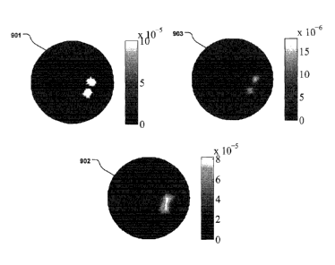
The ground truth is shown as the Input anomaly (901) in Fig. 9. Two separate,
but close
anomalies are shown as the irregular dots in the larger circle.
In the reconstruction using a linear fluorophore (902) the two closely
situated anomalies
can not be distinguished, as is evident from Fig. 9.
However, a reconstruction using a quadratic fluorophore (902) shows a good
separation
between the two closely situated anomalies. This can clearly be seen in Fig.
9.
This comparison illustrates the advantageous effect that the use of non-linear
fluorophores provides, namely a higher contrast and resolution, than with
linear fluorophores. The
enhancement is due to the more narrow sensitivity while using the quadratic
source term as seen
in equation (1) below. This can be visualized by considering the collected
signal for different
source positions. Using a quadratic fluorophore, the signal will only be
strong if the source
position is in the vicinity of the fluorophore itself. Thus the signal can
provide more information
CA 02760783 2011-11-02
WO 2010/128090 23 PCT/EP2010/056127
about the location of the fluorophore than for the case of a linear
fluorophore. This may also give
the possibility of resolving, for example, two closely situated fluorophores
that are not resolvable
using a linear fluorophore, as shown in Fig. 9.
Multi-beam fluorescence diffuse optical tomography using upconverting
nanoparticles
Additionally, this disclosure demonstrate a method in Fluorescence diffuse
optical
tomography to exploit the unique nonlinear power dependence of upconverting
nanoparticles to
further increase the amount of information in a raster-scanning setup by
including excitation with
two beams simultaneously. It was found that the increased information led to
more accurate
1 0 reconstructions.
Fluorescence diffuse optical tomography (FDOT) is a relatively new modality
which
seeks to reconstruct the spatial distribution of the concentration of
fluorescent probes inside
turbid material. As an imaging tool, it has a good prospect in biomedical
studies to image, for
example, tumors, proteases, and drug effects.
FDOT has numerically very ill-posed issues. In this issue, the quality of the
reconstructions for the
fluorescent target is directly determined by the amount and quality of
fluorescence information
obtained from boundary measurements. Instrumental noise and tissue
autofluorescence are the
main perturbations of the measurements, resulting in poor signal quality, and
can cause severe
artifacts in the reconstructed results. In order to overcome this, one could,
for example, employ
2 0 low-noise equipment, use background subtraction or spectral unmixing.
However, such methods
cannot resolve all issues, since they essentially are only utilizing the
present information in a
better way rather than adding new constraints for the reconstructions, i.e.,
adding new
independent information, which is critical to improve the quality of the
reconstructions.
In a noncontact CCD-based FDOT system, one preferred way to gain more
information is by
2 5 increasing the number of excitation positions. However, in order to
keep the intensity of the
excitation beam within reasonable levels, there is a limit on the minimum size
of the excitation
beam. This implies a practical upper limit to the highest excitation-position
density, since distinct,
i.e., non-overlapping, excitation positions are desired for reconstructions.
It is also possible to
employ an anatomical imaging modality such as magnetic-resonance imaging to
provide a-priori
3 0 structural information. However, this is at the cost of significantly
increased complexity and
reduced flexibility of the system.
In this disclosure, we present an approach to exploit the quadratic power
dependence
of upconverting nanoparticles to gain additional information by utilizing two
beams simultaneously
for excitation in FDOT. The effect of the images taken with dual-beam
excitation (named type-D
35 images) on the reconstructions of the nanoparticle number density
distribution, n, is
CA 02760783 2011-11-02
WO 2010/128090 2 4 PCT/EP2010/056127
demonstrated. In addition, comparisons of reconstructed results between the
linear Rhodamine
6G and the quadratic upconverting nanoparticles are made.
The excitation and emission fields can be modeled by two coupled diffusion
equations
[Ref. 1], For quadratic fluorophores, the fluorescence signal detected at a
fixed detector position
under excitation of the k:th beam;
can be described by the forward model (1);
=_- E ri)n(r,)[LTC(rSr,)]2AVi, (1)
where N denotes the number of voxels,
rs,d,i denotes the coordinates for source, detector, and
k
voxel, respectively, and;
AV, is the volume of voxel I.
2 0 The forward solution of the excitation light is represented by;
[ue(r,, r,)]2
while the adjoint solution to the forward fluorescence problem is represented
by;
25 U.f(rd,
When exciting the medium using two beams simultaneously, the detected signal
is given by (2);
rkk3 =U (rd, ri)n(ri)[Lre(rsõri) Ere(rsj,ri)]2AV,
=rk +F+
2 E Uf*(rd,ri)n(r,) Ue(rsõ. rz)U, (1'53 , rt)Airi,
(2)
CA 02760783 2011-11-02
WO 2010/128090 2 5 PCT/EP2010/056127
which reveals the involvement of cross-terms. In a raster-scanning setup (500,
507), if two
images are taken sequentially with one excitation beam scanning over two
positions (named
type-S images), and a third image is taken with two-beam excitation (type-D)
above the previous
two positions, the involvement of cross-terms implies that the type-D image
cannot be obtained
by any mathematical manipulation from the existing type-S images, indicating
that it is
independent and contains additional information. However, for linear
fluorophores, e.g.,
Rhodamine 6G, the type-D image is only linear combinations of the existing
type-S images, and
will not add more constraints for the inverse problem. For nonlinear
fluorophores, it is deduced
that Eq. (2) can be generalized to include more simultaneous excitation beams.
The significance of the measurements with dual-beam excitation in the
reconstructions was
confirmed by the singular-value analysis of the weight matrix, W, whose
elements are given by
(3) [Ref. 1];
Tir(s.d),i uf*(rd, 1.0 [Ue(rs, r,)PAV,, (3)
with;
2 for quadratic fluorophores and;
1 for linear fluorophores.
25 Calculations were performed using the NIRFAST package implementing the
finite element
method. W was factorized according to (4);
TV = ETEIT*, (4)
3 0 where U and V are unitary matrices containing the left and right
singular vectors of W, and;
is a diagonal matrix containing the singular values of W. The column-space of
V is spanned by
3 5 the image-space modes, while the column-space of U is spanned by the
detection-space modes.
CA 02760783 2011-11-02
WO 2010/128090 2 6
PCT/EP2010/056127
The singular values of Wdenote how effectively a given image-space mode can be
detected by
an experimental setup [Ref. 2].
Figure 12 shows the normalized singular-value distribution of W. The x-axis
shows the
singular value index (1120) and the y-axis shows the normalized singular value
intensity (1121).
For clarity, only every second singular value are shown. The cross (1122) and
plus (1124) signs
represent the linear fluorophore (T-1), the former for the single-beam
excitation (1122), while the
latter for the combined single-beam excitation and dual-beam excitation
(1124). As seen, the
normalized intensities of the additional sigular values due to dual-beam
excitation (1124) have
dropped to machine precision, which indicates that the measurements with dual-
beam excitation
0 may not alleviate the ill-posed ness of FDOT. In other words, the type-D
images may not provide
more information than the existing type-S images. Hence, it may not improve
the quality of the
reconstructions. However, for the quadratic fluorophore (denoted by asterisk
(1123) and dot
(1125) signs in Fig. 12, the intensities of the additional singular values
(1125) are still significant.
This implies that type-D images will contribute to the quality of the
reconstructions.
The experiments were carried out in a gelatin phantom with optical properties
of pa=
0.29 cm-1 and p's= 10.0 cm-1 at 660 nm, measured with a time-of-flight
spectroscopy system [Ref.
3]. Two capillary tubes, filled with solutions of Rhodamine 6G (c =0.1pM) and
NaYF4: Yb3i-/Tm34-
nanoparticles (c = 0.1wt%), respectively, were used to simulate the
fluorescent lesions. The
experimental setup and corresponding running parameters were similar with
those used in our
previous work [Ref. 1]. Due to the limited area of the phantom under
investigation, only 9
excitation positions (3 x 3 grid) were used in the present disclosure, The
separation of two
nearest-neighboring positions was 3.5 mm, and each excitation beam had a
diameter of
approximately 2.6 mm. During the experiments, a single excitation beam was
first used to scan
over the 3 x 3 grid, and one image was captured for each scanned position by a
CCD camera. In
the next step, two excitation beams, located at two nearest-neighboring sites
of the same grid,
were simultaneously employed to illuminate the phantom, giving 6 extra type-D
images.
Figures 13A-13B shows the three-dimensional rendering of the reconstructed
upconverting nanoparticles. The red cylinders in the subfigures are identical
and represent the
true fluorescent lesions. In the reconstruction of Fig. 13 (a), only type-S
images were used. As
can be seen, the shape of the fluorescent lesion is overestimated. This
overestimation may be
explained by the ill-posedness of the inverse problem. When adding type-D
images, the
reconstruction of the fluorescent lesion shape is improved remarkably, as
shown in Fig. 13B. In
order to emphasize the difference between the two reconstructions, cross-
sectional slices of the
reconstructed relative fluorophore distribution are shown in Figs. 14A-14F.
Although the depth is
3 5 relatively well reconstructed at the center of the fluorescent lesion
(represented by the circles) for
CA 02760783 2011-11-02
WO 2010/128090 27 PCT/EP2010/056127
both reconstructions, the reconstructed fluorescent lesion is more confined
for the case of using
both type-S-and-D images. This result confirms that the images of type D
indeed contribute to the
inverse problem and lead to better reconstructions for the quadratic
upconverting nanoparticles.
The corresponding reconstructions for the linear Rhodamine 6G were also
carried out, whose
cross-sectional slices are presented in Figs. 15A-15F. Compared with the
results for the
nanoparticles, the reconstructions for Rhodamine 6G do not benefit from adding
the type-D
images, which is in agreement with the theory. The true depth of the
fluorescent lesion is also
poorly reconstructed.
It is disclosed an additional unique advantage of the nonlinear power
dependence of
upconverting nanoparticles. This advantage enables the possibility to obtain
additional
information for the inverse problem by using images taken with two or more
excitation beams
simultaneously. We found that this resulted in improved reconstructions. The
same advantage
could not be found when using linear fiuorophores, e.g., Rhodamine 6G.
This disclosure presents embodiments of non-linear luminescence imaging and
tomography. In experiments it was shown that imaging with upconverting
nanocrystals is possible
in scattering media resembling biological tissue. Furthermore, simulations
showed that it is
possible to adapt the theory used in fluorescent optical tomography, to work
with the upconverting
nanocrystals.
The upconverting nanocrystal particles used in this disclosure, in comparison
with
2 0 organic fluorophores, have a variety of applications, such as for
biological markers thanks to their
unique optical properties.
The present invention has been described above with reference to specific
embodiments. However, other embodiments than the above described are equally
possible within
the scope of the invention. The different features and steps of the invention
may be combined in
other combinations than those described. The scope of the invention is only
limited by the
appended patent claims.
The method may be performed in-vivo at a living human or animal body. In this
case,
the markers may be preintroduced into the body in any manner, such as by
injection into the
blood stream or subcutaneously or directly into a tumour, or alternatively by
topical application,
pulmonary and other non-invasive methods. Such preintroduction can be
performed separately
from the remaining method. Such preintroduction can be performed in connection
with the
remaining method but shortly therebefore.
Alternatively or additionally, the method may be performed at a human or
animal body,
which is sacrifized after the method is performed.
= CA 02760783 2016-12-16
28
Alternatively or additionally, the method may be performed in vitro at a non-
living human
or animal body or part of a body, for example a brain-dead human or animal
body.
Alternatively or additionally, the method may be performed at non-medical
fields, such as
filters or tablets.