Note: Descriptions are shown in the official language in which they were submitted.
CA 02761310 2016-06-01
CA2761310
ANTIBODIES AND METHODS OF USE THEREOF
CROSS-REFERENCE TO RELATED APPLICATIONS
[0001] <DELETED>
STATEMENT REGARDING FEDERALLY SPONSORED RESEARCH
[0002] This invention was made with government support under grants nos.
CA072006 and
132 CA108462 awarded by the National Institutes of Health. The government has
certain rights in this
invention.
INTRODUCTION
[0003] Investigation into the use of antibodies as therapeutics has
increased significantly over
the past decade. The high specificity and tight binding characteristics
inherent to antibodies give them
enormous potential for use as therapeutics. Their specificity allows for
precise targeting of protein
functions, which may minimize side effects resulting from off-target binding.
Therapeutic antibodies
currently in use function through three modes of action: as inducers of the
immune system cytotoxicity,
as carriers of a specific cytotoxic agent, or as inhibitors of the target
protein function.
[0004] To date, the majority of therapeutic antibodies have fallen into
this last grouping, acting
as antagonists of proteins in disease related signaling pathways such as VEGF
(Avastin), EGFR
(Erbitux) and TNF (Humira). By combining selectivity and a large binding
footprint, antibodies have
proven to be ideal for creating the steric hindrance necessary to block
ligand/receptor interactions and
inhibit the signaling cascade and downstream functions involved in disease
progression.
[0005] Many diseases have also been found to be dependent upon
misregulated enzyme
function, including proteases. In particular, proteases have been implicated
in a number of functions
essential for cancer progression. These include extracellular matrix
remodeling, release of cytokines,
and loss of apoptotic response. One particular protease that has been
implicated in cancer progression is
the trypsin-fold serine protease MT-SP I (membrane type-serine protease 1,
matriptase) (Uhland K Cell
Mol Life Sci 2006, 63: 2968-78). MT-SP1 is over-expressed on the surface of
epithelial cells involved
in a variety of cancers, including breast, colon and prostate cancers. The
protease is involved in the
activation of other proteases, growth factors and receptors all of which
result in extracellular matrix
remodeling, angiogenesis and invasive growth.
[0006] Recent studies have investigated the use of antibodies as
inhibitors of protease function
(Farady CJ et al. J Mol Biol 2008, 380: 351-60; Farady CJ et al. J Mol Biol
2007, 369: 1041-51; Sun J et
1
CA 02761310 2011-11-07
WO 2010/129609 PCT/US2010/033624
al. Biochemistry 2003, 42, 892-900). The inhibitors were found to either block
substrate binding through
steric hindrance or cause conformational changes due to binding at allosteric
sites. More recently, the
molecular basis of three antibody inhibitors have been determined from crystal
structures of the
antibody/protease complexes (Farady CJ et al. J Mol Biol 2008, 380: 351-601).
LITERATURE
[0007] Sun J et al. Biochemistry 2003, 42, 892-900; Farady CJ et al. J
Mol Biol 2007, 369: 1041-
5; Farady CJ et al. J Mol Biol 2008, 380: 351-601; Foltz et al. (US Patent
Publication No. 2006/0171884);
Foltz et al. American Society of Hematology Annual Meeting Abstracts 2005,
106:Abstract 4816.
SUMMARY
[0008] The present disclosure relates to protease-binding agents (e.g.
antibodies) that bind to and
modulate the activity of a protease, compositions comprising the antibodies,
and methods involving use
of the antibodies or compositions.
[0009] Also provided by the disclosure is an isolated protease-binding
agent comprising a heavy
chain variable region comprising a CDR; and a light chain variable region
comprising a CDR, in which a
hypervariable loop of said heavy chain variable region is capable of binding
the 51 pocket of a Pl-Arg-
specific protease to position a scissile bond in the active site of said
protease in an orientation opposite to
a cleavable substrate of said protease; and in which the heavy chain variable
region and the light chain
variable region provide for antigen specificity so as to position the
hypervariable loop for binding to said
51 pocket. Other agents can include those that bind to the protease in such a
away that the scissile bond of
the binding agent is positioned away from the active site of said protease,
particularly away from the
active site nucleophile.
[0010] Methods of the present disclosure include administering a
composition comprising a
protease-binding agent that inhibits a protease of interest to treat diseases,
such as cancer or infection.
[0011] Methods also may employ the protease-binding agent for diagnosis
of diseases.
[0012] Methods of screening are also provided to identify or engineer a
protease-binding agent
that specifically inhibits a protease of interest.
[0013] Kits containing one or more compositions of the present
disclosure, as well as
those with instructions for use in a method of the present disclosure also are
provided.
[0014] Other features of the invention are described herein, and will
also be readily apparent to
the ordinarily skilled artisan upon reading the present disclosure.
2
CA 02761310 2016-07-25
CA 2761310
[0014A] Various embodiments of the claimed invention relate to an isolated
protease-binding
antibody, comprising: a VLCDR1 comprising the amino acid sequence of SEQ ID
NO: 16; a
VLCDR2 comprising the amino acid sequence of SEQ ID NO: 17; a VLCDR3
comprising the
amino acid sequence of SEQ ID NO: 18; a VHCDR1 comprising the amino acid
sequence of
SEQ ID NO: 19; a VHCDR2 comprising the amino acid sequence of SEQ ID NO: 20;
and a
VHCDR3 comprising the amino acid sequence of SEQ ID NO: 10. Such antibodies
may be useful
in inhibiting a serine protease.
10014B1 Various embodiments of the claimed invention relate to a
pharmaceutical composition
comprising the protease-binding antibody as described herein and a
pharmaceutical acceptable
excipient.
[0014C] Various embodiments of the claimed protease-binding antibodies may
be useful in
diagnostically effective amounts for detecting a cancer cell comprising a cell
surface serine protease in a
subject.
[00141)] Various embodiments of the claimed protease-binding antibodies may
be useful as
components of a kit for detecting cancer cells in a subject comprising: the
protease-binding antibody as
described herein; and reagents for performing the method as described herein.
2a
CA 02761310 2011-11-07
WO 2010/129609 PCT/US2010/033624
BRIEF DESCRIPTION OF FIGURES
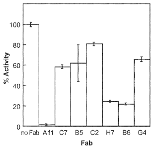
[0015] Figure 1 depicts the relative inhibition of MT-SP1 by seven Fabs
identified from the
phage display library.
[0016] Figure 2, panel A shows the amino acid sequences of the All and E2
heavy and light
chain polypeptides, with the CDRs underlined in the sequences. CDRS are
defined by the Kabat
numbering system (Johnson et al. Nucleic Acids Research, 2000, 28: 214-218).
Panel B shows the nucleic
acid sequences that encode heavy and light chains of All and E2
antibodies.Nucleic acid sequences
encoding the CDRs are bolded. Panel C shows the CDRs of All separately from
the rest of the amino
acid sequences.
[0017] Figure 3 shows the various MT-SP1 alanine scanning mutants.
[0018] Figure 4 depicts the structure of the All/MT-SP1 complex.
[0019] Figure 5 shows Interaction of the All variable loops with MT-SP1.
Panel A, The All
H3 loop interacting with the MT-SP1 surface accounts for the majority of the
buried surface area
contributed by the heavy chain variable loops. The loop inserts Arg 100b into
the active site while
making very few additional contacts. Panel B, The H1 and H2 loops contact
residues in the 60s and 90s
loops of MT-SP1. Panel C, The long L3 loop of All makes a number of contacts
with the surface of MT-
SP1, burying nearly as much surface area as the H3 loop. Panel D, The Ll loop
contacts both the 170s
and 220s loops while the L2 loop makes no contacts with MT-SP1. Panel E,
Together the H2, H3 and L3
loops of All utilize Phe97 of MT-SP1 as an anchor point for binding and
recognition, an interaction that
is crucial to formation of the complex. The heavy and light chains loops are
shown as ribbons and the
MT-SP1 side chains that interact with each variable loop are shaded gray in
the space-filled model.
[0020] Figure 6 shows the insertion of All H3 loop into the MT-SP1 active
site. Panel A shows
that the H3 hypervairable loop of All inserts an arginine (ArgH100b) into the
active site of MT-SP1.
Panel B compares the binding of All to MT-SP1 (right) with the binding of the
bovine pancreatic trypsin
inhibitor (BPTI) on the left. Binding of the E2 antibody to MT-SP1 is also
shown below in Panel B.
[0021] Figure 7 depicts the result of a surface plasmon resonance
experiment. Binding curves of
All Fab to MT-SP1 is black and the binding curve of All Fab to the inactive
mutant zymogen R15A is
gray.
[0022] Figure 8, panel A shows the inhibition of E2 on MT-SP1 activity in
various cell lines.
Panel B shows the inhibition of All on MT-SP1 activity in various cell lines.
[0023] Figure 9 shows All (panel A) and E2 (panel B) Fabs bound to the
recombinant catalytic
domain of MT-SP1.
3
CA 02761310 2011-11-07
WO 2010/129609 PCT/US2010/033624
[0024] Figure 10 shows the fluorescent micrographs of E2 scFv incubated
with MT-SP1-
positivie cells and a negative control cell in culture. HT29 (panel A), MCF7
(panel B), and LNCaP (panel
C). MDA-MD-231 express little to no MT-SP1 (panel D).
[0025] Figure 11 shows the fluorescent micrographs of E2 scFv incubated
with HT29 cells
incubated with recombinant hepatocyte growth factor activator inhibitor-1 (HAT-
1) (panel A) or HT29
cells alone (panel B).
[0026] Figure 12 shows E2 diabody (panel A) and E2 Fab (panel B)
inhibitors in MCF7
xenograph mice with tumor cells circled. Black stains indicate boundaries
around the presence of
luciferase or alexa flour.
[0027] Figure 13 shows that All IgG antibodies selectively target MT-SP1
positive tumors in
vivo. Panel A shows a MCF7 xenograph mouse with tumor indicated by double
arrows. Panel B shows a
MDA-MB-231 (MT-SP1-negative) xenograph mouse as a negative control. Panel C
shows the signal of
active luciferase expressed by MDA-MB-231 cells after luciferin was injected.
Black stains indicate
boundaries around the presence of luciferase or alexa flour.
[0028] Figure 14, panel A shows that All IgG antibodies selectively
target MT-SP1 positive
tumors in vivo. The first row shows xenograph mice with tumors (arrows)
derived from various cell lines.
Second row shows the signal of active luciferase, presence of which is
indicated by surrounding black
stains, after luciferin was injected. Panel B compares the tumor siganal using
percentages of injected dose
in tumor at 48 hours divided by tumor volume (mm3).
[0029] Figure 15, panel A shows the tumor volume over time for various
groups of mice having
PC-3 tumor xenographs. The body weights of the mice are shown as a small
inset. Panel B shows the
tumor volume over time for various groups of mice having H29 tumor xenographs.
The body weights of
the mice are shown as a small inset. Panel C shows a pilot study using a
smaller group of mice than the
experiment shown in Panel B.
[0030] Figure 16 is an Amira processed representation of an HT29
xenograft mouse imaged with
111In-DOTA- All at 48 hr post-injection. Injected dose: 15 pg IgG, 250 pCi.
The CT skeletal image can
be seen in white. For SPECT, dark gray represents the bilateral HT29 tumors
and non-specific uptake can
be seen in the chest cavity in black. A) coronal view at 0 ; B) sagittal view
at 90 ; C) coronal view at
180 ; D) sagittal view at 270 .
[0031] Figure 17, panel A is an 111In-DOTA-All SPECT/CT image of a HT29
bilateral
xenograph at 48 hour post injection. Signals are represented by regions with
gray topographic boundaries.
Injection was done with 15 pg of All IgG (250 pCi). Panel B is an 111In-DOTA-
Palivizumab SPECT/CT
image of a PC3 xenograph at 48 hour post injection. Panel C is an 111In-DOTA-
All SPECT/CT image of
4
CA 02761310 2016-06-01
= CA2761310
a HT29 bilateral xenograph without (left) or with Ecotin blocking (right) at
48 hour post injection. Panel
D is 111In-DOTA-A1l SPECT/CT image of a MT-SP1 negative MDA-MB-23 1 xenograph.
DETAILED DESCRIPTION OF EXEMPLARY EMBODIMENTS
[0032] The present disclosure relates to antibodies that bind to and
modulate the activity of a
protease, compositions comprising the antibodies, and methods involving use of
the antibodies or
compositions.
[0033] Certain of the antibodies disclosed herein were first found by
screening a human Fab
phage display library for inhibition of a type II transmembrane multidomain
serine protease MT-
SP1/matriptase. Structural studies of the complex between the antibody and the
protease reveal that the
antibody comprises features that enable potent inhibition of the protease as
well as other features that
render the antibodies specific for a protease of interest. The data presented
herein support the
application of the antibodies in methods and compositions, including the
diagnosis and treatment of
multiple types of human diseases (e.g. cancer).
[0034] Before the present invention and specific exemplary embodiments
of the invention are
described, it is to be understood that this invention is not limited to
particular embodiments described, as
such may, of course, vary. It is also to be understood that the terminology
used herein is for the purpose
of describing particular embodiments only, and is not intended to be limiting,
since the scope of the
present invention will be limited only by the appended claims.
[0035] Where a range of values is provided, it is understood that each
intervening value, to the
tenth of the unit of the lower limit unless the context clearly dictates
otherwise, between the upper and
lower limit of that range and any other stated or intervening value in that
stated range is encompassed
within the invention. The upper and lower limits of these smaller ranges may
independently be included
in the smaller ranges is also encompassed within the invention, subject to any
specifically excluded limit
in the stated range. Where the stated range includes one or both of the
limits, ranges excluding either
both of those included limits are also included in the invention.
[0036] Unless defined otherwise, all technical and scientific terms used
herein have the same
meaning as commonly understood by one of ordinary skill in the art to which
this invention belongs.
Although any methods and materials similar or equivalent to those described
herein can also be used in
the practice or testing of the present invention, exemplary methods and
materials are now described.
CA 02761310 2011-11-07
WO 2010/129609 PCT/US2010/033624
[0037] It must be noted that as used herein and in the appended claims,
the singular forms "a",
"an," and "the" include plural referents unless the context clearly dictates
otherwise. Thus, for example,
reference to "an antigen" includes a plurality of such antigens and reference
to "the peptide" includes
reference to one or more peptides and equivalents thereof known to those
skilled in the art, and so forth.
[0038] The publications discussed herein are provided solely for their
disclosure prior to the
filing date of the present application. Nothing herein is to be construed as
an admission that the present
invention is not entitled to antedate such publication by virtue of prior
invention. Further, the dates of
publication provided may be different from the actual publication dates which
may need to be
independently confirmed.
DEFINITIONS
[0039] When describing the compositions, pharmaceutical formulations
containing such, and
methods of producing and using such compositions, the following terms have the
following meanings
unless otherwise indicated. It should also be understood that any of the
moieties defined forth below may
be substituted with a variety of substituents, and that the respective
definitions are intended to include
such substituted moieties within their scope.
[0040] The terms "polypeptide" and "protein" are used interchangeably
throughout the
application and mean at least two covalently attached amino acids, which
includes proteins, polypeptides,
oligopeptides, peptides, and fragments thereof. The protein may be made up of
naturally occurring amino
acids and peptide bonds, or synthetic peptidomimetic structures. Thus "amino
acid", or "peptide residue",
as used herein means both naturally occurring and synthetic amino acids. For
example, homo-
phenylalanine, citrulline and noreleucine are considered amino acids for the
purposes of the invention.
"Amino acid" also includes imino acid residues such as proline and
hydroxyproline. The side chains may
be in either the (R) or the (S) configuration. Normally, the amino acids are
in the (S) or L-configuration.
If non-naturally occurring side chains are used, non-amino acid substituents
may be used, for example to
prevent or retard in vivo degradation. Naturally occurring amino acids are
normally used and the protein
is a cellular protein that is either endogenous or expressed recombinantly.
The terms includes fusion
proteins, including, but not limited to, fusion proteins with a heterologous
amino acid sequence, fusions
with heterologous and homologous leader sequences, with or without N-terminal
methionine residues;
immunologically tagged proteins; fusion proteins with detectable fusion
partners, e.g., fusion proteins
including as a fusion partner a fluorescent protein, I3-galactosidase,
luciferase, etc.; and the like.
Polypeptides may be of any size, and the term "peptide" refers to polypeptides
that are 5-50 residues (e.g.,
8-20 residues) in length.
6
CA 02761310 2011-11-07
WO 2010/129609 PCT/US2010/033624
[0041] As used herein, the term "endogenous," refers to biomolecules that
originate within an
organism in nature. For example, an endogenous substrate of a protease is a
protein that originates from
the same organism as the source of the protease and is capable of specifically
binding to the protease
under physiological conditions and of which a peptidic bond is cleaved by the
protease. As another
example, an endogenous substrate of a serine protease is a protein that
originates from the same organism
as the source of the serine protease and is capable of specifically binding to
the serine protease under
physiological conditions and of which a peptidic bond is cleaved by the serine
protease.
[0042] As used herein, the term "cleavable," refers to protease
substrates, of which one or more
peptidic bonds can be hydrolyzed by the protease.
[0043] As used herein, the term "orientation," refers to the positional
relationship of a protease
substrate relative the protease to which it is bound. By convention, the
orientation of a substrate to its
protease is specified from N-terminus to C-terminus based on sites named Pn,
..., P3, P2, P1, P1', P2',
P3',.., Pn', where Pi¨Pi' denotes the scissile bond to be cleaved by the
protease and n is the number of
the feature relative to the scissile bond. Their respective binding sites on
the protease are named Sn, ...,
S3, S2, Sl, 5 l' , S2', S3', ..., Sn ' and n is the number of the feature
relative to the active site. In
accordance with this nomenclature, the scissile bond of a cleavable substrate
is presented to the active site
in an N-terminus to C-terminus orientation relative to sites 51 and SF. If the
scissile bond of a substrate
is presented to the active site in a C-terminus to N-terminus orientation
relative to sites 51 and SF , the
scissile bond is considered to be in the "reversed orientation."
[0044] By "nucleic acid" herein is meant either DNA or RNA, or molecules
which contain both
deoxy- and ribonucleotides. Nucleic acid may be naturally occurring or
synthetically made, and as such,
includes analogs of naturally occurring polynucleotides in which one or more
nucleotides are modified
over naturally occurring nucleotides.
[0045] The term "analog" or "analogue" refers to without limitation any
compound which has
structural similarity to the compounds of the present disclosure and would be
expected, by one skilled in
the art, to exhibit the same or similar utility as the claimed and/or
referenced compounds.
[0046] The term "carrier" as used in the context of a carrier conjugated
to an antibody includes a
peptide or protein carrier, a non-peptide or protein carrier (e.g. a non-
peptide polymer).
[0047] The term "cell surface antigen" (or "cell surface epitope") refers
to an antigen (or
epitope) on surface of a cell that is extracellularly accessible at any cell
cycle stage of the cell, including
antigens that are extracellularly accessible during all stages of the cell
cycle. "Extracellularly accessible"
in this context refers to an antigen that can be bound by an antibody provided
outside the cell without
need for permeabilization of the cell membrane.
7
CA 02761310 2011-11-07
WO 2010/129609 PCT/US2010/033624
[0048] The term "chemotherapy" as used herein refers to use of an agent
(e.g., drug, antibody,
etc.), particularly an agent(s) that is selectively destructive to a cancerous
cell, in treatment of a disease,
with treatment of cancer being of particular interest.
[0049] A "cancer cell" as used herein refers to a cell exhibiting a
neoplastic cellular phenotype,
which may be characterized by one or more of, for example, abnormal cell
growth, abnormal cellular
proliferation, loss of density dependent growth inhibition, anchorage-
independent growth potential,
ability to promote tumor growth and/or development in an immunocompromised non-
human animal
model, and/or any appropriate indicator of cellular transformation. "Cancer
cell" may be used
interchangeably herein with "tumor cell" or "cancerous cell", and encompasses
cancer cells of a solid
tumor, a semi-solid tumor, a primary tumor, a metastatic tumor, and the like.
[0050] The term "conjugated" generally refers to a chemical linkage,
either covalent or non-
covalent, usually covalent, that proximally associates one molecule of
interest with second molecule of
interest.
[0051] The terms "antigen" and "epitope" are well understood in the art
and refer to the portion
of a macromolecule (e.g., a polypeptide) which is specifically recognized by a
component of the immune
system, e.g., an antibody or a T-cell antigen receptor. As used herein, the
term "antigen" encompasses
antigenic epitopes, e.g., fragments of an antigen which are antigenic
epitopes. Epitopes can be recognized
by antibodies in solution, e.g. free from other molecules. Epitopes can be
recognized by T-cell antigen
receptor when the epitope is associated with a class I or class II major
histocompatibility complex
molecule.
[0052] The terms "derivative" and "variant" refer to without limitation
any compound or
antibody which has a structure or sequence derived from the compounds and
antibodies of the present
disclosure and whose structure/sequence is sufficiently similar to those
disclosed herein and based upon
that similarity, would be expected, by one skilled in the art, to exhibit the
same or similar activities and
utilities as the claimed and/or referenced compounds or antibody.
[0053] The term "effective amount" of a composition as provided herein is
intended to mean a
non-lethal but sufficient amount of the composition to provide the desired
utility. For instance, for
eliciting a favorable response in a subject to treat a disorder or infection,
the effective amount is the
amount which eliminates or diminishes the symptoms associated with the
disorder, e.g., so as to provide
for control of cancer metastatis, to eliminate cancer cells, decrease
bacterial or viral infection. As will be
pointed out below, the exact amount required will vary from subject to
subject, depending on the species,
age, and general condition of the subject, the severity of the condition or
disease that is being treated, the
particular composition used, its mode of administration, and the like. Thus,
it is not possible to specify an
8
CA 02761310 2011-11-07
WO 2010/129609 PCT/US2010/033624
exact "effective amount." However, an appropriate effective amount may be
determined by one of
ordinary skill in the art using only routine experimentation.
[0054] The term "immunotherapy" refers to treatment of disease (e.g.,
viral or bacterial
infection, or cancer) by modulating an immune response to a disease antigen.
In the context of the present
application, immunotherapy refers to providing an antibacterial and/or anti-
cancer immune response in a
subject by administration of an antibody (e.g., a monoclonal antibody).
[0055] The term "in combination with" as used herein refers to uses
where, for example, a first
therapy is administered during the entire course of administration of a second
therapy; where the first
therapy is administered for a period of time that is overlapping with the
administration of the second
therapy, e.g. where administration of the first therapy begins before the
administration of the second
therapy and the administration of the first therapy ends before the
administration of the second therapy
ends; where the administration of the second therapy begins before the
administration of the first therapy
and the administration of the second therapy ends before the administration of
the first therapy ends;
where the administration of the first therapy begins before administration of
the second therapy begins
and the administration of the second therapy ends before the administration of
the first therapy ends;
where the administration of the second therapy begins before administration of
the first therapy begins
and the administration of the first therapy ends before the administration of
the second therapy ends. As
such, "in combination" can also refer to regimen involving administration of
two or more therapies. "In
combination with" as used herein also refers to administration of two or more
therapies which may be
administered in the same or different formulations, by the same or different
routes, and in the same or
different dosage form type.
[0056] The term "isolated" is intended to mean that a compound is
separated from all or some of
the components that accompany it in nature. "Isolated" also refers to the
state of a compound separated
from all or some of the components that accompany it during manufacture (e.g.,
chemical synthesis,
recombinant expression, culture medium, and the like).
[0057] The term "antibody" (also used interchangeably with
"immunoglobulin") encompasses
polyclonal and monoclonal antibody preparations where the antibody may be of
any class of interest (e.g.,
IgM, IgG, and subclasses thereof), as well as preparations including hybrid
antibodies, altered antibodies,
F(ab')2 fragments, F(ab) molecules, Fv fragments, single chain fragment
variable displayed on phage
(scFv), single chain antibodies, single domain antibodies, diabodies, chimeric
antibodies, humanized
antibodies, and functional fragments thereof which exhibit immunological
binding properties of the
parent antibody molecule. In some embodiments, e.g., cancer therapy,
antibodies that provide for
complement-mediated killing and/or antibody- dependent cellular cytotoxicity
(ADCC) are of particular
interest. The antibodies described herein may be detectably labeled, e.g.,
with a radioisotope, an enzyme
9
CA 02761310 2011-11-07
WO 2010/129609 PCT/US2010/033624
which generates a detectable product, a fluorescent protein, and the like. The
antibodies may be further
conjugated to other moieties, such as a cytotoxic molecule or other molecule
(e.g., to provide for delivery
of an anti-cancer drug to a cancer cell), members of specific binding pairs,
e.g., biotin (member of biotin-
avidin specific binding pair), and the like. The antibodies may also be bound
to a support (e.g., a solid
support), such as a polystyrene plate or bead, test strip, and the like.
[0058] Immunoglobulin polypeptides include the kappa and lambda light
chains and the alpha,
gamma (IgGi, IgG2, IgG3, Igat), delta, epsilon and mu heavy chains or
equivalents in other species. Full-
length immunoglobulin "light chains" (usually of about 25 kDa or about 214
amino acids) comprise a
variable region of about 110 amino acids at the NH2-terminus and a kappa or
lambda constant region at
the COOH-terminus. Full-length immunoglobulin "heavy chains" (of about 50 kDa
or about 446 amino
acids), similarly comprise a variable region (of about 116 amino acids) and
one of the aforementioned
heavy chain constant regions, e.g., gamma (of about 330 amino acids).
[0059] An immunoglobulin light or heavy chain variable region is composed
of a "framework"
region (FR) interrupted by three hypervariable regions, also called
"complementarity determining
regions" or "CDRs". The extent of the framework region and CDRs have been
precisely defined (see,
"Sequences of Proteins of Immunological Interest," E. Kabat et al., U.S.
Department of Health and
Human Services, 1991, and Lefranc et al. IMGT, the international
ImMunoGeneTics information
system . Nucl. Acids Res., 2005, 33, D593-D597)). A detailed discussion of the
Kabat numbering
system is provided on the World Wide Web at kabatdatabase.com/index.html. The
sequences of the
framework regions of different light or heavy chains are relatively conserved
within a species. The
framework region of an antibody, that is the combined framework regions of the
constituent light and
heavy chains, serves to position and align the CDRs. The CDRs are primarily
responsible for binding to
an epitope of an antigen.
[0060] The term "monoclonal antibody" refers to an antibody composition
having a
homogeneous antibody population. The term is not limited by the manner in
which it is made. The term
encompasses whole immunoglobulin molecules, as well as Fab molecules, F(ab')2
fragments, Fv
fragments, single chain fragment variable displayed on phage (scFv), fusion
proteins comprising an
antigen-binding portion of an antibody and a non-antibody protein, and other
molecules that exhibit
immunological binding properties of the parent monoclonal antibody molecule.
Methods of making
polyclonal and monoclonal antibodies are known in the art and described more
fully below.
[0061] The term "specific binding of an antibody" or "antigen-specific
antibody" in the context
of a characteristics of an antibody refers to the ability of an antibody to
preferentially bind to a particular
antigen that is present in a homogeneous mixture of different antigens. In
certain embodiments, a specific
binding interaction will discriminate between desirable and undesirable
antigens (or "target" and "non-
CA 02761310 2011-11-07
WO 2010/129609 PCT/US2010/033624
target" antigens) in a sample, in some embodiments more than about 10 to 100-
fold or more (e.g., more
than about 1000- or 10,000-fold). In certain embodiments, the affinity between
an antibody and antigen
when they are specifically bound in an antibody-antigen complex is
characterized by a KD (dissociation
constant) of less than 10-6M, less than 10-7 M, less than 10-8 M, less than 10-
9 M, less than 10-9 M, less
than 10-11 M, or less than about 10-12 M or less.
[0062] "Conservative amino acid substitution" refers to a substitution of
one amino acid residue
for another sharing chemical and physical properties of the amino acid side
chain (e.g., charge, size,
hydrophobicity/hydrophilicity). "Conservative substitutions" are intended to
include substitution within
the following groups of amino acid residues: gly, ala; val, ile, leu; asp,
glu; asn, gln; ser, thr; lys, arg; and
phe, tyr. Conservative amino acid substitutions in the context of an antibody
disclosed herein are selected
so as to preserve the interaction between the antibody and the protease of
interest.
[0063] The term "pharmaceutically acceptable" refers to a material that
is not biologically or
otherwise undesirable, i.e., the material is of a medically acceptable quality
and composition that may be
administered to an individual along with the selected active pharmaceutical
ingredient without causing
any undesirable biological effects or interacting in a deleterious manner with
any of the other components
of the pharmaceutical composition in which it is contained.
[0064] The term "pharmaceutically acceptable excipient" as used herein
refers to any suitable
substance which provides a pharmaceutically acceptable vehicle for
administration of a compound(s) of
interest to a subject. "Pharmaceutically acceptable excipient" can encompass
substances referred to as
pharmaceutically acceptable diluents, pharmaceutically acceptable additives
and pharmaceutically
acceptable carriers.
[0065] The term "purified" is intended to mean a compound of interest has
been separated from
components that accompany it in nature and provided in an enriched form.
"Purified" also refers to a
compound of interest separated from components that can accompany it during
manufacture (e.g., in
chemical synthesis, recombinant expression, culture medium, and the like) and
provided in an enriched
form. Typically, a compound is substantially pure when it is at least 50% to
60%, by weight, free from
organic molecules with which it is naturally associated or with which it is
associated during manufacture.
Generally, the preparation is at least 75%, more usually at least 90%, and
generally at least 99%, by
weight, of the compound of interest. A substantially pure compound can be
obtained, for example, by
extraction from a natural source (e.g., bacteria), by chemically synthesizing
a compound, or by a
combination of purification and chemical modification. A substantially pure
compound can also be
obtained by, for example, enriching a sample having a compound that binds an
antibody of interest. Purity
can be measured by any appropriate method, e.g., chromatography, mass
spectroscopy, HPLC analysis,
etc.
11
CA 02761310 2016-06-01
CA2761310
[0066] The term "subject" is intended to cover humans, mammals and other
animals which
contain serine proteases in any fashion. The terms "subject," "host,"
"patient," and "individual" are used
interchangeably herein to refer to any mammalian subject for whom diagnosis or
therapy is desired,
particularly humans. Other subjects may include cattle, dogs, cats, guinea
pigs, rabbits, rats, mice,
horses, and so on.
[0067] In the context of cancer therapies and diagnostics described
herein, "subject" or
"patient" is used interchangeably herein to refer to a subject having,
suspected of having, or at risk of
developing a tumor, where the cancer is one associated with cancerous cells
expressing an active and/or
dysregulated serine protease. Samples obtained from such subject are likewise
suitable for use in the
methods of the present disclosure.
[0068] As used herein, the terms "determining," "measuring," and
"assessing," and "assaying"
are used interchangeably and include both quantitative and qualitative
determinations.
[0069] It is further noted that the claims may be drafted to exclude any
optional or alternative
element. As such, this statement is intended to serve as antecedent basis for
use of such exclusive
terminology as "solely", "only" and the like in connection with the recitation
of claim elements, or the
use of a "negative" limitation.
[0070] The citation of any publication is for its disclosure prior to the
filing date and should not
be construed as an admission that the present invention is not entitled to
antedate such publication by
virtue of prior invention. Further, the dates of publication provided may be
different from the actual
publication dates which may need to be independently confirmed. To the extent
a definition of a term
set out in a document incorporated herein by reference conflicts with the
definition of a term explicitly
defined herein, the definition set out herein controls.
Exemplary methods and compositions employable therein are described first in
greater detail, followed
by a review of the various specific compositions, formulations, kits and the
like that may find use in the
methods of the present disclosure, as well as a discussion of representative
applications in which the
methods and compositions of the present disclosure find use.
PROTEASE BINDING AGENTS
[0071] The present disclosure provides a protease binding agent, where
protease binding agents
include a whole antibody, an antigen-binding fragment thereof, and synthetic
protease binding agents
that
12
CA 02761310 2011-11-07
WO 2010/129609 PCT/US2010/033624
comprises portions of an antibody. A subject protease-binding agent binds a
member of a protease family
such as the P1 Arg-specific protease family (e.g. trypsin-like serine
proteases). An example of a protease
family is the chymotrypsin-fold family, which is also called the peptidase
family Si. A subject protease
binding agent (e.g., antibody) finds use in a variety of applications,
including use in various methods of
treating a host suffering from a disease or condition, as well as in diagnosis
of various diseases and
conditions. For example, in some embodiments, a subject antibody is highly
specific for active
membrane-type serine protease I (MT-SP1), which is often found on cancer
cells. More exemplary uses
of a subject antibody will be described later.
[0072] As noted above, a subject protease-binding agent binds
specifically to a member of a
protease family such as the P1 Arg-specific protease family (e.g. serine
protease family). A subject
protease-binding agent exhibits features that allow not only potent inhibition
of a specific protease but
also specific recognition of the protease. Serine proteases are a group of
enzymes that share structural and
functional features discussed below. Members of the P1 Arg-specific protease
family are also discussed.
Protease Targets
[0073] The target of a subject protease-binding agent is a protease that
catalyzes the hydrolysis
of covalent peptidic bonds.
Serine proteases
[0074] A subject protease-binding agent includes an agent specific for a
serine protease. The
mechanism of catalysis is based on the nucleophilic attack of the peptidic
bond by a serine. Cysteine,
threonine or water molecules associated with aspartate or metals can also paly
the role of a nucleophile. In
many cases, the nucleophilic property of the group is improved by the presence
of a histidine, held by an
aspartate in a basic state, so as to readily accept a proton. The aligned
catalytic group of serine, histidine
and aspartate is a common feature to most serine proteases. The substrate
binding groove containing the
active site is shaped as a cleft. In order to better describe the interaction
between the polypeptide substrate
and its respective serine protease, the polypeptide substrate is labled from N-
terminus to C-terminus as
Pn, ..., P3, P2, Pl, P1', P2', P3',.., Pn' while their respective binding
sites on the protease Sn, ..., S3, S2,
51, 51', S2', S3', ..., Sn'. In accordance with this nomenclature, Pi¨Pi'
denotes the hydrolyzed peptidic
bond of the polypeptide substrate.
Chymotrypsin-fold serine proteases
[0075] A subject protease-binding agent includes an agent specific for a
chymotrypsin-fold
serine protease. There is a large number of serine proteases that catalyze
hydrolysis of peptidic bond in
the manner described above. Many of them are further grouped together and
collectively referred to as the
chymotrypsin-fold serine protease. Chymotrypsin-fold serine proteases make up
a protease family that
13
CA 02761310 2011-11-07
WO 2010/129609 PCT/US2010/033624
has been extensively characterized and studied. They are often synthesized as
non-active zymogens in a
cell and are activated upon cleavage in a highly conserved activation motif to
produce a mature protease.
In addition to high degrees of amino acid sequence identity among the family
members, they also share
identical folds in conserved motifs. Several notable structural and functional
features shared among serine
proteases of this family were first characterized in the chymotrypsin
protease. One prominent feature of
this protein family is a structural fold containing two I3-barrels, with the
catalytic Ser, His, and Asp amino
acids found at the interface of the two domains. Another common feature
includes five enzyme-substrate
hydrogen bonds at positions P1 and P3 that juxtagpose the scissile peptide
bond adjacent to the Ser-His
catalytic couple, such that the nucleophilic Ser 0-7 is accurately positioned
for the nucleophilic attack.
[0076] While very similar pockets and clefts make up the structure of the
active sites of different
chymotrypsin-like serine proteases, the members of this family diverge in
parts of the protease distal from
the active sites. Protein sequences surrounding the active site that differ
among proteases within the
family provide for diversity in the substrate-binding groove, and hence, the
specificity for each respective
proteolytic substrate. One way in which specificity is provided is based on
surface loops of the protease in
the substrate binding groove.
[0077] Although the protease-substrate interactions may be characterized
by sequence
divergence, all the structures responsible for substrate specificity (e.g.
surface loops surrounding the
active site) are still aligned so as to accurately position the scissile bond
of the substrate in the conserved
active site. As such, the positions of the catalytic amino acids in the active
site, such as Ser, His, and Asp,
remain the anchor that defines the common structural framework of chymotrypsin-
like serine protease.
Chymotrypsin-fold serine protease with a trypsin-like Si pocket
[0078] A subject protease-binding agent includes an agent specific for a
member a subfamily of
the chomotrypsin-like serine protease. The chymotrypsin-fold serine protease
family may be subdivided
depending on the sequence of the Pl/S1 site. In certain cases, the 51 of the
protease specifically binds P1
containing an Arg or Lys so as to provide peptidic cleavage following an Arg
or Lys residue (e.g. 51 in
trypsin). In other embodiments, the 51 pocket is hydrophobic and specifcally
binds P1 containing one or
more amino acid(s) having hydrophobic side chains (e.g. 51 in chymotrypsin).
In this case, the peptidic
cleavage occurs after the hydrophobic amino acid residue. In certain cases,
the 51 pocket specifically
binds P1 containing an Ala and cleaves the peptidic bond after the Ala residue
(e.g. 51 in elastase).
[0079] Depending on the structural features of the protease-binding agent
described in more
detail below, the agent may be specific for serine proteases having a 51
pocket similar to trypsin. For
simplicity, serine proteases in the chymotrypsin-fold serine protease family
with a 51 pocket similar to
that of trypsin would be referred herein as trypsin-like serine proteases. In
certain cases, the protease
target of the antibody of the present disclosure is not a serine protease but
has an 51 pocket similar to that
14
CA 02761310 2011-11-07
WO 2010/129609 PCT/US2010/033624
of trypsin. The proteases with a similar Si pocket to that of trypsin
regardless of whether the protease is
categorically a serine protease would be referred herein as "Pi-Arg-specific
proteases".
Type II transmembrane serine proteases
[0080] A subject protease-binding agent includes an agent specific for a
Type II transmembrane
serine protease. A protease-binding agent may be specific for a small group
proteases within the
subfamily of trypsin-like serine proteases. The protease-binding agent can be
capable of binding to type II
transmembrane serine proteases (TTSPs). Aside from possessing the structural
framework, conserved
motifs, and the Si pocket of trypsin-like serine proteases, this group of
proteases share additional
features. The shared features are described in the following from N-terminus
to C-terminus. At the N-
terminus, a segment of a length about 12 to about 112 amino acid residues
resides intracellularly and
plays a putative role in protein sorting and/or intracellular signal
transduction comprises. The intracellular
segment is followed by a hydrophobic domain that spans the plasma membrane,
making up the
transmembrane domain. C-terminal to the transmembrane domain are the
extracellular domains of the
protein. One extracellular domain is the stem regions, which may comprise one
or more of the following:
low density lipoprotein (LDL) receptor class A domains, Group A scavenger
receptor (SR) domains,
frizzled domains, Cls/Clr, urchin embryonic growth factor and bone morphogenic
protein 1 (CUB)
domains, etc. Lastly, the proteolytic domain is presented at or near the C-
terminus of the TTSP. See
Hooper et al. J. Biol. Chem. 2001, 276:857-860 for more detail. There are
about 17 members of TTSPs
found in mammals, of which seven are found in human. See, e.g., Table 1 below.
CA 02761310 2011-11-07
WO 2010/129609 PCT/US2010/033624
Table 1
Name Organism Other name Accession number
Corin Human AF133845
Mouse LRP4 AB013874
Enteropeptidase Human Enterokinase U09860
Bovine U09859
Mouse U73378
Rat 1589367
Porcine D30799
MT-SP1 Human Matriptase AF133086/AF118224
Mouse Epithin AF042822
HAT Human AB002134
Hepsin Human M18930
Mouse AF030065
Rat X70900
Stubble-Stubloid Drosophila L11451
TMPRSS2 Human U75329
Mouse Epitheliasin AF113596
TMPRSS4 Human AF179224
Membrane-type serine protease I
[0081] A subject protease-binding agent can specifically bind and inhibit
membrane-type serine
protease I (MT-SP1). MT-SP1 is a serine protease known to facilitate cellular
invasiveness and may
activate oncogenic pathways. Polypeptide substrates of MT-SP1 have the
following preferred residues N-
terminal to the cleavage site: either an Arg or Lys residue at P4, a non-basic
residue at P3, Ser at P2, and
Arg at Pl. At P1', the position C-terminal to the cleavage site, the preferred
residue is Ala. See Uhland K
Cell. Mol. Life Sci. 2006, 63:2968-2978 for more detail. Based on this
profile, a protease-binding agent
can be designed to be similar to the substrate with those preferred residues
and so would be capable of
binding to the substrate binding groove of MT-SP1.
Structural features of the protease-binding agent
[0082] A subject protease-binding agent binds a protease, as described
above, by specifically
interacting with various parts of the protease, including the substrate
binding groove. The agent comprises
16
CA 02761310 2011-11-07
WO 2010/129609 PCT/US2010/033624
a feature for inhibiting the protease while maintaining certain level of
specificity. The features responsible
for inhibition and specificity are described in greater detail below.
Protease inhibition feature
[0083] A protease-binding agent possesses inhibitory activity against a
specific protease. A
subject protease-binding agent may inhibit more than one type of proteases.
Protease-binding agents of
the present disclosure include a structural loop that is provided by a
hypervariable loop of a heavy chain
variable region, which loop is capable of binding an Si pocket of a protease
so as to inhibit cleavage of a
scissile bond in the protease-binding agent by the active site of said
protease. Inhibition of cleavage of the
scissile bond can be provided by positioning of the scissile bond in the
active site of said protease in an
orientation opposite to that of a cleavable substrate complexed to said
protease or by positioning of the
scissile bond away from the active site of said protease, particularly away
from the active site nucleophile.
For example, an agent, e.g., All antibody, can inhibit a protease by binding
to a Sn site so as to reverse
the orientation of the peptidic bond relative to that of a cleavable substrate
when complexed with the
protease. In an example of an antibody that is a protease-binding agent,
certain amino acid residues or
structure of the antibody may be similar to a cleavable substrate except that
the scissle bond (hydrolizable
peptidic bond) that is normally presented to the active site in a cleavble
substrate is in a reversed
(opposite) orientation. Such an antibody is described in more detail below in
relation to an exemplary
endogenous substrate.
[0084] As described above, in a cleavable substrate bound to a protease,
the segment N-terminal
to the scissile bond (Pn, ..., P2, Pi) would bind to Sn, ..., S2, and Si of
the protease, while the segment C-
terminal to the scissle bond (P1', P2',.., Pn') would bind to Si', S2', ...,
Sn'. The scissile bond is C-
terminal to the P1 site and is presented to the active site in an N-terminus
to C-terminus orientation
relative to Si and S l' . In this orientation, the scissile bond is in a
cleavable conformation. In a complex
between the protease and an inhibitory antibody that positions a scissile bond
in a reversed orientation,
the one or more loops of the antibody C-terminal to a scissile bond may bind
in the Si, S2, .., or Sn
pocket, as opposed to the Si', S2',..., or Sn' pocket. In a related
enbodiment, one or more loops of the
antibody N-terminal to a scissile bond may bind in the Si', S2', ..., or Sn'
pocket respectively as opposed
to the Si, S2, .., or Sn pocket. As a result, the scissile bond is presented
to the active site in a C-terminus
to N-terminus orientation, relative to the positions of Si and Si' of the
protease. This orientation of the
scissile bond is opposite to, or reversed relative to the scissile bond of a
cleavable substrate in complex
with the protease. By presenting a reversed scissile bond in the active site
of a protease, a subject
protease-binding agent (e.g., an antibody) would inhibit the proteolytic
acitivity of the protease. Utilizing
the placement of a reversed scissile bond, a subject protease-binding agent
can inhibit one or more
members of the protease families described above. See schematic for All in
Fig. 6B.
17
CA 02761310 2011-11-07
WO 2010/129609 PCT/US2010/033624
[0085] An exemplary inhibition feature of a protease-binding agent is a
hypervariable region of
an antibody comprising a loop acting as a P1 site. When bound to the Si
pocket, the P1-like loop
positions a scissile bond N-terminal to the loop in the active site of the
protease. As a result, the scissile
bond is presented in a reversed orientation (C-terminus to N-terminus relative
to the position of Si and
Si' of the protease), and since the protease cannot hydrolyze a reversed
scissile bond, the protease is
inhibited by the antibody. The hypervariable region comprising the loop may
reside in the heavy chain. In
other embodiments, the loop may reside in the light chain.
[0086] In another example, protease binding agent can also inhibit a
protease by binding to a Sn
site so as to position the peptidic bond away from the active site,
particularly away from the active site
nucleophile, at a distance further away than that of a cleavable substrate
when complexed with the
protease. Stated differently, the cleavable peptidic bond is positioned at
distance far enough away from
the active site such that the peptidic bond cannot be cleaved. In such an
exemplary agent (e.g. E2
antibody), certain amino acid residues or structure of the antibody may be
similar to a cleavable substrate
except that the scissile bond that is normally presented to the active site in
a cleavble substrate is held at a
distant position from the active site. Such an antibody is described in more
detail below in relation to an
exemplary endogenous substrate.
[0087] In a complex between the protease and an inhibitory antibody that
positions the scissile
bond away from the active site, one or more loops (e.g. P2' and P3') of the
antibody C-terminal to the
scissile bond may bind in the S2, S3.., or Sn pocket, as opposed to the S2',
S3',..., or Sn' pocket while the
P1 loop stays inserted in the Si pocket. In another embodiment, one or more
loops of the antibody N-
terminal to a scissile bond may bind in the S2', S3', ..., or Sn' pocket
respectively as opposed to the 51, S2,
.., or Sn pocket, while the P1' loop stays bound in the Si' pocket. In either
of these embodiments, the
scissile bond is positioned at a distance away from the active site because
loops on one side of the scissile
bond (either C- or N-temrinal) have been flipped to interact with the pockets
on the other side of the
active site. In any of these two exemplary conformations, the scissile bond
ends up being positioned away
from the active site of a protease. Utilizing the placement of this distant
scissile bond, a subject protease-
binding agent can inhibit one or more members of the protease families
described above. See schematic
for E2 in Fig. 6B. The inhibition feature may include the P1-like loop and the
other loops and turns
responsible for flipping the loops to interact with pockets on the other side
of the active site.
[0088] Depending on the type of amino acid residues residing on the P1-
like loop, the loop may
be engineered such that a subject protease-binding agent specifically inhibits
a protease of interest. Since
the Si pocket of a trypsin-like serine protease comprises an Asp of which the
side chain is usually
negative, a P1-like loop containing amino acid residues that have positive
side chains would interact
favorably with the Asp residue in the Si pocket. Accordingly, to be specific
for binding to and/or
18
CA 02761310 2011-11-07
WO 2010/129609 PCT/US2010/033624
inhibiting trypins-like serine proteases or P1 -Arg-specific proteases, an
antibody comprising a P1-like
loop may contain amino acid residues such as Arg or Lys. The loop that binds
to the Si pocket of the
protease of interest may be about 10, 15, 20, 25, 30 or more amino acids in
length. A consensus sequence
of this P1-like loop that would be specific for binding to a protease with an
Si pocket of a trypsin is RR.
Another consensus sequence of this P1-like loop is GIAARRF (SEQ ID NO:9). Yet
another consensus
sequence of a P1-like loop is PxRRGP, such as PQRRGP (SEQ ID NO:11).
Alternatively, these amino
acid sequences may be modified such that one or both of the double arginines
are substituted with
methionines. Where one or more argines are substituted with methionines, the
consensus sequence of the
P1-like loop may be GIARMF (SEQ ID NO:13), GIAAMRF (SEQ ID NO:14), GIAAMMF
(SEQ ID
NO:15), PxRMGP, PxMRGP, or PxMMGP, in which x may be any amino acid residue.
[0089] For example, antibody All comprises a heavy chain variable region,
in which there is a
P1-like loop (named H3 in the crystal structure presented in Example 9)
comprising two Arg residues.
The loop binds to the Si pocket of serine protease MT-SP1 when All is bound to
MT-SP1 and positions
a scissile bond in the reversed orientation in the active site of MT-SP1.
[0090] In a similar vein, a protease-binding agent may be specifc for
other subfamilies other than
P1 -Arg-specific proteases (e.g. trypsin-like serine protease), such as serine
protease with an 51 pocket
similar to that of chymotrypsin or of elastase. For example, to specifically
bind to and/or inhibit
chymotrypsin, the P1-like loop of a subject protease-binding agent (e.g., an
antibody) would comprise
amino acid(s) of hydrophobic side chains in order to bind to the hydrophobic
51 pocket of chymotrypsin.
In view of the above, varying the amino acid(s) of the loop of a subject
protease-binding agent that is
capable of interacting with the 51 pocket of a specific protease determines
whether the agent can
bind/inhibit the protease but not a protease with a different 51 pocket.
Protease specificity feature
[0091] As noted above, a subject protease-binding agent may exhibit
potent inhibitory activity
against a protease while maintaining protease specificity. In addition to
providing the specificity, one or
more of the specificity features described below also function to position the
inhibitory feature for
binding to the protease in the desired orientation. For example, several
surface loops on the heavy chain
and/or light chains of a subject protease-binding agent can provide specific
binding to the binding agent
and in the same time, interact with the protease in such a way to place the P1-
like loop of a hypervariable
region in the desired orientation into the 51 pocket of a serine protease.
Accordingly, many features may
be engineered as part of a subject protease-binding agent in order to confer
specificity while maintaining
the inhibitory features described previously.
[0092] One way to confer specificity is to engineer one or more CDRs to
bind to one or more of
the Sn pockets similar to how a cleavable substrate would bind. A protease-
binding agent may comprise
19
CA 02761310 2011-11-07
WO 2010/129609 PCT/US2010/033624
structural features that mimic one or more Pn sites of a cleavable substrate.
One or more structural
features may present the same amino acid residues, or conservative
subsitutions thereof, as one or more of
the Pn sites of a cleavable substrate. The specificity of a subject protease-
binding agent for a protease
would then be determined by the sequence of one or more CDRs in hypervariable
regions. For example, a
specific CDR of a subject protease-binding agent may present amino acid
residues that can form
favorable interactions with the Sn sites of one protease but not the same Sn
site of a different protease.
The specificity of a protease-binding agent for a protease may also be
determined by the sequence of
CDR that interacts with surface loops surrounding the active site. The surface
loops contacted by a
subject protease-binding agent may be proximal or distal to the active site.
The surface loops contacted by
a protease-binding agent may not be the same loops contacted by a respective
cleavable substrate. Some
of the favorable interaction between an antibody acting as a protease-binding
agent and the protease
include but not limited to hydrogen bonding, water-mediated bonding, and
hydrophobic interactions.
Specificity feature for chymotrypsin-fold serine protease
[0093] Where a subject protease-binding agent specifically binds to and
inhibits a chymotrypsin-
fold serine protease but not other serine proteases, the binding agent
comprises structural features that
would be specific for the structural framework shared by the family members of
chymotrypsin-like serine
protease. For example, a subject protease-binding agent can comprise
hypervariable loops that are capable
of binding to the framework shared by chymotrypsin-fold serine protease. The
structural framework
includes the signature structural fold of two I3-barrels with the catalytic
Ser, His, and Asp amino acids in
between. Hypervariable regions may be designed or screened for specific
binding to that structural fold. A
subject protease-binding agent may also contain CDRs that participate in P1
and P3 hydrogen bonding
that is common among the chymotrypsin-fold serine proteases, describe
previously.
[0094] In order to differentiate among chymotrypsin-fold serine
proteases, the hypervariable
loops in the heavy and light chains are further varied in accordance with
sequence variation among the
proteases of this family. As discussed previously, although having the same
framework, many
chymotrypsin-like serine proteases have divergent amino acid sequence away
from the active site. As a
result, one or more hypervariable loops may comprise different amino acid
residues to complement the
difference in amino acid sequences so to provide inhibitor specificity.
Depending on the type of amino
acid residues that are interacting with the protease beyond the active site,
the antibody can bind to or
inhibit one protease but not another. As noted above for inhibiting serine
proteases in general, a protease-
binding agent that inhibits chymotrypsin-fold serine protease would also
comprise one or more CDRs that
are capable of positioning a scissile bond in a reversed orientation in the
active site of the chymotrypsine-
fold serine protease.
CA 02761310 2011-11-07
WO 2010/129609 PCT/US2010/033624
Specificity feature for trypsin-like serine protease
[0095] A subject protease-binding agent that is specific for the trypsin-
like serine protease
subfamily (chymotrypsin-fold serine protease that has the same 51 pocket as
that of trypsin) would
comprise many features discussed above. For example, a protease-binding agent
can comprise one or
more CDRs that make up a structural feature that is capable of binding to the
conserved structural regions,
such as the chymotrypsin fold discussed above. The specificity structures are
also capable of interacting
with the substrate binding cleft so as to position a scissile bond in a
reversed orientation relative to that of
a cleavable substrate complexed to the protease of interest. A protease-
binding agent may comprise CDRs
that are capable of fitting in a structural framework shared by this subfamily
of trypsin-like serine
protease. In the example of the H3 loop of All provided above, the inhibitory
feature provided by the
loop that is capable of binding to the Si pocket of trypsin can also confer
specificity. The loop in the
hypervariable region of the antibody may be similar to the P1 site of a
cleavable substrate, either in
structural features or amino acid sequence. The loop may reside in the heavy
chain or the light chain.
Varying the sequence of this loop may provide specific binding to one protease
but not to another with a
different Si pocket.
Specificity feature for type II transmembrane serine protease
[0096] A subject protease-binding agent can specifically bind and inhibit
a type II
transmembrane serine protease (TTSP). For example, a subject protease-binding
agent can comprise one
or more CDRs that make up a structural feature that is capable of binding to
the conserved structural
regions in TTSPs, such as the transmembrane region, the stem region, or the
proteolytic region. A
protease-binding agent may comprise CDRs that are capable of fitting in a
structural framework shared by
this specific group of serine protease. Like the previous antibodies described
above, the CDRs of the
hypervariable regions may be changed to conform to a specific TTSP of
interest. The CDRs may form
surface loops that interact with segments of the TTSP surrounding the active
site and beyond. In addition
to the specificity features, a protease-binding agent can comprise a loop that
is capable of binding to the
Si pocket similar to the Si pocket of trypsin. This loop of such a protease-
binding agent may be similar
to the P1 site of their respecitve cleavable substrate. Like the binding agent
described previously, the
hypervariable loop is inserted into the Si pocket such that the scissile bond
is presented to the active site
in a reversed orientation relative to a cleavable substrate.
Specificity feature for membrane-type serine protease I
[0097] Where a subject protease-binding agent specifically binds to or
inhibits an active MT-
SP1, the protease-binding agent does not bind to MT-SP1 bound to its cognate
inhibitor (e.g. hepatocyte
growth factor activator inhibitor type I, HAI-1) but to an active, mature MT-
SP1 not bound to HAT-i. The
substrate binding groove of MT-SP1 described above has certain preferred
residues identified for the
21
CA 02761310 2011-11-07
WO 2010/129609 PCT/US2010/033624
substrate: Arg or Lys at P4, a non-basic residue at P3, Ser at P4, Arg at Pl,
and Ala at P1'. Based on this
profile, a protease-binding agent may be similar to a substrate with respect
to these preferred residues and
so would be capable of binding to the substrate binding groove of MT-SP1. The
preferred residues may
be incorporated into hypervariable regions of an antibody engineered to
interact with the substrate binding
groove. For example, the antibody All has an Arg in the H3 loop, which acts
like a P1-loop when All is
bound to MT-SP-1. All also inhibits MTSP-1 with high specificity. See Example
9 for details on other
loop interactions between All and MT-SP1.
[0098] Features of a protease-binding agent that are specific for MT-SP1
may also be
incorporated into binding agents engineered to bind to or inhibit other
proteases having similar substrate
binding groove. Some of such serine proteases include protease-activated
receptor 2 (PAR-2), the
urokinase-type plasminogen activator (active or the inactive zyomogen form,
pro-uPA), and the
hepatocyte growth factor (active or the inactive form, HGF). PAR-2, pro-uPA,
and HGF all have
substrates with similar preferred residues at the corresponding locations as
the substrates of MT-SP1.
These proteases are also implicated in the process of invasive cancerous
growth as MT-SP1. An
exemplary sequence of a P1-like loop that would be specific for binding to S1
pockets of proteases
containing a substrate binding cleft similar to that of MT-SP1 is GIAARRF (SEQ
ID NO:9). Variants of
protease-binding agent that is specific for MT-SP1 are contemplated herein so
that with the same
framework containing conserved motifs, protease-binding agents may be
generated to specifically bind
these different serine proteases that share similarities.
Amino acid sequences
[0099] A subject protease-binding agent comprises a first polypeptide
region (e.g. P1-like loop)
that binds the S1 pocket of a protease and inhibits catalytic activity; and at
least a second polypeptide
region that binds the protease at a site other than the S1 pocket of a
protease and provides for binding
specificity. The first and second polypeptide regions may or may not be
contiguous. For example, the first
polypeptide region and the second polypeptide region may be contained within a
single polypeptide chain
and are separated from one another by one or more amino acids. A protease-
binding agent can comprise a
first polypeptide region (e.g. P1-like loop) that binds the S1 pocket of a
protease and inhibits catalytic
activity; and at least one other polypeptide region (e.g., at least a second
polypeptide region and a third
polypeptide region) that bind the protease at a site other than the S1 pocket
of a protease and provides for
binding specificity. The first polypeptide region and the second polypeptide
region may also be present as
separate polypeptide chains.
[00100] In some embodiments, the protease-binding agent may be represented
by X1-A-X2-B-X3,
in which A represents the first polypeptide region, B represents the second
polypeptide region, and each
of X1, X2, and X3, if present, independently represents optional amino acid
residue(s) or linker(s). The
22
CA 02761310 2011-11-07
WO 2010/129609 PCT/US2010/033624
first polypeptide region and the second polypeptide region may be contained
within a single polypeptide
chain, separated from one another by one or more amino acids, as represented
by X2. The first
polypeptide region and second polypeptide region may be present in the context
of a scaffold provided by
X1, X2, and X3 where each of X1, X2, and X3 is independently a polymeric form
of amino acids, or a
polymeric form of moieties other than amino acids (e.g., non-polypeptide
polymers), where each of X1,
X2, and X3 comprises from 0 to about 100 monomers.
[00101] The first polypeptide region may have a length of from about 1 aa
to about 100 aa, e.g.,
from about 7 aa to about 10 aa, from about 10 aa to about 15 aa, from about 15
aa to about 20 aa, from
about 20 aa to about 25 aa, from about 25 aa to about 30 aa, from about 30 aa
to about 50 aa, from about
50 aa to about 75 aa, or from about 75 aa to about 100 aa. The first
polypeptide region comprises an RR
(Arg-Arg) sequence. The first polypeptide region may comprise an amino acid
sequence having at least
about 85% or 100%, amino acid sequence identity to the amino acid sequence
GIAARRF (SEQ ID
NO:9). The first polypeptide region may comprise an amino acid sequence having
at least about 80%, at
least about 85%, at least about 90%, or 100% amino acid sequence identity to
the amino acid sequence
DLGIAARRFVSGAFDI (SEQ ID NO:10). The first polypeptide region may comprise an
amino acid
sequence having 100% amino acid sequence identity to the amino acid sequence
PxRRGP, such as
PQRRGP (SEQ ID NO: 11), in which x may be any amino acid sequence. The first
polypeptide region
may comprise an amino acid sequence having at least about 80%, at least about
85%, at least about 90%,
at least about 94%, or 100% amino acid sequence identity to the amino acid
sequence
PYLTYPQRRGPQNVSPFDN (SEQ ID NO:12). Alternatively, these amino acid sequences
may be
modified such that one or both of the double arginines are substituted with
methionines. For example,
GIAARRF (SEQ ID NO:9) or PxRRGP and sequences that contain thereof may be
modified to contain
GIAARMF, GIAAMRF, GIAAMMF, PxRMGP, PxMRGP, or PxMMGP. Conservative amino acid
substitutions may also be contemplated for these amino acid sequences.
[00102] Optional linkers of polypeptide region or within polypeptide
features may comprise
amino acid residues or non-peptide polymers. The linkers may have a length of
from about 1 to about 100
monomers, e.g., from about 7 to about 10, from about 10 to about 15, from
about 15 to about 20, from
about 20 to about 25, from about 25 to about 30, from about 30 to about 50,
from about 50 to about 75, or
from about 75 to about 100 monomers.
[00103] As noted above, in some embodiments, one or more of X1, X2, and X3
is a non-
polypeptide polymer, e.g., a synthetic polymer. Exemplary synthetic polymers
include, but are not limited
to, polymers or copolymers derived from polydioxane, polyphosphazene,
polysulphone resins,
poly(acrylic acid), poly(acrylic acid) butyl ester, poly(ethylene glycol),
poly(propylene), polyurethane
resins, poly(methacrylic acid), poly(methacrylic acid)-methyl ester,
poly(methacrylic acid)-n butyl ester,
23
CA 02761310 2011-11-07
WO 2010/129609 PCT/US2010/033624
poly(methacrylic acid)-t butyl ester, polytetrafluoroethylene,
polyperfluoropropylene, poly N-vinyl
carbazole, poly(methyl isopropenyl ketone), poly alphamethyl styrene,
polyvinylacetate,
poly(oxymethylene), poly(ethylene-co-vinyl acetate), a polyurethane, a
poly(vinyl alcohol), and
polyethylene terephthalate; ethylene vinyl alcohol copolymer (commonly known
by the generic name
EVOH or by the trade name EVAL); polybutylmethacrylate; poly(hydroxyvalerate);
poly(L-lactic acid);
polycaprolactone; poly(lactide-co-glycolide); poly(hydroxybutyrate);
poly(hydroxybutyrate-co-valerate);
polydioxanone; polyorthoester; polyanhydride; poly(glycolic acid) (PGA);
poly(D,L-lactic acid) (PLA);
copolymers of PGA and PLA; poly(glycolic acid-co-trimethylene carbonate);
polyphosphoester;
polyphosphoester urethane; poly(amino acids); cyanoacrylates;
poly(trimethylene carbonate);
poly(iminocarbonate); copoly(ether-esters) (e.g., PEO/PLA); polyalkylene
oxalates; polyphosphazenes;
polyurethanes; silicones; polyesters; polyolefins; polyisobutylene and
ethylene-alphaolefin copolymers;
acrylic polymers and copolymers; vinyl halide polymers and copolymers, such as
polyvinyl chloride;
polyvinyl ethers, such as polyvinyl methyl ether; polyvinylidene halides, such
as polyvinylidene fluoride
and polyvinylidene chloride; polyacrylonitrile; polyvinyl ketones; polyvinyl
aromatics, such as
polystyrene; polyvinyl esters, such as polyvinyl acetate; copolymers of vinyl
monomers with each other
and olefins, such as ethylene-methyl methacrylate copolymers, acrylonitrile-
styrene copolymers, ABS
resins, and ethylene-vinyl acetate copolymers; polyamides, such as Nylon 66
and polycaprolactam; alkyd
resins; polycarbonates; polyoxymethylenes; polyimides; polyethers; epoxy
resins; polyurethanes; rayon;
rayon-triacetate; cellulose; cellulose acetate; cellulose butyrate; cellulose
acetate butyrate; cellophane;
cellulose nitrate; cellulose propionate; cellulose ethers; amorphous Teflon;
and carboxymethyl cellulose.
[00104] The protease-binding agent of the present disclosure includes one
or more parts of the
agent to be cyclic. Methods of cyclizing a peptide are known in the art, and
any of a variety of established
methods can be used to cyclize a peptide. For example, a peptide can be
synthesized to include a Cys at or
near the amino terminus and a Cys at or near the carboxyl terminus, and a
disulfide bond can be formed
between the two Cys residues.
[00105] Where the subject protease-binding agent is an antibody, the
subject antibody may
comprises a light chain polypeptide having an amino acid sequence having at
least about 90%, at least
about 95%, at least about 98%, or at least about 99%, amino acid sequence
identity to a contiguous stretch
of the amino acid sequence set forth in SEQ ID NO: 1.
[00106] A subject protease-binding agent may comprise a light chain
polypeptide having an
amino acid sequence having at least about 85%, at least about 90%, at least
about 95%, at least about
98%, or at least about 99%, amino acid sequence identity to a contiguous
stretch of the amino acid
sequence set forth in SEQ ID NO: 5.
24
CA 02761310 2011-11-07
WO 2010/129609 PCT/US2010/033624
[00107] A subject protease-binding agent may comprise a heavy chain
polypeptide having an
amino acid sequence having at least about 85%, at least about 90%, at least
about 95%, at least about
98%, or at least about 99%, amino acid sequence identity to a contiguous
stretch of the amino acid
sequence set forth in SEQ ID NO: 3.
[00108] A subject protease-binding agent may comprise a heavy chain
polypeptide having an
amino acid sequence having at least about 80%, at least about 85%, at least
about 90%, at least about
95%, at least about 98%, or at least about 99%, amino acid sequence identity
to a contiguous stretch of
the amino acid sequence set forth in SEQ ID NO:7.
[00109] For example, a subject protease-binding agent can include the same
CDRs and
framework regions (FRs) as the sequences depicted in Figure 2. In another
embodiment, a subject
antibody has a light or a heavy chain polypeptide sequence as depicted in
Figure 2.
[00110] The CDRs and FRs of a subject antibody may be determined by
methods routine in the
art, as noted previously. In one example, the CDRs and FRs are determined
based on the Kabat
numbering system and are detailed as the following. For the All light chain,
CDR1 has amino acid
sequence of RASQSVSSSYLA (SEQ ID NO:16). CDR2 of the light chain has aminio
acid sequence of
GASTRAT (SEQ ID NO: 17). CDR3 of the light chain has an amino acid sequence of
QQRSNWPPGYT
(SEQ ID NO: 18). For the All heavy chain, CDR1 has an amino acid sequence of
GFTFSSYAMS (SEQ
ID NO:19). CDR2 of the heavy chain has an aminio acid sequence of AISGSGGSTY
(SEQ ID NO:20).
CDR3 of the heavy chain has an amino acid sequence of DLGIAARRFVSGAFDI (SEQ ID
NO:10).
[00111] In another example, the CDRs of a subject antibody may be the same
as one or more
CDRs of E2 light chain and the amino acid sequences of the CDRs are as
follows. CDR1 has amino acid
sequences of RASQGISSYLA (SEQ ID NO: 21). CDR2 has amino acid sequences of
AASSLQS (SEQ
ID NO:22). CDR3 has an amino acid sequence of QQHGNLPYT (SEQ ID NO: 23). For
the E2 heavy
chain, CDR1 has amino acid sequences of GFTFSSYAMS (SEQ ID NO: 24). CDR2 of
the light chain
has aminio acid sequences of AISGSGGSTY (SEQ ID NO:25). In the case of CDR3 of
the light chain,
the amino acid sequence is PYLTYPQRRGPQNVSPFDN (SEQ ID NO: 12). The CDRs of
heavy and
light chains of All and E2 antibodies are summarized in the table below.
[00112] Table 2 Complementarity determining regions of All and E2
according to the Kabat
database.
Light Chain All E2
CDR1 RASQSVSSSYLA (SEQ ID RASQGISSYLA (SEQ ID
NO:16) NO: 21)
CDR2 GASTRAT (SEQ ID NO: 17) AASSLQS (SEQ ID NO: 22)
CDR3 QQRSNWPPGYT (SEQ ID QQHGNLPYT (SEQ ID NO:
NO: 18) 23)
CA 02761310 2011-11-07
WO 2010/129609 PCT/US2010/033624
Light Chain All E2
Heavy Chain
CDR1 GFTFSSYAMS (SEQ ID NO: GFTFSSYAMS (SEQ ID NO:
19) 24)
CDR2 AISGSGGSTY (SEQ ID NO: AISGSGGSTY (SEQ ID NO:
20) 25)
CDR3 DLGIAARRFVSGAFDI PYLTYPQRRGPQNVSPFDN
(SEQ ID NO: 10); (SEQ ID NO: 12)
[00113] Examples of a protease-binding agent include those having a light
chain polypeptide
comprising one or more CDRs (CDR1, CDR2 or CDR3) of the variable region of an
All light chain
polypeptide described above and a heavy chain polypeptide comprising one or
more CDRs (CDR1,
CDR2, or CDR3) of the variable region of the All heavy chain polypeptide
described above. One to five
amino acid residues in one or more of the CDRs set forth above may be deleted,
inserted, or substituted in
the subject protease-binding agent. Conservative substitutions may also be
present.
[00114] In certain embodiments, the heavy chain hypervariable region of a
subject antibody
excludes the following sequences: GFTFSSYAMS (SEQ ID NO:26), GVTGSSYAMS (SEQ
ID NO:27),
AISGSGGSTYYADSVKG (SEQ ID NO:28), AISSSGVNTHYADSVKG (SEQ ID NO:29),
AISSGGNTHYADSVKG (SEQ ID NO:30), IASIALRGYYFDY (SEQ ID NO:31), and
IASIATRGYFFNY (SEQ ID NO:32). In certain embodiments, the light chain
hypervariable region of a
subject antibody excludes the following sequences: RASQSVSSYLA (SEQ ID NO:33),
RASQTFGSSYLA (SEQ ID NO:34), RASQIFSSNSLA (SEQ ID NO:35), GASSRAT(SEQ ID
NO:36),
and QQYGSSPWT (SEQ ID NO:37).
[00115] A subject antibody may be presented as a monoclonal antibody of
various subclasses
(e.g. IgG or IgM). The antibody may also be a humanized monoclonal antibody.
Chimeric antibodies
may also be provided, especially if the antibodies are to be used in
preventive or therapeutic
pharmaceutical preparations. Chimeric antibodies composed of human and non-
human amino acid
sequences may be formed from the mouse monoclonal antibody molecules to reduce
their
immunogenicity in humans by standard techniques known in the art. Antibodies
of the present disclosure
encompass fragments that are capable of exhibiting immunological binding
properties of the parent
antibody molecule. The fragments include, but are not limited to, Fab, Fab'
and F(ab')2, Fd, single-chain
Fvs (scFv), single-chain immunoglobulins (e.g., wherein a heavy chain, or
portion thereof, and light
chain, or portion thereof, are fused), disulfide-linked Fvs (sdFv), diabodies,
triabodies, tetrabodies, scFv
minibodies, Fab minibodies, and dimeric scFv and any other fragments
comprising a VL and a VH domain
in a conformation such that a specific antigen binding region is formed.
Antibody fragments, including
single-chain antibodies, may comprise the variable region(s) alone or in
combination with the entire or
26
CA 02761310 2011-11-07
WO 2010/129609 PCT/US2010/033624
partial of the following: a heavy chain constant domain, or portion thereof,
e.g., a CH1, CH2, CH3,
transmembrane, and/or cytoplasmic domain, on the heavy chain, and a light
chain constant domain, e.g., a
Ckappa or Clambda domain, or portion thereof on the light chain. Also included
in the invention are any
combinations of variable region(s) and CH1, CH2, CH3, Ckappa, Clambda,
transmembrane and cytoplasmic
domains. One or more fragments of the antibody may also be provided as
cyclized forms.
[00116] The present disclosure provides compositions comprising a subject
protease-binding
agent. A subject composition can comprise, in addition to a subject protease-
binding agent, one or more
of: a salt, e.g., NaC1, MgC1, KC1, MgSO4, etc.; a buffering agent, e.g., a
Tris buffer, N-(2-
Hydroxyethyl)piperazine-N'-(2-ethanesulfonic acid) (HEPES), 2-(N-
Morpholino)ethanesulfonic acid
(MES), 2-(N-Morpholino)ethanesulfonic acid sodium salt (MES), 3-(N-
Morpholino)propanesulfonic acid
(MOPS), N-tris[Hydroxymethyl]methy1-3-aminopropanesulfonic acid (TAPS), etc.;
a solubilizing agent;
a detergent, e.g., a non-ionic detergent such as Tween-20, etc.; a protease
inhibitor; glycerol; and the like.
[00117] The disclosure also provides agents (e.g. antibodies) that are
modified by conjugation to a
moiety that can provide for a desired characteristic (e.g., increase in serum
half-life, anti-cancer activity,
etc.). Such antibody conjugates are exemplified below.
[00118] The protease-binding agent, such as an antibody, may be detectably
labeled, either
directly or indirectly. Labels include radioisotopes (e.g., 1251; 35s, 111-rn,
1 99mTC, and the like);
enzymes
whose products generate a signal (e.g., luciferase, I3-galactosidase, horse
radish peroxidase, alkaline
phosphatase, and the like); fluorescent labels (e.g., fluorescein
isothiocyanate, rhodamine, phycoerythrin,
and the like); fluorescence emitting metals, e.g., Eu or others of the
lanthanide series, attached to the
antibody through metal chelating groups such as EDTA; chemiluminescent
compounds, e.g., luminol,
isoluminol, acridinium salts, and the like; bioluminescent compounds, e.g.,
luciferin; fluorescent proteins;
and the like. Indirect labels include second antibodies specific for a subject
antibody, wherein the second
antibody is labeled as described above; and members of specific binding pairs,
e.g., biotin-avidin, and the
like.
Recombinant antibody
[00119] A protease-binding agent may be recombinant. Where the protease-
binding agent is an
antibody, the antibody may contain a light or heavy chain that is encoded by a
polynucleotide having a
nucleotide sequence that is at least 80% identical to (e.g., at least 85%, at
least 90%, at least 95%, at least
98%) to a contiguous sequence of an All light or heavy chain-encoding nucleic
acid, SEQ ID NO:2 and
NO:4, respectively, or that of an E2 light or heavy chain-encoding nucleic
acid, SEQ ID NO:6 and NO:8,
respectively. The percentage identity is based on the shorter of the sequences
compared. Well known
programs such as BLASTN (2Ø8) (Altschul et al. (1997) Nucl. Acids. Res.
25:3389-3402) using default
parameters and no filter may be employed to make a sequence comparison.
27
CA 02761310 2011-11-07
WO 2010/129609 PCT/US2010/033624
[00120] Methods for producing recombinant antibodies are known in the art.
For example, the
nucleic acids encoding the antibody, or at least a CDR of a heavy chain
polypeptide or at least a CDR of a
light chain polypeptide, are introduced directly into a host cell, and the
cell incubated under conditions
sufficient to induce expression of the encoded antibody. The recombinant
antibody may be glycosylated
by the endogenous glycosylase in the host cells, unglycosylated, or may have
an altered glycosylation
pattern.
[00121] Where the antibody is recombinant, the antibody may be chimeric.
Chimeric antibodies
are immunoglobulin molecules comprising human and non-human portions. More
specifically, the
antigen combining region (or variable region) of a humanized chimeric antibody
is derived from a non-
human source (e.g. murine), and the constant region of the chimeric antibody
(which confers biological
effector function to the immunoglobulin) is derived from a human source. The
chimeric antibody can
have the antigen binding specificity of the non-human antibody molecule and
the effector function
conferred by the human antibody molecule. A large number of methods of
generating chimeric antibodies
are well known to those of skill in the art (see, e.g., U.S. Pat. Nos.
5,502,167, 5,500,362, 5,491,088,
5,482,856, 5,472,693, 5,354,847, 5,292,867, 5,231,026, 5,204,244, 5,202,238,
5,169,939, 5,081,235,
5,075,431 and 4,975,369). An alternative approach is the generation of
humanized antibodies by linking
the CDR regions of non-human antibodies to human constant regions by
recombinant DNA techniques.
See Queen et al., Proc. Natl. Acad. Sci. USA 86: 10029-10033 (1989) and WO
90/07861.
[00122] The invention contemplates recombinant fusion antibody that is
specific for a serine
protease, in which the antibody is modified to include a heterologous protein,
i.e., is linked to a
polypeptide to that is not part of the All antibody. For example, an All heavy
chain polypeptide or All
light chain polypeptide may be joined to a reporter protein or to a protein
having a desired anti-cancer
effect. The reporter protein may be a fluorescent protein. The antibody may
also be conjugated to a
second antibody (or at least an antigen-binding portion thereof), e.g., an
antibody that specifically binds
an angiogenic or proliferative factor, such as an antibody that is directed
against vascular enthothelial
growth factor (VEGF), which is key mediator of angiogenesis, where the
antibody targets the conjugate to
specific cancer cells and the anti-VEGF antibody inactivates VEGF thus
inhibiting angiogenesis. Methods
for producing a fusion protein of interest when provided a nucleic acid
sequence are well known in the
art.
Humanized and human antibodies
[00123] A subject antibody includes humanized antibodies. Amino acids may
be substituted in the
framework regions of a parent non-human antibody to produce a modified
antibody that is less
immunogenic in a human than the parent non-human antibody. Antibodies can be
humanized using a
variety of techniques known in the art including, for example, CDR-grafting
(EP 239,400; PCT
28
CA 02761310 2011-11-07
WO 2010/129609 PCT/US2010/033624
publication WO 91/09967; U.S. Pat. Nos. 5,225,539; 5,530,101; and 5,585,089),
veneering or resurfacing
(EP 592,106; EP 519,596; Padlan, Molecular Immunology 1991, 28:489-498;
Studnicka et al., Protein
Engineering 1994, 7:805-814; Roguska. et al., 1994, PNAS 91:969-973), and
chain shuffling (U.S. Pat.
No. 5,565,332). Framework substitutions are identified by modeling of the
interactions of the CDR and
framework residues to identify framework residues important for antigen
binding and sequence
comparison to identify unusual framework residues at particular positions
(see, e.g., U.S. Pat. No.
5,585,089; Riechmann et al., Nature 1988, 332:323). Additional methods for
humanizing antibodies
contemplated for use in the present disclosure are described in U.S. Pat. Nos.
5,750,078; 5,502,167;
5,705,154; 5,770,403; 5,698,417; 5,693,493; 5,558,864; 4,935,496; and
4,816,567, and PCT publications
WO 98/45331 and WO 98/45332. The antibody may also be humanized according to
the methods set
forth in published U.S. published patent application nos. 20040086979 and
20050033031. In view of the
above, a subject antibody may be humanized using methods that are well known
in the art.
[00124] The protease-binding agent may also be a fully human antibody.
Human antibodies are
primarily composed of characteristically human polypeptide sequences. A
subject human antibody can be
produced by a wide variety of methods (see, e.g., Larrick et al., U.S. Patent
No. 5,001,065). Human
antibodies can be produced initially in trioma cells (descended from three
cells, two human and one
mouse). Genes encoding the antibodies are then cloned and expressed in other
cells, particularly non-
human mammalian cells. The general approach for producing human antibodies by
trioma technology has
been described by Ostberg et al. Hybridoma 1983, 2: 361-367, Ostberg, U.S.
Patent No. 4,634,664, and
Engelman et al., U.S. Patent No. 4,634,666. Triomas have been found to produce
antibody more stably
than ordinary hybridomas made from human cells.
[00125] Accordingly, the present disclosure contemplates a DNA molecule
comprising a nucleic
acid sequence encoding an antibody that binds to a protease (e.g. a nucleic
acid encoding All). An
example of nucleic acid sequence encoding a heavy chain of an antibody that
binds to a protease includes
SEQ ID NO:4 and NO:8 . An example of nucleic acid sequence encoding a light
chain of an antibody that
binds to a serine protease includes SEQ ID NO:2 and NO:6. The invention
further contemplates
recombinant host cells containing an exogenous polynucleotide encoding at
least a CDR of a heavy chain
polypeptide or at least a CDR of a light chain polypeptide of the subject
antibody.
Polyethylene glycol (PEG)-modified antibodies
[00126] A subject antibody may comprise one or more poly(ethylene glycol)
(PEG) moieties.
Such antibodies are referred to as "PEGylated antibodies." Antibodies
contemplated herein include
PEGylated antibodies, e.g., PEGylated recombinant antibodies that bind
specifically to a protease.
Methods and reagents suitable for PEGylation of an antibody are well known in
the art. In general, PEG
suitable for conjugation to an antibody is generally soluble in water at room
temperature, and has the
29
CA 02761310 2011-11-07
WO 2010/129609 PCT/US2010/033624
general formula R(O-CH2-CH2)110-R, where R is hydrogen or a protective group
such as an alkyl or an
alkanol group, and where n is an integer from 1 to 1000. Where R is a
protective group, it generally has
from 1 to 8 carbons.
[00127] The PEG may have at least one hydroxyl group modified to generate
a functional group
that is reactive with an amino group, e.g., an epsilon amino group of a lysine
residue, a free amino group
at the N-terminus of a polypeptide, or any other amino group such as an amino
group of asparagine,
glutamine, arginine, or histidine.
[00128] PEG may also be derivatized so that it is reactive with free
carboxyl groups in the
antibody polypeptide. Suitable derivatives of PEG that are reactive with the
free carboxyl group at the
carboxyl-terminus of a heavy chain or light chain polypeptide include, but are
not limited to PEG-amine,
and hydrazine derivatives of PEG (e.g., PEG-NH-NH2).
[00129] Additional derivatives of PEG comprises a terminal thiocarboxylic
acid group, -COSH,
which selectively reacts with amino groups to generate amide derivatives. In
other embodiments, the PEG
comprises a reactive ester such as an N-hydroxy succinimidate at the end of
the PEG chain. Such an N-
hydroxysuccinimidate-containing PEG molecule reacts with select amino groups
at particular pH
conditions such as neutral 6.5-7.5.
[00130] The PEG can be conjugated directly to an amino acid residue of the
antibody, or through
a linker. In some embodiments, a linker is added to an antibody polypeptide,
forming a linker-modified
antibody polypeptide. Such linkers provide various functionalities, e.g.,
reactive groups such sulfhydryl,
amino, or carboxyl groups to couple a PEG reagent to the linker-modified
antibody polypeptide.
[00131] The PEG may be conjugated to the antibody polypeptide is linear.
In other embodiments,
the PEG conjugated to the antibody polypeptide is branched. Branched PEG
derivatives such as those
described in U.S. Pat. No. 5,643,575, "star-PEG's" and multi-armed PEG's such
as those described in
Shearwater Polymers, Inc. catalog "Polyethylene Glycol Derivatives 1997-1998."
Star PEGs are
described in the art including, e.g., in U.S. Patent No. 6,046,305.
Conjugates
[00132] The subject antibody may be conjugated to a second (non-antibody)
molecule. An
antibody conjugated to a second molecule is referred to as an "antibody
conjugate." A subject antibody
conjugate may be useful for modifying the growth of cells, particularly
bacterial and cancer cells. The
compositions encompasse aggregates of conjugates, as they are readily taken up
by cells.
[00133] A subject antibody conjugate retains the desired activity, while
exploiting properties of
the second molecule of the conjugate to impart an additional desired
characteristic. For example, a subject
antibody can be conjugated to a second molecule that aids in solubility,
storage or other handling
properties, cell permeability, half-life, controls release and/or distribution
such as by targeting a particular
CA 02761310 2011-11-07
WO 2010/129609 PCT/US2010/033624
cell (e.g., neurons, leucocytes etc.) or cellular location (e.g., lysosome,
endosome, mitochondria etc.),
tissue or other bodily location (e.g., blood, neural tissue, particular organs
etc.). Other examples include
the conjugation of a dye, fluorophore or other detectable labels or reporter
molecules for assays, tracking
and the like. More specifically, a subject antibody can be conjugated to a
second molecule such as a
peptide, polypeptide, dye, fluorophore, nucleic acid, carbohydrate, lipid and
the like (e.g., at either the
reducing or non-reducing end), such as the attachment of a lipid moiety,
including N-fatty acyl groups
such as N-lauroyl, N-oleoyl, fatty amines such as dodecyl amine, oleoyl amine,
and the like (e.g., see US
6,638,513)).
[00134] The present disclosure further provides an antibody conjugate that
comprises a moiety
that modifies cellular uptake relative to unconjugated material. The antibody
conjugate may exhibit
increased cellular uptake relative to unconjugated material. In alternative
embodiments, the conjugate
exhibits decreased cellular uptake relative to unconjugated material. In this
aspect, the efficiency of
cellular uptake can be increased or decreased by linking to peptides or
proteins that facilitate endocytosis.
For example, a given antibody can be linked to a ligand for a target receptor
or large molecule that is
more easily engulfed by endocytotic mechanisms, such as another antibody. The
antibody or other ligand
can then be internalized by endocytosis and the payload released by acid
hydrolysis or enzymatic activity
when the endocytotic vesicle fuses with lysosomes. As such, the conjugate may
be one that increases
endocytosis relative to unconjugated antibody. To decrease cellular uptake,
the conjugate can include a
ligand that retains the antibody on the surface of a cell, which can be useful
as a control for cellular
uptake, or in some instances decrease uptake in one cell type while increasing
it in others.
[00135] Other features of a conjugated antibody may include one where the
conjugate reduces
toxicity relative to unconjugated antibody. Another feature is that the
conjugate may target a cancer cell
more efficiently than an unconjugated material. Additional examples include an
antibody of the present
disclosure conjugated with one or more molecules that complement, potentiate,
enhance or can otherwise
operate synergistically in connection with the antibody of the present
disclosure. For instance, the
antibody can optionally have attached an anti-cancer drug for delivery to a
site of a cancer or bacterial cell
to further facilitate cell killing or clearance, e.g., an anti-proliferation
moiety (e.g., VEGF antagonist, e.g.,
an anti-VEGF antibody), a toxin (e.g., an anti-cancer toxin, e.g., ricin,
Pseudomonas exotoxin A, and the
like), radionuclide (e.g. 90Y, 1311, 177L, 10B for boron neutron capture, and
the like), anti-cancer drugs
(e.g. doxorubicin, calicheamicin, maytansinoid DM1, auristatin caupecitabine,
5-fluorouricil, leucovorin,
irinotercan, and the like), and/or can optionally be modified to provide for
improved pharmacokinetic
profile (e.g., by PEGylation, hyperglycosylation, and the like).
31
CA 02761310 2011-11-07
WO 2010/129609 PCT/US2010/033624
PHARMACEUTICAL COMPOSITIONS
[00136] Also provided herein are pharmaceutical compositions containing a
subject protease-
binding agent (e.g., a subject antibody). A subject pharmaceutical composition
can be provided in a
pharmaceutically acceptable excipient, which can be a solution such as an
aqueous solution, often a saline
solution or they can be provided in powder form. A subject composition may
comprise other components,
such as pharmaceutical grades of mannitol, lactose, starch, magnesium
stearate, sodium saccharin, talcum,
cellulose, glucose, sucrose, magnesium, carbonate, and the like. The
compositions may contain
pharmaceutically acceptable auxiliary substances as required to approximate
physiological conditions
such as pH adjusting and buffering agents, toxicity adjusting agents and the
like, for example, sodium
acetate, sodium chloride, potassium chloride, calcium chloride, sodium lactate
and the like.
[00137] A subject protease-binding agent, e.g., in the form of a
pharmaceutically acceptable salt,
can be formulated for oral, topical or parenteral administration for use in
the methods, as described above.
In certain embodiments, e.g., where an antibody is administered as a liquid
injectable (such as in those
embodiments where they are administered intravenously or directly into a
tissue), an antibody formulation
is provided as a ready-to-use dosage form, or as a reconstitutable storage-
stable powder or liquid
composed of pharmaceutically acceptable carriers and excipients.
[00138] The concentration of a protease-binding agent in the
pharmaceutical formulations can
vary from less than about 0.1%, usually at or at least about 2% to as much as
20% to 50% or more by
weight, and will be selected primarily by fluid volumes, viscosities, etc., in
accordance with the particular
mode of administration selected and the patient's needs. The resulting
compositions may be in the form of
a solution, suspension, tablet, pill, capsule, powder, gel, cream, lotion,
ointment, aerosol or the like.
[00139] Compositions of the present disclosure can include a
therapeutically effective amount of
a subject protease-binding agent, as well as any other compatible components,
as needed. By
"therapeutically effective amount" is meant that the administration of that
amount to an individual, either
in a single dose, as part of a series of the same or different antibody or
compositions, is effective to inhibit
the growth of a cancerous cell or a bacterial/viral infection in a subject.
Such therapeutically effective
amount of a protease-binding agent and its impact on cell growth or bacterial
infection includes
cooperative and/or synergistic inhibition of cell growth in conjunction with
one or more other therapies
(e.g., immunotherapy, chemotherapy, radiation therapy etc.) As noted below,
the therapeutically effective
amount can be adjusted in connection with dosing regimen and diagnostic
analysis of the subject's
condition (e.g., monitoring for the present or absence of a cell surface
epitopes using an antibody specific
for a serine protease) and the like.
[00140] The amount of composition administered to an animal, e.g., a
human, in the context of
the present disclosure should be sufficient to effect a prophylactic or
therapeutic response in the animal
32
CA 02761310 2011-11-07
WO 2010/129609 PCT/US2010/033624
over a reasonable time frame, and varies depending upon the goal of the
administration, the health and
physical condition of the individual to be treated, age, the taxonomic group
of individual to be treated
(e.g., human, non-human primate, primate, etc.), the degree of resolution
desired, the formulation of the
antibody composition, the treating clinician's assessment of the medical
situation, and other relevant
factors. One skilled in the art will also recognize that dosage will depend on
a variety of factors including
the strength of the particular compound employed, the condition of the animal,
and the body weight of the
animal, as well as the severity of the illness and the stage of the disease.
The size of the dose will also be
determined by the existence, nature, and extent of any adverse side-effects
that might accompany the
administration of a particular compound. Thus it is expected that the amount
will fall in a relatively broad
range, but can nevertheless be routinely determined through various features
of the subject such as note
above.
[00141] Also, suitable doses and dosage regimens can be determined by
comparisons to
anticancer or immunosuppressive agents that are known to affect the desired
growth inhibitory or
immunosuppressive response. Such dosages include dosages which result in the
low dose inhibition of
cell growth, without significant side effects. In proper doses and with
suitable administration of certain
compounds, the compounds of the present disclosure can provide for a wide
range of intracellular effects,
e.g., from partial inhibition to essentially complete inhibition of cell
growth. Dosage treatment may be a
single dose schedule or a multiple dose schedule (e.g., including ramp and
maintenance doses). As
indicated below, a subject composition may be administered in conjunction with
other agents, and thus
doses and regiments can vary in this context as well to suit the needs of the
subject.
[00142] Any of a wide variety of cancer therapies can be combined in a
composition with a
subject protease-binding agent. For example, agents used in chemotherapeutic
treatment or biological
response modifier treatment may be present in the pharmaceutical composition
comprising the antibody.
Certain agents are discussed in more detail below.
[00143] Chemotherapeutic agents are non-peptidic (i.e., non-proteinaceous)
compounds that
reduce proliferation of cancer cells, and encompass cytotoxic agents and
cytostatic agents. Non-limiting
examples of chemotherapeutic agents include alkylating agents, nitrosoureas,
antimetabolites, antitumor
antibiotics, plant (vinca) alkaloids, and steroid hormones.
[00144] Agents that act to reduce cellular proliferation are known in the
art and widely used. Such
agents include alkylating agents, such as nitrogen mustards, nitrosoureas,
ethylenimine derivatives, alkyl
sulfonates, and triazenes, including, but not limited to, mechlorethamine,
cyclophosphamide
(CYTOXANTm), melphalan (L-sarcolysin), carmustine (BCNU), lomustine (CCNU),
semustine (methyl-
CCNU), streptozocin, chlorozotocin, uracil mustard, chlormethine, ifosfamide,
chlorambucil,
33
CA 02761310 2011-11-07
WO 2010/129609 PCT/US2010/033624
pipobroman, triethylenemelamine, triethylenethiophosphoramine, busulfan,
dacarbazine, and
temozolomide.
[00145] Antimetabolite agents include folic acid analogs, pyrimidine
analogs, purine analogs, and
adenosine deaminase inhibitors, including, but not limited to, cytarabine
(CYTOSAR-U), cytosine
arabinoside, fluorouracil (5-FU), floxuridine (FudR), 6-thioguanine, 6-
mercaptopurine (6-MP),
pentostatin, 5-fluorouracil (5-FU), methotrexate, 10-propargy1-5,8-
dideazafolate (PDDF, CB3717), 5,8-
dideazatetrahydrofolic acid (DDATHF), leucovorin, fludarabine phosphate,
pentostatine, and
gemcitabine.
[00146] Suitable natural products and their derivatives, (e.g., vinca
alkaloids, antitumor
antibiotics, enzymes, lymphokines, and epipodophyllotoxins), include, but are
not limited to, Ara-C,
paclitaxel (TAXOLO), docetaxel (TAXOTEREO), deoxycoformycin, mitomycin-C, L-
asparaginase,
azathioprine; brequinar; alkaloids, e.g. vincristine, vinblastine,
vinorelbine, vindesine, etc.;
podophyllotoxins, e.g. etoposide, teniposide, etc.; antibiotics, e.g.
anthracycline, daunorubicin
hydrochloride (daunomycin, rubidomycin, cerubidine), idarubicin, doxorubicin,
epirubicin and
morpholino derivatives, etc.; phenoxizone biscyclopeptides, e.g. dactinomycin;
basic glycopeptides, e.g.
bleomycin; anthraquinone glycosides, e.g. plicamycin (mithramycin);
anthracenediones, e.g.
mitoxantrone; azirinopyrrolo indolediones, e.g. mitomycin; macrocyclic
immunosuppressants, e.g.
cyclosporine, FK-506 (tacrolimus, prograf), rapamycin, etc.; and the like.
[00147] Other anti-proliferative cytotoxic agents are navelbene, CPT-11,
anastrazole, letrazole,
capecitabine, reloxafine, cyclophosphamide, ifosamide, and droloxafine.
[00148] Microtubule affecting agents that have antiproliferative activity
are also suitable for use
and include, but are not limited to, allocolchicine (NSC 406042), Halichondrin
B (NSC 609395),
colchicine (NSC 757), colchicine derivatives (e.g., NSC 33410), dolstatin 10
(NSC 376128), maytansine
(NSC 153858), rhizoxin (NSC 332598), paclitaxel (TAXOLO), TAXOLO derivatives,
docetaxel
(TAXOTEREO), thiocolchicine (NSC 361792), trityl cysterin, vinblastine
sulfate, vincristine sulfate,
natural and synthetic epothilones including but not limited to, eopthilone A,
epothilone B,
discodermolide; estramustine, nocodazole, and the like.
[00149] Hormone modulators and steroids (including synthetic analogs) that
are suitable for use
include, but are not limited to, adrenocorticosteroids, e.g. prednisone,
dexamethasone, etc.; estrogens and
pregestins, e.g. hydroxyprogesterone caproate, medroxyprogesterone acetate,
megestrol acetate, estradiol,
clomiphene, tamoxifen; etc.; and adrenocortical suppressants, e.g.
aminoglutethimide; 17a-
ethinylestradiol; diethylstilbestrol, testosterone, fluoxymesterone,
dromostanolone propionate,
testolactone, methylprednisolone, methyl-testosterone, prednisolone,
triamcinolone, chlorotrianisene,
hydroxyprogesterone, aminoglutethimide, estramustine, medroxyprogesterone
acetate, leuprolide,
34
CA 02761310 2011-11-07
WO 2010/129609 PCT/US2010/033624
Flutamide (Drogenil), Toremifene (Fareston), and ZOLADEXO. Estrogens stimulate
proliferation and
differentiation; therefore compounds that bind to the estrogen receptor are
used to block this activity.
Corticosteroids may inhibit T cell proliferation.
[00150] Other chemotherapeutic agents include metal complexes, e.g.
cisplatin (cis-DDP),
carboplatin, etc.; ureas, e.g. hydroxyurea; and hydrazines, e.g. N-
methylhydrazine; epidophyllotoxin; a
topoisomerase inhibitor; procarbazine; mitoxantrone; leucovorin; tegafur;
etc.. Other anti-proliferative
agents of interest include immunosuppressants, e.g. mycophenolic acid,
thalidomide, desoxyspergualin,
azasporine, leflunomide, mizoribine, azaspirane (SKF 105685); IRESSAO (ZD
1839, 4-(3-chloro-4-
fluorophenylamino)-7-methoxy-6-(3-(4-morpholinyl)propoxy)quinazoline); etc.
[00151] "Taxanes" include paclitaxel, as well as any active taxane
derivative or pro-drug.
"Paclitaxel" (which should be understood herein to include analogues,
formulations, and derivatives such
as, for example, docetaxel, TAXOLTm, TAXOTERETm (a formulation of docetaxel),
10-desacetyl analogs
of paclitaxel and 3'N-desbenzoy1-3'N-t-butoxycarbonyl analogs of paclitaxel)
may be readily prepared
utilizing techniques known to those skilled in the art (see also WO 94/07882,
WO 94/07881, WO
94/07880, WO 94/07876, WO 93/23555, WO 93/10076; U.S. Pat. Nos. 5,294,637;
5,283,253; 5,279,949;
5,274,137; 5,202,448; 5,200,534; 5,229,529; and EP 590,267), or obtained from
a variety of commercial
sources, including for example, Sigma Chemical Co., St. Louis, Mo. (T7402 from
Taxus brevifolia; or T-
1912 from Taxus yannanensis).
[00152] Paclitaxel should be understood to refer to not only the common
chemically available
form of paclitaxel, but analogs and derivatives (e.g., TAXOTERETm docetaxel,
as noted above) and
paclitaxel conjugates (e.g., paclitaxel-PEG, paclitaxel-dextran, or paclitaxel-
xylose).
[00153] Also included within the term "taxane" are a variety of known
derivatives, including both
hydrophilic derivatives, and hydrophobic derivatives. Taxane derivatives
include, but not limited to,
galactose and mannose derivatives described in International Patent
Application No. WO 99/18113;
piperazino and other derivatives described in WO 99/14209; taxane derivatives
described in WO
99/09021, WO 98/22451, and U.S. Patent No. 5,869,680; 6-thio derivatives
described in WO 98/28288;
sulfenamide derivatives described in U.S. Patent No. 5,821,263; and taxol
derivative described in U.S.
Patent No. 5,415,869. It further includes prodrugs of paclitaxel including,
but not limited to, those
described in WO 98/58927; WO 98/13059; and U.S. Patent No. 5,824,701.
[00154] Antibody which finds use in the present disclosure is not limited
to those with appropriate
specificity and antigenicity in order to affect growth of a cancer or
bacterial cell. As such, antibody with
such specificity aid in achieving the intended end result of modifying
cellular proliferation of a cancer cell
or a bacterial cell while minimizing unwanted side effects and toxicity in
accordance with the methods.
Put differently, the antibody employed need not be identical to those
disclosed in the Examples section
CA 02761310 2011-11-07
WO 2010/129609 PCT/US2010/033624
below, so long as the antibody is able to elicit a response against and/or
inhibit growth of a cancerous cell
or a bacterial cell. Thus, one of skill will recognize that a number of
antibody derivatives, can be made
without substantially affecting the activity of the antibody. This includes
compositions of
pharmaceutically acceptable salts (e.g., hydrochloride, sulfate salts),
solvates (e.g., mixed ionic salts,
water, organics), hydrates (e.g., water).
METHODS OF PRODUCTION
[00155] As discussed above, the present disclosure provides binding agents
(e.g., antibodies) that
bind to a protease (e.g. trypsin-like serine protease). A subject protease-
binding agent is highly specific
for binding and inhibiting a specific protease. Exemplary methods of making a
subject protease-binding
agent are presented below.
[00156] Antibodies can be prepared using a wide variety of techniques
known in the art including
the use of hybridoma, recombinant, and phage display technologies, or a
combination thereof. For
example, antibody may be made and isolated using methods of phage display. The
antibody may also be
isolated from sera of an animal host immunized with an immunogenic composition
comprising a serine
protease protein, which encompasses whole proteins and fragments thereof.
Exemplary antibodies include
an isolated antibody capable of binding to an Si pocket of trypsin-like serine
protease (e.g. All).
[00157] The antigen that coats the wells for phage display panning or the
immunogenic
composition used to elicit the antibody of the present disclosure may comprise
an aggregate of one or
more antigens. The method may involve exposing antigens to an aggregating
condition so as to form an
aggregate. Thus the methods of production described above may further include
a step of forming an
aggregate of the isolated antigens. Examples of the aggregating conditions
include heating, addition of an
excipient that facilitates aggregation, and the like.
[00158] Antigens used to coat the wells for phage panning or to elicit
antibodies of the present
disclosure may be conjugated to another molecule. For example, the antigen can
be conjugated to a
second molecule such as a peptide, polypeptide, lipid, carbohydrate and the
like that aids in solubility,
storage or other handling properties, cell permeability, half-life, controls
release and/or distribution such
as by targeting a particular cell (e.g., neurons, leucocytes etc.) or cellular
location (e.g., lysosome,
endosome, mitochondria etc.), tissue or other bodily location (e.g., blood,
neural tissue, particular organs
etc.).
[00159] A particular embodiment of of an antigen conjugated to a second
molecule is where the
second molecule is an immunomodulator. "Immunomodulator" is a molecule that
directly or indirectly
modifies an immune response. A specific class of immunomodulators includes
those that stimulate or aid
36
CA 02761310 2016-06-01
CA2761310
in the stimulation of an immunological response. Examples include antigens and
antigen carriers such as
a toxin or derivative thereof, including tetanus toxoid.
Phage display
[00160] Phage display is used for the high-throughput screening of protein
interactions. Phages
may be utilized to display antigen-binding domains expressed from a repertoire
or combinatorial
antibody library (e.g., human or murine). Phage expressing an antigen binding
domain that binds the
protease of interest can be selected or identified with the protease of
interest, e.g., using labeled serine
protease or serine protease bound or captured to a solid surface or bead.
Phage used in these methods
are typically filamentous phage including fd and M13 binding domains expressed
from phage with Fab,
Fv (individual Fv region from light or heavy chains) or disulfide stabilized
Fv antibody domains
recombinantly fused to either the phage gene III or gene VIII protein.
Exemplary methods are set forth,
for example, in EP 368 684 BI; U.S. Pat. No. 5,969,108, Hoogenboom, H. R. and
Chames, Immunot
Today 2000, 21:371; Nagy etal. Nat. Med. 2002, 8:801; Huie et al., Proc. Natl.
Acad. Sci. USA 2001,
98:2682; Lui etal., BioL 2002, 315:1063. Several publications (e.g., Marks
et al.,
Bio/Technology 1992, 10:779-783) have described the production of high
affinity human antibodies by
chain shuffling, as well as combinatorial infection and in vivo recombination
as a strategy for
constructing large phage libraries. In another embodiment, ribosomal display
can be used to replace
bacteriophage as the display platform (see, e.g., Hanes et al., Nat.
Biotechnol. 2000, 18:1287; Wilson et
al., Proc. Natl. Acad Sci. USA 2001, 98:3750; or Irving et al., J. Immunol
Methods 2001, 248:31). Cell
surface libraries may be screened for antibodies (Boder et al., Proc. Natl.
Acad. Sci. USA 2000,
97:10701; Daugherty et at., ImmunoL Methods 2000, 243:211). Such procedures
provide alternatives
to traditional hybridoma techniques for the isolation and subsequent cloning
of monoclonal antibodies.
See methods and materials in Example section below.
[00161] In phage display methods, functional antibody domains are
displayed on the surface of
phage particles which carry the polynucleotide sequences encoding them. For
example, DNA sequences
encoding heavy chain variable (VH) and light chain variable (VL) regions are
amplified or otherwise
isolated from animal cDNA libraries (e.g., human or murine cDNA libraries of
lymphoid tissues) or
synthetic cDNA libraries. The DNA encoding the VH and VL regions may be joined
together by an
scFv linker by PCR and cloned into a phagemid vector (e.g., p CANTAB 6 or
pComb 3 HSS). The
vector is electroporated in E. coil and the E. coli is infected with helper
phage. The VH or VL regions
are usually recombinantly fused to either the phage gene III or gene VIII.
Phage expressing an antigen
binding domain that binds to an antigen of interest (i.e., a serine protease)
can be selected or identified
with antigen, e.g., using labeled antigen or antigen bound or captured to a
solid surface or bead.
37
CA 02761310 2016-06-01
CA2761310
[00162] Additional examples of phage display methods that can be used to
make the antibodies
include those disclosed in PCT Application No. PCT/GB91/01134; PCT
publications WO 90/02809;
WO 91/10737; WO 92/01047; WO 92/18619; WO 93/11236; WO 95/15982; WO 95/20401;
and U.S.
Pat. Nos. 5,698,426; 5,223,409; 5,403,484; 5,580,717; 5,427,908; 5,750,753;
5,821,047; 5,571,698;
5,427,908; 5,516,637; 5,780,225; 5,658,727; 5,733,743 and 5,969,108.
[00163] As described in the references listed above, after phage
selection, the antibody coding
regions from the phage can be isolated and used to generate whole antibodies,
including human
antibodies, or any other desired antigen binding fragment, and expressed in
any desired host, including
mammalian cells, insect cells, plant cells, yeast, and bacteria. For example,
techniques to recombinantly
produce Fab, Fab' and F(ab')2 fragments can also be employed using
methods known in the art such
as those disclosed in PCT publication WO 92/22324; Mullinax et al.,
BioTechniques 1992, 12:864-869;
and Sawai et al., AJRI 1995, 34:26-34; and Better etal., Science 1988,
240:1041-1043.
Immunization and antibody production
[00164] The method of eliciting antibodies in a host animal involves
administering an effective
amount of serine protease as antigens described above to the host animal
(i.e., a suitable mammal such
as a mouse, rabbit or guinea pig, or a suitable avian, such as a chicken) to
elicit production of an
antibody that specifically binds and inhibit a serine protease. Methods of
immunizing animal, including
the adjuvants used, booster schedules, sites of injection, suitable animals,
etc. are well understood in the
art, e.g., Harlow et al. (Antibodies: A Laboratory Manual, First Edition
(1988) Cold spring Harbor,
N.Y.), and administration of living cells to animals has been described for
several mammals and birds,
e.g., McKenzie et al (Oncogene 4:543-8, 1989), Scuderi et al (Med. Oncol.
Tumor Pharmacother 2:233-
42, 1985), Roth et al (Surgery 96:264-72, 1984) and Drebin et al (Nature
312:545-8, 1984). Next, a
population of antibody producing cells is generated. In one embodiment, the
population of cells is
produced using hybridoma methods that well known to one of skill in the art
(see, e.g., Harlow
Antibodies: A Laboratory Manual, First Edition (1988) Cold Spring Harbor,
N.Y.). Cells are fused to
immortalized cells, such as myeloma cells or transformed cells, which are
capable of replicating
indefinitely in cell culture, thereby producing an immortal, immunoglobulin-
secreting cell line. The
immortal cell line utilized can be selected to be deficient in enzymes
necessary for the utilization of
certain nutrients. Many such cell lines (such as myelomas) are known to those
skilled in the art, and
include, for example: thymidine kinase (TK) or hypoxanthine-guanine
phosphoriboxyl transferase
(HGPRT). These deficiencies allow selection for fused cells according to their
ability to grow on, for
38
CA 02761310 2011-11-07
WO 2010/129609 PCT/US2010/033624
example, hypoxanthine aminopterinthymidine medium (HAT). In alterative
embodiments, populations of
cells expressing monoclonal antibodies may be made using phage display
methods.
[00165] Anti-protease antibodies, including antigen binding fragments of
anti-protease antibodies,
may also be produced by genetic engineering. In this technique, as with the
standard hybridoma
procedure, antibody-producing cells are sensitized to the desired antigen or
immunogen. The messenger
RNA isolated from the immune spleen cells or hybridomas is used as a template
to make cDNA using
PCR amplification. A library of vectors, each containing one heavy chain gene
and one light chain gene
retaining the initial antigen specificity, is produced by insertion of
appropriate sections of the amplified
immunoglobulin cDNA into the expression vectors. A combinatorial library can
be constructed by
combining the heavy chain gene library with the light chain gene library. This
results in a library of
clones which co-express a heavy and light chain (resembling the Fab fragment
or antigen binding
fragment of an antibody molecule). The vectors that carry these genes are co-
transfected into a host (e.g.
bacteria, insect cells, mammalian cells, or other suitable protein production
host cell.). When antibody
gene synthesis is induced in the transfected host, the heavy and light chain
proteins self-assemble to
produce active antibodies that can be detected by screening with the antigen
or immunogen.
Phage panning and screening
[00166] Once the population of antibody-producing cells or phages is
produced, the antibodies are
screened using one or a combination of a variety of assays. In general, these
assays are functional assays,
and may be grouped as follows: assays that detect an antibody's binding
affinity or specificity, and assays
that detect the ability of an antibody to initialize or inhibit a process.
[00167] For example, the antigen is coupled to beads or wells or other
solid support and incubated
with phage displaying the antibody of interest. After washings, bound phage is
then recovered by
inoculation of log phase E. coli cells. The cells are grown and expanded with
helper phage. Steps are
repeated for the amplification of tightly bound phages. The phage-infected E.
coli colonies after several
round of enrichment are harvested and Fab antibodies are purified from the
periplasmic fractions. The
purified antibodies are then analyzed in accordance with methods known in the
art. Certain exemplary
examples are detailed below.
[00168] The population of antibody isolated from phage-infected cells or
hybridomas is further
analyzed and/or screened for binding to a single antigen (i.e., antigens that
are not mixed with other
antigens of the plurality of antigens) of the plurality of antigens in vitor
or in situ (e.g. on cells).
Immunospecific binding may be carried out according to methods routine and
known in the art. The
immunoassays which can be used include, but are not limited to, competitive
and non-competitive assay
systems using techniques such as western blots, radioimmunoassays, ELISA
(enzyme linked
immunosorbent assay), "sandwich" immunoassays, immunoprecipitation assays,
precipitin reactions, gel
39
CA 02761310 2016-06-01
CA2761310
diffusion precipitin reactions, immunodiffusion assays, agglutination assays,
complement-fixation assays,
immunoradiometric assays, fluorescent immunoassays, and protein A
immunoassays, to name but a few. See,
e.g., Ausubel et al, eds, 1994, Current Protocols in Molecular Biology, Vol.
1, John Wiley & Sons, Inc., New
York.
[00169] In addition to binding assays, the cells and antibodies may be
screened based on the ability
of the antibody in the supernatant to perform a specific function (e.g.,
activate complement deposition on
cells).
[00170] Antibodies of the present disclosure may also be screened in vivo.
The method involves
administering a subject antibody to an animal model for a disease or condition
and determining the effect of
the antibody on the disease or condition of the model animal. In vivo assays
of the invention include controls,
where suitable controls include a sample in the absence of the antibody.
Generally, a plurality of assay
mixtures is run in parallel with different antibody concentrations to obtain a
differential response to the
various concentrations. Typically, one of these concentrations serves as a
negative control, i.e., at zero
concentration or below the level of detection.
(00171] A monoclonal antibody of interest is one that modulates, i.e.,
reduces or increases a
symptom of the animal model disease or condition by at least about 10%, at
least about 20%, at least about
25%, at least about 30%, at least about 35%, at least about 40%, at least
about 45%, at least about 50%, at
least about 55%, at least about 60%, at least about 65%, at least about 70%,
at least about 80%, at least about
90%, or more, when compared to a control in the absence of the antibody. In
general, a monoclonal antibody
of interest will cause a subject animal to be more similar to an equivalent
animal that is not suffering from
the disease or condition. Antibodies that have therapeutic value that have
been identified using the methods
and compositions of the invention are termed "therapeutic" antibodies.
[00172] Selected monoclonal antibodies of interest can be expanded in
vitro, using routine tissue
culture methods, or in vivo, using mammalian subjects. For example, pristane-
primed mice can be inoculated
with log phase hybridoma cells in PBS for ascites production. Ascites fluid
can be stored at -70 C. prior to
further purification.
Production of isolated antibodies
[00173] Once obtained, the antibody can be isolated and, where desired,
purified, for use in the
assays and therapies disclosed herein. Examples of techniques which can be
used to produce single-chain Fvs
and antibodies include those described in U.S. Pat. Nos. 4,946,778 and
5,258,498; Huston et al., Methods in
Enzymology 1991, 203:46-88; and Skerra et al., Science 1988, 240:1038-1040
(1988). Antibodies can be
humanized using a variety of techniques known in the art including, for
example, CDR-grafting (EP 239,400;
PCT publication WO 91/09967; U.S. Pat. Nos. 5,225,539; 5,530,101; and
5,585,089), veneering or
resurfacing (EP 592,106; EP 519,596; Padlan, Molecular Immunology 1991,
CA 02761310 2011-11-07
WO 2010/129609 PCT/US2010/033624
28:489-498; Studnicka et al., Protein Engineering 1994, 7:805-814; Roguska. et
al., PNAS 1994, 91:969-
973), and chain shuffling (U.S. Pat. No. 5,565,332). Isolation and
purification of antibodies can be
accomplished using these and other techniques known in the art, and can
provide for antibody-containing
preparations at least 50% to 60%, by weight, free from organic molecules with
which the antibody is
naturally associated or with which it is associated during manufacture.
Antibody preparations include
those that contain antibody in an amount of at least 75%, more usually at
least 90%, and generally at least
99%, by weight. See methods and material in Example section below.
[00174] According to the methods described above, in one embodiment, the
isolated antibody of
the present disclosure is produced by a phage display method where MT-SP1 is
the antigen used in phage
panning. Given the know amino acid sequence, nucleic acid coding sequence may
be inferred. The
antibody may then be produced using recombinant methods in a bacteria or
mammalian tissue culture
directed toward high level of protein production. See Example 2 for detail.
Nucleic acid encoding the antibody
[00175] Cell expressing a monoclonal antibody of interest contains the
immunoglobulin heavy
and light chain-encoding expression cassettes. As such, the nucleic acids
encoding the monoclonal
antibody of interest may be identified. Accordingly, the subject nucleic acids
may be identified by a
variety of methods known to one of skill in the art. Similar methods are used
to identify host cell cultures
in monoclonal antibody production using hybridoma technology (Harlow et al.,
Antibodies: A Laboratory
Manual, First Edition (1988) Cold spring Harbor, N.Y.), and rely on an
"addressable" host cell and an
"addressable" monoclonal antibody, such that once a monoclonal antibody of
interest is identified, a host
cell address may be determined and the nucleic acid encoding the antibody of
interested isolated from the
cell.
[00176] The nucleic acids encoding a monoclonal antibody of interest may
be recovered,
characterized and manipulated from a cell expressing the antibody using
techniques familiar to one of
skill in the art (Ausubel, et al, Short Protocols in Molecular Biology, 3rd
ed., Wiley & Sons, (1995) and
Sambrook, et al, Molecular Cloning: A Laboratory Manual, Third Edition, (2001)
Cold Spring Harbor,
N.Y.).
[00177] For example, a monoclonal antibody produced in the method
described above has a CDR
polypeptide sequence selected from a CDR polypeptide sequence depicted in
Figure 2. In another
embodiment, the monoclonal antibody has a light and heavy chain
complementarity determining region
(CDR) polypeptide sequence as depicted in Figure 2.
41
CA 02761310 2011-11-07
WO 2010/129609 PCT/US2010/033624
METHODS OF SCREENING
[00178] A screening method provided by the present disclosure may involve
the use of a phage
library to screen for a protease-binding agent. The binding agent may be
selected for its potent inhibition
of a protease of interest and/or its specific binding affinity. The method may
be executed according to the
phage display method described above.
[00179] Briefly, the protease of interest (e.g. a P1-Arg-specific
protease) may be immobilized on
an ELISA plate or on beads through a covalent or non-covalent interaction,
such as hydrophobic
adsorption, biotin-avidin interaction, and Ni2+-6xHis interaction. The phage
library is then incubated with
the immobilized antigen/protease, washed, and recovered. During panning and
selection, the bound
phage is recovered and amplified in E. coli. Multiple successive selection
rounds ensure a selection of a
phage displaying a polypeptide that acts as a binding agent or inhibiting
agent specific for the protease of
interest. The stringency of the washes increases over a number of rounds (e.g.
three). Many techniques
well known in the art may be employed to increase the specificity of the
recovered phage. Examples
include increased wash times, increased detergent concentrations, increased
salt concentrations, and
inclusion of known macromolecular inhibitors (e.g. BPTI, Ecotin, and/or
previously identified antibody
inhibitors). Identification of inhibitory antibodies may include ELISAs and
inhibition assays. Details on
the assays to be performed in the method for selecting and isolating a
polypeptide that can act as a
protease-binding agent are discussed above.
[00180] Proteases of interest that may be used to screen for potential
protease-binding agent
include the protease targets described previously. Exemplary proteases include
but not limited to
chymotrypsin-fold serine proteases or Pl-Arg-specific proteases (e.g.
Kallikrein-2, Kallikrein-6, HGFA,
transmembrane protein serine 2 (TMPRSS2), urokinase-type plasminogen activator
(uPA), tissue kinase
plasminogen activator (tPA), etc.).
[00181] A population candidate protease binding agents that are used in
the screening methods
may be engineered so that each contains the inhibitor and specificity features
described above. For
example, certain parts of the protease binding agent may be held constant
(e.g. P1-like loop, CDR3 of the
heavychain hypervariable region) while others may be randomized (specificity
features) for specificity.
The P1-like loop found in the heavy chain hypervariable region may also be
maintained or modified
according to the type of protease for which the candidate agent is designed.
[00182] Also contemplated by the present disclosure is a library of
nucleic acid constructs
encoding the candidate protease binding agents described herein. The library
encodes a plurality of
candidate protease binding agents that may have one or more polypeptide
regions in common (e.g. a
heavy chain CDR3) and at least one other polypeptide region that varies among
the population. One
variation may be the length of the hypervariable loop for a desired
orientation to allow for surface loop
42
CA 02761310 2011-11-07
WO 2010/129609 PCT/US2010/033624
contacts. The length of the loop may be varied by no more than 5, 3, 2, or 1
residue relative to the P1-like
loop of the All antibody. The length may be changed by adding or deleting
amino acid residues starting
at the N-terminus, C-terminus or at both terminus of the loop. The candidate
binding agents may be
engineered to have a hypervariable loop comprising one of the amino acid
sequences discussed above for
the P1-like loop that is inserted into the Sl-pocket of a protease when
complexed with the protease, such
as GIAARRF (SEQ ID NO: 9), DLGIAARRFVSGAFDI (SEQ ID NO: 10), PQRRGP (SEQ ID
NO: 11),
PxRRGP, in which x stands for any amino acid residue, or PYLTYPQRRGPQNVSPFDN
(SEQ ID NO:
12). Candidates containing these exemplary hypervariable regions may be
encoded by the library of
nucleic acid constructs. These amino acid sequences encoded by the nucleic
acids may be modified such
that the double arginines are substituted with double methionines. The amino
acid sequences listed here
can also contain conservative amino acid substitutions for one or more of the
amino acid residues.
DIAGNOSTICS METHODS
[00183] The present disclosure provides a method of detecting a protease
in a biological sample in
situ or isolated from a subject. The methods are useful to both diagnostic and
prognostic pruposes. A
subject method generally involves contacting a sample comprising a cell with a
subject protease binding
agent; and detecting binding of a subject protease-binding agent to a cell in
the sample. The cell can be in
vitro, where the cell is in a biological sample obtained from a subject
suspected for having cancer cells, a
subject suspected of having cells infected with a pathogen, a subject
undergoing treatment, or a subject
being tested for susceptibility to treatment. The cell can be in vivo, e.g.,
the cell is in a subject suspected
for having cancer cells, a subject suspected of having cells infected with a
pathogen, a subject undergoing
treatment, or a subject being tested for susceptibility to treatment.
[00184] Antibodies reactive with a specific protease (e.g. a serine
protease) can be used to detect
the protease in a biological sample of a subject having or suspected of having
cancerous cells or
pathogens using anti-protease antibodies in immunodiagnostic techniques. The
present disclosure
provides additional antibodies suitable for the purpose of detection of cancer
cells given their ability to
recognize and bind an activie protease commonly found on both cancer cells
(e.g. active MT-SP1). Such
diagnostics can be useful to identify patients amenable to the therapies
disclosed herein, and/or to monitor
response to therapy. Further, such antibodies can have or be selected to have
antigen-binding properties
such that the antibodies exhibit little or no detectable binding to non-active
serine proteases or different
types of serine proteases, thereby providing for decreased risk of false
positive results.
[00185] Suitable immunodiagnostic techniques include, but are not
necessarily limited to, both in
vitro and in vivo (imaging) methods. The phrase "in vivo imaging" as used
herein refers to methods of
detecting the presence of a protein (e.g. detectably labeled All) in whole,
live mammal. Optically
43
CA 02761310 2011-11-07
WO 2010/129609 PCT/US2010/033624
detectable proteins such as fluorescent antibodies and luciferases-conjugated
antibodies may be detected
by in vivo imaging. . Methods for using luciferases for real-time imaging of
luciferase expression in live
animals can be readily adapted for use in the subject methods disclosed herein
(e.g., Greer LF et al.,
Luminescence 2002, 17: 43-74). In vivo imaging of fluorescent proteins in live
animals is described in,
e.g., Hoffman, Cell Death and Differentiation 2002, 9:786-789. See Example 13
for details. In vivo
imaging may be used to provide 2-D as well as 3-D images of a mammal.
Radiolabeled antibodies, for
example, may be administered to a subject and the subject imaged with a gamma
camera. Charge-coupled
device cameras, CMOS, or 3D tomographers may used to carry out in vivo
imaging. For example,
Burdette JE Journal of Mol. Endocrin., 40: 253-261, 2008, reviews utilizing
computed tomography,
magnetic resonance imaging, ultrasonography, positron emission tomography,
single-photon emission
computed tomography (SPECT), etc., for in vivo imaging. SPECT can also be used
with an integrated x-
ray CAT (CT) scanner (SPECT/CT) in the subject methods. The information from
many in vivo imaging
methods as those described above can provide 3D distribution of the antibodies
in the subject. See
Example 16 for more detail.
[00186] Where the methods are in vitro, the biological sample can be any
sample in which an
active protease may be present, including but not limited to, blood samples
(including whole blood,
serum, etc.), tissues, whole cells (e.g., intact cells), and tissue or cell
extracts. For example, the assay can
involve detection of a protease on cells in a histological tissue sample. For
example, the tissue sample
may be fixed (e.g., by formalin treatment) and may be provided embedded in a
support (e.g., in paraffin)
or frozen unfixed tissue.
[00187] Assays can take a wide variety of forms, such as competition,
direct reaction, or
sandwich type assays. Exemplary assays include Western blots; agglutination
tests; enzyme-labeled and
mediated immunoassays, such as enzyme-linked immunosorbent assays (ELISAs);
biotin/avidin type
assays; radioimmunoassays; immunoelectrophoresis; immunoprecipitation, and the
like. The reactions
generally include detctable labels such as fluorescent, chemiluminescent,
radioactive, enzymatic labels or
dye molecules, or other methods for detecting the formation of a complex
between antigen in the sample
and the antibody or antibodies reacted therewith.
[00188] The assays can involve separation of unbound antibody in a liquid
phase from a solid
phase support to which antigen-antibody complexes are bound. Solid supports
which can be used include
substrates such as nitrocellulose (e.g., in membrane or microtiter well form);
polyvinylchloride (e.g.,
sheets or microtiter wells); polystyrene latex (e.g., beads or microtiter
plates); polyvinylidine fluoride;
diazotized paper; nylon membranes; activated beads, magnetically responsive
beads, and the like.
[00189] Where a solid support is used, the solid support is usually first
reacted with a solid phase
component (e.g., an anti-serine protease antibody) under suitable binding
conditions such that the
44
CA 02761310 2011-11-07
WO 2010/129609 PCT/US2010/033624
component is sufficiently immobilized to the support. Sometimes,
immobilization to the support can be
enhanced by first coupling the antibody to a protein with better binding
properties, or that provides for
immobilization of the antibody on the support with out significant loss of
antibody binding activity or
specificity. Suitable coupling proteins include, but are not limited to,
macromolecules such as serum
albumins including bovine serum albumin (BSA), keyhole limpet hemocyanin,
immunoglobulin
molecules, thyroglobulin, ovalbumin, and other proteins well known to those
skilled in the art. Other
molecules that can be used to bind antibodies to a support include
polysaccharides, polylactic acids,
polyglycolic acids, polymeric amino acids, amino acid copolymers, and the
like, with the proviso that the
molecule used to immobilize the antibody does not adversely impact the ability
of the antibody to
specifically bind antigen. Such molecules and methods of coupling these
molecules to the antibodies, are
well known to those of ordinary skill in the art. See, e.g., Brinkley, M. A.
Bioconjugate Chem. (1992)
3:2-13; Hashida et al., J. Appl. Biochem. (1984) 6:56-63; and Anjaneyulu and
Staros, International J. of
Peptide and Protein Res. (1987) 30:117-124.
[00190] After reacting the solid support with the solid phase component,
any non-immobilized
solid-phase components are removed from the support by washing, and the
support-bound component is
then contacted with a biological sample suspected of containing a serin
protease under suitable binding
conditions. After washing to remove any non-bound ligand, a secondary binder
moiety is added under
suitable binding conditions, wherein the secondary binder is capable of
associating selectively with the
bound ligand. The presence or absence of the secondary binder can then be
detected using techniques well
known in the art.
[00191] An ELISA method can be used, wherein the wells of a microtiter
plate are coated with a
subject anti- protease antibody. A biological sample containing or suspected
of containing a protease
(e.g., a tumor cell expressing active MT-SP1), is then added to the coated
wells. After a period of
incubation sufficient to allow antibody binding, the plate(s) can be washed to
remove unbound moieties
and a detectably labeled secondary binding molecule added. The secondary
binding molecule is allowed
to react with any captured antigen, the plate washed and the presence or
absence of the secondary binding
molecule detected using methods well known in the art.
[00192] Where desired, the presence or absence of bound serine protease
from a biological
sample can be readily detected using a secondary binder comprising an antibody
directed against the
antibody ligands. For example, a number of anti-bovine immunoglobulin (Ig)
molecules are known in the
art which can be readily conjugated to a detectable enzyme label, such as
horseradish peroxidase, alkaline
phosphatase or urease, using methods known to those of skill in the art. An
appropriate enzyme substrate
is then used to generate a detectable signal. In other related embodiments,
competitive-type ELISA
techniques can be practiced using methods known to those skilled in the art.
CA 02761310 2016-06-02
[00193] Assays can also be conducted in solution, such that the antibodies
and serine protease form
complexes under precipitating conditions. For example, the antibody can be
attached to a solid phase particle
(e.g., an agarose bead or the like) using coupling techniques known in the
art, such as by direct chemical or
indirect coupling. The antibody-coated particle is then contacted under
suitable binding conditions with a
biological sample suspected of containing a serine protease to provide for
formation of particle-antibody-
serine protease complex aggregates which can be precipitated and separated
from the sample using washing
and/or centrifugation. The reaction mixture can be analyzed to determine the
presence or absence of
antibody-antigen complexes using any of a number of standard methods, such as
those immunodiagnostic
methods described above.
[00194] The test sample used in the diagnostics assays can be any sample
in which a serine protease
may be present, including but not limited to, blood samples (including whole
blood, serum, etc.), tissues,
whole cells (e.g., intact cells), and tissue or cell extracts containing cells
(e.g., tissue, isolated cells, etc.), a
cell lysate (i.e., a sample containing non-intact cells), where each type of
sample can contain elements of
both types (e.g., a sample of cells can contain cell lysates, and vice versa).
In some embodiments, particularly
as in embodiments involving detection of cancer cells, it may be desirable to
conduct the assay using a
sample from the subject to be diagnosed that contains intact, living cells.
Serine protease detection can then
be assessed on an extracellular surface of the cells, and can further be
assessed during cell division.
[00195] Diagnostic assays can also be conducted in situ. For example, anti-
serine protease
antibodies can be detectably labeled, administered to a subject suspected of
having a cancer characterized by
cell surface expression of a serine protease, and bound detectably labeled
antibody detected using imaging
methods available in the art.
[00196] The diagnostic assays described herein can be used to determine
whether a subject has a
cancer that is more or less amenable to therapy using antibody-based therapy,
as well as monitor the progress
of treatment in a subject. It also may be used to assess the course of other
combination therapies (e.g., anti-
serine protease antibody therapy as described in (US Patent Application
Publication No. 20100260762 and
WO 2007/075921). Thus, the diagnostic assays can inform selection of therapy
and treatment regimen by a
clinician.
[00197] The protease of interest can be detected by detection of specific
binding of an antibody, e.g.,
a monoclonal antibody (mAb) that has the antigen-binding specificity of All or
E2. In this embodiment, the
A 11-reactive antigen or E2-reactive antigen may be present on the cell
surface at any stage of the cell cycle,
including during cell division. Of note is that in some instances, cancers
that present thle antigen during cell
division may present a lower or no detectable level of the antigen when the
cell is quiescent (i.e., not
undergoing cell division). The antigen can also be detected in a
46
CA 02761310 2011-11-07
WO 2010/129609 PCT/US2010/033624
permeabilized test cell. For example, a test cancer cell that exhibits a
pattern of staining with an All
antibody (or an antibody having the antigen binding specificity of All) that
is distinct from a pattern of
antibody staining in a normal cell is identified as a cancerous cell that
exhibits an All-reactive antigen.
Such cancers are thus amenable to therapy with an antibody that specifically
binds the All-reactive
antigen (e.g., the mAb All).
[00198] The above-described assay reagents, including the antibodies
generated by immunization
with a serine protease according to the methods described previously, can be
provided in kits, with
suitable instructions and other necessary reagents, in order to conduct
immunoassays as described above.
The kit can also contain, depending on the particular immunoassay used,
suitable labels and other
packaged reagents and materials (i.e. wash buffers and the like). Standard
immunoassays, such as those
described above, can be conducted using these kits.
THERAPEUTIC METHODS
[00199] A subject protease-binding agent finds therapeutic use in a
variety of diseases. For
example, a subject protease-binding agent may be used in therapies for cancer
or for pathogen infections
(including prevention (e.g., vaccine) and post-diagnosis therapy) or
diagnostics for cancers/infectious
pathogen having a protease. Subjects having, suspected of having, or at risk
of developing a tumor or
contracting an infection are contemplated for therapy and diagnosis described
herein. Samples obtained
from such subject are likewise suitable for use in the methods of the
invention.
[00200] By "treatment" is meant that at least an amelioration of the
symptoms associated with the
condition afflicting the host is achieved, where amelioration is used in a
broad sense to refer to at least a
reduction in the magnitude of a parameter, e.g. symptom, associated with the
condition being treated. As
such, treatment also includes situations where the pathological condition, or
at least symptoms associated
therewith, are completely inhibited, e.g., prevented from happening, or
stopped, e.g. terminated, such that
the host no longer suffers from the condition, or at least the symptoms that
characterize the condition.
Thus treatment includes: (i) prevention, that is, reducing the risk of
development of clinical symptoms,
including causing the clinical symptoms not to develop, e.g., preventing
disease progression to a harmful
state; (ii) inhibition, that is, arresting the development or further
development of clinical symptoms, e.g.,
mitigating or completely inhibiting an active disease, e.g., so as to decrease
tumor load, which decrease
can include elimination of detectable cancerous cells, or so as to protect
against disease caused by
bacterial infection, which protection can include elimination of detectable
bacterial cells; and/or (iii)
relief, that is, causing the regression of clinical symptoms.
[00201] A variety of hosts are treatable according to the methods.
Generally such hosts are
"mammals" or "mammalian," where these terms are used broadly to describe
organisms which are within
47
CA 02761310 2011-11-07
WO 2010/129609 PCT/US2010/033624
the class mammalia, including the orders carnivore (e.g., dogs and cats),
rodentia (e.g., mice, guinea pigs,
and rats), and primates (e.g., humans, chimpanzees, and monkeys). In many
embodiments, the hosts will
be humans.
[00202] In the context of anti-bacterial/viral methods, of interest are
hosts that are susceptible to
disease that can be caused by infection by a pathogen containing a serine
protease, such as coronavirus or
Staphylococcus aureus. In the methods of treatment of cancer, administering of
the antibody specific for
the serine protease, or an immunogenic composition that including the antibody
facilitates a reduction in
viability or metastatis of cancerous cells exposed to the antibody. The method
involves administering to
the subject an effective amount of a pharmaceutically acceptable formulation
that comprises an antibody
specific for a serine protease. Advantages of these methods are that the
antibody can be directly or
indirectly cytotoxic to cancer cells or pathogen expressing the serine
protease of interest. Thus, the
antibody can have the effect of retarding or otherwise arresting cell growth,
and even inducing apoptosis,
leading to cell death. Another advantage is that the cytotoxicity of the
antibody can be dose dependent,
and thus adjustable.
[00203] In a related embodiment, the subject being treated possesses an
overly active serine
protease. The serine protease can be present inside a cell or expressed on the
cell surface, such as a cancer
cell or a pathogen. This aspect can be beneficial in the context of the
methods of the present disclosure in
that cells expressing or presenting serine protease can be more amenable to
treatment with an antibody of
the present disclosure. The antibody can be administered to a subject, for
example, where therapy is
initiated at a point where presence of the serine protease is not detectable,
and thus is not intended to be
limiting. It is also possible to initiate antibody therapy prior to the first
sign of disease symptoms, at the
first sign of possible disease, or prior to or after diagnosis of a disease.
[00204] Prodrugs of the antibody composition of the present disclosure are
also contemplated in
the methods described herein. Such prodrugs are in general functional
derivatives of the compounds that
are readily convertible in vivo into the required compounds. Thus, in the
methods of the present
disclosure, the term "administering" encompasses administering the compound
specifically disclosed or
with a compound which may not be specifically disclosed, but which converts to
the specified compound
in vivo after administration to the subject in need thereof. Conventional
procedures for the selection and
preparation of suitable prodrug derivatives are described, e.g., in Wermuth,
"Designing Prodrugs and
Bioprecursors" in Wermuth, ed. The Practice of Medicinal Chemistry, 2d Ed.,
pp. 561-586 (Academic
Press 2003). Prodrugs include esters that hydrolyze in vivo (e.g., in the
human body) to produce a
compound described herein. Suitable ester groups include, without limitation,
those derived from
pharmaceutically acceptable, aliphatic carboxylic acids, particularly
alkanoic, alkenoic, cycloalkanoic and
alkanedioic acids, in which each alkyl or alkenyl moiety has no more than 6
carbon atoms. Illustrative
48
CA 02761310 2011-11-07
WO 2010/129609 PCT/US2010/033624
esters include formates, acetates, propionates, butyrates, acrylates,
citrates, succinates, and
ethylsuccinates.
Cancer
[00205] More particularly, antibody compositions described herein can be
administered to a
subject (e.g. a human patient) to, for example, facilitate reduction of
viability of cancerous cells, e.g., to
reduce tumor size, reduce tumor load, and/or improve the clinical outcome in
patients. In particular,
antibody compositions can be used to disrupt the cell cycle of the cancer
cell, and facilitate entry of the
cell into apoptosis, e.g., by inducing cancerous cells to enter the pre-GO
cell cycle phase. The methods
relating to cancer contemplated herein include, for example, use of antibody
therapy alone or in
combination with anti-cancer vaccine or therapy, as well as use of antibodies
generated using serine
protease antigens in anti-cancer vaccines (e.g., by passive immunization) or
therapies. In certain cases, the
method involves administering to a subject an antibody that specifically binds
a serine protease. The
methods are useful in the context of treating or preventing a wide variety of
cancers, including
carcinomas, sarcomas, leukemias, and lymphomas.
[00206] In certain embodiments, the antibody compositions may be
advantageously used in an
anti-cancer therapy, particularly where the cancerous cells present an active
serine protease on an
extracellularly accessible cell surface (e.g., an active MT-SP1). One example
is a cancer that presents an
All-reactive antigen. Cancers that present an All-reactive antigen can be
identified by methods known
in the art. Exemplary methods of detection and diagnosis will be described
later below.
[00207] Cancers particularly amenable to antibody therapy can be
identified by examining
markers of cellular proliferation (e.g., Ki-67 antigen) and/or by examining
the presence / accessibility of
the active serine protease bound by All or by other antibodies specific for
the serine protease (e.g., as in
an in vitro assay).
[00208] For example, the presence of an active membraine-type serine
protease type I (MTSP-1)
in normal human tissue appears to be transient and low abundance. It is
prevalent only in abnormal cells,
such as metastasing cancer cells of epithelial origin. Since expression of
high levels of active MTSP-1
exists predominantly in cancer cells, treatment with antibody compositions can
be used to detect the
presence and localize cancer growth, induce cytotoxicity, and can block tumor
growth. In addition,
antibody compositions can be used therapeutically to effect / prevent adhesion
and invasion of cancer
cells in other tissues.
Types of cancer
[00209] Where the anti-cancer therapy comprises administration of an
antibody composition
described previously, the anti-cancer therapy can be particularly directed to
dividing (replicating,
49
CA 02761310 2011-11-07
WO 2010/129609 PCT/US2010/033624
proliferating) cancerous cells. For example, antibodies generated using a
phage display library, such as
All, may bind an active serine protease associated with a cancerous cell with
an improved binding
affinity. As illustrated in the examples, All was highly effective in binding
as well as inhibiting the
activity of MTSP-1.
[00210] Exemplary cancers presenting an active serine protease include but
not limited to cancer
cells of epithelial origin. Some examples are squamous carcinomas, gastric
cancer, lymph node,
colorectal cancer, and prostate cancer.
[00211] Antibody compositions can be used to treat cancers that present an
All-reactive antigen
on a cell surface, including cancers that exhibit an extracellularly
accessible All-reactive antigen during
cell division or during cell rest.
[00212] It should be noted that while active serine proteases and/or All-
reactive antigens may be
expressed at higher levels on a cancer cell compared to a non-cancerous cell,
this is not a limitation of the
therapies disclosed herein. For example, where the cancer involves a cell type
that can be replenished
(e.g., B cell, T cell, or other cell of hematopoietic origin, as in leukemias
and lymphomas), inhibition of
normal cell growth can be acceptable since damage to a subject by depleting
such cells can be treated
(e.g., with drugs to stimulate repopulation of normal cells, e.g., GM-CSF,
EPO, and the like).
[00213] Carcinomas that can be amenable to therapy by a method disclosed
herein include, but
are not limited to, esophageal carcinoma, hepatocellular carcinoma, basal cell
carcinoma (a form of skin
cancer), squamous cell carcinoma (various tissues), bladder carcinoma,
including transitional cell
carcinoma (a malignant neoplasm of the bladder), bronchogenic carcinoma, colon
carcinoma, colorectal
carcinoma, gastric carcinoma, lung carcinoma, including small cell carcinoma
and non-small cell
carcinoma of the lung, adrenocortical carcinoma, thyroid carcinoma, pancreatic
carcinoma, breast
carcinoma, ovarian carcinoma, prostate carcinoma, adenocarcinoma, sweat gland
carcinoma, sebaceous
gland carcinoma, papillary carcinoma, papillary adenocarcinoma,
cystadenocarcinoma, medullary
carcinoma, renal cell carcinoma, ductal carcinoma in situ or bile duct
carcinoma, choriocarcinoma,
seminoma, embryonal carcinoma, Wilm's tumor, cervical carcinoma, uterine
carcinoma, testicular
carcinoma, osteogenic carcinoma, epithelieal carcinoma, and nasopharyngeal
carcinoma.
[00214] Sarcomas that can be amenable to therapy by a method disclosed
herein include, but are
not limited to, fibrosarcoma, myxosarcoma, liposarcoma, chondrosarcoma,
chordoma, osteogenic
sarcoma, osteosarcoma, angiosarcoma, endotheliosarcoma, lymphangiosarcoma,
lymphangioendotheliosarcoma, synovioma, mesothelioma, Ewing's sarcoma,
leiomyosarcoma,
rhabdomyosarcoma, and other soft tissue sarcomas.
[00215] Other solid tumors that can be amenable to therapy by a method
disclosed herein include,
but are not limited to, glioma, astrocytoma, medulloblastoma,
craniopharyngioma, ependymoma,
CA 02761310 2011-11-07
WO 2010/129609 PCT/US2010/033624
pinealoma, hemangioblastoma, acoustic neuroma, oligodendroglioma, menangioma,
melanoma,
neuroblastoma, and retinoblastoma.
[00216] Leukemias that can be amenable to therapy by a method disclosed
herein include, but are
not limited to, a) chronic myeloproliferative syndromes (neoplastic disorders
of multipotential
hematopoietic stem cells); b) acute myelogenous leukemias (neoplastic
transformation of a multipotential
hematopoietic stem cell or a hematopoietic cell of restricted lineage
potential; c) chronic lymphocytic
leukemias (CLL; clonal proliferation of immunologically immature and
functionally incompetent small
lymphocytes), including B-cell CLL, T-cell CLL prolymphocytic leukemia, and
hairy cell leukemia; and
d) acute lymphoblastic leukemias (characterized by accumulation of
lymphoblasts). Lymphomas that can
be treated using a method include, but are not limited to, B-cell lymphomas
(e.g., Burkitt's lymphoma);
Hodgkin's lymphoma; non-Hodgkin's lymphoma, and the like.
[00217] Other cancers that can be amenable to treatment according to the
methods disclosed
herein include atypical meningioma (brain), islet cell carcinoma (pancreas),
medullary carcinoma
(thyroid), mesenchymoma (intestine), hepatocellular carcinoma (liver),
hepatoblastoma (liver), clear cell
carcinoma (kidney), and neurofibroma mediastinum.
[00218] Further exemplary cancers that can be amenable to treatment using
a methods disclosed
herein include, but are not limited to, cancers of epithelial and
neuroectodermal origin. Examples of
epithelial origin include, but are not limited to, small cell lung cancer,
cancers of the breast, eye lens,
colon, pancreas, kidney, liver, ovary, and bronchial epithelium. In some
embodiments, the methods do not
include treatment of melanoma (i.e., the cancer is other than melanoma). In
other embodiments, the
methods do not include treatment of lymphoma (i.e., the cancer is other than
lymphoma). The methods of
the present disclosure may be used to treat cancer cells known to overexpress
MTSP-1 or have
dysregulated, active MTSP-1
[00219] Examples of cancers of neuroectodermal origin include, but are not
limited to, Ewings
sarcoma, spinal tumors, brain tumors, supratenbrial primative neuroectodermal
tumors of infancy,
tubulocystic carcinoma, mucinous tubular and spindle cell carcinoma, renal
tumors, mediastinum tumors,
neurogliomas, neuroblastomas, and sarcomas in adolescents and young adults.
Combinations with other cancer therapies
[00220] As noted above, another feature of the methods is that a subject
protease-binding agent
can be administered to the subject in combination with one or more other
therapies. For example, a
therapy or treatment other than administration of antibody composition can be
administered anywhere
from simultaneously to up to 5 hours or more, e.g., 10 hours, 15 hours, 20
hours or more, prior to or after
administration of a subject protease-binding agent. In certain embodiments, a
subject protease-binding
agent and other therapeutic intervention are administered or applied
sequentially, e.g., where a subject
51
CA 02761310 2016-06-02
protease-binding agent is administered before or after another therapeutic
treatment. In yet other
embodiments, a subject protease-binding agent and other therapy are
administered simultaneously, e.g.,
where a subject protease-binding agent and a second therapy are administered
at the same time, e.g., when
the second therapy is a drug it can be administered along with a subject
protease-binding agent as two
separate formulations or combined into a single composition that is
administered to the subject. Regardless of
whether administered sequentially or simultaneously, as illustrated above, the
treatments are considered to be
administered together or in combination for purposes of the present
disclosure.
1002211 Additional standard anti-cancer therapeutics that may or may not
be administered in
conjunction with a subject protease-binding agent, include but not limited to
immunotherapy,
chemotherapeutic agents and surgery (e.g., as those described further below).
In addition, therapeutic
administration of a subject protease-binding agent can also be post-
therapeutic treatment of the subject with
an anti-cancer therapy, where the anti-cancer therapy can be, for example,
surgery, radiation therapy,
administration of chemotherapeutic agents, and the like. Use of monoclonal
antibodies, particularly
monoclonal antibodies that can provide for complement-mediated killing, and/or
antibody-dependent cellular
cytotoxicity-mediated killing, of a target cell are of particular interest
(e.g., treatment with an anti-serine
protease antibody (e.g., All or an antibody specific for a serine protease of
the present disclosure) after
identification of a primary tumor composed of cells expressing an active
serine protease (e.g., MT-SP1).
Cancer therapy using a subject protease-binding agent in combination with
immunotherapy that employs
anti-serine protease antibodies is of particular interest.
1002221 For example, a subject protease-binding agent can be administered
in combination with one
or more chemotherapeutic agents (e.g., cyclophosphamide, doxorubicin,
vincristine and prednisone (CHOP)),
and/or in combination with radiation treatment and/or in combination with
surgical intervention (e.g., pre- or
post-surgery to remove a tumor), radiation therapy, bone marrow
transplantation, biological response
modifier treatment, and certain combinations of the foregoing. Radiation
therapy includes, but is not limited
to, X-rays or gamma rays that are delivered from either an externally applied
source such as a beam, or by
implantation of small radioactive sources.
[002231 Particular applications in which the methods and compositions find
use include those
described in U.S. Patent Nos. 2,512,572; 3,892,801; 3,989,703; 4,057,548;
4,067,867; 4,079,056; 4,080,325;
4,136,101; 4,224,446; 4,306,064; 4,374,987; 4,421,913; 4,767,859; 3,981,983;
4,043,759; 4,093,607;
4,279,992; 4,376,767; 4,401,592; 4,489,065; 4,622,218; 4,625,014; 4,638,045;
4,671,958; 4,699,784;
4,785,080; 4,816,395; 4,886,780; 4,918,165; 4,925,662; 4,939,240; 4,983,586;
4,997,913; 5,024,998;
5,028,697; 5,030,719; 5,057,313; 5,059,413; 5,082,928; 5,106,950; 5,108,987;
4,106,488; 4,558,690;
4,662,359; 4,396,601; 4,497,796; 5,043,270; 5,166,149; 5,292,731; 5,354,753;
5,382,582; 5,698,556;
5,728,692; and 5,958,928.
52
CA 02761310 2011-11-07
WO 2010/129609 PCT/US2010/033624
Pathogen Infection
[00224] In the context of anti-pathogen methods, the treatment involves
administering an
effective amount of a protease-binding agent (e.g. antibodies) to a subject in
order to decrease symptoms
associated with an infection caused by bacteria or viruses that bear active
proteases. The administering of
the antibody to a subject can be directly or indirectly cytotoxic to the
pathogen containing the protease.
Thus, the method can have the effect of retarding or otherwise arresting
pathogen growth, and even
leading to pathogen death.
[00225] Exemplary pathogens include bacteria in the Achromobacter genus
(e.g. Burkholderia
and Bordetella.), Staphylococcus aureus bacteria, Mycobacterium tuberculosis,
plasmodium, retrovirus
(e.g., HIV), herpesvirus (e.g., KSHV), coxsackievirus, coronavirus (e.g.,
SARS), and piconarvirus.
[00226] In addition, a subject protease-binding agent can be used to
provide for passive
immunotherapy in mammalian subjects. For example, a subject protease-binding
agent can be provided in
a pharmaceutical composition suitable for administration to a subject, so as
to provide for passive
protection of the subject against diseases or as a therapy to improve the
clinical outcome in patients with
established disease caused by the pathogen (e.g. decreased complication rate
such as shock, decreased
mortality rate, or decreased morbidity)
Dosage
[00227] In the methods, an effective amount of a subject protease-binding
agent is administered to
a subject in need thereof. For example, in some embodiments, a subject
protease-binding agent inhibits
growth of a cancer cell in a host when the subject protease-binding agent is
administered in an effective
amount. The amount administered varies depending upon the goal of the
administration, the health and
physical condition of the individual to be treated, age, the taxonomic group
of individual to be treated
(e.g., human, non-human primate, primate, etc.), the degree of resolution
desired, the formulation of a
subject protease-binding agent, the treating clinician's assessment of the
medical situation, and other
relevant factors. It is expected that the amount will fall in a relatively
broad range that can be determined
through routine trials. For example, the amount of subject protease-binding
agent employed to inhibit
cancer cell growth is not more than about the amount that could otherwise be
irreversibly toxic to the
subject (i.e., maximum tolerated dose). In other cases the amount is around or
even well below the toxic
threshold, but still in an immunoeffective concentration range, or even as low
as threshold dose.
[00228] Individual doses are typically not less than an amount required to
produce a measurable
effect on the subject, and may be determined based on the pharmacokinetics and
pharmacology for
absorption, distribution, metabolism, and excretion ("ADME") of the antibody,
and thus based on the
disposition of the composition within the subject. This includes consideration
of the route of
53
CA 02761310 2011-11-07
WO 2010/129609 PCT/US2010/033624
administration as well as dosage amount, which can be adjusted for topical
(applied directly where action
is desired for mainly a local effect), enteral (applied via digestive tract
for systemic or local effects when
retained in part of the digestive tract), or parenteral (applied by routes
other than the digestive tract for
systemic or local effects) applications. For instance, administration of a
subject protease-binding agent is
typically via injection and often intravenous, intramuscular, intratumoral, or
a combination thereof.
[00229] A subject protease-binding agent may be administered by infusion
or by local injection,
e.g. by infusion at a rate of about 50 mg/h to about 400 mg/h, including about
75 mg/h to about 375 mg/h,
about 100 mg/h to about 350 mg/h, about 150 mg/h to about 350 mg/h, about 200
mg/h to about 300
mg/h, about 225 mg/h to about 275 mg/h. Exemplary rates of infusion can
achieve a desired therapeutic
dose of, for example, about 0.5 mg/m2/day to about 10 mg/m2/day, including
about 1 mg/m2/day to about
9 mg/m2/day, about 2 mg/m2/day to about 8 mg/m2/day, about 3 mg/m2/day to
about 7 mg/m2/day, about
4 mg/m2/day to about 6 mg/m2/day, about 4.5 mg/m2/day to about 5.5 mg/m2/day.
Administration (e.g, by
infusion) can be repeated over a desired period, e.g., repeated over a period
of about 1 day to about 5 days
or once every several days, for example, about five days, over about 1 month,
about 2 months, etc.It also
can be administered prior, at the time of, or after other therapeutic
interventions, such as surgical
intervention to remove cancerous cells. The antibody can also be administered
as part of a combination
therapy, in which at least one of an immunotherapy, a cancer chemotherapy or a
radiation therapy is
administered to the subject (as described in greater detail below).
[00230] Disposition of the antibody and its corresponding biological
activity within a subject is
typically gauged against the fraction of antibody present at a target of
interest. For example, an antibody
once administered can accumulate with a glycoconjugate or other biological
target that concentrates the
material in cancer cells and cancerous tissue. Thus dosing regimens in which
the antibody is administered
so as to accumulate in a target of interest over time can be part of a
strategy to allow for lower individual
doses. This can also mean that, for example, the dose of antibody that are
cleared more slowly in vivo can
be lowered relative to the effective concentration calculated from in vitro
assays (e.g., effective amount in
vitro approximates mM concentration, versus less than mM concentrations in
vivo).
[00231] As an example, the effective amount of a dose or dosing regimen
can be gauged from the
IC50 of a given antibody for inhibiting or binding a serine protease. By
"IC50" is intended the
concentration of a drug required for 50% inhibition in vitro. Alternatively,
the effective amount can be
gauged from the EC50 of a given antibody concentration. By "EC50" is intended
the plasma
concentration required for obtaining 50% of a maximum effect in vivo.
[00232] In general, with respect to the antibody of the present
disclosure, an effective amount is
usually not more than 200X the calculated IC50. Typically, the amount of an
antibody that is
administered is less than about 200X, less than about 150X, less then about
100X and many embodiments
54
CA 02761310 2011-11-07
WO 2010/129609 PCT/US2010/033624
less than about 75X, less than about 60X, 50X, 45X, 40X, 35X, 30X, 25X, 20X,
15X, 10X and even less
than about 8X or 2X than the calculated IC50. In one embodiment, the effective
amount is about 1X to
50X of the calculated IC50, and sometimes about 2X to 40X, about 3X to 30X or
about 4X to 20X of the
calculated IC50. In other embodiments, the effective amount is the same as the
calculated IC50, and in
certain embodiments the effective amount is an amount that is more than the
calculated IC50.
[00233] An effecteve amount may not be more than 100X the calculated EC50.
For instance, the
amount of antibody that is administered is less than about 100X, less than
about 50X, less than about
40X, 35X, 30X, or 25X and many embodiments less than about 20X, less than
about 15X and even less
than about 10X, 9X, 9X, 7X, 6X, 5X, 4X, 3X, 2X or 1X than the calculated EC50.
In one embodiment,
the effective amount is about 1X to 30X of the calculated EC50, and sometimes
about 1X to 20X, or
about 1X to 10X of the calculated EC50. In other embodiments, the effective
amount is the same as the
calculated EC50, and in certain embodiments the effective amount is an amount
that is more than the
calculated EC50.
[00234] Effective amounts can readily be determined empirically from
assays, from safety and
escalation and dose range trials, individual clinician-patient relationships,
as well as in vitro and in vivo
assays such as those described herein and illustrated in the Experimental
section, below.
[00235] The IC50 may be calculated by inhibiting antibody binding in
vitro. This aspect can be
carried out by assessing the ability of the antibody of interest to inhibit
All antibody binding to a serine
protease (e.g. MT-SP1). In general, the procedure is carried out by standard
ELISA in which the plates
are coated with a serine protease as described in the examples at a
concentration of about 10 g/ml, and
then processed and employed as described in the experimental examples to
determine inhibition of
antibody binding and the IC50. These antibodies and others suitable for
various aspects of this purpose
can be employed.
Routes of administration
[00236] In practicing the methods, routes of administration (path by which
a subject protease-
binding agent is brought into a subject) may vary, where representative routes
of administration for a
subject protease-binding agent are described in greater detail below. A
subject protease-binding agent
alone or in combinations described above can be administered systemically
(e.g., by parenteral
administration, e.g., by an intravenous route) or locally (e.g., at a local
tumor site, e.g., by intratumoral
administration (e.g., into a solid tumor, into an involved lymph node in a
lymphoma or leukemia),
administration into a blood vessel supplying a solid tumor, etc.).
[00237] Formulations suitable for oral administration can consist of (a)
liquid solutions, such as
an effective amount of the compound dissolved in diluents, such as water,
saline, or orange juice; (b)
capsules, sachets or tablets, each containing a predetermined amount of the
active ingredient, as solids or
CA 02761310 2011-11-07
WO 2010/129609 PCT/US2010/033624
granules; (c) suspensions in an appropriate liquid; and (d) suitable
emulsions. Tablet forms can include
one or more of lactose, mannitol, corn starch, potato starch, microcrystalline
cellulose, acacia, gelatin,
colloidal silicon dioxide, croscarmellose sodium, talc, magnesium stearate,
stearic acid, and other
excipients, colorants, diluents, buffering agents, moistening agents,
preservatives, flavoring agents, and
pharmacologically compatible excipients. Lozenge forms can comprise the active
ingredient in a flavor,
usually sucrose and acacia or tragacanth, as well as pastilles comprising the
active ingredient in an inert
base, such as gelatin and glycerin, or sucrose and acacia, emulsions, gels,
and the like containing, in
addition to the active ingredient, such excipients as are known in the art.
[00238] The formulations of the present disclosure can be made into
aerosol formulations to be
administered via inhalation. These aerosol formulations can be placed into
pressurized acceptable
propellants, such as dichlorodifluoromethane, propane, nitrogen, and the like.
They may also be
formulated as pharmaceuticals for non-pressured preparations such as for use
in a nebulizer or an
atomizer.
[00239] Formulations suitable for parenteral administration include
aqueous and non-aqueous,
isotonic sterile injection solutions, which can contain anti-oxidants,
buffers, bacteriostats, and solutes that
render the formulation isotonic with the blood of the intended recipient, and
aqueous and non-aqueous
sterile suspensions that can include suspending agents, solubilizers,
thickening agents, stabilizers, and
preservatives. The formulations can be presented in unit-dose or multi-dose
sealed containers, such as
ampules and vials, and can be stored in a freeze-dried (lyophilized) condition
requiring only the addition
of the sterile liquid excipient, for example, water, for injections,
immediately prior to use.
Extemporaneous injection solutions and suspensions can be prepared from
sterile powders, granules, and
tablets of the kind previously described.
[00240] Formulations suitable for topical administration may be presented
as as transdermal
compositions or transdermal delivery devices ("patches"), creams, gels,
pastes, or foams, containing, in
addition to the active ingredient, such carriers as are known in the art to be
appropriate.
[00241] Suppository formulations are also provided by mixing with a
variety of bases such as
emulsifying bases or water-soluble bases. Formulations suitable for vaginal
administration may be
presented as pessaries, tampons, creams, gels, pastes, foams.
[00242] Unit dosage forms for oral or rectal administration such as
syrups, elixirs, and
suspensions may be provided wherein each dosage unit, for example,
teaspoonful, tablespoonful, tablet or
suppository, contains a predetermined amount of the composition containing the
antibody compositions.
Similarly, unit dosage forms for injection or intravenous administration may
comprise the antibody in a
composition as a solution in sterile water, normal saline or another
pharmaceutically acceptable carrier.
56
CA 02761310 2011-11-07
WO 2010/129609 PCT/US2010/033624
[00243] The term "unit dosage form," as used herein, refers to physically
discrete units suitable as
unitary dosages for human and animal subjects, each unit containing a
predetermined quantity of
compounds of the present disclosure calculated in an amount sufficient to
produce the desired effect in
association with a pharmaceutically acceptable diluent, carrier or vehicle.
The specifications for the novel
unit dosage forms depend on the particular compound employed and the effect to
be achieved, and the
pharmacodynamics associated with each compound in the host.
KITS & SYSTEMS
[00244] Also provided are kits and systems that find use in practicing the
methods, as described
above. For example, kits and systems may include one or more of the
compositions described herein, such
as an anti-serine protease antibody (e.g. All or E2), a nucleic acid encoding
the same (especially a
nucleic acid encoding a CDR of a heavy and/or light chain of All or E2), or a
recombinant cell
containing the same. Other optional components of the kit include: buffers,
etc., for administering the
anti-serine protease antibody, and/or for performing a diagnostic assay. The
recombinant nucleic acids of
the kit may also have restrictions sites, multiple cloning sites, primer
sites, etc to facilitate their ligation to
constant regions of non-All encoding nucleic acids. The various components of
the kit may be present in
separate containers or certain compatible components may be precombined into a
single container, as
desired.
[00245] The kits and systems for practicing the methods may include one or
more pharmaceutical
formulations that include the antibody compositions described herein. As such,
the kits may include a
single pharmaceutical composition present as one or more unit dosages. In yet
other embodiments, the
kits may include two or more separate pharmaceutical compositions.
[00246] In addition to the above components, the kits may further include
instructions for
practicing the methods. These instructions may be present in the kits in a
variety of forms, one or more of
which may be present in or on the kit. One form in which these instructions
may be present is as printed
information on a suitable medium or substrate, e.g., a piece or pieces of
paper on which the information is
printed, in or on the packaging of the kit, in a package insert, etc. Yet
another means would be a computer
readable medium, e.g., diskette, CD, etc., on which the information has been
recorded. Yet another means
that may be present is a website address which may be used via the internet to
access the information at a
removed site. Any convenient means may be present in the kits.
[00247] A kit may be provided for use in treating a host suffering from a
cellular proliferative
disease or pathogenic infection. This kit includes a pharmaceutical
composition comprising antibody
specific for an active serine protease, and instructions for the effective use
of the pharmaceutical
composition in a method of treating a host suffering from a cancerous
condition by inhibiting the growth
57
CA 02761310 2016-06-01
CA2761310
of a cancer cell in a subject. Such instructions may include not only the
appropriate handling properties,
dosing regiment and method of administration, and the like, but can further
include instructions to
optionally screen the subject for an active serine protease associated with
the disease. This aspect can
assist the practitioner of the kit in gauging the potential responsiveness of
the subject to treatment with
an antibody of the present disclosure , including timing and duration of
treatment relative to the type and
growth stage of the cancer. Thus in another embodiment, the kit may further
include an antibody or
other reagent for detecting an active serine protease on an extracellularly
accessible surface of a cancer
cell, such as Al I. In another embodiment, the kit includes antibody that
comprises a conjugate with a
detectable label, such as a fluorophore.
[00248] A kit may also be provided for use in treating a host at risk of,
or having, a disease or
disease symptom of infection by bacteria or virus bearing a protease (e.g. Pl-
Arg-specific protease).
This kit includes a pharmaceutical composition comprising an antibody specific
thereto, and instructions
for the effective use in treatment of a host having, or at risk of,
bacterial/viral infection. Such
instructions may include not only the appropriate handling properties, dosing
regiment and method of
administration, and the like, but can further include instructions to
optionally screen the subject for the
bacterial/viral specific protease. This aspect assists the practitioner of the
kit in gauging the potential
responsiveness of the subject to treatment with an antibody of the present
disclosure. The kit may
further include an antibody or other reagent, such as All or E2, for detecting
a serine protease on an
extracellularly accessible surface of a bacterial cell.
[00249] The term "system" as employed herein refers to a collection of
antibodies described
herein and one or more second therapeutic agents, present in single or
disparate compositions that are
brought together for the purpose of practicing the methods. For example,
separately obtained antibody
specific to serine proteases and chemotherapy dosage forms brought together
and coadministered to a
subject are a system according to the present disclosure.
[00250] The following examples further illustrate the present invention
and should not be
construed as in any way limiting its scope.
EXAMPLES
[00251] It is understood that the examples and embodiments described
herein are for illustrative
purposes only and that various modifications or changes in light thereof will
be suggested to persons
skilled in the art and are to be included within the spirit and purview of
this application and scope of the
appended claims.
58
CA 02761310 2011-11-07
WO 2010/129609 PCT/US2010/033624
MATERIALS AND METHODS
[00252] The following methods and materials were used in the Examples
below.
[00253] Phage display library construction. A Fab library was used to
identify inhibitory
antibodies against the human MT-SP1 protease domain (hMT-SP1). A fully human
naive Fab phage
display was constructed using methods described in de Haard HJ et al. (I Biol
Chem 1999, 274: 18218-
30). Peripheral blood lymphocyte RNA was converted to cDNA by reverse
transcriptase primed with
random hexa-nucleotides. cDNA encoding the heavy or light chains of the Fabs
were amplified by PCR
using the primers of Haard HJ et al., supra. The resulting library was cloned
into a phagemid vector which
fuses a C-terminal hexa-histidine and c-myc tag to the heavy chain. Large
scale phage rescue was
performed using M13K07 helper phage. The library was stored at -80 C.
[00254] Phage display panning and identification of inhibitory Fab. Active
MT-SP1 was bound
to wells of a 96-well ELISA plate. The panning was accomplished in three
rounds with increasing
stringency against hMT-SP1 adsorbed to wells. ELISAs were performed to verify
binding of the
identified Fabs to hMT-SP1. ELISA positive clones were expressed, purified and
tested for inhibition of
MT-SP1. Individual clones were sequenced to verify their uniqueness.
[00255] Protein expression and purification from E. coli. MT-SP1 and MT-
SP1 mutants were
expressed in Escherichia coli and purified as previously described (Farady CJ
et al. J Mol Biol 2007, 369:
1041-51, Takeuchi et al. Proc Nall Acad Sci USA 1999, 96: 11054-611999). All
was expressed in E. coli
BL21 DE3 cells. Cultures were grown in 1 L of 2xYT containing 100 ig/m1
ampicillin and 0.1% glucose
at 37 C and 250 rpm to an 0D600 of 0.6-0.8. The temperature was then reduced
to 25 C and the cultures
were induced with the addition of 0.5 mM IPTG. After 18 hours of growth, the
bateria was harvested and
pelleted by centrifugation. The cells were resuspended in 25 mL of buffer
containing 0.2 M Tris pH 8.0,
0.5 mM EDTA and 0.5 M sucrose. The resuspended solution was left on ice for 1
hour. The solution was
then pelleted and the periplasmic fraction was run over a Ni2+ column
prewashed with wash buffer (50
mM Tris pH 8.0, 250 mM NaC1). The Ni column was then washed with 10 column
volumes of the wash
buffer and the Fab was eluted with 250 mM imidazole in 50 mM Tris pH8.0, 100
mM NaCl. Size
exclusion chromatography was carried out on the eluted All using a Superdex
S75 26/60 with a 50 mM
Tris pH 8.0, 100 mM KC1, 5% glycerol buffer.
[00256] Mutagenesis of All. All mutants ArgH100aAla, ArgH100bAla, and
ArgH115bLys
were all created using the Quikchange kit from Stratagene. Sequences were
verified by DNA sequencing.
Expression and purification of All mutants was carried out as described above.
[00257] Trichoderma reesei expresison vector construction. Two independent
expression vectors
were constructed, one for expression of the Fab heavy chain (pCBHIxFabAll H1)
and one for the light
59
CA 02761310 2011-11-07
WO 2010/129609 PCT/US2010/033624
chain (pCBHIxFabA 11 L1). In each case, the Fab chains were produced as fusion
proteins with the T.
reesei CBHI (cellobiohydrolase I, cel7a) catalytic core and linker region. A
Kex2 cleavage site (Val Ala
Val Tyr Lys Arg) was positioned between the CBHI and the Fab chain to allow
cleavage of the fusion
protein after the Arg residue and release of the Fab chain during secretion.
[00258] The following segments of DNA were assembled in the construction
of pCBHIxFabA 11
H1 and pCBHIxFabA 11 Li. The T. reesei cbhl promoter and coding region,
starting at a naturally
occurring Xbal site approximately 1500 bp upstream of the coding region. The
synthetic, codon optimized
coding region for each Fab chain was fused to the end of the CBHI linker
region at a created Spa
restriction site (see below). Immediately after the Fab stop codon was an
Asc.' restriction site followed by
the T. reesei cbhl terminator region (356 bp). This was followed by a 2.75 kb
fragment of Aspergillus
nidulans genomic DNA, including the promoter, coding region and terminator of
the amdS (acetamidase)
gene. The above DNA fragments were inserted in pNEB193 (New England Biolabs,
Inc., USA) between
the Xbal and Kpnl sites of the multiple cloning site.
[00259] The following changes were made within the cbhl open reading
frame. The codon for
amino acid 212 of the mature CBHI protein was changed from GAG (Glutamic acid)
to CAG (Glutamine)
resulting in production of an inactive form of CBHI. Within the coding region
for the CBHI linker a
change was made to create a Spa restriction site. This altered the DNA
sequence from ACCCAG to
ACTAGT, changing the amino acid sequence at the end of the CBHI linker region
from Thr Gln to Thr
Ser. The Gln in this sequence represents the first amino acid of the cellulose
binding domain of CBHI.
[00260] T. reesei transformation. Trichoderma reesei GICC20000150 was
derived from strain
RL-P37 (Sheir-Neiss et al. Applied Microbiology and Biotechnology 1984, 20:46)
by sequential deletion
of the genes encoding the four major secreted cellulases (cel7a, cel6a, cel7b
and cel5a). Transformation
was performed using a Bio-Rad Laboratories, Inc. (Hercules, CA) model PDS-
1000/He biolistic particle
delivery system according the manufacturer's instructions. Transformants were
selected on solid medium
containing acetamide as the sole nitrogen source. For antibody production,
transformants were cultured in
a liquid minimal medium containing lactose as carbon source as described
previously (Ilmen et al. Appl
Environ Microbial 1997, 63: 1298-306), except that 100 mM piperazine-N, N-
bis(3-propanesulfonic acid)
(Calbiochem) was included to maintain the pH at 5.5. In order to produce Fab
it was necessary for
transformants to have taken up both the heavy and light chain expression
vectors. However, both
expression vectors had the same amdS selectable marker so it was not
immediately possible to recognize
co-transformants. Culture supernatants were analyzed by SDS-PAGE under
reducing conditions and those
that contained the highest level of a 25 kDa band (representing heavy and/or
light chain) and an apparent
60 kDa band (representing the CBHI core and linker) were selected for further
analysis.
CA 02761310 2011-11-07
WO 2010/129609 PCT/US2010/033624
[00261] Purification of All from T. reesei expression. Media from the
Trichoderma expression
was adjusted to pH 5.5. For an initial crude purification, the media was run
over an SP sepharose column
equilibrated with Wash Buffer 1 (100 mM MES pH 5.5, 50 mM NaC1). The column
was then washed
with 5 column volumes of Wash Buffer 1, followed by 5 column volumes of Wash
Buffer 2 (50 mM
Tricine pH 8.0). All was eluted with 3 column volumes of 50 mM Tricine pH 8.0,
500 mM NaCl. The
elution was buffer exchanged into 100 mM MES pH 5.5, 50 mM NaC1 and loaded
onto a MonoS HR 5/5
column. The column was washed with Wash Buffer 1 followed by Wash Buffer 2.
Elution was then
carried out in a 0-100% gradient of Wash Buffer 2 to Wash buffer 2 containing
500 mM NaCl. Further
purification was carried out on a Superdex 75 26/60 size exclusion column with
a 50 mM Tris pH 8.0,
100 mM KC1, 5% glycerol buffer.
[00262] Steady State Kinetics. Kinetics were carried out as previously
described (Farady CJ et a.
.1 Mol Biol 2007, 369: 1041-51). Briefly, all reactions were carried out in 50
mM Tris, pH 8.8, 50 mM
NaC1, 0.01% Tween-20 in 96-well, medium binding, flat-bottomed plate
(Corning), and cleavage of
substrate (Spectrazyme-tPA (hexahydrotyrosyl-Gly-Arg-pNA), American
Diagnostica, Greenwich, CT)
was monitored in a UVmax Microplate Reader (Molecular Devices Corporation,
Palo Alto, CA.). ICI's
were measured using the tight-binding inhibition equations of Williams and
Morrison (Methods Enzymol
1979, 63: 437-67). When measuring the effect mutations to MT-SP1 had on the
strength of the
interaction between the protease and inhibitor, IC50 values were used instead
of ICI's. Reactions to
determine the IC50's were were carried out by incubating 0.2 nM enzyme with
inhibitor for > 5 hours to
assure steady-state behavior of the system. Relative ICI's were then
calculated from IC50 values as shown
previously in order to normalize the IC50 with respect to the strength of the
protease/substrate interaction
(Chou T et al. Mol Pharmacol 1974, 10: 235-47). Inhibitory activity against
related proteases was
measured using a similar assay monitoring the cleavage of a p-nitroanilide
substrate. 10 nM Thrombin,
fXa, and plasmin (Haematologic Technologies, Inc., Essex Junction, VT.) were
incubated with 1 pM Fab,
and the reaction was monitored using 1 mM of the substrate T1637 (Sigma, St.
Louis, MO.). 10 nM tPA
and uPA (American Diagnostica) were incubated with 1 pM Fab, and the reaction
was monitored using 1
mM Spectrazyme-tPA and 400 mM Spectrazyme-UK (American Diagnostica),
respectively. Inhibitor
activity was also measured using the chromogenic substrate Spectrafluor tPA
(American diagnostica,
Inc.). In that experiment, All antibody, of which concentration was varied
from 0-250 nM, was incubated
with recombinant MT-SP1 for five hours and proteolysis measured through
activation of Spectrafluor tPA
(America Diagnostica Inc). Kaleidagraph 3.6 was used to fit all graphs and
equations (Synergy Software,
Reading, PA).
[00263] MT-SPI/All Digestion. The digestion of All by MT-SP1 was carried
out as previously
described (Farady CJ et al. .1 Mol Biol 2007, 369: 1041-51). All was incubated
at 2 pM with 0.1 nM
61
CA 02761310 2011-11-07
WO 2010/129609 PCT/US2010/033624
MT-SP1 in either 100 mM MES pH 6.0, 100 mM NaC1 buffer or 50 mM Tris pH 8.0,
100 mM NaCl.
After 120 hours, the samples were run on a 4-20% Tris-Glycine SDS-PAGE gel
(Invitrogen) to visualize.
[00264] Crystallization and Data Collection. All was incubated with MT-SP1
at 1:1 molar ratio
and the complex was purified by gel filtration using a Superdex S75 26/60
column in a buffer containing
50 mM Tris pH8.0, 100 mM NaC1, 5% glycerol. The purified complex was then
concentrated to ¨15
mg/ml. Initial crystalization conditions were discovered using a nanoliter-
scale Mosquito robot (TTP
Labtech). The All/MT-SP1 complex was crystallized in 16% PEG 3350, 0.23 M
MgSO4, 0.4%
isopropanol, 3% glycerol and 0.12 M AmSO4 in hanging drop by vapor diffusion.
Crystals belonging to
the hexagonal space group P64 (a=b=130.6A and c=96.94A) grew in three days and
were cryoprotected in
the mother liquor supplemented with 30% sucrose. Diffraction data were
collected at beamline 8.3.1 at
the Advanced Light Source at LBNL. All/MT-SP1 data were reduced and scaled
using MOSFLM and
scala in the CCP4 suite of programs (Acta Clystallogr D Biol Clystallogr 1994,
50: 760-3).
[00265] Structure determination and refinement. The structure of All/MT-
SP1 was solved by
molecular replacement using Phaser (Read RJ Acta Clystallogr D Biol Cr-
ystallogr 2001, 57: 1373-82) in
CCP4 (Acta Cr-ystallogr D Biol Cr-ystallogr, supra), first searching for MT-
SP1 (using lEAX as search
model), then searching for the Fab fragment with its H3 loop truncated (using
2HFF as search model).
Following molecular replacement, automatic building in ARP/wARP (Evrard GX et
al. Acta Clystallogr
D Biol Clystallogr 2007, 63: 108-17) and manual building yielded the final
structures. Restrained
refinements cycles were done using REFMAC5 (Murshudov GN et al. Acta Cr-
ystallogr D Biol
Cr-ystallogr 1999, 55: 247-55) for the All/MT-SP1 structure. TLS refinement
was applied at the last
stage of the refinement. In the final structure there was no density for the
heavy chain residues 129-133,
213-215, or protease A1a204. There was no side chain density for All light
chain residues Glul, G1u143,
Lys188, and G1u213, or heavy chain residues Glul, Lys201, and Lys210, so the
side chains were not
modeled. These regions are often disordered in Fab structures, and make no
interactions with the
protease. The quality of the final structures was assessed using Molprobity
(Lovell SC et al. Proteins
2003, 50: 437-50). Buried surface area calculations were performed using PISA
(Krissinel E et al. .1 Mol
Biol 2007, 372: 774-97).
[00266] Fluorescent labeling. scFv, diabody, Fab and IgG were labeled with
AlexaFluor 594 (for
microscopy) and Alexafluor 680 (in vivo imaging) (Invitrogen) according to the
manufacturer's protocol.
Proteins were purified from unreacted dye on a Superdex 75 FPLC column (GE
Healthcare). Degree of
labeling was determined using UVNIS spectrometry as directed in manufacturer's
protocol. In
fluorescent experiments, concentrations refer to dye molecules rather than the
labeled protein.
[00267] Surface plasmon resonance. The association and dissociation curves
for MT-SP1 and
the inactive zymogen MT-SP1 R15A were obtained by surface plasmon resonance
using a BIAcore
62
CA 02761310 2011-11-07
WO 2010/129609 PCT/US2010/033624
Biosensor T100 (GE Healthcare). The All Fab (ligand), in 25 mM sodium acetate
buffer, pH=5.0, was
covalently immobilized onto a CM5 chip according to the manufacturer's
protocol with a final
immobilization level of ¨70 RU. The reference channel was treated using the
same chemistry as the
ligand coupled surface. Enzymes (analytes) were washed in HBS-EP buffer (10 mM
HEPES pH=7.4,
150 mM NaC1, 3 mM EDTA and 0.005% [v/v] Tween 20) and injected in
concentrations varying from 50
nM to 1 pM across the chip surface at 20 pl/min to minimize mass transfer
effects. Surface
regenerations were performed with 100 mM Glycine pH=2.2, allowing a complete
return to baseline . The
sensorgram of the reference surface was subtracted from the ligand conjugated
surface for each injection.
No binding was observed for MT-SP1 R15A at concentrations up to 1 pM.
[00268] Cell culture. Human cancer cell lines HT29, PC3, MDA-MB-231, MCF7,
MDA-MB-
468, and LNCaP were obtained from the American Type Culture Collection
(www.atcc.org) and
maintained in the recommended media. Activity Assays: 70-90% confluent
adherent cells were rinsed in
PBS and lifted using Enzyme-Free Cell Dissociation Buffer (Invitrogen). Cells
were washed twice in
serum-free media and counted, then resuspended in serum-free media and
aliquoted into round-bottomed
96-well plates, ranging from 30,000-60,000 cells per well, depending on the
cell line. E2 Fab and serum-
free media were added for a final volume of 95 pl and final inhibitor
concentration of 200 nM. For total
inhibition, 5 pl of 25x Complete Inhibitor Cocktail (Roche) in water was added
along with 90 pl of
serum-free media. After 1 hour incubation at 37 C, 5% CO2, Spectrofluor tPA
(American Diagnostica
Inc.) was added to a final concentration of 500 pM. Fluorescence was measured
on a SpectraMax Gemini
EM plate reader (MDS Inc.) with an excitation/emission wavelengths of 380/460
nm. Fluorescence was
measured for one hour or until proteolysis ceased to be linear. Fluorescence
was also measured in media
only to correct for non-proteolytically-mediated substrate hydrolysis. The
prostate cancer cell line PC3
showed the largest amount of Pi-Arg specific proteolysis, but very little of
it was attributable to MT-SP1,
so this data was not included. Prior to inhibition assays, these experiments
were carried out with 10,000-
100,000 cells per well to ensure that we were working in the range where
fluorescence increased linearly
with cell number. Activity assays were conducted in sextuplicate.
[00269] Fluorescent imaging of cells. Round glass microscope cover slips
(Fisher Scientific)
were flame-sterilized and placed in 12 well plates. Cells were passaged into
these wells and grown to a
confluency of 40-90%. 12-16 hours prior to imaging, cells were switched to
serum-free media. One hour
prior to imaging, fresh serum¨free media was added with enough Alexa Fluor 594-
labeled E2 scFv to
obtain a final fluorphore concentration of 300 nM. Cells were returned to the
incubator for 1-2 hours after
which slides were removed, rinsed in PBS, and immediately imaged on a Nikon
Eclipse E800 fluorescent
microscope outfitted with a G-2E/C filter combination. All cells were imaged
within 10 minutes of
removal from incubator. For the HAI-1 blocking experiment, recombinant human
HAI-1 (R&D Systems)
63
CA 02761310 2011-11-07
WO 2010/129609 PCT/US2010/033624
was diluted to 1 pM in PBS and added along with fresh serum-free media to a
final concentration of 200
nM and cells incubated under normal conditions for three hours. At this time,
fluorescently labeled E2
scFv was added to the blocked cells, as well as to unblocked cells, for a
final dye concentration of 100
nM. Cells were incubated for another hour and then rinsed in PBS and imaged.
[00270] Fluorescent imaging of mice. Mice were fed an alfalfa-free diet of
Harlan Teklad Global
2018 rodent feed to minimize background fluorescence. Mice were anesthetized
with 1.5-2% isofluorane
and imaged prior to injection. Alexa Fluor 680-labeled All IgG, E2 Fab, and E2
diabody were injected to
the tail veins with total amount of injected dye ranging from 0.5-2 nanomoles.
Images were collected on
an IVIS 50 (Caliper Life Sciences) at set intervals depending on the antibody
construct injected. For the
IgG studies, two MCF7 mice were injected with approximately 2 nanomoles of dye
and anesthetized and
imaged periodically for 50 hours. Two MDA-MB-231/Luc+ tumor-bearing mice were
injected with
approximately 0.7-1 nanomoles of dye and imaged in the same manner. In the
images presented, ROT
analysis of the entire mouse using Living Image 2.50.2 software (Caliper Life
Sciences) indicated the
relative signal coming from each mouse four hours after injection. Intensity
minima and maxima were
adjusted to compensate for the difference in total signal from the mice. All
in vivo studies were performed
as directed under institutional approval (IACUC approval # AN077922-02).
EXAMPLE 1 IDENTIFICATION OF All FAB
[00271] A phage-displayed Fab library created from human naive B-cells was
used to identify
inhibitory antibodies against human MT-SP1 protease domain (de Haard HJ et al.
.1- Biol Chem 1999, 274:
18218-30). Seven unique antibodies were identified that exhibited inhibitory
activity against MT-SP1 in
preliminary activity assays with purified protein (Fig. 1). Of these, the Fab
All demonstrated the most
potent inhibition of MT-SP1. Analysis of the amino acid sequence shows that
All has a VH3 heavy chain
template and a V,(3 light chain. The amino acid sequences of the hypervariable
regions are shown in Fig.
2. All has a 17 residue H3 loop, which is longer than the average 12-14
residue H3 loop found in human
antibodies (Zemlin M et al. .1 Mol Biol 2003, 334: 733-49; Wu TT et al.
Proteins 1993, 16: 1-7).
EXAMPLE 2 EXPRESSION AND PURIFICATION OF All
[00272] The recombinant All Fab was periplasmically expressed in E. coli
BL21(DE3) cells
utilizing the original phagemid vector (de Haard HJ et al. .1 Biol Chem 1999,
274: 18218-30). The
periplasmic fraction was initially passed over a Ni2+ column, followed by a
Superdex S75 26/60 size
exclusion column to yield ¨3 mg/L protein which was determined to be >98% pure
by SDS-PAGE
analysis. This expression level was sufficient to perform the biochemical
assays.
64
CA 02761310 2011-11-07
WO 2010/129609 PCT/US2010/033624
[00273] To boost the production levels of the Fab, a Trichodenna reesei
system was used for All
expression. The All light and heavy chains were cloned into individual
expression vectors and expressed
as CBH1 fusion proteins with a Kex2 cleavage site between the domains.
Expression as the fusion
protein allowed for secretion of the expressed protein with cleavage from the
CBH1 domain upon
secretion. This expression system significantly increased the yield of All
compared to the E. coli system,
resulting in the purification of ¨200 mg/L of culture from the growth media.
Purification of the secrested
protein from the media was accomplished with a simple three step purification
and yielded protein that
was >98% pure by SDS-PAGE analysis. This expression level is higher than the
majority of expression
levels reported for Fabs and at the high end of Fab expression in T. reesei,
affirming that T. reesei offers a
simple system for high expression of Fab antibodies (Arbabi-Ghahroudi M et al.
Cancer Metastasis Rev
2005, 24: 501-19; Keranen S et al. Curr Opin Biotechnol 1995, 6: 534-7). The
expression system
produced a sufficient quantity of antibody to simplify crystallization of the
All/MT-SP1 complex and is
useful for in vivo applications.
EXAMPLE 3 STEADY STATE KINETICS
[00274] Steady state kinetics experiments were performed to investigate
the inhibition of MT-SP1
by All. All Fab binds tightly to MT-SP1 and competitively inhibits small-
molecule substrate turnover
(Spectrazyme tPA) with a ICI of 720 pM (Table 3) or ¨50 pM when converted to
IgG. E2, another anti-
MT-SP1 Fab antibody has a ICI of 12 pM. To determine the specificity of All,
assays were performed
with the related serine proteases factor Xa (fXa), thrombin, plasmin, tissue
plasminogen activator (tPA),
urokinase plasminogen activator (uPA) and hepatocyte growth factor activator
(HGFA). All showed no
inhibiton of these proteases at a concentration of 1 pM. Additionally, the
K1measured against epithin, the
mouse homologue of MT-SP1 that shares 86% sequence homology in the protease
domain, was nearly
1000-fold higher. These results demonstrate that All inhibition of MT-SP1 is
potent and selective.
EXAMPLE 4 MT-SP1 POINT MUTANTS
[00275] All inhibition of a library of MT-SP1 alanine mutants was tested
in this example. The ICI
values of All against the MT-SP1 point mutants were determined to locate
residues critical for the
protease /antibody interaction. The results indicate that side chains from
four of the six MT-SP1 surface
loops are important to binding (Fig. 3). The change in ICI values upon
mutation of the residue to alanine is
indicated on the surface of the MT-SP1 structure. Red inidicates an increase
in ICI of >100 fold; pink 3-10
fold; and gray <3 fold change. The mutation of Asp96 and Phe97 had the largest
effect on ICI, increasing
to >1 M in both cases, more than 104 fold over wild-type. The high selectivity
of All for MT-SP1 over
the other related serine proteases tested may be imparted by Phe97. Of the
seven other proteases tested,
CA 02761310 2011-11-07
WO 2010/129609 PCT/US2010/033624
only HGFA and epithin (the mouse homologue of MT-SP1) have a Phe residue at
position 97. Mutation
of Asp217 also showed a large increase in ICI to 99 nM, a ¨250 fold increase,
appearing to have a large
role in All binding. In addition, the point mutants identified contacts
between All and MT-SP1 residues
160, D60a, D60b, N95, Y146, E169, and K224. These residues resulted in a
change in K1 of between 3-
and 6-fold. This binding footpint indicates the overall importance of the MT-
SP1 surface loops in All
binding. While these point mutants indicate that binding to the surface loops
of MT-SP1 is critical for the
antibody inhibitor, this is not the case for the canonical serine protease
inhibitor BPTI (Farady CJ et al. J
Mol Biol 2007, 369: 1041-51). Mutations to the MT-SP1 surface loops minimally
affected the inhibitory
activity of BPTI, lending to the broad specificity of these Kunitz-type
inhibitors against serine proteases.
In contrast, the protease surface loops for All binding provides highly
specific interactions with MT-
SP1.
EXAMPLE 6 All H3 LOOP MUTATIONS
[00276] In this example, ArgH100a and ArgH100b in All H3 loop are found to
play a role in the
binding of MT-SP1, interacting directly with the protease S1 pocket (Farady CJ
et al. J Mol Biol 2008,
380: 351-60). Mutations were made to the double Arg motif to investigate the
possible role of these
residues in MT-SP1 active site binding. The mutations ArgH100aAla, ArgH100bAla
and ArgH100bLys
were made and the ICI's were measured. See table 3 below.
[00277] Table 3. The K1 values of All and various point mutants.
Antibody K1 (nM)
All 0.72
All H 180
R100bA
All H 180
R100bK
All H 1.5
R100aA
All (Epithin) 87
[00278] For ArgH100aAla, the ICI was determined to be 1.5 nM, about 2-fold
greater than the
wild-type inhibitor, indicating that the mutant made fewer contacts with MT-
SP1 than the wild-type All.
Both of the point mutants ArgH100bAla and ArgH100bLys had Kr s of 180 nM
against MT-SP1. These
ICI values were 250-fold higher than for the wild-type All, indicating that
ArgH100b was important in
MT-SP1 binding. Since the residue binds in the S1 pocket, the mutation
ArgH100bAla caused a big
change in ICI. A conservative mutation of ArgH100bLys also resulted in a big
change in ICI. As such, the
loop does not bind the active site in a canonical fashion. The lysine mutation
could place the side chain in
a position that destabilized the interacting with the S1 pocket. In constrast,
the similar mutation of Lys to
66
CA 02761310 2011-11-07
WO 2010/129609 PCT/US2010/033624
Arg at the P1 position of the standard mechanism inhibitor BTPI had a very
minimal effect on the ICI
against trypsin (Krowarsch D et al. J Mol Biol 1999, 289: 175-86).
EXAMPLE 8 MT-SP1 DIGESTION OF All
[00279] Based on Example 7, the H3 loop of All was found to interact with
the substrate binding
pocket in MT-SP1 in a noncanonical orientation. To further investigate this
interaction, the Fab was
incubated with MT-SP1 at both pH 8.0 and pH 6Ø Previous experiments had
shown that incubation of
standard mechanism inhibitors with the target protease at low pH resulted in
cleavage (Farady CJ et al. J
Mol Biol 2007, 369: 1041-51; Ozawa K et al. J Biol Chem 1966, 241: 3955-61;
McGrath ME et al. J Biol
Chem 1991, 266: 6620-5). Different from the standard mechanism inhibitors, MT-
SP1 was unable to
cleave All at both the standard reaction pH (8.0) and a more acidic pH (6.0).
This indicated that All
either did not insert a loop into the active site of MT-SP1 or that it was not
bound in a substrate-like
manner. Based on these results and the previous examples, the H3 loop of All
may bind to the active site
in a non-canonical fashion, thereby avoiding cleavage.
EXAMPLE 9 STRUCTURE OF THE MT-SP1-INHIBIT0R COMPLEX
[00280] The crystal structure of All in complex with MT-SP1 was also
determined.
Details are shown in table 4 below.
[00281] Table 4. Data collection and refinement statistics (molecular
replacement) for All
complexed with MT-SP1.
Data collection Refinement .
Space group P64 Resolution (A) 20-2.1 (2.155-2.1)
Cell dimensions No. reflections 51,934 (3,815)
a, b, c (A) 130.6, 130.6, 96.9 Rwork Rfree 16.1 / 19.5
a, R, 'Y ( ) 90, 90, 120 No. atoms
Resolution (A) 113-2.10 (2.21-2.1) Protein 5,108
Rsym or Rmerge 0.087 (0.349) Ligand/ion 36 (1 sucrose, 2
glycerols)
11(31 11.7 (2.2) Water 462
Completeness (%) 100% (100%) B-factors
Redundancy 7.3 (6.7) Protein 59.5
Ligand/ion 53.7
Water 63.3
R.m.s. deviations
Bond lengths (A) 0.017
Bond angles ( ) 1.64
Values in parentheses are for highest-resolution shell.
Test set was 5% of total reflections for All dataset.
1Rpim value reported for high resolution shell.
[00282] The All/MT-SP1 complex crystallized with only one copy of the
complex in the
asymmetric unit, and the structure was determined by molecular replacement to
2.1 A. Consistent with
67
CA 02761310 2011-11-07
WO 2010/129609 PCT/US2010/033624
the alanine scanning results presented in Example 4, the inhibitor All bound
to MT-SP1 and capped the
active site through numerous interactions with the protease surface loops
(Fig. 4). These loops
surrounded the substrate-binding groove of the protease and modulated
macromolecular substrate
recognition. The protease surface loops are sites of high diversity among the
well-conserved family of
trypsin-like serine proteases (Perona JJ et al. J Biol Chem 1997, 272: 29987-
90). The heavy chain of All
bound to the protease at the 60's and 90's loops while the light chain
interacted with the 90's, 170's and
220's loops (Fig. 5). The H3 loop (dark ribbon) inserts ArgH100binto the
active site while making very
few additional contacts while E2 H3 loop (lower panel, dark ribbon) by
comparison makes more
interaction with the substrate binding cleft (Fig. 5, panel A). In total, All
buried 1,216 A2 of surface area.
Both the light chain and heavy chain loops made significant contacts with the
protease surface. The light
chain buried 538 A2 of surface area (44% of total), while the heavy chain a
similar 678 A2 of surface area
(56% of total). Of this, the long H3 loop is responsible for the majority of
contacts that the heavy chain
had with MT-SP1, burying 466 A2 of surface area (69% buried surface attributed
to the heavy chain).
[00283] The structure agreed well with the alanine scanning data and
confirmed Asp96 and Phe97
of the MT-SP1 are important for inhibitor binding. The long L3 loop (dark
ribbon in Fig. 5, panel C) of
All makes a number of contacts with the surface of MT-SP1, burying nearly as
much surface area as the
H3 loop, while the L3 loop of E2 (dark ribbon in Fig. 5, panel D) is much
shorter and makes very little
contact with the surface of MT-SP1. The hypervariable loops L3 and H2 combined
to grab the 90's loop,
with the Phe97 side chain of MT-SP1 binding in a hydrophobic cavity formed by
TyrH58 of H2 and L3
residue ProL95a. The ArgL91 of L3 side chain and its phenyl ring formed a pi-
stacking interaction. The
H2, H3, and L3 loops of All utilize Phe97 of MT-SP1 as an anchor point for
binding and recognition
(Fig. 5, panel E). Furthermore, Asp96 was rotated 180 from its position in
the apo MT-SP1 structure,
where it formed the bottom of the S4 pocket. Asp96 also forms hydrogen bonds
between SerH52,
SerH54, and SerH56. As for the second important resiude revealed by the MT-SP1
alanine scanning
mutations, Asp217 formed a hydrogen bond with the SerL31. A mutation of Asp217
might cause
structural changes to the entire 220s loop, thereby interrupting additional
interactions with All.
[00284] The V,(3 architecture of the All light chain allows for an
extended L3 loop, which bound
in a groove between the 90's and 170's loop on the protease (Fig. 5, panel C).
In particular, TrpL94 made
significant interactions with the protease 170's loop. The indole side chain
stacked on the side chains of
Thr177 and Pro178, while the backbone amide made a strong hydrogen bond with
the backbone carbonyl
of G1n175 (2.9 A). In addition, Tyr96 made a hydrophobic interaction with
Phe97. The Ll loop also
made several contacts with the protease 170's and 220's loop. The 07 of SerL31
hydrogen bound to
Asp217 062 (2.7 A) and made a water mediated hydrogen bond to Lys224 on the
protease 220's loop.
68
CA 02761310 2011-11-07
WO 2010/129609 PCT/US2010/033624
SerL27a formed a 2.8 A hydrogen bond with NE of the protease Gln174. The L2
loop was positioned so
that it made no interactions with MT-SP1.
[00285] The heavy chain loops of All made contacts with the 60s and 90s
loops while inserting
the H3 loop into the S1 binding pocket (Fig. 5, panels A and E). The H1 loop
interacted mainly with
Asp60b, with the 07 of Ser30 and Ser31 forming hydrogen bonds with the Asp6Ob
side chain. The side
chain of A1a33 was involved in a hydrophobic interaction with Phe97. The H2
loop also made
hydrophobic interactions with Phe97 through A1a50 and Tyr58. In addition,
hydrogen bonds were made
to the side chains of Asn95 and Asp96 through the 07 of 5er52, 5er53 and 5er56
as well as the side chain
of Tyr58.
[00286] The H3 loop of All (dark ribbon in Fig. 5, panel A) adopted a
unique conformation in
the protease active site (Fig. 6). It formed a I3-hairpin turn that reaches
into the protease active site while
inserting an arginine residue (ArgH100b) into the active site of MT-SP1, but
made few other contacts
with the protease. A1aH99, AlaH100, and ValH100d combined to bury 174 A2 of
surface area in
hydrophobic interactions with the protease as the beta-hairpin strand extended
into the active site. At the
apex of the turn, All has two arginines. The C-terminal arginine was bound in
the S1 specificity pocket,
while the first (N-terminal) arginine extended towards the prime side of the
protease active site. This
conformation resulted in the putative scissile bond binding in a reverse
orientation in the active site,
rendering the protease incapable of cleavage at this position. Comparison with
the BPTI demonstrates that
All H3 loop is presented in the opposite direction to standard binding
substrates, as highlighted by the
model in Fig. 6, panel B. The H3 loop is 4.0 A away from Asp189 at the bottom
of the S1 pocket. This
distance forces ArgH100b to make a 2.8 A water-mediated hydrogen bond with
Asp189 of MT-SP1.
This conformation is different from the preferred salt bridge with Asp189
formed by P1 arginine
substrates and mimics, but similar to the binding of a shorter P1 Lys. As
shown in Example 6,
ArgH100bLys mutation was deleterious to All binding to MT-SP1. This is because
lysine side chain is
one carbon shorter than an arginine side chain, and thus cannot make a similar
water mediated hydrogen
bond to the Asp residue at the bottom of the S1 pocket.
EXAMPLE 10 ANTIBODIES SPECIFIC FOR THE ACTIVE FORM OF MT-SP1
[00287] In order to evaluate MT-SP1 activity as a biomarker, probes were
developed which
exclusively targeted the active form of the enzyme. The probes are referred
herein as E2 and All. E2 and
All are also the antibody inhibitors of MT-SP1 disclocsed herein. E2 Fab was
shown to be selective of
the active form of the enzyme based on data from surface plasmon resonance
(Farady CJ et al. J Mol Biol
2008, 380: 351-60). All Fab was also found to selectively bind to the active
form of MT-SP1 (Fig. 7).
While binding of the catalytically active MT-SP1 can be clearly seen at 200
nM, no binding was observed
69
CA 02761310 2011-11-07
WO 2010/129609 PCT/US2010/033624
for the inactive zymogen at concentrations up to 1 pM. That antibody binding
is reliant on the activation-
dependent stabilization of the active site is consistent with structural data
(Farady CJ et al. J Mol Biol
2008, 380: 351-60). Because they share similar mechanisms of inhibition and
potencies against MT-SP1,
we use both E2 and All interchangeably in these examples below.
EXAMPLE 11 INHIBITION OF MT-SP1 IN CELL CULTURE
[00288] The in vivo utility of inhibitor antibodies against MT-SP1MT-SP1
was explored in this
and the following examples. MT-SP1 is a mosaic protein with several domains N-
terminal to the protease
domain that regulate protein-protein interactions. In this example, the
ability of antibodies to bind to and
inhibit full-length MT-SP1 was investigated. To look for inhibition of native
MT-SP1 in a number of
human cancer cell lines, an assay was created to measure proteolysis with and
without E2 antibody
inhibitors. To monitor cell-associated proteolysis, a substrate that
fluoresces upon cleavage by any P1-
arginine specific protease named Spectrofluor tPA was added to human cancer
cells in 96-well plates.
Proteolysis was measured over time in a fluorescent plate reader. A decrease
in the rate of proteolysis
upon preincubation with MT-SP1-specific inhibitory antibodies confirms that
not only was MT-SP1
active on these cells, but the antibodies were capable of inhibiting the
native form of the protease. Fig. 8,
panel A shows the results of this assay as performed with five different human
cancer cells. Black bars
represent total Prarginine proteolytic activity of uninhibited cells, while
the gray bars represent the
activity after cells have been incubated with E2 Fabs. All lines, with the
exception of the breast cancer
cell line MDA-MB-231, express measureable amounts of MT-SP1 mRNA (Bhatt AS et
al. Biol Chem
2003, 384: 257-266). The MT-SP1-positive cells showed a decrease in
proteolysis upon the addition of
MT-SP1-specific antibody-based inhibitors, while the MT-SP1-negative line MDA-
MB-231 showed no
significant decrease in activity. Additionally, the cells showed complete or
near complete inhibition of
proteolysis in the presence of a broad spectrum inhibitor cocktail. The colon
cancer cell line HT29, which
expresses the most MT-SP1 mRNA of all of the cell lines examined, also showed
the largest change in
activity specific to the enzyme.
[00289] The experiment was repeated with All IgG antibody in 5 different
cells line: MCF7
(breast cancer), HT29 (colon cancer), DU145 (prostate cancer), OVCAR5 (ovarian
cancer) and PC3
(prostate). The results are shown in Fig. 8, panel B. In terms of percentage
of Pl-Arg proteolytic activity
that can be inhibited, HT29 and MCF7-ml showed the largest difference. For raw
numbers, PC3 shows
the greatest quantitative change in activity (though there remains a
significant amount of Pl-Arg
proteolysis even when MTSP1 was inhibited).MCF7-ml and HT29 were later picked
to be potential
targets for further study in the later examples.
CA 02761310 2011-11-07
WO 2010/129609 PCT/US2010/033624
[00290] The results of these assays demonstrated that MT-SP1 is active on
the surface of these
cancer cells and that the inhibitor antibodies were able to bind to and
inhibit the full-length protease.
EXAMPLE 12 EX VIVO LABELING OF MT-SP1
[00291] For the purposes of molecular imaging of MT-SP1 activity, the
antibody probes were
labeled while maintaining the probe-enzyme interaction. For fluorescent
detection, commercially
available dye-succinimidyl ester conjugates were used to nonspecifically label
the antibody inhibitors via
accessible lysines. Based on structural data of both All and E2 Fabs bound to
recombinant MT-SP1, the
labeling of free lysines (yellow) should not greatly interfere with enzyme
binding (Fig. 9). Heavy chains
of antibody are labeled magenta and light chains cyan in Fig. 9. Lysine
residues are equite distal from the
binding interface and labeling of the protein via non-specific succinimidyl
ester conjugation to these sides
chains resulted in small decreases in inhibition. Depending on the construct ¨
scFv, diabody, Fab or IgG ¨
an average of 1-6 dye molecules were conjugated per protein. Inhibition assays
using conjugated antibody
probes showed minor (0-5 fold) increases in IC50 values, and given the high
potency of these inhibitors,
such a decrease was not a barrier to probe binding. To test the functionality
of the scFv against full length
protein, human cancer cells were incubated with labeled inhibitor antibody E2
and fluorescently imaged
to look for probe association with the membranes of these cells. The results
of these experiments are
shown in Fig. 10. Three MT-SP1-positive cell lines - HT29, MCF7, and LNCaP ¨
were visibly labeled
with the fluorescent probes, while the negative control line MDA-MB-231 was
not. The labeling also had
the following difference: the HT29 signal appeared to be distributed evenly on
the membrane of the cell
while the signal on PC3 and MCF7 cells was more punctate. These results showed
that the inhibitors (e.g.
E2) were successful in selectively targeting MT-SP1-positive cells.
[00292] MT-SP1 is putatively present on the surface of epithelial cells in
at least three different
isoforms ¨ the inactive zymogen, active protease, and HAI-1-inactivated
protease. To confirm whether
the antibodies are binding to only active proteases and if the antibodies are
capable of displacing HAI-1,
the immunofluorescent cell labeling was carried out after HT29 cells were pre-
incubated with
recombinant HAI-1(Fig. 11). Cells which were incubated with HAI-1 before the
addition of fluorescently
labeled E2 scFv showed much lower labeling than those which were not. The
result of this experiment
demonstrated that the antibodies do not displace HAI-1 bound to active
protease, indicating that the signal
from the bound antibodies derived from free active MT-SP1.
EXAMPLE 13 TARGETING MT-SP1 IN VIVO
[00293] The probes were evaluated in vivo in this example. Using cell
lines validated in the cell
culture experiments in Example 11, xenograft mouse models were generated using
MCF7 and MDA-MB-
71
CA 02761310 2011-11-07
WO 2010/129609 PCT/US2010/033624
231-luc breast cancer cell lines. MDA-MB-231-luc cells are MDA-MB-231 cells
which were modified to
stably express luciferase, so that the tumor can also be imaged via
bioluminescent detection of injected
luciferin. These mouse models were injected with fluorescently labeled E2
diabody, E2 Fab or All IgG,
and imaged for up to 50 hours to assess biodistribution of antibodies and any
tumor localization. All IgG
was labeled with AlexaFlour 680 and the mouse was imaged using a 2-D
fluorescent imager. The a-MT-
SP1 E2 diabody and E2 Fab showed tumor localization in MCF7 xenograft mice,
but failed to achieve
high tumor/background contrast due to high levels of signal retained in the
excretory system (Fig. 12)
Tumors are indicated by arrows. The All IgG, however, localized to the tumor
and remained so until free
protein was cleared, achieving excellent tumor to background contrast by 50
hours (Fig. 13, panel A).
Similar injections in to MDA-MB-231 tumor-bearing mice showed no tumor
associated signal over the
same time period (Fig. 13, panel B). Approximately 2 mg of luciferin injected
into these mice generated a
tumor-specific signal (Fig. 13, panel C), validating both the presence of the
MDA-MB-231 cells at this
location and sufficient vasculature to deliver probe to the tumor. These
results indicated that MT-SP1
were active in the tumors which were positive for MT-SP1 expression, and that
this activity can be
targeted in vivo for non-invasive imaging of cancer using these antibody-based
inhibitors.
[00294] The same experiment was repeated with additional xenographs shown
in Fig. 14, in
which the models are generated using cell lines derived from DU145, HT29,
OVCAR5, PC3, and MDA-
MB-231. Various mouse xenografts were imaged with AlexaFluor 680 labeled All
on a Xenogen IVIS-
50 bioluminescence / fluorescence optical imager. In panel A of Fig. 14, the
arrows point to location of
tumors in the mice in the first row. The concentric black circles in the
second row are the signals
indicating the localization of the All antibody. To compare tumor signals the
percentage of initial dose
that remains in the tumor at 48 hours were measured and then divided by the
volume of the tumor. Data
from multiple tumors in multiple mice are collected and shown in Panel B.
Signals were the most robust
in xenograph models containing tumor cells from HT29 and PC3. PC3 tumor is
slow growing while
HT29 was the cell line that showed a large inhibition of MT-SP1 in cell
culture by All antibody.
EXAMPLE 14 STUDY OF ANTI-TUMOR EFFICACY ACTIVITY IN VIVO
[00295] The first step in the efficacy trial of All in vivo was to
determine the appropriate mosue
xenograft model to be used. Since MT-SP1 was implicated in a myriad of cancers
of epithelial origin, a
diverse array of cell lines were surveyed in the examples above in order to
find cell lines that are suitable
for MT-SP1 targeting. Out of the candidate cell lines, PC3 and HT29 displayed
the good localization of
fluorescence signal in the area of the tumors so PC3 and HT29 were selected
for the in vivo efficacy study
in this Example.
72
CA 02761310 2011-11-07
WO 2010/129609 PCT/US2010/033624
[00296] Five nude/SCID mice were implanted per cell line with two tumors
per mouse. When the
tumors were around 300 mm3 in size, each mouse was injected with 2 nmoles of
AlexaFluor labeled All
and imaged with out to 48 hr.
[00297] To evaluate the anti-tumor efficacy of All in PC3 and HT29, a four
arm study approach
was used for each cell line. In each xenograft study the four arms consisted
of one arm dosed with All,
another with a control antibody (Palivizumab), a third with a standard of care
therapeutic (Docetaxel or
Cisplatin) and the fourth arm a vehicle control. Each arm enrolled ten mice
for a total of forty mice per
xenograft study. The mice were weighed and their tumor volumes were measured
twice a week for three
weeks. At the end of the study, the tumors and lungs of five mice per group
were collected and fixed for
further analysis.
[00298] For the PC3 study the dosing regimen was Group 1: vehicle (PBS
buffer 100 1, iv, q7d x
3), Group 2: Palivizumab (30 mg/kg, iv, q7d x 3),Group 3: All (30 mg/kg, iv,
q3d x 3), and Group 4:
Docetaxel (4 mg/kg, iv, q7d x 3). The results from the PC3 study (Fig. 15,
panel A) showed a miniscule
therapeutic effect in the All treated arm compared to vehicle. The control
antibody, Palivizumab, was
slightly more efficacious than All, but it was not statistically significant.
As expected, the arm treated
with Docetaxel showed the highest lack of tumor growth and the most
significant loss in body weight.
[00299] The regimen for HT29 was Group 1: vehicle (PBS buffer 100 1, iv,
q7d x 3), Group 2:
Palivizumab (30 mg/kg, iv, q7d x 3), Group 3: All (30 mg/kg, iv, q3d x 3), and
Group 4: Cisplatin (4
mg/kg, iv, q7d x 3). In the HT29 study (Fig. 15, panel B), the results were
similar in that All did not
show significant therapeutic benefit and the growth curve for that arm was
similar to the curve for the
control antibody. The results of a pilot study of only 5 mice per group for
H29 are shown in Fig. 15,
panel C.
EXAMPLE 15 LABELING All WITH 1, 4, 7, 10-TETRAAZACYCLODODECANE-1, 4, 7, 10-
TETRAACETIC ACID (DOTA)
[00300] In order to use the All IgG for nuclear imaging, the macrocyclic
transition metal-chelate
group DOTA had to be introduced to the structure of the antibody. This was
accomplished by modifying
exposed lysine side chain groups on the surface of the IgG with an activated
ester form of DOTA. Briefly,
a 35 M aliquot of All IgG, in a conjugation buffer of 0.1M NaHCO3, 1M NaC1, pH
8.3, was reacted
with a twenty-five fold molar excess of DOTA-NHS ester. The DOTA-NHS ester
(Macrocyclics) in
DMSO, was added to the All IgG aliquot and incubated at room temperature on a
shaker in the dark for
90 minutes. The final total volume for the reaction was 500 1. On completion,
the sample was diluted
with 2 ml of 1X PBS and the excess unreacted DOTA-NHS ester was removed by
size-exclusion
chromatography. Concentrating the sample down to 500 1 with a 50 kDa spin
filter yielded an All IgG
73
CA 02761310 2011-11-07
WO 2010/129609 PCT/US2010/033624
sample of 29 M. According to similar labeling protocol from Invitrogen, it is
estimated that four lysines
per IgG will be labeled with DOTA molecules. The labeled IgG was stored at 4 C
until further use.
EXAMPLE 16 RADIOLABELING DOTA-All IGG WITH "IN AND IN VIVO SPECT/CT IMAGING
[00301] For radiolabeling, the DOTA-All IgG was diluted to 2uM with 1X
PBS. This
corresponds to a weight by volume concentration of around 250ug/ml. A 200u1
DOTA-All IgG aliqout
(50 ug of IgG) was incubated with 12 1 of 111InC13 (2.59 mCi) in 0.01N HC1 for
50 minutes at 37 C.
Using radio TLC, the labeling efficiency of the "In with the DOTA chelate was
determined to be 90%.
The radiolabeled antibody was separated from unreactedlilInC13 by size-
exclusion chromatography using
a PD-10 column pre-equilibrated with 1X PBS buffer. 0.5 ml fractions were
collected from the column
and were assayed for the presence of radiolabeled IgG by radio TLC. Fractions
with high radioactive
purity were then injected into the tail vein of six-week old nude mice bearing
HT29 human colon cancer
xenografts of approximately 400mm3 in size. Normally for each injection,
around 10 ug of IgG is
administered corresponding to an "In activity of 200 uCi to 450 uCi. The mice
were then imaged
serially at 24 hr, 48 hr and 72 hr using a Gamma Medica Ideas X-SPECT SPECT/CT
scanner. The CT
was acquired using 512 slices per scan at 75 keV. The SPECT imaging consisted
of 64 x 64 matrix
images at 120 stops (images obtained at 3 intervals), 30 seconds per stop
with a region of interest of 4.5
cm. A pinhole collimator (0.5 mm) was used to provide high resolution SPECT
images. CT and SPECT
images were reconstructed and fused together using the software provided by
the manufacturer. The data
were then analyzed using Visage Imaging Amira software. A processed image of
the HT29 xenograph
labeled with 1 llIn-DOTA-All is shown in Figure 16 with four different views.
As indicated by regions
that are dark gray, All specifically labeled HT29 tumors while the non-
specific uptake was seen in the
chest cavity shown in black. Transverse and coronal images of theHT29
bilateral xenograph are also
shown in Fig. 17, panel A. Injection was done with 15 pg of All IgG (250 pCi).
[00302] The experiments were repeated for Palivizumab in PC3 tumor
xenographs and for other
controls. As shown in Fig. 17, panel B, In-DOTA-Palivizumab was found to
localize to tumors in a
PC3 xenograph at 48 hour post injection.
[00303] Ecotin is a serine protease inhibitor known to block binding of
All to MT-SP1. Ecotin
was then used in the experiment shown in Fig. 17, panel C to test for the
specificity of All's localization
to tumors. Control mouse on the left received 15 pg of "In-DOTA-All (201 pCi)
while the mouse
blocked with ecotin was dosed with 200 pg of ecotin 24 hour i.p. prior to
injection of 18 pg of "In-
DOTA-All (220 pCi). As seen in the figure, the blocked mouse failed to show
localization of "In-
DOTA-All signal at the tumor site while the control has the expected
localization of All at the tumor
site.
74
CA 02761310 2011-11-07
WO 2010/129609 PCT/US2010/033624
[00304] Lastly, in a mouse that is negative for MT-SP1, there was also no
localization of All
signals, further demonstrating the specificity of All for HT29 tumors. Fig.
17, Panel D shows several
views of the MT-SP1 negative MDA-MB-231 xenograph mouse.
EXAMPLE 17 SCREENING FOR SPECIFIC ANTIBODY INHIBITOR OF CANDIDATE PROTEASE
[00305] Phage display libraries may be used to screen candidate protein-
binding agents that could
act as inhibitors for a specific protease of interest.
[00306] The protease of interest is immobilized on an ELISA plate or on
beads through a number
of possible interactions including hydrophobic adsorption, biotin-avidin
interaction and Ni2+-6xHis
interaction. The phage library is the incubated with the immobilized
antigen/protease, washed, and
recovered. The recovered phage is amplified in E. coli and used in successive
selection rounds. The
stringency of the washes increases with subsequent selections (e.g. three
total selection rounds). Selection
techniques include increased wash times, increased detergent concentrations,
increased salt
concentrations, and inclusion of known macromolecular inhibitors, such as
BPTI, Ecotin, and/or
previously identified antibody inhibitors. Various assays described above,
such as binding and inhibition
assays, are used to identifiy inhibitory antibodies.
[00307] Although the foregoing invention has been described in some detail
by way of illustration
and example for purposes of clarity of understanding, it is readily apparent
to those of ordinary skill in the
art in light of the teachings of this invention that certain changes and
modifications may be made thereto
without departing from the spirit or scope of the appended claims.