Note: Descriptions are shown in the official language in which they were submitted.
CA 02761530 2016-07-14
- 1 -
SYSTEMS AND METHODS TO PLACE ONE OR MORE
LEADS IN TISSUE FOR PROVIDING FUNCTIONAL
AND/OR THERAPEUTIC STIMULATION
10
Field of Invention
= This invention relates to systems and methods for
placing one or more electrode leads in tissue for
providing electrical stimulation to tissue.
Background of the Invention
Neurostimulation, i.e., neuromuscular stimulation
(the electrical excitation of nerves and/or muscle to
directly elicit the contraction of muscles) and
neuromodulation stimulation (the electrical excitation of
nerves, often afferent nerves, to indirectly affect the
stability or performance of a physiological system) and
brain stimulation (the stimulation of cerebral or other
central nervous system tissue) can provide functional
and/or therapeutic outcomes. While existing systems and
methods can provide remarkable benefits to individuals
CA 02761530 2011-11-08
WO 2010/065143
PCT/US2009/006403
- 2 -
requiring neurostimulation, many quality of life issues
still remain. For example, existing systems include
complicated procedures to place electrodes and pulse
generators, and issues remain with the migration of
electrodes which eventually reduce the effectiveness of
the neurostimulation. Furthermore, these systems are, by
today's standards, relatively large and awkward to
manipulate, transport, and adhere to the patient.
There exist both external and implantable devices
for providing neurostimulation in diverse therapeutic and
functional restoration indications. These neuro-
stimulators are able to provide treatment and/or therapy
to individual portions of the body. The operation of
these devices typically includes the use of an electrode
placed either on the external surface of the skin and/or
=a surgically implanted electrode. In the case of external
neurostimulators, surface electrode(s) and/or percu-
taneous lead(s) having one or more electrodes may be used
to deliver electrical stimulation to the select portion
of the patient's body.
One example of an indication where therapeutic
treatment may be provided is for the treatment of pain,
such as to provide a therapy to reduce pain in
individuals with amputated limbs. Amputation leads to
chronic pain in almost all (95%) patients, regardless of
how much time had passed since the amputation (Ephraim et
al. 2005). The pain can be extremely bothersome to
amputees, significantly decrease their quality of life,
correlate with increased risk of depression, and
negatively affect their inter-personal relationships and
their ability to return to work (Kashani et al 1983;
Blazer et al. 1994; Cansever et al. 2003). The present
methods of treatment, which are primarily medications,
are unsatisfactory in reducing amputation-related pain, '
have unwanted side effects, offer a low success rate, and ,
CA 02761530 2011-11-08
W02010/065143
PCT/US2009/006403
- 3 -
often lead to addiction.
Most amputees have two types of pain: residual limb
(stump) pain and phantom pain. Approximately 72-85% of
amputees have phantom pain and 68-76% of amputees have
residual limb (stump) pain (Sherman and Sherman 1983;
Sherman et al. 1984; Ehde et al. 2000; Ephraim et al.
2005). Both stump pain and phantom limb pain are chronic
pains experienced after an amputation, and they are
easily distinguished by the perceived location of the
pain. Stump pain is sensed in the portion of the limb
that remains after amputation, and phantom limb pain is
perceived in the portion of the limb that has been
removed. Typically, amputee patients with severe stump
pain also have severe phantom limb pain, but it is
recommended that their responses to treatment be measured
independently (Jensen et al. 1985; Kooijman et al. 2000).
Stump and phantom pain can be severe and debilitating to
a large proportion of persons with amputations, who will
unfortunately often progress through a battery of
management techniques and procedures without finding
relief from their pain (Bonica 1953; Sherman et al. 1980;
Ehde et al. 2000; Loeser 2001a; Ephraim et al. 2005).
An estimated 80-95% of 1.7 millions persons who
currently live with amputations plus the additional
185,000 persons expected to undergo amputation each year
in the United States will suffer from stump and/or
phantom pain at an annual direct cost of $1.4-2.7 billion
and overall associated costs of $13 billion (Sherman and
Sherman 1983; Sherman et al. 1984; Ehde et al. 2000;
Mekhail et al. 2004; Ephraim et al. 2005). Severe post-
amputation pain often leads to further disability,
reduced quality of life, and frequently interferes with
the simple activities of daily life more than, the
amputation itself (Millstein et al. 1985; Schoppen et al.
2001; Marshall et al. 2002; Whyte and Carroll 2002; Rudy
CA 02761530 2011-11-08
WO 2010/065143
PCMJS2009/006403
- 4 -
e t al. 2003), and no available therapy is sufficient to
manage it (Sherman et al. 1980; Jahangiri et al. 1994;
Rosenquist and Haider 2008).
Many techniques have been developed to treat post-
amputation pain, but all of them are ultimately
insufficient (Jahangiri et al. 1994). A review in 1980
found that none of the 68 treatments available for post-
amputation pain were uniformly successful (Sherman et al.
1980), and more recent reviews have found that little has
changed and there remains a large need for an effective
method of treating stump and phantom pain (Davis 1993;
Wall et al. 1994; Loeser 2001a; Halbert et al. 2002;
Rosenquist and Haider 2008). Some studies report that as
few as 1% of amputees with severe phantom and stump pain
receive lasting benefit from any of the available
treatments (Sherman and Sherman 1983; Sherman et al.
1984). Presently, most patients are managed with
medications, but approximately a third of amputees still
report severe (intensity of 7-10 on a scale of 0-10)
phantom and stump pain.
Non-narcotic analgesics, such as acetaminophen or
non-steroidal anti-inflammatory drugs (NSAIDS), have
relatively minor side effects and are commonly used for
several types of pain. However, they are not specific to
stump or phantom pain and are rarely sufficient in
managing moderate to severe chronic post-amputation pain
(Sherman et al. 1980; Loeser 2001a; Rosenquist and Haider
2008).
The use of narcotic analgesics, such as N-methyl-D-
aspartate (NDMA) antagonists, has shown only minor
success with inconsistent results. Narcotics carry the
risk of addiction and side effects, such as nausea,
confusion, vomiting, hallucinations, drowsiness,
dizziness, headache, agitation, and insomnia. Several
trials of multiple narcotic agents have failed to show
CA 02761530 2011-11-08
W02010/065143
PCT/US2009/006403
- 5 -
statistically significant improvement in phantom pain
(Stangl and Loeser 1997; Nikolajsen et al. 2000; Loeser
2001a; Maier et al. 2003; Hayes et al. 2004; Wiech et al.
2004; Rosenquist and Haider 2008).
Physical methods such as adjusting the prosthesis
may be helpful, but only if the pain is due to poor
prosthetic fit. Other physical treatments, including
acupuncture, massage, and percussion or heating/cooling
of the stump, have few complications but also have
limited data to support their use and have not been well
accepted clinically (Russell and Spalding 1950; Gillis
1964; Monga and Jaksic 1981; Loeser 2001a).
Psychological strategies, such as biofeedback and
psychotherapy, may be used as an adjunct to other
therapies but are seldom sufficient, and there are few
studies demonstrating efficacy and these approaches are
not specific to stump or phantom pain (Dougherty 1980;
Sherman 1980). Mirror-box therapy has demonstrated mixed
results and is not widely used in clinical practice
(Ramachandran and Rogers-Ramachandran 1996; Brodie et al.
2007; Chan et al. 2007; Rosenquist and Haider 2008).
Many surgical procedures have been attempted, but
few are successful and most are contraindicated for the
majority of the amputee patients (Loeser 2001a). Because
neuromas are implicated with stump and phantom pain,
there have been many attempts to remove them surgically,
but ultimately a new neuroma will develop each time a
nerve is cut and the pain relief only lasts for the 3
weeks that it takes for a new neuroma to form (Sturm
1975; Sunderland 1978; Sherman 1980). Furthermore,
neuroablative procedures carry the risk of producing
deafferentation pain, and any surgical procedure has a
greater chance of failure than success (Loser 2001a;
Rosenquist and Haider 2008). Thus, present medical
treatments of stump and phantom pain are inadequate, and
CA 02761530 2011-11-08
WO 2010/065143
PCT/US2009/006403
- 6 -
most sufferers resort to living with pain that is poorly
controlled with medications.
Electrical stimulation systems hold promise for
relief of post-amputation pain, but widespread use of
available systems is limited.
Transcutaneous electrical nerve stimulation (TENS)
has been cleared by the FDA for treatment of pain and may
be successful in reducing post-amputation pain. TENS
systems are external neurostimulation devices that use
electrodes placed on the skin surface to activate target
nerves below the skin surface. TENS has a low rate of
serious complications, but it also has a relatively low
(i.e., less than 25%) long-term rate of success.
Application of transcutaneous electrical nerve
stimulation (TENS) has been used to treat stump and
phantom pain successfully, but it has low long-term
patient compliance, because it may cause additional
discomfort by generating cutaneous pain signals due to
the electrical stimulation being applied through the
skin, and the overall system is bulky, cumbersome, and
not suited for long-term use (Nashold and Goldner 1975;
Sherman 1980; Finsen et al. 1988).
Spinal cord stimulation (SCS) systems are FDA
approved as implantable neurostimulation devices marketed
in the United States for treatment of pain. Similar to
TENS, when SCS evokes paresthesias that cover the region
of pain, it confirms that the location of the electrode
and the stimulus intensity should be sufficient to
provide pain relief and pain relief can be excellent
initially, but maintaining sufficient paresthesia
coverage is often a problem as the lead migrates along
the spinal canal (Krainick et al. 1980; Sharan et al.
2002; Buchser and Thomson 2003).
Lead migration is the most common complication for -
spinal cord stimulators occurring in up to 45-88% of the
CA 02761530 2011-11-08
WO 2010/065143
PCT/US2009/006403
- 7 -
cases (North et al. 1991; Andersen 1997; Spincemaille et
al. 2000; Sharan et al. 2002). When the lead migrates,
the active contact moves farther from the target fibers
and loses the ability to generate paresthesias in the
target area. SCS systems attempt to address this problem
by using leads with multiple contacts so that as the lead
travels, the next contact in line can be selected to be
the active contact.
Spinal cord stimulation is limited by the invasive
procedure and the decrease in efficacy as the lead
migrates. When it can produce paresthesias in the region
of pain, spinal cord stimulation is typically successful
initially in reducing stump and phantom pain, but over
time the paresthesia coverage and pain reduction is often
lost as the lead migrates away from its target (North et
al. 1991; Andersen 1997; Loeser 2001a).
Brain stimulation systems are limited by the lack of
patient selection criteria and the lack of studies
demonstrating long-term efficacy.
Peripheral nerve stimulation may be effective in
reducing post-amputation pain, but it previously required
specialized surgeons to place cuff- or paddle-style leads
around the nerves in a time consuming procedure.
Immediately following amputation, all patients
experience short-term (postoperative) pain, but it
usually resolves within a month as the wound heals. In
contrast, a long-term pain often develops and persists in
the stump and phantom limb after the amputated limb has
healed into a healthy stump. Stump and phantom pain are
thought to have a peripheral and central component, and
both components may be mediated by stimulating the
peripheral nerves that were transected during amputation.
Neuromas develop when a peripheral nerve is cut and
the proximal portion produces new axon growth that fotms
a tangled mass as it fails to connect with the missing
CA 02761530 2011-11-08
W02010/065143
PCT/US2009/006403
- 8 -
distal portion of the nerve. All amputations produce
neuromas and not all neuromas are painful, but neuromas
are thought to be a major source of pain after amputation
(Burchiel and Russell 1987; Loeser 2001a; Rosenquist and
Haider 2008). Neuromas may generate spontaneous activity
(Wall and Gutnick 1974), and the level of activity in
afferent fibers innervating the region of pain has been
linked to the level of post-amputation pain (Nystrom and
Hagbarth 1981).
As previously described, electrical stimulation has
been used and shown to be effective in treating amputee
pain, but present methods of implementation have
practical limitations that prevent widespread use.
External systems are too cumbersome, and implanted spinal
cord stimulation systems often have problems of lead
migration along the spinal canal, resulting in either the
need for frequent reprogramming or clinical failure.
It is time that systems and methods for providing
neurostimulation address not only specific prosthetic or
therapeutic objections, but also address the quality of
life of the individual requiring neurostimulation,
including a need to treat amputee pain with minimally-
invasive systems and methods that may not require
reprogramming, and include lead(s) that can be inserted
percutaneously near target peripheral nerve(s) and
resist (s) migration.
Summary of the Invention
The invention provides improved systems and methods
for placing one or more electrode leads in tissue for
providing electrical stimulation to tissue to reduce
pain.
One aspect of the invention provides lead placement
procedures that may be used -for placing a single
electrode lead to activate a target nerve and/or nerves
and/or nerve bundles (e.g., the brachial plexus, sciatic
Attorney Ref.: 1147P010CA01
-9-
nerve, and/or femoral nerve, and/or their roots or
branches) that carry the pain signal(s) in a system for
the relief of ,neuropathic pain, such as post-amputation
pain, but is not exclusive to this application. For
example, if the pinky finger hurts, the systems and
methods are well adapted to stimulate the ulnar nerve
(which innervates the pinky finger). The procedures
optimally allow using only a single lead, although it is
to be appreciated that more than one lead(s) may be used,
to activate a greater range of target nerves and/or nerve
bundles.
According to one aspect of the present invention,
there is provided use of neurostimulation by electrode
to a targeted tissue region for reduction of post-
amputation pain.
According to another aspect of the present
invention, there is provided a system comprising: a
single lead having at least one electrode configured for
placement on, in, or near a brachial plexus, and a
stimulator adapted to provide neurostimulation to the
lead and to stimulate the brachial plexus to reduce pain.
In another aspect, this document discloses the use
of an electrode for treatment of post-amputation pain by
paresthesia in a targeted tissue region, wherein the
electrode comprises a coiled lead having an anchoring
member for preventing migration of the electrode; and
wherein the electrode is for activation of a peripheral
nerve in the targeted tissue region, the peripheral nerve
innervating an area of post-amputation pain, the
peripheral nerve being outside of the area of post-
amputation pain.
CA 2761530 2018-06-29
Attorney Ref.: 1147P010CA01
-9a-
In another aspect, this document discloses a system
comprising: a single percutaneous lead having at least
one electrode for placement in vivo on, in, or near a
brachial plexus, the lead being a coiled wire having an
anchoring member preventing migration of the lead; and
an ex vivo stimulator for percutaneously delivering
neurostimulation through the lead and electrode to
stimulate the brachial plexus to cause paresthesia to
reduce pain in a region of pain, wherein the brachial
plexus is outside of the region of pain.
Other features and advantages of the inventions are
set forth in the following specification and attached
drawings.
Brief Description of the Drawings
Fig. 1 is an anatomical view of a patient utilizing
one embodiment of the present invention, including a
percutaneous electrode lead coupled to an external pulse
generator.
Fig. 2 is an anatomical view of a patient utilizing
another embodiment of the present invention, including
an implanted electrode lead coupled to an implanted pulse
generator.
Figs. 3A and 3B are anatomical views of a patient's
shoulder showing the anatomical landmarks useful to
guide the placement of a needle electrode as a component
and/or step of the present invention.
Fig. 4 is an anatomical view of the shoulder as shown
in Fig. 3B, showing infraclavicular and subcoracoid
CA 2761530 2018-06-29
Attorney Ref.: 1147P010CA01
-9b-
neuroanatomy with a needle introducer depicting a
direction of lead insertion toward the brachial plexus.
Fig. 5 is an anatomical view similar to Fig. 4,
except showing greater detail of the brachial plexus and
the lead insertion, and showing an anticipated region of
activation.
CA 2761530 2018-06-29
CA 02761530 2011-11-08
W02010/065143
PCT/US2009/006403
- 10 -
Fig. 6 is an anatomical cross-sectional view
(perpendicular to the axis of the lead insertion) of the
brachial plexus and surrounding tissue.
Fig. 7 is an anatomical view of the shoulder as
shown in Fig. 3B, showing the percutaneous lead inserted
through the skin in the target area in the shoulder via
an introducer needle.
Fig. 8 is an anatomical view of the shoulder as
shown in Fig. 7, showing the percutaneous lead coupled to
the external pulse generator and the return electrode.
Fig. 9 is a view of a possible electrode lead for
use with the systems and methods of the present
invention.
Figs. 10 and 11 are perspective views of another
possible electrode lead for use with the systems and
methods of the present invention, the lead including
anchoring members.
Fig. 12 is a plan view of a kit packaging the
systems and methods components for use, along with
instructions for use.
Fig. 13 is a plan view of an additional kit
packaging the systems and methods components for use,
along with instructions for use.
Description of the Preferred Embodiment
Although the disclosure hereof is detailed and exact
to enable those skilled in the art to practice the
invention, the physical embodiments herein disclosed
merely exemplifythe invention which may be embodied in
other specific structures. While the desired embodiment
has been described, the details may be changed without
departing from the invention.
The various aspects of the invention will be
described in connection with the placement of one or more
leads 12 having one or more electrodes 14, in tissue,
e.g., on, in, or near nerves and/or muscles, for improved
CA 02761530 2011-11-08
W02010/065143
PCT/1JS2009/006403
- 11 -
recruitment of targeted nerves or muscles for prosthetic
or therapeutic purposes, such as for the treatment of
post-amputation pain. That is because the features and
advantages that arise due to the invention are well
suited to this purpose. Still, it should be appreciated
that the various aspects of the invention can be applied
to achieve other objectives as well.
I. Reduction of Post-Amputation Pain
The present novel invention provides systems and
methods for the reduction of pain. Most amputees have two
types of pain: residual limb (stump) pain and phantom
pain. The systems and methods of the present invention
are adapted to reduce either and/or both types of pain by
stimulating target nerves, generally on the same side of
the body as the amputation, i.e., the nerves that
innervate the regions of pain. It is to be appreciated
that amputation can include any or all portions of a
limb, including arms and legs in both humans and animals.
The present novel invention provides systems and
methods that place percutaneous electrode lead(s) 12
appropriately in patients with amputations to
electrically activate a target nerve and/or nerves and/or
. nerve bundles (e.g., the brachial plexus, sciatic nerve,
and/or femoral nerve, and/or their roots or branches)
that carry the pain signal(s). For example, if the pinky
finger hurts, the systems and methods are well adapted to
stimulate the ulnar nerve (which innervates the pinky
finger). If electrical stimulation activates the target
nerve sufficiently at the correct intensity, then the
patient will feel a comfortable tingling sensation called
a paresthesia in the same region as their pain. It is to
be appreciated that the sensation could be described with
other words such as buzzing, thumping, etc. Just as the
patient can have pain in the stump and/or the phantom
limb, electrical stimulation can evoke paresthesias that
CA 02761530 2011-11-08
WID2010/065143
PCT/US2009/006403
- 12 -
the patient also feels in the stump and/or phantom limb.
Evoking paresthesias in the regions of pain confirms
correct lead placement and indicates stimulus intensity
is sufficient to reduce pain.
The ability to insert the lead 12 percutaneously
near a target peripheral nerve simplifies the approach to
a quick (e.g., 5, or 10, or 20 minute) procedure, such as
an out-patient procedure that can be performed in a
standard community-based clinic, allowing widespread use
and providing a minimally-invasive screening test to
determine if patients will benefit from the systems and
methods of the present invention, including a
percutaneous system 10 and/or a fully implanted system 11
(see Figs. 1 and 2).
The systems and methods of the present invention are
well suited to place a percutaneous lead 12 on, in, or
near the brachial plexus with a quick procedure to
generate electrically a comfortable (tingling) sensation
of paresthesia in the regions of stump and phantom pain
and reduce the patients' pain.
In a percutaneous system 10, the lead 12 may be
percutaneously placed near the brachial plexus and exit
at the skin puncture site 16 and coupled to an external
pulse generator 26. The percutaneously placed lead 12 and
external pulse generator 26 may provide a screening test
function to confirm paresthesia coverage and/or pain
relief of the painful areas. If the screening test is
successful, the patient may proceed to a home-trial
(e.g., a day, week, month, year) to determine if pain
relief can be sustained in the home environment. If
either the screening test or home trial is unsuccessful,
the lead 12 may be quickly and easily removed. It is to
be appreciated that a home-trial is not a requirement for :
either the percutaneous system or a fully implanted
system.
CA 02761530 2011-11-08
WO 2010/065143
PCT/US2009/006403
- 13 -
However, if the screening test and/or home-trial are
successful, the patient's percutaneous system may be
converted into a fully implanted system 11 by replacing
the external pulse generator 26 with an implantable pulse
generator 28 that is implanted in a convenient area
(e.g., the subclavicular area), and coupling a new
sterile lead 12, or a sterile lead extension, to the
implantable pulse generator 28.
Inserting the lead 12 percutaneously allows the lead
12 to be placed quickly and easily, and placing the lead
12 in a peripheral location, where it is less likely to
be dislodged, addresses the lead migration problems of
spinal cord stimulation that result in decreased
paresthesia coverage, decreased pain relief, and the need
for frequent patient visits for reprogramming.
In the exemplary embodiment of the present
invention, placing the percutaneous lead 12 in adipose
tissue of the infraclavicular and subcoracoid space near
the brachial plexus (to be described in greater detail
below), may minimize complications related to lead
movement. Perineural catheters connected to infusion -
pumps have been placed in similar locations for use by
ambulatory patients in their home environment and have a
low rate of catheter dislocations and complications
(Wilson et al 1998; Ekatodramis and Borgeat 2000; Ilfeld
et al. 2002).
In the percutaneous system 10, an electrode lead 12,
such as a coiled fine wire electrode lead may be used
because it is minimally-invasive and previous studies
suggest it will perform well in this location and tissue
type during use.
In the fully implanted system 11, the same or
different electrode lead 12 may be used, such as a
slightly larger electrode lead that may be sized and
configured to withstand greater mechanical forces and
CA 02761530 2011-11-08
WID2010/065143 PCT/US2009/006403
- 14 -
resist migration during long-term use. A larger electrode
lead 12 may be sized and configured to withstand forces
in excess of those anticipated near the brachial plexus,
and other similarly flexible regions of the body.
II. Implanting the Electrode Lead
A. The Anatomic Landmarks
As already described, certain components of the
systems and methods of the present invention are well
adapted to be implanted in a particular location near the
patient's shoulder, where it has been discovered that -
effective stimulation of the nerves of the brachial
plexus can be achieved with a single electrode lead 12 to
reduce pain. As can be seen in Figs. 3A and 3B, the main
anatomic landmarks guiding the unique placement of these
components are the clavicle and the coracoid process.
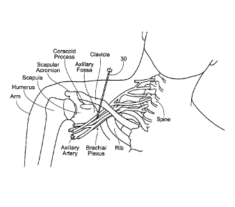
Fig. 4 shows the clavicle as a doubly curved short
bone that connects the arm (upper limb) = to =the body =
(trunk), located directly above the first rib. It acts as
a shunt to keep the scapula in position so the arm can
hang freely. The coracoid process is a small finger-like
structure on the upper lateral corner of the scapula.
Pointing laterally forward, it, together with the
acromion, serves to stabilize the shoulder joint. It is
palpable in the deltopectoral groove between the deltoid
and pectoralis major muscles.
Guided by these landmarks, the brachial plexus can
be identified. Referring to Figs. 4 and 5, the brachial
plexus comprises an arrangement of nerve fibers, running
from the spine, formed by the ventral rami of the lower
cervical and upper thoracic nerve roots, specifically
from above the fifth cervical vertebra to underneath the
first thoracic vertebra (C5-T1). It proceeds through the
neck, under the clavicle and generally anterior to the
scapula, through the armpit region and into the arm. The
brachial plexus is generally responsible for cutaneous
CA 02761530 2011-11-08
W02010/065143 PCT/US2009/006403
- 15 -
and muscular innervation of the entire upper limb, with
only two exceptions; the trapezius muscle is innervated
by the spinal accessory nerve and an area of skin near
the armpit is innervated by the intercostobrachialis
nerve.
Fig. 6 is a cross-sectional view (perpendicular to
=the axis of the lead insertion) of the brachial plexus
and surrounding tissue (Moayeri et al. 2008). As can be
seen in Fig. 6, the brachial plexus is surrounded by a
large amount of adipose tissue 54 in the infraclavicular
and subcoracoid regions, where the electrode lead 12 will
be placed, and is well suited for use in adipose tissue.
In the infraclavicular and subcoracoid sections of
cadavers studied, the brachial plexus was surrounded by
about 6.90 + 1.82 cm2 to about 7.06 + 1.48 cm2, which is
ample area to place the electrode lead 12.
B. Implantation Methodology
Representative lead insertion techniques will now be
described to place an electrode lead 12 in a desired
location in adipose tissue 54 at or near the brachial
plexus. It is this desired placement that makes possible
the stimulation of the brachial plexus with a single lead
12 to provide pain relief.
Figs. 7 and 8 show representative embodiments of the
steps that representative instructions for use 58 can
incorporate or direct for the placement of an electrode
lead 12 in a targeted tissue region for the relief of
pain, such as post-amputation pain. The instructions may
include a series of steps that can be followed to carry
out portion or portions of the procedure. These steps may
include, but are not limited to:
1) Place the patient in a supine position with head
turned away from the lead insertion site 16 and forearm
laid to rest in a neutral position beside the body.
2) Prepare the lead insertion site with antiseptic
CA 02761530 2011-11-08
W02010/065143
PCT/US2009/006403
- 16 -
and local subcutaneous anesthetic (e.g., 2% lidocaine).
3) Locate the site of skin puncture 16 with
landmarks as necessary, such as those previously
described, e.g., approximately 2 cm medial and caudal to
'5 the coracoid process.
4) Insert a sterile percutaneous electrode lead 12
at a predetermined angle based on landmarks used, e.g.,
approximately 45 degrees towards the top of the axillary
fossa in relation to the axillary artery. The lead 12 may
be preloaded in the introducer needle 30 (see Fig. 7).
5) Place a surface stimulation return electrode 24
in proximity of the area in which the percutaneous lead
12 has been placed. Test stimulation will be applied to
the lead 12, with the surface electrode 24 providing a
return path. The surface electrode 24 may be placed
adjacent to the lead. Its position is not critical to
the therapy and it can be moved throughout the therapy to
reduce the risk of skin irritation.
6) Couple the lead 12 to the external pulse
generator 26 and to the return electrode 24 (see Fig. 8).
Set the desired stimulation parameters. Test stimulation
may be delivered using a current-regulated pulse
generator, for example. The external pulse generator 26
may be programmed to 4 mA, 100 ps, 100 Hz, and an on-off
duty cycle of 0.25 sec., as a non-limiting example.
7) Advance the introducer slowly until the subject
reports the first evoked sensation in the stump or
phantom upper limb (e.g., hand). Progressively reduce the
stimulus amplitude and advance the introducer more slowly
until the sensation can be evoked in the phantom upper
limb at a predetermined stimulus amplitude (e.g., 1 mA).
Stop the advancement of the introducer, and increase the
stimulus amplitude in small increments (e.g., 0.1 mA)
until the stimulation-evoked tingling . sensation
(paresthesia) expands to overlay the entire region of
CA 02761530 2011-11-08
WO 2010/065143
PCT/US2009/006403
- 17 -
pain in the subject's stump and phantom limb.
It is expected to locate the brachial plexus after
inserting the introducer approximately 4 cm from the site
of skin puncture 16. At this depth, it is expected that a
low stimulus intensity may evoke comfortable sensations
(paresthesia) without generating muscle contraction
(Nashold and Goldner 1975; Picaza et al. 1975; Nashold et
al. 1982).
8) Withdraw the introducer 30, leaving the
percutaneous lead 12 in proximity to the brachial plexus.
9) Cover the percutaneous exit site and lead 12 with
a bandage 32. A bandage 34 may also be used to secure
the external portion of the lead 12 (or an extension
cable used to couple the lead 12 to the external pulse
generator) to the skin (see Fig. 1). It is expected the
length of time to place the lead 12 to be less than 10
minutes, although the process may be shorter or longer.
10) Vary the stimulus amplitude in small steps
(e.g., 0.1 - 0.5 mA) to determine the thresholds at which
stimulation evokes first sensation (TsEN), sensation
(paresthesia) superimposed on the region of pain (Tsup)
muscle twitch (Tmus) of the triceps brachii (innervated by
the radial nerve branch of the brachial plexus), and
maximum comfortable sensation (Twa). Query the subject at
each stimulus amplitude to determine sensation level, and
visually monitor muscle response. Record the results.
11) It is possible that stimulation intensity may
need to be increased slightly during the process due to
causes such as habituation or the subject becoming
accustomed to sensation, but the need for increased
intensity is unlikely and usually only occurs after
several days to weeks to months as the tissue
encapsulates and the subject accommodates to stimulation
(Nashold 1975; Krainick and Thoden 1981; Goldman et al.
2008). It is to be appreciated that =the need for
CA 02761530 2011-11-08
WO 2010/065143
PCT/US2009/006403
- 18 -
increased intensity could happen at any time, even years
out, which would likely be due to either lead migration
or habituation, but may also be due reasons ranging from
nerve damage to plasticity/reorganization in the central
nervous system.
12) If paresthesias cannot be evoked with the
.initial lead placement, redirect the introducer 30 either
caudal or cephalad, but avoid the lung by never directing
the needle introducer 30 medially.
13) If sensations still cannot be evoked in a given
subject, then the muscle twitch response of the triceps
brachii may be used to guide lead placement and then
increase stimulus intensity until sufficient paresthesias
are elicited in the stump and phantom limb. Minimal
muscle contraction may be acceptable if it is well
tolerated by the amputee patient in exchange for
significant pain relief and if it does not lead to
additional discomfort or fatigue (Long 1973).
14) If stimulation evokes muscle contraction at a
lower stimulus threshold than paresthesia (e.g. if Tms..
Taw) and contraction leads to discomfort, then a lower
stimulus frequency (e.g., 12 Hz) may be used because low
frequencies (e.g., 4-20 Hz) have been shown to minimize
discomfort due to muscle contraction and provide >5094
relief of shoulder pain in stroke patients while still
inhibiting transmission of pain signals in the central
nervous system in animals (Chung et al. 1984; Yu et al.
2001, 2004; Chae et al. 2005). If continued muscle
contraction leads to pain due to fatigue, change the duty
cycle, using parameters shown to reduce muscle fatigue
and related discomfort in the upper extremity (e.g. 5 s
ramp up, 10 s on, 5 s ramp down, 10 s off) (Yu et al.
2004; Chae et al. 2005).
15) If stimulation fails to elicit paresthesia in
all areas of pain, then a second percutaneous lead 12'
CA 02761530 2011-11-08
V)13201(065143
PCT/US2009/006403
- 19 -
(not shown) may need to be placed to stimulate the nerves
that are not activated by the first lead 12. If
paresthesia coverage is incomplete, it may likely be due
to insufficient activation of the musculocutaneous nerve
because it has the most proximal branch point relative to
the other nerves and is the most likely to be missed
during single-injection nerve blocks of the brachial
plexus. To place a lead near the musculocutaneous nerve,
use the modified coracoid approach (a double-stimulation
technique) that targets the musculocutaneous nerve in
addition to the main trunk of the brachial plexus, as
described above (Desroches 2003; Minville et al. 2005).
16) If stimulation is successful, i.e., if the
screening test and/or home-trial are successful, the
patient's percutaneous system 10 (see Fig. 1) may be
converted into a fully implanted system 11 by replacing
the external pulse generator 26 with an implantable pulse
generator 28 that is implanted in a convenient area
(e.g., the subclavicular area). In one embodiment, the
electrode lead 12 used in the screening test and/or home-
trial may be totally removed and discarded, and a new
completely implantable lead may be tunneled
subcutaneously and coupled to the implantable pulse
generator. In an alternative embodiment, a two part lead
may be incorporated in the screening test and/or home-
trial where the implantable part is completely under the
skin and connected to a percutaneous connector (i.e.,
extension) that can be discarded after removal. The
implantable part may then be tunneled and coupled to the
implantable. pulse generator, or a new sterile extension
may be used to couple the lead to the implantable pulse
generator.
III. Electrode Lead Configurations
It is to be appreciated that the configuration of
one or more leads 12 and electrodes 14, and the manner in
CA 02761530 2011-11-08
W02010/065143
PCT/US2009/006403
- 20 -
which they are implanted can vary. Stimulation may be
applied through an electrode lead 12, such as a fine wire
electrode, paddle electrode, intramuscular electrode, or
general-purpose electrode, inserted via a needle
introducer or surgically implanted in proximity of the
target site. Once proper placement is confirmed, the
needle may be withdrawn, leaving the electrode in place.
Stimulation may also be applied through a penetrating
electrode, such as an electrode array comprised of any
number (i.e., one or more) of needle-like electrodes that
are inserted into the target site. In both cases, the
lead may placed using a needle-like introducer, allowing
the lead/electrode placement to be minimally invasive.
The electrode 14 may be electrically insulated
everywhere except at one (monopolar), or two (bipolar),
or three (tripolar), for example, conduction locations
near its distal.tip. Each of the conduction locations
may be connected to one or more conductors that run the
length of the electrode and lead 12, proving electrical
continuity from the conduction location through the lead
12 to the stimulator 26 or 28.
The electrode lead 12 is desirably provided in a
sterile package, and may be pre-loaded in the introducer
needle 30. The lead 12 desirably possess mechanical
properties in terms of flexibility and fatigue life that
provide an operating life free of mechanical and/or
electrical failure, taking into account the dynamics of
the surrounding tissue (i.e., stretching, bending,
pushing, pulling, crushing, etc.). The material of the
electrode desirably discourages the in-growth of
connective tissue along its length, so as not to inhibit
its withdrawal at the end of its use. However, it may be
desirable to encourage the in-growth of connective tissue
at the distal tip of the electrode, to enhance its
anchoring in tissue.
CA 02761530 2011-11-08
WO 2010/065143
PCT/US2009/006403
- 21 -
One embodiment of the lead 12 shown in Fig. 9 may
comprise a minimally invasive coiled fine wire lead 12
and electrode 14. The electrode 14 may also include, at
its distal tip, an anchoring element 48. In the
illustrated embodiment, the anchoring element 48 takes
the form of a simple barb or bend. The anchoring element
48 is sized and configured so that, when in contact with
tissue, it takes purchase in tissue, to resist
dislodgement or migration of the electrode out of the
correct location in the surrounding tissue. Desirably,
the anchoring element 48 is prevented from fully engaging
body tissue until after the electrode 14 has been
deployed. The electrode may not be deployed until after
it has been correctly located during the implantation
(lead placement) process, as previously described.
An alternative embodiment of an electrode lead 12
shown in Figs. 10 and 11, may also include, at or near
its distal tip or region, one or more anchoring
element(s) 70. In¨the
illustrated embodiment, the
anchoring element 70 takes the form of an array of
shovel-like paddles or scallops 76 proximal to the
proximal-most electrode 14 (although a paddle 76 or
paddles could also be proximal to the distal most
electrode 14, or could also be distal to the distal most
electrode 14). The paddles 76 as shown are sized and
configured so they will not cut or score the surrounding
tissue. The anchoring element 70 is sized and configured
so that, when in contact with tissue, it takes purchase
in tissue, to resist dislodgement or migration of the
electrode out of the correct location in the surrounding
tissue (e.g., soft adipose tissue 54). Desirably, the
anchoring element 70 is prevented from fully engaging
body tissue until after the electrode 14 has been
deployed. The electrode is not deployed until after it
has been correctly located during the implantation (lead
CA 02761530 2011-11-08
W02010/065143
PCT/US2009/006403
- 22 -
placement) process, as previously described. In addition,
the lead 12 may include one or more ink markings 74, 75
to aid the physician in its proper placement.
Alternatively, or in combination, stimulation may be
applied through any type of nerve cuff (spiral, helical,
cylindrical, book, flat interface nerve electrode (FINE),
slowly closing FINE, etc.) that is surgically placed
within muscle at the target site.
In all cases, the lead may exit through the skin and
connect with one or more external stimulators 26, or the
lead(s) may be routed subcutaneously to one or more
implanted pulse generators 28, or they may be connected
as needed to internal and external coils for RF (Radio
Frequency) wireless telemetry communications or an
inductively coupled telemetry to control the implanted
pulse generator. The implanted pulse generator 28 may be
located some distance (remote) from the electrode 14, or
an implanted pulse generator may be integrated with an
electrode(s), eliminating the need to route the lead
subcutaneously to the implanted pulse generator.
Control of the stimulator and stimulation parameters
may be provided by one or more external controllers. In
the case of an external stimulator, the controller may be
integrated with the external stimulator. The implanted
pulse generator external controller (i.e., clinical
programmer) may be a remote unit that uses RF (Radio
Frequency) wireless telemetry communications (rather than
an inductively coupled telemetry) to control the
implanted pulse generator. The external or implantable
pulse generator may use passive charge recovery to
generate the stimulation waveform, regulated voltage
(e.g., 10 mV to 20 V), and/or regulated current (e.g.,
about 10 A to about 50 mA). Passive charge recovery is
one method of generating a biphasic, charge-balanced
pulse as desired for tissue stimulation without severe
CA 02761530 2011-11-08
WO 2010/065143
PCT/US2009/006403
- 23 -
side effects due to a DC component of the current.
The neurostimulation pulse may by monophasic,
biphasic, and/or multi-phasic. In the case of the
biphasic or multi-phasic pulse, the pulse may be
symmetrical or asymmetrical. Its shape may be rectangular
or exponential or a combination of rectangular and
exponential waveforms. The pulse width of each phase may
range between e.g., about 0.1 sec. to about 1.0 sec., as
non-limiting examples.
Pulses may be applied in continuous or intermittent
trains (i.e., the stimulus frequency changes as a
function of time). In the case of intermittent pulses,
the on/off duty cycle of pulses may be symmetrical or
asymmetrical, and the duty cycle may be regular and
repeatable from one intermittent burst to the next or the
duty cycle of each set of bursts may vary in a random (or
pseudo random) fashion. Varying the stimulus frequency
and/or duty cycle may assist in warding off habituation
because of the stimulus modulation.
The stimulating frequency may range from e.g., about
1 Hz to about 300 Hz, and the frequency of stimulation
may be constant or varying. In the case of applying
stimulation with varying frequencies, the frequencies may
vary in a consistent and repeatable pattern or in a
random (or pseudo random) fashion or a combination of
repeatable and random patterns.
IV. System Kits
As Figs. 12 and 13 show, the various devices and
components just described can be consolidated for use in
one or more functional kit(s) 60, 64. The kits can take
various forms-and the arrangement and contents of the
kits can vary. In the illustrated embodiments, each kit
60, 64 comprise a sterile, wrapped assembly. Each kit 60,
64 includes an interior tray 62 made, e.g., from die cut
cardboard, plastic sheet, or thermo-formed plastic
CA 02761530 2011-11-08
WO 2010/065143
PCT/US2009/006403
- 24 -
material, which hold the contents. Kits 60, 64 also
desirably includes instructions for use 58 for using the
contents of the kit to carry out the procedures described
above, including the systems and methods incorporating
the percutaneous system 10 and/or the implanted system
11.
The instructions 58 can, of course vary. The
instructions 58 may be physically present in the kits,
but can also be supplied separately. The instructions 58
can be embodied in separate instruction manuals, or in
video or audio tapes, CD's, and DVD's. The instructions
58 for use can also be available through an internet web
page.
The foregoing is considered as illustrative only of
the principles of the invention. Furthermore, since
numerous modifications and changes will readily occur to
those skilled in the art, it is not desired to limit the
invention to the exact construction and operation shown
and described. While the desired embodiment has been
described, the details may be changed without departing
from the invention.
Various features of the invention are set forth in
the following Claims.