Note: Descriptions are shown in the official language in which they were submitted.
CA 02761630 2011-10-24
METHOD OF DETERMINING A COMPLETE BLOOD COUNT
AND A WHITE BLOOD CELL DIFFERENTIAL COUNT
CROSS-REFERENCE TO RELATED APPLICATIONS
This application claims priority under 35 U.S.C. 119 to United States
Provisional
Application No. 61/047,920, filed April 25, 2008, which is expressly
incorporated herein
by reference in its entirety. Further, the present application expressly
incorporates herein
by reference the application entitled "SYSTEMS AND METHODS FOR ANALYZING
BODY FLUIDS," which is being filed on the same date and by the same inventors
as the
present application.
FIELD OF THE INVENTION
This invention relates to a system and process for determining composition and
components of fluids. More specifically the present invention provides
improved
techniques for viewing cellular morphology, and determining the number of a
particular
type of cell in a portion of a body fluid.
BACKGROUND OF THE INVENTION
Pathology is a field of medicine where medical professionals determine the
presence, or absence of disease by methods that include the morphologic
examination of
individual cells that have been collected, fixed or air-dried, and then
visualized by a stain
that highlights features of both the nucleus and the cytoplasm. The collection
of the cells
often involves capturing a portion of a person's body fluid, placing the body
fluid on a
slide, and viewing the fluid on the slide using a microscope.
One of the most commonly performed pathologic studies is the CBC (the
Complete Blood Count). To perform a CBC, a sample of blood is extracted from a
patient
1
CA 02761630 2011-10-24
and then the cells are counted by automated or manual methods. The CBC is
commonly
performed by using an instrument, based on the principal of flow cytometry,
which
customarily aspirates anticoagulated whole blood and divides it into several
analysis
streams. Using the flow cytometer a number of primary and derived measurements
can be
determined including: i) red blood cell (RBC) count, hemoglobin (Hb),
hematocrit (Het),
red blood cell indices (mean corpuscular volume, MCV, mean corpuscular
hemoglobin,
MCH and mean corpuscular hemoglobin concentration MCHC), red blood cell
distribution width, enumeration of other red blood cells including
reticulocytes and
nucleated red blood cells, and red blood cell morphology; ii) white blood cell
(WBC)
count and WBC "differential" count (enumeration of the different normal white
blood cell
types, including neutrophils, lymphocytes, eosinophils, basophils and
monocytes, and the
probable presence of other normal and abnormal types of WBC that are present
in various
disease conditions); and iii) platelet count, platelet distribution widths and
other features of
platelets including morphological features. In flow cytometers, red blood
cell, WBC, and
platelet morphological characterizations are typically made indirectly, based
on light
absorption and light scattering techniques and/or cytochemically based
measurements.
Some advanced flow cytometers calculate secondary and tertiary measurements
from the
primary measurements.
Flow based CBC instruments generally require extensive calibration and
control,
maintenance, and skilled operators, and they have substantial costs associated
with
acquisition, service, reagents, consumables and disposables. One significant
problem with
these systems in routine use is that a large proportion of blood specimens
require further
testing to complete the assessment of the morphologic components of the CBC.
This
involves placing a sample of blood on a slide, smearing the sample against the
slide to
form a wedge smear, and placing the slide under a microscope. This process is
often done
2
CA 02761630 2011-10-24
manually by skilled medical technologists, which increases the cost and time
to receive
results from the tests. The direct visualization of blood cells on a glass
slide must be
performed whenever the results of the automated test require further
examination of the
blood sample. For example, a "manual" differential count is performed by
direct
visualization of the cells by an experienced observer whenever nucleated
immature RBCs
are found or WBCs suspicious for infection, leukemias or other hematologic
diseases are
found.
The proportion of these specimens requiring further review generally ranges
from
10% to S0 /., depending on the laboratory policy, patient population and
"flagging"
criteria, with a median rate of around 27%. The most frequent reasons for
retesting include
the presence of increased or decreased number of WBCs, RBCs or platelets,
abnormal cell
types or cell morphology, clinical or other suspicion of viral or bacterial
infections.
In addition to additional work involved in performing manual differential
counts,
this process has a number of additional technical limitations. These include
distortions of
cell morphology because of mechanical forces involved in smearing the cells
onto the
slide, and cells overlapping one another, which makes visualization of
individual cell
morphology difficult.
SUMMARY OF THE INVENTION
The present invention provides systems and methods for placing cells on a
slide.
Additionally systems and method for imaging the cells are provided. The images
may be
later used to perform tests such as a complete blood count including image-
based counting
and assessment of the morphology of the formed elements of blood, including
RBCs,
WBCs, and platelets. Embodiments of the present invention may improve the
accuracy of
the CBC by providing improved visualization of the formed elements of blood.
Aspects of
3
CA 02761630 2011-10-24
the present invention may analyze and determine the presence of certain cell
types, such as
abnormal or immature WBCs that are found in cases of abnormal bone marrow
function
including hematological malignancies. Further, the configurations of the
present invention
may decrease costs associated with instrumentation, decrease costs of
consumables and
reagents, require less operator time and reagents, fewer repeated tests, and
fewer moving
parts than other prior art techniques. Configurations of the present invention
may also
reduce the turn around time for many of the CBC tests that currently require
visualization
of blood cells after the instrumental portion of the test is completed, by
allowing cells to
be visualized on a monitor instead of under a microscope.
Aspects of the present invention are effective at preserving cell morphology.
This
may be important for patients with hematological malignancies such as chronic
lymphocytic leukemia (CLL) or acute myeloid leukemia (AML). The systems and
methods relating to applying a monolayer of cells to a slide may enable
detection of a
larger number of morphologically well-preserved blast cells and other immature
or fragile
cells. This would allow their more accurate recognition at an earlier stage of
the leukemic
or other disease process. Certain aspects of the present invention provide for
preparing a
substantially uniform distribution of cells across a test area of a slide.
Aspects of the present invention also relate to collecting cells in a fluid
(such as
blood) from organic tissue, possibly mixing the cells contained in the fluid
with a diluent,
collecting a sub-sample (aliquot) of a known volume from the solution, and
then
depositing the aliquot onto a substratum such as a slide using a dispensing
device or
applicator. The cells may be allowed to air dry or may be fixed (using a
fixative solution)
or both, depending on the examination that is anticipated. The cells may also
be stained.
The stained cells on the substratum may be counted and examined by an
automated
imaging system utilizing a computer or viewed by manual microscopic
examination.
4
CA 02761630 2011-10-24
i 1
Digital images may be shown on a computer display to reduce the need for
manual
microscopic review.
BRIEF DESCRIPTION OF THE DRAWINGS
Fig. 1 A: is a perspective, schematic view of a system for analyzing body
fluids.
Fig. 1B: is a perspective, schematic view of a system for analyzing body
fluids.
Fig. 2: is a perspective view of a slide and slide holder.
Fig. 3: is an enlarged top view of the slide and slide specimen.
Fig. 4: is alternate embodiment of the top view of the slide and slide
specimen.
Fig. 5: is a graph illustrating the correlation between Sysmex RBC counts and
the
RBC counts generated using an embodiment of the instant invention.
Fig. 6: is a graph illustrating the correlation between Sysmex WBC counts and
the
WBC counts generated using an embodiment of the instant invention.
Fig. 7A: is a process flow schematic of the embodiment shown in Fig. 1A.
Fig. 7B: is a process flow schematic of the embodiment shown in Fig. 1B.
DETAILED DESCRIPTION OF THE INVENTION
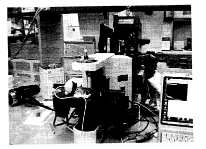
With reference to Fig. 1 A, a system 10 for analyzing body fluids is
disclosed. The
system may comprise a platform 100, a light receiving device 200, a computer
300, an
applicator 400, a gas circulation device 500, a light source 600, a dispenser
800, a
discharge device 900, a slide labeler 1000, and slide label reader 1100. The
following
sections below include capitalized headings which are intended to facilitate
navigation
through the specification. The headings are not intended to be limiting of the
invention in
any manner.
CA 02761630 2011-10-24
THE PLATFORM 100
In embodiments which feature a platform 100, an advancer 110 may be configured
to receive one or more slide apparatuses 700-700". The advancer 110 may be
attached to
a surface, such as the top surface 101, of the platform. The advancer 110 may
take the
form of a belt as shown in Fig. I A, the system may use a mechanical arm,
gravity,
magnetism, hydraulics, gears, or other locomotion techniques to move the slide
apparatus
along the surface 101 of the platform.
The platform 100 may also comprise a feeder 102 and a collector 106 for
respectively feeding and collecting the slide apparatuses 700 from or to a
stack or rack.
The feeder 102 may be equipped with a feeder propulsion mechanism 103 (such as
rubberized wheels) for pushing the slides down a ramp 104 onto the advancer
110. (Of
course embodiments of the invention could be built without a ramp. For
example, if the
feeder is level with advancer 110, no ramp would be needed. Alternatively, a
mechanical
arm could be used to grab the slide apparatus 700 and place the slide
apparatus 700 on the
advancer directly.) Alternate mechanisms to propel the slide out of the feeder
102 may be
used such as magnets or hydraulics. The feeder may comprise a sensor for
determining
how many slides are present. The sensor could measure the weight of the slide
apparatuses 700 for example to determine how many slide apparatuses were
present. Fig.
1A illustrates 3 slide apparatuses 700 stored in the feeder 102. The collector
106 may also
comprise a sensor for determining how many slides are present in the collector
106. The
sensor may inform the computer when a preset number of slides have been
analyzed or
may inform the computer of the receipt of a slide on an ongoing basis.
6
CA 02761630 2011-10-24
THE LIGHT RECEIVING DEVICE 200
The light receiving device 200 may be a microscope (such as brightfield
microscope), a video camera, a still camera, or other optical device which
receives light.
In embodiments using a standard brightfield microscope, one containing an
automated
stage (a slide mover 201) and focus may be selected. In one embodiment, a
microscope
may be attached to a motorized stage and a focus motor attachment. The
microscope may
have a motorized nosepiece, for allowing different magnification lenses to be
selected
under computer 300 control. A filter wheel may allow the computer 300 to
automatically
select narrow band color filters in the light path. LED illumination may be
substituted for
the filters, and use of LEDs may reduce the image acquisition time as compared
to the
time required for filter wheel rotation. A 1600 x 1200 pixel firewire camera
may be used
to acquire the narrow band images.
In some cases, the light receiving device will receive light reflected off
slide
apparatus 700" and store an image of that light. In some embodiments
fluorescent
emission from the cellular objects may be detected in the light receiving
device 200.
However, since the light emission source 600 can be positioned below the
platform, the
light emission source may direct light so that it passes through the platform
100 and the
slide 701 into the light receiving device 200. The light receiving device may
be connected
to a computer through a link 11, and may be capable of X, Y, and Z axial
movement (in
other embodiments a motorized stage or slide mover 201 may provide X, Y, and Z
movement.) The light receiving device may comprise a link 11 such as a wire as
shown in
Fig. I A, or other wireless systems may be used. The light receiving device
200 and any of
the other components may be interfaced with the computer 300 through a link
(11-15)
which may provide energy to the component, provide instructions from the
computer 300
to the component, or allow the component to send information to the computer
300. Light
7
CA 02761630 2011-10-24
receiving device 200 may contain pan, tilt, or locomotive actuators to allow
the computer
300 to position the device 200 in an appropriate position. The light receiving
device may
contain a lens 210 which focuses the light. The light receiving device may
capture black
and white or color images. Alternatively, two or more light receiving devices
could be
used to divide the processing time associated with capturing the images. For
example a
low magnification image station could be followed by a high magnification
image station.
Similarly, in some embodiments, the system 10, platform 100, computer 300, or
light
receiving device 200 may direct a slide mover 201 to move the slide apparatus
700 in
order to store images of all the cells in the slide. Using a slide mover 201
may be
desirable, if for example, the field size of the light receiving device 200 is
smaller than the
specimen zone 710 (Fig. 3).
THE COMPUTER 300
The computer 300 may be a laptop as shown in Fig. IA, or a server,
workstation,
or any other type of computing device. The computer may comprise a processor,
a display
320, and interface 310, and internal memory and/or a disk drive. The computer
300 may
also comprise software stored in the memory or on computer readable media such
as an
optical drive. The software may comprise instructions for causing the computer
to operate
the light receiving device 200, the applicator 400, the applicator controller
490, the fan
500, the platform 100, advancer 110, light source 600, dispenser 450 or 800,
or any
component connected to one of these components. Similarly, the computer may
receive
information from any of these components. For example, the software may
control the
rate of dispersal of slides from the feeder 102, and feeder 102 may inform the
computer
about the number of slides present. In addition, the computer 300 may also be
responsible
for performing the analysis of the images captured by the light receiving
device. Through
8
CA 02761630 2011-10-24
the analysis process, the computer may be able to calculate the number of a
specific type
of cell in a particular volume of blood, for example for blood, red cell,
white cell, and
platelet counts and other measured and derived components of the CBC such as:
hemoglobin content, red blood cell morphology, or WBC differential could be
calculated.
The image analysis software may analyze each individual field and sum the
total red and
white cell counts. To calculate the total counts per microliter in the patient
vial, the
number counted on the slide is multiplied by the dilution ratio and volume of
the sub-
sample. Results of the counts, morphologic measurements, and images of RBCs
and
WBCs from the slide may be shown on the display 320. In some embodiments, the
computer 300 may be able to display numerical data, cell population
histograms,
scatterplots, and direct assessments of cellular morphology using images of
blood cells
displayed on the monitor. The ability to display cellular morphology provides
users of the
system 10, the ability to quickly establish the presence or absence of
abnormalities in cell
morphology that may warrant preparing an additional slide for manual review by
an
experienced technician or other professional. The software may provide the
computer
instructions to display images 331 received from the light receiving device or
may cause
the display 330 to show the results 332 (in perhaps a chart or graph for
example) of an
analysis of the images. Similarly, the computer 300 may be able to enumerate
the number
of cells of a specific type in a particular blood volume or enumerate the
number of
damaged cells, cancerous cells, or lysed cells in a particular volume of
blood. The
memory of the computer may contain software to allow the computer to perform
the
analysis process. The computer may use one or more magnifications during the
analysis.
Although shown as one component, computer 300 may comprise multiple
computers and a first computer could be used for controlling the components
and a second
computer could be used for processing the images from the light receiving
device 200. In
9
CA 02761630 2011-10-24
some embodiments, the various computer may be linked together to allow the
computer to
share information. The computer 300 may also be connected to a network or
laboratory
information system to allow the computer to send and receive information to
other
computers.
THE APPLICATOR 400
In certain embodiments, the applicator 400 may comprise a syringe, a manual or
motor driven pipettor or using a motor controlled pump attached through a tube
to a
pipette tip. While many different types of pipettes or syringes could be used,
test results
have shown improved results can be obtained through using an applicator 400
having
better than 2% accuracy. The pump may be a peristaltic pump, a syringe pump,
or other
similar device that allows small volumes to be aspirated and dispensed through
an orifice.
Typically such an orifice will be contained in a tip 405 that is two to five
millimeters in
'outside diameter with an inner diameter of 0.5 millimeters. The tip 405 may
be disposable
or washable. The tip 405 may be rounded to facilitate insertion and cleaning
of the tip.
Fluid flow through the tip is controlled to allow a thin layer of blood or
diluted blood to be
deposited onto the slide. By optimizing flow rate through the tip and the
relative speed
and height of the tip over the slide an appropriate density of cells can be
deposited onto the
slide. Each of these factors influences the other, so the proper combination
of height, flow
rate through the tip, and speed over the slide must be determined. In one
embodiment the
flow rate through the tip is 0.1 microliters per second while the tip is
moving at a speed of
30 millimeters per second over the slide surface at a height of about 70
microns.
In use, the applicator 400 may comprise a known volume of body fluid such as
30
microliters (ul). The applicator may mix this fluid with a stain or diluent,
and eject a
portion of this fluid onto the slide apparatus 700 (particularly the specimen
zone 710, Fig.
CA 02761630 2011-10-24
(~ .
3). A typical sub-sample would be an aliquot of approximately 1/2 pl to 2 pl,
but may be
in the range of 1/10 to 10 pl. In some embodiments, the system 10 or
applicator 400 may
contain a first reservoir 420 for storing the body fluid and a second
reservoir 430 for
storing diluent. In some embodiments the body fluid will not be diluted.
The system 10 or applicator 400 may contain one or more dispensers 800. The
dispenser 800 (or 450 in Fig. 113) may be used to direct a fixative or a stain
onto the slide
701. In this embodiment, the applicator 400 may contain one or more fluid
chambers 410
to eject body fluid, diluent, stain, and fixative from the applicator 400.
Some dispensers
may be able to store both fluids and direct them sequentially onto the slide,
or in alternate
embodiments, two dispensers may be used (one for the fixative and one for the
stain.)
Excess stain and fixative may be removed from the slide, by tilting the slide
apparatus so
that it is orthogonal (or angled) to the platform surface 101. A slide titter
801 may be used
for this purpose. Slide tilter may comprise a simple wedge as shown, or may
comprise a
mechanical arm to tilt the slide.
In the embodiment shown in Fig IA, the stain dispenser is attached to the
platform
100. Examples of stains compatible with embodiment shown in Figure IA may
include:
Wright-Giemsa stain, Geimsa stains, and Romanowsky stains. Other solutions
that could
be dispensed are fixatives (methanol) and buffer solutions. Other
visualization methods
involving immunocytochemical reagents or other markers of specific cell
components may
also be used. The stain dispenser may also be embodied as a stain reservoir
450 and
attached to the applicator 400 (see Fig. 1B). Examples of stains compatible
with the
embodiment shown in Figure lB may include: Romanowsky stains, reticulocyte
stains,
and stains using specific antibodies. . In the embodiment having dispenser
800, the
dispenser can dispense stain onto the slide apparatus (particularly the
specimen zone 710.)
Dispenser 800 may take the form of a peristaltic pump. In the embodiment
having a stain
11
CA 02761630 2011-10-24
reservoir 450, the stain may be mixed in with the body fluid and the diluent
from
reservoirs 420 and 430. The body fluid and the diluent may be mixed together
by a mixer
440, which can mix the fluid and diluent in certain ratios. In an alternate
embodiment, the
slide could be immersed into one or more baths of the fixation and staining
solutions. In
another embodiment, fixation and staining solutions could be moved across the
slide using
capillary action.
Various fixatives and diluents may be used with the present invention. For
example 85% methanol can be used as the fixative. For some stains an ethyl
alcohol or
formaldehyde based fixative might be used. Diluents useful for diluting whole
blood for
example, may include salt solutions or protein solutions. Salt solutions range
from
"physiological saline" (0.9N), to complex mixtures of salts, to the commercial
preparation
Plasmalyte that simulates virtually all the salts found in human blood serum.
Protein
solutions can range from simple solutions of bovine albumin to Plasmanate, a
commercial
preparation with selected human plasma proteins. Such preparations can vary in
protein
concentrations, buffers, pH, osmolarity, osmalality, buffering capacity, and
additives of
various types. Synthetic or "substitute" versions of these solutions may also
be usable,
including Ficoll or Dextran or other polysaccharides. Other substitutes may be
used. An
example of a diluent is Plasmalyte plus Plasmanate in the proportion of 4:1
(Plasmalyte:Plasmanate)._Another example of a diluent is 5% albumin. When
analyzing
whole blood, a dilution of 2 parts blood to I part diluent can be used, where
the diluent is a
physiologically compatible solution, but a range of dilution from 0:1 (no
dilution) to 10:1
(diluent:blood) may be used in alternate embodiments.
The applicator may comprise a hydraulic piston for pushing the fluid out of
fluid
chamber 410 (like a syringe or a pipette). A tip 405 may be provided for
adjusting the
flow rate of the fluid. While size of the tip does not affect the speed
(pi/sec) in which the
12
CA 02761630 2011-10-24
solution flows out of the tip, generally, the smaller the opening in the tip,
the greater the
force (gg'distance/seconds). Additionally, the size of the tip affects
thickness of the fluid
flows 750 shown in Fig's 2 and 3. A tip having a 0.3 millimeter inner diameter
may
provide for a flow rate of 0.1 microliters per second, and the distance from a
middle point
751 of the first flow to the middle point 752 of the second flow may be 500
microns. In
order to create the flows 750 shown in Fig. 2 and 3, the system 10 may be
configured to
account for the variances in the number of cells in a given blood specimen.
For human
peripheral blood samples, the range is large butwithin one order of magnitude.
In order to
accurately count the blood cells, the overlap between red blood cells should
be minimized.
One method to provide minimal overlapping between cells is to lay down non-
touching
rows of cells from the tip of the applicator. Increasing viscosity of the
diluted fluid or the
type or amount of diluent may affect the width of the final settlement
positions of the
flows 750. By selecting a distance between rows to allow for the typical
variation in
blood samples, all cells can be counted in all samples. For many samples these
gaps will
be seen between the flows; however this does not affect the image analysis and
the row
and gap effect tends not to be noticed during high magnification manual review
under the
microscope. To avoid these gaps, a light receiving device could be attached to
the
applicator or positioned near station A (see Fig. 7A) to allow the computer
300 to
determine the width of the first flow 751 (Fig. 3) formed by directing the
cells onto the
slide. By determining the width of the flow, i.e. how far the blood flows
sideways from
location the fluid was placed on the slide, the computer 300 could cause the
applicator to
adjust the gap size of the flows. The computer 300 calculate the distance the
second flow
752 (Fig. 3) needs to be from the first flow 751, and place the flows so that
they settle
adjacent to one another minimizing the formation of any gaps between the
flows. Using
13
CA 02761630 2011-10-24
this process, a gapless or contiguous flow of cells can be applied to the
specimen zone
710.
To physically place the cells on the slide 701, the computer 300 could direct
the
applicator controller 490 to perform the body fluid application process 7B
(see Fig. 713)
which involves moving the body fluid chamber 410 in the X, Y, or Z directions
to position
the tip 405 so that it tracing the eventual locations of the flows 750. In
some
embodiments, the X, Y, and Z directions are all perpendicular to each other
affording the
applicator the ability to move in any direction in a three dimensional
coordinate system.
The computer 300 may be connected to the applicator controller 490 to control
this
movement. In the embodiment shown in Fig. 3, the controller may position the
tip at the
top left corner of the specimen zone 710 and proceed to place fluid sample
onto the cells
by ejecting the fluid from the fluid chamber 410. While the ejection is
occurring, the
controller 490 may move the tip in the positive X direction to the top right
portion of the
specimen zone 710 (see Fig. 3). Once the top right section is reached, the
controller 490
may move the tip in the negative Y direction one flow width. The flow 750
width may
range from 300 to 1000 microns, and flow thickness increases as the flow rate
of fluid out
of the tip increases and/or the speed of the tip across the slide decreases.
Additionally the
viscosity of the fluid and diluent choice may affect the width of the flow 750
(Fig. 3).
Typically, the cells of the fluid will settle within a few seconds once placed
on the slide.
Once the tip has been moved one flow width, the controller may move the tip in
the
negative X direction to the leftmost side of the specimen zone 710. Once the
leftmost side
is reached, the tip again may be moved one flow width in the negative Y
direction. This
process may be repeated until the entire specimen zone is covered. In
alternate
embodiments, the diluted body fluid could be applied to slide with a fixed
applicator and
slide which moves via the moveable slide controller 760 (this application
process 7A is
14
CA 02761630 2011-10-24
shown on Fig. 7A.) The slide controller 760 may be moveable in the X, Y, Z
direction to
move the slide apparatus in similar positions to allow the applicator to place
flows 750 of
body fluid on the specimen zone 710.
The number of cells placed on the slide 701 using this method will vary
depending
on the type of fluid being examined and the dilution ratio. Assuming whole
blood were
being analyzed with a 1:3 (blood:diluent ratio), about 900,000 red blood
cells, 45,000
platelets, and 1,000 white blood cells would be placed on the slide. Though
Fig. 3 shows
the generation of a uniformly distributed fluid specimen in a rectangular
shape, other
shapes may be constructed in a similar manner. Fig. 4, shows for example, a
fluid flow
comprising a plurality of concentric circles. Like Fig. 3, the fluid flows 750
are placed
adjacent to one another to create a uniform viewing field. This process
provides a highly
uniform distribution of cells across the specimen zone 710, facilitating the
analysis
process. Additionally, the computer 300 can alter the appearance and width of
the fluid on
the zone 710. For example, the computer 300 may control the speed at which the
tip
moves across the specimen zone, which would affect the thickness of the fluid
resting on
the zone. In some embodiments, speeds of 10 to 100 mm/s may be selected in
order to
provide the zone with a specimen which is about one cell thick. The controller
490 also
may select the height of the tip above the slide 700. A height of 70 +/- 40
microns above
the slide may be used in order to minimize damage to fluid cells when they
come into
contact with the slide apparatus 700, and to maintain fluid flow from the tip
to the
substrate.
THE GAS MOVEMENT DEVICE 500
Gas movement device 500 may comprise a fan (such as shown in Fig. 1) or may
comprise other gas movement devices such as a compressor or a bellow for
example. Gas
CA 02761630 2011-10-24
movement device 500 may be connected directly to the computer 300 or may be
connected through another component such as the platform 100 or the applicator
400 (as
shown.) The gas movement device pushes gas (in some cases atmospheric air)
across the
slide to control the rate at which the slide dries. Moving too much air too
quickly (i.e. too
high of a fan speed) across the slide can cause cells in the specimen to burst
due to too
rapid drying, and too little air too slowly (i.e. too low of a fan speed)
across the slide can
cause the cells to dry too slowly and appear to shrink._ The computer 300 may
select the
amount of air that moves across the slide in a period of time (i.e. the cubic
feet of air per
second) based upon the distance the gas movement device is from the slide, the
type of
fluid being analyzed, the width of the flows, and averages thickness of the
flows (this
would be the amount cells in each flow in the Z direction). The gas movement
device 500
may be placed near the slide apparatus 700, and positioned so that the device
directs gas so
that the gas strikes the slide at an angle of 30-60 angle (450 degrees can be
used) for a
period of about 15 to 20 seconds. In some embodiments, the computer can
control of
humidity and temperature settings in the vicinity of the system to allow the
drying process
to occur without the use of a gas movement device 500.
THE LIGHT EMISSION DEVICE 600
Two different embodiments of light emission device 600 are illustrated. In
Fig.
IA, light emission device 600 comprises a housing 601, a multispectrum light
source 610,
a number of light filters 620, 620', and 620", and a filter selector 621. As
shown in Fig.
IA, a portion of the housing has been removed to better show the light source
610. Light
source 610 may comprise a white light source or other multispectrum light
source such as
a halogen bulb, florescent bulb, or incandescent bulb etc. Filters 620-620"
may be used to
filter the multispectrum light into a single wavelength or a narrow band of
wavelengths.
16
CA 02761630 2011-10-24
The filter selector 621 may select which filters appear in front of the light
source 610. In
some embodiments more than one filter may be used to allow a particular range
of light to
illuminate the slide. Filter selector 621, may comprise a rotation motor and a
rod to spin
the filters in and out of the path of the light. In a second embodiment light
source may
comprise one or more lasers or LEDs (630) which emit a narrow band of light
(see Fig.
1B). An advantage for using LEDs in this system 10, is that LEDs can rapidly
be switched
on and off, allowing the light receiving device a single black and white
camera to acquire
the multiple spectral images in a very short time. LEDs also produce narrow
bandwidths
of illumination, typically from 15 to 30 nm full width at half maximum (the
breadth of the
wavelength intensity distribution at half of the peak brightness of the
maximum intensity).
Also, LEDs in the visible range do not project heat-producing infrared energy
into the
optical system and are relatively long lived as compared to conventional
lamps. An
advantage of using narrow-band illumination rather than unfiltered white light
(i.e. broad-
band illumination) is using narrow band illumination increases the sharpness
of the images
generated by the light receiving device 200. If the light receiving device 200
contains a
lens, the presence of the lens may cause some chromatic aberration that result
in slight
focus shifts or image quality degradation when using different colors. With
white light
illumination this can result in an overall degradation of the image quality.
The light
receiving device 200 may capture a black and white image for each narrow-band
of
illumination. The computer 300 may be able to correct focus and image quality
for each
wavelength by adjusting the focal distance or the distance of the lens from
the slide. In
some embodiments, the computer 300 may shift the focus position of the lens
while a
number of light colors are emitted sequentially to improve the quality of the
image.
Various wavelengths of light may be directed by the light emission device 600.
Two - eight or more different wavelengths of light may be directed at the
slide apparatus
17
CA 02761630 2011-10-24
tt `'(
700. Wavelengths such as 430 nm are useful for imaging a hemoglobin-only image
for
assessing RBC morphology and hemoglobin content Using an image taken with such
a
wavelength which is designed to show only red blood cells, it may also show
red blood
cells which are touching white blood cells. The touching red blood cells may
be digitally
removed from images to make it easier for the computer to detect the white
blood cell
borders in order to make more accurate cellular measurements and enumeration.
Light
emitted at 570 nm may be useful to provide high contrast images for platelets
and nuclei.
Other wavelengths may be chosen in order to best discriminate the colors of
basophils,
monocytes, lymphocytes (all shades of blue), eosinophils (red), and
neutrophils (neutral
color). For counting platelets, for example, two colors of illumination may be
used (such
as 430 nm and 570nm). A high contrast image may be obtained by subtracting the
430 run
image from the 570 nm image. Light having a wavelength of 430, 500, 525 and
600 are
particularly effective at showing cell color information, but light at
wavelengths between
400 nm and 700nm inclusive may be used. These wavelengths will also be used
for the
display of the color images if appropriate. Otherwise one or two additional
images may
need to be taken for the 200+ cells that will be analyzed for the differential
count and
which may be shown on the display 320. Typically the narrow-band images will
be
chosen from the range of 400 nm to 750 nm. Test results have shown that 2-8
separate
light colors to work well, with 3-4 separate light colors being optimal. The
computer 300
may be able to further refine the images by compensating for spatial shifts.
Also the
computer may combine the various colored images to generate multi color images
for
display or analysis. Numeric descriptors of the individual images or combined
images can
be used to determine spatial, densitometric, colorimetric and texture features
of the cells
for classification of the cell types. A further advantage of using narrow band
illumination
is that using narrow band illumination allows for the elimination of the use
of oil
18
CA 02761630 2011-10-24
objectives or coverslips. Light is refracted when the light passes from glass
to air. Prior
art systems have used oil objectives or coverslips to minimize this refraction
at air to glass
transitions, but having to add oil or coverslips adds steps to processing the
slides, and
increases the per slide analysis cost. To overcome, the need to use coverslips
or oil, a
combination of narrow band LEDS or filtered light can be used. Reducing the
variance or
bandwidth in the wavelengths of the light decreases the distortion in the
image captured by
the light receiving device 200 when the light passes through the slide 701.
The computer
300 may also instruct the light emission device 600, to focus the light from
the light source
(either 610 or 630) so that the light is properly focuses on the slide. To do
this, the
computer 300 may instruct a focus adjustor to optimize the focus for each
color of light..
THE SLIDE APPARATUS 700
Fig.'s 1A, 2, and 3 illustrate an embodiment of the slide apparatus 700
comprising
a slide 701, a specimen zone 710, a slide frame 720, and a slide holder 730.
However,
other embodiments of the invention may not require the use of a slide holder
730 or slide
frame 720. Additionally the specimen zone 710 boundary mark is optional as
well, and
may comprise one or more hydrophobic rings or other painted marks. These rings
may
help contain the blood sample, and also make reviewing images of the slides
easier by
quickly locating the specimen zone when a slide is viewed manually under a
microscope
(the may also assist the analysis process in interpreting the image.) The
rings may also
assist in facilitating the transfer of the stain onto the slides.
Additionally, while the
specimen zone has been illustrated as a rectangle other shapes such as a
circle or triangle
may be used. Different size specimen zones may be used, including zones having
a total
area of one half to three square centimeters. The slide 701 may be
manufactured from
glass or plastic and may be 1 inch tall by 3 inches wide by 1 mm thick. Also
shown on
19
CA 02761630 2011-10-24
Fig.'s 2 and 3 is a fluid sample dispersed on the slide in flows 750. The
fluid can be
dispersed in flows as shown in Fig. 3, or in a spiral pattern as shown in Fig.
4.
THE DISCHARGE DEVICE 900
With reference to Fig. IB, the system may comprise a discharge device 900 for
pretreating the slide 701. The discharge device may take the form of a corona
discharge
device. The discharge device 900 may clean the slide 701 by creating a high
intensity heat
to burn off small particles to clean the slide to create a hydrophilic
surface. Electro-
Technic Products, Sawicki PA makes a corona discharge device compatible with
the
present invention. To perform the pretreatment, the computer 300 would turn on
the
discharge device 900, and cause the slide apparatus controller 760 to move the
slide in a
spiral or raster motion for about 15 seconds (though a range of 1-20 seconds
could be
used.) The discharge device may be set at an angle from the slide, or may be
positioned
directly above the slide. Typically, the discharge device 900 may be
positioned
approximately 10 to 20 mm above the slide.
THE SLIDE LABELER 1000 AND SLIDE LABEL READER 1100
The system 10 may optionally include a slide labeler 1000 and optionally a
slide
label reader 1100. The slide label reader 1000 may situated on the platform
100 near the
feeder 102 as shown in Fig.'s I A and 1 B or may be free standing or attached
to other
components. Slide labeler 1000 may place a label on the slide. A label 770 may
include
items such as stickers, barcodes, RFID tags, EAS tags, or other type of
markings on the
slide. Fig 3 shows an exemplary slide having a UPC bar code label on it, but
other
markings conventions may be used. Moreover, the markings may be applied
directly to
CA 02761630 2011-10-24
the slide via paint or ink, or may they may be stuck to the slide using a
writing medium
and an adhesive (like a sticker).
The system 10 may comprise a slide label reader 1100. Slide label reader 1100
may read markings placed on the slide from the slide labeler 1000 or by
labelers external
to the system. The slide label reader 1100 could comprise an interrogator, a
bar code
reader, or other optical device. In some embodiments, the system 10 may be
able to
determine information from the labels 770 without a slide label reader 1100 by
using the
light receiving device 200 to capture an image of the label 770. The computer
300 or the
light receiving device (if it contains a processor and memory) could perform
imaging
processing on the image containing the label and determine the information
about the label
770.
BONE MARROW
As discussed above, the present invention may be used to analyze peripheral or
whole blood. The invention can also be used, however, to study cells of
various types of
body fluids. For example, the preparation methods and analysis techniques
described here
can also be applied to bone marrow aspiration samples. Bone marrow samples
have a
higher cellular density and contain many immature red and white blood cell
types that are
seldom found in peripheral blood. The technique of preparing a thin layer of
cells,
staining with a Romanowsky stain and analyzing with image analysis can be
applied to
bone marrow aspirates as well, however more sophisticated image analysis may
be needed
to discriminate the additional types of cells.
As with peripheral blood samples, bone marrow samples may be collected into a
container with an anticoagulant. This anticoagulant may be EDTA or heparin.
Additional
diluting or preserving fluid may be added to the sample. In the instrument
described here
21
CA 02761630 2011-10-24
"{ ct.
a bone marrow sample would be prepared by first agitating the sample to
provide a
thorough mixing. Due to the uncertain cellular density of such samples one or
more
dilutions may be prepared and pipetted onto the slide or slides. In one
embodiment, a
triple dilution process may be used to create three specimens. A first
specimen may be
created by adding 2 parts diluent to one part bone marrow. The first specimen
may then
be ejected onto a first portion of the specimen zone 710 of the slide 701. A
second
specimen may be created by adding four parts diluent to the bone marrow. The
second
specimen may then be ejected onto a second portion of the specimen zone 710 of
the slide
701. A third specimen may be created by adding eight parts of diluent to the
marrow. The
third may then be ejected onto a third portion of the specimen zone 710 of the
slide 701.
For the image analysis, a low magnification assessment of the cellular area on
the
slide could choose the optimum one third for subsequent analysis. Once the
proper area of
the slide is selected, 200+ bone marrow cells would be measured to determine
the
differential count.
RETICUL OCYTES
The system 10 may also count the number of reticulocytes in a blood sample.
Using a Romanowsky stain to mark RNA, the computer 300 can count the number of
reticulocytes present in the specimen. When a Romanowsky stain is used, the
reticulocytes appear slightly bluer than other red blood cells, and are
usually slightly
larger. The computer 300 can use its analysis process (16A or 17B, of Figures
7A and 7B)
to quantify the blue component of the red cells. The analysis process could
measure
integrated optical density of a cell's difference image created by subtracting
one image
taken with blue light of 430 nm (range of 400 to 470 am) from an image taken
with non-
blue light of 600 nm (range of 470 to 750 nm). The analysis process could
correlate the
22
CA 02761630 2011-10-24
number of red blood cells with a defined range of integrated blue component to
a number
of reticulocytes counted manually or by flow methods using special stains. The
accuracy
of the analysis process can be further improved by requiring the analysis
process (16A or
17B) to measure the size, shape, color, and measured characteristics of
cellular objects.
For example, the analysis process could detect the difference between a red
blood cell with
a bluish platelet lying under or over a red blood cell as opposed to a true
reticulocyte.
PROCESS FLOWS
Embodiments of the present invention are contemplated to process multiple
slide
apparatuses 700 in a pipelined series as shown in Fig.'s IA or 1B, but
embodiments which
process the slide apparatuses 700 in parallel may also be constructed.
Embodiments may
be constructed which can process a large number (e.g. 10-20) of slide
apparatuses in series
or in parallel, or smaller volume systems 10 can be constructed (processing 4-
8 slides at a
time.) The following two paragraphs describe an example process flow for Fig's
IA and
1B, but alternate process flows are possible and feasible. These process flows
are also
illustrated in Fig.'s 10 A and 7B. Additionally, other configurations of the
system are
possible, and would likely have different process flows. Moreover, although
the steps are
presented in a series, many of the steps may be presented in a different order
or performed
simultaneously. Finally, most of the following steps are optional, and may be
removed
from the process flow.
In the embodiment shown in Figs. IA and 7A, the software stored in the memory
of the computer 300 may cause the computer to control the order, speed, and
variables
associated with processes IA-I 6A. The process may begin with computer 300
sending an
instruction to the slide labeler 1000 to place a label 770 on the slide 701.
The labeling
process IA may be performed in the feeder 102 or may be performed on the ramp
104 or
23
CA 02761630 2011-10-24
at the slide apparatus controller 760. To move the slide apparatus 700 from
the feeder
102, the computer 300 may send an instruction to the feeder 102 to activate
the feeder
propulsion mechanism 103. The computer 300 may also cause a feeder process 2A
to
begin which may include moving the slide apparatus 700 onto the advancer 110.
The
feeder process may include utilizing the sensor to determine how many slides
are in the
feeder 102. The computer may cause the advancer 110 to initiate an advancing
process
3A including moving the slide to the application station A, and onto the slide
apparatus
controller 760 (if one is present). Once the slide apparatus is on the slide
apparatus
controller, the slide may be pretreated by the discharge device 900. The
pretreatment
process 4A may include the slide controller 760 rotating or spinning the slide
apparatus
700 as the discharge device burns debris off of the slide 701. Once the
optional
pretreatment process 4A is completed the applicator process 5A may begin. The
applicator process 5A may comprise having an operator fill the reservoir tank
420 with
diluent and reservoir tank 430 with body fluid. Body fluids such as peripheral
blood or
bone marrow aspirate may be used.. Alternatively, the fluids may be aspirated
automatically from a patient's sample vial. The mixer 440 may begin the mixing
process
6A to mix the diluent with the body fluid in a certain ratio such as 2:1 (body
fluid:diluent)
to form a diluted body fluid. To apply the diluted body fluid to the slide
701, one of two
body fluid application processes 7A or 7B (Fig's 7A and 78, described above in
conjunction with the applicator 400) may be performed (but either process
could be used
for both embodiments.) After the application completes, the advancer 110 may
continue
the advancing process moving the slide apparatus to a preparation station B.
Once the
body fluid application process 7A is completed, the drying process 8A may
begin. The
drying process may include using the gas movement device 500 to direct gas
onto the slide
for a period of time (such as 20-30 seconds). Once the slide is dried, the
body fluid may
24
CA 02761630 2011-10-24
be fixed using the fixation process 9A. After the body fluid is fixed, it may
be stained
using the staining process 7A. After the body fluid is stained, the excess
stain may be
removed using a stain removing process 1 IA. The stain removing process I IA
may
include a slide tilting process wherein the slide is tilted at least partially
in order to allow
the stain and or fixative to drain off the slide. To capture images of the
specimen, the
advancer 110 may continue advancing the slide apparatus to the imaging station
C. At the
imaging station C, the system may activate specimen illuminating process 12A
and an
imaging process 15A, which uses the light emission device 600 and light
receiving device
200 respectively to illuminate the specimen and to capture images of the
illuminated
specimen. The computer 300 may direct the light emission device to apply to
different
filters to the light to change wavelength of emitted light using the light
filtration process
13A. Alternatively, the LED illumination process of 13B could be used if a
light emission
device 600 comprising LEDs is provided. A slide movement process 14A may be
performed by the slide mover 201 to position the slide 701 in various X, Y, Z
directional
positions. Since in many embodiments, the magnification of the lens of the
light receiving
device will generate a view field which only contains a part of the total area
of the
specimen, the slide movement process 14A may be utilized to move the specimen
into
different X, Y positions allowing the light receiving device 200 to take
multiple images
331 to capture the entire specimen. The slide mover may also be able to move
the slide to
multiple imaging stations allow light receiving devices to take images at
various
magnifications. The slide mover may also be able to move the slide in the Z
direction to
allow the light receiving device to take images at various magnifications. The
system 10
may use the label reader 1100 to read the labels on the slides (using the
label reading
process 16A), or alternatively the computer may recognize symbols on the label
using
image recognition software. The light receiving device may transfer the images
to the
CA 02761630 2011-10-24
~t (t
computer through link 11. The computer may save the images in internal memory
and use
its software to analyze the images (using the analysis process 17A) to count
the cells and
perform calculations on the resulting data. The software may generate results
including
tables, charts, or graph of the results 332, and may display the images 331 on
the display
320 of the computer 300.
A second process flow is shown in Fig. 7B (also refer to Fig. 113). The
process
may begin with computer 300 sending an instruction to the slide labeler 1000
to place a
label 770 on the slide 701. The labeling process 1B may be performed in the
feeder 102 or
may be performed on the ramp 104 or at the slide apparatus controller 760. To
move the
slide apparatus 700 from the feeder 102, the computer 300 may send an
instruction to the
feeder 102 to activate the feeder propulsion mechanism 103. The computer 300
may also
cause a feeder process 2B to begin which may include moving the slide
apparatus 700
down the ramp 104 onto the advancer 110. The feeder process 2B may include
utilizing
the sensor to determine how many slides are in the feeder 102. The computer
may cause
the advancer 110 to initiate an advancing process 3B including moving the
slide to the
application station A, and onto the slide apparatus controller 760 (if one is
present). Once
the slide apparatus is on the slide apparatus controller 760, the slide 701
may be pretreated
by the discharge device 900. The pretreatment process 4B may include the slide
controller
760 rotating or spinning the slide apparatus 700 as the discharge device bums
off any
debris on the slide 701. Once the optional pretreatment process 4B is
completed the
applicator process 5B may begin. The applicator process 5B may comprise having
an
operator fill the reservoir tank 420 with diluent and reservoir tank 430 with
body fluid.
Alternatively, the fluids may be aspirated automatically from a patient's
sample vial.
Body fluids such as peripheral blood or bone marrow aspirate may be used. The
applicator 400 may contain a third reservoir for containing the stain, and
perhaps a fourth
26
CA 02761630 2011-10-24
reservoir for containing fixative (however, in other embodiments the stain and
fixative
could be stored in the same reservoir). The mixer 440 may begin the mixing
process 6B to
mix the diluent with the body fluid (and possibly the stain and fixative) in a
certain ratio
such as 2:1 (body fluid:diluent) to form a diluted body fluid. In these
embodiments, the
applicator 400 would apply the stain and or the fixative after the body fluid
is applied to
the slide using the staining process and fixative process respectively. Once
the slide is
dried, the body fluid may be fixated using the fixation process 9B. After the
body fluid is
fixed, it may be stained using the staining process 7B. To apply the diluted
body fluid to
the slide 701, one of two body fluid application processes 7A or 7B (described
above in
conjunction with the applicator 400) may be performed (but either process
could be used
for both embodiments.) Once the body fluid application process 7B is
completed, the
drying process 8B may begin. The drying process may include using the gas
movement
device 500 to direct gas onto the slide for a period of time (such as 20-30
seconds). After
the body fluid is stained and fixed, the stain may be removing using a stain
removing
process 1 lB. The stain removing process 11B may include a slide tilting
process wherein
the slide is tilted at least partially in order to allow the stain and or
fixative to drain off the
slide. To capture images of the specimen, the advancer 110 may continue
advancing the
slide apparatus to the imaging station C. At the imaging station C, the system
may
activate specimen illuminating process 12B and an imaging process 15B, which
uses the
light emission device 600 and light receiving device 200 respectively to
illuminate the
specimen and to capture images of the illuminated specimen. The computer 300
may
direct the light source 600 to apply a sequence of narrow band light onto the
slide 701
using LED illumination process 13B. Alternatively, if a light emission device
600 with
filters is provided, the computer 300 may direct the light emission device to
radiate light
and apply different filters to the light to change wavelength of emitted light
using a light
27
CA 02761630 2011-10-24
filtration process 13A. Once slides are illuminated, a slide movement process
14B may be
performed by the slide mover 201 to position the slide 701 in various X, Y, Z
positions.
Since in many embodiments, the magnification of the lens of the light
receiving device
will generate a view field which only contains a part of the total area of the
specimen, the
slide movement process 14B may be utilized to move the specimen into different
X, Y
positions allowing the light receiving device 200 to take multiple images to
capture the
entire specimen. The slide mover may also be able to move the slide to
multiple imaging
stations allow light receiving devices to take images at various
magnifications. The slide
mover may also be able to move the slide in the Z direction to allow the light
receiving
device to take images at various magnifications. The system 10 may use the
label reader
1100 to read the labels on the slides (using the label reading process 1613),
or alternatively
the computer may recognize symbols on the label using image recognition
software. The
light receiving device may transfer the images to the computer through link
11. The
computer may save the images in internal memory and use its software to
analyze the
images (using the analysis process 17B) to count the cells and perform
calculations on the
resulting data. The software may generate results including tables, charts, or
graph of the
results, and may display the images 331 or the results 332 on the display 320
of the
computer 300.
TEST RESULTS
To determine the accuracy of this method, computer algorithms were developed
to
count RBCs and WBCs from digital images.
Table 1 below shows a summary of data for 34 slides. "Invention" data
represents
red and white blood cell counts from slides produced using the method
described above,
28
CA 02761630 2011-10-24
and analyzed using image analysis counting algorithms. "Sysmex" data
represents red and
white blood cell counts from a commercial "flow-based" automated CBC analyzer.
Note
that the specimens include very high and very low red blood cell counts and
white blood
cell counts.
Table I
Specimen Invention Sysmex Invention Sysmex
Count Count Count Count
RBC X 106 RBC X 106 WBC X 103 WBC X 103
1 4.97 5.69 5.00 5.64
2 3.66 4.22 5.92 6.99
3 4.32 4.83 4.13 4.00
4 4.00 4.01 3.36 2.91
4.27 4.22 9.66 8.48
6 2.83 3.20 8.60 9.25
7 4.46 4.79 5.80 6.40
8 4.04 4.78 4.02 4.63
9 2.98 3.10 10.02 10.16
4.88 5.04 6.24 6.44
11 2.95 3.29 7.28 8.43
12 4.47 4.97 6.75 7.70
13 2.75 3.01 4.91 4.62
14 4.35 4.73 8.48 9.27
3.82 4.16 6.26 6.06
16 3.16 3.50 14.49 14.97
17 3.87 4.22 5.37 4.67
18 3.69 4.04 3.75 3.50
19 4.08 4.51 11.42 11.22
3.03 3.26 2.00 1.87
21 3.23 3.49 6.68 6.50
22 4.35 4.63 10.09 9.95
23 2.84 3.03 10.28 11.62
24 3.02 3.27 0.59 0.57
2.75 2.87 17.06 16.42
26 2.78 3.01 5.80 5.56
27 2.73 2.90 8.84 8.28
28 2.97 2.98 17.18 17.41
29 3.56 3.75 16.70 16.79
29
CA 02761630 2011-10-24
Specimen Invention Sysmex Invention Sysmex
Count count count count
RBC X 106 RBC X 106 WBC X 103 WBC X 103
30 2.91 3.16 7.05 7.89
31 3.32 3.55 9.80 9.73
32 3.01 3.29 45.00 44.62
33 4.77 5.24 6.11 6.44
34 4.34 4.57 7.01 6.89
Table I shows the raw data from counts performed on 34 vials. The second and
third columns shows the red
blood cell counts expressed as millions per microliter of patient blood for
the invention count and the
Sysmex count, respectively. The fourth and fifth columns shows the white blood
cell counts expressed as
thousands per microliter of patient blood for the invention count and the
Sysmex count, respectively.
TABLE 2
Vial SysmexRBCs RBCcounts RBCscated SysmexWBCs WSCcounts WBCscded
1 5.69 1241785 4.97 5.84 1250 5.00
2 4.22 916262 3.66 6.99 1481 5.92
3 4.83 1080856 4.32 4.00 1033 4.13
4 4.01 998828 4.00 2.91 840 3.36
4.22 1068411 4.27 8.48 2414 9.65
6 320 707250 2.83 9.25 2149 8.60
7 4.79 1116913 4.46 6.40 1451 5.80
8 4.78 1010933 4.04 4.63 1006 4.02
9 3.10 744241 2.98 10.16 2504 10.02
5.04 1220400 4.88 6.44 1559 6.24
11 3.29 736701 2.96 6.43 1819 7.28
12 4.97 1117606 4.47 7.70 1688 6.75
13 3.01 687645 2.75 4.82 1228 4.91
14 4.73 1088737 4.35 9.27 2120 8.48
4.16 955279 3.82 6.06 1564 8.28
16 3.50 789218 3.16 14.97 3622 14.49
17 422 967780 3.87 4.67 1343 5.37
18 4.04 922880 3.69 3.50 937 3.76
19 4.51 1019878 4.08 11.22 2865 11.42
3.26 757606 3.03 1.87 500 2.00
21 3.49 808679 3.23 6.50 1870 8.68
22 4.63 1086451 4.35 9.95 2522 10.09
23 3.03 709164 2.64 11.62 2571 10.28
24 3.27 753952 3.02 0.57 147 0.59
2.87 688731 2.75 10.42 4265 17.06
26 3.01 695059 2.78 5.56 1451 5.80
27 2.90 682449 2.73 8.28 2209 8.84
28 2.98 741274 2.97 17.41 4296 17.18
29 3.75 890278 3.55 18.79 4174 16.70
3.1e 727860 2.91 7.89 1762 7.05
31 3.55 831027 3.32 9.73 2460 9.80
32 3.29 753365 3.01 44.62 11250 46.00
33 5.24 1193348 4.77 6.44 1527 6.11
34 4.57 1085941 4.34 8.89 1763 7.01
ROC Correlation (R"2) 97.95% WBC Correlation (RA2) 99.70%
Table 2. Table 2 shows the raw data from counts performed on 34 vials. The 2"4
column gives the
reference (Sysmex) RBC counts, while the 3i4 column reports the automated
counts from the
microscope slide. The 4 h column scales the counts to million cells per
microliter, assuming a 1:4
dilution. The 54i -7' column show the data for the WBC counts. At the bottom
of the table are the
calculated correlation coefficients (R squared).
CA 02761630 2011-10-24
The data was obtained from 34 patient samples during two sessions of preparing
slides. The data is representative of typical patients, although the tubes
were selected from
patients with a wide distribution of red and white cell counts. Most, if not
all of the 34
samples, were obtained from specimens "archived" during the day in the
refrigerator, and
then pulled and prepared on the instrument in the late afternoon. Once the
tubes were
pulled, they were processed consecutively.
The algorithms were first validated by comparing manually counted microscope
fields to the automated counts. There is a high correlation between the
manually counted
cells and the automatically counted cells.
High correlation between the two methods was found for both the red blood cell
counts and the white blood cells counts (see Tables I and 2 and Fig's 5 and
6). The graph
of Figure 5 shows the correlation between the Sysmex counts and the automated
slide
based counts for the red blood cells. The data points are tightly clustered
and form a line
that indicates that the numbers on the vertical axis (the invention counts)
are similar to the
numbers on the horizontal axis (the Sysmex counts). Typically for such data a
correlation
coefficient (R-squared) can be calculated to show the degree of agreement,
where 100%
would be perfect agreement. An R-squared value of 97.95% was calculated for
this red
blood cell data, indicating a high degree of agreement and similar to what two
different
automated instruments might show. The graph shown in Figure 6 shows the
correlation
between the Sysmex counts and the automated slide counts for the white blood
cells. The
raw counts varied between 147 and 11,250 white blood cells per slide. An R-
squared
value of 99.70% was calculated for this white blood cell data, indicating a
high degree of
agreement and similar to what two different automated instruments might show.
This
31
CA 02761630 2011-10-24
4 c
confirms that the novel approach to quantitative transfer of cells was
successful and that
automated cell counts from computer imaging yielded accurate results.
EXEMPLARY PROCESS FOR CBC AND WHITE
BLOOD CELL (WBC) DIFFERENTIAL:
The following sequence of steps may be performed in any order and some steps
may be
omitted or replaced with other steps.
Step 1. Extract a known volume of blood from a tube filled with a patient's
blood.
Step 2. Dilute the blood if necessary. For example, one may use 5% albumin in
distilled
water as a diluent.
Step 3. Spread a known volume of blood or blood plus diluent over an area on a
glass
microscope slide in a thin layer. The slide may be treated to produce a
hydrophilic surface to spread the cells better. The slide may be treated to
allow
optimal adherence of the blood elements to the slide.
Step 4. Allow the slide to dry in the air, or assist the drying using light
air or heat.
Step 5. Capture an image without a coverslip using a "dry" objective that is
corrected for
no coverslip, for example one may use a l Ox or 20x objective coupled to a
CCD camera. Determine the count in each image frame including Red Blood
Cells (RBCs), and possibly White Blood Cells (WBCs), and platelets. One or
more colors may be used, for example using a color camera or using narrow
band illumination produced by an interference filter or LED. Measurement of
hemoglobin content may be done at this time as well.
Step 6. Fix and stain the cells on the slide. Fixation may be a separate step
or combined
with staining.
Step 7. Capture an image of stained slide without coverslipping, using a "dry"
objective,
32
CA 02761630 2011-10-24
to count RBCs, WBCs, and platelets and hemoglobin. This step may be in
place of or in conjunction with step S.
Step 8. Perform WBC differential count from high resolution images acquired
without a
coverslip, using a "dry" objective, for example with a 40x or 50x objective
that
is not corrected for a coverslip. A color camera or multiple black & white
images taken using color filters or using LED illumination may be used. This
step may be in addition to, or combined with step #7.
Step 9. Calculate desired parameters and derived parameters required for the
CBC.
Step 10. Display all CBC parameters to an operator in a Graphical User
Interface (GUI).
Step 11. Display results of WBC differential to an operator in the GUI.
Step 12. Display images of RBCs, WBCs, platelets and any unusual/abnormal
blood
elements to an operator.
Step 13. Allow an operator to interact with the images and the parameters to
"sign off
the CBC, WBC differential count, and identification of unusual or abnormal
objects.
Step 14. If needed, update results of CBC and WBC counts depending on operator
interaction in step # 13.
Step 15. Optionally, allow objects of interest to be relocated on a microscope
that has a
motorized, computer controllable stage to allow automated relocation of the
objects for viewing.
Step 16. Optionally, update the results of the CBC and WBC counts depending on
the
microscopic operator interaction.
It is claimed:
33