Note: Descriptions are shown in the official language in which they were submitted.
CA 2761798 2017-02-24
- 1 -
Use of N-(R)-lipoyl-L-glutamyl-L-alanine in the Treatment of Ischemic
and Ischemia-Reperfusion Injuries
BACKGROUND OF THE INVENTION
In the United States, cardiovascular disease is the leading cause of death for
both men and women. More than one million people suffer from heart attacks
every
year in the United States alone. Cardiac ischemia, a condition characterized
by
reduced blood flow and oxygen to the heart muscle, or myocardium, is one
hallmark
of cardiovascular disease that can ultimately lead to a heart attack, or
myocardial
infarction. Cardiovascular disease can also result in restricted blood flow
and
reduced oxygen supply to other areas of the body resulting in ischemic
injuries to
various organs and tissues, including the brain, which can lead to stroke.
Re-establishment of blood flow, or reperfusion, and re-oxygenation of the
affected area following an ischemic episode is critical to limit irreversible
damage.
However, reperfusion also brings potentially damaging consequences, such as
reperfusion injury, which is caused by the restoration of coronary blood flow
after
an ischemic episode and results from the generation and accumulation of
reactive
oxygen and nitrogen species during reperfusion. Ischemia-reperfusion injury is
biochemically characterized by a depletion of oxygen during an ischemic event,
a
resultant increase in intracellular calcium levels, followed by reoxygenation
and the
concomitant generation of reactive oxygen species during reperfusion (Piper,
H. M.,
Abdallah, C., Schafer, C., The first minutes of reperfusion: a window of
opportunity
for cardioprotection. Annals of Thoracic Surgery 2003, 75:644; Yellon, D. M.,
Hausenloy, D. J., Myocardial reperfusion injury. New England Journal of
Medicine
2007, 357:1121). Reperfusion injury may be responsible for as much as 50% of
the
damage to the heart following a myocardial infarction (Yellon, D. M.,
Hausenloy, D.
CA 02761798 2011-11-10
WO 2010/132657 PCT/US2010/034701
- 2 -
J., Myocardial reperfusion injury. New England Journal of Medicine 2007,
357:1121).
The prevalence of cardiovascular disease in the United States, and
throughout the world, necessitates the development of therapies and
therapeutic
agents that can effectively prevent, reduce, or counteract ischemia and
ischemia-
reperfusion injury resulting from a heart attack or stroke. Current therapies
for
treating ischemia and ischemia-reperfusion injury caused by myocardial
infarction,
such as mechanical ischemic preconditioning, have proven to be clinically
impractical, while other therapies, such as antagonists to block the influx of
calcium
and scavengers of reactive oxygen species, have yielded disappointing clinical
outcomes (Otani, H., Ischemic preconditioning: From molecule mechanisms to
therapeutic opportunities. Antioxidants & Redox Signaling, 2008, 10:207;
Yellon, D.
M., Hauscnloy, D. J., Myocardial reperfusion injury. New England Journal of
Medicine 2007, 357:1121).
Thus, there is a significant need for new and more effective therapies and
therapeutic agents for the treatment of ischemia and ischemia-reperfusion
injuries
resulting from cardiovascular disease and other conditions.
SUMMARY OF THE INVENTION
The invention described herein addresses a need for treating ischemia,
ischemic injury and ischemia-reperfusion injury, including myocardial ischemia
and
ischemia-reperfusion injury, by activating kinases involved in cell signaling
pathways that inhibit apoptosis and by scavenging reactive oxygen species. In
particular, the present invention relates to compositions comprising the
disclosed
compounds, or pharmaceutically acceptable salts thereof, and their effective
use as
activators of cytoprotective kinases.
In one embodiment, the invention relates to compositions comprising a
substantially pure compound represented by Structural Formula I:
CA 02761798 2011-11-10
WO 2010/132657
PCT/US2010/034701
- 3 -
0 OR1
0
0
OR2
()0
wherein le and R2 are each independently H or a hydrolyzable group, or a
pharmaceutically acceptable salt thereof.
In another embodiment, the invention relates to compositions comprising a
substantially pure compound represented by Structural Formula II:
O OH
O 0
N
S OH (S
0
or a pharmaceutically acceptable salt thereof.
In a further embodiment, the invention relates to compositions comprising a
substantially pure compound represented by Structural Formula III:
0 ONa
,====
N (S ONa
0
In another embodiment, the invention relates to a composition comprising: i)
a pharmaceutically acceptable carrier or diluent; and ii) a substantially pure
compound represented by Structural Formula I:
O OR1
0
0
-01R`
(R) H
0
CA 02761798 2011-11-10
WO 2010/132657
PCT/US2010/034701
- 4 -
wherein Rl and R2 are each independently H or a hydrolyzable group, or a
pharmaceutically acceptable salt thereof.
In an additional embodiment, the invention relates to a method of activating
at least one cytoprotective kinase (e.g., Akt kinase, IRK kinase, IGF1R
kinase, Src
kinase) in a cell, comprising contacting the cell with an effective amount of
a
compound represented by Structural Formula I:
0
0
S,s (R)
0
wherein Rl and R2 are each independently H or a hydrolyzable group, or a
pharmaceutically acceptable salt thereof. In a particular embodiment, the
cytoprotective kinase is Akt kinase, or a kinase that functions in the same
cell
signaling pathway as Akt kinase (e.g., IRK kinase).
In another embodiment, the invention relates to a method of treating an
ischemia or ischemia-reperfusion injury in a mammalian subject, comprising
administering to the subject an effective amount of a compound represented by
Structural Formula I:
0
0
(R) H 0
wherein Rl and R2 are each independently H or a hydrolyzable group, or a
pharmaceutically acceptable salt thereof. In a particular embodiment, the
ischemia
or ischemia-reperfusion injury is a myocardial ischemia or ischemia-
reperfusion
injury.
In yet another embodiment, the invention relates to a method of inhibiting
apoptosis in a subject, comprising administering to the subject an effective
amount
of a compound represented by Structural Formula I:
CA 02761798 2011-11-10
WO 2010/132657
PCT/US2010/034701
- 5 -
0 OR1
O 0
1-)N 0 R2
(R)
0
wherein le and R2 are each independently H or a hydrolyzable group, or a
pharmaceutically acceptable salt thereof.
In a further embodiment, the invention relates to a method of preventing
cytosolic calcium overload in a subject, comprising administering to the
subject an
effective amount of a compound represented by Structural Formula 1:
0 OR1
O 0
N R2
(R)
0
wherein le and R2 are each independently H or a hydrolyzable group, or a
pharmaceutically acceptable salt thereof.
In another embodiment, the invention relates to a method of increasing
peroxyl radical absorbance in a subject (e.g., a subject suffering from
ischemia),
comprising administering to the subject an effective amount of a compound
represented by Structural Formula I:
O OR1
O 0
N`-"=-='.0 R2
S (R)
0
wherein le and R2 are each independently H or a hydrolyzable group, or a
pharmaceutically acceptable salt thereof.
In another embodiment, the invention provides for a method of treating
ischemia, an ischemic injury or an ischemia-reperfusion injury in a mammalian
subject comprising a multi-dose administration of the compounds described
herein.
CA 02761798 2011-11-10
WO 2010/132657 PCT/US2010/034701
- 6 -
In a particular embodiment, the invention relates to a method for treating an
ischemic injury or an ischemia-reperfusion injury in a subject in need
thereof,
comprising administering to the subject at least two doses of a compound
represented by the following structural formula:
O O1
R
O 0
N F1\1R2
(R)
0
or a pharmaceutically acceptable salt thereof, wherein Rl and R2 in the
compound
are each independently H or a hydrolyzable group and the compound, or
pharmaceutically acceptable salt thereof, is at least 90% enantiomerically
pure. The
method comprises administering a non-final dose of the compound or
pharmaceutically acceptable salt thereof and a final dose of the compound or
pharmaceutically acceptable salt thereof to the subject. As described herein,
the
final dose is administered immediately prior to the injury, and the non-final
dose and
the final dose are administered at a dosage interval that potentiates the
efficacy of
the compound for treating the injury relative to a single-dose administration
of the
compound.
In yet another embodiment, the invention provides a method for treating an
ischemic injury or an ischemia-reperfusion injury in a subject in need
thereof,
comprising administering to the subject at least two doses of a compound
represented by the following structural formula:
O OR1
O 0
õ,õõõ=,, H 2
N
OR
0
or a pharmaceutically acceptable salt thereof, wherein R' and R2 in the
compound
are each independently H or a hydrolyzable group and the compound, or
pharmaceutically acceptable salt thereof, is at least 90% enantiomerically
pure. The
CA 02761798 2011-11-10
WO 2010/132657 PCT/US2010/034701
- 7 -
method comprises administering a non-final dose of the compound or
pharmaceutically acceptable salt thereof and a final dose of the compound or
pharmaceutically acceptable salt thereof to the subject. As described herein,
the
final dose is administered to the subject at a time point within about two
half-lives of
the compound prior to the injury and the non-final dose is administered to the
subject at a time point that is at least about four half-lives of the compound
prior to
administration of the final dose.
The compounds and methods described herein are unexpectedly efficacious
in the treatment of ischemia, ischemic injury and ischemia-reperfusion injury.
In
particular, the compounds of the present invention having a lipoyl moiety in
the "R"
configuration are more potent in vivo when each is provided as an
enatiomerically
pure compound than as a racemic mixture and, furthermore, are more efficacious
than their stereoisomeric counterparts having the lipoyl moiety in the "S"
configuration. In addition, although it is well accepted in standard
pharmacological
protocols that administering multiple doses of a drug to a subject at regular
intervals
to maintain a constant plasma concentration of the drug in the subject (e.g.,
oral
administration every 4, or every 12 hours) will generally sustain its
efficacy,
administering multiple doses of a compound of the present invention at the
doses
and time intervals described herein not only sustains, but enhances the
efficacy of
the claimed compounds for treating ischemia, ischemic injury and ischemia-
reperfusion injury. In particular, as described herein, administering multiple
doses
of a claimed compound at dosage intervals that permit the plasma concentration
of
the claimed compound to decrease to well below the compound's initial maximum
concentration before administering a subsequent dose is counter-intuitive to
standard
pharmacological protocols, but results in an unexpected potentiation of the
compound's efficacy.
BRIEF DESCRIPTION OF THE DRAWINGS
Figure 1 is a diagram depicting proposed cell signaling pathways accounting
for the potential mechanisms of action for RLip-EA-OH and related compounds.
Figure 2 is a graph depicting the effect of RLip-EA-OH and RLip-OH at
variant concentrations on the level of Akt phosphorylation in A549 cells. Data
is
CA 02761798 2011-11-10
WO 2010/132657 PCT/US2010/034701
- 8 -
presented as the mean sem (N=4) of the background subtracted ratio of
phosphorylated Akt to total Akt.
Figure 3 is a graph depicting the effect of RLip-EA-OH at variant
concentrations on the level of Akt phosphorylation in A549 cells alone or in
the
presence of LY294002, a known phosphotidylinosito1-3'-kinase inhibitor. Data
is
presented as the mean sem (N=4) of the background subtracted ratio of
phosphorylated Akt to total Akt.
Figure 4 is a graph depicting the effect at variant concentrations of
RLip-EA-OH, Ac-EA-OH and RLip-OH on calcium flux in Chinese hamster ovary
(CHO) cells. Data is presented as the mean sem (N=4) measuring cytosolic
calcium levels as a percentage of a buffer-only control.
Figure 5 is a graph depicting the efficacy of RLip-EA-OH in a rat model of
myocardial ischemia-reperfusion injury. Data is presented as the ratio of
myocardial
infarct size divided by the total area at risk (M1/AR). The results represent
a meta
analysis of animals treated via an intracardial injection with RLip-EA-OH at 1
mg/kg (N=64) vs. saline vehicle (N=54). Animals treated with RLip-EA-OH had
significantly (p < 0.001) reduced (33%) infarct area (dead tissue) to area at
risk ratio
compared to those animals receiving saline vehicle.
Figure 6 is a graph depicting the effect of timing of intracardial
administration of RLip-EA-OH at 1 mg/mL on reduction of myocardial damage in a
rat model of cardiac ischemia-reperfusion injury. Data is presented as the
ratio of
myocardial infarct size divided by the total area at risk (MI/AR) and
displayed as the
mean sem (N=12-15/group). Results indicate that treatment with RLip-EA-OH
significantly (p < 0.05) reduced myocardial tissue death when administered 15
min
pre-occlusion (pre-occlusion, 38%), 15 min after occluding (during occlusion,
24%),
and within 1 min after reperfusion (at reperfusion, 32%) compared to those
animals
receiving saline vehicle 15 min pre-occlusion.
Figure 7 is a graph depicting the effect of different doses of RLip-EA-OH in
a rat model of myocardial ischemia-reperfusion injury. RLip-EA-OH was
administered 15 minutes pre-occlusion by intravenous (IV) or intra left
ventricular
cardiac (IC) administration. Data is presented as the ratio of myocardial
infarct size
divided by the total area at risk (MI/AR) and displayed as the mean sem
(N=10-
CA 02761798 2011-11-10
WO 2010/132657 PCT/US2010/034701
- 9 -
12/group). Results indicate that treatment with RLip-EA-OH significantly (p <
0.05)
reduced myocardial tissue death and was dose dependent.
Figure 8 is a graph comparing the in vivo efficacy of different single and
multiple dosing regimens for intravenously-administered RLip-EA-OH, as
assessed
in a rat model of myocardial ischemia-reperfusion injury.
Figures 9A and 9B are graphs plotting human mean plasma RLip-EA-OH
concentration vs. time data on linear (Figure 9A) and semi-logarithmic (Figure
9B)
scales.
DETAILED DESCRIPTION OF THE INVENTION
DEFINITIONS
The term "alkyl" means a straight or branched hydrocarbon radical having I-
10 carbon atoms and includes, for example, methyl, ethyl, n-propyl, isopropyl,
n-
butyl, sec-butyl, isobutyl, tert-butyl, n-pentyl, n-hexyl, n-heptyl, n-octyl,
n-nonyl, n-
decyl and the like.
The term "cycloalkyl" means a monocyclic, bicyclic or tricyclic, saturated
hydrocarbon ring having 3-10 carbon atoms and includes, for example,
cyclopropyl,
cyclobutyl, cyclopentyl, cyclohexyl, cycloheptyl, cyclooctyl,
bicyclo[2.2.2]octyl,
bicyclo[2.2.1]heptyl, spiro[4.4]nonane, adamantyl and the like.
The term "aryl" means an aromatic radical which is a phenyl group, a
naphthyl group, an indanyl group or a tetrahydronaphthalene group. An aryl
group is
optionally substituted with 1-4 substituents. Exemplary substituents include
alkyl,
alkoxy, alkylthio, alkylsulfonyl, halogen, trifluoromethyl, dialkylamino,
nitro,
cyano, CO2H, CONH2, N-monoakl-substituted amido and N,N-dialkyl-substituted
amido.
The term "heteroaryl" means a 5- or 6-membered heteroaromatic radical
which may optionally be fused to a saturated or unsaturated ring containing 0-
4
heteroatoms selected from N, 0, and S and includes, for example, a
heteroaromatic
radical which is 2- or 3-thienyl, 2- or 3-furanyl, 2- or 3- pyrrolyl, 2-,3-,
or 4-pyridyl,
2-pyrazinyl, 2-, 4-, or 5-pyrimidinyl, 3- or 4-pyridazinyl, 1H-indo1-6-yl, 1H-
indo1-5-
yl, 1H-benzimidazol-6-yl, 1H-benzimidazol-5-yl, 2-, 4-, 5-, 6-, 7- or 8-
quinazolinyl,
CA 02761798 2011-11-10
WO 2010/132657 PCT/US2010/034701
- 10 -
2-, 3-, 5-, 6-, 7- or 8-quinoxalinyl, 2-, 3-, 4-, 5-, 6-, 7- or 8-quinolinyl,
1-, 3-, 4-, 5-,
6-, 7- or 8-isoquinolinyl, 2-, 4-, or 5-thiazolyl, 2-, 3-, 4-, or 5-pyrazolyl,
2-, 3-, 4-, or
5-imidazolyl. A heteroaryl is optionally substituted. Exemplary substituents
include
alkyl, alkoxy, alkylthio, alkylsulfonyl, halogen, trifluoromethyl,
dialkylamino, nitro,
cyano, CO2H, CONH2, N-monoalkyl-substituted amido and N,N-dialkyl-substituted
amido, or by oxo to form an N-oxide.
The term "heterocycly1" means a 4-, 5-, 6- or 7-membered saturated or
partially unsaturated heterocyclic ring containing 1 to 4 heteroatoms
independently
selected from N, 0, and S. Exemplary heterocyclyls include pyrrolidine,
pyrrolidin-
2-one, 1-methylpyrrolidin-2-one, piperidine, piperidin-2-one, 2-pyridone, 4-
pyridone, piperazine, 1-(2,2,2-trifluoroethyl)piperazine, piperazin-2-one, 5,6-
dihydropyrimidin-4-one, pyrimidin-4-one, tetrahydrofuran, tetrahydropyran,
tetrahydrothiophene, tetrahydrothiopyran, isoxazolidine, 1,3-dioxolane, 1,3-
dithiolane, 1,3-dioxane, 1,4-dioxane, 1,3-dithiane, 1,4-dithiane, oxazolidin-2-
one,
imidazolidin-2-one, imidazolidine-2,4-dione, tetrahydropyrimidin-2(11/)-one,
morpholine, N-methylmorpholine, morpholin-3-one, 1,3-oxazinan-2-one,
thiomorpholine, thiomorpholine 1,1-dioxide, tetrahydro-1,2,5-thiaoxazole 1,1-
dioxide, tetrahydro-2H-1,2-thiazine 1,1-dioxide, hexahydro-1,2,6-thiadiazine
1,1-
dioxide, tetrahydro-1,2,5-thiadiazole 1,1-dioxide and isothiazolidine 1,1-
dioxide. A
heterocyclyl can be optionally substituted with 1-4 substituents. Exemplary
substituents include alkyl, haloalkyl and oxo.
Certain of the disclosed compounds may exist in various stereoisomeric
forms. Stereoisomers are compounds that differ only in their spatial
arrangement.
Enantiomers are pairs of stereoisomers that are non-superimposable minor
images
of one another, most commonly because they contain an asymmetrically
substituted
carbon atom that acts as a chiral center. "Enantiomer" means one of a pair of
molecules that are mirror images of each other and are not superimposable.
Diastereomers are stereoisomers that are not related as mirror images, most
commonly because they contain two or more asymmetrically substituted carbon
atoms. The symbol "*" in a structural formula represents the presence of a
chiral
carbon center. "R" and "S" represent the configuration of substituents around
one
CA 02761798 2011-11-10
WO 2010/132657 PCT/US2010/034701
- 11 -
or more chiral carbon atoms. Thus, "R*" and "S*" denote the relative
configurations of substituents around one or more chiral carbon atoms.
"Racemate" or "racemic mixture" means a compound of equimolar
quantities of two enantiomers, wherein such mixtures exhibit no optical
activity;
i.e., they do not rotate the plane of polarized light.
"Levorotatory" signifies that polarized light is rotated to the left when
passed through an asymmetric compound. The prefix to designate levorotary is
"1".
"Dextrorotatory" signifies that polarized light is rotated to the right when
passed through an asymmetric compound. The prefix to designate levorotary is
"d".
"Geometric isomer" means isomers that differ in the orientation of
substituent atoms in relationship to a carbon-carbon double bond, to a
cycloalkyl
ring, or to a bridged bicyclic system. Atoms (other than hydrogen) on each
side of
a carbon-carbon double bond may be in an E (substituents are on opposite sides
of
the carbon-carbon double bond) or Z (substituents are oriented on the same
side)
configuration.
When the stereochemistry of a disclosed compound is named or depicted by
structure, the named or depicted stereoisomer is at least 60%, 70%, 80%, 90%,
99%
or 99.9% by weight relative to the other stereoisomers. When a single
enantiomer
is named or depicted by structure, the depicted or named enantiomer is at
least 60%,
70%, 80%, 90%, 99% or 99.9% by weight optically pure. Percent optical purity
by
weight is the ratio of the weight of the enantiomer over the weight of the
enantiomer plus the weight of its optical isomer.
When a disclosed compound has at least one chiral center and is named or
depicted by structure without indicating the stereochemistry, it is to be
understood
that the name or structure encompasses one enantiomer of the compound free
from
the corresponding optical isomer, a racemic mixture of the compound and
mixtures
enriched in one enantiomer relative to its corresponding optical isomer.
When a disclosed compound has at least two chiral centers and is named or
depicted by structure without indicating the stereochemistry, it is to be
understood
that the name or structure encompasses a diastereomer free of other
diastereomers, a
pair of diastereomers free from other diastereomeric pairs, mixtures of
CA 02761798 2011-11-10
WO 2010/132657
PCT/US2010/034701
- 12 -
diastereomers, mixtures of diastereomeric pairs, mixtures of di astereomers in
which
one diastereomer is enriched relative to the other diastereomer(s) and
mixtures of
diastereomeric pairs in which one diastereomeric pair is enriched relative to
the
other diastereomeric pair(s).
In compounds of the invention that contain one or more double bonds, the
designations "E," "Z," "cis," and "trans" indicate configurations relative to
the core
molecule.
Amino acids may exist in various stereoisomeric forms. The Fischer
convention is commonly used to describe the configuration of the groups around
the
asymmetric carbon atom of an amino acid as compared to the arrangement of the
groups around the asymmetric carbon atom of glyceraldehyde. For a-amino acids,
the amino, carboxyl, R (i.e., the side chain) and H groups around the Ca atom
correspond to the hydroxyl, aldehyde, CH2OH, and H groups, respectively, of
glyceraldehyde:
CHO COOH
HO MIN::=11111H H2N1110^41H
= =
CH2OH
L-Glyceraldehyde L-a-Amino Acid,
CHO COO H
H1111110H
CH2OH
D-Glyceraldehyde D-a-Amino Acid
L- Glyceraldehyde and L-a-amino acids have the same relative configuration and
D-
glyceraldehyde and D-a-amino acids have the same relative configuration. The L
or
D designation does not indicate the amino acid's ability to rotate the plane
of
polarized light. Many L-amino acids are dextrorotatory.
As used herein, "substantially pure" means that the depicted or named
compound is at least about 60% by weight. For example, "substantially pure"
can
mean about 60%, 70%, 72%, 75%, 77%, 80%, 82%, 85%, 86%, 87%, 88%, 89%,
90%, 91%, 92%, 93%, 94%, 95%, 96%, 97%, 98%, 99%, 99.5%, 99.9%, or a
percentage between 70% and 100%. In one embodiment, substantially pure means
CA 02761798 2011-11-10
WO 2010/132657 PCT/US2010/034701
- 13 -
that the depicted or named compound is at least about 75%. In a specific
embodiment, substantially pure means that the depicted or named compound is at
least about 90 % by weight. The substantially pure composition comprising a
compound represented by Structural Formula I can comprise the four compounds
represented by Structural Formulae IV, V, VI or VII, either alone, or in any
combination thereof
As used herein, an "effective amount" is an amount sufficient to achieve a
desired effect under the conditions of administration, in vitro, in vivo or ex
vivo, such
as, for example, an amount sufficient to activate one or more cytoprotective
kinases
in a cell, an amount sufficient to inhibit apoptosis of a cell and an amount
sufficient
to inhibit (e.g., prevent, delay) ischemia and ischemia reperfusion injury
(e.g., in a
subject). The effectiveness of a therapy can be determined by suitable methods
known by those of skill in the art including those described herein.
As defined herein, a -therapeutically effective amount" is an amount
sufficient to achieve a desired therapeutic or prophylactic effect in a
subject in need
thereof under the conditions of administration, such as, for example, an
amount
sufficient to inhibit (e.g., prevent, delay) ischemia and ischemia reperfusion
injury in
a subject (e.g., by inhibiting apoptosis of one or more affected cells in the
subject).
The effectiveness of a therapy can be determined by suitable methods known by
those of skill in the art.
The present invention is based, in part, on Applicants' discovery that the
lipoic acid derivative compounds described herein have cytoprotective and anti-
oxidative properties. In particular, Applicants have shown that a certain
lipoic acid
derivative, RLip-EA-OH, activates Akt kinase and other kinases (e.g., IRK,
IGF1R,
Src) that are known to mediate cell signaling pathways that inhibit apoptosis
and
promote cell survival (Figure 1). Applicants have further shown that RLip-EA-
OH
can reduce the extent of ischemia and ischemia-reperfusion injury in an animal
model of myocardial ischemia-reperfusion injury.
In one embodiment, the invention relates to compositions comprising a
substantially pure compound represented by Structural Formula T:
CA 02761798 2011-11-10
WO 2010/132657
PCT/US2010/034701
- 14 -
0 OR1
0
0
N R2
()0
wherein le and R2 are each independently H or a hydrolyzable group, or a
pharmaceutically acceptable salt thereof.
As used herein, the term "hydrolyzable group" refers to a moiety that, when
present in a molecule of the invention, yields a carboxylic acid or salt
thereof upon
hydrolysis. Hydrolysis can occur, for example, spontaneously under acidic or
basic
conditions in a physiological environment (e.g., blood, metabolically active
tissues
such as, for example, liver, kidney, lung, brain), or can be catalyzed by an
enzyme(s), (e.g., esterase, peptidases, hydrolases, oxidases, dehydrogenases,
lyases
or ligases). A hydrolyzable group can confer upon a compound of the invention
advantageous properties in vivo, such as improved water solubility, improved
circulating half-life in the blood, improved uptake, improved duration of
action, or
improved onset of action.
In one embodiment, the hydrolyzable group does not destroy the biological
activity of the compound. In an alternative embodiment, a compound with a
hydrolyzable group can be biologically inactive, but can be converted in vivo
to a
biologically active compound.
Compounds of the invention that include hydrolyzable groups may act as
prodrugs. As used herein, the term "prodrug" means a compound that can be
hydrolyzed, oxidized, metabolized or otherwise react under biological
conditions to
provide a compound of the invention. Prodrugs may become active upon such
reaction under biological conditions, or they may have activity in their
unreacted
forms. A prodrug may undergo reduced metabolism under physiological conditions
(e.g., due to the presence of a hydrolyzable group), thereby resulting in
improved
circulating half-life of the prodrug (e.g., in the blood). Prodrugs can
typically be
prepared using well-known methods, such as those described by Burger's
Medicinal
Chemistry and Drug Discovery (1995) 172-178, 949-982 (Manfred E. Wolff ed.,
5th
ed).
CA 02761798 2011-11-10
WO 2010/132657
PCT/US2010/034701
- 15 -
In one embodiment, the hydrolyzable group is selected from the group
consisting of (C1-Cio)alkyl, (C2-Cio)alkenyl, (C2-Ci0)alkynyl, (C1-
Cio)alkoxy(Ci-
Cio)alkyl, (Ci-Cio)alkoxy(CI-C10)alkoxy(Ci-C10)alkyl, aryl and aryl(Ci-
Ci0)alkyl,
wherein each is optionally substituted with 1 to 3 substituents selected from
the
group consisting of halo, nitro, cyano, cyclopropyl, cyclobutyl, cyclopentyl,
cyclohexyl, amino, (C1-C6)alkylamino, di(Ci-C6)alkylamino, (Ci-C6)alkyl,
halo(Ci-
C6)alkyl, (Ci-C6)alkoxy, halo(Ci-C6)alkoxy, morpholino, phenyl, and benzyl.
In another embodiment, the hydrolyzable group is selected from the group
consisting of methyl, ethyl, n-propyl, isopropyl, n-butyl, sec-butyl,
isobutyl, tert-
butyl, pentyl, hexyl, heptyl, ally!, ethoxymethyl, methoxyethyl,
methoxyethoxymethyl, methoxyethoxyethyl, benzyl, pentafluorophenyl, 2-N-
(morpoholino)ethyl, dimethylaminoethyl and para-methoxybenzyl.
Hydrolysis of the hydrolyzable group generates a carboxylic acid. For
example, the tert.-butyl in Compound A is cleaved to generate the carboxylic
acid
groups in Compound B in mildly acidic conditions:
oo OOH
0 0 0 0
N
(R) OH
0
0
A
RI and R2 may be different hydrolyzable groups, resulting in compounds
such as Compound C, where two different esters are present. Use of different
hydrolyzable groups can allow for selective hydrolysis of a particular ester.
For
example, either R1 or R2 can be a hydrolyzable group stable to acidic
environments
and the other can be a hydrolyzable group stable to basic environments. In an
alternative embodiment, either Rl or R2 can be a hydrolyzable group cleaved by
a
particular enzyme, while the other is not cleaved by that enzyme. In some
embodiments, the hydrolysis of the two esters may occur simultaneously.
Alternatively, the hydrolysis of the two esters may be step-wise. In another
example, the tert.-butyl group in Compound C is cleaved under mildly acidic
CA 02761798 2011-11-10
WO 2010/132657 PCT/US2010/034701
- 16 -
conditions while the 2-7V-morpolinoethyl moiety may be enzymatically cleaved
with
the lipase from R. niveus:
oo OOH
O 0 0 0
0
S (R) OH
0
Methods for the selection, introduction and subsequent removal of hydrolyzable
groups are well known to those skilled in the art. (T.W. Greene and P. G. M.
Wuts
"Protective Groups in Organic Synthesis" John Wiley & Sons, Inc., New York
1999).
Alternatively, only one of RI or R2 may be present, resulting in a hydrolysis
of a single ester to generate the carboxylic acid or salt thereof:
r,o
o)
o o
=õ==='
O 0 0 0
j( j=L
Sr:).1"*.A 0H
(R) H OH
One skilled in the art will understand that other hydrolyzable protecting
groups can be employed with the compounds of the present invention to obtain
prodrugs encompassed by the present description.
The compounds of the invention may be present in the form of
pharmaceutically acceptable salts. For use in medicines, the salts of the
compounds
of the invention refer to non-toxic pharmaceutically acceptable salts.
The pharmaceutically acceptable salts of the disclosed compounds include
acid addition salts and base addition salts. The term "pharmaceutically
acceptable
CA 02761798 2011-11-10
WO 2010/132657
PCT/US2010/034701
- 17 -
salts" embraces salts commonly used to form alkali metal salts and to form
addition
salts of free acids or free bases. The nature of the salt is not critical,
provided that it
is pharmaceutically acceptable.
Suitable pharmaceutically acceptable acid addition salts of the disclosed
compounds may be prepared from an inorganic acid or an organic acid. Examples
of such inorganic acids are hydrochloric, hydrobromic, hydroiodic, nitric,
carbonic,
sulfuric and phosphoric acid. Appropriate organic acids may be selected from
aliphatic, cyclo aliphatic, aromatic, arylaliphatic, heterocyclic, carboxylic
and
sulfonic classes of organic acids, examples of which are formic, acetic,
propionic,
succinic, glycolic, gluconic, maleic, embonic (pamoic), methanesulfonic,
ethanesulfonic, 2-hydroxyethanesulfonic, pantothenic, benzenesulfonic,
toluenesulfonic, sulfanilic, mesylic, cyclohexylaminosulfonic, stearic,
algenic, 13-
hydroxybutyric, malonic, galactic, and galacturonic acid. Pharmaceutically
acceptable acidic/anionic salts also include, the acetate, benzenesulfonate,
benzoate,
bicarbonate, bitartrate, bromide, calcium edetate, camsylate, carbonate,
chloride,
citrate, dihydrochloride, edetate, edisylate, estolate, esylate, fumarate,
glyceptate,
gluconate, glutamate, glycollylarsanilate, hexylresorcinate, hydrobromide,
hydrochloride, hydroxynaphthoate, iodide, isethionate, lactate, lactobionate,
malate,
maleate, malonate, mandelate, mesylate, methylsulfate, mucate, napsylate,
nitrate,
pamoate, pantothenate, phosphate/diphospate, polygalacturonate, salicylate,
stearate, subacetate, succinate, sulfate, hydrogensulfate, tannate, tartrate,
teoclate,
tosylate, and triethiodide salts.
Suitable pharmaceutically acceptable base addition salts of the disclosed
compounds include, but are not limited to, metallic salts made from aluminum,
calcium, lithium, magnesium, potassium, sodium and zinc or organic salts made
from N,N'-dibenzylethylene-diamine, chloroprocaine, choline, diethanolamine,
ethylenediamine, N-methylglucamine, lysine, arginine and procaine. All of
these
salts may be prepared by conventional means from the corresponding compound
represented by the disclosed compound by treating, for example, the disclosed
compounds with the appropriate acid or base. Pharmaceutically acceptable
basic/cationic salts also include, the diethanolamine, ammonium, ethanolamine,
piperazine and triethanolamine salts.
CA 02761798 2011-11-10
WO 2010/132657 PCT/US2010/034701
- 18 -
In an embodiment, the pharmaceutically acceptable salt comprises a
monovalent cation or a divalent cation. In a particular embodiment, the
pharmaceutically acceptable salt is a lysine salt.
In another embodiment, the monovalent cation is a monovalent metal cation
and the divalent cation is a divalent metal cation. In a particular
embodiment, the
monovalent metal cation is a sodium cation.
In another embodiment, the invention relates to compositions comprising a
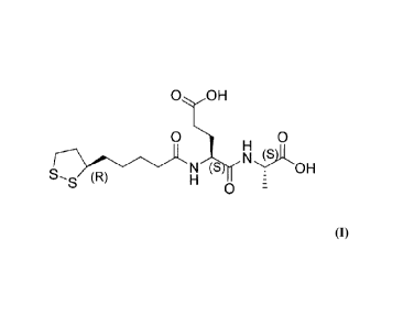
substantially pure compound represented by Structural Formula II:
OOH
0 0
OH
(R)
0
or a pharmaceutically acceptable salt thereof. As used herein, RLip-EA-OH
refers to
Structural Formula II.
In a further embodiment, the invention relates to compositions comprising a
substantially pure compound represented by Structural Formula III:
OONa
0 0
(s)
S _ ONa
0
In another embodiment, the invention relates to compositions comprising a
substantially pure compound represented by Structural Formula TV:
0 0
(S)
S OR2
0
or a pharmaceutically acceptable salt thereof. The values of Rl and R2 are as
defined above for Structural Formula (I).
CA 02761798 2011-11-10
WO 2010/132657
PCT/US2010/034701
- 19 -
In another embodiment, the invention relates to compositions comprising a
substantially pure compound represented by Structural Formula V:
O OR1
O 0
0 (R)
0
or a pharmaceutically acceptable salt thereof. The values of Rl and R2 are as
defined above for Structural Formula (I).
In another embodiment, the invention relates to compositions comprising a
substantially pure compound represented by Structural Formula VI:
O OH
O 0
(R) OH
(R)
0
or a pharmaceutically acceptable salt thereof. The values of Rl and R2 are as
defined above for Structural Formula (I).
In another embodiment, the invention relates to compositions comprising a
substantially pure compound represented by Structural Formula VII:
O OH
O 0
cr-N OR)OH
(R)
0
or a pharmaceutically acceptable salt thereof. The values of Rl and R2 are as
defined above for Structural Formula (I).
A composition of the invention may, alternatively or in addition to the
disclosed compounds, comprise a pharmaceutically acceptable salt of a compound
represented by the disclosed compounds, or a prodrug or pharmaceutically
active
metabolite of such a compound or salt and one or more pharmaceutically
acceptable
carriers and are delivered to a recipient subject (preferably a human) in
accordance
CA 02761798 2011-11-10
WO 2010/132657
PCT/ES2010/034701
- 20 -
with known methods of drug delivery. The compounds of the present invention
may be administered alone or in combination with at least one other agent
known or
believed by the applicants to be useful for the activation of cytoprotective
kinases
and/or the treatment of ischemic injuries or ischemia-reperfusion injuries.
Alternatively, a composition of the invention may comprise a compound
represented by the disclosed compounds or a pharmaceutical salt thereof as the
only
pharmaceutically active agent in the composition.
In another embodiment, the invention relates to a composition comprising: i)
a pharmaceutically acceptable carrier or diluent; and ii) a compound
represented by
Structural Formula I:
0 OR1
0
0
No R2
S (R) 0
wherein Rl and R2 are each independently H or a hydrolyzable group, or a
pharmaceutically acceptable salt thereof.
The pharmaceutically acceptable compositions of the present invention
comprise one or more compounds disclosed herein in association with one or
more
nontoxic, pharmaceutically acceptable carriers and/or diluents and/or
adjuvants
and/or excipients, collectively referred to herein as "carrier" materials,
and, if
desired, other active ingredients.
The present invention also relates to methods of activating a cytoprotective
kinase in a cell. As used herein, the term "cytoprotective kinase" refers to a
kinase
that, when activated, phosphorylates components of one or more cell signaling
pathways that promote cell survival and/or inhibit cell death (e.g.,
apoptosis).
Accordingly, in one embodiment, the invention relates to a method of
activating a
cytoprotective kinase (e.g., insulin receptor kinase, Akt kinase, insulin-like
growth
factor 1 receptor kinase, Src kinase) in a cell, comprising contacting the
cell with an
effective amount of a compound represented by Structural Formula I:
CA 02761798 2011-11-10
WO 2010/132657 PCT/US2010/034701
- 21 -
0 OR1
0
0
OR2
()0
wherein le and R2 are each independently H or a hydrolyzable group, or a
pharmaceutically acceptable salt thereof.
In a particular embodiment, the invention relates to a method of activating a
kinase whose pathway is cytoprotective in a cell, comprising contacting the
cell with
an effective amount of a compound represented by Structural Formula 11:
0 OH
N
H (
OH
(R)
0
or a pharmaceutically acceptable salt thereof.
In another embodiment, the invention relates to a method of activating a
cytoprotective kinase in a cell, comprising contacting the cell with an
effective
amount of a compound represented by Structural Formula III:
0 ONa
N
H (s
ONa
SS (R)
0
Activation of a cytoprotective kinase can lead to activation of one or more
cytoprotective cell signaling pathways that include the cytoprotective kinase.
In a
particular embodiment, activation of one or more cytoprotective kinases in a
cell by
the compounds of the invention can inhibit (e.g., prevent, delay) apoptosis of
the cell
in which the kinase(s) has been activated.
The methods of the invention relating to activation of cytoprotective kinases
can be performed in vitro (e.g., using cultured cells, using isolated cells)
or in vivo
CA 02761798 2011-11-10
WO 2010/132657 PCT/US2010/034701
- 22 -
(e.g., by administering a compound(s) of the invention to a living organism).
In a
particular embodiment, the compounds of the invention are used in a method to
activate one or more cytoprotective kinases in one or more cells in a human.
In one embodiment, the cytoprotective kinase is Akt kinase. Activation of
Akt kinase can lead to activation of one or more Akt cell signaling pathways
that are
cytoprotective. Akt kinase, also known as Akt, PKB and Rac-PK, belongs to the
Akt/PKB family of serine/threonine kinases and has been shown to be involved
in
many diverse signaling pathways (Alessi, and Cohen, Curr. Opin. Genet. Dev. 8
(1998), 55-62) including pathways related to cell survival and proliferation
(Song,
G., Ouyang, G., and Bao, S., The activation of Akt/PKB signaling pathway and
cell
survival. J Cell Hol Med 2005 9:59; Hausenloy, D.J., Yellon, D.M., Reperfusion
injury salvage kinase signaling: taking a RISK for cardioprotection. Heart
Fail Rev
2007, 12:217 .). Akt consists of an N-terminal lipid-binding pleckstrin-
homology
domain and a C-terminal catalytic domain. In resting cells, all Akt iso forms
reside
in the cytoplasm but translocate to the plasma membrane following stimulation
with
external ligands. Translocation and subsequent activation is induced by
several
different ligands including PDGF, IGF, EGF, 13FGF and insulin. This activation
depends on P13-kinase activity and requires hierarchial phosphorylation of
Thr308
and Ser473 of Akt by PDK-1 and PDK-2, respectively (Alessi et al., Cum Biol. 8
(1998), 69-81). Once activated, Akt mediates several different functions,
including
prevention of apoptosis, induction of differentiation and/or proliferation,
protein
synthesis and the metabolic effects of insulin.
As described in Example 2 herein, compounds of the invention increase Akt
phosphorylation in a cell-based in vitro assay. Akt kinase phosphorylation in
a cell
can be assessed using one or more in vitro Akt kinase phosphorylation assays
known
in the art including, for example, kits and assays for testing AKT
phosphorylation in
cells available from commercial suppliers (e.g., Cellomics Phospho-AKT
Activation
Kit, Thermo Scientific; Akt Activity Assay Kit, Bio Vision Incorporated; FACE
'1'1
AKT in-cell Western analysis for phospho AKT (S473), Active Motif; PathScan(R)
Phospho-Akt (Thr308) Sandwich ELISA Kit, Cell Signaling Technology;
AlphaScreen SureFire Phospho-AKT Assay Kits, Perkin Elmer; Akt Activity
Immunoassay Kit, EMD Biosciences). An exemplary assay for assessing Akt kinase
CA 02761798 2011-11-10
WO 2010/132657
PCT/US2010/034701
- 23 -
phosphorylation is described herein in Example 2. (Chen, H., Kovar, J.,
Sissons, S.,
et. at. A cell based immunocytochemical assay for monitoring kinase signaling
pathways and drug efficacy. Analyt Biochem 2005 338: 136)
Akt activation can also be assessed in vivo, e.g., by immunodetection
methods performed on a cell sample obtained from a subject. Several Akt-
specific
antibodies, including phospho-specific Akt antibodies (e.g., specific for
phospho-
Ser473, specific for phospho-Thr308), are commercially available (e.g., Perkin
Elmer).
In another embodiment, the cytoprotective kinase is insulin receptor kinase
("IRK") (Diesel, B., Kulhanek-Heinze, S., Holtje, M., et. al., a-Lipoic Acid
as a
directly binding activator of the insulin receptor: protection from hepatocyte
apoptosis. Biochemistry, 2007 46:2146; Hausenloy, D.J., Yellon, D.M. New
directions for protecting the heart against ischemia-reperfusion injury:
targeting the
reperfusion injury salvage kinase (RISK)-pathway. Cardiovasc Res 2004 61:448).
IRK activation leads to phosphorylation and activation of Akt (Alessi, D.R.,
Andjelkovic, M., Caudwell, B., Cron, P., Morrice, N., Cohen, P., Hemmings,
B.A.
Mechanism of activation of protein kinase B by insulin and IGF-1. EMBO J 1996
15:6541-6551). Activation of IRK can lead to activation of one or more IRK
cell
signaling pathways that are cytoprotective. As described in Example 3 herein,
compounds of the invention activate IRK in a biochemical assay in vitro. IRK
activation can be assessed using one or more in vitro IRK activation assays
known
in the art. An exemplary assay for assessing IRK activation is described
herein in
Example 3. (Mobility Shift Kinase Assay, Caliper Life Sciences, Hanover, MD)
In another embodiment, the cytoprotective kinase is insulin-like growth
factor 1 receptor ("IGF1R") kinase. Activation of IGF1R kinase can lead to
activation of one or more IGF1R cell signaling pathways that are
cytoprotective. As
described in Example 4 herein, compounds of the invention activate IGF1R
kinase
in a biochemical assay in vitro. IGF1R kinase activation can be assessed using
one
or more in vitro IGF1R kinase activation assays known in the art. An exemplary
assay for assessing IGF1R kinase activation is described herein in Example 4.
In a further embodiment, the cytoprotective kinase is Src kinase. Activation
of Src kinase can lead to activation of one or more Src cell signaling
pathways that
CA 02761798 2011-11-10
WO 2010/132657 PCT/US2010/034701
- 24 -
are cytoprotective. As described in Example 4 herein, compounds of the
invention
activate Src kinase in a biochemical assay in vitro. Src kinase activation can
be
assessed using one or more in vitro Src kinase activation assays known in the
art.
IGF1R and Src tyrosine kinases play a role in protecting the heart from
ischemia-reperfusion injury (Buddhadeb, D., Takano, H., Tang, X.-L., et al.
Role of
Src protein tyrosine kinase in late preconditioning against myocardial
infarction. Am
Physiol 2002 283:H549; Pasdois, P., Quinlan, CL., Rissa, A., et al. Ouabain
protects rat hearts against ischemia-reperfusion injury via pathway involving
Src
kinase, mitoKATP, and ROS. Am J Physiol 2006, 292:H1470; Suzuki, Y. J. Growth
factor signaling for cardioprotection against oxidative stress-induced
apoptosis.
Antiox Redox Signal 2003, 5:741; Hausenloy, D.J., Yellon, D.M., New directions
for
protecting the heart against ischaemia-reperfusion injury: Targeting the
Reperfusion
Injury Salvage Kinasc (RISK)-pathway. Cardiovasc Res 2004 61:448).
According to the invention, activation of one or more cytoprotective kinases
in a cell by the compounds of the invention can inhibit (e.g., prevent, delay)
apoptosis of the cell. Methods of assessing apoptosis are well known in the
art.
Microscopic analysis (e.g., light microscopy, electron microscopy, confocal
microscopy, laser-scanning microscopy) for visualizing apoptotic cells (e.g.,
by
detecting morphological changes associated with apoptosis, such as chromatin
condensation and cytoplasmic shrinking) is typically employed to study
apoptotic
cells.
The study of DNA fragmentation in agarose gels is also considered to be
indicative of apoptosis. A number of techniques take advantage of DNA
fragmentation for labeling the fragments and thus for quantifying the
proportion of
apoptotic cells. Each DNA fragment has a 3'-OH terminal portion. This terminal
fragment can be labeled in various ways (for instance, with the help of a
modified
terminal deoxynucleotidyl transferase), so that the labeling rate is
proportional to the
degree of DNA fragmentation.
In particular, TdT-mediated dUTP Nick-End Labeling, or TUNEL, is a
technique for detecting fragmented DNA, which occurs near the final step in
the
apoptotic process. Fragmented DNA of apoptotic cells can incorporate
fluorescein-
dUTP at 3'-OH at DNA ends using the enzyme Terminal Deoxynucleotidyl
CA 02761798 2011-11-10
WO 2010/132657 PCT/US2010/034701
- 25 -
Transferase (TdT), which forms a polymeric tail using the principle of the
TUNEL
assay. The labeled DNA can then be visualized directly by fluorescence
microscopy
or quantitated by flow cytometry.
Some current techniques take advantage of the changes in membrane
phospholipids that occur early in apoptotic cells. The negatively charged
membrane
phospho lipids exposed to the external environment by the apoptotic cell are
labeled
with fluorochrome-conjugated molecules, and the percentage of fluorescent
cells can
be easily quantified.
Apoptosis can also be detected using fluorescently-conjugated Annexin V.
Annexin V is an anticoagulant protein that preferentially binds negatively
charged
phospho lipids. An early step in the apoptotic process is disruption of
membrane
phospholipid asymmetry, exposing phosphatidylserine (PS) on the outer leaflet
of
the cytoplasmic membrane. Fluorescently conjugated Annexin V can be used to
detect this externalization of phosphatidylserine on intact living cells.
Propidium
iodide is often combined as a second fluro chrome to detect necrotic cells.
Induction
of apoptosis leads to procaspase-3 proteolytic cleavage to generate an active
18 kDa
caspase-3 fragment which then targets key modulators of the apoptotic pathway
including poly-ADP-ribose polymerase and other caspases, for cleavage. Assays
for
detecting other active caspases in apoptotic cells are known in the art (e.g.,
Caspase-
Glo Assays, Promega).
Apoptotic cells can also be detected using the active 18 kDa caspase-3
fragment as a marker. Induction of apoptosis leads to procaspase-3 proteolytic
cleavage to generate an active 18 kDa caspase-3 fragment which then targets
key
modulators of the apoptotic pathway, including poly-ADP-ribose polymerase and
other caspases, for cleavage. Several antibodies that recognize only the
active 18
kDa fragment are available from commercial suppliers (e.g., BD Biosciences,
Chemicon, Cell Signaling Technology, Trevigen).
In addition, flow cytometry assays can be employed to monitor and quantify
nuclear changes associated with apoptotic cells.
An exemplary assay for detecting inhibition of apoptosis is described herein
in Example 5.
CA 02761798 2011-11-10
WO 2010/132657 PCT/US2010/034701
- 26 -
The activation of cellular cytoprotective kinases also have utility in the
treatment of conditions resulting from excess or unwanted apoptotic cell death
in an
affected tissue or organ, leading to damage and dysfunction. Such conditions
include, inter alia, ischemia and ischemia-reperfusion injury. Accordingly,
the
invention also relates to methods of treating an ischemia or ischemia-
reperfusion
injury in a mammalian subject, comprising administering to the subject an
effective
amount of a compound represented by Structural Formula I:
0
0
N R2
S,s (R)
0
wherein Rl and R2 are each independently H or a hydrolyzable group, or a
pharmaceutically acceptable salt thereof.
In a particular embodiment, the invention relates to a method of treating an
ischemia or ischemia-reperfusion injury in a mammalian subject, comprising
administering to the subject an effective amount of a compound represented by
Structural Formula II:
0 0
H (s
N
(R) H
0
or a pharmaceutically acceptable salt thereof.
In another embodiment, the invention relates to a method of treating an
ischemia or ischemia-reperfusion injury in a mammalian subject, comprising
administering to the subject an effective amount of a compound represented by
Structural Formula III:
CA 02761798 2011-11-10
WO 2010/132657 PCT/US2010/034701
- 27 -
0 ONa
0 0
Qr N ONa
(R)
0
As used herein, the "injury resulting from ischemia," "injury caused by
ischemia" and "ischemic injury" refer to an injury to a cell, tissue or organ
caused
by ischemia, or an insufficient supply of blood (e.g., due to a blocked
artery), and,
thus, oxygen, resulting in damage or dysfunction of the tissue or organ
(Piper, H.
M., Abdallah, C., Schafer, C., Annals of Thoracic Surgety 2003, 75:644;
Yellon, D.
M., Hausenloy, D. J., .7Vew England Journal of Medicine 2007, 357:1121).
Injuries
that result from ischemia can affect various tissues and organs. Such injuries
may be
treated by the compounds and methods of the invention, including, for example,
injuries caused by cardiovascular ischemia, cerebrovascular ischemia, renal
ischemia, hepatic ischemia, ischemic cardiomyopathy, cutaneous ischemia, bowel
ischemia, intestinal ischemia, gastric ischemia, pulmonary ischemia,
pancreatic
ischemia, skeletal muscle ischemia, abdominal muscle ischemia, limb ischemia,
ischemic colitis, mesenteric ischemia and silent ischemia. Thus, an injury
resulting
from ischemia can affect, for example, a heart, kidney, liver, brain, muscle,
intestine,
stomach, lung or skin.
In a particular embodiment, the injury resulting from ischemia is the result
of
a myocardial ischemia. An injury resulting from a myocardial ischemia can
result
from, for example, a myocardial infarction (e.g., an acute myocardial
infarction) in
an individual.
In another embodiment, the injury resulting from ischemia is an injury
resulting from cerebral ischemia (e.g., a stroke) in an individual.
In another embodiment, the injury resulting from ischemia is an ischemia-
reperfusion injury. As used herein, the term "ischemia-reperfusion injury"
refers to
an injury resulting from the restoration of blood flow to an area of a tissue
or organ
that had previously experienced deficient blood flow due to an ischemic event.
Oxidative stresses associated with reperfusion may cause damage to the
affected
CA 02761798 2011-11-10
WO 2010/132657 PCT/US2010/034701
- 28 -
tissues or organs. Tschemia-reperfusion injury is characterized biochemically
by a
depletion of oxygen during an ischemic event followed by reoxygenation and the
concomitant generation of reactive oxygen species during reperfusion (Piper,
H. M.,
Abdallah, C., Schafer, C., Annals of Thoracic Surgery 2003, 75:644; Yellon, D.
M.,
Hausenloy, D. J., New England Journal of Medicine 2007, 357:1121).
An ischemia-reperfusion injury can be caused, for example, by a natural
event (e.g., restoration of blood flow following a myocardial infarction), a
trauma, or
by one or more surgical procedures or other therapeutic interventions that
restore
blood flow to a tissue or organ that has been subjected to a diminished supply
of
blood. Such surgical procedures include, for example, coronary artery bypass
graft
surgery, coronary angioplasty, organ transplant surgery and the like (e.g.,
cardiopulmonary bypass surgery). In a particular embodiment the compounds and
methods of the invention are useful for treating pen-operative cardiac damage
caused by an ischemia or ischemia-reperfusion injury.
For the treatment of ischemic and ischemia-reperfusion injuries caused by
therapeutic interventions, such as surgical procedures, it is preferable that
a
compound of the invention is administered to a subject undergoing treatment
prior to
the therapeutic intervention (e.g., cardiac surgery, organ transplant). For
example, a
compound of the invention can be administered to a subject undergoing
treatment,
e.g., about 1 hour, about 2 hours, about 3 hours, about 4 hours, about 5
hours, about
12 hours, about 24 hours, or about 48 hours prior to the therapeutic
intervention. A
compound of the invention can also be administered to a subject undergoing
treatment, for example, about 5 minutes, about 10 minutes, about 15 minutes,
about
20 minutes, about 30 minutes or about 45 minutes prior to the therapeutic
intervention.
Alternatively, or in addition, a compound of the invention can be
administered to a subject undergoing treatment at the time of, or during, the
therapeutic intervention. For example, the compound can be administered one or
more times during the course of a therapeutic intervention in intervals (e.g.,
15
minute intervals). Alternatively, a compound can be administered continuously
throughout the duration of a therapeutic intervention.
CA 02761798 2011-11-10
WO 2010/132657 PCT/US2010/034701
- 29 -
Furthermore, a compound of the invention can be administered to a subject
undergoing treatment after a therapeutic intervention. For example, a compound
of
the invention can be administered to a subject undergoing treatment, e.g.,
about 1
hour, about 2 hours, about 3 hours, about 4 hours, about 5 hours, about 12
hours,
about 24 hours, or about 48 hours after the therapeutic intervention. A
compound of
the invention can also be administered to a subject undergoing treatment, for
example, about 5 minutes, about 10 minutes, about 15 minutes, about 20
minutes,
about 30 minutes or about 45 minutes after the therapeutic intervention.
A compound of the invention can also be used to inhibit an ischemia or
ischemia-reperfusion injury to a cell, tissue or organ, ex vivo, prior to a
therapeutic
intervention (e.g., a tissue employed in a graft procedure, an organ employed
in an
organ transplant surgery). For example, prior to transplant of an organ into a
host
individual (e.g., during storage or transport of the organ in a sterile
environment),
the organ can be contacted with a compound of the invention (e.g., bathed in a
solution comprising a compound of the invention) to inhibit ischemia or
ischemia-
reperfusion injury.
As described herein, conditions resulting from ischemia, and injuries caused
by ischemia or ischemia-reperfusion, can induce apoptotic cell death in an
affected
cell, tissue or organ, leading to damage and dysfunction. Accordingly, the
compounds of the invention also have utility in methods of inhibiting
apoptosis in a
cell, a tissue or an organ (e.g., a transplant tissue or organ or a cell,
tissue or organ in
a subject), wherein the cell, tissue or organ has experienced an ischemia or
other
condition or disorder that results in excessive or unwanted apoptosis. The
methods
comprise contacting the cells, tissue, or organ with, or administering to the
subject,
an effective amount of a compound represented by Structural Formula I:
0 OR1
0 0
No R2
S (R) 0
wherein Rl and R2 are each independently H or a hydrolyzable group, or a
pharmaceutically acceptable salt thereof.
CA 02761798 2011-11-10
WO 2010/132657 PCT/US2010/034701
- 30 -
In a particular embodiment, the invention relates to a method of inhibiting
apoptosis in a cell, tissue or organ, wherein the cell, tissue or organ has
experienced
an ischemia or other condition or disorder that results in excessive or
unwanted
apoptosis, comprising administering to the subject an effective amount of a
compound represented by Structural Formula II:
0 OH
N (S OH
0
or a pharmaceutically acceptable salt thereof.
In another embodiment, the invention relates to a method of inhibiting
apoptosis in a cell, tissue or organ, wherein the cell, tissue or organ has
experienced
an ischemia or other condition or disorder that results in excessive or
unwanted
apoptosis, comprising administering to the subject an effective amount of a
compound represented Structural Formula III:
OONa
0 0
H
ONa
(R)
0
Methods for assessing apoptosis in cells, tissues or organs are known in the
art and include those described herein.
Conditions associated with unwanted and/or excess apoptosis that are
treatable by the compounds and methods of the invention include, but are not
limited
to, neurodegenerative diseases associated with excess apoptosis (e.g.,
Parkinson's
Disease, Alzheimer's Disease, amyotrophic lateral sclerosis, retinitis
pigmentosa,
epilepsy), haematologic diseases associated with excess apoptosis (e.g.,
aplastic
anaemia, myelodysplastic syndrome, T CD4+ lymphocytopenia, G6PD deficiency),
tissue damage associated with excess apopotosis (e.g., myocardial infarction,
cerebrovascular accident, ischemic renal damage, polycystic kidney disease),
AIDS,
and preeclampsia.
CA 02761798 2011-11-10
WO 2010/132657
PCT/ES2010/034701
-31 -
One of the hallmarks of ischemia-reperfusion injury is an increase in
cytosolic calcium levels, resulting from a depletion of oxygen during an
ischemic
event (Piper, H. M., Abdallah, C., Schafer, C., Annals of Thoracic Surgery
2003,
75:644; Yellon, D. M., Hausenloy, D. J., New England Journal of Medicine 2007,
357:1121). It has been postulated that the increase in cytosolic calcium
combined
with an increase in free radicals triggers apoptosis (Chen, X., Zhang, X.,
Hubo, H.,
et al., Circ Res 2005, 97:1009; Lopes-Neblina, F., Toledo, A.H., Toledu-
Pereyra,
L.H. J Invest Surg 2005, 18:335). However, to date, treatments of patients
with
acute myocardial infarction with either an antagonist to block the influx of
calcium
or with a scavenger of the reactive oxygen species has each yielded
disappointing
clinical outcomes (Yellon, D. M., Hausenloy, D. J., New England Journal of
Medicine 2007, 357:1121).
In addition, through pro-survival pathways activated by Akt, cytosolic
calcium overload is inhibited (Joseph, S.K., Hajnoczky, G., Apoptosis 2007,
12:951;
Pinton, P., Rizzuto, R., Cell Death Diff2006, 13:1409; Khan, M.T., Wagner, L.
II,
Yule, D.I., Bhanumathy, C., Joseph, .S.K. 2006, Akt kinase phosphorylation of
inositol 1,4,5-triphosphate receptors. J Biol Chem 281:3731). The Akt
dependent
signaling pathway also prevents intracellular calcium overload by regulation
of Bel-
2 (Raphael, J., Abedat, S., Rivo, J., et al., J Pharmacol Exp Ther 2006,
318:186;
Thomenius, M.J. and Distelhorst, C. W., J Cell Sci 2003, 116:4493).
Accordingly, the compounds of the invention, which induce Akt activation,
also have utility in methods of decreasing cytosolic calcium in a cell, tissue
or organ
(e.g., in a subject suffering from an ischemia). The methods comprise
administering
to the subject an effective amount of a compound represented by Structural
Formula
I:
0 OR1
0 0
No R2
S (R) 0
wherein R1 and R2 are each independently H or a hydrolyzable group, or a
pharmaceutically acceptable salt thereof.
CA 02761798 2011-11-10
WO 2010/132657 PCT/US2010/034701
- 32 -
In a particular embodiment, the invention relates to a method of decreasing
cytosolic calcium in a cell, tissue or organ, comprising administering to the
subject
an effective amount of a compound represented by Structural Formula II:
O OH
Ass.r
H (_s
o OH
0
or a pharmaceutically acceptable salt thereof
In another embodiment, the invention relates to a method of decreasing
cytosolic calcium in a cell, tissue or organ, comprising administering to the
subject
an effective amount of a compound represented Structural Formula ITT:
O ONa
O 0
H
NONa
(R)
0
An exemplary assay for detecting levels of cytosolic calcium is described
herein in Example 6.
Compounds of the invention also display an enhanced capacity for peroxyl
radical absorbance. Biological organisms generate harmful reactive oxygen
species
(ROS) and various free radicals in the course of normal metabolic activities
of
tissues such as brain, heart, lung, and muscle tissue (Halliwell, B. and
Gutteridge, J.
M. C., eds. (Oxford: Clarendon Press, 1989)). Recognition of the role of ROS
and
free radicals in a variety of important diseases and drug side effects has
grown
appreciably over recent years. Many studies have demonstrated that a large
number
of disease states and harmful side effects of therapeutic drugs are linked
with a
failure of the antioxidant defense system of an individual to keep up with the
rate of
generation of ROS and various free radicals (see, for example, Chan, et al.,
Adv.
Neurol., 1996, 71:271-279; DiGuiseppi, J. and Fridovich, I., Crit. Rev.
Toxicol.,
1984, 12:315-342). For example, abnormally high ROS levels have been found
under conditions of anoxia elicited by ischemia during a stroke or anoxia
generated
CA 02761798 2011-11-10
WO 2010/132657 PCT/US2010/034701
- 33 -
in heart muscle during myocardial infarction (see, for example, Walton, M. et
al.,
Brain Res. Rev., 1999, 29:137-168; Pulsinelli, W. A. et al., Ann. Neural.,
1982, 11:
499-502; Lucchesi, B. R., Am. J. (ardiol., 1990, 65:141-231). In addition, an
elevation of ROS and free radicals has also been linked with reperfusion
damage
after renal transplants.
Accordingly, an elevation of ROS and free radicals has been linked with the
progression and complications developed in many diseases, drug treatments,
traumas, and degenerative conditions including oxidative stress induced damage
with age, Tardive dyskinesia, Parkinson's disease, Huntington's disease,
degenerative eye diseases, septic shock, head and spinal cord injuries,
Alzheimer's
disease, ulcerative colitis, human leukemia and other cancers, and diabetes
(see, for
example, Ratanis, Pharmaceutical Executive, pp. 74-80 (April 1991)).
For example, elevated levels of ROS and free radicals arc known to be
generated in cells and tissues during reperfusion after an ischemic event.
Such
increased levels of ROS and free radicals can cause considerable damage to an
already stressed or debilitated organ or tissue. The compounds of this
invention,
which display peroxyl radical absorbance capacity, may be used to treat high
levels
of harmful free radicals present after reperfusion injuries that occur in
diseases and
conditions such as stroke, heart attack, or renal disease and kidney
transplants. If the
ischemic event has already occurred, as in stroke and heart attack, a compound
described herein may be administered to the individual to detoxify the
elevated ROS
and free radicals already present in the blood and affected tissue or organ.
Alternatively, if the ischemic event is anticipated as in organ
transplantation,
or other procedures that can lead to ischemic injury or ischemia-reperfusion
injury
(e.g., coronary artery bypass graft surgery, coronary angioplasty,
cardiopulmonary
bypass surgery) then the compounds described herein may be administered
prophylactically, prior to the operation or ischemic event, at dosage
intervals as
described herein to potentiate the efficacy of the claimed compounds.
The compounds described herein may be used to treat any disease or
condition associated with undesirable levels of ROS and free radicals, or to
prevent
any disease, disorder or condition caused by undesirable levels of ROS and
free
radicals. According to the invention, the compounds described herein may also
be
CA 02761798 2011-11-10
WO 2010/132657 PCT/US2010/034701
- 34 -
administered to provide a therapeutic or prophylactic treatment of elevated
ROS and
other free radicals associated with a variety of other diseases and
conditions,
including, but not limited to, oxygen toxicity in premature infants, burns and
physical trauma to tissues and organs, septic shock, polytraumatous shock,
head
trauma, brain trauma, spinal cord injuries, Parkinson's disease, amyotrophic
lateral
sclerosis (ALS), Alzheimer's disease, age-related elevation of ROS and free
radicals,
senility, ulcerative colitis, human leukemia and other cancers, Down syndrome,
arthritis, macular degeneration, schizophrenia, epilepsy, radiation damage
(including
UV-induced skin damage), and drug-induced increase in ROS and free radicals.
A progressive rise of oxidative stress due to the formation of ROS and free
radicals also occurs during aging (see, e.g., Mecocci, P. et al., Free Radio.
Biol.
Med., 2000, 28: 1243-1248). This has been detected by finding an increase in
the
formation of lipid peroxidates in rat tissues (Erdincler, D. S., et al., Clin.
Chim. Acta,
1997, 265: 77-84) and blood cells in elderly human patients (Congi, F., et
al.,
Presse. Med., 1995, 24: 1115-1118). Accordingly, the compounds described
herein,
which are able to absorb peroxyl radicals, are also well suited for use in
methods of
preventing and/or counteracting increased tissue damage and decreased life
expectancy due to elevated levels of ROS and free radicals that accompany the
aging
process.
Thus, the compounds of the invention have utility in the treatment of
conditions and disorders caused by harmful reactive oxygen species (ROS) and
other
free radicals. Accordingly, the invention further relates to methods of
increasing
peroxyl radical absorbance in a tissue in a subject (e.g., a subject suffering
from an
ischemia), comprising administering to the subject an effective amount of a
compound represented by Structural Formula I:
0 OR1
0 0
No R2
S (R) 0
wherein Rl and R2 are each independently H or a hydrolyzable group, or a
pharmaceutically acceptable salt thereof.
CA 2761798 2017-02-24
- 35 -
In a particular embodiment, the invention relates to a method of increasing
peroxyl radical absorbance in a tissue in a subject, comprising administering
to the
subject an effective amount of a compound represented by Structural Formula
II:
0 OH
O 0
N91(
OH
S S (R) 0
or a pharmaceutically acceptable salt thereof.
In another embodiment, the invention relates to a method of increasing
peroxyl radical absorbance in a tissue in a subject, comprising administering
to the
subject an effective amount of a compound represented Structural Formula III:
O ONa
O 0
Nk
S20.L1.1 (S ONa
0
An exemplary assay for detecting peroxyl radical absorbance is described
herein in Example 7. Other methods of detecting free radical absorbance are
described in U.S. Patent No. 6,890,896.
The activation of Akt kinase by a compound of the invention has utility in
the treatment of conditions resulting from reduced or insufficient Akt
activity in a
cell, including, but not limited to, ischemic injuries. Suitable conditions
resulting
from reduced Akt activity for treatment using the compounds and methods of the
invention include, for example, diseases or disorders characterized by
insufficient
vascularization (e.g., diabetic ulcers, gangrene, wounds requiring
neovascularization
to facilitate healing, Buerger's syndrome, hypertension, conditions
characterized by
a reduction in microvasculature), and certain neurological diseases or
disorders (e.g.,
Parkinson's discasc, Alzheimer's disease, depression, anxiety, manic-
depressive
psychosis, post traumatic stress disorder, mild cognition impairment (MCI),
CA 2761798 2017-02-24
- 36 -
amyotrophic lateral sclerosis (ALS), Huntington's disease, spinocerebellar
degenerative disease, multiple sclerosis (MS), Pick's disease, schizophrenia,
anxiety
neurosis, obsessive-compulsive neurosis, head trauma, spinal cord injury,
cerebrovascular disorder, cerebrovascular dementia, asymptomatic brain
infarction,
polyglutamine disease, prion disease, corticobasal ganglionic degeneration,
progressive supranuclear palsy, AIDS encephalopathy, muscular dystrophy,
diabetic
neuropathy).
Other conditions resulting from reduced Akt activity that may be treated
using the compounds and methods of the invention include, but are not limited
to,
diabetic retinopathy, diabetic nephropathy, liver cirrhosis, alcoholic
hepatitis, senile
diseases characterized by a decrease in self-regenerating ability, non-
metabolic bone
diseases, metabolic bone diseases, joint diseases, periodontal diseases,
cytomegalovirus infection, rheumatoid arthritis, Lyme disease, gout, sepsis
syndrome, hyperthermia, ulcerative colitis, enterocolitis, osteoporosis,
periodontal
disease, glomerulonephritis, chronic non-infectious inflammation of the lung,
sarcoidosis, smoker's lung, granuloma formation, fibrosis of the liver,
fibrosis of the
lung, transplant rejection, graft vs. host disease, chronic myeloid leukemia,
acute
myeloid leukemia, neoplastic disease, asthma bronchiale, type I insulin
dependent
diabetes mellitus, arteriosclerosis, atherosclerosis, psoriasis, chronic B
lymphocyte
leukemia, common variable immunodeficiency, disseminated intravascular
coagulation, systemic sclerosis, encephalomyelitis, lung inflammation, hyper
IgE
syndrome, cancer metastasis, cancer growth, adoptive immune therapy, acquired
respiratory distress syndrome, sepsis, reperfusion syndrome, postsurgical
inflammation, organ transplantation, and alopecia.
AKT-mediated disorders resulting from reduced AKT activity are also
disclosed in US 2004/0122077; US 2006/0241168; and US 2007/0219139.
The pharmaceutical preparations disclosed herein are prepared in accordance
with standard procedures and are administered at dosages that are selected to
reduce,
prevent, or eliminate, or to slow or halt the progression of, the condition
being
treated (See, e.g., Remington's Pharmaceutical Sciences, Mack Publishing
Company, Easton, PA, and Goodman and Gilman's The Pharmaceutical Basis of
CA 2761798 2017-02-24
- 37 -
Therapeutics, McGraw-Hill, New York, N.Y., for a general description of the
methods for
administering various agents for human therapy). The compositions of a
compound
represented by the disclosed compounds can be delivered using controlled or
sustained-release delivery systems (e.g., capsules, biodegradable matrices).
Exemplary delayed-release delivery systems for drug delivery that would be
suitable
for administration of the compositions of the disclosed compounds are
described in
U.S. Patent Nos. US 5,990,092 (issued to Walsh); 5,039,660 (issued to
Leonard);
4,452,775 (issued to Kent); and 3,854,480 (issued to Zaffaroni) .
For preparing pharmaceutical compositions from the compounds of the
present invention, pharmaceutically acceptable carriers can either be solid or
liquid.
Solid form preparations include powders, tablets, pills, capsules, cachets,
suppositories, and dispersible granules. For example, the compounds of the
present
invention may be in powder form for reconstitution at the time of delivery. A
solid
carrier can be one or more substances which may also act as diluents,
flavoring
agents, solubilizers, lubricants, suspending agents, binders, preservatives,
tablet
disintegrating agents, or an encapsulating material. In powders, the carrier
is a
finely divided solid which is in a mixture with the finely divided active
ingredient.
In tablets, the active ingredient is mixed with the carrier having the
necessary
binding properties in suitable proportions and compacted in the shape and size
desired.
The powders and tablets preferably contain from about one to about seventy
percent of the active ingredient. Suitable carriers are magnesium carbonate,
magnesium stearate, talc, sugar, lactose, pectin, dextrin, starch, gelatin,
tragacanth,
methylcellulosc, sodium caboxymethylcellulose, a low-melting wax, cocoa
butter,
and the like. Tablets, powders, cachets, lozenges, fast-melt strips, capsules
and pills
can be used as solid dosage forms containing the active ingredient suitable
for oral
administration.
Liquid form preparations include solutions, suspensions, retention enemas,
and emulsions, for example, water or water propylene glycol solutions. For
CA 02761798 2011-11-10
WO 2010/132657
PCT/US2010/034701
- 38 -
parenteral injection, liquid preparations can be formulated in solution in
aqueous
polyethylene glycol solution.
Aqueous solutions suitable for oral administration can be prepared by
dissolving the active ingredient in water and adding suitable colorants,
flavors,
stabilizing agents, and thickening agents as desired. Aqueous suspensions for
oral
administration can be prepared by dispersing the finely divided active
ingredient in
water with viscous material, such as natural or synthetic gums, resins,
methylcellulose, sodium carboxymethylcellulose, and other well-known
suspending
agents.
The pharmaceutical composition is preferably in unit dosage form. In such
form, the composition is subdivided into unit doses containing appropriate
quantities
of the active ingredient. The unit dosage form can be a packaged preparation,
the
package containing discrete quantities of, for example, tablets, powders, and
capsules in vials or ampules. Also, the unit dosage form can be a tablet,
cachet,
capsule, or lozenge itself, or it can be the appropriate amount of any of
these in
packaged form. The quantity of active ingredient in a unit dose preparation
may be
varied or adjusted from about 0.1 mg to about 1000.0 mg, preferably from about
0.1
mg to about 100 mg (e.g., for intravenous administration) or from about 1.0 mg
to
about 1000 mg (e.g., for oral administration). The dosages, however, may be
varied
depending upon the requirements of the patient, the severity of the condition
being
treated, the compound and the route of administration being employed.
Determination of the proper dosage for a particular situation is within the
skill in the
art. Also, the pharmaceutical composition may contain, if desired, other
compatible
therapeutic agents.
In general, the methods for delivering the disclosed compounds and
pharmaceutical compositions of the invention in vivo utilize art-recognized
protocols
for delivering the agent with the only substantial procedural modification
being the
substitution of the compounds represented by any one of the disclosed
compounds
for the drugs in the art-recognized protocols.
The compounds of the present invention may be administered by any route,
preferably in the form of a pharmaceutical composition adapted to such a
route, and
would be dependent on the condition being treated. The compounds and
CA 02761798 2011-11-10
WO 2010/132657 PCT/US2010/034701
- 39 -
compositions may, for example, be administered intravascularly,
intramuscularly,
subcutaneously, intraperitoneally, orally or topically. It will be obvious to
those
skilled in the art that the following dosage forms may comprise as the active
ingredient, either compounds or a corresponding pharmaceutically acceptable
salt of
a compound of the present invention. A preferred method of administration for
the
compounds of the invention is intravenous administration.
In some embodiments, the composition may be administered parenterally via
injection. Parenteral administration can include, for example, intraarticular,
intramuscular, intravenous, intraventricular, intraarterial, intrathecal,
subcutaneous,
or intraperitoneal administration. Formulations for parenteral administration
may be
in the form of aqueous or non-aqueous isotonic sterile injection solutions or
suspensions. These solutions or suspensions may be prepared from sterile
powders
or granules having one or more of the carriers mentioned for use in the
formulations
for oral administration. The compounds may be dissolved in polyethylene
glycol,
propylene glycol, ethanol, corn oil, benzyl alcohol, sodium chloride, and/or
various
buffers (e.g., sodium bicarbonate, sodium hydroxide).
For oral administration, the pharmaceutical compositions may be in the form
of, for example, a tablet, capsule, suspension or liquid. The composition is
preferably made in the form of a dosage unit containing a therapeutically
effective
amount of the active ingredient. Examples of such dosage units are tablets and
capsules. For therapeutic purposes, the tablets and capsules can contain, in
addition
to the active ingredient, conventional carriers such as binding agents, for
example,
acacia gum, gelatin, polyvinylpyrrolidone, sorbitol, or tragacanth; fillers,
for
example, calcium phosphate, glycine, lactose, maize-starch, sorbitol, or
sucrose;
lubricants, for example, magnesium stearate, polyethylene glycol, silica, or
talc;
disintegrants, for example potato starch, flavoring or coloring agents, or
acceptable
wetting agents. Oral liquid preparations generally in the form of aqueous or
oily
solutions, suspensions, emulsions, syrups or elixirs may contain conventional
additives such as suspending agents, emulsifying agents, non-aqueous agents,
preservatives, coloring agents and flavoring agents. Examples of additives for
liquid
preparations include acacia, almond oil, ethyl alcohol, fractionated coconut
oil,
CA 02761798 2011-11-10
WO 2010/132657 PCT/US2010/034701
- 40 -
gelatin, glucose syrup, glycerin, hydrogenated edible fats, lecithin, methyl
cellulose,
methyl or propyl para-hydroxybenzoate, propylene glycol, sorbitol, or sorbic
acid.
For topical use the compounds of the present invention may also be prepared
in suitable forms to be applied to the skin, or mucus membranes of the nose
and
throat, and may take the form of creams, ointments, liquid sprays or
inhalants,
lozenges, or throat paints. Such topical formulations further can include
chemical
compounds such as dimethylsulfoxide (DMSO) to facilitate surface penetration
of
the active ingredient. Suitable carriers for topical administration include
oil-in-water
or water-in-oil emulsions using mineral oils, petrolatum and the like, as well
as gels
such as hydrogel. Alternative topical formulations include shampoo
preparations,
oral pastes and mouthwash.
For application to the eyes or ears, the compounds of the present invention
may be presented in liquid or semi-liquid form formulated in hydrophobic or
hydrophilic bases as ointments, creams, lotions, paints or powders.
For rectal administration the compounds of the present invention may be
administered in the form of suppositories admixed with conventional carriers
such as
cocoa butter, wax or other glyceride. For preparing suppositories, a low-
melting
wax, such as a mixture of fatty acid glycerides or cocoa butter, is first-
melted and
the active ingredient is dispersed homogeneously therein, as by stirring. The
molten
homogeneous mixture is then poured into convenient sized molds, allowed to
cool,
and thereby to solidify.
Delivery can also be by injection into the brain or body cavity of a patient
or
by use of a timed release or sustained release matrix delivery systems, or by
onsite
delivery using micelles, gels and liposomes. Nebulizing devices, powder
inhalers,
and aerosolized solutions are representative of methods that may be used to
administer such preparations to the respiratory tract. Delivery can be in
vitro, in
vivo, or ex vivo.
For example, suitable intravenous dosages for a compound of the invention
can be from about 0.001 mg/kg to about 100 mg/kg, from about 0.01 mg/kg to
about
100 mg/kg, from about 0.01 mg/kg to about 10 mg/kg, from about 0.01 mg/kg to
about 1 mg/kg body weight per treatment. Determining the dosage and route of
administration for a particular agent, patient and ischemia or ischemia-
reperfusion
CA 02761798 2011-11-10
WO 2010/132657 PCT/US2010/034701
- 41 -
injury is well within the abilities of one of skill in the art. Preferably,
the dosage
does not cause or produces minimal adverse side effects.
A therapeutically effective amount of a compound of the invention can be
administered alone, or in combination with one or more other therapeutic
agents .
Suitable therapeutic agents that are useful for treating ischemic injuries,
which can
be administered in combination with a compound of the invention, include, but
are
not limited to, calcium channel blockers, beta blockers, nitroglycerin,
aspirin, anti-
inflammatory agents, natriuretic factors, vasodilators, thrombolytic and
antithrombolic agents.
Thus, a compound of the invention can be administered as part of a
combination therapy (e.g., with one or more other therapeutic agents). The
compound of the invention can be administered before, after or concurrently
with
one or more other therapeutic agents. In some embodiments, a compound of the
invention and other therapeutic agent can be co-administered simultaneously
(e.g.,
concurrently) as either separate formulations or as a joint formulation.
Alternatively, the agents can be administered sequentially, as separate
compositions,
within an appropriate time frame, as determined by the skilled clinician
(e.g., a time
sufficient to allow an overlap of the pharmaceutical effects of the
therapies). A
compound of the invention and one or more other therapeutic agents can be
administered in a single dose or in multiple doses, in an order and on a
schedule
suitable to achieve a desired therapeutic effect (e.g., a reduction in and/or
inhibition
ofjoint inflammation; a reduction in and/or inhibition of ischemia, a
reduction in
and/or inhibition of an ischemic injury; a reduction in and/or inhibition of
an
ischemia-reperfusion injury). Suitable dosages and regimens of administration
can
be determined by a clinician and are dependent on the agent(s) chosen,
pharmaceutical formulation and route of administration, various patient
factors and
other considerations.
As described herein, the present invention is further based on the surprising
discovery that administering multiple doses of a compound of the invention at
non-
standard, extended dosage intervals can potentiate the efficacy of the
compound for
treating an ischemic injury or ischemia-reperfusion injury relative to a
single-dose
administration of the compound. Thus, in certain embodiments, the invention
CA 02761798 2011-11-10
WO 2010/132657 PCT/US2010/034701
- 42 -
relates to a method for treating an ischemic injury or an ischemia-reperfusion
injury
in a subject in need thereof, comprising administering to the subject multiple
(i.e., 2
or more) doses of a compound of the invention. In a particular embodiment, the
method comprises administering to the subject at least two doses of a compound
of
the invention, or a pharmaceutically acceptable salt thereof, prior to the
ischemic
injury or the ischemia-reperfusion injury, wherein the doses comprise a non-
final
dose and a final dose. As used herein, "final dose" refers to the last dose of
a
compound of the invention that is administered to the subject prior to the
ischemic
injury or the ischemia-reperfusion injury. Preferably, the final dose is
administered
immediately prior to the injury. As used herein, the term "immediate" in
association
with "final dose" and "injury' means close or very close in time, wherein
there is no
intervening dose between the final dose and the injury. As defined herein,
"immediately prior to the injury" means a time point in the range of about 0
minutes
(e.g., at the onset of the injury) to about 2 hours prior to the injury.
Exemplary time
points for administration of the final dose include, for example, about 5
minutes,
about 10 minutes, about 15 minutes, about 30 minutes, about 45 minutes, about
60
minutes and about 90 minutes prior to the injury.
The term "non-final dose" refers to a dose of a compound of the invention
that precedes the final dose, wherein there are no intervening doses of the
compound
administered to the subject between the non-final and final doses. In general,
the
non-final dose will be administered to the subject at a time point that is
within about
48 hours prior to the injury, preferably within about 24 hours prior to the
injury,
more preferably within about 12 hours prior to the injury (e.g., about 10
hours, about
8 hours, about 6 hours, or about 4 hours prior to the injury).
According to the invention, the non-final dose and the final dose are
administered at a dosage interval that potentiates the efficacy of the
compound for
treating the injury relative to a single-dose administration of the compound.
As used
herein, the term -potentiate" means to enhance or increase the effect of a
drug, or to
promote or strengthen a biochemical or physiological action or effect of a
drug. As
used herein, the expression "potentiates the efficacy of the compound" means
enhances or increases a biochemical or physiological action or effect of the
compound in the subject, particularly as it relates to treatment of one or
more
CA 02761798 2011-11-10
WO 2010/132657 PCT/US2010/034701
- 43 -
symptoms of the ischemic injury or the ischemia-reperfusion injury for which
treatment is being sought.
Typically, the efficacy of a pharmacological agent is directly related to its
plasma concentration, wherein efficacy is reduced as plasma concentration
falls.
Thus, in standard multi-dosing regimens, a pharmacological agent is
administered
on a dosage schedule that is designed to maintain a pre-determined or optimal
plasma concentration in the subject undergoing treatment. When the agent is
administered at dosage intervals that are longer than the optimal interval(s),
its
plasma concentration can fall to undesirably low levels before the next dose
is
administered, with a concomitant decrease in efficacy. However, as described
herein, administering multiple doses of a compound of the invention at dosage
intervals that are much longer than standard intervals (e.g., at dosage
intervals of at
least about 4 half-lives of the compound) unexpectedly enhances the efficacy
of the
compound.
One of skill in the art can readily assess the efficacy of a compound for
treating an ischemic injury or an ischemia-reperfusion injury by measuring
biochemical or physiological parameters in the subject prior to and after
treatment of
the subject by using standard assays for the parameter(s) being measured. For
example, the efficacy of a compound of the invention can be determined by
analyzing the levels of surrogate cardiac bio markers, including certain
cardiac
enzymes (e.g., creatine kinase (CK-MB), troponin-T, troponin-I), in blood
samples
obtained from a subject at various time points before and after the ischemic
injury or
ischemia-reperfusion injury, wherein a statistically significant reduction in
the levels
of the enzymes is indicative of the compound having efficacy in treating the
injury.
In an exemplary assessment, one or more blood samples are collected from a
subject
prior to the injury (e.g., about 6 to about 48 hours prior to the injury) and
analyzed
for CK-MB and troponin-T levels. Blood samples are then obtained from the
subject at various time points after the injury (e.g., at 6.0, 12.0, 18.0, and
24.0 hours
after the injury) and CK-MB and troponin-T levels are analyzed in one or more
of
these samples.
The efficacy of a compound of the invention can also be determined by
electrocardiogram (ECG) monitoring. For example, standard continuous 12-lead
CA 02761798 2011-11-10
WO 2010/132657 PCT/US2010/034701
- 44 -
ECG monitoring can be performed after fitting the subject with a continuous 12-
lead
ECG monitoring device with electronic data storage prior to dosing (e.g.,
about 5
minutes prior to dosing). The ECG readings can then be obtained before and
after
the injury (e.g., until about 24 hours after the injury). A change from an
abnormal
ECG trace to a normal ECG trace (e.g., a reduction of an elevated ST segment)
is
indicative of the compound having efficacy in treating the injury.
In addition, as described in Example 8 herein, the efficacy of a compound of
the invention can also be assessed by determining the ratio of the myocardial
infarct
area (MI) to the ischemic area at risk (AR), wherein a statistically
significant
reduction of MI/AR ratio is indicative of the compound having efficacy in
treating
the injury.
Using any suitable measure of efficacy, including those described above
(e.g., cardiac enzyme levels, ECG traces, MI/AR ratios), one of skill in the
art can
determine whether a compound's efficacy is greater when the compound is
administered as multiple doses relative to a single dose administration. For
example, a measure of a compound's efficacy for a multiple dosing regimen can
be
compared to a corresponding measure of its efficacy for a single dose
administration
by the same route. The corresponding efficacy measurement for a given dosage
amount (e.g., 5.0 mg/kg intravenous) when administered as a single dose can
be, for
example, a typical or standard efficacy measurement based on a prior
population
study (e.g., a clinical trial). Alternatively, the efficacy of a single dose
administration of a given dose of compound can be determined experimentally in
another patient being treated for the same injury.
More specifically, data from clinical studies following standard protocols
that measure these parameters can be used to determine suitable dosages and
dosage
intervals for potentiating the efficacy of the compounds of the invention in a
given
population (e.g., a population comprising subjects in need of treatment, such
as
patients undergoing certain surgical procedures described herein). A
statistically
significant decrease or increase in a measured parameter in the subject or
population
of subjects is indicative of a dose(s) and a dosage interval(s) that
potentiates the
efficacy of the compound for treatment of an ischemic injury or an ischemic-
reperfusion injury.
CA 02761798 2011-11-10
WO 2010/132657 PCT/US2010/034701
- 45 -
Dosage intervals between the non-final dose and final dose that potentiate the
efficacy of a compound of the invention in multiple dosing methods relative to
a
single dose administration of the compound are typically at least about 4
hours in
length, for example, in the range of about 4 to about 12 hours, preferably
about 4 to
about 8 hours, more preferably about 4 to about 6 hours.
As described above, the efficacy of a pharmacological agent is generally
correlated to its plasma concentration, wherein efficacy is reduced as plasma
concentration falls. As a compound's plasma concentration is directly related
to its
half-life, the half-life of the compound is often used to determine an
appropriate
dosage interval for maintaining a pre-determined or optimal plasma
concentration in
the subject undergoing treatment. Therefore, dosage intervals for potentiating
the
efficacy of a compound can also be defined in terms of the compound's half-
life.
Thus, in another embodiment, the invention relates to a method for treating an
ischemic injury or an ischemia-reperfusion injury in a subject in need
thereof,
comprising administering to the subject two doses of a compound of the
invention,
or a pharmaceutically acceptable salt thereof, prior to the injury, wherein
the two
doses comprise a non-final dose and a final dose, and wherein:
(a) the final dose is administered to the subject at a time point
within
about two half-lives of the compound prior to the injury; and
(b) the non-final dose is administered to the subject at a time point that
is
at least about four half-lives of the compound prior to administration
of the final dose.
Preferably, the final dose of the compound is administered to the subject at a
time point in the range of about 0.25 half-lives to about 2 half-lives of the
compound
prior to the injury, more preferably about 0.25 to about 1 half-life of the
compound
prior to the injury. Thus, the final dose can be administered to the subject
at about
0.25, 0.5, 0.75, 1.0, 1.25, 1.5, 1.75 or about 2.0 half-lives of the compound
prior to
the injury.
The non-final dose is administered to the subject at a time point that is at
least about 4 half-lives of the compound prior to administration of the final
dose,
including, but not limited to, about 4 half-lives, about 5 half-lives, about 6
half-lives,
about 7 half-lives, about 8 half-lives, about 9 half-lives, about 10 half-
lives, about 12
CA 02761798 2011-11-10
WO 2010/132657
PCT/US2010/034701
- 46 -
half-lives, about 15 half-lives, about 20 half-lives, and about 30 half-lives
prior to
administration of the final dose. The exemplary intervals are longer than
those
typically employed in standard dosage regimens for reasons described herein.
The half-life of a compound is a measure of the time required for the
compound's concentration to fall to half (50%) of its (initial) maximum value
and
can be determined by one of skill in the art using standard methodologies.
Thus, for
example, at 1 half-life, the relative concentration of the compound is 50% of
its
initial (i.e., time 0) concentration, while at 2 half-lives the compound is
present at
25% its initial concentration, at 3 half-lives the compound is present at
12.5% its
initial concentration, at 4 half-lives the compound is present at 6.25% its
initial
concentration, at 5 half-lives the compound is present at 3.12% its initial
concentration, at 6 half-lives the compound is present at 1.56% its initial
concentration, etc. Accordingly, the half-life of a compound serves as a
measure of
the rate of decline of its concentration in the blood of a subject that has
been
administered the compound.
In a first order process, the half-life is calculated as follows:
Ln 2 0.693
t - - ___________________________________
1 k k
where k is any first order rate constant.
One of skill in the art can determine the half-life of a compound of the
invention, for example, by administering the compound to a subject, obtaining
blood
samples from the subject at various time points after administration of the
compound, analyzing the levels and/or concentration of the compound in the
blood
sample (e.g., by mass spectrometry) and processing the data using any suitable
software program or algorithm. For example, the half-life of a compound and
other
PK parameters can be readily calculated using WinNolin0 software products
(Pharsight0; Mountain View, California).
Data from clinical studies following standard protocols that measure half-life
can be used to determine the half-life of any of the compounds described
herein.
This information can be used by a clinician or physician in determining a
suitable
dose(s) and dosage interval(s) that potentiates the efficacy of the compound
for
treatment of an ischemic injury or an ischemic-reperfusion injury.
CA 02761798 2011-11-10
WO 2010/132657 PCT/US2010/034701
- 47 -
Half-lives for different dosages of the compound RLip-EA-OH in rats and
humans are described herein in Tables 15-17 in Examples 12 and 13.
Preferably, the concentration of a compound, or pharmaceutically acceptable
salt thereof, of the invention is less than or equal to about 10% of its
maximum
concentration (C.), or maximum observed plasma (or blood) level following
dosing, in a blood sample (e.g., serum, plasma, whole blood) obtained from the
subject at the time of administration of the final dose. For example, the
concentration of the compound, or pharmaceutically acceptable salt thereof,
can be
about 0%, about 1%, about 2%, about 3%, about 4%, about 5%, about 6%, about
7%, about 8%, about 9 or about 10% of its maximum concentration (Cr.) in a
sample obtained from the subject at the time of administration of the final
dose. The
C. of a compound can be determined, for example, by taking blood samples from
a subject that was administered the compound and then analyzing the samples
for
the concentration of the compound using an appropriate analytical method
(e.g.,
mass spectrometry). Using the data obtained, the C. is typically calculated
using a
suitable algorithm or software program (e.g., WinNolin software products
(Pharsight*; Mountain View, California)). Determining the Cm ax of a compound
in
a subject can be easily practiced by a skilled clinician or physician.
In one embodiment, the non-final and final doses are equivalent.
Alternatively, the non-final dose and final dose can differ in amount (e.g., a
non-
final dose of about 1.0 mg/kg and a final dose of about 0.5 mg/kg, wherein
both
doses are provided intravenously) and/or in route of administration (e.g., an
intravenous non-final dose and intracardiac final dose. Preferably, the non-
final and
final doses are administered to a subject intravenously at a dosage in the
range of
about 0.5 mg/kg to about 5.0 mg/kg. Other preferred routes of administration
for the
compounds of the invention include, for example, intracardiac (IC),
intraperitoneal
(IP) and oral (PO). As described herein, the compound RLip-EA-OH was
efficacious in a rat model of FR injury when administered intravenously (by
bolus
IV and by infusion), orally and by intra-cardiac administration.
The dosages of a compound of the invention provided to a subject may be
varied depending upon the requirements of the patient, the severity of the
condition
being treated, the route of administration and the compound being employed.
CA 02761798 2011-11-10
WO 2010/132657 PCT/US2010/034701
- 48 -
Determination of the proper dosage for a particular situation is within the
skill in the
art. For example, suitable dosages for administration to humans can be
extrapolated
from data obtained in experiments performed on animal (e.g., rat) models.
Guidance
for extrapolating non-human animal model dosage data to human dosages can be
found, for example, in FDA Draft Guidance: Estimating the Safe Starting Dose
in
Clinical Trials for Therapeutics in Adult Healthy Volunteers (2005).
The multiple dosing methods of the invention described herein, in certain
embodiments, can also comprise the step of administering one or more
additional
doses of a compound of the invention. Such additional doses can be
administered
prior to, but not between, the non-final dose and final dose (i.e., prior to
the non-
final dose). Such prior doses are typically administered within about 7 days
prior to
the injury, for example, within about 6 days, within about 5 days, within
about 4
days, within about 3 days, within about 2 days or within about 1 day prior to
the
injury. Other suitable time points for administering a compound of the
invention
prior to administration of the non-final dose in a multiple dosing regimen
include,
but are not limited to, about 18 hours, about 15 hours, about 12 hours, about
10
hours and about 8 hours prior to the injury.
Alternatively, or in addition, one or more doses of the compound can be
administered to the subject in a multiple dosing regimen of the invention
after the
ischemic injury or the ischemia-reperfusion injury has occurred. Suitable time
points for administering doses of a compound of the invention after an
ischemic
injury or ischemia-reperfusion injury include those described previously
herein, for
example, about 1, about 2, about 3 or about 4 hours after the injury.
Accordingly, a compound of the invention can be administered to a subject
in need thereof in 2 or more doses, such as, for example, 2 doses, 3 doses, 4
doses, 5
doses, 6 doses, 7 doses, 8 doses, 9 doses or 10 doses. Such doses can span a
time
period of about 7 days prior to an ischemic injury or ischemia-reperfusion
injury to
about 7 days after an ischemic injury or ischemia-reperfusion injury,
preferably in
the range of about 2 days prior to an ischemic injury or ischemia-reperfusion
injury
to about 1 day after an ischemic injury or ischemia-reperfusion injury.
The additional doses prior to the non-final dose and/or after the injury can
be
administered at the same dosage interval and/or route of administration as the
non-
CA 02761798 2011-11-10
WO 2010/132657 PCT/US2010/034701
- 49 -
final and final doses or may be provided at a dosage interval and/or route of
administration that differs from that of the non-final and final doses. For
example, a
compound of the invention can be administered to a subject in need thereof in
three
doses, wherein the doses are provided at 8 hours (i.e., additional dose prior
to non-
final dose), 5 hours (i.e., non-final dose) and 0.25 hours (i.e., final dose)
prior to the
injury.
Similarly, each of the additional doses administered prior to the non-final
dose or after the injury can be provided either at a dose that is equivalent
to the non-
final or final doses or at one or more other different doses. For example, a
compound of the invention can be administered to a subject in need thereof in
three
doses, comprising a non-fmal dose, a final dose and one additional dose,
wherein the
non-final and final doses are provided intravenously at 0.5 mg/kg and the
additional
dose is provided intravenously at 1.0 mg/kg.
Determining an appropriate multiple dosage regimen for a particular
compound of the invention, patient and ischemia or ischemia-reperfusion injury
is
well within the abilities of one of skill in the art.
CA 02761798 2011-11-10
WO 2010/132657 PCT/US2010/034701
- 50 -
EXAMPLES
Example 1: Synthesis of Compounds
Materials and Methods:
Synthesis of RLip-EA-OH (Lip-EA)
RLipoic Acid (RLip-OH, 1.00 g) was dissolved in dioxane. The solution was
protected from direct light by covering the reaction flask with foil. DIEA
(0.845
mL) and DSC (1.24 g) were added sequentially and the reaction was stirred
vented
overnight at room temperature to form Lip-NHS in situ. An aqueous solution of
glutamyl-alanine (H-EA-OH, 1.11 g) and DIEA (2.65 mL) was prepared and added
to the solution of Lip-NHS. The combined solution was stirred overnight and
then
transferred to a separatory funnel. Ethyl acetate followed by 5% KHSO4(aq) was
added to the reaction mixture. The organic phase was collected and washed with
5%
KHSO4(aq) followed by saturated NaCl(aq). The organic phase was again
collected, dried with anhydrous Na2504 and after filtration evaporated in
vacuo to
yield crude RLip-EA-OH as a light yellow foam. A portion of the crude foam was
dissolved for purification by RP-HPLC with 2:1 water-acetonitrile (0.5% HOAc)
and the product isolated on a YMC Pack Pro C18 reverse phase column using a
gradient of increasing acetonitrile (0.5% acetic acid) in water (0.5% acetic
acid).
Product containing fractions were identified by analytical HPLC, pooled,
frozen, and
lyophilized to provide RLip-EA-OH (165 mg) at 100% HPLC purity (area % at 220
nm). The product NMR was consistent with structure and had an observed mass of
405 (M-1), calculated 406.
R/SLip-EA-OH, and SLip-EA-OH were prepared using a similar procedure.
Optically pure RLip-OH and SLip-OH starting materials were obtained for the
preparation of RLip-EA-OH and SLip-EA-OH, respectively. Optical purity was
assayed by optical rotation and met release specifications. Product structure
and
identity was confirmed by MS, HPLC retention time shift (relation to Lip-OH
starting material) and in most cases NMR. Compound structures, names, and
appropriate abbreviations for the compounds described herein are contained in
Table
1. Analytical data on the compounds described in this Example are shown in
Table
2.
CA 02761798 2011-11-10
WO 2010/132657 PCT/US2010/034701
-51 -
RLip-OH was obtained commercially (Labochim, Milan, Italy). R/sLip-Ea-
OH and Ac-EA-OH were obtained by contract.
Preparation of the dilysine salt of RLip-EA-OH (Lip-EA)
RLip-EA-OH (20.0 g) was dissolved in ethanol-water (19:1). Two
equivalents of lysine (14.4 g) were added to the ethanolic solution of RLip-EA-
OH
and the slurry was warmed to reflux. After refluxing for 30 minutes, the
solution
was allowed to cool to room temperature. The product dilysine salt of RLip-EA-
OH
was recovered by filtration, rinsed with absolute ethanol and dried to a
constant
weight for a 96% recovery of dilysine salt of RLip-EA-OH.
Table 1. Compound Abbreviations, Structures, and Chemical Names.
Compound
Compound Structure Full Name*
Abbreviations
RLip-OH s r-> c 02H s (R)-lipoic acid
002H
RLip-LG1u-LA1a-OH;
RLip-EA-OH
N-(R)-lipoyl-L-glutamyl-L-
H
r--\/õ.....L..),,rN co, H alanine
s,s
S
002H
R/SLip-LG1U-LA1a-OH;
0 N-(R/S)-lipoyl-L-glutamyl-
r\ N 02H L-alanine
RisLip-EA-OH s,s 0 =
R/S
002H
SLip-W1U-LAta-OH;
0 N-(S)-lipoyl-L-glutamyl-L-
H
N
2 alanine
SLip-EA-OH COH s 0 =
CO2 H
Ri'sLip-LG1u-DAla-OH;
N-(R/S)-lipoyl-L-glutamyl-
RiSLip-Ea-OHsr:)s N D-alanine
s o IR
CO2H
Ac-LG1u-LAla-OH;
N-acetyl-L-glutamyl-L-
)1N2 1ILJl 11 C 02 H alanine
Ac-EA-OH H s 0 zs
*Standard nomenclature was used for natural amino acids and common analogs V.
Biol. Chem.1972, 247:977 -983]; Lipoyl = 1,2-dithiolane-3-pentanoyl
CA 02761798 2011-11-10
WO 2010/132657 PCT/US2010/034701
- 52 -
Table 2. Analytical Data for Compounds Listed in Table 1.
HPLC Mass Optical
Compound NMR*
Purity# Spectroscopy Rotation
RLip-OH1. NA 99.9% NA +121.7
Dithiolane -CH-S-
m, 1H, 6 3.10 Calc: 406
RLip-LG1u-LA1a-OH 100% +36.1
Glutamyl, Alaninyl Found (M-1): 405
aC-H m, 2H, 6 4.38
Dithiolane -CH-S- m,
.1. 1H, 6 3.10 Calc: 406
R/sLip-LG1u-LAla-OH 99.6% -24.2
Glutamyl, Alaninyl Found: 406
aC-H m, 2H, 6 4.38
Calc: 406
SLip-LG1u-LA1a-OH NA 96.5% Found (M-1): -
69.5
405
Calc: 406
R/sLip-LG1u-DAla-OH1- NA 99.6% NA
Found: 406
Calc: 261
Ac-LG1u-LAla-OH1- NA 99.6% NA
Found: 261
* 1H NMR obtained in either Methanol (d4) or DMSO (d6)
Area percent at k=220 nm
1- Purchased commercially or via contract, analytical data from certificate of
analysis.
NA = not available
Example 2: RLip-EA-OH treatment induces Akt phosphorylationiactivation in a
dose dependent manner in cultured cells
Akt is activated via phosphorylation by activation of signal transduction
pathways through known receptors in the plasma membrane of cells.
Phosphorylated Akt is readily detected with specific antibodies in situ in
fixed and
permeabilized cells.
CA 02761798 2011-11-10
WO 2010/132657 PCT/US2010/034701
- 53 -
Materials and Methods:
Cytoblot Assay for Akt Activation
The ability of RLip-EA-OH to increase phosphorylated Akt was assayed
using an in-cell western blot, or cytoblot. A549 cells (human non-small cell
lung
cancer cell line) were selected because these cells can be manipulated to
increase or
decrease the level of phosphoryated Akt. The cells were inoculated onto
culture
plates and allowed to adhere to the bottom, then treated, fixed, and
permeabilized.
Following permeabilization, the cells were treated with antibodies specific
for either
phospho-Akt or total Akt. The cells were then treated with fluorescent
secondary
antibodies to quantify the amount of bound primary antibody. Total Akt and
phospho-Akt were simultaneously detected.
A549 cells were plated in 384 well black-wall, clear-bottom, cell culture-
treated microtiter plates at 70% confluence. Cells were incubated overnight to
allow
cell attachment. The media was changed to low serum (0.1%, fetal bovine serum
[FBS]) and the cells were incubated for another 24 hours. Cells were treated
with
test compounds, fixed and permeabilized for the Akt assay. The fixative was
3.7%
formaldehyde in phosphate buffered saline. The permeabilization buffer was
0.5%
Triton X-100 in phosphate buffered saline. After permeabilization of cells,
the
amounts of total Akt and phosphorylated Akt at either of the two
phosphorylation
sites (threonine-308 and serine-473) were determined at 7 test concentrations,
each
performed in quadruplicate. The phospho-Akt data were normalized to total Akt
and
background subtracted for analysis.
Following overnight serum starvation to reduce basal Akt phosphorylation,
cells were stimulated with vehicle, RLip-EA-OH, or RLip-OH. Cells were treated
for
45 minutes or 3 hours and studied for phosphorylation at serine-473 or
threonine
308, respectively.
lmmunohistochemistry Assay for Phosphorylated Akt
The ability of RLip-EA-OH to increase phosphorylated Akt was assayed in
H9c2 cells by immunocytochemistry followed by visualization using microscopy.
H9c2 cells are a cell line derived from rat cardiac myocytes and can be
manipulated
to increase or decrease the level of phosphoryated Akt. Cells were inoculated
onto
CA 02761798 2011-11-10
WO 2010/132657 PCT/US2010/034701
- 54 -
culture plates and allowed to adhere to the bottom, treated, fixed, and
permeabilized.
Following permeabilization, cells were treated initially with specific phospho-
Akt
antibodies followed by fluorescent secondary antibodies to quantify the amount
of
bound primary antibody.
H9c2 cells were plated in 12 well cell culture treated plates at 70%
confluence. Cells were incubated overnight to allow cell attachment. The media
was changed to low serum (0.5%, fetal bovine serum [FBS]) and cells incubated
for
another 48 hours. Cells were treated for 3 hours with either vehicle, RLip-EA-
OH
(50 uM), or co-treatment with LY294002 (25 iuM), fixed with 3.7% formaldehyde
in
phosphate buffered saline and permeabilized with 0.5% Triton X-100 in
phosphate
buffered saline. After permeabilization, the cells were treated with antibody
specific
for Akt phosphorylated at threonine-308, followed by treatment with a
fluorescent
labeled secondary antibody.
Result:
The effect of RLip-EA-OH and RLip-OH on Akt phosphorylation relative to
total Akt was assessed in A549 cells using a cytoblot assay as described. A 3-
fold
and 2-fold increase in phosphorylated Akt at senile 473 was observed following
45
minutes of treatment with RLip-EA-OH and RLip-OH, respectively (Figure 2).
Both
RLip-EA-OH and RLip-OH increased the amount of phosphorylated Akt relative to
total Akt in a dose dependent manner. RLip-EA-OH was more effective than RLip-
OH at most dose levels.
RLip-EA-OH treatment in the presence and absence of LY294002, a known
phosphotidylinosito1-3'-kinase inhibitor (Vlahos, C.J., Matter, W.F., Hui,
K.Y.,
Brown, R.F. A specific inhibitor of phosphatidylinositol 3-kinase, 2-(4-
morpholiny1)-8-pheny1-4H-1-benzopyran-4-one (LY294002). J Biol Chem 1994,
269:5241-5248), was also evaluated. Cells were treated for 3 hours and studied
for
phosphorylation at tyrosine-308. Increased Akt phosporylation was observed
following 3 hours of treatment with RLip-EA-OH alone. The increased
phosphorylation of Akt in response to RLip-EA-OH treatment was completely
inhibited by cotreatment with 5 iuM LY294002 (Figure 3).
CA 02761798 2011-11-10
WO 2010/132657 PCT/US2010/034701
- 55 -
In addition, the effect of RLip-EA-OH on Akt phosphorylation was assessed
in H9c2 cells using an immunohistochemistry assay as described. RLip-EA-OH
treatment was compared to either vehicle treatment or RLip-EA-OH treatment in
the
presence of LY294002. Cells treated with vehicle showed little fluorescence.
Fluorescence intensity was much brighter in cells that were treated for 3
hours with
RLip-EA-OH. Co-treatment of cells with LY294002 for an additional 30 minutes
prior to the addition of RLip-EA-OH diminished the fluorescence intensity from
Akt
phosphorylation.
Example 3: RLip-EA-OH activates Insulin Receptor Tyrosine Kinase (IRK)
Materials and Methods: IRK Activation Assay
Insulin receptor kinasc activity was readily measured by a mobility shift
assay using a Caliper LabChip 3000 and a 12-sipper LabChip to detect both
phosphorylated and unphosphorylated substrate (Caliper Life Sciences,
Discovery
Alliances and Services Division, Hanover, MD). The mobility-shift kinase assay
uses a micro fluidic chip to measure the conversion of a fluorescent peptide
substrate
to a phosphorylated product. The reaction mixture from a microtiter plate well
was
introduced through a capillary sipper onto the chip and the nonphosphorylated
substrate and phosphorylated product were separated by electrophoresis and
detected
via laser-induced fluorescence. The signature of the fluorescence signal over
time
revealed the extent of the reaction. The catalytic subunits of tyrosine
kinases have
endogenous levels of activity in the presence of ATP and substrate, thus the
results
are presented as a % of control.
Specifically, each test compound was diluted to 25 times its assay
concentration with 100% DMSO and added to 12 L of an assay buffer solution
(100 mM HEPES, 10 mM MnC12, 5 mM B-GP, and 0.002% Brij) containing
dithiotheritol (DTT, 2 mM) and insulin receptor kinase domain (Millipore
catalog #
14-466, 80 nM) prior to pre-incubation at room temperature for 15 minutes.
Following the pre-incubation, assay buffer (12 [iL) containing 3 i_LM of a
fluorescent
peptide substrate and ATP (1620 04) was added and the mixture further
incubated
at room temperature for an additional 1.5 hours at which point approximately
50%
CA 02761798 2011-11-10
WO 2010/132657 PCT/US2010/034701
- 56 -
of the substrate was converted to product. The samples were placed on the
LabChip
3000 to measure the amount of parent substrate and phosphorylated product.
Results:
Using the IRK activation assay described above, compounds were evaluated
at 100 uM for their ability to activate the insulin receptor tyrosine kinase
(IRK). The
results of this assay are shown in Table 3.
Table 3. The Effect of Compounds at 100 uM on the Activity of the Insulin
Receptor Kinase.
AVG '1/0 activity above
Compound
control ( SEM)
RLip-EA-OH 78 + 15
R/sLip-EA-OH 14 1
sLip-EA-OH 4 12
RiSLip-Ea-OH 1 6
Example 4: RLip-EA-OH activates IGF1R kinase and Src tyrosine kinase
Materials and Methods: IGF1 and Src tyrosine kinase activation assay.
IGF1R and Src tyrosine kinases have endogenous levels of activity in the
presence of ATP and substrate. This activity was readily measured using a
mobility
shift assay using a Caliper LabChip0 3000 and a 12-sipper LabChip0 to detect
both
phosphorylated and unphosphorylated substrate (Caliper Life Sciences,
Discovery
Alliances and Services Division). Enzyme, substrate and ATP concentrations
were
optimized for each assay. For the 1GF1R assay, the final concentrations of the
enzyme, peptide and ATP were 20 nM, 1.5 i_tM, and 1220 !_tM, respectively. For
the
Src assay, the final concentrations of the enzyme, peptide and ATP were 2.5
nM, 1.5
M, and 17 M, respectively.
RLip-EA-OH was evaluated at multiple concentrations for an effect on
activation of IGF1 receptor kinase (IGF1R) and Src. The effects of a 100 M
concentration of different Lip-EA compounds on kinase activation are shown in
CA 02761798 2011-11-10
WO 2010/132657 PCT/US2010/034701
- 57 -
Table 4a. The compounds tested activated IGF1R and Src with different
selectivities.
The effect of 300 uM RLip-EA-OH or RLip-OH on activation of different
tyrosine kinases was also assessed (Table 4b). At this concentration, RLip-EA-
OH
induced a significantly greater activation of both IRK and Src tyrosine
kinases than
RLip-OH, relative to vehicle control.
Table 4a. The Effect of Compounds at 100 ),I.M on the Activity of IGF1R and
Src
kinases.
IGF1R Src
Compound % activity above control % activity above
control
( SEM) ( SEM)
RLip-EA-OH 42 1 70 1
R/SLip-EA-OH 27 3 43 + 1
RISLip-Ea-OH 17 + 12 29 1
SLip-EA-OH -4 + 10 19 + 1
Table 4b. The Effect of RLip-EA-OH and RLip-OH at 300 VI on the activity of
IRK, IGF1R and Src Tyrosine Kinases. The data are the % increase in activity
above vehicle control.
% Increase in activity at 300 NI
Tyrosine Kinase
RLip-EA-OH R-a-Lip-OH
IRK 77 7 48 1
IGF1R 33 3 37 4
Src 78 2 15 1
Example 5: RLip-EA-OH prevents apoptosis and promotes cell survival in
cultured
cells
Materials and Methods: Cell survival assay in Jurkat cells
The ability of RLip-EA-OH to prevent apoptosis and promote cell survival
was assessed using Jurkat cells deficient in Receptor-Interacting Protein
(RIP), a cell
death mediating protein. The Jurkat cell line is derived from human T-
CA 02761798 2011-11-10
WO 2010/132657
PCT/US2010/034701
- 58 -
Lymphocytes. RIP-deficient Jurkat cells are susceptible to apoptosis when
treated
with tumor necrosis factor alpha (TNFa). These cells were treated with either
vehicle or RLip-EA-OH and then apoptosis was triggered with TNFa. Cell
survival
was assessed to evaluate if RLip-EA-OH protected the cells against TNFa
induced
apoptosis.
RIP-deficient Jurkat cells were seeded into 96 well plates at 20,000 cells per
well and treated for 2 hours with RLip-EA-OH (6 wells for each concentration)
or
DMSO. After pretreatment, 3 wells for each dose of drug were exposed to 10
ng,/mL human recombinant TNFa (TNFa was not added to the other 3 wells).
Twenty four hours after TNFa treatment, ATP cell viability was determined
(CellTiter-Glo, Promega) and the values were used to calculate % survival of
the
cells.
In the absence of RLip-EA-OH, approximately 20% of the cells did not
survive TNFa treatment. Treatment of cells with RLip-EA-OH prevented TNFa
induced cell death in a dose dependent manner (Table 5).
Table 5. The effect of RLip-EA-OH on TNFa-induced apoptosis in RIP-deficient
Jurkat cells.
RLip-EA-OH
0* 0.006 0.012 0.05 0.1 0.2 1.5
(i-LM)
% Survival
82 84 95 96 98 101 106
CYO
SD 0.1 3 7 5 3 6 3
* Control
Example 6: RLip-EA-OH inhibits carbachol-stimulated increases in intracellular
calcium in a dose-dependent manner in cultured cells
Materials and Methods: Cytosolic Calcium Overload Assay
Cytosolic calcium increases in the CHO M1-WT3 cells when stimulated with
carbachol([Molecular Devices, FlexStation Application Note 2, Comparison of
FLIPRO and FLEXstationTM for Calcium Mobilization Assays]) and may be
CA 02761798 2011-11-10
WO 2010/132657 PCT/ES2010/034701
- 59 -
detected with a fluorescent dye that binds calcium. An increase in
fluorescence
following carbachol stimulation is interpreted as an increase in cytosolic
calcium.
Chinese hamster ovary (CHO) cells were allowed to adhere to the bottom of a 96-
well culture plate. The fluorescent dye and a test sample were placed on the
plate
and allowed to be taken up by the cells. The fluorescence level was measured
every
two seconds using a plate reader (Flexstation II, Molecular Devices, Sunnyvale
CA).
The cells were stimulated with carbachol and flourescent data reported as the
peak
increase in fluorescence above baseline following carbachol stimulation. Data
were
normalized to the peak carbachol response in the control sample.
CHO cells from the cell line CHO-M1-WT3 were grown in Hams F12
medium supplemented with 10% FBS and 5 [tg/mL G418 to maintain expression of
the MI muscarinic receptor. Cells were seeded the night before the experiment
at a
concentration of 30,000 cells/well in a volume of 1001AL per well of black
walled,
clear bottomed, 96-well microplates (-assay plates"). Cells were incubated at
37 C
and 5% CO, overnight. The next day, the cells were incubated at 37 C for 60
minutes with Fluo-4 NW or Calcium-3 in Hank's Balanced Salt Solution along
with
2.5 mM water soluble probeneeid and the test compound at the indicated
concentrations or vehicle. The final volume in each well was 200 ittL. The
cells
were placed into the FlexStation system to monitor fluorescence before and
after the
addition of 50 [t1_, of 1 JAM carbachol for a final concentration of 200 nM.
Fluorescence was measured for 17 seconds prior to carbachol addition and 43
seconds following carbachol addition. The Fluo-4 dye was excited at a
wavelength
of 485 nm and emission measured at 525 nm. The Calcium-3 dye was excited at a
wavelength of 494 nm and emission measured at 525 nm. The calcium response
was reported as the peak fluorescence minus the baseline fluorescence
calculated as
the average of the fluorescence prior to carbachol addition.
Result:
The ability of RLip-EA-OH, RLip-OH and Ac-EA-OH to prevent calcium
overload in CHO Ml-WT3 cells was determined. The peak rise in cytosolic
calcium
of CHO cells expressing muscarinic M1 receptor was measured in response to
carbachol stimulation. RLip-EA-OH diminished the flux in cytosolic calcium in
a
CA 02761798 2011-11-10
WO 2010/132657 PCT/US2010/034701
- 60 -
dose dependent manner, whereas RLip-OH and Ac-EA-OH had minimal effect
(Figure 4). RLip-EA-OH has an inhibitory effect on carbachol stimulated
increases
in cytosolic calcium and the inhibition is dose dependent. RLip-OH and Ac-EA-
OH
had only modest cytosolic calcium diminishing activity.
Example 7: RLip-EA-OH demonstrates a greater peroxyl radical absorbance
capacity than RLip-OH
Materials and Methods: Oxygen radical absorbance capacity (ORAC) assay
Peroxyl radicals are one species of reactive oxygen produced by cells during
reperfusion. The presence of peroxyl radicals can be detected by fluorescein
oxidation. In the presence of peroxyl radicals, fluorescein fluorescence will
decay
over time. In the presence of an oxygen radical scavenger, the rate of decay
is
diminished. The change in the rate of decay between control and in the
presence of
scavengers is used to measure the peroxyl radical scavenging ability of a test
compound.
Each compound tested was diluted to a concentration ranging from 25 uM to
250 uM in 10 mM phosphate buffer (pH = 7.4) containing 10 nM fluorescein. The
buffer and compound were incubated at 37 C for 10 minutes. After the
incubation,
flourescein fluorescence was measured using a plate reader (Molecular Devices
Flexstation II, Ex=485, Em=520). Baseline fluorescence measurements were
recorded for 15 minutes before 2,2'-azobis(2-methylpropionamidine)
dihydrochloride (AAPH) was added. The fluorescence decay was recorded for 90
minutes. The fluorescence decay without compound was subtracted from the
fluorescence decay with compound. The slope of the decay vs. concentration is
reported as the absorbance capacity.
CA 02761798 2011-11-10
WO 2010/132657 PCT/US2010/034701
- 61 -
Results:
The peroxyl radical absorbance capacity for lipoyl-containing compounds
was determined (Tables 6a and 6b). RLip-EA-OH displayed a greater peroxyl
radical absorbance capacity than RLip-OH. The results also indicate that the-
lipoyl
moiety is critical for scavenging peroxyl radicals in the ORAC assay, as
acetyl-
glutamylalanine had no appreciable peroxyl radical absorption capability.
Table 6a. ORAC assay results relative to RLip-OH.
Compound ORAC Value
RLip-OH 100 6 *
RLip-EA-OH 131 +6
Ac-EA-OH 8 5 t
Values are the mean of 3-4 experiments.
Table 6b. ORAC assay results relative to RLip-EA-OH
Compound ORAC Value
RLip-EA-OH 100 11
R/sLip-EA-OH 102 10
SLip-EA-OH 89+4
Values are the mean of 3-4 experiments.
Example 8: RLip-EA-OH protects the myocardium against ischemia-reperfusion
injury in vivo
Materials and Methods: Rat model of myocardial PR injury
A rat model of myocardial PR injury was used as an in vivo screen to
determine if certain lipoic acid derivative compounds are cardio-protective
(e.g.,
against mycoardial ischemia-reperfusion injury). This model is analogous to
the
ischemia-reperfusion injury observed in cardiac patients following coronary
occlusions and cardiac surgery procedures, such as coronary artery bypass
grafting
(CABG) (Matsui, T., Tao, J., del Monte, F., Lee, K.-H. et al., Akt Activation
CA 02761798 2011-11-10
WO 2010/132657 PCT/US2010/034701
- 62 -
Preserves Cardiac Function and Prevents Injury After Transient Cardiac
Ischemia In
vivo, Circulation 2001, /04:330).
General Procedure
The circumflex branch of the left coronary artery was ligated temporarily to
induce regional ischemia in the left ventricular mass, followed by the
injection of
fluorescent microspheres to delineate the ischemic region. The animals were
sacrificed about 24 hours after reperfusion and the hearts were excised,
sectioned
and stained with triphenyltetrazolium. The direct impact of the pharmacologic
intervention was determined by measuring the myocardial infarct area (MI), the
ischemic area at risk (AR) and the left ventricular area (LV). The reduction
of MI
over the AR (MI/AR ratio) was used as the primary measure of drug efficacy
relative to vehicle controls.
Detailed procedure
Male Sprague-Dawley rats between 300 and 350 gm were used for these
experiments. Anesthesia was induced with 3-4% isoflurane in an induction
chamber. After induction, anesthesia was maintained at a surgical plane with
1.5 ¨
2.0% isoflurane, administered by a Rodent Ventilator through a 16-gauge
angiocatheter introduced orally into the trachea. The ventilator was set at
2.5 cc at a
rate of 60-65 breaths per minute to maintain ventilation during surgery. The
core
temperature of the animal was monitored and maintained at 37 C using a rectal
probe and a heating lamp attached to a temperature controller.
A left anterior thoracotomy was performed and the heart was exposed using
a vertical pericardotomy. The circumflex branch of the left coronary artery
(LCx)
was ligated approximately 4 mm from the aorta using a cardiovascular 7.0
mono filament suture on an 11 mm needle to induce ischemia in the left
ventricle.
Fluorescent microspheres (300 IAL) were injected into the left ventricular
cavity 10-20 minutes after the ligation to delineate the ischemic area. The
suture
was removed 30 minutes after ligation to reperfuse the ischemic area and the
ischemic area was checked for reperfusion.
CA 02761798 2011-11-10
WO 2010/132657 PCT/US2010/034701
- 63 -
The chest was then closed in layers using absorbable suture (Dexon 5-0) for
the muscle layers and monofilament Nylon 5-0 suture was used to close the
cutaneous layer. The animals were allowed to recover, then were returned to
the
colony.
Twenty-four hours after reperfusion, anesthesia was induced using ketamine
hydrochloride and the chest was opened. The animals were sacrificed with 15%
potassium chloride aqueous solution (w/v) injected into the LV cavity to
arrest the
heart in diastole. The heart was excised distal to the aortic valve and washed
with
saline to remove the blood. Sagittal slices of the heart were obtained between
the
base of the ventricle and the apex. Five slices of heart tissue were obtained,
each 2
mm thick. The slices were immersed in a 1% 2,3,5-tripheny1-2H-tetrazolium
chloride (TTC) in saline solution and then stored in the dark for 30 minutes
to stain.
Images of the slices were obtained under bright field (to observe the TTC
staining) and under fluorescence (to observe the microspheres). The area at
risk was
determined by the absence of microspheres and the infarct area was determined
by
the absence of TTC staining.
Result:
A meta analysis of RLip-EA-OH treated animals (n=64) vs. saline
vehicle-treated (n=54) animals in the myocardial ischemia-reperfusion model
demonstrated that RLip-EA-OH administered as an intraventricular cavity
injection
(1 mg/kg IC), effectively reduced the myocardial infarct (MI) size relative to
the
area at risk (AR). The MI:AR ratios for vehicle and RLip-EA-OH treated animals
were 0.373 (n=54) and 0.250 (n=64), respectively corresponding to a 33%
difference
between groups (p < 0.001)(Figure 5). A significant reduction in the area of
cardiac
damage was observed in myocardial tissue sections following RLip-EA-OH
treatment.
The timing of administration of RLip-EA-OH was investigated in the rat
myocardial ischemia-reperfusion model. RLip-EA-OH was administered at 1 mg/kg
IC pre-ischemia (15 minutes pre-occlusion), intra-ischemia (15 minutes after
occluding), or post-ischemia (within 1 minute after reperfusion). RLip-EA-OH
significantly (p < 0.05) reduced myocardial tissue death pre-occlusion (38%),
during
CA 02761798 2011-11-10
WO 2010/132657
PCT/US2010/034701
- 64 -
occlusion (24%), and at reperfusion (32%) compared to those animals receiving
saline vehicle (Figure 6). RLip-EA-OH was effective at 1 mg/kg IC in reducing
myocardial damage whether administered prophylactically or therapeutically. In
addition, RLip-EA-OH was effective at various dosages when administered
intravenously at 15 minutes pre-occlusion (Figure 7). These results indicate
that
RLip-EA-OH administration effectively decreases the damage to the heart due to
ischemia-reperfusion injury.
Example 9: The RLip-EA-OH enantiomer is more effective than the parent moiety
RLip-OH, the racemic mix R/SLip-EA-OH, and SLip-EA-OH enantiomer for
reducing myocardial ischemia/reperfusion injury.
The rat model of myocardial ischemia-reperfusion injury described in
Example 8 herein was employed to compare the efficacy of the pure RLip-EA-OH
enantiomer with the efficacy of the parent moiety RLip-OH, pure SLip-EA-OH and
the racemic mixture RisLip-EA-OH in the treatment of myocardial I/R injury.
Results:
A meta analysis of animals treated with either RLip-OH (2 mg/kg IC) or
RLip-EA-OH (1 mg/kg IC) is shown in Table 7 as a % reduction compared to a
saline vehicle control. Ac-EA-OH, a non-lipoyl-containing compound, was also
evaluated and found to be statistically similar to vehicle (data not shown).
This
result demonstrates that cardioprotection from I/R injury following treatment
with
RLip-EA-OH was better than treatment with RLip-OH.
Table 7. An Efficacy Comparison of RLip-OH to RLip-EA-OH Examined in Rat
Model of Myocardial Ischemia-Reperfusion Injury.
Reduction of MI:AR Ratio
Compound
Relative to Vehicle (%)
RLip-EA-OH 31 3*
RLip-OH 19 74
CA 02761798 2011-11-10
WO 2010/132657
PCT/US2010/034701
- 65 -
* Results based upon a meta analysis of RLip-EA-OH treated animals (n=75) vs.
vehicle-treated (n=89) animals
# Results based upon a meta analysis of RLip-OH treated animals (n=18) vs.
vehicle-treated (n=19) animals
An analysis of animals treated with either RLip-EA-OH (1 mg/kg IC) or
R/sLip-EA-OH (1 mg/kg IC) is shown in Table 8 as a % reduction compared to a
saline vehicle control. This result demonstrates that cardioprotection from FR
injury
following treatment with RLip-EA-OH was better than treatment with R/sLip-EA-
OH.
Table 8. An Efficacy Comparison of RLip-EA-OH to R/SLip-EA-OH Examined in
Rat Model of Myocardial Ischemia-Reperfusion Injury.
Reduction of MI:AR Ratio
Compound
Relative to Vehicle (%)
RLip-EA-OH 31 3*
R/SLip-EA-OH 19+ 5#
* Results based upon a meta analysis of RLip-EA-OH treated animals (n=75) vs.
vehicle-treated (n=89) animals
# Results based upon a meta analysis of R/SLip-EA-OH treated animals (n=26)
vs.
vehicle-treated (n=25) animals
The results of study comparing the single isomer compounds RLip-EA-OH
and SLip-EA-OH is shown in Table 9. Compounds were administered at either 1
mg/kg or 2 mg/kg with a single bolus (IC) 15 minutes prior to the ischemic
episode.
A racemic mixture was prepared by adding equal amounts of RLip-EA-OH and
SLip-EA-OH. Raccmic R/SLip-EA-OH was also evaluated. Data is presented as the
reduction of myocardial infarct (MI) size relative to the area at risk (AR).
This
result demonstrates that cardioprotection from I/R injury following treatment
with
RLip-EA-OH was better than treatment with the corresponding sLip-EA-OH.
CA 02761798 2011-11-10
WO 2010/132657 PCT/US2010/034701
- 66 -
Table 9. Reduction of myocardial infarct (MI) size relative to the area at
risk (AR)
in Lip-EA-OH treated animals. Results based upon 9-10 animals/group.
Treatment
Compound MI/AR SEM
(mg/kg)
RLip-EA-OH 2 0.290 0.038
RLip-EA-OH + sLip-EA-OH 1 + 1 0.331 0.035
RLip-EA-OH 1 0.337 0.02
sLip-EA-OH 2 0.400 0.042
sLip-EA-OH 1 0.401 0.026
Example 10: Increasing the dosage of RLip-EA-OH by administering the compound
in multiple doses at non-standard, extended dosage intervals potentiates its
in vivo
efficacy
Materials and Methods:
Detailed procedure
In vivo experiments employing a rat model of myocardial I/R injury were
performed as described in Example 8. Several single and multiple dosing
regimens
were tested as shown in Table 10. Catheterized (jugular vein) rats were used
for
intravenous dose administration at time points prior to and/or including 0.25
h.
Results:
Administration of a single RLip-EA-OH bolus demonstrated efficacy in
reducing MI damage across a broad time period relative to the I/R injury
(i.e., prior
to surgery at 15 min, 3 h, 6 h, or 24 h; at the time of vessel occlusion; and
at the time
of reperfusion into the vessel). The drug was also shown to be efficacious at
the
time of artery occlusion and at the time of reperfusion, but not quite to the
degree as
seen at certain earlier time points (e.g., -15 min). This broad time period of
efficacy
CA 02761798 2011-11-10
WO 2010/132657 PCT/US2010/034701
- 67 -
relative to the FR injury in the rat model is paradoxical when the short serum
half-
life of RLip-EA-OH (i.e., about 9 minutes in rats at a Cmax of 20 [tg/mL at a
dose of
30 mg/kg) is taken into consideration.
RLip-EA-OH exhibited a bell-shaped dose response curve in the rat model of
I/R injury when administered at different doses prior to ischemia as a single
IV
bolus (Table 10, Entries 1, 2a, 2b, 3a, 3b, 4a, and 4b). A single 10 mg/kg IV
bolus
dose administered 15 min before ischemia or at 3 h before ischemia yielded
about a
33% reduction in MI damage in the rat model (see Table 10, Entry 4a vs 7),
while
administration of the same dose at earlier time points (6 h or 24 h) before
ischemia
resulted in slightly lower reductions in MI damage (Table 10, Entries 8, 9).
Lower
single doses of RLip-EA-OH (1, 3 or 5 mg/kg) were not as effective and higher
single doses of RLip-EA-OH (20 mg/kg or 40 mg,/kg) did not improve the
reduction
in MI damage seen with optimal lower doses when administered at similar time
points and, in some cases, had a detrimental effect on efficacy (Table 10,
compare
Entries 4a vs 6, 7 vs 10 or 11). Thus, a 10 mg/kg dose administered prior to
the
procedure exhibited better efficacy than lower and higher single doses in the
rat
model. At the highest doses tested, the efficacy did not fall below the
placebo level.
Administration of RLip-EA-OH at 15 min before ischemia as a single IV bolus or
as
a 60 min infusion prior to ischemia provided a similar reduction in MI damage
(Table 10, compare Entries 2a vs 2b, 3a vs 3b, 4a vs 4b).
Unexpectedly, increasing the dosage of RLip-EA-OH by administering
multiple (2 or 3) doses improved the reduction in MI damage compared to
optimal
single doses (Table 10, Entries 12 vs. 15, 7 vs 15; Table 11, Entries la vs
lb; Entries
2a vs 2b; Table 12, Entries B vs C). In particular, administration of 2 or 3
doses of
RLip-EA-OH provided improved reduction in MI damage relative to single doses
when the final dose was administered at about 0.25 hours prior to the ischemia
(Table 10, Entries 12 13, and 15 vs 14, 16 and 17) and not at the time of
reperfusion
(Table 10, compare Entries 12 and 15 vs 16 and 17).
Meta-analysis revealed that the optimal single bolus dose (10 mg/kg at -3 h)
reduced MT by 29% while multiple dosing (10 mg/kg at -3 h and -0.25 h) reduced
MI by 43% (Table 12).
CA 02761798 2011-11-10
WO 2010/132657 PCT/US2010/034701
- 68 -
Table 10. Efficacy of RLip-EA-OH compared to vehicle controls in the rat model
of
I/R injury
Entry Dose (mg/kg) Timing (h) % Reduction in MI/AR
Compared to Vehicle
1 1 -0.25 3
2a 3 (bolus) -0.25 6
2b 3 (1 h infusion) - 1 h 12
3a 5 (bolus) -0.25 27*
3b 5 (1 h infusion) - 1 h 27*
4a 10 (bolus) 33*
-0.25
4b 10 (1 h infusion) 33*
15 -0.25 30*
6 20 -0.25 9
7 10 -3 33*
8 10 -6 24*
9 10 -24 24*
20 -3 26*
11 40 -3 17
12 10 and 10 -3 and -0.25 43*
13 20 and 10 -3 and -0.25 38*
14 10 and 10 -24 and -3 32*
10 and 10 and 10 -24 and -3 and -0.25 42*
16 10 and 10 and 10 -3 and -0.25 and at
reperfusion
17 10 and 10 -3 and at reperfusion 34*
5 * Results were significantly better than vehicle, p < 0.05.
Table 11. Comparative efficacy of RLip-EA-OH single and multi-dose regimens in
rat model of I/R injury
Entry Dose (mg/kg) Timing (h) A Reduction in MI/Ale
la 10 and 10 -3 and -0.25 43*
lb 10 -3 29
2a 10 and 10 -3 and -0.25 43*
2b 10 -0.25 28
* Results were significantly better than single dose, p<0.05.
Percentage reduction in MI/AR was compared to vehicle controls.
CA 02761798 2011-11-10
WO 2010/132657
PCT/US2010/034701
- 69 -
Table 12. Meta-analysis of RLip-EA-OH efficacy for single and multiple doses
in
rat model of I/R injury
Entry MI/AR Number SD SE T-T est P-value
A- Vehicle 0.366 25 0.076
0.015 B vs. A 7.66E-06
B- Single ( 10_0) 0.261 26 0.073
0.014 C vs. A 3.17E-11
C- Multiple (10_10) 0.207 25 0.052 0.010
C vs. B 0.0039
Example 11: Administration of multiple doses of RLip-EA-OH at non-standard,
extended dosage intervals reduces the incidence of cardiac dysfunction and
mortality
Materials and Methods
The incidence of acute cardiac dysfunction associated with arrhythmia and
cardiac arrest requiring intervention and the incidence of mortality due to
these co-
morbidities was examined in the rat model of myocardial I/R injury described
herein
for different dosages and dosage regimens for administering RLip-EA-OH. In
vivo
experiments were performed as described in Example 8 using dosage intervals
described in Example 10. Treatments (i.e., vehicle, RLip-EA-OH) were
administered as a left intra-ventricular injection, IV bolus, IV infusion, or
orally.
Specifically, the incidence of non-self-resolving cardiac dysfunction (i.e.,
extended
fibrillation
arrhythmia, sudden cardiac arrest) during ischemia and requiring intervention
(direct
mechanical cardiac resuscitation), and mortality associated with cardiac
dysfunction
were observed.
Results:
Multiple and single administration of RLip-EA-OH reduced the incidence of
life threatening cardiac dysfunctions and the associated need for intervention
to
alleviate them, as well as mortality associated with these life threatening
cardiac
dysfunctions relative to vehicle control (Table 14). Multi-dosing regimens
exhibited
significantly better efficacy than single dose administration.
CA 02761798 2011-11-10
WO 2010/132657 PCT/US2010/034701
- 70 -
Table 14. Incidence of acute cardiac dysfunction and associated mortality in
the rat
model of FR injury following single and multi-dose administration of RLip-EA-
OH
Multi-dose Multi-dose Single
Treatment Vehicle (3x) (2x) dose
Intervention due to
Arrhythmia/Arrest
14/20 (70) 4\18 (22) 3\19 (16) 6\17 (35)
(%)
Mortality (%) 5/20 (25) 1\18 (5) 1\19 (5) 2\17 (12)
Example 12: Pharmacokinetic evaluation of RLip-EA-OH in rats
To evaluate systemic exposure to RLip-EA-OH, groups of rats were given
single intravenous infusion doses of vehicle (2/sex) or RLip-EA-OH at the same
dose levels of 30, 100, or 200 mg/kg (6/sex/group). For the vehicle-treated
group,
blood samples were collected from all rats at pre-dose and at 30 minutes (+2
minutes) post-dose only. For RLip-EA-OH -treated groups, blood samples were
collected from three rats/sex/group at 10 minutes, 30 minutes, 45 minutes and
1, 2,
3, and 4 hours ( 2 minutes) after dosing.
Administration was by IV infusion into a surgically-placed catheter in the
femoral vein using a syringe pump at a rate of injection no greater than 0.2
mL/min.
The dose volume for each animal was based on the most recent body weight
measurement and doses were rounded to the nearest 0.1 mL.
Whole venous blood samples of approximately 0.5 mL were collected from
the tail vein of toxicokinetic animals to provide plasma in which RLip-EA-OH
concentrations were measured.
Samples were placed in tubes containing K3EDTA and stored on ice until
centrifuged, under refrigeration, for at least ten minutes at 3000 rpm. After
centrifugation, plasma was removed and stored frozen at or below minus 70 C
until
shipped to BASi in West Lafayette, IN for analysis.
Plasma RLip-EA-OH concentrations were measured at BASi in West
Lafayette, N, using a validated LC/MS/MS method with positive turbo ion spray,
in
the multiple reaction-monitoring mode. The quantifiable range for the assay
was
CA 02761798 2011-11-10
WO 2010/132657 PCT/US2010/034701
- 71 -
0.05 to 50 pg/mL and QC samples were included at 0.15, 10 and 37.5 pg/mL.
Toxicokinetic analysis was performed using WinNonlin version 5.1 by
calculation
of standard parameters.
The results of this analysis are shown in Table 15.
Table 15. Rat Pharmacokinetic Data for RLip-EA-OH for a 30 mg/kg Dose
Cmax t1/2 AUCo-cc CL Vss
( g/mL) (h) (h*I_Lg/mL) (L/h/kg) (L/kg)
20 0.15 ¨7.0 4.5 1.2
Example 13: Pharmacokinetic evaluation of RLip-EA-OH in humans
Materials and Methods:
Bioanalytical Methodology
Plasma and urine RLip-EA-OH concentrations were measured at BASi, Inc.
(West Lafayette, IN) using a LC/MS/MS method with positive turbo ion spray, in
the
multiple reaction-monitoring mode. The quantifiable range for human plasma was
1.00 to 500 ng/mL and QC samples were included at 3.00, 75.0 and 375 ng/mL.
Data Sets Analyzed
The PK population included subjects who received RLip-EA-OH and had? 1
post-dose PK measurement. If any subject was found to be noncompliant with
respect to dosing or to have incomplete data, a decision was made on a case-by-
case
basis as to their inclusion in the analysis. Plasma samples from 24 subjects
(6 RLip-
EA-OH subjects per cohort) were analyzed (Tables 16 and 17).
Results:
Mean plasma RLip-EA-OH concentration versus time data are displayed in
Figures 9A and 9B on linear and semi-logarithmic scales, respectively. Plasma
concentrations decayed rapidly in a multi-phasic manner.
A summary of PK parameters for both noncompartmental and
compartmental methods is provided in Table 14 and Table 15. The mean terminal-
CA 02761798 2011-11-10
WO 2010/132657
PCT/US2010/034701
- 72 -
phase tp2ofRLip-EA-OH was short and ranged from an average of 0.86 hours at
the
20 mg dose to 1.45 hours at the 300 mg dose. The AUCinf increase was slightly
greater than the increase in dose and ranged from 583 to 16483 h*ng/mL. The CL
decreased slightly with increasing dose and ranged from 19.3 L/h for the 300
mg
dose to 35.3 L/hr for the 20 mg dose. The volume of distribution of the
central
compartment (Vc) was relatively small, approximately 6 liters. The Vss was 2-
to 3-
fold greater, ranging from approximately 12 to 18 liters, and decreased with
increasing dose.
Urinary excretion data indicated that approximately 30 to 45% of the
administered dose was excreted unchanged in the urine over a 16-hr collection
interval. Most of the urinary excretion occurred in the first 4 hours.
CA 02761798 2011-11-10
WO 2010/132657
PCT/US2010/034701
- 73 -
Table 16. Summary of Noncompartmental Pharmacokinetic Parameters
RLip-EA-OH RLip-EA-OH RLip-EA-OH RLip-EA-OH
20 mg 60 mg 150 mg 300 mg
(N = 6) (N = 6) (N = 6)
(N = 6)
Cmax (ng/mL)
1827 6557 17067 42233
Mean (SD)
(475) (1086) (1980) (9569)
Median 1845 6840 16950 42150
Minimum, maximum 1220, 2420 5190, 7990 14900,20000
32000, 58100
AUCo_t (ng-hr/mL)
581 2252 6976 16453
Mean (SD)
(108) (586) (1473) (4383)
Median 556 1975 6901 16516
Minimum, maximum 466, 717 1718, 3109 4972, 9328
11633, 23073
AUCiõf (ng-hr/mL)
583 2256 6989 16463
Mean (SD)
(108) (587) (1478) (4383)
Median 558 1978 6922 16524
Minimum, maximum 467, 718 1722, 3114 4977, 9341
11644, 23082
Võ (L)
18.5 14.9 13.6 11.7
Mean (SD)
(3.1) (2.4) (2.4) (2.2)
Median 18.7 14.8 13.2 11.1
Minimum, maximum 14.4,22.4 11.3, 18.6 9.9, 16.6 8.9, 15.1
CL (L/hr)
35.3 28.0 22.3 19.3
Mean (SD)
(6.3) (6.4) (4.8) (5.1)
Median 35.9 30.3 21.8 18.2
Minimum, maximum 27.9, 42.8 19.3, 34.9 16.1, 30.1
13.0, 25.8
11/2 (hr)
0.86 1.21 1.23 1.45
Mean (SD)
(0.31) (0.23) (0.19) (0.58)
Median 0.76 1.26 1.25 1.14
Minimum, maximum 0.61, 1.41 0.84, 1.50 0.97, 1.52
0.99, 2.35
% Excreted Unchanged
28.5 39.1 32.5 43.6
Mean (SD)
(9.6) (4.0) (13.9) (8.8)
Median 31.0 40.6 33.3 46.3
Minimum, maximum 16.2, 37.6 32.4, 43.0 17.5, 48.3
32.4, 54.9
Renal clearance (L/hr)
Mean (SD) 10.09 (3.94) 10.85 (2.36) 7.44 (4.29)
8.47 (2.59)
Median 11.15 11.25 5.88 8.42
Minimum, maximum 4.85, 15.28 8.09, 13.18 3.51, 14.56 5.11,
11.98
CA 02761798 2011-11-10
WO 2010/132657
PCT/US2010/034701
- 74 -
Table 17. Summary of Compartmental Pharmacokinetic Parameters
RLip-EA-OH RLip-EA-OH RLip-EA-OH RLip-EA-OH
20 mg 60 mg 150 mg 300 mg
(N = 6) (N = 6) (N = 6) (N = 6)
Estimated Cmax (ng/mL)
2358 6867 18085 46257
Mean (SD)
(771) (1441) (2342) (13787)
Median 2477 6288 18442 43432
Minimum, maximum 1365, 3337 5525, 9048 14178, 20643
31037, 70140
V1 (L)
6.61 6.62 6.09 4.91
Mean (SD)
(3.20) (1.65) (1.42) (1.56)
Median 6.13 6.69 5.86 5.59
Minimum, maximum 2.56, 10.7 4.68, 8.68 4.04, 8.32
2.50, 6.53
AUCinf (ng-hr/mL)
621 2259 7034 16450
Mean (SD)
(130) (612) (1581) (4422)
Median 614 1912 6925 16424
Minimum, maximum 474, 787 1771,3163 4889,9608 11503,22598
V. (L)
16 14 13 11
Mean (SD)
(4) (2) (3) (3)
Median 16 15 13 11
Minimum, maximum 11,20 10,17 9,17 8,16
CL (L/hr)
33.4 28.0 22.2 19.4
Mean (SD)
(7.0) (6.5) (5.1) (5.3)
Median 32.7 31.4 21.8 18.3
Minimum, maximum 25.4, 42.2 19.0, 33.9 15.6, 30.7
13.3, 26.1
t12 (hr)
1.0 1.5 1.8 1.5
Mean (SD)
(0.4) (0.5) (0.8) (0.6)
Median 0.8 1.4 1.4 1.2
Minimum, maximum 0.6, 1.6 1.1,2.6 1.1, 3.3
1.0,2.5
CA 02761798 2011-11-10
WO 2010/132657
PCT/US2010/034701
- 75 -
REFERENCES
Alessi, D.R., Andjelkovic, M., Caudwell, B., Cron, P., Morrice, N., Cohen, P.,
Hemmings, B.A. Mechanism of activation of protein kinase B by insulin
and IGF-1. EMBO J, 1996, 15,6541-6551.
Alessi and Cohen, Curr. Opin. Genet. Dev. 8 (1998), 55-62.
Alessi et al., Cum Biol. 8 (1998), 69-81.
Burger's Medicinal Chemistry and Drug Discovery, 5th Ed., Wolff, M.E., ed.,
New
York: John Wiley, and Sons, Inc., 1995, 172-178, 949-982..
Bird TD, In: Harrison's Principles of Internal Medicine, 14th Ed., Fauci AS et
al.,
eds, New York: McGraw-Hill, 1998, Chapter 26.
Buddhadeb, D., Takano, H., Tang, X.-L., et al., Role of Src protein tyrosine
kinase
in late preconditioning against myocardial infarction. Am J Physiol 2002,
283:H549.
Chan et al., Adv. Neurol., 71:271-279 (1996);
Chen, X., Zhang, X., Hubo, H., et al., Ca2' influx-induced sarcoplasmic
reticulum
Ca2+ overload causes mitochondrial-dependent apoptosis in ventricular
myocytes. Circ Res 2005, 97:1009.
Congi, F., et al., Presse. Med., 24: 1115-1 118 (1995).
Datta, K., Bellacosa, A., Chan, TO., Tsichlis, P.N. Akt is a direct target of
the
phosphatidylinositol 3-kinase: Activation by growth factors, v-src and vHa-
ras, in 519 and mammalian cells. J Biol Chem 1996, 271: 30835.
Diesel, B., Kulhanek-Heinze, S., Holtje, M., et. al., a-Lipoic Acid as a
directly
binding activator of the insulin receptor: protection from hepatocyte
apoptosis. Biochemistg, 2007 46:2146.
DiGuiseppi, J. and Fridovich, I., Crit. Rev. Toxicol., 12:315-342 (1984)
(Erdincler, D. S., et al., Clin. Chim. Acta, 265: 77-84 (1997).
Goodman and Gilman's The Pharmaceutical Basis of Therapeutics, McGraw-Hill,
New York, N.Y.
Greene, T.W., Wuts, P.G.M., "Protective Groups in Organic Synthesis" John
Wiley
& Sons, Inc., New York, 1999.
Halliwell, B. and Gutteridge, J. M. C., eds. (Oxford: Clarendon Press, 1989)
CA 02761798 2011-11-10
WO 2010/132657 PCT/US2010/034701
- 76 -
Hausenloy, D.J., Yellon, D.M. Reperfusion inury salvage kinase signaling:
taking a
RISK for cardioprotection. Heart Fail Rev 2007, 12:217.
Hausenloy, D.J., Yellon, D.M., New directions for protecting the heart against
ischaemia-reperfusion injury: targeting the Reperfusion Injury Salvage
Kinase (RISK)-pathway. Cardiovasc Res 2004, 61:448.
Joseph, S.K., Hajnoczky, G., IP3 receptors in cell survival and apoptosis:
Ca2+
release and beyond. Apoptosis 2007, 12:951.
Khan, M.T., Wagner, L. II, Yule, D.I., Bhanumathy, C., Joseph, .S.K. 2006, Akt
kinase phosphorylation of inosito11,4,5-triphosphate receptors. J Biol Chem
281:3731.
Kulik, G., Klippel, A., Weber, M.J. Antiapoptotic signaling by the insulin-
like
growth factor I receptor, phosphatidylinositol 3-kinase and Akt. Mol Cell
Biol 1997, 17:1595.
Lopes-Neblina, F., Toledo, A.H., Toledu-Pereyra, L.H. Molecular biology of
apoptosis in ischemia and reperfusion../ Invest Surg 2005, 18:335.
Lucchesi, B. R., Am. J. Cardiol., 65:141-231 (1990).
Mecocci, P. et al., Free Radio. Biol. Med., 28: 1243-1248 (2000).
Matsui, T., Tao, J., del Monte, F., Lee, K.-H. et al, Akt Activation Preserves
Cardiac
Function and Prevents Injury After Transient Cardiac Ischemia In vivo,
Circulation 2001, /04:330.
Molecular Devices, FlexStation Application Note 2, Comparison of FLIPRO and
FLEXstationTM for Calcium Mobilization Assays
Moini, H., Tirosh, 0., Park, Y.C., et al., R-a-Lipoic acid action on cell
redox status,
the insulin receptor and glucose uptake in 3T3-L1 adipocytes. Arch Biochem
Biophys 2002, 397:384.
Otani, H., Ischemic preconditioning: From molecule mechanisms to therapeutic
opportunities. Antioxidants & Redox Signaling, 2008, 10:207.
Packer, L., Witt, E.H., and Tritschler, H.J., Alpha-lipoic acid as a
biological
antioxidant. Free Radic Biol Med 1995, 19:227.
Pasdois, P., Quinlan, C.L., Rissa, A., et al., Ouabain protects rat hearts
against
ischemia-reperfusion injury via pathway involving Src kinase, mitoKATP,
and ROS. Ant J Physiol 2006, 292:H1470.
CA 02761798 2011-11-10
WO 2010/132657 PCT/US2010/034701
- 77 -
Pinton, P., Rizzuto, R., Bc1-2 and Ca2-' homeostasis in the endoplasmic
reticulum.
Cell Death Diff 2006, 13:1409.
Piper, H. M., Abdallah, C., Schafer, C., The first minutes of reperfusion: a
window
of opportunity for cardioprotection. Annals of Thoracic Surgery 2003,
75:644.
Pulsinelli, W. A. etal., Ann. Neurol., 11: 499-502 (1982).
Raphael, J., Abedat, S., Rivo, J., et at., Volatile anesthetic preconditioning
attenuates
myocardial apoptosis in rabbits after regional ischemia and reperfusion via
Akt signaling by modulation of Bc1-2 family proteins. J Pharmacol Exp Ther
2006,318:186.
Ratanis, Pharmaceutical Executive, pp. 74-80 (April 1991).
Remington's Pharmaceutical Sciences, Mack Publishing Company, Easton, PA.
Rosamond W, Flegal K, Furic K, et at., Heart disease and stroke statistics ¨
2008
update: A report from the American Heart Association Statistics Committee
and Stroke Statistics Subcommitte. Circulation 2008, 117:e25-e146.
Suzuki, Y.J., Growth factor signaling for cardioprotection against oxidative
stress-
induced apoptosis. Antiox Redox Signal 2003, 5:741.
Thomenius, M.J. and Distelhorst, C. W., Bc1-2 on the endoplasmic reticulum:
protecting the mitochondria from a distance. J Cell Sci 2003, 116:4493.
U.S. Patent Nos. US 3,854,480 (issued to Zaffaroni).
U.S. Patent Nos. US 4,452,775 (issued to Kent).
U.S. Patent Nos. US 5,039,660 (issued to Leonard).
U.S. Patent Nos. US 5,990,092 (issued to Walsh).
U.S. Patent No. 6,890,896.
US 2004/0122077.
US 2006/0241168.
US 2007/0219139
Vlahos CJ, Matter WF, Hui KY, Brown RF. A specific inhibitor of
phosphatidylinosito13-kinase, 2-(4-morpholiny1)-8-pheny1-4H-1-
benzopyran-4-one (LY294002). JBiol Chem 1994, 269:5241-5248
Walton, M. et al., Brain Res. Rev., 29:137-168 (1999)
CA 2761798 2017-02-24
- 78 -
Yellon D. M., Hausenloy D. J., Myocardial reperfusion injury. New England
Journal of Medicine 2007, 357:1121.
While this invention has been particularly shown and described with
references to example embodiments thereof, it will be understood by those
skilled in
the art that various changes in form and details may be made therein without
departing from the scope of the invention encompassed by the appended claims.
_