Note: Descriptions are shown in the official language in which they were submitted.
CA 02762193 2011-12-14
HEMOSTATIC PATCH
BACKGROUND
10002] The present disclosure relates to implants and, more particularly, to
patches suitable for
achieving hemostasis,
[0003] In situ hemostatic therapy has primarily focused on the transformation
of precursor
solutions into solids within a patients body. The transformation of these
precursors may be
achieved in a variety of ways, including precipitation, polymerization,
crosslinking, and
desolvation. However, limitations exist when using solutions for in situ
hemostatic therapy. For
example, solutions of low viscosity may flow away and be cleared from an
application site
before transformation and solidification occurs. Furthermore, formulation of
the solutions may
be complex, as their preparation may require reconstitution of precursors, or,
when the solutions
are stored frozen, thawing. Moreover, certain surgeries, including those
dealing with the joining
of tubular structures in the body, (e.g., anastomoses), do not lend themselves
to the use of
liquid hemostatic therapies.
[0004] It would thus be beneficial to provide an implantable device capable of
adhering and
providing hemostatic therapy to physiological structures to which a solid
device may not easily
adhere.
1
CA 02762193 2011-12-14
SUMMARY
[0005] The present disclosure relates to surgical patches, cutting templates
suitable for
customizing the shapes of the surgical patches, and methods of forming
surgical patches with
these templates.
[0006] In embodiments, a cutting template of the present disclosure may
include a top portion
possessing at least one slit forming a desired pattern; a bottom portion
possessing openings
corresponding to the pattern in the top portion, the bottom portion further
including a recessed
region capable of holding a surgical patch therein; and a means for connecting
the top portion to
the bottom portion.
[0007] In other embodiments, a cutting template of the present disclosure may
include a top
portion possessing slits forming a star pattern; a bottom portion possessing
openings
corresponding to the star pattern present in the top portion, the bottom
portion further including
a recessed region capable of holding a surgical patch therein; and a means for
connecting the
top portion to the bottom portion.
[0008] As noted above, methods for using cutting templates to form surgical
patches are also
provided. In embodiments, a method of the present disclosure includes
providing a cutting
template including a top portion possessing at least one slit forming a
desired pattern, a bottom
portion possessing openings corresponding to the at least one slit present in
the top portion,
and a means for connecting the top portion to the bottom portion, the bottom
portion further
including a recessed region capable of holding a surgical patch therein;
introducing a surgical
patch into the recessed region in the bottom portion; passing a cutting device
through the at
least one slit in the top portion, the surgical patch, and the openings in the
bottom portion,
thereby cutting the surgical patch in the pattern of the at least one slit and
openings; removing
the top portion of the template from the bottom portion of the template; and
removing the
surgical patch possessing the pattern from the cutting template.
2
CA 02762193 2011-12-14
BRIEF DESCRIPTION OF THE DRAWINGS
[0009] The accompanying drawings, which are incorporated in and constitute a
part of this
specification, illustrate embodiments of the disclosure and, together with a
general description of
the disclosure given above, and the detailed description of the embodiments
given below, serve
to explain the principles of the disclosure.
[0010] FIG. 1 is an illustration of an embodiment of a hemostatic patch of the
present disclosure
possessing flaps;
[0011] FIG. 2 is an illustration of the hemostatic patch of FIG. 1 with some
of the flaps retracted;
[0012] FIG. 3 is an illustration of the hemostatic patch of FIG. 1 with
longitudinal flaps retracted;
[0013] FIG. 4 is an illustration of the hemostatic patch of FIG. 1 with
additional flaps and the
longitudinal flaps retracted;
[0014] FIG. 5A is a side view of the hemostatic patch of FIG. 1, folded with
flaps retracted for
positioning over a surgical anastomosis;
[0015] FIG. 5B is a side view of the surgical anastomosis having the
hemostatic patch of FIG. 1
positioned thereover;
[0016] FIG. 6 is an illustration of yet another embodiment of a hemostatic
patch in accordance
with the present disclosure;
[0017] FIG. 7 is an illustration of a surgical anastomosis having two of the
hemostatic patches
of FIG. 6 applied thereto;
[0018] FIG. 8 is an illustration of a surgical anastomosis with one of the
hemostatic patches of
FIG. 6;
[0019] FIG. 9 is an enlarged illustration of a portion of a hemostatic patch
in accordance with
the present disclosure;
[0020] FIG. 10 is an enlarged illustration of a surgical anastomosis and a
hemostatic patch in
accordance with the present disclosure;
3
CA 02762193 2011-12-14
[0021] FIG. 11 is an enlarged illustration of a surgical anastomisis and a
crosslinked hemostatic
patch in accordance with the present disclosure;
[0022] FIGS. 12 A-C are illustrations of a template for cutting a hemostatic
patch in accordance
with the present disclosure;
[0023] FIGS. 13 A-B are illustrations of a template for cutting a hemostatic
patch in accordance
with the present disclosure;
[0024] FIGS. 14 A-B are illustrations of an embodiment of a hemostatic patch
of the present
disclosure possessing a longitudinal slit capable of forming flaps; and
[0025] FIGS. 15 A-B are illustrations of an embodiment of a hemostatic patch
of the present
disclosure possessing a keyhole configuration, including a longitudinal slit
capable of forming
flaps.
DETAILED DESCRIPTION
[0026] The present disclosure provides surgical implants which, in
embodiments, may be
suitable to promote hemostasis. In embodiments, the present disclosure
provides in situ
hemostatic therapy, which includes implantable devices combined with dry
materials that are
activated by the presence of aqueous physiological fluids. The combination of
an implantable
device with dry materials may ensure in situ hemostatic therapy will occur at
the site of
implantation.
[0027] In embodiments, an implant in accordance with the present disclosure
may be a surgical
patch. The surgical patch may be configured so that it is capable of
surrounding tubular
structures of various sizes in situ. In embodiments, the surgical patch may
include a
longitudinal slit. Additional slits may extend from the longitudinal slit.
These slits may form
retractable flaps that may be retracted for placement in situ and folded back
over the location of,
for example, a bleeding area. In other embodiments, the surgical patch may
include one or
more through-holes or cut-outs for placement of the patch around various
tissues in situ. In
4
CA 02762193 2011-12-14
addition, the patch may be coated and/or impregnated with materials, such as,
precursors, that
will form a hydrogel in situ. These hydrogels may further promote hemostasis
and/or assist in
adhering the patch to tissue.
[0028] Although the following description is with reference to a hemostatic
patch, the patch
described herein may be any surgical patch and is not limited to patches
capable of conferring
hemostasis.
[0029] Referring now in detail to the drawings, in which like reference
numerals are applied to
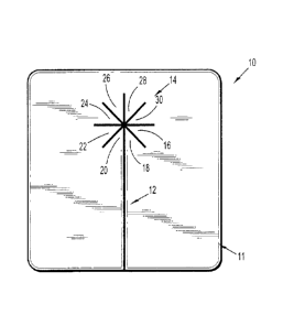
like elements in the various views, FIG. 1 depicts a hemostatic patch 10
including a body 11, a
longitudinal slit 12 bisecting a portion of body 11, and additional slits
extending from the
longitudinal slit 12, forming a star pattern 14, which defines retractable
sections 16, 18, 20, 22,
24, 26. 28, and 30. As is apparent from FIG. 1, the additional slits extending
from the
longitudinal slit may define the number of retractable sections.
[0030] The longitudinal slit 12 and additional slits forming star pattern 14
are cuts through the
body 11 of hemostatic patch 10. These slits may be formed without removing any
portion of the
body 11 of hemostatic patch 10, i.e., the body 11 may be contiguous. In
embodiments, the slits
may be perforated, rather than cut through, so that certain sections may be
retracted while other
sections are more securely maintained in their original position. The
longitudinal slit 12 extends
from an edge of the body 11 and may bisect from about 1% to about 99% of the
length of the
body 11, in embodiments from about 25% to about 75% of the length of the body
11. In
embodiments, the additional slits may be from about 10% to about 75% of the
length of the
longitudinal slit, in embodiments from about 25% to about 50% of the length of
the longitudinal
slit.
[0031] Any number of additional slits may extend from the longitudinal slit.
For example, in
embodiments, the implant may include one additional slit. In other
embodiments, the implant
may include, for example, 20 or more additional slits. In some cases there may
be from about 2
to about 10 additional slits. The additional slits may be at any angle
extending from the
CA 02762193 2011-12-14
longitudinal slit. For example, an additional slit may extend at an angle from
about 1 to about
179 from the longitudinal slit. Where there is more than one additional slit,
the additional slits
may extend from the longitudinal slit at angles that are the same, i.e., each
additional slit may
be angled equally from those to either side of it, or different angles.
[0032] As noted above, the slits form retractable sections or flaps. As shown
in FIG. 2, sections
16, 18, 20, 22, 24, 26, 28, and 30 may be opened (or retracted) to form
retractable flaps 16', 18',
20', 22', 24', 26', 28', and 30', and through-hole 31. In accordance with the
present disclosure a
"through-hole" goes completely through the hemostatic patch, thereby creating
an opening. In
embodiments, no portion of the body 11 is removed in order to create the
through-hole 31;
rather, the through-hole 31 is formed by retracting the retractable flaps 16',
18', 20', 22', 24', 26',
28', and 30'. Although depicted with eight retractable sections, any number of
retractable
sections may be included in the hemostatic patch 10.
[0033] FIG. 3 depicts hemostatic patch 10 with longitudinal slit 12 retracted
or folded back to
form retractable flaps 32 and 34. The retractable flaps allow the hemostatic
patch 10 to
surround tissue prior to contacting the tissue. FIG. 4 depicts all of the
flaps 32, 34, 16', 18', 20',
22', 24', 26', 28', and 30' retracted to create a large opening in the body 11
of the hemostatic
patch 10.
[0034] When folded back, the flaps may prevent hydrogel precursors on the
patch from coming
into contact with moist tissue surface until the surgical patch is in place.
Then the flaps may be
folded back onto the tissue to surround and seal the tubular tissue to prevent
further bleeding.
The cut-outs and through-holes allow for hemostasis around uniquely shaped
tissues in situ.
This function may be useful, for example, during a surgical procedure such as
an anastomosis
procedure. During a surgical anastomosis, two tubular structures or hollow
tissues are joined in
situ. For example, a surgical anastomosis may include: joining two blood
vessels during bypass
surgery, including a procedure known as coronary artery bypass grafting;
resectioning a portion
6
CA 02762193 2011-12-14
of intestine following removal of an intestinal segment; reversal of tubal
ligation or vasectomy
procedures; restoration of continuity to the bladder; and the like.
[0035] A hemostatic patch with a star pattern may be useful, in embodiments,
in an end-to-side
vascular anastomosis. For example, hemostatic patches in sheet form may not be
easily
applied to an end-to-side vascular anastomosis due to the complex geometry
involved at the
site of the anastomosis. Moreover, if the material utilized to form the
hemostatic patch is not
compliant enough, it may be stretched around the anastomosis suture line, but
a risk of
compression and/or stenosis arises. Small strips may be cut and placed on the
suture line, but
this may be very time intensive, and overlapping strips may lead to gaps,
which may allow
bleeding to continue.
[0036] An example of a vascular anastomosis 100 is shown in FIG, 5A. A blood
vessel 52 is
joined to a blood vessel 54 using sutures or staples 56. The flaps 32, 18',
16', 30', 28' (shown)
and 34, 20', 22', 24', 26' (not shown) of the body 11 of the hemostatic patch
10 are retracted in
order to prevent contact with the vessels 52 and 54, prior to locating the
hemostatic patch 10
around the intersection of the vessels 52 and 54. As shown in FIG. 5B, when
placed around the
anastomosis 100, the body 11 surrounds the intersection of vessels 52 and 54
(shown) and 28',
20', 22', 24' and 26' (not shown). The body 11 of the hemostatic patch 10 is
coplanar with
vessel 54. Flaps 16', 18' and 30' (shown) and 28', 20', 22', 24', and 26' (not
shown) are
retracted from the plane of the body 11 and abut vessel 52.
[0037] FIG. 6 depicts yet another embodiment of an implant of the present
disclosure. A
hemostatic patch 80 may include body 82 and arcuate cut-outs 84, 86, 88, and
90. The arcuate
cut-outs 84, 86, 88, and 90 each have a depth A, B, C, and D, respectively.
The depths A, B, C,
D, are the distance between the edge of the body 82 of the hemostatic patch
and the innermost
portion of the arcuate cut-out 84, 86, 88, and 90, respectively, and may be
the same or different
for each arcuate cut-out 84, 86, 88, and 90. For example, where the depth is
different, in
embodiments depth A may be about 2 mm, depth B about 3 mm, depth C about 4 mm,
and
7
depth D about 5 mm. In other embodiments, the depth of, for example, cut-outs
84 and 90, or
88 and 86, may be the same.
[0038] As shown in FIG. 7 a vascular anastomosis 100 may be formed from
tissues 102 and
104. Two hemostatic patches from FIG. 6, 80 and 80', may be aligned so that
arcuate cut-outs
90 and 90' (not shown) encircle tissue 102 and bodies 82 and 82' lie along,
adhere to, and are
coplanar with tissue 104. FIG. 8 depicts an embodiment where the depths A and
D of arcuate
cut-outs 84 and 90, respectively, are the same. The body 82 of hemostatic
patch 80 may
surround tissue 108 and arcuate cut-outs 84 and 90 may encircle tissue 106.
[0039] FIG. 9 depicts a body 111 of a hemostatic patch 110 of the disclosure.
The body 111 is
made of a porous or fabric-like material or substrate 116. The porous
substrate 116 has a first
hydrogel precursor 112 applied to a first portion and a second hydrogel
precursor 120 applied to
a second portion. Such a hemostatic patch 110 is disclosed in U.S. Patent
Application
Publication No. 2010/0100123, filed October 5, 2009. The body 111 of FIG. 9
is shown having a first hydrogel precursor 112 in the form of particles
applied
to a first portion of the porous substrate or fabric-like material 116 and a
second hydrogel precursor 120 in the form of a film applied to a second
portion of the porous
substrate 116.
[0040] During use, the hemostatic patch 110 is oriented with the second
portion of the body
111, to which the second hydrogel precursor 120 is applied, being closer to
the tissue 130, and
the first portion having the first hydrogel precursor 112 applied thereto
further from the tissue
130. In embodiments, the first and second portions may be distinguishable from
one another by
the addition of contrast dyes, surface texturing, coloring or other visual
cues. Upon contact with
tissue, such as, for example, injured tissue 130, the hemostatic patch 110
will soak up
physiological fluid and the second hydrogel precursor 120 may be dissolved by
the fluid. As the
fluid wicks into and migrates across the body 111 of the hemostatic patch 110,
it will carry the
dissolved second hydrogel precursor 120 along through the hemostatic patch
110. Eventually,
8
CA 2762193 2018-04-10
CA 02762193 2011-12-14
the fluid will migrate through the body 111 sufficiently to reach the first
portion to which the first
hydrogel precursor 112 is applied, thereby contacting the first hydrogel
precursor 112. The first
and second hydrogel precursors 112, 120 will then react to form a
biocompatible cross-linked
material, thereby creating hemostasis at the injury site. In some embodiments,
the
biocompatible cross-linked material produced by reaction of the first and
second hydrogel
precursors 112, 120 will not only provide hemostatic properties but also
provide a portion of the
hemostatic patch 110 with adhesive properties.
[0041] The porous substrate 116 of the body 111 of the hemostatic patch 110
has openings or
pores over at least a portion of a surface thereof. The pores may be formed in
the substrate
either before or after implantation. As described in more detail below,
suitable materials for
forming the porous substrate include, but are not limited to fibrous
structures (e.g., knitted
structures, woven structures, non-woven structures, etc.) and/or foams (e.g.,
open or closed cell
foams). In embodiments, the pores may be in sufficient number and size so as
to interconnect
and thus span across the entire thickness of the porous substrate. Woven
fabrics, kitted fabrics
and open cell foam are illustrative examples of structures in which the pores
can be in sufficient
number and size so as to interconnect across the entire thickness of the
porous substrate. In
embodiments, the pores do not interconnect across the entire thickness of the
porous substrate.
Closed cell foam or fused non-woven materials are illustrative examples of
structures in which
the pores may not interconnect across the entire thickness of the porous
substrate. In other
embodiments, the pores of the porous substrate may span across the entire
thickness of porous
substrate. In yet other embodiments, the pores do not extend across the entire
thickness of the
porous substrate, but rather are present at a portion of the thickness
thereof. In embodiments,
the openings or pores are located on a portion of the surface of the porous
substrate, with other
portions of the porous substrate having a non-porous texture.
[0042] In other embodiments, the pores may be formed after implantation in
situ. The in situ
pore formation may be performed using any suitable method. Some non-limiting
examples
9
CA 02762193 2011-12-14
include the use of contact lithography, living radical photopolymer (LRPP)
systems, salt
leaching, combinations thereof, and the like. Those skilled in the art reading
the present
disclosure will envision other pore distribution patterns and configurations
for the porous
substrate.
[0043] Where the porous substrate is fibrous, the fibers may include filaments
or threads
suitable for knitting or weaving or may be staple fibers, such as those
frequently used for
preparing non-woven materials. The fibers may be made from any biocompatible
material.
Thus, the fibers may be formed from a natural material or a synthetic
material. The material
from which the fibers are formed may be bioabsorbable or non-bioabsorbable. It
should be
understood that any combination of natural, synthetic, bioabsorbable and non-
bioabsorbable
materials may be used to form the fibers.
[0044] Some non-limiting examples of materials from which the fibers may be
made include, but
are not limited to, polyesters such as poly(lactic acid) and poly(glycolic
acid) poly(trimethylene
carbonate), poly(dioxanone), poly(hydroxybutyrate), poly(phosphazine),
polyethylene
terephthalate, ultra-high molecular weight polyethylene, polyethylene glycols,
polyethylene
oxides, polyacrylamides, polyhydroxyethylmethylacrylate (pHEMA),
polyvinylpyrrolidone,
polyvinyl alcohols, polyacrylic acid, polyacetate, polycaprolactone,
polypropylene, aliphatic
polyesters, glycerols, poly(amino acids), copoly(ether-esters), polyalkylene
oxalates, poly
(saccharides), polyamides, poly(iminocarbonates), polyalkylene oxalates,
polyoxaesters,
polyorthoesters, polyphosphazenes, biopolymers, polymer drugs and copolymers,
block
copolymers, homopolymers, blends and combinations thereof.
[0045] Where the porous substrate is fibrous, the porous substrate may be
formed using any
method suitable to forming fibrous structures including, but not limited to,
knitting, weaving, non-
woven techniques, wet-spinning, electro-spinning, extrusion, co-extrusion, and
the like. Suitable
techniques for making fibrous structures are within the purview of those
skilled in the art. In
embodiments, the textile has a three dimensional structure, such as the
textiles described in
U.S. Patent Nos. 7,021,086 and 6,443,964.
[0046] In some embodiments, the porous substrate is made from fibers of
oxidized cellulose.
Such materials are known and include oxidized cellulose hemostat materials
commercially
available under the trade name SURGICELe. Methods for preparing oxidized
cellulose
hemostat materials are within the purview of those skilled in the art and are
disclosed, for
example, in U.S. Patent Nos. 3,364,200; 4,626,253; 5,484,913; and 6,500,777.
0047] Where the porous substrate is a foam, the porous substrate may be formed
using any
method suitable to forming a foam or sponge including, but not limited to, the
lyophilization or
freeze-drying of a composition. The foam may be cross-linked or non-cross-
linked, and may
include covalent or ionic bonds. Suitable techniques for making foams are
within the purview of
those skilled in the art.
[0048] As mentioned above, the porous substrate 116 has a first and second
hydrogel
precursor 112, 120 applied thereto. The terms "first hydrogel precursor" and
"second hydrogel
precursor" each mean a polymer, functional polymer, macromolecule, small
molecule, or
crosslinker that can take part in a reaction to form a network of crosslinked
molecules, e.g., a
hydrogel.
[0049] In embodiments, each of the first and second hydrogel precursors 112,
120, include only
one category of functional groups, for example only nucleophilic groups or
only electrophilic
functional groups, so long as both nucleophilic and electrophilic precursors
are used in the
crosslinking reaction. Thus, for example, if the first hydrogel precursor 112
has nucleophilic
functional groups such as amines, the second hydrogel precursor 120 may have
electrophilic
functional groups such as N-hydroxysuccinimides. On the other hand, if first
hydrogel precursor
112 has electrophilic functional groups such as sulfosuccinimides, then the
second hydrogel
precursor 120 may have nucleophilic functional groups such as amines or
thiols. Thus,
11
CA 2762193 2018-04-10
CA 02762193 2011-12-14
functional polymers such as proteins, poly(ally1 amine), styrene sulfonic
acid, or amine-
terminated di- or multifunctional poly(ethylene glycol) ("PEG") can be used.
[0050] The first and second hydrogel precursors 112, 120 may have biologically
inert and water
soluble cores When the core is a polymeric region that is water soluble,
suitable polymers that
may be used include: polyethers, for example, polyalkylene oxides such as
polyethylene
glycol("PEG"), polyethylene oxide ("PEO"), polyethylene oxide-co-polypropylene
oxide ("FPO"),
co-polyethylene oxide block or random copolymers, and polyvinyl alcohol
("PVA"); poly(vinyl
pyrrolidinone) ("PVP"); poly(amino acids); poly (saccharides), such as
dextran, chitosan,
alginates, carboxymethylcellulose, oxidized cellulose, hydroxyethylcellulose,
hydroxymethylcellulose, hyaluronic acid, and proteins such as albumin,
collagen, casein, and
gelatin. The polyethers, and more particularly poly(oxyalkylenes),
poly(ethylene glycol) or
polyethylene glycol, are especially useful. When the core is small in
molecular nature, any of a
variety of hydrophilic functionalities can be used to make the first and
second hydrogel
precursors 112, 120 water soluble. For example, functional groups like
hydroxyl, amine,
sulfonate and/or carboxylate, which are water soluble, may be used to make the
precursor
water soluble. As a further example, the N-hydroxysuccinimide ("NHS") ester of
subaric acid is
insoluble in water, but by adding a sulfonate group to the succinimide ring,
the NHS ester of
subaric acid may be made water soluble, without affecting its reactivity
towards amine groups.
[0051] The first and second hydrogel precursors 112, 120 may be applied to the
porous
substrate 116 using any suitable method within the purview of those skilled in
the art. For
example, the first and second hydrogel precursors 112, 120, may be
incorporated into the
porous substrate 116 prior to forming the porous substrate 116. In another non-
limiting
example, the first or second hydrogel precursors 112, 120 may be positioned in
the pores of the
porous substrate 116 or onto a surface of the porous substrate 116 following
formation of the
substrate. In additional embodiments, the porous substrate 116 may be
calendered prior to
application of the first hydrogel precursor 112 thereby allowing the first or
second hydrogel
12
precursors 112, 120 to penetrate into openings on the substrate which were
created by the
calendaring process.
[0052] In other embodiments, the first or second hydrogel precursors may be in
the form of a
coating which is applied to the substrate in any concentration, dimension and
configuration
capable of forming the hemostatic patch. The coating may form a non-porous
layer or a porous
layer. In embodiments, at least one of the first and second hydrogel
precursors is a cross-linker.
In embodiments, at least one of the first and second hydrogel precursors is a
macromolecule,
and may be referred to herein as a 'functional polymer".
[0053] Each of the first and second hydrogel precursors is multifunctional,
meaning that it
includes two or more electrophilic or nucleophilic functional groups, such
that, for example, a
nucleophilic functional group on the first hydrogel precursor may react with
an electrophilic
functional group on the second hydrogel precursor to form a covalent bond. At
least one of the
first or second hydrogel precursors includes more than two functional groups,
so that, as a
result of electrophilic-nucleophilic reactions, the precursors combine to form
cross-linked
polymeric products.
[0054] In embodiments, a multifunctional nucleophilic polymer such as
trilysine may be used as
a first hydrogel precursor and a multifunctional electrophilic polymer such as
a multi-arm PEG
functionalized with multiple NHS groups may be used as a second hydrogel
precursor. The
multi-arm PEG functionalized with multiple NHS groups can for example have
four, six or eight
arms and have a molecular weight of from about 5,000 to about 25,000. Other
examples of
suitable first and second hydrogel precursors are described in U.S. Patent
Nos. 6,152,943;
6,165,201; 6,179,862; 6,514,534; 6,566,406; 6,605,294; 6,673,093; 6,703,047;
6,818,018;
7,009,034; and 7,347,850.
[0055] While the present disclosure may involve a hemostatic patch, any
surgical patch may be
used. The hemostatic patch may be any size and dimension. In embodiments the
patch may be
13
CA 2762193 2018-04-10
CA 02762193 2011-12-14
capable of transport in a laparoscopic deployment device or capable of
introduction in open
surgery. In embodiments, the hemostatic patch may be about 2 inches square,
although it is
envisioned that the patch may be of varying shapes and sizes. Additionally,
while the substrate
used in forming the patch is described as "porous," the substrate may be
porous or non-porous
in various embodiments.
[0056] Upon application to a site of bleeding tissue, the hemostatic patch may
affect
hemostasis of said tissue. As used herein, the term "hemostasis" means the
arrest of bleeding.
It is believed, without being limited to any theory, that the hemostatic
effect of the hemostatic
patch is due to both intrinsic and extrinsic factors. In embodiments, the
substrate may include a
hemostatic agent providing an intrinsic hemostatic effect. In other
embodiments, the cross-
linking between the hydrogel precursors creates a physical barrier to blood
flow, thereby
providing an extrinsic hemostatic effect.
[0057] Hemostasis may occur, at the site of application of the hemostatic
patch, within less than
about 2 minutes. As stated above, upon contact with tissue, such as, for
example, injured or
bleeding tissue, the hemostatic patch soaks up interstitial and physiological
fluid (e.g., blood,
lymph-fluid, etc.) and the first and second hydrogel precursors are mixed by
the fluid. In order
to prevent the hemostatic patch from taking up fluid prior to use at the
location in need of
hemostasis, the hemostatic patch is retained or sealed in packaging until the
time it is needed
for its application.
[0058] As seen in FIG. 10, during use, the hemostatic patch 110 is oriented
with second portion
of body 111, to which the second hydrogel precursor 120 is applied, being
closer to tissue 130
and with the first portion, to which the first hydrogel precursor 112 is
applied, being disposed
further from the tissue 130. Upon contact with bleeding tissue 130, hemostatic
patch 110 soaks
up physiological fluid or blood 132 and the second portion, having the second
hydrogel
precursor 120 is dissolved by the fluid or blood 132. As the fluid or blood
132 wicks into and
migrates across the body 111 of the hemostatic patch 110, the fluid or blood
carries the
14
CA 02762193 2011-12-14
dissolved second hydrogel precursor 120 along through the body 111
sufficiently to reach the
first portion, to which the first hydrogel precursor 112 is applied, thereby
initiating the cross-
linking reaction between the first and second hydrogel precursors 112, 120. At
this point, as
seen in FIG. 11, first and second hydrogel precursors 112, 120, then react to
form a
biocompatible cross-linked material 134 thereby assisting with the hemostasis
of the tissue 130.
[0059] In use, an individual, such as a nurse or surgeon, using a hemostatic
patch of the
present disclosure may wish to cut the patch to a desired size and shape. For
certain shapes,
this may prove difficult and time consuming, and imperfect cuts may lead to
continued bleeding
where too much material has been cut away, so there is insufficient contact
between the
hemostat and tissue surface; vessel stenosis where too little material is
removed; or a damaged
patch that must be discarded.
[0060] Thus, in embodiments, the hemostatic patch of the present disclosure
may be provided
with a cutting fixture, sometimes referred to herein, in embodiments, as a
cutting template,
which may allow one to cut the hemostatic patch to form a suitable pattern, in
embodiments a
star pattern, for application to a vascular anastomosis. For example, as set
forth in FIGS. 12 A-
C, a template is provided having a top 200 and bottom 210 portions. In use,
the template is
opened by physically separating top 200 from bottom 210. A hemostatic patch
(not shown) is
placed in bottom 210 and top 200 is then affixed to bottom 210 by some means,
including
physical means such as a catch, snap, interference fit (like a twist), and/or
hinge, or other
means including magnets, combinations thereof, and the like. As seen in FIG.
12A, top 200 has
narrow slits 220 cut in the shape of the star pattern, and top 200 can be
thin, as depicted in the
side view of top 200 set forth in FIG. 12B. Slits 220 are just wide enough to
permit a scalpel
blade, or some similar knife or cutting device, to pass through the openings,
thereby permitting
the scalpel blade to cut the hemostatic patch. As seen in FIG. 120, the bottom
210 of the
template has a recessed region 230 that holds the hemostatic patch in place
and centers the
hemostatic patch under the top 200 of the template. Additionally, bottom 210
of the template
CA 02762193 2011-12-14
has a star pattern corresponding to the one in top 200, with openings 240 that
are wider than
the slits 220 included in the top 200 of the template. The openings 240 in
bottom 210 of the
template allow the scalpel blade to penetrate all the way through the
hemostatic patch and
template, while still providing sufficient support to the hemostatic patch.
Once the pattern has
been cut in the hemostatic patch, the top 200 is again removed from bottom
210, and the cut
patch, now having a star pattern, is removed therefrom.
[0061] In other embodiments, as depicted in FIGS. 13A and 13B, top 300 may be
connected to
bottom 310 by hinge 350. Top 300 has slits 320 and bottom 310 has openings
340. A
hemostatic patch (not shown) may be placed in a recessed portion 330 of bottom
310 to center
the patch therein. After placement of the hemostatic patch in recessed portion
330 of bottom
310, top 300 is then closed, thereby holding the hemostatic patch firmly in
place. An individual,
such as a nurse or surgeon, may then pass a blade, including a scalpel or some
similar knife or
cutting device, through slits 320 in top 300, passing through opening 340 in
bottom 310, thereby
cutting a star pattern in the hemostatic patch. The top 300 is then opened,
and the cut
hemostatic patch, now having a star pattern, is removed from the template.
[0062] In embodiments, the template may have a handle or other similar region
(not shown) to
permit an individual to hold the template while keeping the individual's
fingers away from the
cutting zone.
[0063] The template of the present disclosure allows an individual, including
a nurse or surgeon
in an operating room, to cut a star pattern in a hemostatic patch of the
present disclosure on an
as-needed basis. The template is small, inexpensive, and easy to use.
[0064] While the above description has been directed to templates having a
star pattern, thus
permitting the formation of hemostatic patches having the same star pattern,
templates for use
in accordance with the present disclosure may possess any other desirable
pattern (not shown)
in tops 200 and/or 300, with corresponding patterns in bottoms 210 and/or 310,
thereby
permitting an individual to cut a hemostatic patch of the present disclosure
in the desired
16
CA 02762193 2011-12-14
pattern. Suitable patterns include, for example, an elongate longitudinal slit
at least partially
dividing a rectangular patch, or a modified keyhole like pattern, which
includes the elongate slit
as described above with a circular opening where the slit ends within the body
of the patch.
[0065] FIGS. 14A and 14B depict a hemostatic patch 410 of the present
disclosure having
elongate longitudinal slit 412 at least partly dividing patch 410. The
longitudinal slit may be the
same as longitudinal slit 12 described above with respect to hemostatic patch
10. As seen in
FIG. 14A, in embodiments, longitudinal slit 412 may extend from an edge of
hemostatic patch
410 to a point within the body of hemostatic patch 410. FIG. 14B depicts
hemostatic patch 410
with longitudinal slit 412 retracted or folded back to form retractable flaps
432 and 434.
[0066] FIGS. 15A and 15B depict a hemostatic patch 510 of the present
disclosure having
elongate longitudinal slit 512 at least partly dividing patch 510. The
longitudinal slit may be the
same as longitudinal slit 12 described above with respect to hemostatic patch
10. As can be
seen in FIG. 15A, longitudinal slit 512 extends from an edge of patch 510 into
the body of patch
510, terminating at circular opening 560. Thus, hemostatic patch 510 may be
referred to, in
embodiments, as possessing a keyhole configuration. FIG. 158 depicts
hemostatic patch 510
with longitudinal slit 512 retracted or folded back to form retractable flaps
532 and 534.
[0067] Additionally, the hemostatic patch may include biologically acceptable
additives such as
plasticizers, antioxidants, dyes, dilutants, therapeutic agents, and the like
and combinations
thereof, which can be coated on the filaments or fibers, or impregnated into
the fibers or
filaments (e.g., during compounding or extrusion) used to form the hemostatic
patch of the
present disclosure.
[0068] Therapeutic agents include, but are not limited to, drugs, amino acids,
peptides,
polypeptides, proteins, polysaccharides, muteins, immunoglobulins, antibodies,
cytokines (e.g.,
lymphokines, monokines, chemokines), blood clotting factors, hemopoietic
factors, interleukins
(1 through 18), interferons (6-IFN, a-IFN and y-IFN), erythropoietin,
nucleases, tumor necrosis
factor, colony stimulating factors (e.g., GCSF, GM-CSF, MCSF), insulin, anti-
tumor agents and
17
CA 02762193 2011-12-14
tumor suppressors, blood proteins, fibrin, thrombin, fibrinogen, synthetic
thrombin, synthetic
fibrin, synthetic fibrinogen, gonadotropins (e.g., FSH, LH, CG, etc.),
hormones and hormone
analogs (e.g., growth hormone, luteinizing hormone releasing factor), vaccines
(e.g., tumoral,
bacterial and viral antigens); somatostatin; antigens; blood coagulation
factors; growth factors
(e.g., nerve growth factor, insulin-like growth factor); bone morphogenic
proteins, TGF-B,
protein inhibitors, protein antagonists, and protein agonists; nucleic acids,
such as antisense
molecules, DNA, RNA, RNAi; oligonucleotides; polynucleotides; cells, viruses,
and ribozymes.
[0069] In embodiments, the therapeutic agent may include at least one of the
following drugs,
including combinations and alternative forms of the drugs such as alternative
salt forms, free
acid form, free base forms, pro-drugs and hydrates: analgesics/antipyretics
(e.g., aspirin,
acetaminophen, ibuprofen, naproxen sodium, buprenorphine, propoxyphene
hydrochloride,
propoxyphene napsylate, meperidine hydrochloride, hydromorphone hydrochloride,
morphine,
oxycodone, codeine, dihydrocodeine bitartrate, pentazocine, hydrocodone
bitartrate,
levorphanol, diflunisal, trolamine salicylate, nalbuphine hydrochloride,
mefenamic acid,
butorphanol, choline salicylate, butalbital, phenyltoloxamine citrate,
diphenhydramine citrate,
methotrimeprazine, cinnamedrine hydrochloride, and meprobamate);
antiasthmatics (e.g.,
ketotifen and traxanox); antibiotics (e.g., neomycin, streptomycin,
chloramphenicol,
cephalosporin, ampicillin, penicillin, tetracycline, and ciprofloxacin);
antidepressants (e.g.,
nefopam, oxypertine, amoxapine, trazodone, amitriptyline, maprotiline,
phenelzine, desipramine,
nortriptyline, tranylcypromine, fluoxetine, doxepin, imipramine, imipramine
pamoate,
isocarboxazid, trimipramine, and protriptyline); antidiabetics (e.g.,
biguanides and sulfonylurea
derivatives); antifungal agents (e.g., griseofulvin, ketoconazole,
itraconizole, amphotericin B,
nystatin, and candicidin); antihypertensive agents (e.g., propanolol,
propafenone, oxyprenolol,
nifedipine, reserpine, trimethaphan, phenoxybenzamine, pargyline
hydrochloride, deserpidine,
diazoxide, guanethidine monosulfate, minoxidil, rescinnamine, sodium
nitroprusside, rauwolfia
serpentina, alseroxylon, and phentolamine); anti-inflammatories (e.g., (non-
steroidal)
18
CA 02762193 2011-12-14
indomethacin, ketoprofen, flurbiprofen, naproxen, ibuprofen, ramifenazone,
piroxicam,
(steroidal) cortisone, dexamethasone, fluazacort, celecoxib, rofecoxib,
hydrocortisone,
prednisolone, arid prednisone); antineoplastics (e.g., cyclophosphamide,
actinomycin,
bleomycin, dactinomycin, daunorubicin, doxorubicin, epirubicin, mitomycin,
methotrexate,
fluorouracil, gemcitabine, carboplatin, carmustine (BCNU), methyl-CCNU,
cisplatin, etoposide,
camptothecin and derivatives thereof, phenesterine, paclitaxel and derivatives
thereof,
docetaxel and derivatives thereof, vinblastine, vincristine, goserelin,
leuprolide, tamoxifen,
interferon alfa, retinoic acid (ATRA), nitrogen mustard alkylating agents, and
piposulfan);
antianxiety agents (e.g., lorazepam, buspirone, prazepam, chlordiazepoxide,
oxazepam,
clorazepate dipotassium, diazepam, hydroxyzine pamoate, hydroxyzine
hydrochloride,
alprazolam, droperidol, halazepam, chlormezanone, and dantrolene);
immunosuppressive
agents (e.g., cyclosporine, azathioprine, mizoribine, and FK506 (tacrolimus));
antimigraine
agents (e.g., ergotamine, propanolol, isometheptene mucate, and
dichloralphenazone);
sedatives/hypnotics (e.g., barbiturates such as pentobarbital, pentobarbital,
and secobarbital;
and benzodiazapines such as flurazepam hydrochloride, triazolam, and
midazolam); antianginal
agents (e.g., beta-adrenergic blockers; calcium channel blockers such as
nifedipine, and
diltiazem; and nitrates such as nitroglycerin, isosorbide dinitrate,
pentearythritol tetranitrate, and
erythrityl tetranitrate); antipsychotic agents (e.g., haloperidol, loxapine
succinate, loxapine
hydrochloride, thioridazine, thioridazine hydrochloride, thiothixene,
fluphenazine, fluphenazine
decanoate, fluphenazine enanthate, trifluoperazine, chlorpromazine,
perphenazine, lithium
citrate, and prochlorperazine); antimanic agents (e.g., lithium carbonate);
antiarrhythmics (e.g.,
bretylium tosylate, esmolol, verapamil, amiodarone, encainide, digoxin,
digitoxin, mexiletine,
disopyramide phosphate, procainamide, quinidine sulfate, quinidine gluconate,
quinidine
polygalacturonate, flecainide acetate, tocainide, and lidocaine);
antiarthritic agents (e.g.,
phenylbutazone, sulindac, penicillanine, salsalate, piroxicam, azathioprine,
indomethacin,
meclofenamate, gold sodium thiomalate, ketoprofen, auranofin, aurothioglucose,
and tolmetin
19
CA 02762193 2011-12-14
sodium); antigout agents (e.g., colchicine, and allopurinol); anticoagulants
(e.g., heparin,
heparin sodium, and warfarin sodium); thrombolytic agents (e.g., urokinase,
streptokinase, and
alteplase); antifibrinolytic agents (e.g., aminocaproic acid); hemorheologic
agents (e.g.,
pentoxifylline); antiplatelet agents (e.g., aspirin); anticonvulsants (e.g.,
valproic acid, divalproex
sodium, phenytoin, phenytoin sodium, clonazepam, primidone, phenobarbital,
carbamazepine,
amobarbital sodium, methsuximide, metharbital, mephobarbital, mephenytoin,
phensuximide,
paramethadione, ethotoin, phenacemide, secobarbitol sodium, clorazepate
dipotassium, and
trimethadione); antiparkinson agents (e.g., ethosuximide);
antihistamines/antipruritics (e.g.,
hydroxyzine, diphenhydramine, chlorphenira mine, brompheniramine maleate,
cyproheptadine
hydrochloride, terfenadine, clemastine fumarate, triprolidine, carbinoxamine,
diphenylpyraline,
phenindamine, azatadine, tripelennamine, dexchlorpheniramine maleate, and
methdilazine);
agents useful for calcium regulation (e.g., calcitonin, and parathyroid
hormone); antibacterial
agents (e.g., amikacin sulfate, aztreonam, chloramphenicol, chloramphenicol
palirtate,
ciprofloxacin, clindamycin, clindamycin palmitate, clindamycin phosphate,
metronidazole,
metronidazole hydrochloride, gentamicin sulfate, lincomycin hydrochloride,
tobramycin sulfate,
vancomycin hydrochloride, polymyxin B sulfate, colistimethate sodium, and
colistin sulfate);
antiviral agents (e.g., interferon alpha, beta or gamma, zidovudine,
amantadine hydrochloride,
ribavirin, and acyclovir); antimicrobials (e.g., cephalosporins such as
cefazolin sodium,
cephradine, cefaclor, cephapirin sodium, ceftizoxime sodium, cefoperazone
sodium, cefotetan
disodium, cefuroxime e azotil, cefotaxime sodium, cefadroxil monohydrate,
cephalexin,
cephalothin sodium, cephalexin hydrochloride monohydrate, cefamandole nafate,
cefoxitin
sodium, cefonicid sodium, ceforanide, ceftriaxone sodium, ceftazidime,
cefadroxil, cephradine,
and cefuroxime sodium; penicillins such as ampicillin, amoxicillin, penicillin
G benzathine,
cyclacillin, ampicillin sodium, penicillin G potassium, penicillin V
potassium, piperacillin sodium,
oxacillin sodium, bacampicillin hydrochloride, cloxacillin sodium, ticarcillin
disodium, azlocillin
sodium, carbenicillin indanyl sodium, penicillin G procaine, methicillin
sodium, and nafcillin
CA 02762193 2011-12-14
sodium; erythromycins such as erythromycin ethylsuccinate, erythromycin,
erythromycin
estolate, erythromycin lactobionate, erythromycin stearate, and erythromycin
ethylsuccinate;
and tetracyclines such as tetracycline hydrochloride, doxycycline hyclate, and
minocycline
hydrochloride, azithromycin, clarithromycin); anti-infectives (e.g., GM-CSF);
bronchodilators
(e.g., sympathomimetics such as epinephrine hydrochloride, metaproterenol
sulfate, terbutaline
sulfate, isoetharine, isoetharine nnesylate, isoetharine hydrochloride,
albuterol sulfate, albuterol,
bitolterolmesylate, isoproterenol hydrochloride, terbutaline sulfate,
epinephrine bitartrate,
metaproterenol sulfate, and epinephrine); anticholinergic agents such as
ipratropium bromide;
xanthines such as aminophylline, dyphylline, metaproterenol sulfate, and
aminophylline; mast
cell stabilizers such as cromolyn sodium; inhalant corticosteroids such as
beclomethasone
dipropionate (BDP), and beclomethasone dipropionate monohydrate; salbutamol;
ipratropium
bromide; budesonide; ketotifen; salmeterol; xinafoate; terbutaline sulfate;
triamcinolone;
theophylline; nedocromil sodium; metaproterenol sulfate; albuterol;
flunisolide; fluticasone
proprionate; steroidal compounds and hormones (e.g., androgens such as
danazol,
testosterone cypionate, fluoxymesterone, ethyltestosterone, testosterone
enathate,
methyltestosterone); estrogens such as estradiol, estropipate, and conjugated
estrogens;
progestins such as methoxyprogesterone acetate, and norethindrone acetate;
corticosteroids
such as triamcinolone, betamethasone, betamethasone sodium phosphate,
dexamethasone,
dexamethasone sodium phosphate, dexamethasone acetate, prednisone,
methylprednisolone
acetate suspension, triamcinolone acetonide, methylprednisolone, prednisolone
sodium
phosphate, methylprednisolone sodium succinate, hydrocortisone sodium
succinate,
triamcinolone hexacetonide, hydrocortisone, hydrocortisone cypionate,
prednisolone,
fludrocortisone acetate, paramethasone acetate, prednisolone tebutate,
prednisolone acetate,
prednisolone sodium phosphate, and hydrocortisone sodium succinate; and
thyroid hormones
such as levothyroxine sodium); hypoglycemic agents (e.g., human insulin,
purified beef insulin,
purified pork insulin, glyburide, chlorpropamide, glipizide, tolbutarnide, and
tolazamide);
21
CA 02762193 2011-12-14
hypolipidemic agents (e.g., clofibrate, dextrothyroxine sodium, probucol,
pravastitin,
atorvastatin, lovastatin, and niacin); proteins (e.g., DNase, alginase,
superoxide dismutase, and
lipase); nucleic acids (e.g., sense or anti-sense nucleic acids encoding any
therapeutically
useful protein, including any of the proteins described herein); agents useful
for erythropoiesis
stimulation (e.g., erythropoietin); antiulcer/antireflux agents (e.g.,
famotidine, cimetidine, and
ranitidine hydrochloride); antinauseants/antiemetics (e.g., meclizine
hydrochloride, nabilone,
prochlorperazine, dimenhydrinate, promethazine hydrochloride,
thiethylperazine, and
scopolamine); as well as other drugs useful in the compositions and methods
described herein
include mitotane, halonitrosoureas, anthrocyclines, ellipticine, ceftriaxone,
ketoconazole,
ceftazidime, oxaprozin, albuterol, valacyclovir, urofollitropin, famciclovir,
flutamide, enalapril,
mefformin, itraconazole, buspirone, gabapentin, fosinopril, tramadol,
acarbose, lorazepam,
follitropin, glipizide, omeprazole, fluoxetine, lisinopril, tramsdol,
levofloxacin, zafirlukast,
interferon, growth hormone, interleukin, erythropoietin, granulocyte
stimulating factor, nizatidine,
bupropion, perindopril, erbumine, adenosine, alendronate, alprostadil,
benazepril, betaxolol,
bleomycin sulfate, dexfenfluramine, diltiazem, fentanyl, fiecainid,
gemcitabine, glatiramer
acetate, granisetron, lamivudine, mangafodipir trisodium, mesalamine,
metoprolol fumarate,
metronidazole, miglitol, moexipril, monteleukast, octreotide acetate,
olopatadine, paricalcitol,
somatropin, sumatriptan succinate, tacrine, verapamil, nabumetone,
trovafloxacin, dolasetron,
zidovudine, finasteride, tobramycin, isradipine, tolcapone, enoxaparin,
fluconazole,
lansoprazole, terbinafine, pamidronate, didanosine, diclofenac, cisapride,
venlafaxine,
troglitazone, fluvastatin, losartan, imiglucerase, donepezil, olanzapine,
valsartan, fexofenadine,
calcitonin, and ipratropium bromide. In some embodiments, the therapeutic
agent may be water
soluble. In some embodiments, the therapeutic agent may not be water soluble.
[0070] In embodiments, the above therapeutic agents may be applied to a
hemostatic patch of
the present disclosure in a solution. Where the therapeutic agent is water
soluble, water may
be used as a solvent for forming such a solution. Other solvents which may be
used include
22
CA 02762193 2011-12-14
polar and non-polar solvents including, but not limited to, alcohols, such as,
methanol, ethanol,
propanol; chlorinated hydrocarbons such as methylene chloride, chloroform, 1,
2-dichloro-
ethane; and aliphatic hydrocarbons such as hexane, heptene, ethyl acetate; and
the like and
combinations of these.
[0071] It will be understood that various modifications may be made to the
embodiments
disclosed herein. Therefore, the above description should not be construed as
limiting, but
merely as an exemplification of preferred embodiments. Those skilled in the
art will envision
other modifications within the scope and spirit of the present disclosure.
Such modifications and
variations are intended to come within the scope of the following claims.
23