Note: Descriptions are shown in the official language in which they were submitted.
CA 02762351 2011-11-17
WO 2010/132982 PCT/CA2010/000734
TREATMENT OF MUSCLE DISEASE CHARACTERIZED BY INSULIN RESISTANCE
TECHNICAL FIELD
[0001] The present disclosure relates generally to therapeutic agents,
compositions and methods for treating muscle diseases and conditions
characterized by
impaired insulin-dependent signaling in muscle tissue, in essence, a form of
insulin
resistance.
BACKGROUND
[0002] There are numerous diseases and conditions that affect muscle. Examples
include muscle wasting diseases, including cachexia, muscle attenuation or
atrophy,
including sarcopenia, ICU-induced weakness, surgery-induced weakness,
neuromuscular
diseases, and muscle degenerative diseases, such as muscular dystrophies.
[0003] Muscular dystrophy (MD) refers to a group of hereditary, progressive,
degenerative disorders characterized by progressive muscle weakness, defects
in muscle
proteins, and the destruction of muscle fibers and tissue over time. In many
cases, the
histological picture shows variation in fiber size, muscle cell necrosis and
regeneration,
and often proliferation of connective and adipose tissue. The diseases
primarily target the
skeletal or voluntary muscles. However, muscles of the heart and other
involuntary
muscles are also affected in certain forms of muscular dystrophy.
[0004] There are several forms of muscular dystrophy, which differ in their
age of
onset, penetrance, severity, and pattern of muscles affected. Known forms of
muscular
dystrophy include Duchenne muscular dystrophy (DMD), Becker muscular dystrophy
(BMD), Limb-Girdle muscular dystrophies, myotonic dystrophy (Steinert's
disease),
Emery-Dreifuss muscular dystrophy, Landouzy-Dejerine muscular dystrophy,
facioscapulohumeral muscular dystrophy (FSH), von Graefe-Fuchs muscular
dystrophy,
oculopharyngeal muscular dystrophy (OPMD), distal muscular dystrophy, and
congenital
muscular dystrophies. While these are the main forms classified as muscular
dystrophy,
there are more than 100 diseases in total with similarities to muscular
dystrophy. Some
dystrophies may result from different underlying defects than others. Most
types of MD
are multi-system disorders with manifestations in body systems including the
musculoskeletal, gastrointestinal and nervous systems, the heart, endocrine
glands, skin,
eyes and other organs.
[0005] Duchenne Muscular Dystrophy (DMD) is the most common inherited lethal
childhood muscular dystrophy, affecting about 1 in 3000 males. Children with
DMD
usually become wheelchair bound by the age of 11 or 12 years and affected
individuals
- 1 -
CA 02762351 2011-11-17
WO 2010/132982 PCT/CA2010/000734
usually die in the second or third decade of life. DMD originates from
mutations in the
dystrophin gene located on the X chromosome (Xp21), leading to loss of
dystrophin
protein with attendant muscle fiber destruction. Although the role of the
dystrophin protein
in maintaining skeletal myofiber integrity is generally well recognized, the
exact
mechanism that leads to myofiber destruction and loss in dystrophic muscle is
not well
understood. The discovery of the dystrophin gene and the subsequent
characterization of
the protein product have established dystrophin as an integral sarcolemmal
protein,
linking the muscle sarcomere and cytoskeleton to the surrounding extracellular
matrix.
The localization of dystrophin is synonymous with maintaining muscle integrity
and its
absence (as evidenced in DMD) leads to membrane fragility, contraction induced
myofiber damage, and death (Petrof et al. 1993).
[0006] Becker type muscular dystrophy (BMD), also known as Benign
pseudohypertrophic muscular dystrophy is an X-linked recessive inherited
disorder
characterized by slowly progressive muscle weakness of the legs and pelvis,
which is
also caused by mutations in the dystrophin gene, has onset in adolescence or
adulthood
with a less severe course of progression. BMD is related to Duchenne Muscular
Dystrophy in that both result from a mutation in the dystrophin gene, but in
DMD no
functional dystrophin is produced making DMD much more severe than BMD. Both
DMD
and BMD have traditionally been called "X-linked" recessive diseases (Freund
et al.,
2007).
[0007] The limb girdle muscular dystrophies all show a similar distribution of
muscle weakness, affecting both upper arms and legs. Many forms of limb girdle
muscular dystrophy have been identified, showing different patterns of
inheritance:
autosomal recessive (designated LGMD1) or autosomal dominant (LGMD2). In an
autosomal recessive pattern of inheritance, an individual receives two copies
of the
defective gene, one from each parent. In an autosomal dominant disease, the
disorder
can occur in either sex when an individual inherits a single defective gene
from either
parent. The recessive limb girdle muscular dystrophies are more frequent than
the
dominant forms, and may be more severe. Limb girdle muscular dystrophy can
have a
childhood onset, although more often symptoms appear in adolescence or young
adulthood. The dominant limb girdle muscular dystrophies usually show adult
onset.
Some of the recessive forms have been associated with defects in proteins that
make up
the dystrophin-glycoprotein complex. Mutations in one component of the
dystrophin-
glycoprotein complex, the sarcoglycans, can lead to the forms of limb girdle
muscular
dystrophy known as LGMD2C, 2D, 2E, and 2F. Defects in caveolin 3, a protein
that
associates with the dystrophin-glycoprotein complex, lead to LGMD1C, while
mutations in
-2-
CA 02762351 2011-11-17
WO 2010/132982 PCT/CA2010/000734
dysferlin, a protein that is thought to interact with caveolin 3, cause
LGMD2B. Mutations
in genes not related to the dystrophin-glycoprotein complex are implicated in
other forms
of limb girdle muscular dystrophy. For example, mutations in the enzymatic
protein
calpain 3 lead to LGMD2A (Guglieri M. et al., 2008).
[0008] Myotonic dystrophy is the most common form of muscular dystrophy. It is
dominantly inherited and characterized by muscle hyperexcitability (myotonia),
muscle
wasting and weakness, cataracts, hypogonadism, cardiac conduction
abnormalities and
other developmental and degenerative manifestations frequently including
cognitive
dysfunction. Penetrance can be variable. Myotonic dystrophy can be caused by
mutations
in different genes, but the characteristics are quite similar. Type 1 myotonic
dystrophy
(DM1) is caused by expansion of a CTG triplet repeat in an untranslated region
of the
dystrophia myotonica protein kinase gene (DMPK) on chromosome 19, while type 2
(DM2) is caused by expansion of a CCTG repeat in the first intron of the zinc
finger
protein-9 gene (ZNF9) on chromosome 3. Repeat number in the myotonic
dystrophies
increases in subsequent generations (anticipation). DM1 also has congenital
and
childhood onset forms; these early appearing forms of the disease differ
mechanistically
from the adult form only in exhibiting larger CTG repeats that, in turn,
trigger earlier
appearance of symptoms. Those patients that survive early onset DM1 frequently
exhibit
morbidity and mortality in the third and fourth decades relating to
cardiopulmonary
involvement (Liquori CL. et al., 2001; Cho DH. et al., 2007).
[0009] Facioscapulohumeral muscular dystrophy (FSHD), a dominantly inherited
disorder, is the third most common dystrophy after Duchenne and myotonic
muscular
dystrophy. FSHD is an autosomal dominant progressive degenerative disease that
initially affects the muscles of the face (facio), shoulders (scapulo), and
upper arms
(humeral), followed by the muscles of the feet, pelvic girdle, and abdomen.
Affected
individuals may also suffer from hearing loss. Onset and progression of the
disease is
variable and often the weakness is asymmetrical in affected individuals. Life
expectancy
is typically within normal range, but the disease can lead to severe
disability. Nearly all
cases are associated with deletions of tandem repeats, termed D4Z4, in a
distal region of
chromosome 4 (4q35) (Tawil R., 2008).
[0010] The congenital muscular dystrophies are a heterogeneous class of
disorders, and include several disorders with a range of symptoms. Muscle
degeneration
can be mild or severe, and may be restricted to skeletal muscle, or paired
with effects on
the brain and other organs. Defects in the protein merosin are responsible for
about half
of the cases in the U.S. Mutations in one of the integrin proteins gives rise
to another form
of congenital muscular dystrophy. Defects in the proteins called fukutin and
fukutin-
-3-
CA 02762351 2011-11-17
WO 2010/132982 PCT/CA2010/000734
related protein cause the most common forms of congenital muscular dystrophy
found in
Japan. All of these proteins are thought to have some relationship to the
dystrophin-
glycoprotein complex. Some forms of congenital muscular dystrophy, including
Fukuyama
muscular dystrophy, muscle-eye brain disease, and Walker-Warburg syndrome are
due
to defective glycosylation of one of the proteins in the dystrophin-
glycoprotein complex
(alpha-dystroglycan) and show severe brain malformations, such as
lissencephaly (a
"cobblestone" appearance to part of the brain) and hydrocephalus (an excessive
accumulation of fluid in the brain). Other forms, including the merosin-absent
form and
rigid spine syndrome, do not have major brain malformations associated with
the disease.
The molecular basis for many forms of congenital muscular dystrophy remains
unknown
(Sewry CA., 2008).
[0011] Several other forms of muscular dystrophy also occur. Oculopharyngeal
muscular dystrophy, which causes weakness in the eye, throat, and facial
muscles,
followed by pelvic and shoulder muscle weakness, has been attributed to a
short triplet
repeat expansion in the nuclear polyadenylate binding protein 1 gene (PABPN1),
a gene
involved in translating the genetic code into functional proteins. Inheritance
follows either
autosomal dominant or autosomal recessive patterns.
[0012] Polyalanine tract expansion from a norm of 10 to 12-17 residues causes
aggregation of filamentous intranuclear inclusions in skeletal muscle which
appear to
precipitate the disease. This disease is most common in people of French-
Canadian
descent or people of Hispanic descent from certain regions of the Southwest.
Miyoshi
myopathy, one of the distal muscular dystrophies, causes initial weakness in
the calf
muscles, and is caused by defects in the protein dysferlin, which is the same
gene
responsible for LGMD2B, reinforcing the idea that progress against one form of
muscular
dystrophy should be informative to other forms. There are two forms of Emery-
Dreifuss
muscular dystrophy, an X-linked and an autosomal dominant form. Emery-Dreifuss
muscular dystrophy is characterized by weakness in the shoulder girdle and
lower legs,
as well as the development of contractures in regions of the body,
particularly the elbows,
Achilles tendons, and neck. Defects in proteins that make up the nucleus,
including
emerin, and lamin A/C, are implicated in the disorder.
[0013] Several animal models, manifesting phenotypes observed in
neuromuscular diseases, have been identified in nature or generated in
laboratory. These
models generally present physiological alterations observed in human patients
and can
be used as important tools for genetic, therapeutic, and histopathological
studies. The
study of animal models for genetic diseases, in spite of the existence of
differences in
some phenotypes, can provide important clues to the understanding of the
pathogenesis
-4-
CA 02762351 2011-11-17
WO 2010/132982 PCT/CA2010/000734
of these disorders and are also very valuable for testing strategies for
therapeutic
approaches (Vainzof M, et al., 2008).
[0014] The mdx mouse model is a well-accepted animal model of human DMD.
The mdx mouse carries a premature stop codon in exon 23 of the dystrophin gene
and
exhibits no detectable levels of dystrophin in muscle tissue. The progression
of disease
pathology in the dystrophic mdx mouse has been associated with constitutive
activation
of the MAP kinase, JNK1 (Kolodziejczyk et al. 2001), a ubiquitous signaling
molecule.
Once activated, JNK1 can phosphorylate the transcription factor NF-ATc1,
leading to
cytoplasmic accumulation and loss of NF-ATc1 function. Direct inhibition of
JNK1 in
dystrophic muscle, by overexpression of the JNK1 scaffolding protein JIP-1,
was shown
to reduce damage associated with typical disease progression (Kolodziejczyk et
al.
2001). The present inventors have previously shown that the glucocorticoid,
deflazacort,
attenuates DMD pathology by circumventing and limiting the deleterious effects
of JNK1
(St-Pierre et al. 2004). Deflazacort did not directly inhibit JNK1, rather the
beneficial
effects of this compound appear to originate from increasing the activity of
the calcineurin
phosphatase. Once activated, calcineurin then dephosphorylates NF-ATc1,
restoring NF-
ATc1 nuclear localization and transcriptional function (St-Pierre et al.
2004). Other groups
have now demonstrated that increased calcineurin activity alleviates
dystrophic muscle
pathology (Chakkalakal et al. 2004; Chakkalakal et al. 2006; Stupka et al.
2006; Stupka et
al. 2008).
[0015] A general interpretation of these studies is that calcineurin
activation
enhances myofiber integrity by increasing the expression of the dystrophin
homologue
utrophin, which itself provides an effective substitute for dystrophin in
animal models of
DMD. (St-Pierre et al. 2004; Chakkalakal et al. 2004; Chakkalakal et al.
2006). In
agreement with this, enhanced utrophin expression has been shown to be an
effective
therapeutic intervention in a variety of dystrophy models (reviewed in
Chakkalakal et al.
2005).
[0016] Currently, there are no cures for muscular dystrophy. Despite diligent
research efforts to identify new therapeutic agents and new interventions for
the
treatment and management of MD, including of DMD, there has been limited
success to
date. Current treatments for DMD consist primarily of supportive care,
including physical
rehabilitation with braces, wheelchairs and ventilators, which can temporarily
slow
progression of disease and are essential in preventing complications and
improving
quality of life.
[0017] Corticosteroids (e.g., prednisone, prednisolone and deflazacort) are
the
only drugs that have been extensively studied as a pharmacologic therapy for
DMD.
-5-
CA 02762351 2011-11-17
WO 2010/132982 PCT/CA2010/000734
However, controversies exist over their use because of the associated adverse
effects,
which include excessive weight gain, behavioral abnormalities, redistribution
of body fat
to the face and abdomen and away from the limbs, excessive hair growth,
increased
bone thinning and gastric ulceration, among others.
[0018] Prednisone is a synthetic corticosteroid drug that is usually taken
orally,
but can also be delivered by intramuscular injection. It is the corticosteroid
most
commonly prescribed for the treatment of DMD in North America. As with other
steroid
drugs, it is used to treat a number of different diseases and conditions.
Prednisone is a
prodrug that is converted by the liver into prednisolone, which is the active
steroid.
Prednisone can be effective in delaying the onset of symptoms of DMD, although
the
mechanism for the delay of symptoms is unknown.
[0019] Gene therapy offers future hope in the treatment of inherited single
gene
disorders, such as DMD, through targeting genetic defects and helping restore
the
defective protein. Indeed, it is widely believed that in the future, gene
therapy could
provide the cure for disorders such as DMD because it targets the disorder
directly,
whereas most other forms of treatment target only the symptoms of disease.
However, at
the present time, such therapy remains a distant reality and there is an
immediate need
for new and improved treatments.
[0020] It is, therefore, desirable to provide new compositions and methods for
treating muscle diseases and conditions, including but not limited to,
muscular dystrophy.
SUMMARY OF ASPECTS AND EXEMPLARY EMBODIMENTS
[0021] In one aspect, there is provided, a therapeutic agent for treating or
preventing a muscle disease or condition characterized by impaired insulin-
dependent
signaling in muscle tissue. In another aspect, there is provided, a
composition for treating
or preventing a muscle disease or condition characterized by impaired insulin-
dependent
signaling in muscle tissue. The therapeutic agent is an activator of the
insulin signaling
pathway.
[0022] In some embodiments, the therapeutic agent exerts effects downstream of
IRS-1 in the pathway. For example, the therapeutic agent may exert effects
either directly
or indirectly on effector molecules downstream of IRS-1 in the insulin
signaling pathway.
The therapeutic agent may, for example, exert one or more of the following
effects:
inhibition of JNK1; activation of AMPK; activation of AKT; or inhibition of
GSK3R.
[0023] In some embodiments, the therapeutic agent inhibits JNK1. In some
embodiments, the therapeutic agent activates AMPK. In some embodiments, the
-6-
CA 02762351 2011-11-17
WO 2010/132982 PCT/CA2010/000734
therapeutic agent activates AKT. In some embodiments, the therapeutic agent
inhibits
GSK3R.
[0024] In some embodiments, the therapeutic agent is selected from the group
consisting of biguanides, AMPK activators, and analogues and derivatives
thereof. In
some embodiments, the therapeutic agent is a biguanide, such as, metformin or
an
analogue or derivative thereof. In some embodiments, the therapeutic agent is
metformin.
[0025] In some embodiments, the therapeutic agent is a biguanide derivative.
The
biguanide derivative may, for example, be a prodrug. In some embodiments, the
prodrug
is a compound of Formula II:
NH NH2
R1~NAN~N~X~R3
I I
R2 H
Formula II
wherein:
R1 and R2 are independently selected from the group consisting of H, alkyl,
alkoxy,
haloalkyl, hydroxyalkyl, cyanoalkyl, alkenyl, alkynyl, cycloalkyl,
cycloalkenyl,
heterocycloalkyl, heterocycloalkenyl, aryl, heteroaryl, alkylaryl,
alkylheteroaryl, alkylene-
O-alkyl, alkylene-O-cycloalkyl, alkylene-O-heterocycloalkyl, alkylene-O-
alkylene-
cycloalkyl, alkylene-O-alkylene-heterocycloalkyl, C(O)-alkyl, C(OO)-alkyl,
C(O)-cycloalkyl,
C(OO)-cycloalkyl, C(O)-heterocycloalkyl, S(O)2-heterocycloalkyl, alkylene-O-
aryl,
alkylene-O-heteroaryl, alkylene-O-alkylene-aryl, alkylene-O-alkylene-
heteroaryl,
C(O)alkyl, OC(O)alkyl, C(O)Oalkyl, C(O)N(H)alkyl, C(O)N(alkyl)alkyl,
S(O)2N(H)alkyl or
S(O)2N(alkyl)alkyl;
R3 is selected from the group consisting of Cl- to C8-lower alkyl, Cl- to C8-
lower alkoxy,
Cl- to C8-lower alkyl-ester, cycloalkyl, heterocycloalkyl, bicycloalkyl,
heterobicycloalkyl,
aryl, heteroaryl, optionally-substituted aryl, optionally-substituted hetero-
aryl;
hydroxyalkyl, hydroxycycloalkyl, hydroxy-heterocycloalkyl, cyanoalkyl,
alkenyl, alkynyl,
cycloalkyl, cycloalkenyl, heterocycloalkyl, heterocycloalkenyl, alkylaryl,
alkylheteroaryl,
alkylene-O-alkyl, alkylene-O-cycloalkyl, alkylene-O-heterocycloalkyl, alkylene-
O-alkylene-
cycloalkyl, alkylene-O-alkylene-heterocycloalkyl;
-7-
CA 02762351 2011-11-17
WO 2010/132982 PCT/CA2010/000734
X is selected from the group consisting of lower-alkyl, 0, C(O), C(0)2, C(O)N,
S, S(O),
S(0)2 and P(0)3;
and/or a pharmaceutically-acceptable salt, hydrate, solvate, isoform,
tautomer, optical
isomer, or combination thereof.
[0026] In some embodiments, the compound of Formula II is a compound of
Formula IIA, IIB, IIC, IID or HE
NH NH2
NH NH2 0
R1 ,N N N I R4 R1,
I I I N N5 N2 'j R5
R2 H H NH R2 H H
I
PG
Formula IIA Formula IIB
NH NH NH NH2 0 O
11,
R1,N N NA"",O R6 R1 ,N'J~N5N11 P On
I I I Y I I 1 O~)C
R2 H H O R2 H H R7
Formula IIC Formula IID
NH NH2
R1 N N~NR8
I I I
R2 H H
Formula IIE
Wherein:
R1 and R2 are independently selected from the group consisting of H, alkyl,
alkoxy,
haloalkyl, hydroxyalkyl, cyanoalkyl, alkenyl, alkynyl, cycloalkyl,
cycloalkenyl,
heterocycloalkyl, heterocycloalkenyl, aryl, heteroaryl, alkylaryl,
alkylheteroaryl, alkylene-
-8-
CA 02762351 2011-11-17
WO 2010/132982 PCT/CA2010/000734
O-alkyl, alkylene-O-cycloalkyl, alkylene-O-heterocycloalkyl, alkylene-O-
alkylene-
cycloalkyl, alkylene-O-alkylene-heterocycloalkyl, C(O)-alkyl, C(OO)-alkyl,
C(O)-cycloalkyl,
C(OO)-cycloalkyl, C(O)-heterocycloalkyl, S(O)2-heterocycloalkyl, alkylene-O-
aryl,
alkylene-O-heteroaryl, alkylene-O-alkylene-aryl, alkylene-O-alkylene-
heteroaryl,
C(O)alkyl, OC(O)alkyl, C(O)Oalkyl, C(O)N(H)alkyl, C(O)N(alkyl)alkyl,
S(O)2N(H)alkyl or
S(O)2N(alkyl)alkyl;
R4 is selected from the group consisting of H, hydroxy, halogen, cyano, nitro,
carboxylic
ester, carboxylic acid, carboxylic amide, Cl- to C8-lower alkyl, Cl- to C8-
lower alkoxy,
Cl- to C8-lower alkyl-ester, cycloalkyl, heterocycloalkyl, bicycloalkyl,
heterobicycloalkyl,
aryl, heteroaryl, optionally-substituted aryl, optionally-substituted hetero-
aryl;
hydroxyalkyl, hydroxycycloalkyl, hydroxy-heterocycloalkyl, cyanoalkyl,
alkenyl, alkynyl,
cycloalkyl, cycloalkenyl, heterocycloalkyl, heterocycloalkenyl, alkylaryl,
alkylheteroaryl,
alkylene-O-alkyl, alkylene-O-cycloalkyl, alkylene-O-heterocycloalkyl, alkylene-
O-alkylene-
cycloalkyl, alkylene-O-alkylene-heterocycloalkyl;
R5 to R8 are independently selected from the group consisting of Cl- to C8-
lower alkyl,
Cl- to C8-lower alkoxy, Cl- to C8-lower alkyl-ester, halo-alkyl-ester
cycloalkyl,
heterocycloalkyl, bicycloalkyl, heterobicycloalkyl, aryl, heteroaryl,
optionally-substituted
aryl, optionally-substituted hetero-aryl; hydroxyalkyl, hydroxycycloalkyl,
hydroxy-
heterocycloalkyl, cyanoalkyl, alkenyl, alkynyl, cycloalkyl, cycloalkenyl,
heterocycloalkyl,
heterocycloalkenyl, alkylaryl, alkylheteroaryl, alkylene-O-alkyl, alkylene-O-
cycloalkyl,
alkylene-O-heterocycloalkyl, alkylene-O-alkylene-cycloalkyl, alkylene-O-
alkylene-
heterocycloalkyl; and
n is selected from 1 to 4 and m from 0 to 2.
[0027] In some embodiments, R1 and R2 are independently selected from the
group consisting of alkyl, cycloalkyl and heterocycloalkyl. In some
embodiments, R5, R6,
R7 and R8 are independently selected from the group consisting of Cl- to C8-
lower alkyl,
cycloalkyl, heterocycloalkyl, aryl and heteroaryl; and/or a pharmaceutically-
acceptable
salt, hydrate, solvate, isoform, tautomer, optical isomer, or combination
thereof.
[0028] In some embodiments, the therapeutic agent is N1,N1-Dimethyl-S-
cyclohexyl-N4-thiohydroxylbiguanidine. In some embodiments, the therapeutic
agent is
N1,N1-Dimethyl-S-phenyl-N4-thiohydroxylbiguanidine. In some embodiments, the
therapeutic agent is tert-Butyl 4-[(3-(N,N-
Dimethylcarbamimidoyl)guanidino)methyl]phenyl
-carbamate. In some embodiments, the therapeutic agent is 4-{[3-(N,N-
-9-
CA 02762351 2011-11-17
WO 2010/132982 PCT/CA2010/000734
Dimethylcarbamimidoyl)guanidino]methyl}phenyl -octanoate. In some embodiments,
the
therapeutic agent is 4-{[3-(N,N-Dimethylcarbamimidoyl)guanidino]methyl}phenyl-
diethylcarbamate. In some embodiments, the therapeutic agent is 4-[(3-(N,N-
Dimethylcarbamimidoyl)guanidino)methyl]-3-hydroxyphenyl-pivalate. In some
embodiments, the therapeutic agent is 3-[3-(N,N-
Dimethylcarbamimidoyl)guanidino]propyl-acetate. In some embodiments, the
therapeutic
agent is [(N',N'-Dimethylguanidino)iminomethyl]carbamic acid benzyl-ester. In
some
embodiments, the therapeutic agent is [(N',N'-
Dimethylguanidino)iminomethyl]carbamic
acid 2,2,2-trich loroethyl -ester. In some embodiments, the therapeutic agent
is [(N1,N1-
Dimethylcarbamimidoyl)guanidino]-4-phenyl-1,3,2-dioxaphosphoramidate. In some
embodiments, the therapeutic agent is a pharmaceutically-acceptable salt,
hydrate,
solvate, isoform, tautomer, optical isomer, or combination thereof, of any of
the above
compounds.
[0029] In some embodiments, the compositions disclosed herein comprise at
least one pharmaceutically acceptable carrier and/or excipient.
[0030] In some embodiments, the disease or condition characterized by impaired
insulin-dependent signaling in muscle tissue is a muscular dystrophy. In some
embodiments, the muscular dystrophy is Duchenne muscular dystrophy, Becker
muscular
dystrophy, a limb-girdle muscular dystrophy, or a related dystrophy. In some
embodiments, the muscular dystrophy is Duchenne muscular dystrophy.
[0031] In some embodiments, the compositions disclosed herein further comprise
a corticosteroid, for example, prednisone, prednisolone, deflazacort, or a
combination
thereof. In some embodiments, corticosteroid is prednisone. In some
embodiments, the
corticosteroid is prednisolone. In some embodiments, the corticosteroid is
deflazacort.
[0032] In some embodiments, a composition as disclosed herein comprises
metformin and a corticosteroid. In some embodiments, a composition as
disclosed herein
comprises metformin and a prednisone. In some embodiments, a composition as
disclosed herein comprises metformin and a prednisolone. In some embodiments,
a
composition as disclosed herein comprises metformin and a deflazacort.
[0033] In another aspect, there is provided a biguanide derivative selected
from
the group consisting of:
N 1, N 1-Dimethyl-S-cyclohexyl-N4-thiohydroxylbiguanidine;
N1,N1-Dimethyl-S-phenyl-N4-thiohydroxylbiguanidine;
tert-Butyl 4-[(3-(N,N-Dimethylcarbamimidoyl)guanidino)methyl]phenyl -
carbamate;
4-{[3-(N,N-Dimethylcarbamimidoyl)guanidino]methyl}phenyl -octanoate;
4-{[3-(N,N-Dimethylcarbamimidoyl)guanidino]methyl}phenyl-diethylcarbamate;
-10-
CA 02762351 2011-11-17
WO 2010/132982 PCT/CA2010/000734
4-[(3-(N,N-Dimethylcarbamimidoyl)guanidino)methyl]-3-hydroxyphenyl-pivalate;
3-[3-(N,N-Dimethylcarbamimidoyl)guanidino]propyl-acetate;
[(N',N'-Dimethylguanidino)iminomethyl]carbamic acid benzyl-ester;
[(N',N'-Dimethylguanidino)iminomethyl]carbamic acid 2,2,2-trich loroethyl -
ester;
[(N1,N1-Dimethylcarbamimidoyl)guanidino]-4-phenyl-1,3,2-
dioxaphosphoramidate; and/or a pharmaceutically-acceptable salt, hydrate,
solvate,
isoform, tautomer, optical isomer, or combination thereof.
[0034] In another aspect, there is provided a method of treating or preventing
a
muscle disease or condition characterized by impaired insulin-dependent
signaling in
muscle tissue, comprising administering to a subject in need thereof a
therapeutically
effective amount of a composition comprising a therapeutic agent as defined
herein. A
subject in need thereof may be a subject that has, is suspected of having, or
is at risk of
developing a muscle disease or condition characterized by impaired insulin-
dependent
signaling.
[0035] In some embodiments, the therapeutic agent is metformin or an analogue
or derivative thereof.
[0036] In some embodiments, the therapeutic agent is metformin.
[0037] In some embodiments, the method further comprises administering a
corticosteroid to the subject. In some embodiments, the corticosteroid is
prednisone,
prednisolone, deflazacort, dexamethasone or a combination thereof. In some
embodiments, the corticosteroid is prednisone. In some embodiments, the
corticosteroid
is prednisolone. In some embodiments, the corticosteroid is deflazacort. In
some
embodiments, the corticosteroid is dexamethasone. The corticosteroid may be
administered to the subject together with or separately from the therapeutic
agent. For
instance, the corticosteroid may be administered to the subject prior to,
concurrently with,
or subsequent to the therapeutic agent. In some embodiments, the
corticosteroid is
administered to the subject prior to the therapeutic agent. In some
embodiments,
corticosteroid is administered to the subject concurrently with the
therapeutic agent. In
some embodiments, the corticosteroid is administered to the subject subsequent
to the
therapeutic agent.
[0038] The therapeutic agent may be administered by any suitable route of
administration, for example, local or systemic routes of administration. In
some
embodiments, the therapeutic agent is administered orally or parenterally. In
some
embodiments, the therapeutic agent is administered orally. In some
embodiments, the
therapeutic agent is administered parenterally. In some embodiments, the
parenteral
administration is intramuscular, subcutaneous, intravenous, or intraarterial.
In some
- 11 -
CA 02762351 2011-11-17
WO 2010/132982 PCT/CA2010/000734
embodiments, the parenteral administration is intramuscular. In some
embodiments, the
parenteral administration is intravenous. In some embodiments, the parenteral
administration is intraarterial.
[0039] In another aspect, there is provided a method of treating Duchenne
muscular dystrophy comprising administering to a patient a therapeutically
effective
amount of metformin. In some embodiments, the method further comprises
administration
of a corticosteroid. In some embodiments, the metformin and the corticosteroid
are
administered together. In some embodiments, the metformin and the
corticosteroid are
administered separately.
[0040] In another aspect, there is provided a kit or commercial package for
the
treatment of muscular dystrophy comprising metformin and instructions for use
in the
treatment of muscular dystrophy.
[0041] In another aspect, there is provided a kit or commercial package for
the
treatment of muscular dystrophy comprising, metformin and a corticosteroid,
together with
instructions for their administration in a combination therapy.
[0042] In another aspect, there is provided a use of a composition as
described
herein for the treatment or prevention of a muscle disease or condition
characterized by
impaired insulin dependent signaling.
[0043] In another aspect, there is provided a use of the composition of a
composition as described herein for the manufacture of a medicament for the
treatment
or prevention of a muscle disease or condition characterized by impaired
insulin
dependent signaling.
[0044] In another aspect, there is provided a composition as described herein
for
the treatment or prevention of a muscle disease or condition characterized by
impaired
insulin dependent signaling.
[0045] In another aspect, there is provided metformin or an analogue or
derivative
thereof for the treatment or prevention of a muscle disease or condition
characterized by
impaired insulin dependent signaling.
[0046] In another aspect, there is provided a combination of metformin or an
analogue or derivative thereof and a corticosteroid for the treatment or
prevention of a
muscle disease or condition characterized by impaired insulin dependent
signaling.
[0047] In another aspect, there is provided a method of determining whether a
patient suffering from a muscle disease or condition would benefit from
treatment with an
activator of the insulin signaling pathway comprising: obtaining a muscle-
derived
biological sample from the subject; and testing the sample for impaired
insulin dependent
signaling, wherein the identification of impaired insulin dependent signaling
is indicative
-12-
CA 02762351 2011-11-17
WO 2010/132982 PCT/CA2010/000734
that the patient would benefit from treatment with an activator of the insulin
signaling
pathway. In some embodiments, the method further comprises the step of
administering
an activator of the insulin signaling pathway to the patient. In some
embodiments, the
activator of the insulin signaling pathway is metformin or a derivative
thereof.
[0048] Other aspects and features will become apparent to those ordinarily
skilled
in the art upon review of the following description of specific embodiments in
conjunction
with the accompanying figures.
BRIEF DESCRIPTION OF THE DRAWINGS
[0049] Embodiments will now be described, by way of example only, with
reference to the attached Figures, wherein:
[0050] Figure 1 is a Schematic Representation of the Insulin-Dependent
Signaling Pathways. Insulin binds to its receptor (IR) leading to
autophosphorylation,
catalyzing the phosphorylation of insulin receptor substrates (IRS). Upon
tyrosine
phosphorylation, IRS activates phosphoinositide 3-kinase (P13K), PIP2 and PIP.
This, in
turn, activates phosphatidylinositol3 (PtdlnsP3), which subsequently activates
AKT/PKB.
Once active, AKT phosphorylates and thus inactivates glycogen synthase kinase
3
(GSK3). This results in the translocation of the glucose transporter (GLUT4)
from
cytoplasmic vesicles to the cell membrane. Glycogen synthase (GS) is a major
substrate
of GSK3 and catalyses the final step in glycogen synthesis. Phosphorylation of
glycogen
synthase by GSK3 inhibits glycogen synthesis; therefore the inactivation of
GSK3 by AKT
promotes glucose storage as glycogen. Additionally, autophosphorylation of the
IR also
results in activation of the Cpl-CAP-APS complex leading to formation of Crkll-
C3G
complex which stimulates TC1 0 activity. These pathways act to coordinate the
regulation
of vesicle trafficking, protein synthesis and gene expression, which results
in the
regulation of glucose, lipid and protein metabolism.
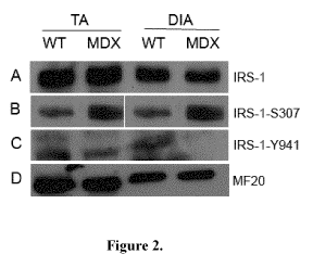
[0051] Figure 2 illustrates that IRS-1 Serine Phosphorylation is Increased in
Dystrophic Skeletal Muscle. Protein lysates were obtained from the tibialis
anterior (TA)
and diaphragm (DIA) muscles of 4, 8 and 10 wk old WT and MDX mice.
Immunoblotting
was performed using anti-IRS-1 (Row A), anti-IRS-1-5307 (Row B) and anti-IRS-
1Y941
(Row C) to compare expression levels. (Row D) MF-20 was used as a loading
control.
(n=3).
[0052] Figure 3 illustrates that normal AKT phosphorylation and GSK-3(3
activity are altered in Dystrophic Skeletal Muscle. A) AKT was
immunoprecipitated
from skeletal muscles (TA and DIA) wild-type (WT) and MDX mice. AKT kinase
assay
was preformed using AKT substrate peptide and [y-32P]ATP. Kinase reaction was
dotted
-13-
CA 02762351 2011-11-17
WO 2010/132982 PCT/CA2010/000734
on p81 paper and radioactivity was measured by scintillation counter (mean
+SE; n > 3
*p< 0.05). B) Active GSK-3R was immunoprecipitated and GSK-3R kinase assay was
preformed using GSK-3R peptide substrate and [y-32P]ATP. Kinase reaction was
dotted
on p81 paper and radioactivity was measured by scintillation counter (mean
+SE; n > 3
*p< 0.05).
[0053] Figure 4 illustrates that Metformin Treatment Restores Normal AKT
phosphorylation and GSK-3(3 inactivity in Dystrophic Skeletal Muscle. A)
Analysis of
AKT and GSK-3R kinase activity. AKT was immunoprecipitated from the skeletal
muscle
(TA and DIA) from MDX-saline (MS) and MDX-metformin treated) mice. AKT kinase
assay was preformed using AKT substrate peptide and [y-32P]ATP. Kinase
reaction was
dotted on p81 paper and radioactivity was measured by scintillation counter
(mean+SE; n
> 3 *p< 0.05). B) Active GSK-3R was immunoprecipitated and GSK-3R kinase assay
was
preformed using GSK-3R peptide substrate and [y-32P]ATP. Kinase reaction was
dotted
on p81 paper and radioactivity was measured by scintillation counter (mean
+SE; n > 3
*p< 0.05).
[0054] Figure 5 illustrates that Metformin Treatment Restores GLUT4
Localization in Dystrophic Skeletal Muscle. Immuno-histochemistry on paraffin
sections of the TA muscles of WT and MDX (saline and metformin treated) mice
to
analyze GLUT4 localization (green) using anti-GLUT4 antibody (1:200).
(Magnification,
20x).
[0055] Figure 6 illustrates that Metformin Treatment Restores Glycogen
Content in Dystrophic Skeletal Muscle. Alterations in glycogen storage were
analyzed
using periodic acid Schiff (PAS) to label glycogen pools on transverse
sections of the TA
from wild-type (WT) and saline and metformin treated MDX mice, MDX-S and MDX-M
respectively. (Magnification, 20x and 40x, n=3).
[0056] Figure 7 illustrates that Metformin Treatment Reduces the
Appearance of Focal Necrosis in Dystrophic Skeletal Muscle. Following 28 days
of
metformin administration, the indicated muscles (tibialis anterior [TA],
diaphragm [DIA],
gastrocnemius [GASTRO] and soleus) were removed, subject to paraffin fixation.
Cross-
sections from MDX-saline (MS) treated and MDX-metformin (MM) treated mice were
stained with H&E to visualize muscle morphology. (Magnification 20x, n=3).
[0057] Figure 8 illustrates that Metformin Treatment Leads to a Reduction in
the Number of Centrally Located Myofiber Nuclei in Dystrophic Skeletal Muscle.
Following 28 days of metformin administration, the indicated muscles (tibialis
anterior [TA]
and diaphragm [DIA]) were removed, subject to paraffin fixation. (A-B) cross-
sections
from TA and DIA in wild-type (WT), MDX-saline (MS) treated and MDX-metformin
(MM)
-14-
CA 02762351 2011-11-17
WO 2010/132982 PCT/CA2010/000734
treated mice were stained with H&E. (C) Quantification of myofibers with
centrally
located nuclei (Magnification 20x, n=3). (D-E) Following treatment, metformin
treated mdx
fibers exhibited a lower proportion of fibres with a smaller cross-sectional
area (CSA)
compared to saline-treated mdx mice.
[0058] Figure 9 illustrates that Metformin Partially Restores the DGC and
Improves Myofiber Fragility. Metformin administration increased protein levels
of
utrophin, R-dystroglycan, y-sarcoglycan and utrophin in the gastrocnemius
compared to
Saline-treated mdx controls (Fig. 9A). Metformin treatment led to a notable
increase in
sarcolemmal distribution of both R-dystroglycan and y-sarcoglycan along the
sarcolemma
in mdx-myofibers (Fig. 9B and C). Immunohistochemical analysis revealed that
metformin
administration led to a robust increase in utrophin along the extrasynaptic
sarcolemma
(Fig. 9D).
[0059] Figure 10 illustrates that Metformin Treatments decreases
sarcolemmal damage in dystrophic skeletal muscle. (A) Macroscopic evidence of
EBD infiltration in TA and gastrocnemius (G) muscles from mdx mice treated
with saline
or metformin for 28 days. (B) Uptake of EBD shown by red fluorescence on
transverse
sections of gastrocnemius muscle fibres. White arrows indicate muscle fibers
that have
taken up significant EBD, which fibres appear lighter shade of gray than
surrounding
fibers in figure. Sarcolemma is visible defining the perimeters of the fibers
(scale bar,
20um). (C) Quantification of EBD-positive fibers. Treated mdx-M (MM) fibers
exhibited
fewer EBD infiltrated fibers compared to mdx-S (MS). (n=6/group)
[0060] Figure 11 illustrates the Metformin treated mdx-mice display
improved running endurance compared to saline-treated mdx-mice. A)
Quantification of fall latency time is represented as the average of each
trial (1-4)
performed, where each of the 4 trials is performed each day for 3 consecutive
days. (n =
9 SEM).
DETAILED DESCRIPTION
[0061] Generally, the present disclosure provides therapeutic agents,
compositions and methods for treating muscle diseases or conditions
characterized by
metabolic disturbance in muscle tissue, in particular, impaired insulin-
dependent
signaling.
[0062] It is demonstrated herein that certain muscle diseases and conditions
are
characterized by metabolic disturbances in the muscle tissue itself. In
particular, it is
demonstrated that dystrophic muscle exhibits impaired insulin-dependent
signaling, in
essence, a form of insulin resistance. It is further demonstrated that
treating the
-15-
CA 02762351 2011-11-17
WO 2010/132982 PCT/CA2010/000734
underlying defect in insulin signaling results in a significant improvement in
disease
pathology, at both the molecular and behavioral levels. Thus, diseases or
conditions
characterized by this metabolic disturbance may be alleviated by administering
a
therapeutic agent for correcting the underlying defect in insulin-dependent
signaling. It is
believed that these findings represent a significant scientific advance in the
understanding
of muscle disease, including muscular dystrophy, and also represent a much-
needed
therapeutic advance in this field.
[0063] Various non-limiting aspects and embodiments are described herein. A
skilled person having regard to the present disclosure will appreciate that
the scope of the
invention is not limited to the exemplary aspects and embodiments disclosed
herein.
[0064] The term "impaired insulin-dependent signaling" refers generally to a
form
of insulin resistance wherein cells become less sensitive to the effects of
insulin. More
particularly, as used herein, "impaired insulin-dependent signaling" refers to
an
impairment that results in elevated phosphorylation of the IRS-1 at serine 307
leading to
inhibition in insulin signaling. The term "defective" insulin signaling is
also used herein.
[0065] In some embodiments, the muscle disease or condition characterized by
impaired insulin-dependent signaling is one or more of a muscle degenerative
disease, a
myopathy, or a disease or condition characterized by muscle wasting or
atrophy. A skilled
person will appreciate that other muscle diseases and conditions besides those
listed
above may be tested and found to be characterized by impaired insulin-
dependent
signaling. Such muscle diseases and conditions are considered within the scope
of the
present disclosure.
[0066] In some embodiments, the muscle disease or condition characterized by
impaired insulin-dependent signaling is a muscle degenerative disease,
including but not
limited to a muscular dystrophy. Muscular dystrophies include, but are not
limited to,
Duchenne muscular dystrophy (DMD), Becker muscular dystrophy, limb-girdle
muscular
dystrophies, myotonic dystrophy (also known as Steinert's disease),
facioscapulohumeral
muscular dystrophy, congenital muscular dystrophies, oculopharyngeal muscular
dystrophy, distal muscular dystrophies and Emery-Dreifuss muscular dystrophy.
See,
e.g., Hoffman et al., N. Engl. J. Med., 318.1363-1368 (1988); Bonnemann, C. G.
et al.,
Curr. Opin. Ped., 8: 569-582 (1996); Worton, R., Science, 270: 755-756 (1995);
Funakoshi, M. et al., Neuromuscul. Disord., 9 (2): 108-114 (1999); Lim, L. E.
and
Campbell, K. P., Cure. Opin. Neurol., 11 (5): 443-452 (1998); Voit, T., Brain
Dev., 20 (2):
65-74 (1998); Brown, R. H., Annu. Rev. Med., 48: 457-466 (1997); Fisher, J.
and
Upadhyaya, M., Neuromuscul. Disord., 7 (1): 55-62 (1997).
-16-
CA 02762351 2011-11-17
WO 2010/132982 PCT/CA2010/000734
[0067] In some embodiments, the muscular dystrophy is Duchenne muscular
dystrophy, Becker muscular dystrophy, a Limb-Girdle muscular dystrophy,
myotonic
dystrophy or a related dystrophy characterized by impaired insulin-dependent
signaling.
In some embodiments, the muscular dystrophy originates from disruptions in the
dystrophin- dystroglycan complex. In some embodiments, the muscular dystrophy
is
Duchenne muscular dystrophy (DMD). In some embodiments, the muscular dystrophy
is
Becker muscular dystrophy (BMD). In other embodiments, the muscular dystrophy
is a
Limb-Girdle muscular dystrophy. In other embodiments, the muscular dystrophy
is
myotonic dystrophy. In some embodiments, the muscular dystrophy is a
congenital
muscular dystrophy characterized by impaired insulin dependent signaling.
[0068] In some embodiments, the muscle disease or condition characterized by
impaired insulin-dependent signaling is a disease or condition characterized
by muscle
wasting or atrophy, for example, a disuse atrophy, for example, sarcopenia or
intensive
care atrophy.
[0069] In some embodiments, the disease or condition is a myopathy. In some
embodiments, the disease or condition is a critical illness myopathy, which
may, for
example, be brought about from bone marrow transplant, sepsis, multi-organ
failure, or
prolonged mechanical ventilation. This is referred to in the literature as
CINMA or critical
illness neuromuscular abnormalities and it affects approximately 50% of all
ICU patients.
[0070] In accordance with the present disclosure, there are contemplated
therapeutic agents, compositions and methods for treating and/or preventing a
disease or
condition characterized by impaired insulin dependent signaling. The
therapeutic agent
targets the underlying signaling defect.
[0071] A "therapeutic agent" is a molecule or group of molecules for eliciting
a
desired therapeutic effect and may include, for example, organic and inorganic
small
molecules, peptides and polypeptides, polymers, fusion proteins,
polynucleotides,
oligonucleotides, antibodies or antibody fragments, macromolecules,
encapsulated
molecules, among others. In accordance with the present disclosure, there are
described
therapeutic agents for treating or preventing a muscle condition or disease
characterized
by impaired insulin-dependent signaling.
[0072] In some embodiments, the therapeutic agent is an "activator of the
insulin
signaling pathway" that preferably exerts its effects, at least in part,
downstream of IRS-1.
When referring to activation of a signaling pathway, "activation" may occur
directly (e.g.
via activation, stimulation or up-regulation of an activating component of a
signaling
pathway) or may occur indirectly (e.g. via inhibition or down-regulation of an
inhibitory
-17-
CA 02762351 2011-11-17
WO 2010/132982 PCT/CA2010/000734
component of the pathway). The converse is also true where "inhibition" may
occur
directly or indirectly.
[0073] Activators of the insulin signaling pathway may act at a genetic level,
for
example, to upregulate or downregulate the expression of a gene of interest,
or they may
act at the protein level, for example, to increase or decrease the activity of
a polypeptide
of interest. Exemplary activators of the insulin signaling pathway may include
activators
or inhibitors of one or more downstream effector molecules in the insulin
signaling
pathway (e.g. IRS-1/AKT/GSK3). Activators of the insulin signaling pathway may
also
target molecules that affect the insulin signaling pathway, such as AMPK and
JNK1. For
example, in some embodiments, the therapeutic agent may be an AKT activator,
AMPK
activator, GSK3R inhibitor, or JNK1 inhibitor, among others.
[0074] In some embodiments, the activator of the insulin signaling pathway is
an
AMPK activator. Exemplary AMPK activators include A-769662 9 (a non-nucleoside
compound from the thienopyridone family), GW-501516 (which activates PPAR-
gamma
and AMPK), and AICAR (aminoimidazole carboxamide ribonucleotide). As a
combination
therapy, GW-501516 has been shown to act synergistically with AICAR. The
thiazolidinedione (activators of PPAR-gamma) class of drugs may also be
considered
AMPK activators.
[0075] In some embodiments, the activator of the insulin signaling pathway is
a
JNK inhibitor. Exemplary JNK inhibitors include, for example, SP600125 and BI-
78D3. BI-
78D3 was recently demonstrated to alleviate insulin resistance in a murine
model of type-
II diabetes (Stebbins et al, 2008) and it therefore predicted to have
beneficial effects in
accordance with the present disclosure.
[0076] In some embodiments, the therapeutic agent which activates the insulin
signaling pathway is an organic or inorganic small molecule. "Small molecule",
as used
herein, generally means a low molecular weight (e.g. less than 1000Da, often
less than
800Da, often than 500Da) organic compound. In some cases, a subunit of a
polymer, or a
small peptide, can be considered within the definition of a small molecule.
[0077] In some embodiments, the therapeutic agent is a biguanide or an
analogue or derivative thereof. Biguanides include, for example, metformin,
phenformin,
buformin, and proguanil. The biguanide or biguanide derivative selected should
be
capable of treating impaired insulin-dependent signaling in a muscle condition
or disease
characterized thereby, for example, by activating the insulin signaling
pathway whether
directly or indirectly.
[0078] In some embodiments, the biguanide is metformin (N,N-
dimethylimidodicarbonimidicdiamide) or an analogue or derivative thereof.
-18-
CA 02762351 2011-11-17
WO 2010/132982 PCT/CA2010/000734
[0079] Metformin has the following structural formula (Formula I):
NH NH
N N NH2
[0080] Metformin has been widely prescribed for over 50 years to treat insulin
resistance in diabetic patients. Thus, in one aspect, the present disclosure
provides for
the re-purposing of a safe and well-established anti-diabetic drug to provide
a new and
effective treatment for a muscle disease or condition characterized by
impaired insulin-
dependent signaling.
[0081] Metformin has a low toxicity profile. The most serious complication
associated with metformin is lactic acidosis, which has an incidence of about
0.03 cases
per 1000 patients years of treatment and a mortality risk of about 0.015 per
1000 patient-
years. Most cases occur in patients with impaired renal function (e.g. serum
creatinine
level >130 pmol/L or >1.5 g/L). Other major contraindications include
congestive heart
failure, hypoxic states and advanced liver disease. Serious adverse events
with
metformin are predictable rather than spontaneous and are potentially
preventable if the
prescribing guidelines are respected. Gastrointestinal adverse effects,
notably diarrhea,
occur in less than 20% of patients and remit when the dosage is reduced. The
life-
threatening risks associated with metformin are rare and could mostly be
avoided by strict
adherence to the prescribing guidelines. Given the 5 decades of clinical
experience with
metformin, its antihyperglycaemic efficacy, and benefits against Syndrome X,
metformin
offers a very favorable risk-benefit assessment when compared with the chronic
morbidity
and premature mortality among patients with type 2 diabetes mellitus.
Metformin is
commercially available from a variety of sources.
[0082] In some embodiments, the therapeutic agent is a derivative of a
biguanide.
As used herein, "derivative" includes, but is not limited to, prodrug forms,
pegylated
forms, etc.
[0083] The prodrug approach is a chemical approach using reversible
derivatives,
which can be useful in optimizing the clinical application of a therapeutic
agent. Prodrugs
have been designed and developed, for example, to overcome pharmaceutical and
pharmacokinetic barriers in clinical drug application, such as low oral drug
absorption,
lack of site specificity, chemical instability, toxicity, and poor patient
acceptance (Han,
-19-
CA 02762351 2011-11-17
WO 2010/132982 PCT/CA2010/000734
2000). As used herein, "prodrug" generally refers to a compound that, upon in
vivo
administration, is metabolized or otherwise converted to the biologically,
pharmaceutically
or therapeutically active form of the compound. To produce a prodrug,
generally the
pharmaceutically active compound is modified such that the active compound is
regenerated, enzymatically or nonenzymatically, to exert a therapeutic effect.
The
prodrug may be designed to alter the metabolic stability or the transport
characteristics of
a drug, to mask side effects or toxicity, to improve the flavor of a drug or
to alter other
characteristics or properties of a drug. By virtue of knowledge of
pharmacodynamic
processes and drug metabolism in vivo, those of skill in this art, once a
pharmaceutically
active compound is known, can design prodrugs of the compound (see, e.g.,
Nogrady1985, Stella et al. 1985, Banerjee et al. 1985).
[0084] In some embodiments, the therapeutic agent is a prodrug of a biguanide.
[0085] In some embodiments, the biguanide prodrug is a compound of Formula II:
NH NH2
R1 1~ NAN~N"XIR3
1 1
R2 H
Formula II
wherein:
R1 and R2 are independently selected from the group consisting of H, alkyl,
alkoxy,
haloalkyl, hydroxyalkyl, cyanoalkyl, alkenyl, alkynyl, cycloalkyl,
cycloalkenyl,
heterocycloalkyl, heterocycloalkenyl, aryl, heteroaryl, alkylaryl,
alkylheteroaryl, alkylene-
O-alkyl, alkylene-O-cycloalkyl, alkylene-O-heterocycloalkyl, alkylene-O-
alkylene-
cycloalkyl, alkylene-O-alkylene-heterocycloalkyl, C(O)-alkyl, C(OO)-alkyl,
C(O)-cycloalkyl,
C(OO)-cycloalkyl, C(O)-heterocycloalkyl, S(O)2-heterocycloalkyl, alkylene-O-
aryl,
alkylene-O-heteroaryl, alkylene-O-alkylene-aryl, alkylene-O-alkylene-
heteroaryl,
C(O)alkyl, OC(O)alkyl, C(O)Oalkyl, C(O)N(H)alkyl, C(O)N(alkyl)alkyl,
S(O)2N(H)alkyl or
S(O)2N(alkyl)alkyl;
R3 is selected from the group consisting of Cj- to C8-lower alkyl, Cj- to C8-
lower alkoxy,
Cj- to C8-lower alkyl-ester, cycloalkyl, heterocycloalkyl, bicycloalkyl,
heterobicycloalkyl,
aryl, heteroaryl, optionally-substituted aryl, optionally-substituted hetero-
aryl;
-20-
CA 02762351 2011-11-17
WO 2010/132982 PCT/CA2010/000734
hydroxyalkyl, hydroxycycloalkyl, hydroxy-heterocycloalkyl, cyanoalkyl,
alkenyl, alkynyl,
cycloalkyl, cycloalkenyl, heterocycloalkyl, heterocycloalkenyl, alkylaryl,
alkylheteroaryl,
alkylene-O-alkyl, alkylene-O-cycloalkyl, alkylene-O-heterocycloalkyl, alkylene-
O-alkylene-
cycloalkyl, alkylene-O-alkylene-heterocycloalkyl;
X is selected from the group consisting of lower-alkyl, 0, C(O), C(O)2, C(O)N,
S, S(O),
S(O)2 and P(O)3;
and/or a pharmaceutically-acceptable salt, hydrate, solvate, isoform,
tautomer, optical
isomer, or combination thereof;
[0086] In some embodiments, the compound of Formula II comprises a
compound of any one of Formula IIA, IIB, IIC, IID or HE
NH NH2
NH NH 0
R1, N N N R4 R1 ,NAN~N OR5
R2 H H NH R2 H H
I
PG
Formula IIA Formula IIB
NH NH2 NH NH2 0 R1, N N N~O"~O R6 R1 ,
N'J~ N~N11P Om
I I I Y I I 1 O
R2 H H O R2 H H R7
Formula IIC Formula IID
NH NH2
R1 N N5~ NR8
I I I
R2 H H
Formula IIE
Wherein:
-21-
CA 02762351 2011-11-17
WO 2010/132982 PCT/CA2010/000734
R1 and R2 are independently selected from the group consisting of H, alkyl,
alkoxy,
haloalkyl, hydroxyalkyl, cyanoalkyl, alkenyl, alkynyl, cycloalkyl,
cycloalkenyl,
heterocycloalkyl, heterocycloalkenyl, aryl, heteroaryl, alkylaryl,
alkylheteroaryl, alkylene-
O-alkyl, alkylene-O-cycloalkyl, alkylene-O-heterocycloalkyl, alkylene-O-
alkylene-
cycloalkyl, alkylene-O-alkylene-heterocycloalkyl, C(O)-alkyl, C(OO)-alkyl,
C(O)-cycloalkyl,
C(OO)-cycloalkyl, C(O)-heterocycloalkyl, S(O)2-heterocycloalkyl, alkylene-O-
aryl,
alkylene-O-heteroaryl, alkylene-O-alkylene-aryl, alkylene-O-alkylene-
heteroaryl,
C(O)alkyl, OC(O)alkyl, C(O)Oalkyl, C(O)N(H)alkyl, C(O)N(alkyl)alkyl,
S(O)2N(H)alkyl or
S(O)2N(alkyl)alkyl;
R4 is selected from the group consisting of H, hydroxy, halogen, cyano, nitro,
carboxylic
ester, carboxylic acid, carboxylic amide, Cj- to C8-lower alkyl, Cj- to C8-
lower alkoxy, Cj-
to C8-lower alkyl-ester, cycloalkyl, heterocycloalkyl, bicycloalkyl,
heterobicycloalkyl, aryl,
heteroaryl, optionally-substituted aryl, optionally-substituted hetero-aryl;
hydroxyalkyl,
hydroxycycloalkyl, hydroxy-heterocycloalkyl, cyanoalkyl, alkenyl, alkynyl,
cycloalkyl,
cycloalkenyl, heterocycloalkyl, heterocycloalkenyl, alkylaryl,
alkylheteroaryl, alkylene-O-
alkyl, alkylene-O-cycloalkyl, alkylene-O-heterocycloalkyl, alkylene-O-alkylene-
cycloalkyl,
alkylene-O-alkylene-heterocycloalkyl;
R5 to R8 are independently selected from the group consisting of Cj- to C8-
lower alkyl,
Cj- to C8-lower alkoxy, Cj- to C8-lower alkyl-ester, halo-alkyl-ester
cycloalkyl,
heterocycloalkyl, bicycloalkyl, heterobicycloalkyl, aryl, heteroaryl,
optionally-substituted
aryl, optionally-substituted hetero-aryl; hydroxyalkyl, hydroxycycloalkyl,
hydroxy-
heterocycloalkyl, cyanoalkyl, alkenyl, alkynyl, cycloalkyl, cycloalkenyl,
heterocycloalkyl,
heterocycloalkenyl, alkylaryl, alkylheteroaryl, alkylene-O-alkyl, alkylene-O-
cycloalkyl,
alkylene-O-heterocycloalkyl, alkylene-O-alkylene-cycloalkyl, alkylene-O-
alkylene-
heterocycloalkyl;
[0087] In some embodiments, n is selected from 1 to 4 and m from 0 to 2;
[0088] In some embodiments of the compounds of Formula II, R1 and R2 are
independently selected from the group consisting of alkyl, cycloalkyl and
heterocycloalkyl;
[0089] In yet other embodiments of the compound of Formula II, R5, R6, R7 and
R8 are independently selected from the group consisting of Cj- to C8-lower
alkyl,
cycloalkyl, heterocycloalkyl, aryl and heteroaryl; and/or a pharmaceutically-
acceptable
salt, hydrate, solvate, isoform, tautomer, optical isomer, or combination
thereof;
-22-
CA 02762351 2011-11-17
WO 2010/132982 PCT/CA2010/000734
[0090] Some exemplary prodrugs include the following compounds, their
pharmaceutically acceptable salts, hydrates, solvates, optical isomers, and
combinations
thereof:
= N',N'-Dimethyl-S-cyclohexyl-N4-thiohydroxylbiguanidine;
= N',N'-Dimethyl-S-phenyl-N4-thiohydroxylbiguanidine;
= tent-Butyl 4-[(3-(N,N-Dimethylcarbamimidoyl)guanidino)methyl]phenyl -
carbamate;
= 4-{[3-(N,N-Dimethylcarbamimidoyl)guanidino]methyl}phenyl -octanoate;
= 4-{[3-(N,N-Dimethylcarbamimidoyl)guanidino]methyl}phenyl-diethylcarbamate;
= 4-[(3-(N,N-Dimethylcarbamimidoyl)guanidino)methyl]-3-hydroxyphenyl-pivalate;
= 3-[3-(N,N-Dimethylcarbamimidoyl)guanidino]propyl-acetate;
= [(N',N'-Dimethylguanidino)iminomethyl]carbamic acid benzyl-ester;
= [(N',N'-Dimethylguanidino)iminomethyl]carbamic acid 2,2,2-Trichloroethyl -
ster;
= [(N1,N1-Dimethylcarbamimidoyl)guanidino]-4-phenyl-1,3,2-
dioxaphosphoramidate;
and/or a pharmaceutically-acceptable salt, hydrate, solvate, isoform,
tautomer, optical
isomer, or combination thereof.
[0091] Acid addition salts of the compounds of Formula II are most suitably
formed from pharmaceutically acceptable acids, and include for example those
formed
with inorganic acids e.g. hydrochloric, sulphuric or phosphoric acids and
organic acids
e.g. succinic, maleic, acetic or fumaric acid. Other non-pharmaceutically
acceptable salts
e.g. oxalates may be used for example in the isolation of compounds of Formula
II for
laboratory use, or for subsequent conversion to a pharmaceutically acceptable
acid
addition salt. Also included within the scope are base addition salts (such as
sodium,
potassium and ammonium salts), solvates and hydrates of compounds disclosed
herein.
The conversion of a given compound salt to a desired compound salt is achieved
by
applying standard techniques, well known to one skilled in the art.
[0092] The conversion of a given compound salt to a desired compound salt is
achieved by applying standard techniques, well known to one skilled in the
art.
[0093] Several methods for preparing compounds of Formula II are illustrated
in
the following Schemes and in the Examples section. Starting materials and the
requisite
intermediates are in some cases commercially available or can be prepared
according to
literature procedures (Huttunen, 2009, Huttunen 2008) or as illustrated
herein.
-23-
CA 02762351 2011-11-17
WO 2010/132982 PCT/CA2010/000734
[0094] Certain prodrug compounds of Formula IIA, wherein R1 and R2 are
independently selected from alkyl group, and X-R3 is either substituted
protected p-amino
or p-hydroxybenzylic groups, can be prepared in accordance with Scheme 1.
NH NH2
R1 \NAN5~ N R4
I I I
R2 H H NH
R1, NH NH2 (B) PG
N~N~NH2
I I
R2 H
(ii)
(A) NH NH2
R1, NAN~N I I I
R2 H H ajR4
O
1
(C) PG
(i) p-PG-Amino-Benzyl halide, DMF or MeCN/0 C to RT
(ii) p-PG-Hydroxy-Benzyl halide, DMF or MeCN/0 C to RT
Scheme 1
[0095] Condensation of N, N-dialkyl-metformin A with an appropriately
substituted
benzyl-halide (e.g. tent-butyl 4-(Chloro or Bromomethyl)phenylcarbamate and 4-
(Chloro
or Bromomethyl)phenyl alcanoate) in polar solvent such as DMF or MeCN at 0 C
under
argon led, after purification by flash chromatography to the targeted prodrug
compounds
B and C respectively.
[0096] In yet another embodiment, there is provided a method of preparing a
compound of Formula IIB, wherein R1 and R2 are independently selected from
alkyl
group, and X-R3 is a carbamic ester group, can be generally prepared in
accordance with
Scheme 2.
NH NH NH NH2 0
R 1 , N~NH (I) R1 ,NAN5~ N'J~ 0 R5
I I 2 I I I
R2 H R2 H H
(A) (D)
-24-
CA 02762351 2011-11-17
WO 2010/132982 PCT/CA2010/000734
(i) R5- chloroformate, DMF or MeCN/0 C to RT
Scheme 2
[0097] Condensation of N, N-dialkyl-metformin derivative A with an appropriate
chloroformate (e.g. Benzyl chloroformate and 2,2,2-Trichloroethyl
chloroformate) in a
polar solvent, such as MeCN at 0 C under argon led, after purification by
flash
chromatography to the targeted prodrug compounds D.
[0098] In yet a further aspect, the biguanide prodrug compounds, described by
Formula IIC, wherein R1 and R2 are independently selected from alkyl group,
and X-R3
is an alkyl-ester, can be generally prepared in accordance with Scheme 3.
R1, NH NH2 0) R1, NH NH2 '-0""0 R6
N N NH2 N N N
R2 H R2 H H O
(A) (E)
(i) haloalkyl-ester, Acetone/Reflux
Scheme 3
[0099] Condensation of an appropriate haloalkyl-ester (e.g. 3-chloropropyl
acetate) with N, N-dialkyl-metformin derivative A in anhydrous acetone under
reflux led,
after purification by flash chromatography, to the targeted biguanide prodrug
compounds
E.
[00100] In yet another aspect, certain biguanide prodrug compounds of Formula
IID, wherein R1 and R2 are independently selected from alkyl group, and X-R3
is a
substituted cyclic phosphate, can be prepared in accordance with Scheme 4.
NH NH2 NH NH2 O O
R1, 0) JL~
N N NH2 R1,
N N N O~0õ
R2 H R2 H H R7
(A) (F)
(i) substituted cyclic phosphoryl-chloride, 1-methylimidazole in MeCN/0 C to
RT
-25-
CA 02762351 2011-11-17
WO 2010/132982 PCT/CA2010/000734
Scheme 4
[00101] Condensation of N, N-dialkyl-metformin derivative A with an
appropriate
substituted cyclic phosphoryl chloride (e.g. 2-Chloro-4-phenyl-[1,3,2]dioxa-
phosphinane
2-oxide) and 1-methylimidazole in MeCN at 0 C under argon to provide, after
stirring
overnight at room temperature and purification by flash chromatography,
biguanide
prodrug cyclic phosphates F.
[00102] In yet another aspect, biguanide prodrug compounds of Formula IE,
wherein R1 and R2 are independently selected from alkyl group, X is Sulfur and
-R3 is
alkyl, cycloalkyl, heterocycloalkyl, aryl, heteroaryl, can generally be
prepared in
accordance with Scheme 5.
NH NH2 0) NH NH2
R1 ,NAN~NH2 R 1 ,
R7
1 1 1 1 1
R2 H R2 H H
(A) (G)
(i) 2-(R7-thio)isoindoline-1,3-dione, DMF, RT/ON
Scheme 5
[00103] Condensation of N, N-dialkyl-metformin derivative A with an
appropriate
substituted 2-(thio)-isoindoline-1,3-dione derivative in anhydrous DMF under
argon to
provide, after stirring 24 hours at room temperature and purification by flash
chromatography, thioxy-biguanide prodrug compounds G;
[00104] In some embodiments, the compound of Formula II is pharmaceutically-
acceptable salt, optical isomer, or combination thereof.
[00105] In some embodiments, the pharmaceutically-acceptable salt comprises an
acid addition salt or a basic addition salt.
[00106] In some embodiments, the prodrug compounds may have one or more
asymmetric centres and it is intended that any optical isomers, as separated,
pure or
partially purified optical isomers or racemic mixtures thereof are included
within the scope
of the present application.
[00107] In some embodiments, some of the compounds disclosed herein may exist
in different tautomeric forms and it is intended that any tautomeric forms
which the
compounds are able to form are included within the scope of the present
application.
-26-
CA 02762351 2011-11-17
WO 2010/132982 PCT/CA2010/000734
[00108] In some embodiments, the acid addition salt is formed from
hydrochloric
acid, hydrobromic acid, sulfuric acid, phosphoric acid, acid metal salt,
monocarboxylic
acids, dicarboxylic acids, or tricarboxylic acids.
[00109] Unless specified otherwise, the chemical nomenclature used herein
generally follows the examples and rules stated in Nomenclature of Organic
Chemistry,
Sections A, B, C, D, E, F, and H, Pergamon Press, Oxford, 1979, which is
incorporated
by reference herein for its exemplary chemical structure names and rules on
naming
chemical structures. Optionally, a name of a compound may be generated using a
chemical naming program, e.g. ACD/ChemSketch, Version 5.09/September 2001,
Advanced Chemistry Development, Inc., Toronto, Canada.
[00110] The compounds disclosed herein may have asymmetric centers, chiral
axes, and chiral planes (e.g., as described in: E. L. Eliel and S. H. Wilen,
Stereo-
chemistry of Carbon Compounds, John Wiley & Sons, New York, 1994, pages 1119-
1190), and occur as racemates, racemic mixtures, and as individual
diastereomers, with
all possible isomers and mixtures thereof, including optical isomers, being
included in the
present disclosure.
[00111] Generally, reference to a certain element such as hydrogen or H is
meant
to, if appropriate, include all isotopes of that element.
[00112] The following terms are meant to encompass unsubstituted and/or
substituted.
[00113] The term "alkyl" as used herein means a straight- or branched-chain
hydrocarbon radical; in one aspect, having from one to eight carbon atoms, and
includes,
for example, and without being limited thereto, methyl, ethyl, propyl,
isopropyl, t-butyl and
the like. As noted above, "alkyl" encompasses substituted alkyl. Substituted
alkyl
includes, for example, and without being limited thereto, haloalkyl,
hydroxyalkyl,
cyanoalkyl, and the like. This is applied to any of the groups mentioned
herein. Groups
such as "alkenyl", "alkynyl", "aryl", etc. encompass substituted "alkenyl",
"alkynyl", "aryl",
etc.
[00114] The term "alkenyl" as used herein means a straight- or branched-chain
alkenyl radical; in one aspect, having from two to eight carbon atoms, and
includes, for
example, and without being limited thereto, ethenyl, 1-propenyl, 1-butenyl and
the like.
The term "alkenyl" encompass radicals having "cis" and "trans" orientations,
or
alternatively,"E" and "Z" orientations.
[00115] The term "alkynyl" as used herein means a straight- or branched-chain
alkynyl radical; in one aspect, having from two to eight carbon atoms, and
includes, for
example, and without being limited thereto, 1-propynyl (propargyl), 1-butynyl
and the like.
-27-
CA 02762351 2011-11-17
WO 2010/132982 PCT/CA2010/000734
[00116] The term "cycloalkyl" as used herein means a carbocyclic system (which
may be unsaturated) containing one or more rings wherein such rings may be
attached
together in a pendent manner or may be fused. In one aspect, the ring(s) may
have from
three to seven carbon atoms, and includes, for example, and without being
limited
thereto, cyclopropyl, cyclohexyl, cyclohexenyl and the like.
[00117] The term "heterocycloalkyl" as used herein means a heterocyclic system
(which may be unsaturated) having at least one heteroatom selected from N, S
and/or 0
and containing one or more rings wherein such rings may be attached together
in a
pendent manner or may be fused. In one aspect, the ring(s) may have a three-
to seven-
membered cyclic group and includes, for example, and without being limited
thereto,
piperidinyl, piperazinyl, pyrrolidinyl, tetrahydrofuranyl and the like.
[00118] The term "alkoxy" as used herein means a straight- or branched-chain
alkoxy radical; in one aspect, having from one to eight carbon atoms and
includes, for
example, and without being limited thereto, methoxy, ethoxy, propyloxy,
isopropyloxy, t-
butoxy and the like.
[00119] The term "halo" as used herein means halogen and includes, for
example,
and without being limited thereto, fluoro, chloro, bromo, iodo and the like,
in both
radioactive and non-radioactive forms.
[00120] The term "alkylene" as used herein means a difunctional branched or
unbranched saturated hydrocarbon radical; in one aspect, having one to eight
carbon
atoms, and includes, for example, and without being limited thereto,
methylene, ethylene,
n-propylene, n-butylene and the like.
[00121] The term "alkenylene" as used herein means a difunctional branched or
unbranched hydrocarbon radical; in one aspect, having two to eight carbon
atoms, and
having at least one double bond, and includes, for example, and without being
limited
thereto, ethenylene, n-propenylene, n-butenylene and the like.
[00122] The term "alkynylene" as used herein means a difunctional branched or
unbranched hydrocarbon radical; in one aspect, having two to eight carbon
atoms, and
having at least one triple bond, and includes, for example, and without being
limited
thereto, ethynylene, n-propynylene, n-butynylene and the like.
[00123] The term "aryl", alone or in combination, as used herein means a
carbocyclic aromatic system containing one or more rings wherein such rings
may be
attached together in a pendent manner or may be fused. In particular
embodiments, aryl
is one, two or three rings. In one aspect, the aryl has five to twelve ring
atoms. The term
"aryl" encompasses aromatic radicals such as phenyl, naphthyl,
tetrahydronaphthyl,
indanyl, biphenyl, phenanthryl, anthryl or acenaphthyl. The "aryl" group may
have 1 to 4
-28-
CA 02762351 2011-11-17
WO 2010/132982 PCT/CA2010/000734
substituents such as lower alkyl, hydroxyl, halo, haloalkyl, nitro, cyano,
alkoxy, lower
alkylamino and the like.
[00124] The term "heteroaryl", alone or in combination, as used herein means
an
aromatic system having at least one heteroatom selected from N, S and/or 0 and
containing one or more rings wherein such rings may be attached together in a
pendent
manner or may be fused. In particular embodiments, heteroaryl is one, two or
three rings.
In one aspect, the heteroaryl has five to twelve ring atoms. The term
"heteroaryl"
encompasses heteroaromatic radicals such as pyridyl, indolyl, furyl,
benzofuryl, thienyl,
benzothienyl, quinolyl, oxazolyl and the like. The "heteroaryl" group may have
1 to 4
substituents such as lower alkyl, hydroxyl, halo, haloalkyl, nitro, cyano,
alkoxy, lower
alkylamino and the like.
[00125] It is understood that substituents and substitution patterns on the
compounds disclosed herein may be selected by one of ordinary skill in the art
to provide
compounds that are chemically stable and that can be readily synthesized by
techniques
known in the art, as well as those methods set forth below. If a substituent
is itself
substituted with more than one group, it is understood that these multiple
groups may be
on the same carbon or on different carbons, as long as a stable structure
results.
[00126] The term "pharmaceutically acceptable salt" means either an acid
addition
salt or a basic addition salt which is compatible with the treatment of
patients.
[00127] A "pharmaceutically acceptable acid addition salt" is any non-toxic
organic
or inorganic acid addition salt of the base compounds represented by Formula
II or any of
its intermediates. Illustrative inorganic acids which form suitable salts
include, but are not
limited thereto, hydrochloric, hydrobromic, sulfuric and phosphoric acid and
acid metal
salts such as sodium monohydrogen orthophosphate and potassium hydrogen
sulfate.
Illustrative organic acids which form suitable salts include the mono-, di-
and tricarboxylic
acids. Illustrative of such acids are, for example, acetic, glycolic, lactic,
pyruvic, malonic,
succinic, glutaric, fumaric, malic, tartaric, citric, ascorbic, maleic,
hydroxymaleic, benzoic,
hydroxybenzoic, phenylacetic, cinnamic, salicylic, 2-phenoxybenzoic, p-
toluenesulfonic
acid and other sulfonic acids such as methanesulfonic acid and 2-
hydroxyethanesulfonic
acid. Either the mono- or di-acid salts can be formed, and such salts can
exist in either a
hydrated, solvated or substantially anhydrous form. In general, the acid
addition salts of
these compounds are more soluble in water and various hydrophilic organic
solvents, and
generally demonstrate higher melting points in comparison to their free base
forms. The
selection criteria for the appropriate salt will be known to one skilled in
the art. Other non-
pharmaceutically acceptable salts e.g. oxalates may be used for example in the
isolation of
-29-
CA 02762351 2011-11-17
WO 2010/132982 PCT/CA2010/000734
compounds of Formula II for laboratory use, or for subsequent conversion to a
pharmaceutically acceptable acid addition salt.
[00128] A "pharmaceutically acceptable basic addition salt" is any non-toxic
organic or inorganic base addition salt of the acid compounds represented by
Formula II
or any of its intermediates. Illustrative inorganic bases which form suitable
salts include,
but are not limited thereto, lithium, sodium, potassium, calcium, magnesium or
barium
hydroxides. Illustrative organic bases which form suitable salts include
aliphatic, alicyclic or
aromatic organic amines such as methylamine, trimethyl amine and picoline or
ammonia.
The selection of the appropriate salt may be important so that an ester
functionality, if any,
elsewhere in the molecule is not hydrolyzed. The selection criteria for the
appropriate salt
will be known to one skilled in the art.
[00129] "Solvate" generally means a compound of Formula II or the
pharmaceutically acceptable salt of a compound of Formula II wherein molecules
of a
suitable solvent are incorporated in a crystal lattice. A suitable solvent is
physiologically
tolerable at the dosage administered as the solvate. Examples of suitable
solvents, but
are not limited thereto, are ethanol, water and the like. When water is the
solvent, the
molecule is referred to as a hydrate.
[00130] The term "stereoisomers" is a general term for all isomers of the
individual
molecules that differ only in the orientation of their atoms in space. It
includes mirror
image isomers (enantiomers), geometric (cis/trans) isomers and isomers of
compounds
with more than one chiral centre that are not mirror images of one another
(diastereomers).
[00131] In some embodiments, the therapeutic agent may be a peptide or
polypeptide (or an active derivative, fragment or variant thereof) that
activates the insulin
signaling pathway in muscle tissue characterized by defective insulin
signaling. For
example, the peptide or polypeptide may activate or upregulate AMPK or AKT, or
may
inhibit JNK.
[00132] The terms "polypeptide" and "peptide, " as used herein, refer to a
sequence of amino acid residues linked together by peptide bonds or modified
peptide
bonds. Typically, a polypeptide is at least six amino acids long and a peptide
is at least 3
amino acids long. The polypeptide or peptide can be naturally occurring,
recombinant,
synthetic, or a combination of these. The polypeptide or peptide can be a
fragment of a
naturally occurring protein or polypeptide. The terms polypeptide and peptide
also
encompass peptide derivatives, peptide analogues, and peptidomimetic
compounds.
Such compounds are well known in the art.
-30-
CA 02762351 2011-11-17
WO 2010/132982 PCT/CA2010/000734
[00133] A "peptide derivative" is a peptide containing additional chemical or
biochemical moieties not normally a part of a naturally occurring peptide.
Peptide
derivatives include peptides in which one or more amino acid side chain and/or
the
amino-terminus and/or the carboxy-terminus has been derivatized with a
suitable
chemical substituent group, as well as cyclic peptides, dual peptides,
multimers of the
peptides, peptides fused to other proteins or carriers glycosylated peptides,
phosphorylated peptides, peptides conjugated to lipophilic moieties and
peptides
conjugated to an antibody or other biological ligand.
[00134] A "peptide analogue" is a peptide comprising one or more non-naturally
occurring amino acids.
[00135] Peptidomimetics are compounds that are structurally similar to
peptides
and contain chemical moieties that mimic the function of the polypeptide or
peptide of the
present disclosure.
[00136] One skilled in the art will appreciate that not all amino acids in a
peptide or
polypeptide need be modified. Similarly not all amino acids need be modified
in the same
way. Polypeptide/peptide derivatives, analogues and peptidomimetics of the
present
disclosure thus include chimeric molecules that contain two or more chemically
distinct
regions, each region comprising at least one amino acid or modified version
thereof.
[00137] A variant polypeptide or peptide is one in which one or more amino
acid
residues have been deleted, added or substituted for those that appear in the
amino acid
sequence of the naturally occurring protein. In the context of the present
disclosure, a
variant also retains substantially the same activity as the naturally
occurring protein.
Typically, when a variant contains one or more amino acid substitutions, they
are
"conservative" substitutions. A conservative substitution involves the
replacement of one
amino acid residue by another residue having similar side chain properties.
[00138] In accordance with the present disclosure, a polypeptide or peptide
analogue, derivative, variant or active fragment has substantially the same or
increased
activity as compared to a naturally occurring target protein. The term
"substantially
identical activity" indicates an activity that is at least about 50%, more
typically at least
about 60%, 75%, 80%, 90% or 99% of the activity of a naturally occurring
protein. In still
another embodiment, the analogue, derivative, variant or active fragment
exhibits
enhanced (increased) activity compared to a naturally occurring protein,
preferably a
human protein.
[00139] As used herein, the term "about" refers to a +/-5% variation from the
nominal value.
-31-
CA 02762351 2011-11-17
WO 2010/132982 PCT/CA2010/000734
[00140] The polypeptides can be prepared by methods known in the art, such as
purification from cell extracts or the use of recombinant techniques.
[00141] Shorter sequences can also be chemically synthesized by methods known
in the art including, but not limited to, exclusive solid phase synthesis,
partial solid phase
synthesis, fragment condensation or classical solution synthesis (Merrifeld
(1963) Am.
Chem. Soc. 85:2149; Merrifeld (1986) Science 232:341). The polypeptides for
use in
accordance with the present disclosure can be purified using standard
techniques such
as chromatography (e.g. ion exchange, affinity, and sizing column
chromatography or
high performance liquid chromatography), centrifugation, differential
solubility, or by other
techniques familiar to a worker skilled in the art.
[00142] The polypeptides can also be produced by recombinant techniques.
Typically this involves transformation (including transfection, transduction,
or infection) of
a suitable host cell with an expression vector comprising a polynucleotide
encoding the
protein or polypeptide of interest. For example, the nucleic acid sequences
for human
JNK1 and AMPK genes and various other components involved in the insulin
signaling
pathway are known in the art. These may be used as a basis for making the
polynucleotides.
[00143] The polynucleotides can be derived or purified from a suitable source
by
standard techniques. The polynucleotides can be genomic DNA or RNA or they can
be
cDNA prepared from isolated mRNA. Alternatively, the known sequences may be
used to
prepare probes to obtain other nucleic acid sequences encoding the polypeptide
from
various sources using standard techniques.
[00144] Polynucleotides encoding fragments or variants of the naturally
occurring
proteins of interest can be constructed by deletion, addition, and/or
substitution of one or
more nucleotides within the coding sequence using standard techniques, such as
site-
directed mutagenesis techniques.
[00145] The polypeptides and peptides can also be produced as fusion proteins.
One use of such fusion proteins is to improve the purification or detection of
the
polypeptide or peptide.
[00146] Specific initiation signals may be required for efficient translation
of cloned
polynucleotide. These signals include the ATG initiation codon and adjacent
sequences.
In cases where an entire wild-type gene or cDNA, including its own initiation
codon and
adjacent sequences, is inserted into the appropriate expression vector,
additional
translational control signals may not be needed. In other cases, exogenous
translational
control signals, including, perhaps, the ATG initiation codon, must be
provided.
-32-
CA 02762351 2011-11-17
WO 2010/132982 PCT/CA2010/000734
[00147] Furthermore, the initiation codon must be in phase with the reading
frame
of the desired coding sequence to ensure translation of the entire insert. The
exogenous
translational control signals and initiation codons can be natural or
synthetic. The
efficiency of expression may be enhanced by the inclusion of appropriate
transcription
enhancer elements and/or transcription terminators (Bittner et al. (1987)
Methods in
Enzymol. 153, 516).
[00148] Signal sequences may also be incorporated into the molecules for
targeting of the expressed polypeptides or peptides.
[00149] Suitable expression vectors for use with the nucleic acid sequences
include, but are not limited to, plasmids, phagemids, viral particles and
vectors, phage
and the like. For insect cells, baculovirus expression vectors are suitable.
For plant cells
viral expression vectors (such as cauliflower mosaic virus and tobacco mosaic
virus) and
plasmid expression vectors (such as the Ti plasmid) are suitable. The entire
expression
vector, or a part thereof, can be integrated into the host cell genome. In
some
circumstances, it is desirable to employ an inducible expression vector as
known in the
art. In some circumstances, it is desirable to employ an inducible expression
vector, e.g.
the LACSWITCH Inducible Expression System (Stratagene, LaJolla, CA).
[00150] Those skilled in the field of molecular biology will understand that a
wide
variety of expression systems can be used to provide the recombinant
polypeptide or
peptide. The precise host cell used is not critical. The polypeptide or
peptide can be
produced in a prokaryotic host (e.g., E. cold or B. subtilis) or in a
eukaryotic host (e.g.,
Saccharomyces or Pichia; mammalian cells, such as COS, NIH 3T3, CHO, BHK, 293,
or
HeLa cells, insect cells, or plant cells). The methods of transformation or
transfection and
the choice of expression vector will depend on the host system selected and
can be
readily determined by one skilled in the art. Transformation and transfection
methods are
described, for example, in Ausubel et al. (1994) CurrentProtocols in Molecular
Biology,
John Wiley & Sons, New York; and various expression vectors may be chosen from
those
provided, e.g. in Cloning Vectors: A Laboratory Manual (Ponwels et al., 1985,
Supp.
1987) and by various commercial suppliers.
[00151] In addition, a host cell may be chosen which modulates the expression
of
the inserted sequences, or modifies and processes the gene product in a
specific, desired
fashion. Such modifications (e.g. glycosylation) and processing (e.g.
cleavage) of protein
products may be important for the activity of the protein. Different host
cells have
characteristic and specific mechanisms for the post-translational processing
and
modification of proteins and gene products. Appropriate cell lines or host
systems can be
-33-
CA 02762351 2011-11-17
WO 2010/132982 PCT/CA2010/000734
chosen by one skilled in the art to ensure the correct modification and
processing of the
expressed heterologous protein.
[00152] The host cells harboring the expression vehicle can be cultured in
conventional nutrient media adapted as needed for activation of a chosen gene,
repression of a chosen gene, selection of transformants, or amplification of a
chosen
gene according to known procedures.
[00153] The present disclosure also contemplates antisense oligonucleotides to
various genes of interest, for example, JNK1. In one embodiment, the
therapeutic agent
is a JNK1 antisense oligonucleotide Inhibition of JNK, which inhibits the
insulin-signaling
pathway, would indirectly activate the pathway by restoring the response to
insulin.
[00154] The present disclosure also contemplates oligonucleotide modulators
that
are short interfering double-stranded RNA molecules (siRNAs). RNA interference
mediated by siRNAs is known in the art to play an important role in post-
transcriptional
gene silencing [Zamore, Nature Struc. Biol., 8:746-750 (2001)] In nature,
siRNA
molecules are typically 21-22 base pairs in length and are generated when long
double-
stranded RNA molecules are cleaved by the action of an endogenous
ribonuclease.
Recently, it has been demonstrated that transfection of mammalian cells with
synthetic
siRNA molecules having a sequence identical to a portion of a target gene
leads to a
reduction in the mRNA levels of the target gene.
[00155] The oligonucleotide modulators can be prepared by conventional
techniques well-known to those skilled in the art. For example, the
oligonucleotides can
be prepared using solid-phase synthesis using commercially available
equipment, such
as the equipment available from Applied Biosystems Canada Inc. (Mississauga,
Canada).
[00156] Alternatively, the oligonucleotide modulators can be prepared by
enzymatic digestion and/or amplification of the naturally occurring target
gene or mRNA,
or of cDNA synthesized from the mRNA, using standard techniques known in the
art.
[00157] When the oligonucleotide inhibitors comprise RNA, they can be prepared
by in vitro transcription methods also known in the art. As indicated above,
siRNA
molecules can also be conveniently prepared using commercially available in
vitro
transcription kits. Oligonucleotides can also be prepared using recombinant
DNA
techniques.
[00158] In some embodiments, the therapeutic agent is an anti-diabetic agent
selected from the group consisting of insulin, insulin mimetics, and insulin
secretagogues.
Non-limiting examples of antidiabetic agents include insulin, insulin
mimetics, insulin
analogues, biguanides (e.g. metformin, phenformin), meglitinides (e.g.
repaglinide,
nateglinide), biguanide/glyburide combinations (e.g., Glucovance ), insulin
-34-
CA 02762351 2011-11-17
WO 2010/132982 PCT/CA2010/000734
secretagogues, incretins, insulin sensitizers (e.g., metformin, glitazones,
and
thiazolidinediones), sulfonylureas (e.g., glimepiride, glyburide, gliclazide,
chlorpropamide,
glipizide, gliamilide, acetohexamide, glibenclamide, tolazamide, and
tolbutamide),
thiazolidinediones (e.g., troglitazone, rosiglitazone and pioglitazone).
[00159] In some embodiments, the insulin secretagogue is a sulfonylurea or a
meglitinide. In some embodiments, the anti-diabetic agent is selected from
insulin, an
insulin analogue, a co-secreted agent, pramlinitide, and a DPP4 antagonist.
[00160] In some embodiments, the anti-diabetic drug may be a PPAR-a agonist,
PPAR-y agonist, or PPAR- a/y dual agonist. However, there have been reported
cardiac
problems with such compounds and, given that dystrophy patients are known to
have
cardiac complications, patients would have to be closely monitored for
potential cardiac
issues.
[00161] In some embodiments, the therapeutic agent is a an insulin sensitizers
(e.g., metformin, glitazone, and thiazolidinedione).
[00162] In some embodiments, the therapeutic agent is a modulator of insulin
signaling. The term "modulator" as used herein refers to both activators and
inhibitors of a
signaling event. The modulator may include, for example, any drug that
improves insulin
resistance and/or enhances muscle metabolism. Modulators may, for example,
include
activators of insulin signaling.
[00163] The therapeutic agents disclosed herein may be administered alone but,
more typically, will be incorporated into a composition for the treatment
and/or prevention
of a muscle condition or disease characterized by impaired insulin-dependent
signaling.
Pharmaceutical compositions and methods of preparing them are known in the art
and
are described, for example, in "Remington: The Science and Practice of
Pharmacy"
(formerly "Remingtons Pharmaceutical Sciences"); Gennaro, A., Lippincott,
Williams &
Wilkins, Philadelphia, PA (2000).
[00164] Thus, in accordance with the present disclosure, there are
contemplated
compositions for the treatment and/or prevention of a muscle condition or
disease
characterized by impaired insulin-dependent signaling. The compositions
comprise one or
more therapeutic agents that act, at least in part, by activating the insulin
signaling
pathway, which is now known to be impaired in certain muscle diseases and
conditions.
The therapeutic agent may be any one or more of the therapeutic agents
described
herein.
[00165] The pharmaceutical composition may comprise one or more therapeutic
agents, or active ingredients, together with one or more pharmaceutically
acceptable
diluents, carriers or excipients. A "pharmaceutically acceptable" diluent,
carrier or
-35-
CA 02762351 2011-11-17
WO 2010/132982 PCT/CA2010/000734
excipient is a material that is mixed with the therapeutic agent in order to
permit the
formation of a pharmaceutical composition, i.e., a dosage form capable of
administration
to a patient, the material being generally non-toxic when administered to a
patient.
Pharmaceutically acceptable diluents, carriers, and excipients are well known
to those of
skill in the art. Furthermore, a skilled person can formulate a suitable
combination of
pharmaceutically acceptable carriers, diluents and/or excipients depending on,
for
example, the properties of the therapeutic agent, their concentration in the
composition,
the dosage form, and the route of administration, among other factors.
[00166] The composition may comprise one or more additional therapeutic
agents.
In some embodiments, the additional therapeutic agent comprises an agent for
promoting
muscle regeneration or repair. For example, the composition may comprise one
or more
stem cell modulators, one or more muscle stem cells, or a combination thereof.
[00167] In some embodiments, the additional therapeutic agent is a
corticosteroid
(e.g., prednisone, prednisolone or deflazacort, dexamethasone, among others).
Corticosteroids are currently the first-line therapy for certain muscle
diseases, e.g.
muscular dystrophies, and it is expected that the combination of a
corticosteroid with a
therapeutic agent that activates the insulin signaling pathway would provide
significant
benefit to patients.
[00168] Corticosteroids include natural and synthetic compounds having
glucocorticoid and/or mineralocorticoid activity. Natural corticosteroids are
synthesized
from cholesterol within the adrenal cortex. Synthetic drugs with
corticosteroid-like effect
are used to treat a variety of conditions and diseases. Dexamethasone and its
derivatives
are almost pure glucocorticoids, while prednisone and its derivatives have
some
mineralocorticoid action in addition to the glucocorticoid effect.
Deflazacort, a prodrug
with glucocorticoid properties, has approximately 70-90% potency compared to
prednisone.
[00169] In some embodiments, the corticosteroid is prednisone. In some
embodiments, the corticosteroid is prednisolone. In some embodiments, the
corticosteroid is deflazacort. In some embodiments, the corticosteroid is
dexamethasone.
[00170] The optimal dosage of corticosteroid, when used in combination with a
therapeutic agent that activated the insulin signaling pathway, can be
determined by a
person of skill in the art, for example, by titration.
[00171] Where multiple therapeutic agents are to be administered, they may be
administered together or separately. For example, they may be administered
together in a
single composition, together in separate compositions, or separately in
separate
compositions. In some embodiments, a single composition comprises two or more
-36-
CA 02762351 2011-11-17
WO 2010/132982 PCT/CA2010/000734
therapeutic agents. In one embodiment, a single composition comprises
metformin and a
corticosteroid (e.g. one or more of prednisone, prednisolone, deflazacort, or
dexamethasone). In another embodiment, metformin and a corticosteroid are
comprised
in two separate compositions. The metformin may be administered prior to,
concurrently
with, or subsequent to the corticosteroid in a combination therapy.
[00172] Administration of the pharmaceutical compositions disclosed herein may
be by any of a number of routes depending upon whether local or systemic
treatment is
desired, and upon the area to be treated. In some embodiments, the
compositions are
administered locally. In some embodiments, the compositions are administered
systemically.
[00173] Where multiple therapeutic agents are to be administered, they may be
administered by the same or different routes of administration.
[00174] The pharmaceutical composition may be formulated, for example, for
enteral administration, topical administration, parenteral administration, or
pulmonary
administration.
[00175] Enteral administration may comprise, for example, oral, sublingual,
rectal
or vaginal administration. In some embodiments, the composition is for oral
administration. For the oral mode of administration, the compositions may be
formulated
in the form of tablets, capsules, lozenges, chewing gum, troches, powders,
syrups, elixirs,
aqueous solutions and suspensions, and the like. In the case of tablets,
carriers may be
used, including, for example, lactose, sodium citrate and salts of phosphoric
acid. Various
disintegrants, such as starch, and lubricating agents such as magnesium
stearate and
talc, are also commonly used in tablets. For oral administration in capsule
form, useful
diluents include, for example, lactose and high molecular weight polyethylene
glycols. If
desired, certain sweetening and/or flavoring agents can be added.
[00176] Parenteral administration may comprise, for example, intramuscular,
subcutaneous, intravenous, intrarterial, intracerebral, intraperitoneal,
intracardiac,
intrathecal or intraosseous administration. In some embodiments, the
parenteral
administration is intramuscular administration. In some embodiments, the
parenteral
administration is subcutaneous administration. In some embodiments, the
parenteral
administration is intravenous administration. In some embodiments,
compositions are
administered by injection or infusion. For parenteral administration, sterile
solutions are
usually prepared, and the pHs of the solutions are suitably adjusted and
buffered. For
intravenous use, the total concentration of solutes should be controlled to
render the
preparation isotonic.
-37-
CA 02762351 2011-11-17
WO 2010/132982 PCT/CA2010/000734
[00177] The compositions for parenteral administration can be administered,
for
example, by injection as a solution or suspension in a pharmaceutically
acceptable liquid
medium, e.g. oil or aqueous medium or emulsion. Alternatively, the composition
can be
administered in a biocompatible medium which is, or becomes in site, a semi-
solid or
solid matrix. For example, the matrix maybe an injectable liquid which forms a
semi-solid
gel at the site of tissue damage or degeneration, such as matrices comprising
collagen
and/or its derivatives, polylactic acid or polyglycolic acid, or it may
comprise one or more
layers of a flexible, solid matrix that is implanted, such as impregnated
fibrous matrices.
Such matrices are known in the art (for example, Gelfoam available from
Upjohn,
Kalamazoo, Mich.) and may act to hold the active ingredients in place at a
target location.
In some embodiments, parenteral administration may include the use of a pump
for
periodic or continuous delivery.
[00178] In some embodiments, the composition is formulated for topical
administration. Topical administration may include, for example, delivery via
the skin or
the mucous membranes of, for example, the eyes, nose, urethra, rectum or
vagina. The
topical composition may be in the form of, for example, a cream, lotion, gel,
paste or
ointment. In some embodiments, topical administration, may include, for
example, the use
of a patch or other transdermal delivery device. For ocular administration,
ointments or
droppable liquids may be delivered by ocular delivery systems known to the art
such as
applicators or eye droppers. Suppository dosage forms are useful for vaginal,
urethral
and rectal administration. Such suppositories will generally be constructed of
a mixture of
substances that is solid at room temperature but melts at body temperature.
The
substances commonly used to create such vehicles include the obroma oil,
glycerinated
gelatin, hydrogenated vegetable oils, mixtures of polyethylene glycols of
various
molecular weight and fatty acid esters of polyethylene glycol. Analogous gels
or creams
can be used for vaginal, urethral and rectal administrations.
[00179] In some embodiments, the composition is for administration by
pulmonary
route, for example, by inhalation or insufflation of powders or aerosols, for
example, using
a nebulizer. For aerosol administration, diluents and/or carriers selected
will be
appropriate to allow the formation of an aerosol. In some embodiments, the
composition
is for intranasal administration.
[00180] The compositions described herein may be delivered in combination with
a
pharmaceutically acceptable vehicle. Preferably, such a vehicle would enhance
the
stability and/or delivery properties. Numerous administration vehicles will be
apparent to
those of ordinary skill in the art, including without limitation, slow release
formulations,
liposomal formulations, microparticles, microcapsules and polymeric matrices.
-38-
CA 02762351 2011-11-17
WO 2010/132982 PCT/CA2010/000734
[00181] In some embodiments, the composition comprises a pharmaceutically
acceptable acid addition salt or a pharmaceutically acceptable base addition
salt.
Examples of pharmaceutically acceptable acid addition salts include those
derived from
mineral acids, such as hydrochloric, hydrobromic, phosphoric, metaphosphoric,
nitric and
sulfuric acids, and organic acids, such as tartaric, acetic, citric, malic,
lactic, fumaric,
benzoic, glycolic, gluconic, succinic, p-toluenesulphonic and arylsulphonic
acids, for
example. Examples of pharmaceutically acceptable base addition salts include
those
derived from non-toxic metals such as sodium or potassium, ammonium salts and
organo-amino salts such as triethylamine salts. Numerous appropriate such
salts will be
known to those of ordinary skill.
[00182] The dosage regimen for a composition as disclosed herein may be
selected or optimized n accordance with a variety of factors including type,
species, age,
weight, sex and medical condition of the patient; the severity of the
condition to be
treated; the route of administration; the renal and hepatic function of the
patient; and the
particular therapeutic agent(s) being administered. A physician or
veterinarian of ordinary
skill can readily determine and prescribe the effective amount of the
therapeutic agent or
composition required to treat and/or prevent the disease or condition
characterized by
impaired insulin dependent signaling. Where a standard dosage is not known,
treatment
will generally be initiated with small dosages less than the optimum dose of
each
therapeutic agent. Thereafter, the dosage is increased until the optimum
effect under the
circumstances is reached. In general, the pharmaceutical compositions are
administered
at a concentration that will generally afford effective results without
causing harmful or
deleterious side effects. Administration can be either as a single unit dose
or, if desired,
the dosage can be divided into convenient subunits that are administered at
suitable
times throughout the day.
[00183] In general, dosages are often selected to maintain a serum level of
the
therapeutic agent between about 0.01 pg/cc and about 1000 pg/cc, or between
about 0.1
pg/cc and about 100 pg/cc. For parenteral administration, an alternative
measure of
administration amount is from about 0.001 mg/kg to about 10 mg/kg (e.g. from
about 0.01
mg/kg to about 10 mg/kg), or from about 0.01 mg/kg to about 1 mg/kg (e.g. from
about
0.1 mg/kg to about 1 mg/kg), will be administered. For oral administration, an
alternative
measure of administration amount is from about 0.001 mg/kg to about 10 mg/kg
(e.g.
from about 0.1 mg/kg to about 10 mg/kg), or from about 0.01 mg/kg to about 1
mg/kg
(e.g. from about 0.1 mg/kg to about 1 mg/kg). For administration in
suppository form, an
alternative measure of administration amount is from about 0.1 mg/kg to about
10 mg/kg,
e.g. from about 0.1 mg/kg to about 1 mg/kg.
-39-
CA 02762351 2011-11-17
WO 2010/132982 PCT/CA2010/000734
[00184] Thus, in another aspect, the present disclosure contemplates methods
for
the prevention and/or treatment of a muscle condition or disease characterized
by
impaired insulin-dependent signaling. The treatment methods may be applied to
any
muscle disease or condition that is characterized by the signaling defect.
[00185] In some embodiments, the method comprises administering to a subject
in
need thereof a therapeutic agent as described herein. A subject in need
thereof may be a
subject that has, is suspected of having, or is at risk of developing a muscle
disease or
condition characterized by impaired insulin-dependent signaling. The
therapeutic agent
preferably activates the insulin signaling pathway downstream of IRS-1, either
directly or
indirectly, thereby targeting the underlying signaling defect in the muscle
tissue.
[00186] The term "treat" or "treating" generally means to alleviate symptoms
or
pathology of a muscle disease or condition characterized by impaired insulin-
dependent
signaling, eliminate the causation of the symptoms or pathology, either on a
temporary or
permanent basis, or to inhibit or delay the onset of symptoms or pathology of
the named
disease or condition.
[00187] The term "prevent" generally means to inhibit or delay the onset of
symptoms or pathology of a muscle disease or condition characterized by
impaired
insulin-dependent signaling.
[00188] The term "therapeutically effective amount" means an amount of the
therapeutic agent which is effective in treating or preventing the named
disorder or
condition without eliciting significant adverse effects.
[00189] Tests can be performed by those of skill in the art, including those
tests
described in the Examples section, to determine whether a given muscle disease
or
condition is characterized by defective insulin signaling in muscle tissue,
and also to
determine whether a particular patient would benefit from a treatment method
disclosed
herein.
[00190] Correction of the underlying signaling defect may result in one or
more of
the following: reduced clinical pathology, reduced symptoms, delayed onset or
progression of disease, enhanced mobility, increased muscle mass, increased
muscle
strength, improved contractile properties, increased muscle regeneration,
increased
repair of damaged or defective tissue, and/or prevention of muscle atrophy.
[00191] The selected therapeutic agent or composition may be administered as a
monotherapy or as part of a combination therapy. In some embodiments, the
therapeutic
agent is administered in a monotherapy. In other embodiments, the therapeutic
agent for
activating the insulin signaling pathway is administered as part of a
combination therapy
-40-
CA 02762351 2011-11-17
WO 2010/132982 PCT/CA2010/000734
with another therapeutic agent, in particular, a therapeutic agent that
targets other
pathways. Some overlap in the pathways targeted is, of course, permitted.
[00192] In some embodiments, method comprises administration of metformin or
an analogue or derivative thereof, in a combination therapy. For example,
metformin may
be co-administered with a corticosteroid, such as prednisone, prednisolone,
deflazacort,
or dexamethasone, among others. The corticosteroid may be administered to the
subject
prior to, concurrently with, or subsequent to the therapeutic agent.
[00193] The combination therapy with metformin is expected to provide a
significant advantage in that standard therapeutic doses of corticosteroids
may be
reduced, thereby reducing the side effects associated with these drugs. It is
predicted, for
example, that corticosteroid treatment in combination with metformin treatment
will
provide an effective combinatorial drug regime to treat DMD and related
diseases.
[00194] Evaluation of the therapeutic agents described herein may be
accomplished through in vitro, ex vivo and/or in vivo assays that are well
known in the art,
including the assays described below. Additionally, various screening methods
known in
the art can be employed to identify candidate activators of the insulin
signaling pathway.
For example, activators that up- or down-regulate a target gene can be
identified by
monitoring cells treated with the candidate activator for an increase or
decrease in the
expression of the target gene. Methods such as Northern blot analysis,
quantitative RT-
PCR or microarray analysis can be used for this purpose. Alternatively, an
increase or
decrease in the corresponding protein level can be monitored, for example, by
Western
blot analysis. Activators or inhibitors that modulate the activation state of
a signaling
molecule, e.g. phosphorylation state, can also be identified using appropriate
assays. For
polypeptide or peptide activators (or analogues, derivatives, variants or
peptidomimetic
compounds corresponding to the polypeptides) that bind a specific protein, the
binding
ability can be determined using one of a variety of binding assays known in
the art (see,
for example, Coligan et al., (eds.) Current Protocols in Protein Science, J.
Wiley & Sons,
New York, NY). For antibody or antibody fragment activators, various
immunoassays can
be used.
[00195] The ability of potential therapeutic agents to rescue tissue from
impaired
insulin dependent signaling, or to improve behavioral outcomes, can be tested
in a
suitable animal model. One well-established animal model of muscular dystrophy
is the
mdx mouse model. There are other available mouse models of muscular dystrophy
besides the mdx mouse model. Mouse models for congenital MD include the dy/dy
(dystrophia-muscularis) mouse and the allelic mutant dy(2J)/dy(2J) mouse, both
presenting significant reduction of alpha2-laminin in the muscle and a severe
phenotype.
-41-
CA 02762351 2011-11-17
WO 2010/132982 PCT/CA2010/000734
The myodystrophy mouse (Large(myd)) harbors a mutation in the
glycosyltransferase
Large, which leads to altered glycosylation of alpha-DG, and also a severe
phenotype.
More recently, using the homologous recombination technique in embryonic stem
cell,
several mouse models have been developed with null mutations in each one of
the four
SG genes. All sarcoglycan-null animals display a progressive muscular
dystrophy of
variable severity and share the property of a significant secondary reduction
in the
expression of the other members of the sarcoglycan subcomplex and other
components
of the Dystrophin-glycoprotein complex. Other informative models for muscle
proteins
include the knockout mouse for myostatin, which demonstrated that this protein
is a
negative regulator of muscle growth. Additionally, the stress syndrome in
pigs, caused by
mutations in the porcine RYR1 gene, helped to localize the gene causing
malignant
hypertermia and Central Core myopathy in humans.
[00196] The canine golden retriever MD model (the best characterized dog
model),
the disease results from a single base pair change in the 30 consensus splice
site of
intron 6, leading to skipping of exon 7 and alteration of the reading frame in
exon 8, which
creates a premature stop (Sharp NJ, et al. Genomics 1992 13(1):115-121). This
model
represents a clinically similar model of DMD due to the large size of the
animal and
significant muscle weakness. Autosomal recessive limb-girdle MD forms models
include
the SJL/J mice, which develop a spontaneous myopathy resulting from a mutation
in the
Dysferlin gene, being a model for LGMD2B. For the human sarcoglycanopahties
(SG),
the B1014.6 hamster is the spontaneous animal model for delta-SG deficiency,
whereas
some canine models with deficiency of SG proteins have also been identified
(Willmann
R. et al. Neuromuscular Disorders 2009 19 241-249; Vainzof M. et al. J Mot
Neurosci.
2008 34(3):241-8).
[00197] These and other animal models may be used to investigate impairments
in
insulin signaling in various forms of muscle disease and the effects of
therapeutic agents.
Activators of the insulin signaling pathway, such as metformin and analogues
and
derivatives thereof, are expected to provide benefit in forms of disease
characterized by
impaired insulin-dependent signaling.
[00198] The ability of the compound(s) and treatments to repair damaged muscle
tissue can be tested, for example, by administering the compound(s) or
treatments to
mice exposed to freeze-induced or cardiotoxin-induced muscle damage, and
monitoring
repair of the damaged muscle (see Megeney et al., (1996) Genes Dev., 10:1173-
1183;
Asakura et al., (2000) J: Cell Biol., 159:123- 134).
[00199] In another aspect, there is provided a method of determining whether a
subject suffering from a muscle disease or condition would benefit from
treatment with an
-42-
CA 02762351 2011-11-17
WO 2010/132982 PCT/CA2010/000734
activator of the insulin signaling pathway. The method comprises obtaining a
biological
sample from the subject; and testing the sample for impaired insulin dependent
signaling,
wherein the identification of impaired insulin dependent signaling is
indicative that the
patient would benefit from treatment with an activator of the insulin
signaling pathway.
The sample may be, for example, a cell or tissue sample. The cell or tissue
may be
blood-derived or muscle-derived. In some embodiments, the sample is a muscle
cell or
muscle tissue.
[00200] In another aspect, there are provided commercial packages and kits
comprising the therapeutic agents and/or compositions disclosed herein.
[00201] The commercial packages or kits may comprise one or more therapeutic
agents together with instructions for use in the prevention and/or treatment
of a muscle
disease or condition characterized by impaired insulin dependent signaling.
[00202] In some embodiments, therapeutic kits are provided comprising one or
more therapeutic agents in pharmaceutical compositions.
[00203] In some embodiments, the kit is a diagnostic kit, for example, to
identify a
patient that may benefit from treatment with a particular therapeutic agent or
composition.
[00204] Individual components of the kit could be packaged in separate
containers
and, associated with such containers, can be a notice in the form prescribed
by a
governmental agency regulating the manufacture, use or sale of pharmaceuticals
or
biological products, which notice reflects approval by the agency of
manufacture, use or
sale for human or animal administration.
[00205] When the components of the kit may be provided in one or more liquid
solutions, the liquid solution can be an aqueous solution, for example, a
sterile aqueous
solution. In this case, the container means may be a sealed pouch or vial, an
inhalant,
syringe, pipette, eye dropper, or other such like apparatus, from which the
composition
may be administered to a patient.
[00206] The components of the kit may also be provided in dried or lyophilized
form and the kit can additionally contain a suitable solvent for dissolution
or reconstitution
of the components. Irrespective of the number or type of containers, the kits
also may
comprise an instrument for assisting with the administration of the
composition to a
patient.
[00207] In other aspects, there are provided uses of the therapeutic agents
and
compositions disclosed herein for the treatment and/or prevention of a muscle
disease or
condition characterized by impaired insulin dependent signaling. In some
embodiments,
there is provided a use of a therapeutic agent disclosed herein for the
manufacture of a
-43-
CA 02762351 2011-11-17
WO 2010/132982 PCT/CA2010/000734
medicament for the treatment and/or prevention of a muscle disease or
condition
characterized by impaired insulin dependent signaling.
[00208] DISCUSSION OF EXPERIMENTAL FINDINGS
[00209] This section is a discussion of insulin signaling as it relates to the
experimental findings described below in the Examples. The scope of the
invention is not
intended to be limited by the content of this discussion, which is merely
provided to aid
the reader in understanding the present disclosure.
[00210] A schematic representation of insulin-dependent signaling pathways is
shown in Figure 1. Following the binding of insulin to its cognate receptor,
the receptor is
subject to tyrosine autophosphorylation, which recruits and activates
adapter/signaling
proteins such as IRS-1. IRS-1 serves as a focal point for the activation of
multiple insulin-
dependent signaling cascades, i.e. as a major docking site for
phosphatidylinositol 3-
kinase (PI 3 kinase), which in turn promotes the recruitment of various
kinases to
membrane locations (AKT) followed by their activation (reviewed in White et
al. 2002).
AKT activation is known to act as a prerequisite for insulin-stimulated
glucose transport
through the recruitment and translocation of the insulin regulated glucose
transporter
protein, GLUT4, from intracellular storage membranes to the plasma membrane
proper,
although the precise mechanism remains unknown (Lizcano and Alessi 2002). In
addition, IRS-1/AKT signaling has been established as a crucial pro-survival
pathway in
skeletal muscle (Sandri et al. 2004; Stitt et al. 2004) and one study has
reported that AKT
phosphorylation may protect myotube integrity against the loss of dystroglycan
function in
vitro (Langenbach and Rando et al. 2002).
[00211] The progression of disease pathology in the dystrophic mdx mouse has
been associated with constitutive activation of the MAP Kinase, JNK1
(Kolodziejczyk et
al. 2001), a ubiquitous signaling molecule. In the present studies, it was
postulated that
the increased activity of JNK1 in dystrophic muscle could be suggestive of
metabolic
perturbations, since elevated JNK1 activity in skeletal muscle is known to be
a key event
in the development of insulin resistance in diabetes (Hirosumi et al. 2002;
Chung et al.
2008). Studies were thus conducted to test the hypothesis that dystrophic
skeletal muscle
has a disruption in normal metabolic function and that this deficit
exacerbates the disease
pathology. In looking to other effectors of insulin signaling, two recent
studies were
identified that demonstrated that a transgenic strain over-expressing AKT, a
ubiquitous
signaling intermediate, bred to the mdx background alleviated the dystrophic
pathology
associated with the mdx strain (Peter et al. 2009), while improving
contractile parameters
(Blaauw et al. 2008). However, this finding on its own does not implicate an
underlying
defect in insulin signaling. The interpretation of these observations was that
elevation of
-44-
CA 02762351 2011-11-17
WO 2010/132982 PCT/CA2010/000734
AKT signaling restored the expression of utrophin and DGC components leading
to a
reduction in muscle pathology (Peter et al. 2009). Although the metabolic
status of the
combinatorial transgenic strain was not investigated, the present inventors
now postulate
that the phenotypic correction may have derived, at least in part, from a
correction in an
underlying metabolic disturbance.
[00212] In general, IRS-1 is subject to inhibitory serine phosphorylation or
activating tyrosine phosphorylation (Y941). JNK1 can directly phosphorylate
the IRS-1
protein at serine 307 leading to inhibition of insulin signaling (Aguirre et
al. 2000; Hilder et
al. 2004). It is demonstrated herein that JNK1 targets and phosphorylates the
insulin
receptor substrate IRS-1 at serine 307 in dystrophic muscle with attendant
disruptions in
insulin mediated signal events and insulin sensitive metabolic responses. This
serine
phosphorylation of IRS-1 is known from the literature to induce insulin
resistance and
metabolic disturbance in a variety of diabetes-related conditions and obesity.
As such, the
present inventors postulated that dystrophic skeletal muscle may suffer from
similar
metabolic limitations. In carrying out their studies, the inventors have now
determined that
dystrophic myofibers are indeed characterized by metabolic perturbations,
including loss
of glycogen content, altered distribution of the insulin sensitive glucose
transporter protein
GLUT4 and inhibition in key metabolic regulatory factors such as AKT, AMPK
etc., all of
which are consistent with an insulin resistant state.
[00213] Based on these observations, the inventors postulated that dystrophic
muscle pathology may be attenuated by use of compounds or drugs that treat or
alleviate
insulin resistance. The most commonly prescribed drug for treating insulin
resistance in
Type II diabetes is metformin. To test this hypothesis, the impact of
metformin delivery in
dystrophic mice (the mdx model) was explored. Metformin was continuously
delivered for
either a 14-day or 28-day period via the use of implanted osmotic mini-pumps
(Alzet
minipumps). The animals were closely monitored throughout the study for
adverse
events and at the end of the indicated time period, animals were euthanized
and a variety
of skeletal muscles were collected and analyzed for dystrophic muscle
characteristics.
Metformin-treated animals displayed a significant correction in the dystrophic
muscle
phenotype. For example, metformin treatment led to a reduction in myofiber
damage,
reduced numbers of centrally located myonuclei (indicative of degenerating and
regenerating myofibers), restoration of glycogen concentrations, normal
distribution of
GLUT4 and upregulation in the metabolic regulatory kinase AKT and down
regulation of
the catabolic kinase GSK3R.
[00214] Thus, these findings demonstrate that dystrophy patients may derive
significant benefit from a metformin treatment regime. Moreover, any compound
that
-45-
CA 02762351 2011-11-17
WO 2010/132982 PCT/CA2010/000734
improves skeletal muscle insulin resistance may be useful as a treatment for
Duchenne
muscular dystrophy (DMD) and its related genetic disorders such as Becker's
muscular
dystrophy, limb girdle muscular dystrophies, among others.
[00215] The development of dystrophic muscle pathology has been understood to
arise primarily from mechanical insufficiency, which in turn leads to
increased
susceptibility of the affected muscle fibers to work related damage. However,
the work of
the present inventors now indicates that dystrophic muscle also suffers from a
general
metabolic disturbance which itself may augment or accelerate the evolving
pathology.
Importantly, it has now been demonstrated that therapeutic agents that act to
correct the
underlying metabolic disturbance, such as metformin and its derivatives, can
significantly
limit the development of dystrophic muscle damage and improve behavioral
outcomes,
opening an exciting new avenue for therapeutic intervention. Furthermore,
patients
suffering from other muscle diseases and conditions characterized by impaired
insulin-
dependent signaling may also derive benefit from treatments that target the
signaling
defect.
EXAMPLES
[00216] Example 1. Inhibition of Insulin-Signaling in Dystrophic Skeletal
Muscle
[00217] To begin to address the hypothesis that dystrophic muscle pathology
develops (in part) from perturbations in metabolic control, the
phosphorylation status of
the insulin receptor substrate 1 (IRS-1) was measured in normal and mdx
skeletal
muscle. Importantly, western blot analysis revealed that dystrophic muscle
exhibited a 3-
fold increase in IRS-1 serine307 phosphorylation, and a significant decrease
in IRS-1
tyrosine941 phosphorylation in the TA and DIA, respectively (Fig 2, lanes B
and C).
Modifications in IRS-1 phosphorylation were not reflective of changes in total
expression
levels of IRS-1, which remained constant (lane A). These results suggested
that insulin-
mediated signal events may be compromised in dystrophic muscle through the
activation
of JNK1. Therefore, the activity of IRS-1 responsive signal components, AKT
and GSK3R,
were examined. Under normal circumstances, following insulin stimulation, AKT
becomes
activated, leading to the subsequent inhibition of GSK-3(3 activity.
Additionally, the
allosteric activation of AKT and GSK3(3 by GSK-3 and GS, respectively has been
well
established. Thus, to examine the relative contributions of these factors,
kinase activity in
skeletal muscle from wild-type (WT) and mdx animals was analyzed. AKT kinase
activity
was significantly decreased by - 53% (TA) and -33% (DIA) in dystrophic muscle
compared to wild-type muscles (Fig. 3A). Consequently, GSK-3(3 kinase activity
was
-46-
CA 02762351 2011-11-17
WO 2010/132982 PCT/CA2010/000734
elevated by - 45% (TA) and -24% (DIA) in mdx animals compared to wild-type
control
animals (Fig. 3B). These changes in kinase activity are consistent with an
inhibition of
insulin/IRS-1 signaling (See Fig.1) and suggest that the activity of
downstream metabolic
regulatory factors is altered in dystrophic skeletal muscle. Taken together,
these
observations imply that dystrophic skeletal muscle suffers from an inherent
metabolic
deficiency that may arise from inhibition or reduction in insulin-mediated
signaling events.
[00218] These data imply that JNK1 antagonizes insulin signaling in dystrophic
mdx skeletal muscle. As such, insulin mimetics, particularly those that act
downstream of
the insulin receptor, may provide an amenable therapeutic intervention to
circumvent or
limit the signaling defects seen in dystrophic muscle to thereby alleviate the
pathology or
progression of the dystrophic muscle phenotype. Although insulin itself may
provide an
effective mechanism to curtail dystrophic muscle pathology, exogenous insulin
may be
insufficient to counteract the degree of inhibitory IRS1 serine 307
phosphorylation. It is
also possible that the insulin dose required for efficacy would induce
hypoglycemia.
[00219] Example 2. Glycogen Deposition and Glut4 Translocation are Altered
in Dystrophic Muscle
[00220] Given that dystrophic skeletal muscle displayed a significant
inhibition of
insulin/IRS-1 mediated signals, it was postulated that the metabolic status of
the muscle
would be similarly disrupted. In skeletal muscle, the vast majority of polymer
glucose is
stored as glycogen dispersed in granules. The present study therefore
investigated
glycogen deposition in wild-type and mdx skeletal muscles using periodic acid
schiff
staining. Under normal conditions, wild-type muscles show glycogen distributed
uniformly
throughout the cytoplasm of the myofiber. In contrast, dystrophic muscle
revealed an
asymmetrical distribution (pooling or loss) of glycogen content in the TA.
Formation of
glycogen stores is contingent on glucose uptake, a process dependent on the
recruitment
and translocation of the glucose transporter, GLUT4, which is mediated by
insulin
signaling. As such, the present study investigated whether defects in insulin
signaling
would reflect alterations in GLUT4 translocation in dystrophic (TA) muscle.
Immunohistochemistry revealed that GLUT4 was localized predominantly in a peri-
nuclear fashion in mdx muscles compared to wild-type control muscles (Fig. 5).
The mis-
localization of GLUT4 is consistent with repression of IRS-1 signals, as such,
the
observations of altered glycogen deposition and GLUT4 localization further
confirmed the
hypothesis that insulin-dependent signaling is impaired in dystrophic muscle.
[00221] Example 3. Metformin Treatment Improves the Metabolic Profile and
limits Pathology in Dystrophic Skeletal Muscle
-47-
CA 02762351 2011-11-17
WO 2010/132982 PCT/CA2010/000734
[00222] Based on the results in Examples 1 and 2, studies were conducted to
determine whether an insulin mimetic would circumvent the signaling defect and
alter the
progression of the dystrophic muscle pathogenesis, thereby providing a novel
pharmacologic intervention to treat or prevent dystrophic muscle pathology.
The mimetic
selected for testing was metformin. Metformin is a standard intervention for
type 2
diabetes/insulin resistance and has a post-insulin receptor mode of action
(reviewed in
Musi and Goodyear 2006). Although metformin and its chemical derivatives have
been in
clinical use since 1957, the mode of action for this drug has remained unknown
until the
recent past. Metformin is known to stimulate cross-talk between insulin-
dependent and
insulin-independent signaling pathways, leading to substantial upregulation of
AKT and
AMPK kinase activities, respectively. Specifically, metformin stimulates the
kinase AMPK
and, once activated, AMPK activates many insulin-dependent cellular functions,
such as
enhancing glucose transport, restoring glycogen levels etc., through as yet
undefined
mechanisms (reviewed in Misra 2008; Zhou et al. 2001; Musi et al. 2002 Suwa et
al.
2006). Since the molecular target of metformin lies downstream of the insulin
receptor
and IRS-1, it was predicted that metformin administration would effectively
bypass the
inherent insulin resistance in dystrophic muscle that may originate from the
JNK1 serine
phosphorylation of IRS1, thereby limiting dystrophic muscle pathology.
[00223] Studies were conducted in which metformin was administered to 4-week
old mdx mice followed by extensive biochemical and morphologic analysis. The 4-
week
age group coincides with the early muscle degeneration/regeneration cycle and
as such
tests the ability of metformin to limit the degeneration process that begins
at 3-4 weeks of
age in this murine model of DMD. Metformin delivery was accomplished with the
use of
osmotic pump technology (Alzet). Alzet osmotic pumps are produced in a variety
of
formats ideal for use in murine models. The pumps were preloaded with
metformin and
then surgically implanted in a subcutaneous position suitable for long-term
delivery, i.e.
mid-scapular region. The Alzet osmotic pump was loaded with a metformin
concentration
selected to deliver 100mg/kg/bodywt per day, per standard diabetic treatment
regimes, at
28 days of continuous infusion (Misra 2008). A skilled person will appreciate
that
metformin could be given orally or by other routes of administration to humans
but the
osmotic pump is a convenient way to regulate administration over the 28 days
of the
murine model. Control animals were implanted with osmotic pumps containing
saline.
Given that metformin is an anti-hyperglycemic agent, blood glucose levels were
measured. Animals were closely monitored and no evidence of hypoglycemia or
adverse
health effects were observed throughout drug treatment.
-48-
CA 02762351 2011-11-17
WO 2010/132982 PCT/CA2010/000734
[00224] As the mechanism of metformin action is known to occur, in part, by
directly stimulating AMPK activity (and to a lesser extent AKT activity), AMPK
was
measured in wild-type and dystrophic muscles (mdx-S, mdx-M). Metformin induced
a
marked elevation in AMPK kinase activity by 75% (TA) and 55% (DIA) over
baseline
levels in muscles from mdx-S control (not shown). This confirmed that
metformin was
active and eliciting a physiological response in dystrophic skeletal muscle.
[00225] Example 4. Metformin Rescues Phosphorylation/Activity of Insulin
Signaling Intermediates in Dystrophic Muscle
[00226] Based on the above evidence of impaired insulin-dependent signaling,
it
was predicted that metformin would improve glucose metabolism independently of
IRS-1
phosphorylation. As such, the affects of metformin on insulin signaling
intermediates, AKT
and GSK-3(3, in dystrophic muscle were investigated. Metformin administration
in mdx
mice led to a substantial increase in skeletal muscle AKT kinase activity in
mdx-M (MM)
treated muscles compared to mdx-S (MS) control muscles (83% TA and -80% DIA;
Fig.
4A). In addition, GSK-3(3 activity was significantly reduced by 31% (TA) and
66% (DIA) in
mdx-M compared to observations in the mdx-S control group (Fig. 4B). These
findings
suggest that metformin rescues the activity of the insulin signaling
intermediates in
dystrophic skeletal muscle.
[00227] Example 5. Metformin Preserves Glycogen Deposition and Enhances
GLUT4 Translocation in Dystrophic Muscle
[00228] The intracellular localization of GLUT4 was measured using immuno-
histochemistry and confocal analysis. GLUT4 is the AKT-responsive insulin
regulated
glucose transporter protein. Metformin administration, in addition to AMPK,
has been
reported to stimulate glucose uptake by promoting translocation of glucose
transporters
from intracellular pools to the sarcolemma. In normal healthy skeletal muscle,
GLUT4
was localized to discrete regions of the plasma membrane/sarcolemma, along
with a
diffuse cytoplasmic distribution (Fig. 5, WT-S). However, in mdx dystrophic
skeletal
muscle, GLUT4 localization was disrupted, as evidenced by minimal sarcolemmal
staining, concurrent with a concentrated deposition to vacuolar like
structures in the
cytoplasm (Fig. 5, MDX-S). Metformin treatment of mdx mice led to a
restoration of
GLUT4 protein to that observed in wild-type skeletal muscle (Fig. 5, MDX-M).
In addition
to GLUT4 distribution, the effect of metformin administration on skeletal
muscle glycogen
levels was also measured. In comparison to wild-type skeletal muscle (Fig. 6,
WT-S),
mdx skeletal muscle displayed abnormal glycogen deposition, with limited or
depleted
glycogen content in most fibers and excess glycogen content in a small number
of fibers
(Fig.6, MDX-S). These data are consistent with the observations described
above that
-49-
CA 02762351 2011-11-17
WO 2010/132982 PCT/CA2010/000734
show dystrophic skeletal muscle has enhanced activity of GSK3R (an event that
will
dramatically impair glycogen deposition through targeted inactivation of
glycogen
synthase). Subsequently, metformin treatment led to a complete normalization
of
glycogen content in mdx skeletal muscle (Fig.6, MDX-M). Taken together, these
observations strongly imply that dystrophic skeletal muscle suffers from an
inherent
metabolic deficiency, arising from inhibition or reduction in insulin-mediated
signaling
events, and that metformin administration can alleviate this pathology.
[00229] Example 6. Metformin Treatment Protects Against Myofiber
Degeneration in Dystrophic Muscle
[00230] Given that metformin treatment reversed the metabolic deficit in
dystrophic
skeletal muscle, it was predicted that metformin use may also limit or reverse
dystrophic
myofiber pathology. Using the same 28-day treatment regime, a dramatic
improvement in
myofiber pathology was observed following metformin (MM) versus saline (MS)
treatment
in mdx mice (Fig. 7). Specially, a significant decline in the focal necrosis
was noted
throughout a number of skeletal muscles, including the TA, DIA, gastrocnemius
(GASTRO) and the soleus.
[00231] In reference to Fig. 8, H & E stained sections on skeletal muscles
from
WT-S, mdx-S and mdx-M mice were evaluated for analysis of modifications in
muscle
pathology/morphology. Dystrophic muscle pathology in the mdx strain is
characterized by
variability in myofiber size with extensive myofiber regeneration (as
evidenced by
myofibers with centrally located nuclei). Without drug treatment, dystrophic
muscles (MS)
exhibited a large number of centrally located myonuclei with variable myofiber
size
compared to wild-type TA and DIA skeletal muscle (Fig. 8 A, B, and illustrated
in C).
Metformin treatment resulted in a significant reduction in central nuclei with
only -38% of
mdx-M dystrophic fibers displaying central nuclei compared to (55%) untreated
mdx-S
fibers (Fig. 8C). The beneficial effect of metformin in dystrophic muscle was
also apparent
from analyses of fiber size distribution (based on cross-sectional area).
Untreated
dystrophic muscles exhibited a greater proportion, 22% (TA) and 38% (DIA), of
smaller
myofibers (CSA < 1500 m), whereas mdx-M displayed fewer myofibers, 14% (TA)
and
23% (DIA), in the same size range (Fig. 8 D and E). Similar observations were
apparent
in other muscles such as gastrocnemius and soleus following metformin
treatment (not
shown). These findings suggest that metformin treatment provides a protective
effect
against continual degeneration of muscle fibers, leading to a reduction in the
appearance
of smaller caliber regenerating myofibers.
[00232] Example 7. Metformin Partially Restores the DGC and Improves
Myofiber Fragility
-50-
CA 02762351 2011-11-17
WO 2010/132982 PCT/CA2010/000734
[00233] Sarcolemma fragility is commonly known to occur subsequent to the loss
of dystrophin/DGC in DMD. Since metformin treatment diminished the
degeneration of
dystrophic muscle, it was investigated whether the improvement in sarcolemmal
integrity
was associated with restoration of the DGC. It was found that R-dystroglycan
and y-
sarcoglycan protein expression were increased compared to the mdx-Saline (MS)
control
(Fig. 9A, first and second rows). Similarly, metformin administration
increased utrophin
protein levels in the gastrocnemius compared to mdx-Saline control (Fig. 9A,
third row).
Immunohistochemical analysis revealed that metformin treatment led to a
notable
increase in sarcolemmal distribution of both R-dystroglycan and y-sarcoglycan
along the
sarcolemma in mdx-myofibers (Fig. 9C and C). Similarly, metformin
administration led to
a robust increase in utrophin along the extrasynaptic sarcolemma (Fig. 9D).
[00234] The restoration of DGC components suggested that metformin treatment
may lead to an improvement in the sarcolemmal fragility of dystrophic skeletal
muscle.
As such, the infiltration of blood serum albumin into damage muscle fibers
bound to the
impermeable compound Evans blue dye (EBD) was examined (Fig 10A). EBD is a
standard reagent used in the dystrophy research field to monitor myofiber
damage. EBD
stained fibers were significantly reduced following metformin treatment
compared to mdx-
Saline (MS) control (Fig. 10B). Quantification revealed a significant
reduction in
sarcolemma damage where 42% of untreated (MS) myofibers were positively
stained for
EBD compared to 22% of treated (MD) fibers (Fig. 10C). Collectively, these
results
suggest that metformin treatment decreases dystrophic myofiber pathology, in
part, by
increasing DGC protein content and thereby improving sarcolemmal integrity.
[00235] Example 8. Metformin treated mdx-mice display improved motor
performance compared to saline-treated mdx-mice. Mice (mdx) 3-4 weeks of age
were treated with metformin or saline for 28 days according to the protocol
described
above. At the end of the treatment phase, a rotorod test was performed, which
was
repeated daily for 3 consecutive days. The mice were 7-8 weeks of age when
tested, thus
young adults. The initial rotorod speed for each trial was 15 rpm with gradual
acceleration
at a rate of 0.1 rpm per second. The results of the study are shown in Figure
11 (n = 9
SEM). Mean fall latency time refers to the average amount of time the mice
could run on
the rotating rod (seconds). As soon as the animals fell from the rod, the time
was
stopped. The average value shown represents the average of each trial (1-4)
performed
for 3 consecutive days. The rotorod test is a behavioural test of motor
coordination or
performance and fatigue resistance or exercise tolerance, where the earlier
time points
are essentially an adaptation to the rotorod. The results indicate that, by
the 4th trial, the
metformin-treated mice successfully adapt to the rotorod with a significant
improvement in
-51-
CA 02762351 2011-11-17
WO 2010/132982 PCT/CA2010/000734
running time. No comparable improvement was seen in the saline-treated mdx
mice. The
results indicate that correcting the underlying insulin signalling defect in
dystrophic
muscle, with metformin treatment, improves motor performance and exercise
tolerance
(behavioural outcomes) in the diseased animals. This strongly suggests that
metformin
treatment would alleviate disease pathology in humans suffering from a muscle
disease
or condition characterized by impaired insulin-dependent signalling.
[00236] Example 9. Insulin Treatment does not Limit Dystrophic Myofiber
Damage
[00237] A study was conducted to determine if insulin itself would alleviate
the
dystrophic muscle phenotype. It was predicted that insulin would not be as
effective as
metformin, as both JNK1 activity and IRS-1 phosphorylation would remain
elevated
thereby blocking the insulin signal (in contrast to metformin which targets
insulin signaling
intermediates downstream of IRS-1, i.e. effectors of insulin signaling.
Dystrophic skeletal
muscle showed no significant change in AKT kinase activity and only marginal
changes in
GSK3R kinase activity following 28-day insulin delivery (not shown). GSK3R
kinase
activity in insulin-treated mdx mice (mdx-I ) was slightly more responsive to
insulin.
Comparison of myofiber morphology using H & E staining revealed that both TA
and DIA
fibers of the mdx-I displayed no reduction in fiber size variability and
protection against
degeneration. Morphometric analysis revealed no overall change in fiber size
distribution,
in particular the DIA, although a proportion of fibers in the TA of mdx-I were
smaller.
However, the number of centrally located nuclei remained comparable between
mdx-S
and mdx-I treatment groups. In addition, GLUT4 remained localized in a
perinuclear
domain following insulin treatment. These observations confirm that IRS-
1/insulin-
dependent signaling is impaired in dystrophic muscle and that circumventing
this pathway
with insulin mimetics such as metformin, which target JNK1 or effectors of
insulin
signaling downstream of IRS-1, may provide a readily accessible therapy to
treat and limit
dystrophic muscle damage.
[00238] Example 10. Metformin and Corticosteroid Combinatorial Treatment
Regime
[00239] It was predicted that corticosteroid treatment in combination with
metformin
treatment would provide an effective combinatorial drug regime to treat muscle
disease
characterized by impaired insulin-dependent signaling, such as DMD. A study
was
conducted to assess the impact of prednisolone alone, metformin alone and
prednisolone
+ metformin delivery in the murine mdx model of DMD and the impact on
dystrophic
muscle pathology.
-52-
CA 02762351 2011-11-17
WO 2010/132982 PCT/CA2010/000734
[00240] All drug interventions were tested in both juvenile (3 weeks old) and
adult
(16 weeks) mdx mice, each drug intervention consisted of 2 subgroups of a 14
day and a
28 day treatment regime (n= 7 for each subgroup). The 3 week age group
coincides with
the early muscle degeneration/regeneration cycle and used to test the ability
of each
compound to limit the degeneration process, while 16 weeks of age represents a
time of
decelerated disease progression, which tested the ability of each compound to
reverse or
limit the existing pathology. As such, the 3 week old mdx treatment group
comprised a
prednisolone alone group (n=7), a metformin alone group (n=7), a
predisolone/metformin
group (n=7), a saline injected control group (n=7) and an osmotic pump/saline
control
group (n=7). Untreated wild type mice from the same genetic background were
also
used as additional controls for each treatment regime, for a total of 5 mdx
groups (n=35)
and 5 wild type groups (n=35) in the juvenile treatment group and a similar
number in the
adult treatment group (n=70 total). For the prednisolone only treatment
regime,
prednisolone was administered by intraperitoneal injection every second day at
a
concentration of 1 uL (approximately 1 mg/kg/day body weight), as in St-Pierre
et al. 2004.
Metformin delivery was accomplished with the use of osmotic pump technology
(Alzet) as
described above. Alzet osmotic pumps are produced in a variety of formats
ideal for use
in murine models. The pumps were preloaded with metformin and then surgically
implanted in a subcutaneous position that was suitable for long-term delivery,
i.e. mid-
scapular region. This enabled delivery of a sustained low dosage over a period
of 14 to
28 days at a concentration of 100mg/kg/day. Daily doses of about 50 -150
mg/kg/day
may also be administered.
[00241] No signs of toxicity were observed in any of the treatment groups.
[00242] A variety of individual skeletal muscles will be collected,
representing the
different fiber constituencies (TA, EDL, soleus, gastrocnemius). Muscles will
be analyzed
and characterized for baseline metabolic status, the phosphorylation and
activity status of
insulin signaling components and for standard pathologic indices. Baseline
metabolic
analysis will include a measure of glycogen distribution using periodic acid
Schiff staining
on muscle sections and glycogen concentrations through standard biochemical
analysis
(Megeney et al. 1992) and immuno-localization of the insulin-regulated glucose
transporter GLUT4. Skeletal muscle will also be prepared to measure the
phosphorylation status of the insulin receptor substrate (IRS-1), and kinase
activity for
JNK-1, AKT, AMPK and GSK3R, all of which are altered in the dystrophic muscle
milieu.
Muscle fiber integrity will also be monitored by counting the number of
degenerated/regenerated myofibers (central nucleation), myofiber size and
immunoglobin
-53-
CA 02762351 2011-11-17
WO 2010/132982 PCT/CA2010/000734
infiltration in each of the conditions outlined above. Appropriate statistical
analysis will be
conducted (ANOVA, posthoc analysis) to determine significance.
[00243] Example 11: Preparation of prodrug N',N'-Dimethyl-S-cyclohexyl-N4
Thiohydroxylbiguanidine.
NH NHz O NH NHz
Me, N 1 N NHz + \ DMF, RT/ON Me~ N 1 N N S
Me H ~S-N I / Me H H
O
[00244] Example 12: In a similar manner N',N'-Dimethyl-S-phenyl-N4
thiohydroxylbiguanidine was synthesized.
[00245] Example 13: Preparation of tent-Buty14-[(3 (N,N-
Dimethylcarbamimidoyl)guanidino)methyl]phenyl -carbamate.
Br
NH NHz NH NHz
Me, I McCN, RT/ON Me,N N N O
N N NHz +
Me H Me H H N O
NHBoc I
H
[00246] In a similar manner 4-{[3-(N,N-
Dimethylcarbamimidoyl)guanidino]methyl}phenyl -octanoate (Example 14) and 4-
{[3-
(N,N Dimethylcarbamimidoyl)guanidino]methyl}phenyl-diethylcarbamate (Example
15)
were synthesized.
[00247] Example 16: Preparation of 3-[3-(N,N-
Dimethylcarbamimidoyl)guanidino]propyl-acetate.
NH NH2 NH NH2
CI(CH2)3000Me
Mew 0
Me~N N NH2 Acetone, Reflux/ON N N NO
Me H Me H H
-54-
CA 02762351 2011-11-17
WO 2010/132982 PCT/CA2010/000734
[00248] Example 17: Preparation of [(N',N'-
Dimethylguanidino)iminomethyl]carbamic acid benzyl-ester.
NH NH Me I 2 NH NH 0
'j~ O
2 CICOZBn I\
Mew , N N N
N N NH2
MeCN, O C/2hrs I I I
Me H Me H H
_"~c
[00249] Example 18: In a similar manner [(N',N'-
Dimethylguanidino)iminomethyl]carbamic acid 2,2,2-Trichloroethyl -ester.
[00250] Example 19: Preparation of [(/V,/V-Dimethylcarbamimidoyl)guanidino]-4-
phenyl-1,3,2-dioxaphosphoramidate.
NH NH2 NH NH2 O O
Me A O O McCN, Me i P
N N NH2 + \p' _ ~N N N~
I I CI/ \ o C, RT/ON I I 1 O
Me H O Me H H
Materials and Methods
[00251] Animal Models. Wild-type(C57BL-10) and mdx mice (C57BL/10ScSn-
DMD/mdx) were purchased from Jackson Laboratories (Bar Harbour, ME) and housed
in
the University of Ottawa Animal Care Facility. Mice aged 3-4 weeks (peak
necrotic stage)
were surgically implanted with an (Alzet) osmotic mini-pump containing
metformin or
insulin for 14 and 28 days. Animals were randomly assigned to experimental or
control
treatment groups. Depending on the experiment, Mdx mice either received
100mg/kg/d of
metformin (Aldrich) (mdx-M) or.75 U of insulin (Sigma) (mdx-I). Drug
concentrations were
based on the effective dose range used in diabetic mouse models (Bailey and
Puah,
1986; Cohen et al., 2004)
[00252] Control groups were surgically implanted with the mini-pump containing
saline (wild type [WT-S], mdx-S). Following completion of drug treatment,
animals were
sacrificed by cervical dislocation and skeletal muscles (tibialis anterior TA,
diaphragm
DIA, gastrocnemius Gastro, and Soleus) were harvested. For analysis of
biochemistry,
skeletal muscles were snap frozen in liquid nitrogen. For preparation of
paraffin sections
skeletal muscles were fixed in formalin.
[00253] Immunofluorescence. Paraffin embedded muscles were section
transversely (10 ^m) and deparaffinized in descending alcohols, blocked with
3% BSA
-55-
CA 02762351 2011-11-17
WO 2010/132982 PCT/CA2010/000734
and permeabilized in 0.1% triton in PBS for 1 h at RT. Sections were then
incubated in
primary antibodies against GLUT4 (Chemicon), Laminin (Sigma), Utrophin, R-
Dystroglycan and y-Sarcoglycan were purchased from Vector Laboratories. In
order to
amplify primary antibody signal the avidin/biotin complex technique was
performed on
muscle sections. As well primary mouse antibodies were diluted in M.O.M
diluent (M.O.M.
Basic Kit, Vector Laboratories). In brief, sections were incubated with
primary antibody
overnight at 4 C then incubated in biotinylated secondary antibody for 30 mins
at RT,
followed by incubation in FITC-streptoavidin conjugated secondary antibody
(Molecular
Probes) for 1 h at RT. Sections were then washed and mounted using
fluorescence
mounting medium (DAKO).
[00254] Histological Analysis
[00255] Hematoxylin and Eosin (H&E). Muscle sections were rehydrated in xylene
followed by descending alcohols (100%, 95%, 80%), incubated in Shandon Instant
Hematoxylin (Fisher Scientific) for 3 mins, washed 1 min, incubated in Eosin
(Fisher
Scientific) for 3 mins and then dehydrated in ascending alcohols followed by
incubation in
xylene. Stained sections were mounted with permount (Fisher Scientific).
Quantification
of morphometric parameters was performed on 1000 fibers per muscle.
[00256] Periodic Acid Schiff (PAS). To measure glycogen deposition
deparaffinized, rehydrated muscle sections were stained according to
manufacturer's
instructions (Periodic Acid Schiff Staining Kit; Polysciences, Inc). Briefly,
muscle sections
were incubated in 0.5% periodic acid for 5 mins, washed, incubated in Schiff's
reagent for
15 mins, and then rinsed in 0.55% potassium metabisulfite. Next sections were
counterstained with hematoxylin, washed, dehydrated in ascending alcohols and
then
mounted with permount (Fisher Scientific).
[00257] Evans Blue Dye. To measure sarcolemmal integrity, treated and
untreated
mdx mice were injected intravenously through the dorsal tail vein with 50ul/1
Og of 1 %
Evans Blue Dye (EBD, Sigma) in sterilized phosphate-buffered saline (PBS 7.4).
Skeletal
muscle was harvested 6-8 hrs post-injection and prepared for
immunofluorescence as
previously described. To visualize sarcolemmal membrane, EBD infiltrated
fibers sections
were incubated in anti-laminin (Sigma) at 4 C overnight and then mounted in
fluorescent
mounting medium (DAKO).
[00258] Immunoblot Analysis. Muscle samples were homogenized, lyzed and
quantified for protein concentration using Brafford Assay. Equal
concentrations of protein
samples were resolved by 6-8% SDS-PAGE and transferred to nitrocellulose
membranes. Primary antibodies against IRS, IRSy941, IRSS307 anti-MHC (clone
MF20)
were used at a dilution of 1:500. Rabbit polyclonal antibodies were detected
using
-56-
CA 02762351 2011-11-17
WO 2010/132982 PCT/CA2010/000734
horseradish peroxidase-conjugated anti-rabbit at dilution of 1:5000.
Immunoblots were
developed using with Supersignal West Pico Chemiluminescence substrate.
[00259] Kinase Activity Assay. Muscle samples were homogenized and quantified
for protein concentration using Brafford Assay. Equal concentrations of
protein samples
were immunoprecipitated for AKT and GSK3-R overnight at 4 C. Sepharose beads
containing the immunoprecipitated AKT or GSK3-b were washed and then incubated
in
reaction buffer a specific peptide substrate ARKRERTYSFGHHA and
RRRPASVPPSPSLSRHSSHQRR, respectively with [y-32P]-ATP at 37 C. After
incubation, beads were pelleted and equal amounts of supernatant was spotted
on p-81
phosphocellulose paper, washed in phosphoric acid, then washed in acetone and
air-
dried. Incorporation of radioactivity was counted using liquid scintillation
counter.
[00260] Immunoprecipitation and Kinase Activity Assay Muscle samples were
prepared and 200 pg of total protein was incubated with specific antibodies
against
AMPK, AKT, and GSK-3R overnight at 4 C. Protein G sepharose beads (Amersham
Biosciences) were then added to the lysates and rotated for 4 hr at 4 C. The
lysate-bead
mixture was clarified by centrifugation at 1100 rpm for 1 min and prepared for
kinase
reaction as described below. The immunoprecipitated beads were washed twice
with
modified RIPA lysis buffer and once in kinase buffer: (100 mmol/L MOPS pH 7.2,
125
mmol/L R-glycerol phosphate, 25 mmol/L EGTA, 5 mmol/L sodium orthovanadate,
and 5
mmol/L DTT) for AKT kinase activity and 50 mM Tris/HCL pH 7.5, 0.1 mM EGTA,
0.1 %
(by volume) 2-mercoptoethanol, 10mM magnesium acetate for the AMPK kinase
assay.
The kinase buffer for GSK-3R assay consisted of 25 mmol/L HEPES pH 7.4, 10
mmol/L
MgCl2, and 1 mmol/L DTT. Immunoprecipitates were then incubated in the
specific kinase
buffer supplemented with [y-32P]ATP and either RPRAATF (including PKA
inhibitor
peptide from Aktl/PKBa immunoprecipitation kinase assay; Upstate
Biotechnology),
YRRAAVPPSPSLSRHSSPHQPS (Upstate, Biotechnology) or SAMS (Upstate,
Biotechnology) peptides as target substrates for AKT/SGK, GSK3-f3 and AMPK,
respectively. After incubation at 37 C for 20-30 mins, beads were pelleted and
equal
amounts of supernatant was spotted on P81 phosphocellulose squares (Upstate,
Biotechnology). Papers were washed twice in 0.75% phosphoric acid, acetone and
air-
dried. Incorporation of radioactivity was counted using a liquid scintillation
counter and
expressed as counts per minute (cpm) per 200 microgram of protein.
[00261] Viewing and imaging. H & E stained muscle sections were viewed using
bright-field light microscopy on a Nikon Eclipse 80i microscope (Nikon
Instruments, Inc).
To image immunofluorescence muscle, sections were analyzed using Axioplan 2
-57-
CA 02762351 2011-11-17
WO 2010/132982 PCT/CA2010/000734
fluorescent microscope with appropriate filters. Images were created using
Axiovision 4.5
software (Carl Zeiss, Inc).
[00262] Statistics. All data are expressed as mean SEM. Statistical
significance
was determined using two-tailed Student's t test, statistical significance
defined as *p<
.05.
[00263] Abbreviations:
AMPK AMP (adenosine monophosphate)-activated protein kinase
atm Atmosphere
aq. Aqueous
BINAP 2,2'-bis(diphenylphosphino)-1,1'-binaphthyl
Boc tert-butoxycarbonyl
CDI N,N'-Carbonyldiimidazole
DCC N,N-Dicyclohexylcarbodiimide
DCM Dichloromethane
DBU Diaza(1,3)bicyclo[5.4.0]undecane
DEA N,N-Diisopropyl ethylamine
DIA Diaphragm
DIBAL-H Diisobutylaluminium hydride
DIC N,N'-Diisopropylcarbodiimide
DMAP N,N-Dimethyl-4-aminopyridine
DMF Dimethylformamide
DMSO Dimethylsulfoxide
DPPF Diphenylphosphinoferrocene
EA Ethyl acetate
EBD Evans blue dye
EDCI N-[3-(dimethylamino)propyl]-N'-ethylcarbodiimide hydrochloride
EDC 1-Ethyl-3-(3-di methylaminopropyl)carbodiimide
Et20 Diethylether
EtOAc Ethyl acetate
EtOH Ethanol
Etl lodoethane
Et Ethyl
Fmoc 9-fluorenylmethyloxycarbonyl
GLUT4 glucose transporter type 4
GP Protecting group
GS Glycogen synthase
-58-
CA 02762351 2011-11-17
WO 2010/132982 PCT/CA2010/000734
GSK3R Glycogen synthase kinase 3R
h hour(s)
HetAr Heteroaryl
HOBt N-Hydroxybenzotriazole
HBTU O-(Benzotriazol-1-yl)-N,N,N',N'-tetramethyluronium hexafluorophosphate
HPLC High performance liquid chromatography
IRS-1 Insulin receptor substrate-1
JNK-1 Jun kinase 1
LAH Lithium aluminium hydride
LCMS HPLC mass spec
MCPBA m-Chlorbenzoic acid
MeCN Acetonitrile
MeOH Methanol
min Minutes
Mel lodomethane
MeMgCI Methyl magnesium chloride
Me Methyl
MM metformin-treated mdx mice (also M-mdx, Met-mdx or mdx-Met, etc)
n-BuLi 1-Butyllithium
NaOAcSodium acetate
NMR Nuclear magnetic resonance
NMP N-Methyl pyrrolidinone
nBuLi 1-Butyl lithium
o.n. Over night
PI 3 kinase phosphatidylinositol 3-kinase
RT, rt, r.t. Room temperature
TEA Triethylamine
THE Tetrahydrofurane
nBu normal Butyl
OMs Mesylate or methane sulfonate ester
OTs Tosylate, toluene sulfonate or 4-methylbenzene sulfonate ester
PCC Pyridinium chlorochromate
PIP3 Phosphatidylinositol (3,4,5)-triphospohate
PPTS Pyridinium p-toluenesulfonate
TA Tibialis anterior
TBAF Tetrabutylammonium fluoride
-59-
CA 02762351 2011-11-17
WO 2010/132982 PCT/CA2010/000734
pTsOH p- Toluenesulfonic acid
SPE Solid phase extraction (usually containing silica gel for mini-
chromatography)
sat. Saturated
SM saline-treated mdx mice (also S-mdx, Sal-mdx or mdx-Sal, etc)
WT Wild-type
[00264]
References
[00265] Abramovici, H., Hogan, A.B., Obagi, C., Topham, M.K., and Gee, S.H.
(2003). Diacylglycerol kinasezeta localization in skeletal muscle is regulated
by
phosphorylation and interaction with syntrophins. Mol Biol Cell. 14, 4499-
4511.
[00266] Aguirre, V., Uchida, T., Yenush, L., Davis, R., and White, M.F.
(2000). The
c-Jun NH(2)-terminal kinase promotes insulin resistance during association
with insulin
receptor substrate-1 and phosphorylation of Ser(307). J Biol Chem. 275, 9047-
9054.
[00267] Bailey CJ, Puah JA (1986) Effect of metformin on glucose metabolism in
mouse soleus muscle. Diabete Metab 12 :212-8.
[00268] Bundgaard H, ed., New York: Elsevier; 1985: 93-133
[00269] Barton, E.R., Morris, L., Musaro, A., Rosenthal, N., Sweeney, H.L.
(2002).
Muscle-specific expression of insulin-like growth factor I counters muscle
decline in mdx
mice. J Cell Biol. 157, 137-148.
[00270] Barton, E.R. (2006). The ABCs of IGF-I isoforms: impact on muscle
hypertrophy and implications for repair. Appl Physiol Nutr Metab. 31, 791-797.
Review.
[00271] Blaauw, B., Mammucari, C., Toniolo, L., Agatea, L., Abraham, R.,
Sandri,
M., Reggiani, C., Schiaffino, S.. (2008). Akt activation prevents the force
drop induced by
eccentric contractions in dystrophin-deficient skeletal muscle. Hum Mol Genet.
17, 3686-
3696.
[00272] Chakkalakal, J.V., Harrison, M.A., Carbonetto, S., Chin, E., Michel,
R.N.,
and Jasmin, B.J. (2004). Stimulation of calcineurin signaling attenuates the
dystrophic
pathology in mdx mice. Hum Mol Genet. 13, 379-388.
[00273] Chakkalakal, J.V., Thompson, J., Parks, R.J., and Jasmin, B.J. (2005).
Molecular, cellular, and pharmacological therapies for Duchenne/Becker
muscular
dystrophies. FASEB J. 19, 880-891.Review.
[00274] Chakkalakal, J.V., Michel, S.A., Chin, E.R., Michel, R.N., and Jasmin,
B.J.
(2006). Targeted inhibition of Ca2+ /calmodulin signaling exacerbates the
dystrophic
phenotype in mdx mouse muscle. Hum Mol Genet. 15, 1423-1435.
-60-
CA 02762351 2011-11-17
WO 2010/132982 PCT/CA2010/000734
[00275] Chen, Y.W. Zhao, P., Borup, R., and Hoffman, E.P. (2000). Expression
profiling in the muscular dystrophies: identification of novel aspects of
molecular
pathophysiology. J Cell Biol. 151, 1321-1336.
[00276] Chin, E.R., Olson, E.N., Richardson, J.A., Yang, Q., Humphries, C.,
Shelton, J.M., Wu, H., Zhu, W., Bassel-Duby, R., and Williams, R.S. (1998). A
calcineurin-dependent transcriptional pathway controls skeletal muscle fiber
type. Genes
Dev. 12, 2499-2509.
[00277] Cho DH. et al., Myotonic dystrophy: emerging mechanisms for DM1 and
DM2, Biochim Biophys Acta. 2007 1772(2):195-204
[00278] Chung, J., Nguyen, A.K., Henstridge, D.C., Holmes, A.G., Chan, M.H.,
Mesa, J.L., Lancaster, G.I., Southgate, R.J., Bruce, C.R., Duffy, S.J.,
Horvath, I., Mestril,
R., Watt, M.J., Hooper, P.L. Kingwell, B.A., Vigh, L., Hevener, A., and
Febbraio, M.A.
(2008). HSP72 protects against obesity-induced insulin resistance. Proc Natl
Acad Sci U
S A. 105, 1739-1744.
[00279] Cohen SE, Tseng YH, Michael MD, Kahn CR (2004) Effects of insulin-
sensitising agents in mice with hepatic insulin resistance. Diabetologia
47:407-11.
[00280] Ervasti, J.M., and Sonnemann, K.J. (2008). Biology of the striated
muscle
dystrophin-glycoprotein complex. Int Rev Cytol. 265, 191-225. Review.
[00281] Even, P.C., Decrouy, A., and Chinet, A. (1994). Defective regulation
of
energy metabolism in mdxmouse skeletal muscles. Biochem J. 304, 649-654.
[00282] Freund AA. et al., Arg. Neuro Arg. Neuropsiqiatr. 2007, 65(1):73-6
[00283] Gregorevic, P., Plant, D.R., Lynch, G.S. (2004). Administration of
insulin-
like growth factor-I improves fatigue resistance of skeletal muscles from
dystrophic mdx
mice. Muscle Nerve. 30, 295-304.
[00284] Guglieri M. et al., Limb-Girdle muscular dystrophies. Curr Opin
Neurol.,
2008, 21(5): 576-84
[00285] Hasegawa, M., Cuenda, A., Spillantini, M.G., Thomas, G.M., Buee-
Scherrer, V., Cohen, P., and Goedert, M. (1999). Stress-activated protein
kinase-3
interacts with the PDZ domain of alphal-syntrophin. A mechanism for specific
substrate
recognition. J Biol Chem. 274, 12626-12631.
[00286] Hilder, T.L., Tou, J.C., Grindeland, R.E. Wade, C.E., Graves, L.M.
(2003).
Phosphorylation of insulin receptor substrate-1 serine 307 correlates with JNK
activity in
atrophic skeletal muscle. FEBS Lett. 553, 63-67.
[00287] Hirosumi, J., Tuncman, G., Chang, L., Gorgun, C.Z., Uysal, K.T.,
Maeda,
K., Karin, M., Hotamisilgil, G.S. (2002). A central role for JNK in obesity
and insulin
resistance. Nature. 420, 333-336.
-61-
CA 02762351 2011-11-17
WO 2010/132982 PCT/CA2010/000734
[00288] Han, Hyo-Kyung, AAPS Pharmsci. 2000, 2(1) article 6.
[00289] Huttunen KM. et al., J. Med. Chem. 2009, 52, (14):4142-8,
[00290] Huttunen KM. et al., Synthesis 2008, 22, 3619-3624
[00291] Khairallah, M., Khairallah, R., Young, M.E., Dyck, J.R., Petrof, B.J.,
Des
Rosiers, C. (2007). Metabolic and signaling alterations in dystrophin-
deficient hearts
precede overt cardiomyopathy. J Mol Cell Cardiol. 43, 119-129.
[00292] Langenbach, K.J., and Rando, T.A. (2002). Inhibition of dystroglycan
binding to laminin disrupts the P13K/AKT pathway and survival signaling in
muscle cells.
Muscle Nerve. 26, 644-653.
[00293] Liquori CL. et al., Myotonic dystrophy type 2 caused by a CCTG
expansion
in intron 1 of ZNF9. Science. 2001 3;293(5531): 864-7
[00294] Lizcano, J.M., and Alessi, D.R. (2002). The insulin signalling
pathway. Curr
Biol. 12, R236-238. Review.
[00295] Lumeng, C., Phelps, S., Crawford, G.E., Walden, P.D., Barald, K., and
Chamberlain, J.S. (1999). Interactions between beta 2-syntrophin and a family
of
microtubule-associated serine/threonine kinases. Nat Neurosci. 2, 611-617.
[00296] Nogrady (1985) Medicinal Chemistry A Biochemical Approach, Oxford
University Press, New York, pages 388-392.
[00297] Madhavan, R., and Jarrett, H.W. (1994). Calmodulin-activated
phosphorylation of dystrophin. Biochemistry. 33, 5797-5804.
[00298] Madhavan, R., and Jarrett, H.W. (1999). Phosphorylation of dystrophin
and
alpha-syntrophin by Ca(2+)-calmodulin dependent protein kinase 11. Biochim
Biophys
Acta. 1434, 260-274.
[00299] Misra, P. (2008). AMP activated protein kinase: a next generation
target for
total metabolic control. Expert Opin Ther Targets. 12, 91-100. Review.
[00300] Mokhtarian, A., Decrouy, A., Chinet, A., and Even, P.C. (1996).
Components of energy expenditure in the mdx mouse model of Duchenne muscular
dystrophy. Pflugers Arch. 431, 527-532.
[00301] Musaro, A., McCullagh, K.J., Naya, F.J., Olson, E.N., and Rosenthal,
N.
(1999). IGF-1 induces skeletal myocyte hypertrophy through calcineurin in
association
with GATA-2 and NF-ATc1. Nature. 400, 581-585.
[00302] Musi N, Hirshman MF, Nygren J, Svanfeldt M, Bavenholm P, Rooyackers
0, Zhou G, Williamson JM, Ljunqvist 0, Efendic S, Moller DE, Thorell A,
Goodyear LJ.
(2002). Metformin increases AMP-activated protein kinase activity in skeletal
muscle of
subjects with type 2 diabetes. Diabetes. 51, 2074-2081.
-62-
CA 02762351 2011-11-17
WO 2010/132982 PCT/CA2010/000734
[00303] Musi N, Goodyear LJ. (2006). Insulin resistance and improvements in
signal transduction. Endocrine. 29, 73-80. Review.
[00304] Naya, F.J., Mercer, B., Shelton, J., Richardson, J.A., Williams, R.S.,
Olson,
E.N. (2000). Stimulation of slow skeletal muscle fiber gene expression by
calcineurin in
vivo. J Biol Chem. 275, 4545-4548.
[00305] Noguchi, S. (2005). The biological function of insulin-like growth
factor-I in
myogenesis and its therapeutic effect on muscular dystrophy. Acta Myol. 24,
115-118.
Review.
[00306] Peter, A.K., Ko, C.Y., Kim, M.H, Hsu, N., Ouchi, N., Rhie, S.,
Izumiya, Y.,
Zeng, L., Walsh, K., Crosbie, R.H. (2009). Myogenic Akt signaling upregulates
the
utrophin-glycoprotein complex and promotes sarcolemma stability in muscular
dystrophy.
Hum Mol Genet. 18, 318-327.
[00307] Petrof, B.J., Shrager, J.B., Stedman, H.H., Kelly, A.M., and Sweeney,
H.L.
(1993). Dystrophin protects the sarcolemma from stresses developed during
muscle
contraction. Proc Natl Acad Sci USA., 90, 3710-3714.
[00308] St-Pierre, S.J.G., Kolodziejczyk, S.M., Knudson, J.C., Chakkallal, J.,
Jasmin, B., and Megeney LA. (2004). Glucocorticoid treatment alleviates
dystrophic
myofiber pathology by activation of the calcineurin/NF-AT pathway. FASEB J.
18, 1937-
1939.
[00309] Sandri, M., Sandri, C., Gilbert, A., Skurk, C., Calabria, E., Picard,
A.,
Walsh, K., Schiaffino, S., Lecker, S.H., and Goldberg, A.L. (2004). Foxo
transcription
factors induce the atrophy-related ubiquitin ligase atrogin-1 and cause
skeletal muscle
atrophy. Cell. 117, 399-412.
[00310] Semsarian, C., Wu, M.J., Ju, Y.K., Marciniec, T., Yeoh, T., Allen,
D.G.,
Harvey, R.P., and Graham, R.M. (1999). Skeletal muscle hypertrophy is mediated
by a
Ca2+-dependent calcineurin signaling pathway. Nature. 400, 576-581.
[00311] Sewry CA., J Muscle Res Cell Motil. 2008, 29(6-8):231-8
[00312] Schertzer, J.D., and Lynch, G.S. (2006). Comparative evaluation of IGF-
I
gene transfer and IGF-I protein administration for enhancing skeletal muscle
regeneration
after injury. Gene Ther. 13, 1657-1664.
[00313] Schertzer, J.D., Ryall, J.G., and Lynch, G.S. (2006). Systemic
administration of IGF-I enhances oxidative status and reduces contraction-
induced injury
in skeletal muscles of mdx dystrophic mice. Am J Physiol Endocrinol Metab.
291, E499-
E505.
[00314] Stella VJ, et al. Drugs. 1985, 29:455-473
[00315] Stebbins et al, 2008 PNAS, October 28, vol 105 (43), p. 16809-16813.
-63-
CA 02762351 2011-11-17
WO 2010/132982 PCT/CA2010/000734
[00316] Stitt, T.N., Drujan, D., Clarke, B.A., Panaro, F., Timofeyva, Y.,
Kline, W.O.
Gonzalez, M., Yancopoulos, G.D., Glass, D.J. (2004). The IGF-1/PI3K/Akt
pathway
prevents expression of muscle atrophy-induced ubiquitin ligases by inhibiting
FOXO
transcription factors. Mol Cell. 14, 395-403.
[00317] Stupka, N., Plant, D.R., Schertzer, J.D., Emerson, T.M., Bassel-Duby,
R.,
Olson, E.N., and Lynch, G.S. (2006). Activated calcineurin ameliorates
contraction-
induced injury to skeletal muscles of mdx dystrophic mice. J Physiol. 575, 645-
656.
[00318] Stupka, N., Schertzer, J.D., Bassel-Duby, R., Olson, E.N., and Lynch,
G.S.
(2008). Stimulation of calcineurin A{alpha} activity attenuates muscle
pathophysiology in
mdx dystrophic mice. Am J Physiol Regul Integr Comp Physiol. 294, R983-R992.
[00319] Suwa M, Egashira T, Nakano H, Sasaki H, Kumagai S. (2006). Metformin
increases the PGC-1 alpha protein and oxidative enzyme activities possibly via
AMPK
phosphorylation in skeletal muscle in vivo. J Appl Physiol. 101,1685-1692.
[00320] Tawil R. Facioscapulohumeral muscular dystrophy. Neurotherapeutics.
2008 5(4):601-6
[00321] Vainzof M, et al., J Mol Neurosci. 2008, 34(3):241-8
[00322] Wu, H., Naya, F.J., McKinsey, T.A., Mercer, B., Shelton, J.M., Chin,
E.R.,
Simard, A.R., Michel, R.N., Bassel-Duby, R., Olson, E.N., and Williams, R.S.
(2000).
MEF2 responds to multiple calcium regulated signals in the control of skeletal
muscle
fiber type. EMBO J. 19, 1963-1973.
[00323] Yang, B., Jung, D., Motto, D., Meyer, J., Koretzky, G., and Campbell,
K.P.
(1995). SH3 domain mediated interaction of dystroglycan and Grb2. J Biol Chem.
270,
11711-11714.
[00324] Zhou G, Myers R, Li Y, Chen Y, Shen X, Fenyk-Melody J, Wu M, Ventre J,
Doebber T, Fujii N, Musi N, Hirshman MF, Goodyear LJ, Moller DE. (2001). Role
of AMP-
activated protein kinase in mechanism of metformin action. J Clin Invest. 108,
1167-
1174.
[00325] The above-described embodiments are intended to be examples only.
Alterations, modifications and variations can be effected to the particular
embodiments by
those of skill in the art without departing from the scope of the invention,
which is defined
solely by the claims appended hereto.
-64-