Note: Descriptions are shown in the official language in which they were submitted.
CA 02764345 2011-12-02
WO 2010/141220 PCT/US2010/035414
1
Description
METHOD AND APPARATUS FOR TREATING
SLEEP APNEA
Technical Field
[0001] This invention relates to sleep apnea, and more particularly, to an
apparatus for
diagnosing and treating sleep apnea, including the method of measuring the
airway with the
mandible in precisely displaced positions until the airway allows a maximum
airflow within the
comfort zone of the patient, and then fabricating an appliance to maintain the
mandibular
position during sleep.
Background of the Invention
[0002] Although sleep apnea has been recognized as a critical life-
endangering problem,
there are still a limited number of doctors or dentists trained to treat this
disorder and a limited
number of treatments. The traditional options for treating obstructive sleep
apnea, which is a
medical condition where there is a blockage of the airway usually caused by
the soft tissue in the
rear of the throat, which collapses and closes during sleep, are with the
nasal continuous positive
air pressure (nCPAP), an oral airway dilator or with surgery. The treatments
using positive air
pressure and the oral airway dilators (OAD) are not cures, but reversible
management tools.
[0003] Some of the common design features in the first generation (OADs)
airway dilator
resulted in minimal success and gave them a negative image in sleep medicine.
The current
designs of the OADs have increased their successes, and they are now more
widely accepted for
managing sleep apnea and are far more user-friendly and socially acceptable.
[0004] The current CPAPs on the market are expensive, difficult to adjust
and largely
uncomfortable, and therefore disagreeable to the patient during the diagnosis
and fitting process.
Further, a percentage of these devices are non-compliant.
SUBSTITUTE SHEET (RULE 261)
CA 02764345 2011-12-02
WO 2010/141220 PCT/US2010/035414
2
[0005]
Current publications which are not necessarily directly related to sleep apnea
but
may be considered prior art to the current invention include:
[0006]
U.S. Patent No. 1,800,714 granted to Clapp April 14, 1931, which discloses a
stepped device for determining the particular bite when artificial dentures
are needed, and the
device includes a scale intended to measure the angle of a line drawn from the
crest of the upper
edentulous ridge to the crest of the lower edentulous ridge.
[0007]
U.S. Patent No. Re 23,442 granted to George November 20, 1990, teaches the use
of
an appliance designed to prevent the occlusion of the oro-pharngeal airway,
which includes a
front beak.
[0008]
U.S. Patent No. 4,997,368 granted to Mayer et al March 5, 1991, discloses an
oral
measuring device for determining the size of the mouth opening and the range
of motion to
determine the success or nature of the operation performed following facial or
oral surgery.
[0009]
U.S. Patent No. 5,154,609 granted to George October 15, 1992, discloses an
instrument including calibrations and extensions for registration of the
dental bite of a patient,
and includes as a combination a bite fork.
Disclosure of the Invention
[0010]
With the above-noted prior art and inadequacies in mind, it is the goal of the
present invention to provide a simple, straightforward diagnostic tool for
obstructive sleep apnea,
as well as a simple, accurate tool for determining the optimal position of the
mandibular to
maximize the air flow while not compromising the comfort of the patient. The
diagnostic tool
allows the dental personnel to quickly measure the position of the patient's
lower jaw when
maximizing the air flow to determine the suitability of the possible use of an
oral airway dilator.
[0011] The
airway dilution simulator comprises a plurality of sequentially sized elements
for positioning the mandible as guided by the patients' subjective feedback on
their airway
changes and comfort. The utilization of the present invention also quickly
interfaces with
acoustic reflection pharyngometry that measures the upper airway dimension in
any of the
SUBSTITUTE SHEET (RULE 26)
CA 02764345 2011-12-02
WO 2010/141220 PCT/US2010/035414
3
mandibular positions with this invention, thus permitting the dental personnel
to determine the
position for maximum air flow.
[0012] Following the determination of maximum airway dimension, the dental
personnel
can fabricate an oral device to retain the mandible in the optimal position
during sleep. The
process of achieving the optimal position of the mandible may take more than
one measurement
and more than one adjustment, spread out over a period of time, to allow the
patient to
comfortably adjust to the proscribed changes.
Brief Description of the Drawings
[0013] Figure 1 is an elevationa1 view of the snoring/airway screener.
[0014] Figure 2 is a side elevational view of one of a series of airway
dilation simulators.
[0015] Figure 3 is a plan view of the bite fork.
[0016] Figure 4 is an illustration of the portion of a set of a series of
the airway dilation
simulator as seen in Figure 2.
[0017] Figure 5 is an illustration of some of the representative apparatus
in a storage case.
Best Mode for Carrying Out the Invention
[0018] As seen in Figure 1, the snoring/airway screener which empirically
utilizes the
snoring sound generated by the patient and is relatively low tech, but
empirically shows a high
correlation with the airway size in the airway segment that obstructive sleep
apnea airway
dilators falter. Therefore, it is an effective screening tool. The screener
serves as a quick way to
narrow the more definitive analysis zone for optimum airway flow. The system
enables
repeatability in any position.
[0019] Historically, after the standard muscle joint and range of motion
screening, there
were no devices or accepted techniques for predicting success with an oral
airway dilator. Sonar
technology has been adapted to measure the upper airway by sending an echo
through the mouth
and into the airway while the patient holds a snorkel-like mouthpiece between
their teeth and
SUBSTITUTE SHEET (RULE 261)
CA 02764345 2011-12-02
WO 2010/141220 PCT/US2010/035414
4
breathes naturally. This process is actually comfortable, cost-effective and
also produces data
and graphical output for analysis. This modality is called acoustic reflective
phargynometry,
where the instrument effectively measures the status quo of the patient but
nothing exists to show
the phargynometry to simulate jaw positions to determine whether they are a
candidate for the
oral airway dilator treatment and which device would be more effective.
[0020] The snoring/airway screener as shown in Figure 1, as noted
hereinabove, is an
effective screening tool for detecting airway dilator candidacy, having a high
correlation with a
pharyngometer. The device itself is a relatively thin piece of material which
is sterilizable,
having a bottom surface 2 calibrated, a rear surface 4 likewise calibrated,
and a front surface 6 of
a stair-step design, such that in use, the patient places his upper incisor on
the desired step (as
designated by the calibration) and moves the lower jaw to the position wherein
the snore noise is
least, giving the clinician the quick determination and accurate location
indicating that this type
of correction could and would be useful as a reference for further treatment
and perhaps
installation of an oral airway dilator.
[0021] As seen in Figure 2, one of the airway dilation simulators is
shown. It comprises
a relatively thin piece of material which is sterilizable and, as explained
hereinafter, is shown as
only one of a series of simulators that are used to allow the pharyngometer
testing of the jaw and
airway dimension in simulated positions. The simulator is a rectangular piece
of material having
a bottom 8, including an indentation 10 for the lower incisor, a top 12 having
one or more
indentations 14 for the placement of the upper incisor to selectively find the
maximum air flow
position through the sequential use of the set until the clinician determines
which relative
position maximizes the flow and least comprises comfort. The mandibular
positioning simulator
includes inscribed information telling the clinician precisely the relative
location, vertical and
horizontal, of the incisors to reduce the chance of error and permitting an
accurate repetition of
positions. The simulator includes two end portions 16 and 18, each including a
slot 20-22 for
reasons to be explained hereinafter.
[0022] Reference is now had to Figure 3, wherein an instrument, generally
known as a
bite fork, is shown, which includes a horseshoe-shaped main body portion 24
having a plurality
SUBSTITUTE SHEET (RULE 26)
CA 02764345 2016-08-26
of spaced openings, and a rearwardly projecting mounting member 26 having an
inwardly
projecting slot 28.
[0023] As seen in Figure 4, the simulator as shown in Figure 2 is shown in
conjunction
with the fork of Figure 3, such that when the optimal relative jaw position
both vertical and
horizontal is determined using the simulator and the pharyngometer, then the
fork is attached to
the simulator. This provides the correct jaw position and a fitting substance
is spread evenly on
the fork so that the impression of the patient's teeth can be reproduced in
the oral airway dilator,
which is fabricated using the information achieved through the combination of
the
pharyngometer and the sequential use of the simulators.
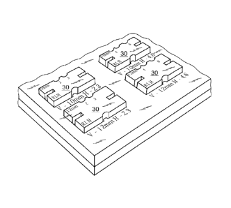
[0024] Reference is now had to Figure 5, wherein there is shown a portion
of a kit,
including a plurality of the simulators 15 in a case. It is important to note
that the simulators vary
in height from 4 mm to 12 mm and the horizontal displacement of the lower
mandible varies
from 2 mm to 7 mm, allowing the clinician to place a series of the simulator
in the mouth of the
patient with or without the pharyngometer to determine the maximum and minimum
discomfort
as plotted on a graph. It is to be understood that the simulators are
constructed such that they can
be reversed as desired, thereby providing over 50 selected jaw positions.
[0025] Thus, as can be seen, the present invention provides a quick and
simple,
straightforward method and apparatus for a snoring/airway screener and a kit
which allows the
clinician to use the pharyngometer to determine airway dimensions, predicting
air flow while
noting the precise position, horizontally and vertically, of the mandibles.
[0026] Mandibular positioning system for optimum airway includes fifteen
simulators
which provide 40 horizontal and vertical combinations that enable bite
registration in the
selected position. Simulators, easily placed by patient, create simple,
effective, repeatable, and
predictable position for device effectiveness and quickly interface with
pharyngometry.
Accessories enable screening and device tuning.
[0027] The scope of the invention should not be limited by the preferred
embodiments
set forth in the examples but should be given the broadest interpretation
consistent with the
description as a whole. The claims are not to be limited to the preferred or
exemplified
embodiments of the invention.
What is claimed is: