Note: Descriptions are shown in the official language in which they were submitted.
CA 02764380 2011-12-02
W02010/142020
PCT/CA2010/000850
ALIGNMENT DEVICE FOR BITEWING RADIOGRAPH
TECHNICAL FIELD
[0001] The present invention relates generally to dentistry
and, in particular, to techniques for taking oral radiographs.
BACKGROUND
[0002] Current
x-ray technologies work well for patients with
straight teeth and nicely curving arches. However,
many
patients have teeth that are crooked. The
result is x-rays
that are non-diagnostic and cannot be used. Dentists and x-
ray operators must repeatedly guess the angle of the mal-
alignment and match that to the x-ray cone.
pow] While the well-known Rinn holder (see, e.g.
http://www.rinncorp.com/) is useful for preventing cone cuts,
this device does not address the specific problem of aligning
the x-ray cone and radiographic film for patients having
crooked teeth or patients with unusually curved arches.
glom As a result of this problem, productive billing time
for the dentist or x-ray operator is lost. It is
estimated
that each x-ray takes approximately two minutes to take and
seven minutes to develop and analyze. Dentists may typically
only bill for the clear x-ray images. Having to re-take the
bitewing x-rays means not only lost billing time but also
added cost of wasted x-ray films. For the
patient, this
represents inconvenience and a delay in diagnosis and
treatment, not to mention extra exposure to x-ray radiation.
Overall, the inability to reliably position a bitewing for x-
raying crooked teeth is frustrating for dentists, x-ray
operators and patients alike. There is
thus a need in
dentistry for a device and method that addresses this
technical problem.
-1-
CA 02764380 2011-12-02
W02010/142020
PCT/CA2010/000850
SUMMARY
[0005] In broad
terms, the present invention is an alignment
device for bitewing radiographs. This alignment device is a
tool that a dentist, hygienist, dental assistant or other x-
ray operator can easily use to determine the proper angle or
orientation of an x-ray cone for the taking of a bitewing
radiograph. This
novel alignment device thus enables a new
method of taking bitewing radiographs in which this alignment
device is placed between crooked teeth to determine a proper
angle for subsequently taking a bitewing radiograph.
[0006]
Accordingly, one main aspect of the present invention
is an alignment device for a bitewing radiograph. The device
comprises a longitudinally extending handle having a first end
and a second end and a tip disposed at the second end and
extending orthogonally to the handle, wherein a length of the
tip is substantially less than a length of the handle and
wherein a cross-sectional area of the tip is substantially
less than a cross-sectional area of the handle.
[0007] In particular embodiments of this invention, the
alignment device may further include a marking ring, for
example made of rubber or equivalent, that can be slid along
the handle for marking the radius of the x-ray cone being
used.
[0008] In other
embodiments of the invention, the alignment
device has a movable hub that is dimensioned to be manually
displaced along the length of the handle. A rod is affixed to
the movable hub and extends orthogonally from the handle and
parallel to the tip.
[0009] A
further main aspect of the present invention is a
method of taking a bitewing radiograph. This method entails
inserting into a mouth of a patient an alignment device
comprising a longitudinally extending handle having a first
-2-
CA 02764380 2011-12-02
' W02010/142020
PCT/CA2010/000850
end and a second end and a tip disposed at the second end and
extending orthogonally to the handle, wherein a length of the
tip is substantially less than a length of the handle and
wherein a cross-sectional area of the tip is substantially
less than a cross-sectional area of the handle. The
method
further entails aligning the tip between two adjacent teeth,
the handle thereby providing visual guidance for positioning
an x-ray cone for taking the bitewing radiograph.
[0010] Yet a
further main aspect of the present invention is
a method of taking a bitewing radiograph. This
method
comprises inserting into a mouth of a patient an alignment
device comprising a longitudinally extending handle having a
first end and a second end and a tip disposed at the second
end and extending orthogonally to the handle, aligning the tip
between two adjacent teeth, the handle thereby protruding
outwardly from the mouth of the patient, positioning an x-ray
cone, instructing the patient to hold still, removing the
device, inserting the film, taking the bitewing radiograph
using the x-ray cone, and developing the bitewing radiograph.
[0011] Other
aspects, features and advantages of this novel
technology will become apparent with reference to the
following description and drawings.
BRIEF DESCRIPTION OF THE DRAWINGS
[0012] Further features and advantages of the present
invention will become apparent from the following detailed
description, taken in combination with the appended drawings,
in which:
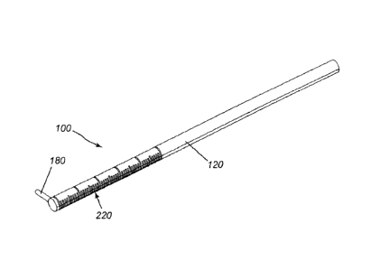
[0013] FIG. 1
is an isometric view of an alignment device for
taking a bitewing radiograph in accordance with one embodiment
of the present invention;
-3-
CA 02764380 2011-12-02
' W02010/142020
PCT/CA2010/000850
[0014] FIG. 2
is a side elevation view of an alignment device
having a hub and rod in accordance with another embodiment of
the present invention;
[0015] FIG. 3
is an enlarged front view of the hub and rod
shown in FIG. 2;
[0016] FIG. 4
depicts how the alignment device presented in
FIG. 1 is used to align an x-ray cone for taking a bitewing
radiograph;
[0017] FIG. 5
depicts how the alignment device presented in
FIG. 2 is used to align an x-ray cone for taking a bitewing
radiograph; and
[0018] FIG. 6
is a top plan view of an alignment device in
accordance with another embodiment of the present invention;
[0019] FIG. 7 is a side elevation view of the alignment
device of FIG. 6; and
[COM FIG. 8
is an isometric view of the alignment device of
FIG. 6.
g021] It will be noted that throughout the appended
drawings, like features are identified by like reference
numerals. It
should furthermore be noted that the drawings
are not necessarily to scale.
DETAILED DESCRIPTION
[0022] In general, and by way of overview, the present
invention provides an alignment device that facilitates the
task of taking a bitewing radiograph of crooked teeth. In
operation, a tip of the alignment device is placed between two
adjacent teeth in the patient's mouth. The handle orthogonal
to the tip thus provides visual guidance to facilitate
-4-
CA 02764380 2011-12-02
. W02010/142020
PCT/CA2010/000850
orientation and alignment of the x-ray cone being used to take
the bitewing radiograph.
[0023] Main illustrative embodiments of this invention are
now described below having regard to the appended figures.
[0024] FIG. 1
is an isometric view of an alignment device for
taking a bitewing radiograph in accordance with one embodiment
of the present invention. The
alignment device, which is
generally designated by reference numeral 100 in this figure,
comprises a longitudinally extending handle 120 having a first
end 140 and a second end 160. The device 100 also includes a
tip 180 disposed at the second end and extending orthogonally
to the handle. As illustrated, a length of the tip (or
"probe") is substantially less than a length of the handle. A
cross-sectional area of the tip is substantially less than a
cross-sectional area of the handle.
[0025] The
alignment device may optionally include a marking
ring 200 that is dimensioned to be manually displaced along
the length of the handle for marking the radius of an x-ray
cone being used to take the bitewing radiograph. The marking
ring in one embodiment may be a coloured rubber ring.
Optionally, the handle may have length markings 220 (like on a
ruler) extending from the second end toward the first end.
The ring thus facilitates lining up of the x-ray cone.
[0026] The
handle of the alignment device may optionally be
made of a soft plastic for patient comfort. The handle may
also have a ribbed or bumpy outer surface, making the handle
more comfortable for the patient to bite.
[0027] A
disposable variant of the alignment device may also
be provided. Whereas the version shown in FIG. 1 is meant to
be reusable, a disposable version may be discarded after
usage. Whereas
the embodiment shown in FIG. 1 may have a
-5-
CA 02764380 2011-12-02
õ
' W02010/142020
PCT/CA2010/000850
metal tip, the disposable version would preferably have a
plastic tip.
[00U] FIG. 3
is a front view of an alignment device having a
slidable hub 240 and a 260 rod in accordance with another
embodiment of the present invention. In
other words, the
alignment device in accordance with this variant has a movable
hub 240 that is dimensioned to be manually displaced along the
length of the handle and a rod 260 affixed to the movable hub
and extending orthogonally from the handle and parallel to the
tip. Preferably, a length of the rod is greater than a length
of the tip.
[0on] As
shown in the embodiment depicted in FIG. 3, the hub
comprises first and second sockets 280, 300 disposed on
opposing sides 320, 340 of the hub 240, each of the first and
second sockets 280, 300 being dimensioned to securely hold the
rod 260.
[0030] FIG. 4
depicts an occlusal view of mal-aligned teeth
and how the alignment device 100 presented in FIGS. la-2b is
used to align an x-ray cone 360 for taking a bitewing
radiograph 380. FIG. 4
shows teeth 33, 34, 35, 36, 37
(labelled according to international tooth codes). The
alignment device may be used as follows: (1) measure the
diameter of the x-ray cone and divide by two to obtain the
radius of the x-ray cone. Set the
rubber ring to mark the
radius. As will
be appreciated in light of the foregoing
discussion, use of the rubber ring is optional; (2) instruct
the patient to hold still; (3) line up the tip ("probe end")
on the contact in the direction needed for the x-ray beam to
pass through; (4) line up the x-ray cone perpendicular to the
handle; (5) bring the edge of the cone in line with the rubber
ring to prevent cone cut (as mentioned above, use of the
rubber ring is optional); (6) instruct the patient to slowly
-6-
CA 02764380 2011-12-02
W02010/142020
PCT/CA2010/000850
open without moving his or her head; (7) insert the film with
a tab or holder, but not with a Rinn; and (8) take the
bitewing radiograph and expose film. As shown in FIG. 4, the
alignment device helps the user (dentist, dental assistant,
etc.) to find the correct angle to open up the 36/37 contact.
Once the correct angle is found, the bitewing is inserted and
the radiograph taken to provide a clear and unobstructed (non-
overlapping) image of teeth 36 and 37.
gmq FIG. 5
depicts an occlusal view of mal-aligned teeth
and how the alignment device 100 presented in FIGS. 3a-3b
(having hub 240 and rod 260) is used to align an x-ray cone
360 for taking a bitewing radiograph 380. As was
the case
with FIG. 4, FIG. 5 also shows teeth 33, 34, 35, 36, 37
(labelled according to international tooth codes). The
procedure for using this version of the alignment device is
similar to what was described above except that the rod and
hub is employed in lieu of the rubber ring.
[0032] FIGS. 6 to 8 depict a further embodiment of the
present invention. In this further embodiment, the handle 120
of the alignment device 100 has a circular cross-section. A
ring (or hub and rod) with a circular inside profile may
optionally be added to this embodiment of the alignment
device. However,
it should be noted that this alignment
device has been found to function perfectly well without a
ring (or hub and rod).
[0033] This novel alignment device greatly facilitates the
taking of diagnostic bitewing radiographs. The
alignment
device helps to ensure that the bitewing radiograph is
perpendicular to the x-ray cone, thus greatly improving the
prospects of obtaining a clear image that enables the dentist
to identify or rule out decay. This new
technology saves
both time and cost to the dentist, and enables a more rapid
-7-
CA 02764380 2011-12-02
,
W02010/142020
PCT/CA2010/000850
diagnosis of mal-aligned teeth by eliminating the guesswork in
taking bitewing radiographs of mal-aligned teeth.
[0014] In
addition, this novel alignment device may be used
for taking a periapical film.
[0035] The
present invention has been described in terms of
specific embodiments, examples, implementations and
configurations which are intended to be exemplary or
illustrative only. Other variants, modifications, refinements
and applications of this innovative technology will become
readily apparent to those of ordinary skill in the art who
have had the benefit of reading this disclosure. Such
variants, modifications, refinements and applications fall
within the ambit and scope of the present invention.
Accordingly, the scope of the exclusive right sought by the
Applicant for the present invention is intended to be limited
solely by the appended claims and their legal equivalents.
-8-