Note: Descriptions are shown in the official language in which they were submitted.
CA 02764635 2016-08-26
r
WO 2010/1-14194 PCINS2010/033779
Tr FLE
TOPICAL, DRUG DELIVERY SYSTEMS FOR OPHTHALMIC LSE
Fl ELI)
The embodiments disclosed herein relate to topical drug delivery systems, and
more
particularly to mixed nanomicellar formulations of corticosteroids and methods
of treating
diseases affecting the posterior ocular segments.
1 0 BACKGROUND
The ophthalmology market includes front-of-eye conditions such as glaucoma,
where
drugs can be delivered using eye drops and other conventional ophthalmic
formulations; and
retinal diseases affecting the vitreous or back-of-the-eye, such as age-
related macular
degeneration (A MD) and diabetic macular edema (DME), which are the leading
causes of vision
loss in the western world.
Disease and injury to the anterior surface of the eye are the leading causes
of visits to
physicians for medical eye care in the United States. These diseases and
injuries rank among the
most painful of eye conditions and can lead to disability and blindness. Major
clinical problems
of the surface of the eye include ocular surface drying. tear film
abnormalities, and related
complications; ocular surface wounds with resultant pathology and scarring:
cortical dysfunction
dystrophies and inherited disease; inflammatory disease; and external ocular
infections. Eye
diseases and injuries can have symptoms ranging from itchy, runny eyes to
impaired vision.
Therefore, it is important to address eye problems right away, as some
diseases can progressively
worsen or even trigger other serious problems. Most phammeologic management of
ocular
disease includes the topical application of solutions to the surface of the
eye as drops, Despite
the relatively small proportion of a topically applied drug dose that
ultimately reaches anterior
segment ocular tissues, topical .formulations remain effective, largely
because of the very high
concentrations of drugs that arc administered.
Disease and injury to tissues of the posterior segment of the eye, including
the retina and
choroid, is involved in many of the most common blinding diseases in the
industrialized world.
CA 02764635 2011-12-06
WO 2010/144194 PCT/US2010/033779
Age-related macular degeneration (AMD) alone impacts more than 10 million
Americans.
Severe vision loss from AMD and other diseases affecting the posterior
segment, including
diabetic retinopathy, glaucoma, and retinitis pigmentosa account for most
cases of irreversible
blindness world wide. Currently, the treatment of posterior segment disease is
to a significant
extent limited by the difficulty in delivering effective doses of drugs to
target tissues in the
posterior eye. While new drugs have emerged for the treatment of these
diseases, the current
standard of care is administration by direct injection into the vitreous. This
kind of regime is not
only hard for patients to endure but carries a growing risk of tissue damage
and infection.
Topical drops rarely make it to the back-of-the-eye and a blood-ocular barrier
prevents
systemically administered drugs from penetrating ocular tissue.
SUMMARY
The embodiments disclosed herein relate to topical drug delivery systems for
ophthalmic
use, including mixed nanomicellar formulations of corticosteroids and methods
of treating
diseases affecting the posterior ocular segments.
According to aspects illustrated herein, there is disclosed an aqueous
ophthalmic solution
that includes nanomicelles in a physiologically acceptable buffer, having a pH
of 5.0 to 8.0,
wherein a corticosteroid at a concentration from about 0.01 % w/v to about
1.00 % w/v is
solubilized through entrapment in a mixed micellar hydrophobic core with a
corona composed of
hydrophilic chains extending from the hydrophobic core, wherein the
nanomicelles comprise
vitamin E TPGS at a concentration ranging from about 3.0 % w/v to about 5.0 %
w/v stabilized
with octoxyno1-40 at a concentration ranging from about 1.0 % w/v to about 3.0
% w/v. In an
embodiment, the aqueous ophthalmic solution has a pH of 6.6 to 7Ø
According to aspects illustrated herein, there is disclosed an eye drop
formulation that
includes a cortico steroid at a concentration ranging from about 0.01 % Aviv
to about 1.00 % w/v;
vitamin E TPGS at a concentration ranging from about 3.0 % w/v to about 5.0 %
w/v; and
octoxyno1-40 at a concentration ranging from about 1.0 `)/0 w/v to about 3.0
`)/0 w/v, wherein the
cord costeroid is solubilized through entrapment in a mixed micellar
hydrophobic core of the
vitamin E TPGS and the octoxyno1-40. In an embodiment, after administration of
a single dose
of the eye drop formulation to a rabbit, dexamethasone tissue levels in
posterior retina-choroid
are equivalent to concentrations of at least 30 ng/g.
2
CA 02764635 2011-12-06
WO 2010/144194 PCT/US2010/033779
According to aspects illustrated herein, there is disclosed a kit that
includes a unit dose of
an aqueous ophthalmic solution comprising nanomicelles in a physiologically
acceptable buffer,
having a pH of 5.0 to 8.0, wherein a corticosteroid at a concentration from
about 0.01 % w/v to
about 1.00 % w/v is solubilized through entrapment in a mixed micellar
hydrophobic core with a
corona composed of hydrophilic chains extending from the hydrophobic core,
wherein the
nanomicelles comprise vitamin E TPGS at a concentration ranging from about 3.0
% w/v to
about 5.0 % w/v stabilized with octoxyno1-40 at a concentration ranging from
about 1.0 % w/v to
about 3.0 % w/v, wherein the unit dose is contained within a vial prepared
from a
pharmaceutically acceptable packaging material. In an embodiment, the unit
dose is about 50
4.
According to aspects illustrated herein, there is disclosed a method of
treating a back-of-
the-eye ocular condition that includes administering to an eye of a patient an
effective amount of
an aqueous ophthalmic solution comprising nanomicelles in a physiologically
acceptable buffer,
having a pH of 5.0 to 8.0, wherein a corticosteroid at a concentration ranging
from about 0.01 %
w/v to about 1.00 % w/v is solubilized through entrapment in a mixed micellar
hydrophobic core
with a corona composed of hydrophilic chains extending from the hydrophobic
core, wherein the
nanomicelles comprise vitamin E TPGS at a concentration ranging from about 2.0
% w/v to
about 5.0 % w/v stabilized with octoxyno1-40 at a concentration ranging from
about 1.0 % w/v to
about 3.0 "A w/v.
According to aspects illustrated herein, there is disclosed a method of
treating a back-of-
the-eye disease that includes topically applying a formulation of the present
disclosure to the
eye, the formulation comprising an aqueous solution of corticosteroid-loaded
nanomicelles;
transporting the corticosteroid-loaded nanomicelles by passive diffusion
through the aqueous
channels/pores of the sclera; transporting the corticosteroid-loaded
nanomicelles by endocytosis
through the choroid to the basolateral side of the retinal pigment epithelium;
discharging the
corticosteroid from the nanomicelles into the retinal pigment epithelium; and
treating the back-
of-the-eye disease.
BRIEF DESCRIPTION OF THE DRAWINGS
The presently disclosed embodiments will be further explained with reference
to the
attached drawings, wherein like structures are referred to by like numerals
throughout the several
3
46,628,217v1
CA 02764635 2016-08-26
WO 2010/144194 PCIYUS2010/033779
views. The drawings shown are not necessarily to scale, with emphasis instead
generally being
placed upon illustrating the principles of the presently disclosed
embodiments.
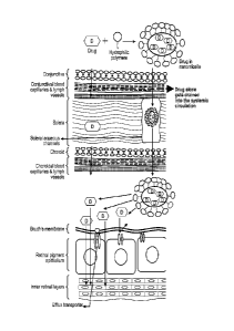
FIG. I is a schematic representation of an embodiment of the permeation of a
topically
applied hydrophobic drug through water channels./pores of the sclera of the
eye and evasion of
eonjunctivaliehoroidal blood vessels and lymphatics using a formulation of the
present
disclosure.
While the above-identified drawings set Fonh presently disclosed embodiments,
other
embodiments are also contemplated, as noted in the discussion. This disclosure
presents
illustrative embodiments by way of representation and not limitation. The
scope of the
claims should not be limited by the preferred embodiments set forth in the
examples. but
should be given the broadest interpretation consistent with the description as
a whole.
DETAILED DESCRIPTION
The presently disclosed embodiments relate to nonirritating mixed
nanoinieelles
comprising water-insoluble (hydrophobic) drugs. Solubilization of the
hydrophobic drug is
achieved through entrapment in a mixed micellar hydrophobic core with a corona
composed of
hydrophilic chains extending from the hydrophobic core. In an embodiment, the
nanomicelles
arc composed of two non-ionic surfactants; a first non-ionic surfactant with
an li.LB index
greater than about 10 and a second non-ionic surfactant with an HUI index of
greater than about
13. at a defined ratio. In an embodiment, the absolute difference between the
11LB index of the
first non-ionic surfactant and the 11111 index of the second non-ionic
surfactant is greater than
about 3, In an embodiment, the first non-ionic surfactant acts as the main
spherical stnicture and
the second non-ionic surfactant adds strength to the nanomicellar structure by
inserting itself
between two polymeric chains of the first non-ionic surfactant. In an
embodiment, a suitable
carrier for the nanomicelles is an aqueous solution. Such stabilization is
believed to result in the
formation of aqueous solutions of extremely hydrophobic drugs that have
optical clarity. An
aqueous solution of nanomicelles of the present disclosure may comprise other
components,
including, but not limited to, a buffering agent, an isotonicity, a
surfactant, a ehelating agent, an
antibacterial agent, an anti-infective agent, a diagnostic agent and a
preservative. Collectively, an
aqueous solution of mixed nanomicelles of the present disclosure, which
optionally include other
components (as described above), is known as a "formulation" of the present
disclosure.
4
46.628,217v1
CA 02764635 2011-12-06
WO 2010/144194 PCT/US2010/033779
In an embodiment, mixed nanomicelles of the present disclosure carry at least
one
corticosteroid. In an embodiment, the at least one corticosteroid is selected
from the group
consisting of prednisolone, methylprednisolone, prednisone, triamcinolone,
betamethasone,
budesonide, and dexamethasone. In an embodiment, mixed nanomicelles of the
present
disclosure carry the corticosteroid dexamethasone. In an embodiment, mixed
nanomicelles of the
present disclosure can substantially improve the solubility and
bioavailability of the
corticosteroid. In an embodiment, mixed nanomicelles of the present disclosure
comprising
dexamethasone can improve the solubility of dexamethasone by up to about 10
fold.
In an embodiment, mixed nanomicelles of the present disclosure, with a size
ranging
from about 10 nm to about 20 nm, allow for efficient accumulation of the
corticosteroid into
targeted diseased tissues. In an embodiment, an aqueous solution of mixed
nanomicelles of the
present disclosure can be used as a topically applied drug delivery platform
for delivery of a
corticosteroid to the back of the eye. Solutions may be manually delivered to
the eye in suitable
dosage form, e.g., eye drops, or delivered by suitable microdrop or spray
apparatus typically
affording a metered dose of medicament. It has been found that after topical
administration of a
formulation of the presently disclosed embodiments, the corticostcroid is able
to reach the back
of the eye. As will be shown in the Examples below, significantly high levels
of dexamethasone
were found at the back of the eye without resulting in significant levels in
the lens and vitreous
humor, suggesting the corticosteroid is not reaching the back of the eye via a
conventional
pathway of going through the eye. Instead, it is believed that the drug is
being transferred to the
back of the eye via an unconventional pathway of going around the eye.
Therefore, a formulation
of the presently disclosed embodiments is particularly useful for topical
application to the eye of
a patient for the treatment of back-of-the-eye ocular conditions.
In an embodiment, a formulation of the present disclosure is applied topically
to an eye.
In an embodiment, a formulation of the present disclosure is used to treat,
reduce, prevent,
ameliorate and alleviate ocular conditions in a patient or subject. In an
embodiment, a
formulation of the present disclosure is used in the treatment of a posterior
segment disorder and
disease. In an embodiment, a formulation of the present disclosure is used for
the treatment of a
back-of-the-eye (posterior) ocular condition, including, but not limited to,
idiopathic uveitis,
ocular surface inflammation, age-related macular degeneration (AMD, wet and
dry), diabetic eye
conditions including diabetic retinopathy and maculopathy, macular edema,
glaucoma, optic
5
46,628,217v1
CA 02764635 2011-12-06
WO 2010/144194 PCT/US2010/033779
neuritis, ocular hypertension, post-operative eye pain and inflammation,
posterior segment
neovascularization (PSNV), proliferative vitreoretinopathy (PVR), hypertensive
retinopathy,
cytomegalovirus retinitis (CMV), choroidal neovascular membranes (CNVM),
vascular
occlusive diseases, retinitis pigmentosa, neuralgia, aging (e.g. muscle
relaxants and other
aesthetic products), cicatrizing ocular surface diseases, ocular infections,
inflammatory ocular
diseases, ocular surface diseases, corneal diseases, retinal diseases, ocular
manifestations of
systemic diseases, hereditary eye conditions, and ocular tumors.
It has been found that after topical administration of a formulation of the
present
disclosure, the drug is able to reach the posterior of the eye. There are two
potential pathways for
molecules to reach posterior eye tissues following topical administration: (1)
intraocular route
through the cornea, aqueous humor, lens, vitreous humor and finally retina;
and (2) trans-scleral
route around the conjunctiva, through the sclera, choroid, and retina. For
hydrophobic drugs, the
intraocular route is often unsuccessful since the hydrophilic stroma becomes a
rate limiting
barrier for trans-corneal absorption. Moreover, aqueous humor in the anterior
and posterior
segments flow in opposite directions and hinder the passage of molecules from
the aqueous
humor to the lens and, subsequently, through the lens zonular spaces to the
vitreous humor, thus
making this an unfavorable pathway. The trans-scleral route offers a more
viable pathway for
back-of-the-eye delivery of hydrophobic molecules by passive diffusion through
the scleral
water channels/pores.
Water-insoluble drugs, like dexamethasone, encapsulated in mixed nanomicelles,
form
spherical structures of amphiphilic molecules in water. FIG. 1 is a schematic
representation of an
embodiment of the permeation of a topically applied water-insoluble drug
through water
channels/pores of the sclera of the eye and evasion of conjunctival/choroidal
blood vessels and
lymphatics using an aqueous solution of mixed nanomicelles of the present
disclosure. In an
embodiment, a method of treating a back-of-the-eye ocular condition includes
administering (for
example, topically applying) a formulation of the present disclosure to the
eye, the formulation
comprising an aqueous solution of water-insoluble drug-loaded nanomicelles;
transporting the
water-insoluble drug-loaded nanomicelles by passive diffusion through the
aqueous
channels/pores of the sclera; transporting the water-insoluble drug-loaded
nanomicelles by
endocytosis through the choroid to the basolateral side of the retinal pigment
epithelium;
discharging the water-insoluble drug from the nanomicelles into the retinal
pigment epithelium;
6
46,628,217v1
CA 02764635 2011-12-06
WO 2010/144194 PCT/US2010/033779
and treating the back-of-the-eye disease. In an embodiment, hydrophilic chains
in the
nanomicellar corona partially evade wash-out by the conjunctival/choroidal
blood vessels and
lymphatics. In an embodiment, limited intraocular drug penetration is achieved
as a result of the
topical application of the formulation of the present disclosure. In an
embodiment, the limited
intraocular drug penetration results in negligible concentration of the water-
insoluble drug in the
lens and vitreous humor of the eye. In an embodiment, the limited intraocular
drug penetration
results in the intraocular pressure (lOP) remaining substantially the same,
i.e., no increase in TOP
due to the topical application of the formulation. In an embodiment, the
limited intraocular drug
penetration results in no cataract development.
The outer surface of the mixed nanomicellar structure protrudes hydrophilic
¨OH groups
to the outside environment. It is believed that due to their hydrophilic
corona, these micellar
nanocarriers can pass through the aqueous channels/pores of the sclera, which
range from about
30 nm to about 300 nm in size. Nanomicelles may then be absorbed onto the
basolateral side of
the Retinal Pigment Epithelium (RPE) through endocytosis. The contents of the
micellar
nanocarriers are discharged inside the cell after fusion with the cell
membrane. It is also believed
that during the transit, the hydrophilic nanomicellar corona helps to evade
drug wash-out into the
systemic circulation by the conjunctival/choroidal blood vessels and
lymphatics. In an
embodiment, a formulation of the present disclosure is able to carry
hydrophobic drug molecules
in therapeutic amounts to the retina, Bruch's membrane and RPE. Since the
mixed nanomicelles
can carry the hydrophobic drugs preferentially across conjunctiva and sclera
rather than across
cornea, lens, and vitreous, negligible levels or no detectable levels are
observed in the lens and
vitreous. Therefore, the formulations of the presently disclosed embodiments
are particularly
useful for topical application to the eye of a patient for the treatment of
back-of-the-eye
(posterior) ocular conditions.
A patient or subject to be treated by a formulation of the present disclosure
can mean
either a human or a non-human animal. In an embodiment, the disclosure
provides methods for
treatment of back-of-the-eye ocular conditions in a human patient in need
thereof In an
embodiment, the disclosure provides methods for treatment of back-of-the-eye
ocular conditions
in a veterinary patient in need thereof, including, but not limited to dogs,
horses, cats, rabbits,
gerbils, hamsters, rodents, birds, aquatic mammals, cattle, pigs, camelids,
and other zoological
animals.
7
46,628,217v1
CA 02764635 2011-12-06
WO 2010/144194 PCT/US2010/033779
As used herein, the terms "micelle" and "nanomicelle" refer to an aggregate
(or cluster)
of surfactant molecules. Micelles only form when the concentration of
surfactant is greater than
the critical micelle concentration (CMC). Surfactants are chemicals that are
amphipathic, which
means that they contain both hydrophobic and hydrophilic groups. Micelles can
exist in
different shapes, including spherical, cylindrical, and discoidal. A micelle
comprising at least
two different molecular species is a "mixed micelle". Nanomicelles are
colloidal particles with
nanometer size ranges, forming spherical structures of amphiphilic molecules
in water.
Polymeric micelles are exploited as pharmaceutical nanocarriers for the
delivery of
poorly water-soluble drugs, which can be solubilized in the hydrophobic inner
core of a micelle.
Micelles can therefore serve to improve solubility and bioavailability of
various hydrophobic
(water-insoluble) drugs. (Lukynov et al., Polyethylene glycol-diacyllipid
micelles demonstrate
increased accumulation in subcutaneous tumors in mice. Pharm. Res. 2002,
19:1424-1429.) The
small size (typically about 10 to 100 nm) of micelles allows the advantage of
sterilization of
micelles by filtration through membranes with the cut off size 0.22 gm.
Another example of a
mixed micellar formulation is described, for example, by Mu et al., 2005,
comprising
polyethylene glycol phosphatidylethanolamine conjugate and vitamin E TPGS in a
mixed
micellar formulation of the poorly soluble anticancer drug camptothecin. (Mu
et al., Int. J.
Pharmaceutics 2005, 306:142-149). Micelles can be formed from one or more
polymeric
nonionic surfactants. Since the micelle size is smaller than light
wavelengths, it is believed that
the light is not scattered by the small micelles resulting in a transparent
optically clear solution.
As used herein, the term "LX214" refers to a 0.2% topical formulation of the
potent
calcineurin inhibitor, voclosporin. The topical formulation is a non-
irritating aqueous solution of
mixed nanomicelles, the nanomicelles comprising a mixture of defined amounts
of octoxyno1-40
with vitamin E TPGS. In an embodiment, the topical formulation has optical
clarity. In an
embodiment, the average nanomicelle size ranges from about 10 nm to about 30
nm, from about
12 nm to about 28 nm, from about 14 nm to about 26 nm, from about 16 nm to
about 24 nm,
from about 18 nm to about 22 nm.
As used herein, the term "optical clarity" is defined as 90% or greater
transmission of
light of 400 nm wavelength in a 1.0 centimeter path. An aqueous ophthalmic
solution of the
present disclosure has a high degree of optical clarity. The optical clarity
of the solution results
8
46,628,217v1
CA 02764635 2011-12-06
WO 2010/144194 PCT/US2010/033779
from the nanomiceller size which is typically smaller than the smallest
wavelength of a visible
light radiation (about 350 nm). In an embodiment, the formulations of the
present disclosure are
substantially clear with an absorption loss less then about 0.1% per micron of
path length. In an
embodiment, the formulations of the present disclosure are substantially clear
with an absorption
loss less then about 0.05 % per micron of path length measured at 400 urn.
The HLB (hydrophilicIlipophilic balance) index value is a concept introduced
by Griffin
in 1950 as a measure of the hydrophilicity or lipophilicity of nonionic
surfactants. The HLB
index value can be determined experimentally by the phenol titration method of
Marszall; see
"Parfumerie, Kosmetik", Vol. 60, 1979, pp. 444-448; further literature
references can be found in
Rompp, Chemistry Lexicon, 8th Edition 1983, p. 1750. See also, for example, US
Pat. No.
4,795,643 (Seth).
By "treating" or "treatment" is meant medically managing a subject (e.g., a
patient) with
the intent that a prevention, cure, stabilization, or amelioration of the
symptoms will result.
Treatment includes active treatment, that is, treatment directed specifically
towards improvement
of the disease; palliative treatment, that is, treatment designed for the
relief of symptoms rather
than the curing of the disease; preventive treatment, that is, treatment
directed to prevention of
the disease; and supportive treatment, that is, treatment employed to
supplement another specific
therapy directed toward the improvement of the disease. As such, "treatment"
also refers to
delaying the onset of the disease or disorder, or inhibiting the disease or
disorder, thereby
providing a prophylactic benefit.
In the embodiments disclosed herein, a therapeutically effective amount is
applied
topically to the eye of a subject in need of treatment. A "therapeutically
effective amount" refers
to an amount of the therapeutic agent either as an individual compound or in
combination with
other compounds that is sufficient to induce a therapeutic effect or
prophylactic benefit on the
disease or condition being treated. This phrase should not be understood to
mean that the dose
must completely eradicate the ailment. A therapeutically effective amount will
vary depending
on, inter alia, the pharmacological properties of the compound used in the
methods, the condition
being treated, the frequency of administration, the mode of delivery,
characteristics of the
individual to be treated, the severity of the disease, and the response of the
patient.
9
46,628,217v1
CA 02764635 2011-12-06
WO 2010/144194 PCT/US2010/033779
To treat the ocular disease, the formulations of the present disclosure can be
applied
topically to the affected eye(s). In some embodiments, the ocular formulation
can be applied in
defined volumes, such about 10, 20, 35, 50, 75, 100, 150, or 200 ).1.1 or
more. The frequency of
application will depend on, among others, the type of ocular disease being
treated, the severity of
the condition, age and sex of the patient, the amount of the water-insoluble
drug in the
formulation, and the pharmacokinctic profile in the ocular tissue to be
treated. In some
embodiments, the formulation can be administered more than one times per day.
When the
formulations are administered more than once per day, the frequency of
administration can be
two, three, four, up to eight times per day. In some embodiments, the
formulation can be
administered one to four times daily. In some embodiments, the formulation can
be applied once
every two days. In some embodiments, the formulation can be applied once every
four days. In
some embodiments, the formulation can be administered once every week.
Determining the
frequency and amount to be administered for a particular ocular disorder is
well within the skill
and judgment of the attending practitioner.
In some embodiments, an ocular formulation of the present disclosure can be
provided in
the form of a kit. As such, the kit can contain the ocular formulation in a
container, a vial, as
single dose unit or as a single solution reservoir. The kit can also contain a
dispenser for
dispensing measured doses as well as instructions for dosing and use of the
formulations. In an
embodiment, the kit assembly also provides a sequential dispenser means
containing a plurality
of daily sets of kit sub-assembly components, such as a series of jars,
bottles, containers or
ampoules containing a supply (unit dose) of formulation.
In an embodiment, a formulation of the present disclosure comprises from about
0.01 %
w/v to about 80 % w/v, from about 0.05 % w/v to about 16 % w/v, from about
0.10 % w/v to
about 3.2 % w/v, from about 0.15 % w/v to about 0.60 % w/v of the water-
insoluble drug. In an
embodiment, a formulation of the present disclosure comprises about 0.20 % w/v
of the water-
insoluble drug. In an embodiment, a formulation of the present disclosure
comprises about 0.10
% w/v of the water-insoluble drug. Suitable classes of water-insoluble drugs
include, but are not
limited to, peptides, cyclic peptides (e.g., some calcineurin inhibitors),
eicosanoids (e.g.
prostacyclins and prostaglandins), anti-inflammatory drugs, immunosuppressive
drugs (e.g.,
some mTOR inhibitors), autonomic drugs (e.g. beta-blockers, alpha-blockers,
beta-agonists, and
46,628,217v1
CA 02764635 2011-12-06
WO 2010/144194 PCT/US2010/033779
alpha-agonists), antiangiogenic drugs, biologics, gene therapy agents (e.g.,
viral vectors), anti-
infectives (e.g antifungals, antibiotics, and antivirals), monoclonal
antibodies and fragments
thereof, retinoids, RNAi, photo sensitizers, steroids (e.g., corticosteroids),
mixture drugs,
immuno-modulators, chemotherapeutic agents, G-coupled protein receptor
antagonists, receptor
tyrosine kinase (RTK) inhibitors, growth hormone inhibitors, integrin
inhibitors, Sdfl/CXCR4
pathway inhibitors, nACh receptor antagonists, analogs thereof or
pharmaceutically acceptable
salts, esters or prodrugs. In an embodiment, the water-insoluble drug is
selected from one of
cyclosporin A, voclosporin, ascomycin, tacrolimus (FK506), sirolimus
(rapamycin),
pimecrolimus, dexamethasone, an analog thereof or a pharmaceutically
acceptable salt, ester or
prodrug. In an embodiment, the water-insoluble drug is a calcineurin
inhibitor. In an
embodiment, the calcineurin inhibitor is voclosporin. In an embodiment, the
water-insoluble
drug is an mTOR inhibitor. In an embodiment, the mTOR inhibitor is rapamycin.
In an
embodiment, the water-insoluble drug is a corticosteroid. In an embodiment,
the corticosteroid
is dexamethasone. The formulations can further include pharmaceutical
excipients, including,
but not limited to, antibacterial agents, anti-infective agents, diagnostic
agents and preservatives.
Calcineurin Inhibitors
Calcineurin is a calcium/calmodulin-regulated protein phosphatase involved in
intracellular signaling. For reviews on calcineurin, see e.g. Rusnak and
Mertz, Physiol. Rev. 80,
1483-1521 (2000) and Feske et al., Biochem. Biophys. Commun. 311, 1117-1132
(2003).
Calcineurin inhibitors are substances which block calcineurin
dephosphorylation of appropriate
substrates, by targeting calcineurin phosphatasc (PP2B, PP3), a cellular
enzyme that is involved
in gene regulation. Calcineurin inhibitors have been found to be effective in
inhibiting, among
other things, T-cell proliferation, and this mode of action contributes to
clinical effects of
calcineurin inhibitors to treat chronic inflammation and act as
immunosuppressants.
A calcineurin inhibitor of the present disclosure is preferably an
immunophilin-binding
compound having calcineurin inhibitory activity. Immunophilin-binding
calcineurin inhibitors
are compounds forming calcineurin inhibiting complexes with immunophilins,
e.g. cyclophilin
and macrophilin. Examples of cyclophilin-binding calcineurin inhibitors are
cyclosporins or
cyclosporin derivatives (hereinafter cyclosporins) and examples of macrophilin-
binding
calcineurin inhibitors are ascomycin and ascomycin derivatives (hereinafter
ascomycins), see
11
46,628,217v1
CA 02764635 2016-08-26
WO 291(1/144194 PCIliS2010/033779
e.g. Liu et al., Cell 66, 807-815 (1991) and Dumont et al., J. Exp. Med., 176,
751-780 (1992).
Ascomycins and their preparation are known. Ascomycin (FR 520) is a macrolide
antibiotic
disclosed e.g. in U.S. Pat. No. 3,244,592 and in EP 349061. A wide range of
ascomycin
derivatives are known, which are either naturally occurring among lunaal
species or arc
obtainable by manipulation of fermentation procedures or by chemical
derivatization.
Ascomycin-type macrolides include ascornycin, tacrolimus (11(506) and
pimecrolimus.
Cyclosporin is a fungal peptide composed of 11 amino acids. It has been in use
since
1983 as an immunosuppressive drug. Cyclosporin inhibits the production of 1L-
2. Cyclosporin
binds to the cytosolic protein cyclophilin (an immunophilin) of
immunocompetent lymphocytes,
especially T-Iymphoeytes. The complex of cyclosporin and cyclophilin inhibits
calcineurin,
which under normal circumstances induces the transcription of interleukin-2.
Cyclosporin also
inhibits lymphokine production and interleukin release, leading to a reduced
function of effector
[-cells. Cyclosporin is used in the treatment of acute rejection reactions,
but has been
increasingly substituted with newer immunosuppressants due to nephrotoxicity.
Cyclosporins and their preparation arc e.g. disclosed in U.S. Pat. No.
4,117,118.
Cyclosporin, originally extracted from the soil fungus Putypaciadium
infilattim, has a cyclic 11-
amino acid structure and includes e.g. Cyclosporins A through I, such as
Cyclosporin A, B, C. D
and G. Cyclosporin A tcyclosporine; cyclo(L-alanyl-D-alanyl-N-methyl-L-leucyl-
N-methyl-L-
leucyl-N-methyl-L-val y1-((3R,4R,6E)-6,7-didehydro-3-hydroxy-N,4-dimethyl-L-2-
aminooctanoyl-E-2-aminobutanoyi-N-methylglycyl-N-meihyl-1,-leucyl-L-valyl-N-
methylleucy1)) is utilized in a commercially available ophthalmic emulsion
(Ding et al., US Par.
No. 5,474,979, Restasis4) used in the treatment of dry eye syndrome. Domb (US
Pat. No.
7,026.290) discloses a dispersible concentrate for the delivery of cyclosporin
including a
surfactant with an FHB (hydrophilicilipophilic balance)of at least about 8 and
a surfactant with a
low HER of less than about 5.
Cyclosporins of the present disclosure also include eyclosporin analogs. For
example,
cyclosporin analogs disclosed in Naicker et al.. US Pat. No. 6,998,385.
A preferred example of a cyclosporin analog is voclosporin
(Cyelosporin A. 6((2S,3R,4R)-3-hydroxy-4-methy1-2-(methylamino)-6,8-
nonadicnoie acid)-;
including specifically the trans-version 1SA1,247, trans-1SA247 CAS RN 368455-
04-3). which is
1,
46,628,217v1
CA 02764635 2016-08-26
WO 2010/144194 PC17US2010/033779
described in, for example, Naickcr et al., US Patent 7.429,562,
Further compositions of voclosporin are described, For example. in Naicker et
al.. US Pat. No.
7,060,672.
Voclosporin (-VCS") is a next-generation calcineurin inhibitor. Like other
molecules of
this class, VCS reversibly inhibits immunocompetent lymphocytes, particularly
T-lymphocytcs,
and also inhibits lymphokine production and release. This action is primarily
mediated through
inhibition of calcineurin, a phosphatase enzyme found in the cytoplasm of
cells. VCS is a more
potent and less toxic semi-synthetic derivative of Cyclosporine A.
Tacrolimus (FK506) is another caleinemin inhibitor which is also a fungal
product, but
has a macrolide lactone structure. Tacrolimus (Prografk, oral, injectable,
Astellas Pharma US;
and Protonic'''. topical. Astellas Pharrna US) is used as an immtmosuppressant
in conjunction
with liver, kidney. heart, lung and heart/lung transplants. Tacrolimus also
inhibits the production
of 1L-2. Tacrolimus binds to an immunophilin (FK-binding protein 12, FKBP12),
followed by
binding of the complex to calcineurin to inhibit its phosphatase activity.
Ascomycin, also called Immunomyein, FR-900520, FK520, is an ethyl analog of
tacrolitnus (12K506) with strong immunosupprosant properties. Ascomycin acts
by binding to
immunophilins, especially macrophilin-12. It appears that Ascomycin inhibits
the production of
Thl (interferon- and 1L-2) and Th2 (IL-4 and IL-10) cytokines. Additionally,
ascomycin
preferentially inhibits the activation of mast cells, an important cellular
component of the ample
response. Ascomycin produces a more selective immunomodulatory effect in that
it inhibits the
elicitation phase of allergic contact_dermatitis but does not impair the
primary immune response
when administered systemically.
Pimecrolimus is an immunomodulating agent used in the treatment of atopic
dermatitis
(eczema). It is currently available as a topical cream, once marketed by
Novartis (however
Galderma is promoting the molecule in Canada since early 2007) under the trade
name Flidel.
Pimecrolimus is an ascomycin macrolactam derivative. Pimecrolimus, like
tacrolimus, belongs
to the ascomvcin class of macrolactam immunosuppressives, acting by the
inhibition of T-cell
activation by the calcineurin pathway and inhibition of the release of
numerous inflammatory
cytokincs, thereby preventing the cascade of immune and inflammatory signals.
13
46,628:217v1
CA 02764635 2011-12-06
WO 2010/144194 PCT/US2010/033779
In an embodiment, a calcineurin inhibitor such as cyclosporin A, voclosporin,
ascomycin,
tacrolimus, pimecrolimus, an analog thereof, or a pharmaceutically acceptable
salt thereof is
utilized in an aqueous nonirritating mixed nanomicellar formulation of the
present disclosure. In
an embodiment, the calcineurin inhibitor is voclosporin.
mTOR Inhibitors
Another class of compounds that exhibit this general therapeutic profile are
the mTOR
inhibitors. MTOR inhibitors target a molecular target known as "mammalian
target of
rapamycin" (mTOR). A prototypical compound of this class is sirolimus.
Sirolimus (rapamycin, Rapamune , oral, Wyeth Pharmaceuticals, Inc.) is a
microbial
product isolated from the actinomycete Streptornyces hygravcopicus. Sirolimus
was initially
discovered as an antifungal agent in the 1970's, but because of its
immunosuppressive effects,
was not developed for use as an antibiotic. Structural similarities with
tacrolimus eventually led
researchers to investigate immunosuppressive properties of sirolimus in
experimental organ
transplantation. (Gummert et al., 1999). Sirolimus binds to an immunophilin
(FK-binding
protein 12, FKBP12), but the complex inhibits the mammalian target of
rapamycin (mTOR)
pathway through directly binding the mTOR Complex 1 (mTORC1). Sirolimus
inhibits the
response to interleukin-2 (IL-2) and thereby blocks activation of T- and B-
cells. By contrast,
tacrolimus and cyclosporine inhibit the production of 1L-2. Sirolimus
(rapamycin) is disclosed in
a method of treating ocular inflammation in Kulkarni in US Pat. No. 5,387,589.
Formulations
for ocular treatment comprising sirolimus are disclosed in Dor et al., WO
2006/086744.
In an embodiment, an immunosuppressive mTOR inhibitor, such as sirolimus
(rapamycin), temsirolimus, everolimus, an analog thereof, or a
pharmaceutically acceptable salt
thereof is utilized in an aqueous nonirritating mixed nanomicellar formulation
of the present
disclosure.
Corticosteroids
Corticosteroids are a family of compounds that include the adrenal steroid
hormone
cortisol (hydrocortisone) and related synthetic drugs, including, but not
limited to, prednisolone,
methylprednisolone, prednisone, triamcinolone, betamethasone, budesonide, and
14
46,628,217v1
CA 02764635 2011-12-06
WO 2010/144194 PCT/US2010/033779
dexamethasone. Adrenal corticosteroids are hormones extracted from the adrenal
cortex or a
synthetic substance similar in chemical structure and biologic activity to
such a hormone.
Corticosteroids have similar mechanisms of action: they bind to specific
corticosteroid binding
proteins in the cytoplasm. These complexes are then transported into the
nucleus where they
bind to discrete portions of the cell's DNA. Corticosteroids are generally
grouped into four
classes, based on chemical structure.
Prednisolone is a corticosteroid drug with predominantly glucocorticoid and
low
mineralocorticoid activity, making it useful for the treatment of a wide range
of inflammatory
and auto-immune conditions including, but not limited to, asthma, uveitis,
rheumatoid arthritis,
ulcerative colitis and Crohn's disease, Bell's palsy, multiple sclerosis,
cluster headaches, and
Systemic Lupus Erythematosus. Prednisolone acetate ophthalmic suspension is an
adrenocortical
steroid product prepared as a sterile ophthalmic suspension, used to reduce
swelling, redness,
itching, and allergic reactions affecting the eye.
Methylprednisolone is a synthetic glucocorticoid drug. Like most
adrenocortical steroids,
methylprednisolone is typically used for its anti-inflammatory effects. The
list of medical
conditions for which methlyprednisolone is prescribed is rather long, and is
similar to other
corticosteroids such as prednisolone.
Prednisone is a synthetic corticosteroid drug that is usually taken orally but
can be
delivered by intramuscular injection and can be used for a number of different
conditions. It has
a mainly glucocorticoid effect. Prednisone is a prodrug that is converted by
the liver into
prednisolone, which is the active drug and also a steroid.
Triamcinolonc is a synthetic corticosteroid given orally, by injection,
inhalation, or as a
topical ointment or cream.
Hydrocortisone is a steroid hormone produced by the adrenal cortex.
Hydrocortisone is
commonly used for the short-term treatment of inflammation in the eye (due to
allergy, injury or
infection) or ear (due to eczema).
Betamethasone is a moderately potent glucocorticoid steroid with anti-
inflammatory and
immunosuppressive properties. Betamethasone is applied as a topical cream,
ointment, foam,
lotion or gel to treat itching (e.g. from eczema).
46,628,217v1
CA 02764635 2011-12-06
WO 2010/144194 PCT/US2010/033779
Dexamethasone is a potent synthetic corticosteroid. It has been demonstrated
by animal
and human studies based on an oral application to possess approximately six to
seven times the
potency of prednisolone and at least 30 times the potency of cortisone. The
potency of this
compound is accomplished by the addition of a methyl radical and a fluorine
atom to the
prednisolone radical. MAXIDEXO 0.1% (dexamethasone ophthalmic suspension) is
an
adrenocortical steroid prepared as a sterile topical ophthalmic suspension.
In an embodiment, corti co steroi ds such as prednisolone, m ethylprednisolon
e, pre dni sone,
triamcinolone, hydrocortisone, betamethasone and dexamethasone are utilized in
an aqueous
nonirritating mixed nanomicellar formulation of the present disclosure. In an
embodiment, the
cortico steroid is dexamethasone.
In an embodiment, a mixed nanomicellar formulation of the present disclosure
includes
two non-ionic surfactants. In an embodiment, a mixed nanomicellar formulation
of the present
disclosure includes a first non-ionic surfactant with an HLB index greater
than about 10, and a
second non-ionic surfactant with an HLB index of greater than about 13. In an
embodiment, the
first non-ionic surfactant having a HLB greater than about 10 is selected from
various chemical
derivatives of vitamin E with ester and ether linkages of various chemical
moieties to
polyethylene glycol of various lengths. Particularly preferred are vitamin E
tocopherol
polyethylene glycol succinate (TPGS) derivatives with PEG molecular weights
between about
500 and 6000 Da. In an embodiment, the vitamin E polymeric derivative with an
HLB index
greater than about 10 is vitamin E tocopherol polyethylene glycol 1000
succinate (Vitamin E
TPGS, tocophcrsolan). In an embodiment, the Vitamin E TPGS contributes to the
solubilization
of the water-insoluble drug and may reduce ocular discomfort in aqueous
conditions. In an
embodiment, the vitamin E TPGS is present in from about 0.01 % w/v to about 20
')/0 w/v of the
composition. In an embodiment, the vitamin E TPGS is present in from about 1.0
% w/v to
about 7.0 % wiv of the composition. It will be understood that throughout the
specification the
term weight percent (wt%) refers to mass per unit volume, unless otherwise
specified.
Vitamin E Tocopherol Polyethylene Glycol 1000 Succinate (Vitamin E TPGS,
tocopherlosan, MW, approximately 1,513 g/mol, Eastman Chemical Co., Kingsport,
Tenn) is an
amphipathic excipient which is a water soluble derivative of natural-source
vitamin E. Vitamin
E TPGS, or PEGylated vitamin E, is a vitamin E derivative in which
polyethylene glycol
16
46,628,217v1
CA 02764635 2011-12-06
WO 2010/144194 PCT/US2010/033779
subunits are attached by a succinic acid diester at the ring hydroxyl of the
vitamin E molecule.
Vitamin E TPGS is an amphipathic non-ionic surfactant with an HLB index of
about 13.
Various chemical derivatives of vitamin E TPGS including ester and ether
linkages of various
chemical moieties are included within the definition of vitamin E TPGS. In
addition to serving
as a source of water-soluble vitamin E, vitamin E TPGS has been suggested for
use as an
emulsifier, solubilizer, absorption enhancer, an a vehicle for lipid-soluble
drug delivery
formulations. Vitamin E TPGS is a component in an FDA approved product,
Agenerse(Amprenavir, an antiviral HIV protease inhibitor) of Glaxo SmithKline
Pharmaceuticals.
In an embodiment, the second non-ionic surfactant having a HLB greater than
about 13 is
an amphipathic polyethylene glycol (PEG)-alkyl ether surfactant or
polyethylene glycol (PEG)-
alkyl aryl ether surfactant. In one aspect, this surfactant is selected from a
PEG 5-100 octyl
phenyl ether which has an HLB greater than about 13. In this aspect, the PEG
octylphenyl
compound is selected from octoxyno1-9, octoxynol-10, octoxynol-11, octoxynol-
12, octoxynol-
13, octoxynol-16, octoxyno1-20, octoxyno1-25, octoxyno1-30, octoxyno1-33,
octoxyno1-40,
octoxyno1-70. In a specific aspect, the PEG-alkyl phenyl ether surfactant is
octoxyno1-40. In an
embodiment, the octoxyno1-40 contributes to the reduction of ocular
discomfort, and to the
formation of a stable, mixed micellar formulation that is optically clear. In
another aspect, the
surfactant with an HLB greater than about 10 is selected from a PEG-5-100
nonyl phenyl ether;
tyloxapol (ethoxylated p-tert-octylphenol formaldehyde polymer), a PEG- fatty
acid monoester
surfactant, a PEG- glycerol fatty acid ester, and a PEG- sorbiton fatty acid
ester. PEG- Fatty acid
monoester surfactants include, but are not limited to, PEG-15 oleate, PEG-20
laurate, PEG-20
oleate, PEG-20 stearate, PEG-32 laurate, PEG-32 oleate, PEG-32 stearate, PEG-
40 laurate, PEG-
40 oleate, and PEG-40 stearate. PEG- Glycerol fatty acid esters include, but
are not limited to,
PEG-15 glyceryl laurate PEG-20 glyceryl laurate, PEG-30 glyceryl laurate, PEG-
40 glyceryl
laurate, and PEG-20 glyceryl stearate. PEG- sorbiton fatty acid esters
include, but are not
limited to, PEG-4 sorbiton monolauratc, PEG-4 sorbiton monostearate, PEG-5
sorbiton
monooleate, PEG-20 sorbiton monolaurate, PEG-20 sorbiton monopalmitate, PEG-20
sorbiton
monostearate, and PEG-20 sorbiton monooleate. In an embodiment, the second non-
ionic
surfactant with HLB greater than about 13 is octoxyno1-40. Octoxyno1-40 is
used as a surfactant
in a marketed formulation (Acular , and Acular LS of Allergan, Inc., CA).
Octoxyno1-40
17
46,628,217v1
CA 02764635 2011-12-06
WO 2010/144194 PCT/US2010/033779
(IGEPAL CA-897) has an HLB index of about 18. In an embodiment, the octoxyno1-
40 is
present in from about 0.001 % w/v to about 10 % w/v of the composition. In an
embodiment, the
octoxyno1-40 is present in from about 0.01 % w/v to about 5.0 % w/v of the
composition.
In an embodiment, a formulation of the present disclosure comprising a water-
insoluble
(i.e., hydrophobic) drug can be topically applied to an eye in a method to
treat a back-of-the-eye
ocular condition. As will be shown in the Examples that follow, it has been
found that after
topical administration of a formulation of the present disclosure, the water-
insoluble drug is able
to reach the back of the eye, thus providing a treatment for back-of- the-eye
ocular conditions. In
an embodiment, the water-insoluble drug is present in the formulation at
concentrations from
about 0.01 % w/v to about 10 % w/v, preferably from about 0.1 % w/v to about
3.0 % w/v .In an
embodiment, the water-insoluble drug is voclosporin, and the voclosporin is
present in the
formulation at a concentration from about 0.02 % w/v to about 0.5 % w/v. In an
embodiment,
the water-insoluble drug is dexamethasone, and the dexamethasone is present in
the formulation
at a concentration from about 0.1 % w/v to about 1.0 % w/v. In an embodiment,
Vitamin E
TPGS is present in the formulation at concentrations from about 0.001 % w/v to
about 20 % w/v,
from about 0.1 % w/v to about 5 % w/v. In an embodiment, Octoxyno1-40 or its
homolog
mixtures are present in the formulation at concentrations from about 0.001 %
w/v to about 10 %
w/v, preferably from about 0.01 % w/v to about 3.0 % w/v. Preferably, the
total amount of
surfactants in a formulation of the presently disclosed embodiments is about
30 percent or less of
the total formulation with the remaining major component being water.
In an embodiment, a formulation of the present disclosure comprises about 0.2
% w/v of
voclosporin, about 2.5 % w/v of vitamin E TPGS, and about 2.0 % w/v octoxyno1-
40. In an
embodiment, a formulation of the present disclosure comprises about 0.5 `)/0
w/v of voclosporin,
about 3.5 % Aviv of vitamin E TPGS, and about 2.0 % w/v octoxyno1-40. In an
embodiment, a
formulation of the present disclosure comprises voclosporin at about 2.0 %
w/v. In an
embodiment, a formulation of the present disclosure comprises about 0.1 % w/v
of
dexamethasone, about 4.5 % w/v of vitamin E TPGS, and about 2.0 % w/v
octoxyno1-40. In an
embodiment, a formulation of the present disclosure comprises about 0.2 % w/v
of rapamycin,
about 4.5 % w/v of vitamin E TPGS, and about 2.0 % w/v octoxyno1-40.
18
46,628,217v1
CA 02764635 2011-12-06
WO 2010/144194 PCT/US2010/033779
It should be understood that the formulations of the present disclosure can
also comprise
other components such as, but not limited to, buffers, lubricating agents,
tonicity agents, anti-
infective agents, antibacterial agents, antioxidants, bioadhesive polymers,
viscosity enhancing
agents, wetting agents, and preservatives. In any of the mixed formulations of
the present
disclosure for topical administration to the eye, the mixtures are preferably
formulated at about
pH 5 to about pH 8. This pH range may be achieved by the addition of buffers
to the mixtures as
described in the examples. In an embodiment, the pH range in the mixtures in a
formulation is
about pH 6.6 to about pH 7Ø It should be appreciated that the formulations
of the present
disclosure can be buffered by any common buffer system such as phosphate,
borate, acetate,
citrate, carbonate and borate-polyol complexes, with the pH and osmolality
adjusted in
accordance with well-known techniques to proper physiological values. The
formulations of the
presently disclosed embodiments are stable in buffered aqueous solution. That
is, there is no
adverse interaction between the buffer and any other component that would
cause the
compositions to be unstable.
Tonicity agents include, for example, mannitol, dextrose, sodium chloride,
xylitol and
glycerol. These tonicity agents can be used to adjust the osmolality of the
compositions. In an
embodiment, the osmolality of a formulation of the present disclosure is
adjusted to be in the
range of about 75 to about 350 mOsm/kg.
In an embodiment, a formulation of the present disclosure further comprises
one or more
bioadhesive polymers. Bioadhesion refers to the ability of certain synthetic
and biological
macromolecules and hydrocolloids to adhere to biological tissues. Bioadhcsion
is a complex
phenomenon, depending in part upon the properties of polymers, biological
tissue, and the
surrounding environment. Several factors have been found to contribute to a
polymer's
bioadhesive capacity: the presence of functional groups able to form hydrogen
bridges (--OH,
COOH), the presence and strength of anionic charges, sufficient elasticity for
the polymeric
chains to interpenetrate the mucous layer, and high molecular weight.
Bioadhesion systems have
been used in dentistry, orthopedics, ophthalmology, and in surgical
applications. However, there
has recently emerged significant interest in the use of bioadhesive materials
in other areas such
as soft tissue-based artificial replacements, and controlled release systems
for local release of
bioactive agents. Such applications include systems for release of drugs in
the buccal or nasal
cavity, and for intestinal or rectal administration.
19
46,628,217v1
CA 02764635 2011-12-06
WO 2010/144194 PCT/US2010/033779
In an embodiment, bioadhesive polymers are optionally incorporated in the
formulation
to enhance the viscosity and thereby to increase residence time in the eye.
Bioadhesive polymers
of the present disclosure include, for example, carboxylic polymers like
Carbopol (carbomers),
Noveon (polycarbophils), etc.; cellulose derivatives including alkyl and
hydroxyalkyl cellulose
like methylcellulose, hydroxypropylcellulose, carboxymethylcellulose, etc.;
gums like locust
beam, xanthan, agarose, karaya, guar, etc.; and other polymers including but
not limited to
polyvinyl alcohol, polyvinyl pyrollidone, polyethylene glycol, Pluronic
(Poloxamers),
tragacanth, and hyaluronic acid; phase-transition polymers for providing
sustained and controlled
delivery of enclosed medicaments to the eye (e.g., alginic acid, carrageenans
(e.g., Eucheuma),
xanthan and locust bean gum mixtures, pectins, cellulose acetate phthalate,
alkylhydroxyalkyl
cellulose and derivatives thereof, hydroxyalkylated polyacrylic acids and
derivatives thereof,
poloxamers and their derivatives, etc. Physical characteristics in these
polymers can be mediated
by changes in environmental factors such as ionic strength, pH, or temperature
alone or in
combination with other factors. In an embodiment, the optional one or more
bioadhesive
polymers is present in the formulation from about 0.01 wt% to about 10
wt%/volume; preferably
0.1 to about 5 wt%/volume. In an embodiment, the mixed nanomicellar
formulation optionally
further comprises hydrophilic polymer excipients selected from, for example,
PVP-K-30, PVP-
K-90, HPMC, HEC, and polycarbophil. In an embodiment, the polymer excipient is
selected
from PVP-K-90, PVP-K-30 or HPMC. In an embodiment, the polymer excipient is
selected
from PVP-K-90 or PVP-K-30.
In an embodiment, if a preservative is desired, the formulations may
optionally be
preserved with any well-known system such as benzyl alcohol with/without EDTA,
benzalkonium chloride, chlorhexidine, Cosmocil CQ, or Dowicil 200.
Pharmaceutically acceptable packaging materials for the formulations can
include, but
are not limited to polypropylene, polystyrene, low density polyethylene
(LDPE), high density
polyethylene (HDPE), polycarbonate, polyvinylidine chloride, and other
materials known to
those skilled in the art. In an embodiment, the formulations are packaged
aseptically employing
blow-fill-seal technology. Blow-fill-seal (BFS) describes an aseptic filling
process in which
hollow containers are blow molded, filled with sterile product, and sealed,
all in one continuous
machine cycle. The technology is an alternative to conventional aseptic
filling and capping
operations, often providing cost savings through high output and process
efficiency.
46,628,217v1
CA 02764635 2011-12-06
WO 2010/144194 PCT/US2010/033779
In an embodiment, the formulations disclosed herein are filled to single-use
bottles,
packets, LDPE BFS vials, ampoules, LDPE BFS containers, or HDPE BFS
containers.
In an embodiment, multiple doses can be supplied as a plurality of single-use
packages.
In an embodiment, the formulations are conveniently packaged in a bottle,
container or device
that allows for metered application, including containers equipped with a
dropper for topical
ophthalmic application.
While the precise regimen is left to the discretion of the clinician, it is
recommended that
the formulations of the presently disclosed embodiments be topically applied
by placing one to
two drops, or more, in each eye 1 to 4 times daily. For example, the
formulation may be applied
1, 2, 3, 4 or 8 times a day, or more. In an embodiment, a formulation of the
present disclosure is
topically applied by placing one to two drops in each eye once or twice daily.
In an embodiment, a formulation of the present disclosure is topically applied
to an eye
of a patient, transported into the eye, and releases a drug at a posterior
portion of the eye with
negligible drug accumulation in the middle portion of the eye. In an
embodiment, negligible
drug accumulation in the middle portion of the eye refers to negligible drug
accumulation in at
least one of the aqueous humor, lens, and vitreous humor. It is believed that
the formulations of
the present disclosure can significantly reduce side effects associated with
current therapy to
treat back-of-the-eye conditions. Adverse side effects of current
corticosteroid therapy includes,
but are not limited to, cataracts and ocular hypertension. These side effects
often cause lowering
therapeutic efficacy and discontinuation of therapy.
In an embodiment, a formulation of the present disclosure can be used as a
topically
applied drug delivery platform for delivery of a hydrophobic, water-insoluble
drug to the back of
the eye. In an embodiment, a formulation of the present disclosure is applied
topically to an eye.
In an embodiment, a formulation of the present disclosure is used to treat,
reduce, prevent,
ameliorate and/or alleviate ocular conditions in a patient or subject. In an
embodiment, a
formulation of the present disclosure is used to treat, reduce, prevent,
ameliorate and/or alleviate
a back-of-eye disease. Examples of "back-of-eye" disease include, among
others, macular
edema such as angio graphic cystoid macular edema; retinal ischemia and
choroidal
neovascularization; macular degeneration; retinal diseases (e.g., diabetic
retinopathy, diabetic
retinal edema, retinal detachment); inflammatory diseases such as uveitis
(including panuveitis)
21
46,628,217v1
CA 02764635 2011-12-06
WO 2010/144194 PCT/US2010/033779
or choroiditis (including multifocal choroiditis) of unknown cause
(idiopathic) or associated with
a systemic (e.g., autoimmune) disease; episcleritis or scleritis; Birdshot
retinochoroidopathy;
vascular diseases (retinal ischemia, retinal vasculitis, choroidal vascular
insufficiency, choroidal
thrombosis); neovascularization of the optic nerve; and optic neuritis.
In an embodiment, an aqueous ophthalmic solution includes nanomicelles in a
physiologically acceptable buffer, having a pH of 5.0 to 8.0, wherein a
corticosteroid at a
concentration from about 0.01 % w/v to about 1.00 % w/v is solubilized through
entrapment in a
mixed micellar hydrophobic core with a corona composed of hydrophilic chains
extending from
the hydrophobic core, wherein the nanomicelles comprise vitamin E TPGS at a
concentration
ranging from about 3.0 % w/v to about 5.0 % w/v stabilized with octoxyno1-40
at a concentration
ranging from about 1.0 % w/v to about 3.0 % w/v. In an embodiment, the aqueous
ophthalmic
solution has a pH of 6.6 to 7Ø
In an embodiment, an eye drop formulation includes a corticosteroid at a
concentration
ranging from about 0.01 % w/v to about 1.00 % w/v; vitamin E TPGS at a
concentration ranging
from about 3.0 % w/v to about 5.0 % w/v; and octoxyno1-40 at a concentration
ranging from
about 1.0 % w/v to about 3.0 % w/v, wherein the corticosteroid is solubilized
through entrapment
in a mixed micellar hydrophobic core of the vitamin E TPGS and the octoxyno1-
40. In an
embodiment, after administration of a single dose of the eye drop formulation
to a rabbit,
dexamethasone tissue levels in posterior retina-choroid are equivalent to
concentrations of at
least 30 ng/g.
In an embodiment, a kit includes a unit dose of an aqueous ophthalmic solution
comprising nanomicelles in a physiologically acceptable buffer, having a pH of
5.0 to 8.0,
wherein a corticosteroid at a concentration from about 0.01 % w/v to about
1.00 % w/v is
solubilized through entrapment in a mixed micellar hydrophobic core with a
corona composed of
hydrophilic chains extending from the hydrophobic core, wherein the
nanomicelles comprise
vitamin E TPGS at a concentration ranging from about 3.0 % w/v to about 5.0 %
w/v stabilized
with octoxyrto1-40 at a concentration ranging from about 1.0 % w/v to about
3.0 % w/v, wherein
the unit dose is contained within a vial prepared from a pharmaceutically
acceptable packaging
material. In an embodiment, the unit dose is about 50 4.
A method of preparing nanomicelles of the present disclosure includes mixing a
22
46,628,217v1
CA 02764635 2011-12-06
WO 2010/144194 PCT/US2010/033779
corticosteroid with a first surfactant having an HLB index greater than about
10 and a second
surfactant having an HLB index of greater than about 13 in a solvent to form a
solvent solution;
evaporating the solvent solution to form a near-solid matter; hydrating the
near-solid matter with
an aqueous solution; and dissolving the near-solid matter to produce the
nanomicelles, wherein
the nanomicelles are optically clear. In an embodiment, the corticosteroid is
selected from one of
a prednisolone, methylprednisolone, prednisone, triamcinolone, hydrocortisone,
betamethasone,
dexamethasone, analog thereof or a combination thereof In an embodiment, the
corticosteroid is
dexamethasone.
A method for treating, reducing, ameliorating, or alleviating an ocular
condition in a
subject includes providing an aqueous ophthalmic solution that includes
nanomicelles in a
physiologically acceptable buffer, having a pH of 5.0 to 8.0, wherein a
corticosteroid at a
concentration from about 0.01 % w/v to about 1.00 % w/v is solubilized through
entrapment in a
mixed micellar hydrophobic core with a corona composed of hydrophilic chains
extending from
the hydrophobic core, wherein the nanomicelles comprise vitamin E TPGS at a
concentration
ranging from about 3.0 % w/v to about 5.0 % w/v stabilized with octoxyno1-40
at a concentration
ranging from about 1.0 % w/v to about 3.0 % w/v; and administering to the
subject an amount of
the aqueous ophthalmic solution at a frequency sufficient to treat, reduce,
ameliorate, or alleviate
the ocular condition. In an embodiment, the ocular condition is a back-of-the-
eye condition or
disorder. In an embodiment, the corticosteroid is selected from one of a
prednisolone,
methylprednisolone, prednisone, triamcinolone, hydrocortisone, betamethasone,
dexamethasone,
analog thereof or a combination thereof. In an embodiment, the corticosteroid
is dexamethasone.
A method for treating, reducing, ameliorating, or alleviating an ocular
condition in a
subject includes providing an aqueous ophthalmic solution that includes
nanomicelles in a
physiologically acceptable buffer, having a pH of 5.0 to 8.0, wherein a
corticosteroid at a
concentration from about 0.01 % w/v to about 1.00 % w/v is solubilized through
entrapment in a
mixed micellar hydrophobic core with a corona composed of hydrophilic chains
extending from
the hydrophobic core, wherein the nanomicelles comprise vitamin E TPGS at a
concentration
ranging from about 3.0 % w/v to about 5.0 % w/v stabilized with octoxyno1-40
at a concentration
ranging from about 1.0 % w/v to about 3.0 % w/v; and administering to the
subject an amount of
the aqueous ophthalmic solution at a frequency sufficient to treat, reduce,
ameliorate, or alleviate
the ocular condition. In an embodiment, the corticosteroid is selected from
one of a
23
46,628,217v1
CA 02764635 2011-12-06
WO 2010/144194 PCT/US2010/033779
prednisolone, methylprednisolone, prednisone, triamcinolone, hydrocortisone,
betamethasone,
dexamethasone, analog thereof or a combination thereof. In an embodiment, the
corticosteroid is
dexamethasone.
A method of treating a back-of-the-eye disease includes topically applying a
formulation
of the present disclosure to the eye, the formulation comprising an aqueous
solution of
corticosteroid-loaded nanomicelles; transporting the corticosteroid-loaded
nanomicelles by
passive diffusion through the aqueous channels/pores of the sclera;
transporting the
corticosteroid-loaded nanomicelles by endocytosis through the choroid to the
basolateral side of
the retinal pigment epithelium; discharging the corticosteroid from the
nanomicelles into the
retinal pigment epithelium; and treating the back-of-the-eye disease. In an
embodiment, the
corticosteroid is selected from one of a prednisolone, methylprednisolone,
prednisone,
triamcinolone, hydrocortisone, betamethasone, dexamethasone, analog thereof or
a combination
thereof. In an embodiment, the corticosteroid is dexamethasone.
The presently disclosed embodiments are described in the following Examples,
which are
set forth to aid in the understanding of the disclosure, and should not be
construed to limit in any
way the scope of the disclosure as defined in the claims which follow
thereafter. The following
examples are put forth so as to provide those of ordinary skill in the art
with a disclosure and
description of how to make and use the described embodiments, and are not
intended to limit the
scope of what the inventors regard as their invention nor are they intended to
represent that the
experiments below are all or the only experiments performed. Efforts have been
made to ensure
accuracy with respect to numbers used (e.g. amounts, temperature, etc.) but
some experimental
errors and deviations should be accounted for. Unless indicated otherwise,
parts are parts by
weight, molecular weight is weight average molecular weight, temperature is in
degrees
Centigrade, and pressure is at or near atmospheric.
EXAMPLES
In general, all reagents were commercially available and used without further
purification unless indicated otherwise. Voclosporin was obtained from
Isotechnika, Inc.,
Edmonton, Alberta, Canada. The stock obtained from Isotechnika was stored by
Lux
Biosciences at the New Jersey Center for Biomaterials at Rutgers University;
Cyclosporin A was
obtained from Xenos Bioresources, Inc., Santa Barbara, CA; Sirolimus and
Tacrolimus were
24
46,628,217v1
WO 2010/144194 PCI1US201nin33779
obtained from Haorui Pharma-Chem, Inc., NJ; Dexamethasone was obtained from
Biomol,
Plymouth, PA. Vitamin E TPGS (NE Grade) was obtained from Eastman Chemical
Company,
TM
IGEPAL CA-897 (Octoxyno1-40) was obtained from Rhodia. Inc., Distilled
Dcionized Water
was prepared in house at LIMK.C. (University of Missouri, Kansas City) by use
of EASY Pure
UV Compact Ultra Pure Water System, (Banistead, IA). Kollidoe 30 (PVP), and
Kollidon' 90
F (Povidone K 90) were obtained from BASE. flydroxyethyl Cellulose, 100 cps,
and 5000 cps
were obtained from Spectrum, Methocer, HPMC was obtained from Colorcon,
Noveoe,
Polycarbophil was obtained from Lubrizol Advanced Materials.
Example 1
Preparation of Mixed Nanomicellar Formulations
Mixed nanomicellar formulations of the present disclosure having drug
concentrations of
0.02 wt%, 0.2 wt%, 0.4 wt%, 0.5 wt%, and 1.0 wt% were fabricated as described
below. Basic
2X drug formulations were made in the ratios shown in 'fable I. In one
protocol. for example.
caleineurin inhibitor and vitamin .E TPGS required fly approximately 50 mi..
were calculated,
weighed, then mixed in about 5 mL 95% ethanol, until a clear solution was
obtained. The
ethanolic solution was evaporated under vacuum to get a thin film near-solid
matter. Deionized
water, approximately 25 rat., was mixed with octoxyno1-40 and the solution was
added to the
thin film near-solid matter and sonicated for approximately 20 minutes to
ensure complete
formation of mixed micelles. The prepared 2X drug formulations were stored at
room
temperature. Alternatively, amounts of drug, vitamin E TPGS and oetoxyno1-10
required for
approximately 50 rriL were calculated, weighed, then mixed in about 5 mL 95%
ethanol, and
evaporated, under vacuum to form a thin film near-solid matter, The thin film
near-solid matter
was then dissolved in approximately 25 mt., &ionized water and sonicated or
mixed by rotary
motion in a rotary evaporator for approximately 20 minutes to ensure complete
fbrmation of
mixed micelles. The prepared 2X form.olations were stored at room temperature.
TABLE 1
Labe Ingredients 1 2 3
Drug, 0.4 0,8 1.0
Vitamin F. TINGS 4.0 6.0 7.0
Octoxyn01-40 IA) 1.0 1 0
. _
Basic 2X Formulations shown in 'Fable I were prepared as described in the
alternative
protocol described in Example I. Basic formulations were prepared where the
caleincurin
46.628,2170
CA 2764635 2017-08-09
CA 02764635 2011-12-06
WO 2010/144194 PCT/US2010/033779
inhibitor or mTOR inhibitor was voclosporin, cyclosporin A, sirolimus or
tacrolimus. In one
preparation for 50 mL of formulation: a buffer mixture was prepared by
dissolving amounts of
components shown in Table 2 in 25 mL of deionized water to prepare a 2X
buffer. The 2X
buffer mixture was prepared both with and without (N/A) added preservatives.
TABLE 2
Components Amount for Amount for Amount for Amount for
50 mL 50 mL 50 mL 50 mL
Sodium Phosphate, Dibasic 0.4048 g 0.4048 g 0.4048 g 0.4048 g
Sodium Phosphate, 0.4645 g 0.4645 g 0.4645 g 0.4645 g
Monobasic
EDTA 10 mg N/A 10 mg N/A
Benzalkonium chloride 10 mg N/A N/A 10 mg
The required amount of polymer excipient shown in Table 3A was dispersed in
2.5 mL
2X buffer mixture and gently vortexed to get a clear solution. The basic 2X
formulation was
added in equal volume and mixed to get uniform solution. The pH of the
solution was adjusted
with NaOH or HC1 to a target of about pH 6.8. The osmolality of the solution
was adjusted with
NaC1 to be in the range of about 280-300 mOsmol/kg. The formulation was
sterilized by a nylon
membrane filter (0.22 pm) and then stored at room temperature until use.
TABLE 3A
Label/ Ingredients 1 2 3 4 5 6
Basic Formulation (2X) 2.5 mL 2.5 mL 2.5 mL 2.5 mL 2.5 mL 2.5 mL
Buffer Mixture (2X) 2.5 mL 2.5 mL 2.5 mL
2,5 mL 2.5 mL
PVP- K-30 (1.8%) 90 mg
PVP-K-90 (1.2%) 60 mg
HPMC (0.5%) 25 mg
HEC (0.5%) 25 mg
Polycarbophil (0.5%) 25 mg
Water 2.5 mL
Total Approx. Vol. 5 mL 5 mL 5 mL 5 mL 5 mL 5
mL
In an alternative procedure for the preparation of 100 mL formulations, the
basic 2X
formulations shown in Table 1 were prepared using voclosporin ("VCS"). In
order to make
formulations at VCS concentrations of 0.2 wt%/volume, 0.4 wt%/volume and 0.5
wt%/volume,
appropriate amounts of drug, vitamin E TPGS and octoxyno1-40 required for 100
mL were
calculated, weighed, then mixed in 10 mL 95% ethanol, and evaporated under
vacuum for
approximately 12 hours to form a thin film near-solid matter. The thin film
near-solid matter
was then dissolved in 50 mL deionized water and sonicated, or mixed by rotary
motion in a
rotary evaporator, for approximately 20 minutes to ensure complete formation
of mixed
26
46,628,217v1
CA 02764635 2011-12-06
WO 2010/144194 PCT/US2010/033779
micelles; then stored at room temperature. The required amount of polymer
excipient shown in
Tables 3B and 3C was dispersed in 40 mL deionized water and stirred to get a
clear polymer
solution. The other components shown in Tables 3B and 3C were added to the 50
mL basic 2X
formulation and stirred well to get clear buffered solution. The clear
buffered solution was
slowly transferred into the clear polymer solution and mixed well. The pH of
the solution was
adjusted with NaOH or HO to a target of about pH 6.8. The osmolality of the
solution was
maintained in the range of 280-300 mOsmol/kg. The volume was brought up to 100
mL with
water. The formulation was sterilized by a nylon membrane filter (0.22 ttm)
and then stored at
room temperature until use.
TABLE 3B
Label/ Ingredients 1 2 3 4 5 6
Basic Formulation (2X) 50 mL 50 mL 50 mL 50 mL 50 mL
50 mL
Povicione-K-30 1.8g
Povidone-K-90 1.2g
Hydroxy propyl methyl 0.5g
cellulose
Hydroxyethyl cellulose 0.5g
Polycarbophil 0.9g
Sodium phosphate, dibasic 0.81g 0.81g 0.81g 0.81g 0.81g
0.81g
heptahydrate
Sodium phosphate, 0.93g 0.93g 0.93g 0.93g 0.93g
0.93g
monobasic
Sodium chloride 0.2g 0.2g 0.2g 0.2g 0.2g 0.2g
Water up to 100 mL 100 mL 100 mL 100 mL 100 mL 100
mL
TABLE 3C
Label/ Ingredients 1 2 3 4 5 6
Basic Formulation (2X) 50 mL 50 mL 50 mL 50 mL
50 mL 50 mL
Povidonc- K-30 1.8g
Povidone-K-90 1.2g
Hydroxy propyl methyl 0.5g
cellulose
Hydroxyethyl cellulose 0.5g
Polycarbophil 0.9g
Sodium phosphate, 0.81g 0.81g 0.81g 0.81g 0.81g
0.81g
dibasic
heptahydrate
Sodium phosphate, 0.93g 0.93g 0.93g 0.93g 0.93g
0.93g
monobasic
Sodium chloride 0.2g 0.2g 0.2g 0.2g 0.2g 0.2g
Benzylkonium chloride 0.02g 0.02g 0.02g 0.02g 0.02g
0.02g
EDTA 0.02g 0.02g 0.02g 0.02g 0.02g
0.02g
Water up to 100 mL 100 mL 100 mL 100 mL 100 mL
100 mL
27
46,628,217v1
CA 02764635 2011-12-06
WO 2010/144194 PCT/US2010/033779
In an embodiment, a nanomicellar formulation of the present disclosure having
a VCS
concentration of 0.2 % w/v, is an ophthalmic solution having the composition
found in Table
3D:
TABLE 3D
Ingredient Amount
Voclosporin 0.2 g
Vitamin E TPGS 2.0 g
Octoxynol -40 2.0 g
PVP-K-90 1.2 g
Sodium Phosphate, Dibasic 0.81 g
Sodium Phosphate, Monobasic 0.93 g
Sodium Chloride 0.2 g
Water up to 100 mL
In an embodiment, a nanomicellar formulation of the present disclosure having
a
Cyclosporin A ("CsA") concentrations of 0.05 % w/v, is an ophthalmic solution
having the
composition found in Table 4:
TABLE 4
Label/ Ingredients w0/0/vol
Drug (CsA) 0.05
Vitamin E TPGS 3
Octoxynol -40 0.02
Hydroxy Ethyl Cellulose 0.2
Benzalkonium Chloride 0.01
EDTA 0.01
Sodium Chloride 0.86
Water up to 100
The formulation shown in Table 4 was fabricated using a similar fashion as
described in the
alternative protocol in Example 1. The CsA formulation was adjusted to a pH of
about 6.88 and
osmolality was 320 mOsmikg.
Example 2
Ocular Distribution and Pharmacokinetics of 14C-Voclosporin after Topical
Administration of a
0.2 wt%/vol. Voclosporin Nanomicellar Formulation (LX214)
NZW rabbits (30 females / 8 males) were used in a single dose (SD) and 7-day
repeat
dose (RD) study (Table 5A). DB rabbits (16 females) were used in a single dose
study (Table
5B). Animals were either not treated (controls) or given a single or a daily
topical ocular dose for
7 days (35 IA of 0.2% 14C-LX214 solution to one or both eyes). Blood and
ocular tissue
radioactivity levels were assessed at designated time points via combustion
followed by liquid
28
46,628,217v1
CA 02764635 2011-12-06
WO 2010/144194 PCT/US2010/033779
scintillation counting. Voclosporin concentrations were also measured in blood
using a validated
liquid chromatography coupled with atmospheric pressure ionization mass
spectrometry (LC-
API/MS/MS) method.
TABLE 5A
Group No. of 14C-Dose Sample Collection Time
a Matrices Collected
ID Animals/group Administration (Time of euthanasia)
b
2 Tear, Blood, Ocular
None Pre-dose
2 5 \ Tissues/Fluids
u: 0 5 1 2 4 8 and 24 hr
Tear, Blood, Ocular ¨ " ' "
12 y 35 L/eye, once,
Tissues /Fluids a: 1,4, and 24 hr
6 (5' Ocular (bilateral) After the dose
administration
(SD group)
(2 animals/time point)
35 'IL/eye, once, Tear, Blood, Ocular
3 2 y 1 hr after the dose
administration
Ocular (unilateral) Tissues/Fluids
35 L/eye, once
4 d 2 5) daily, bilateral for Tear, Blood Ocular Just
prior to 7th dose administration in
Tissues/Fluids the next group
6 days
35 L/eye, once Tear, Blood Ocular 0.5, 1,
2, 4, 8, and 24 hr after the last
C 12 daily, bilateral for Tissues/Fluids dose
administration
7 days (RD group) (2 animals/time point)
5 a The topical dose formulation contained 0.2% voclosporin. The target
dose was ¨3 Ci/35 1_, and 70 ng
voclosporin.
h Used as predose concentration for Treatment Group 2 (SD group).
c Used for pharmacokinctic assessment (SD group).
d Used as predose concentration for Treatment Group 5 (RD group).
e Used for pharmacokinetic assessment (MD group).
TABLE 5B
Group No. of 14C-Dose Sample Collection Time
Matrices Collected
a
ID Animals/group Administration (Time of Euthanasia)
lb 2 y None Tear, Blood, Ocular
Pre-dose
Tissues/Fluids
Tear, Blood, Ocular 0.5, 1, 2, 4, 8, and 24 hr after the
35 L/eye, once,
,c 12 nu' Tissues/Fluids dose
administration
Ocular (bilateral)
(SD group) (2 animals/time point)
35 L/eye, once, Tear, Blood, Ocular
3 2 y 1 hr after dose
administration
Ocular (unilateral) Tissues/Fluids
a The topical dose formulation contained 0.2% voclosporin. The target dose was
¨3 aCi/35 ILL and 70 ng
voclosporin/dose.
b Used as predose concentration for Treatment Group 2 (SD group).
c Used for pharmacokinctic assessment (SD group).
At each sampling point, a t-test was used to compare the tissue concentrations
within or
between the two strains of rabbits. SigmaStae) 3.5 (Systat, Inc., San Jose,
CA) was used for the
statistical analyses (p < 0.05). Non-compartmental analysis was performed on
the mean tissue
29
46,628,217v1
CA 02764635 2011-12-06
WO 2010/144194 PCT/US2010/033779
14C-LX214 concentration ¨ time data. Pharmacokinetic analysis was performed
using
WinNonlin 5.2 (Pharsight, Corporation, Mountain View, CA). C. and T., and
where
calculable AUC and ti/2, are reported.
Selected pharmacokinetic parameters (C., AUC, T., and t112) for 14C-LX214-
derived
radioactivity following a single dose (SD) or repeat dose (RD) (once-a-day for
7 days), bilateral
ocular administration are summarized in Tables 6 and 7 for NZW female and DB
female rabbits,
respectively. After a single dose, there was rapid penetration of drug
(measured as radioactivity)
into ocular tissues with the highest concentrations (>1 mg eq/g tissue)
occurring in the eyelids,
conjunctiva, cornea, nictitating membrane and tears, and the lowest
concentrations (1-11 ng eq/g
tissue) in the aqueous and vitreous humor, and the lens. The remaining ocular
tissues achieved
various levels (20-223 ng eq/g tissue) of voclosporin and/or related residue.
Following repeat
dosing of up to 7 days, there was no apparent change in 14C-LX214 tp2,
suggesting minimal
tissue accumulation (Table 6). In the posterior portions of the eye (for
example, the
choroid/retina and the optic nerve), the measured level at each time point was
substantially
higher than the presumed therapeutic concentration of 30 eq ng/g. However, no
significant drug
accumulation, in comparison with the tissue levels post single dose, was
observed after repeat
dosing for 7 days. High levels of drug are achievable with one topical
application (single dose)
of the formulation of the present disclosure. More particularly, high drug
levels were maintained
in ocular tissues for up to, and beyond, 24 hours post-administration,
suggesting that QD (once-
a-day) dosing is achievable using the compositions of the present disclosure.
The concentration
of drug is high in tissues in the front of the eye (cornea, conjunctiva,
sclera) and at the back of
the eye (retina, optic nerve) but minimal in the middle of the eye (aqueous
and vitreous humor),
suggesting transport of the drug by a mechanism other than passively through
the eye. The high
drug levels achieved at the back of the eye make topical administration of the
compositions of
the present disclosure feasible for the treatment of diseases of the back-of-
the-eye (e.g., retinal,
diseases involving optic nerve such as glaucoma). Various water-insoluble
drugs can be used
with the compositions of the present disclosure, including, but not limited
to, calcincurin, MTOR
inhibitors, and corticosteroids. High levels, especially in back-of-the-eye
tissues such as
choroid/retina and optic nerve, have been shown with the formulations of the
present disclosure.
30
46,628,217v1
CA 02764635 2011-12-06
WO 2010/144194
PCT/US2010/033779
TABLE 6
Ocular C.õ (ng eq./g) AUC (hr*ng eq./g) Tniax (hr) t112
(hr)
Tissue(s)/Fluids SD RD Ratio SD RD Ratio SD RD SD RD
Aqueous Humor 6 13 2.3 45 96 2.1 0.5 0.5 - 14
Choroid/Retina 48 76 1.6 472 897
1.9 1.0 2.0 23 -
Cornea 1203 3382 2.8 23166
54624 2.4 8.0 0.5 -
Iris/Ciliary Body 20 119 5.8 382 1952 5.1 24.0 1.0
- -
Lacrimal Gland 31 120 3.9 416 1109 2.7 2.0 4.0 -
6
Lens 4 26 6.7 47 356 7.5 24.0 0.5 - -
Lower Bulbar
1810 2929 1.6 12029 16585 1.4 0.5 0.5
10 7
Conjunctiva
Lower Eyelid 20814 41635 2.0 207630 358791 1.7 1.0 0.5
- -
Nictitating
1716 2468 1.4 12135 15964 1.3 0.5 0.5
7 8
Membrane
Optic Nerve 83 164 2.0 569 1805 3.2 0.5 0.5 -
16
Sclera 223 367 1.6 2646 3825 1.4 0.5 0.5 -
16
Submandibular
74 120 1.6 893 1190 1.3 2.0 2.0 -
Lymph Node
Tear 20246 30904 1.5
168259 230878 1.4 0.5 0.5 - 7
Upper Bulbar
2235 3170 1.4 14782 19944 1.3 0.5 0.5
7 7
Conjunctiva
Upper Eyelid 9896 17500 1.8 114651 98656 0.9 1.0 0.5
- 4
Vitreous Humor 2 2 1 27 23 0.9 8.0 4.0 - -
Blood BQL BQL NC NC NC NC NC NC NC NC
SD= Single dose; RD= Repeat Dose; Ratio = Repeat Dose/Single Dose. ; -=
Insufficient tissue
concentrations to determine ti/2; BQL= Below Quantifiable Limit (<0.1ng/mL);
NC= Not calculated.
TABLE 7
Ocular Tissue(s)/Fluids Cmax Tmax tin. AUC
& Blood (ng eq./g) (hr) (hr) (hr*ng
eq./g)
Aqueous Humor 11 0.5- 56
Choroid/Retina 49 1.0- 92
Cornea 1519 8.0- 27844
Iris/Ciliary Body 30 24.0- 541
Lacrimal Gland 75 1.0 335
Lens 2 24.0- 26
Lower Bulbar Conjunctiva 2080 1.0 15 13107
Lower Eyelid 69055 4.0- 512473
Nictitating Membrane 2400 1.0 19 13091
Optic Nerve 192 1.0 16 1127
Sclera 220 1.0- 3502
Submandibular Lymph Node 86 4.0- 635
Tear 57476 1.0 262299
Upper Bulbar Conjunctiva 2491 1.0 14 14296
Upper Eyelid 8245 4.0- 68063
Vitreous Humor 1 1.0- 16
Blood BQL NC NC NC
31
46,628,217v1
CA 02764635 2011-12-06
WO 2010/144194 PCT/US2010/033779
Table 8 shows comparative C. of 14C-voclosporin derived radioactivity in NZW
and
DB rabbits after single topical ocular administration of 14C-voclosporin.
TABLE 8
New Zealand White Dutch Belted
Ocular Tissue(s)/Fluids (Study No. S08861) (Study No. S08862)
& Blood C.õ C.õ
(ng eq./g) (ng eq./g)
Aqueous humor 6 11
Choroid/Rctina 48 49
Cornea 1203 1519
Iris/Ciliary Body 20 30
Lacrimal Gland 31 75
Lens 4 2
Lower Bulbar Conjunctiva 1810 2080
Lower Eyelid 20814 69055
Nictitating membrane 1716 2400
Optic Nerve 83 192
Sclera 223 770
Submandibular Lymph Node 74 86
Tear 20246 57476
Upper Bulbar Conjunctiva 2235 2491
Upper Eyelid 9896 8245
Vitreous humor t.) 1
Blood BQL BQL
Example 3
Preparation of a Dexamethasone Mixed Nanomicellar Formulation
The aqueous solubility of corticosteroids, such as dexamethasone, is
approximately 0.159
mg/mL. In this example, the solubility of the drug, dexamethasone, was
improved by about 6.2
fold (1 mg/mL). In an embodiment, in order to make a composition at a drug
concentration
(dexamethasone) of 0.1 wt%, the following protocol was employed. Drug basic
formulation was
made in the ratios shown in Table 9. In this protocol, dexamethasone, vitamin
E TPGS and
octoxyno1-40 were calculated, weighed, then mixed in 6 mL 95% ethanol for a
final 50 mL
formulation, until a clear solution was obtained. The ethanolic solution was
evaporated under
vacuum to get a thin film near-solid matter. Deionized water, 25 mL, was added
to the thin film
near-solid matter and sonicated for approximately 20 min to ensure complete
formation of mixed
micelles. The prepared 2X formulation was stored at room temperature.
32
46,628,217v1
WO 20101144194 PCT/US2010/033779
TABLE 9
Label/ Ingredients Wt%
Drug 0.1
Vitamin E TPGS 4.5
Octoxyno1-40 2.0
General Preparation of Composition:
Basic 2X formulation shown in Table 9 was prepared as described in the
protocol basic
formulation where the employed drug was the steroid dexamethasonc. In the
preparation of a 50
mL formulation: a buffer mixture was prepared by dissolving amounts of
components shown in
Table 10 in 25 mL of deionized water to prepare a 2X buffer.
TABLE 10
Amount for 50 niL final
Components
______________________________________ formulation
Sodium phosphate dibasic (0.81%) 0.2025 g
Sodium phosphate monobasie (0.93%) 0.2325 g
Sodium chloride (0.18%) 0.045 g
Polymer excipient (PVP-K-90, 1.2%, 0.3 g for 50 mL) was dispersed in 25 mL 2X
buffer
mixture and gently vortexed to get a clear solution. [he basic 2X formulation
was added in equal
volume and mixed to get uniform solution. The pH of the solution was adjusted
with NaOH or
HC1 to a target pH 6.8. The osmolality of the solution was adjusted with NaC1
to be in the range
of about 280-300 mOsmol/kg. The formulation was sterilized by a nylon membrane
filter (0.22
iim) and then stored at 4 C until use.
In an embodiment, a dexamethasonc mixed nanomicellar formulation of the
present
disclosure include the components listed in Table 11:
TABLE 11
Components (wiv Weight (g)
Dexamethasone (0.1%) 0.025 g
Vii. H TP(iS (4.5%) 1.125 g
Octoxynol -40 (2.0%) 0.5 g
Sodium phosphate dibasic (0.81%) 0.2025 g
Sodium phosphate monobasic (0.93%) 0.2325 g
Sodium chloride (0.18%) 0.045 g
PVP K-90 (1.2%) 0.3 g
33
CA 2764635 2017-09-06
CA 02764635 2011-12-06
WO 2010/144194 PCT/US2010/033779
Stability tests showed that after about 40 days in the refrigerator (¨ 4 C)
the
dexamethasone concentration in the composition remained about constant
(samples were diluted
1000 times before analysis). The analysis of dexamethasone in the stability
samples were
analyzed using reverse phase high performance liquid chromatography. Mobile
phase consisted
of 40% acetonitrile: 60% water: 0.1% trifluoroacetic acid, pumped at a flow
rate of 1 ml/min.
Stationary phase consisted of a C18-column, 250 x 4.6 mm (Phenomenex,
Torrance, CA).
Particle size of the mixed micellar compositions comprising 0.1% dexamethasone
showed
micelle size of approximately 20 nm. The viscosity of 0.1% mixed micellar
formulation was
measured to be 2.79 centi poise. The optical clarity was assessed by measuring
the absorbance
of the formulation at 400 nm using UV-Visible spectrophotometer, (Model:
Biomatc-3, Thermo
Spectronic, Waltham, MA), with water as blank. Formulation was clear, since
the UV
absorbance of the formulation containing the drug was similar to water
(blank).
Example 4
Ocular Distribution and Pharmacokinetics of "C-Dexamethasone after Topical
Administration
of a 0.1 wt%/vol. Dexamethasone Nanomicellar Formulation
This study was carried out to assess the temporal distribution of a 0.1%
wt%/vol. topical
formulation of dexamethasone of the present disclosure after ocular
application (single dose of
50 L per eye) by determining concentration in ocular tissues, tears, and blood
in male New
Zealand White (NZW) rabbits.
Male NZW rabbits (n=4) weighing between 2.0 and 2.5 kilograms were used in a
single
dose (SD) study. Animals were anesthetized prior to the experiment by means of
ketamine HC1
(35 mg/kg) and xylazine (3.5 mg/kg) administered intramuscularly. Anesthesia
was maintained
throughout the experiment. Fifty microliters of 0.1% dexamethasone mixed
micellar formulation
(Vitamin E TPGS - 4.5% and Octoxyno1-40 ¨ 2.0%) was instilled topically. After
a period of 60
min, euthanasia was performed under deep anesthesia with an intravenous
injection of sodium
pentobarbital through the marginal ear vein. Following euthanasia, the eye
ball was enucleated
immediately (on an average within 150 seconds) and transferred to a beaker
containing ice-cold
phosphate buffer (pH 7.4). Repetitive washings were carried out in cold
phosphate buffer to
remove any drug adsorbed on to the surface. Aqueous humor was withdrawn by
limbal
paracentesis and then vitreous humor was aspirated using a 1 ml tuberculin
syringe after making
34
46,628,217v1
WO 2010/144194 PCT/US2010/033779
a tiny incision at sccral limbus junction. The enucleated eyeball was cut open
and the following
tissues were dissected: cornea, iris-ciliary body (1CB), lens, retina-choroid
(12.C) and sclera. After
dissection, the tissues were dried with Kirnwipee and weighed. Protein content
in the aqueous
and vitreous humor was measured by the method of Bradfbrd (Bio-Rad protein
estimation kit,
Hercules, CA). All tissue samples were stored at -80 C before further
analysis.
Tissues were homogenized in approximately 500 pl chilled (4 C) phosphate
buffer (pH
7A) for about 4 mm with a tissue homogenizer (Tissue "fearor, Model 985-370;
Drente(
Multipro, Racine, WI) in an ice bath, with the exception of sclera which
required 1.5 nil of
buffer. Two hundred microliters of aqueous humor (AH) and vitreous humor (VH)
were used for
analysis as such without further processing. Subsequently, 200 p.1 of the
tissue homogenates
(cornea, iris-ciliary body, lens, retina-choroid and sclera). aqueous humor
and vitreous humor
were used for further sample processing.
Dexa.methasone was extracted from the ocular tissue homogenates using simple
liquid-
liquid extraction. Prednisolone was used as an internal standard (IS) in the
quantitative LC-
MS/MS assay for Dexamethasone. Twenty five microliters of IS at a
concentration of 10 pnitril
was added to all the tissue homogenate samples and vortexed for 30 seconds.
Five hundred
microliters of t-butyl methyl ether was added to the samples and then vortexed
vigorously for 1
min. Samples were then centrifuged at 10,000 rpm for 25 min at 4 C. The
supernatant was then'
separated and evaporated using SpeedVae (SAVANT Instruments, Inc., Holbrook,
NY). The
dry residues were dissolved in 100 pl of acetonitrile:water (70:30) containing
0.05% formic acid
and then the sample was vortesed for 1 min. Calibration standards were
prepared by spiking
appropriate control tissues (tissues obtained from non-treated animals) with
varying
concentrations of dexamethasone. All standards and the samples were subjected
to the same
extraction procedure. All standards and samples were analyzed using LC-MS/MS.
Th.c analysis of dexamethasonc from the ocular tissue homogenates was
performed using a
triple quadrupole mass spectrometer with electrospray ionization (ESI) on a
turbo ionspray
source (API 2000; Applied Biosystems, Foster City, CA, USA) coupled to a
liquid
'I Tr
chromatography system (Agi en 1100, Agilent Technology Inc., Palo Alto, CA,
USA) and
C18-column 50 x 4.6 mm (Phenomenex, Torrance, ('A). The mobile phase consisted
of 70%
acetonitrile and 30% water with 0.1% formic acid and was pumped at a flow rate
of 0.2 milmin.
46.628,2170
CA 2764635 2017-08-09
CA 02764635 2011-12-06
WO 2010/144194 PCT/US2010/033779
The sample volume injected was 25 pi and the analysis time was 7 min. Multiple
reaction
monitoring (MRM) mode was utilized to detect the compound of interest. The
limit of
quantitation was found to be 2.7 ng/ml. Table 12 shows the concentration of
dexamethasone
achieved in ocular fluids and tissues following a single topical eye drop
administration of 0.1%
dexamethasone mixed nanomicellar formulation of the present disclosure.
TABLE 12
Ocular Tissue(s)/Fluids C SD
& Blood (ng eq./g)
Choroid/Retina 48.5 23.1
Cornea 1050.7 446.8
Iris/Ciliary Body 529.6 309.1
Aqueous humor 344.0k 116.7
Sclera 103.9 67.1
Lens Not Detectable
Vitreous humor Not Detectable
Reports indicate that the therapeutic concentration levels of dexamethasone
required in
the vitreous humor for the treatment of posterior uveitis is 0.01-4.0 g/mL.
It is believed that the
disclosed nanomicellar formulations can significantly reduce the side effects
associated with
current therapy to treat back-of-the-eye conditions. For example, such adverse
side effects of
current steroid therapy include cataract and ocular hypertension. As a result
dose limiting long
term toxicities such as cataract and ocular hypertension or glaucoma can be
reduced or
completely avoided. These side effects often cause lowering therapeutic
efficacy and
discontinuation of therapy. Using a nanomicellar formulation of the present
disclosure, a
concentration of dexamethasone achieved in the retina-choroid (RC ¨50 ng/g
tissue) after a
single eye drop administration (50 iuL) falls in the therapeutic concentration
range for the
treatment of posterior segment diseases. It is believed that higher
concentration levels might be
achieved in RC following multiple dosing or by higher drug loading within the
nanomicelles.
Interestingly, the concentration of dexamethasone in lens and vitreous humor
was below the
detection limit (LOD = 2.5 ng/mL). This interesting result suggests that upon
topical
administration, the nanomicelles permeate through the scleral aqueous
channels/pores reaching
the RPE transclerally (around the globe) and not intraocularly through the
lens and vitreous
humor. Though intravitreal injection/implant directly delivers the compounds
to the posterior
segment of the eye, their inherent potential side effects like increased
intraocular pressure,
hemorrhage, retinal detachment, cataract, endophthalmitis, lead to
complications limiting long
36
46,628,217v1
CA 02764635 2011-12-06
WO 2010/144194 PCT/US2010/033779
term therapy. Negligible concentration levels in lens and vitreous humor
suggests that these
side-effects could be greatly diminished or even eliminated using the newly
developed mixed
nanomicellar formulation.
Example 5
Preparation of a Rapamycin (Sirolimus) Mixed Nanomicellar Formulation
Rapamycin (sirolimus) is a USFDA approved mTOR inhibitor. It is a hydrophobic
drug
with a low aqueous solubility (2.6 [tg/mL). In this example, the solubility of
the drug,
rapamycin, was improved by up to about 2000 fold (4 mg/mL).
Nanomixed micelle formulation of rapamycin (sirolimus) of approximately 25 nm
was
prepared by a solvent evaporation method. The preparation of formulation was
divided into two
steps: 1. Preparation of basic formulation and 2. rehydration. Briefly, 200 mg
of rapamycin, 4.5
gm of Vit E TPGS and 2 gm of octoxyno1-40 were dissolved in 10mL of ethanol
separately. All
the three solutions were mixed together in a round bottom flask. The solution
was mixed to get a
homogenized solution. Solvent was evaporated by rotary evaporation to obtain a
solid thin film.
The residual ethanol in the formulation was removed under high vacuum
overnight at room
temperature. The resultant thin film was hydrated with 50 mL of double
distilled water. The
rehydrated formulation was subjected to sonication, for approximately 20 mins.
The obtained
rehydrated formulation was made up with phosphate buffer solution, pH 6.8, to
100 mL which
was then filtrated through 0.2 [tm nylon filter membrane to sterilize and
remove any other
foreign particulate matter.
Example 6
Ocular Distribution and Pharmacokinetics of14C-Rapamycin after Topical
Administration of a
0.2 wr/o/vol. Rapamycin Nanomicellar Formulation
For animal studies, 0.2% rapamycin formulations (2 mg/mL), which showed an
¨1000
fold increase in solubility, was used. The animal protocol for this experiment
was approved by
University of Missouri Kansas City Institutional Animal Care and Use committee
(UMKC
IACUC). NZW male rabbits weighing approximately 2.5 kg were obtained from
Myrtle's
Rabbitry (Thompson Station, TN). Animals were acclimated for 24 hours in the
UMKC animal
facility. For treatment, N=3 animals were used. Animals were anesthetized
prior to the
experiment by means of ketamine HCI (35 mg/kg) and xylazine (3.5 mg/kg)
administered
37
46,628,217v1
CA 02764635 2011-12-06
WO 2010/144194 PCT/US2010/033779
intramuscularly. Anesthesia was maintained throughout the experiment. Fifty
microliters of
0.2% rapamycin mixed nanomicellar formulation (Vit. E TPGS - 4.5% and
Octoxyno1-40 ¨
2.0%) was instilled topically into conjuctival sac of left eye. One minute
prior to the instillation
of formulation, fifty microliters of buffer was instilled topically into the
conjuctival sac of right
eye as control treatment. After a period of 60 min, euthanasia was performed
under deep
anesthesia with an intravenous injection of sodium pentobarbital through the
marginal ear vein.
Following euthanasia, the eye ball was enucleated immediately and transferred
to a
beaker containing ice-cold phosphate buffer (pH 7.4). Enucleated eye balls
were washed twice in
cold phosphate buffer to remove any drug adsorbed on to the surface. Aqueous
humor (AH) was
withdrawn by timbal paracentesis and then vitreous humor (VH) was aspirated
using a 1 mL
tuberculin syringe after making a tiny incision at sceral limbus junction. The
enucleated eyeball
was cut open and the following tissues were dissected: cornea, iris-ciliary
body (TCB), lens,
retina-choroid (RC) and sclera. After dissection, the tissues were dried with
Kimwipes and
weighed. All tissues were homogenized using the following procedure described
below and were
stored at -80 C. Tissue homogenates were thawed. Protein content was
determined on an
aliquot and another aliquot of each homogenate was utilized for rapamycin
content as described
below.
Tissue homogenization:
Four tissues( retina-choroid, lens, cornea and iris ciliary body) per animal
were
homogenized in 500 1i1_, chilled (4 C) phosphate buffer (pH 7.4) for about 4
min with a tissue
homogenizer (Tissue Tearor, Model 985-370; Dremel Multipro, Racine, WI) in an
ice bath.
Sclera required 2.0 mL of buffer for homogenization. Aqueous humor and
vitreous humor did
not require homogenization.
Protein Determination:
Protein content in the tissue extracts (cornea, lens, sclera, retina-choroid,
iris ciliary
bodies), aqueous and vitreous humor was measured by the method of Bradford
(Bio-Rad protein
estimation kit, Hercules, CA) following the manufacturers guidelines.
Extraction for rapamycin determination:
Two hundred [LL of each tissue homogenate (cornea, iris-ciliary body, lens,
retina-
choroid and sclera), aqueous humor (100 nL) and vitreous humor (200 [EL) were
extracted for
38
46,628,217v1
CA 02764635 2011-12-06
WO 2010/144194 PCT/US2010/033779
rapamycin analysis. Erythromycin was used as an internal standard (IS). Twenty
five microliters
of IS at a concentration of 5 [ig/mL was added to all the tissue homogenate
samples except blank
and vortexed for 60 seconds. Rapamycin and the IS were extracted from the
ocular tissue
homogenates using protein precipitation method. Twenty five microliters of 50
% triethyl amine
in methanol (v/v) was added to all the samples and then vortexed vigorously
for 2 min. Proteins
were precipitated by adding to the above mixture with 800 L of methanol and
vortexed mixed
for another 2 minutes. Samples were then centrifuged at 10,000 rpm for 30 min
at 4 C. The
supernatant, 500 AL, was then separated and evaporated using SpeedVac (SAVANT
Instruments, Inc., Holbrook, NY). The dry residues were reconstituted in 100
[tL of HPLC
mobile phase (acetonitrile-water (80:20 v/v) with 0.1% formic acid) followed
by vortexing for 2
min. Calibration curve standards for each individual tissue homogenates were
prepared by
spiking respective control tissues homogenates (b 1 an k) with varying
concentrations (calibration
curve range 10.48-1000 ng/mL) of rapamycin. Seven calibration curves (one per
tissue) were
prepared and analyzed along with each tissue homogenate sample analysis. One
sample matrix
per tissue was extracted from control treated eyes. All standards and samples
were analyzed
using LC-MS/MS.
Analytical method for rapamycin analysis:
The analysis of rapamycin from the ocular tissue homogenates was performed
using a
triple quadrupole mass spectrometer with electrospray ionization (ESI) on a
turbo ionspray
source (API 3200; Applied Biosystems, Foster City, CA, USA) coupled to a
liquid
chromatography system (Prominence HPLC shimadzu, Riverwood Drive, Columbia
Maryland-
21046, USA) and reversed phase C 8-column, 5mm, 50 x 4.6 mm (Waters
Corporation US ) and
column temperature was maintained at 40 C( Flatron CH-30 column heater,
flatiron systems
Inc., USA). The mobile phase consisted of acetonitrile-water (80:20 v/v) with
0.1% formic acid
and was pumped at a flow rate of 0.25 mL/min. The sample volume injected was
20 [ti, and the
analysis run time was 7 min. Multiple reaction monitoring (MRM) mode was
utilized to detect
the compound of interest. MRM transition for rapamycin ni/z[M+Na]+ :
936.4/409.3 and for IS
nilz[M+H] : 734.4/576.5 were optimized. The calibration curve consisted of
10.48 ng/mL,
29.95 ng/mL, 187.20 ng/mL, 312.00 ng/mL, 480.00 ng/mL, 640.00 ng/mL, 800.00
ng/mL and
1000.00 ng/mL of rapamycin in respective tissue sample extracts. The lower
limit of
quantitation was determined to be 10.48 ng/mL.
39
46,628,217v1
CA 02764635 2011-12-06
WO 2010/144194 PCT/US2010/033779
Rapamycin levels in isolated ocular tissues is provided in Table 13. Units of
ng/mL in
the tissue extract was converted to ng/g tissue based on the actual weight of
the tissue sample.
Rapamycin was not detected in AH, VH and in the lens. The highest
concentration was detected
in cornea followed by iris-ciliary body, sclera and then retina/choroids (back
of the eye tissue). It
is noted that much higher than therapeutic levels of rapamycin with topical
application is
achieved in the retina/choroid (target for diabetic macular edema, retinal
neovascularization, wet
age related macular degeneration) when the formulation is applied topically.
Micellar
nanocarriers, due to their hydrophilic chains on the exterior may utilize the
aqueous
channels/pores of the sclera to permeate efficiently, the diameter of which
ranges from 30 nm to
300 nm. In addition, the hydrophilic micellar corona helps evade the wash out
of drugs into the
systemic circulation by the conjunctival/choroidal blood vessels and
lymphatics. Utilizing the
sclera aqueous channels rapamycin traverses transscleral pathway and reaches
the back of the
eye (retina choroid). Here, the diffusion of drug into the aqueous humor, lens
and vitreous
humor is avoided due to hydrophobic nature of the drug. Therefore, side
effects associated with
repeated intravitreal injections can be avoided.
TABLE 13
Ocular Tissue(s)/Fluids C SD
& Blood (ng/g)
Cornea 2260.74 +507.11
Iris ciliary muscles 585.48 +80.06
Sclera 486.39 +89.99
Choroid/Retina 362.35 +56.17
For the preparation of mixed nanomicellar formulations of the present
disclosure, two
non-ionic surfactants, vitamin E TPGS and octoxyno1-40, were selected which
readily formed
mixed nanomicelles. In an embodiment, 2.5% vitamin E TPGS and 2.0% octoxyno1-
40 was
used for the fabrication of a voclosporin nanomicellar formulation of the
present disclosure. In
an embodiment, 4.5% vitamin E TPGS and 2.0% of octoxyno1-40 was used for the
fabrication of
a rapamycin nanomicellar formulation of the present disclosure and a
dexamethasone
nanomicellar formulation of the present disclosure. Osmolality of the prepared
formulations
ranged from about 75 mOsm/kg to about 325 mOsm/kg. Mixed nanomicelles size,
0.2%
rapamycin formulation, was determined with a particle size analyzer and was
found to be
25.3+1.2 nm with a polydispersity index of 0.206. There was an increase in the
size of
rapamycin micelles when compared to voclosporin (-12nm) and dexamethasone (-
18nm).
Mixed nanomicelles dissociated and released the hydrophobic rapamycin to form
a milky
46,628,217v1
WO 2010/144194 PeT/US201111033779
aqueous suspension at the temperature range of about 83 C to about 90 C, with
upon cooling, a
regeneration time of approximately 2 min to approximately 3 min to form a
clear solution again.
Compared to a voclopsorin nanomicellar formulation, mixed nanomicellar
formulations of
dexamethasone and rapamyein dissociated at a much higher temperatures.
Absorbance of the
mixed nanomicclics was observed at 400 rim in a UV-Visible spectrophotometer
using water as
blank. The absorbance of the formulation was found to be negligible (0.033
AU). This shows
that the formulation was clear and no drug precipitation was observed.
Entrapment of raparnyein
in a nanomicellar formulation was determined with UV-1-1PLC at a wavelength of
278 rim using
C-8 column for separation. The column temperature was maintained at
approximately 40T.
Entrapment efficiency of voclosporin or dexamethasone was determined with UV-
11PLC at a
11,4
wavelength of 2-10 rim and 254 nm respectively using Zorbax SB-phenyl column
for separation.
For voclosporin analysis the column temperature was maintained at 70 C.
The 0.2% mixed nanomicellar formulation containing rapamyein, administered
topically,
was used to study the level of raparnycin in various ocular tissues. The
nanornicellar formulation
showed therapeutic drug levels in retinarchoroid (posterior segment of eye).
No drug was
detected in the vitreous humor. The drug levels were below limit of detection
in the aqueous
humor and lens. The hitthest concentration was detected in cornea followed by
iris-ciliary body,
sclera and then retina/ehoroid, Similar trend in drug delivery to the
retinalchoroid was observed.
with the .calcineurin inhibitor (voclosporin), and corticosteroid
(dexamethazone). Ocular tissue
distribution data for voclosporin and dexamethasone showed approximately 252
ngigni and
approximately 50 rig/gm of tissue drug level respectively in the
retinalehoroid. Ra.pamycin
formulation showed a tremendous increase in drug coucentrations in retina and
ehoroid. The
concentration of drug detected in ietina/choroid was approximately 370 ngig of
tissue (-1.5 fold
and -7.5 fold more than voclosporin and dexamethasone respectively). "fable 14
below compares
physical properties for a 0.2% voclosporin nanomicellar formulation of the
present disclosure, a
0.1% dexamethasone nanomicellar formulation of the present disclosure and a
0.2% rapatnycin
nanomicellar formulation of the present disclosure.
41
40,628.2170
CA 2764635 2017-08-09
CA 02764635 2016-08-26
WO 2910/1-1-1194 PCT/US21110/033779
TABLE 14
= Particle Poly disp ersity Dissociation
Re v,eneration
.Formulation index Temperature( time (mini
Absorbance
0.2% VoCummin 12.5 0.156 55 3.3 0.026
0.1%
Duxitinelhasone 18 0 0.21R N.D N.1) 0.075
0.2% Rapainvem 25.0 0.206 90 2.9 0.033
N.D Not determined (above 100 "C)
It will be appreciated that various of the above
-
disclosed and other features and functions, or alternatives thereof, may be
desirably combined
into many other different systems or applications. The scope of the claims
should not be
limited by the preferred embodiments set forth in the examples, but should be
given the
broadest interpretation consistent with the description as a whole.
42
46.628,217v1