Note: Descriptions are shown in the official language in which they were submitted.
CA 02765376 2016-09-30
ORTHOPAEDIC IMPLANT AND FASTENER ASSEMBLY
BACKGROUND
Technical Field
This disclosure relates to orthopaedic implants and fastener assemblies.
Description of the Related Art
There are a variety of devices used to treat fractures of the femur, humerus,
tibia,
and other long bones. For example, fractures of the femoral neck, head, and
intertrochanteric region have been successfully treated with a variety of
compression
screw assemblies, which include generally a compression plate having a barrel
member, a
lag screw and a compressing screw. Examples include the AMBI and CLASSICTm
compression hip screw systems offered by Smith & Nephew, Inc. In such systems,
the
compression plate is secured to the exterior of the femur, and the barrel
member is
inserted in a predrilled hole in the direction of the femoral head. The lag
screw has a
threaded end, or another mechanism for engaging bone, and a smooth portion.
The lag
screw is inserted through the barrel member so that it extends across the
break and into
the femoral head. The threaded portion engages the femoral head. The
compression
screw connects the lag screw to the plate. By adjusting the tension of the
compression
screw, the compression (reduction) of the fracture can be varied. The smooth
portion of
the lag screw is free to slide through the barrel member to permit the
adjustment of the
compression screw. Some assemblies of the prior art use multiple screws to
prevent
rotation of the lag screw relative to the compression plate and barrel member
and also to
prevent rotation of the femoral head on the lag screw.
CA 02765376 2016-09-30
Intramedullary nails in combination with lag screws or other screw assemblies
have been successfully used to treat fractures of the femur, humerus, tibia,
and other long
bones as well. A significant application of such devices has been the
treatment of femoral
fractures. One such nailing system is the IMHS system offered by Smith &
Nephew,
Inc., and covered at least in part by U.S. Pat. No. 5,032,125 and various
related
international patents. Other seminal patents in the field include U.S. Pat.
Nos. 4,827,917,
5,167,663, 5,312,406, and 5,562,666, which are all assigned to Smith & Nephew,
Inc.
. A typical prior art intramedullary
nail may have one or more transverse apertures through its distal end to allow
distal bone
screws or pins to be screwed or otherwise inserted through the femur at the
distal end of
the intramedullary nail. This is called "locking" and secures the distal end
of the
intramedullary nail to the femur. In addition, a typical intramedullary nail
may have one
or more apertures through its proximal end to allow a lag screw assembly to be
screwed
or otherwise inserted through the proximal end of the intramedullary nail and
into the
femur. The lag screw is positioned across the break in the femur and an end
portion of
the lag screw engages the femoral head. An intramedullary nail can also be
used to treat
shaft fractures of the femur or other long bones.
As with compression hip screw systems, intramedullary nail systems are
sometimes designed to allow compression screws and/or lag screws to slide
through the
nail and thus permit contact between or among the bone fragments. Contact
resulting
from sliding compression facilitates faster healing in some circumstances. In
some
systems, two separate screws (or one screw and a separate pin) are used in
order, among
other things, to prevent rotation of the femoral head relative to the
remainder of the
femur, to prevent penetration of a single screw beyond the femoral head, and
to prevent a
single screw from tearing through the femoral neck and head. When an
additional screw
or pin is used, however, unequal forces applied to the separated screws or
pins can cause
the separate screws or pins to be pressed against the sides of the holes
through which the
separate screws or pins are intended to slide. This may result in binding,
which reduces
the sliding of the screws or pins through the nail. Conversely, a problem can
result from
excessive compression of the femoral head toward or into the fracture site. In
extreme
cases, excessive sliding compression may cause the femoral head to be
compressed all the
way into the trochanteric region of the femur.
2
CA 02765376 2011-12-13
WO 2011/002903
PCT/US2010/040631
SUMMARY
One or both of a structure configured to be implanted in or stabilize a first
bone
fragment and a fastening assembly can be used to treat bone fractures. The
structure may
take the form of a plate or other device for at least partial application to
the outer surface
of bone, or an implant for at least partial implantation within bone. Such
implants may
include a proximal section having a transverse aperture, and an aperture
substantially
along their length. Preferably, they include at least one cross-section in
their proximal
portions which features a shape that imparts additional strength and
resistance to tension.
Such shapes can be provided, for instance, by (1) adding additional mass in
lateral
io portions of the cross section, and/or (2) strategically adding
and reducing mass in the
cross section to take advantage of flange effects similar to the way flanges
add structural
benefits to 1-beams and channels. One way to characterize such cross-sections,
which can
but need not be asymmetrical with respect to at least one axis, is that they
generally
feature a moment of inertia extending in a lateral direction from a point that
is the
midpoint of a line from a lateral tangent to a medial tangent of the cross
section. In some
structures, that line is coplanar with the axis of the transverse aperture and
coplanar with
the cross section and thus defined by the intersection of those planes. The
endpoints of
that line can be defined as the intersection of the line with tangents to the
medial aspect
and the lateral aspect of the cross section, respectively. Such implants also
typically
include a distal section and a transition section that provides a coupling
between the
proximal section and the distal section.
Fastening assemblies can include an engaging member and a compression device.
The fastening assemblies are adapted to be received in the transverse aperture
of the
implant in a sliding relationship, so that the fastening assembly is adapted
to slide with
respect to the transverse aperture, and thus apply compression to a fracture
and for any
other desired purpose. The engaging member is adapted to gain purchase in a
second
bone fragment. The engaging member and the compression device are configured
so that
the compression device interacts with a portion of the implant and also with a
portion of
the engaging member so that adjustment of the compression device controls
sliding of the
engaging member relative to the implant and thereby enables controlled
movement
between the first and second bone fragments. In some implementations, the
compression
device at least partially directly contacts the second bone fragment when
implanted.
3
CA 02765376 2011-12-13
WO 2011/002903
PCT/US2010/040631
In one general aspect, a femoral intramedullary nail includes a shaft having a
proximal region, a distal region, a medial side, a lateral side, and a
longitudinal axis
extending proximally and distally, the proximal region having a non-circular
cross-
sectional shape perpendicular to the longitudinal axis. A reconstruction
aperture is
located in the proximal region for receiving two members in a reconstruction
mode and
the aperture is oriented to target the femoral head and neck. An antegrade
aperture is
located in the proximal region for receiving a member in an antegrade mode.
The
antegrade aperture is oriented to target the lesser trochanter. The
reconstruction aperture
extends from the medial side to the lateral of the nail and the antegrade
aperture is
o radially offset from the reconstruction aperture.
Implementations can include one or more of the following features. For
example,
the antegrade aperture includes an exit opening located within the
reconstruction aperture.
The reconstruction aperture comprises two overlapping apertures. The shaft
comprises a
head portion in the proximal region, the head portion having a cross-sectional
shape
perpendicular to the longitudinal axis that is different from a cross-
sectional shape
perpendicular to the longitudinal axis of the distal region of the shaft. The
longitudinal
axis within the head portion is angled from the longitudinal axis in the
distal region.
In another general aspect, a femoral intramedullary nail includes a shaft
having a
proximal region, a distal region, a medial side, a lateral side, and a
longitudinal axis
extending proximally and distally. A reconstruction aperture is located in the
proximal
region for receiving two members in a reconstruction mode, and the
reconstruction
aperture is oriented to target the femoral head and neck. An antegrade
aperture is located
in the proximal region for receiving a member in an antegrade mode, and the
antegrade
aperture is oriented to target the lesser trochanter. A distal aperture is
located in the distal
region, and the reconstruction, antegrade, and distal apertures each have a
central through
axis. The central through axis of the antegrade aperture lies within an
antegrade plane,
with the antegrade plane being parallel to the longitudinal axis. The through
axis of the
distal aperture lies in the antegrade plane or a plane parallel to the
antegrade plane, and
the central through axis of the reconstruction aperture intersects the
antegrade plane.
Implementations can include one or more of the following features. For
example,
the reconstruction aperture comprises two overlapping apertures. The
reconstruction
aperture comprises two discrete apertures. The shaft comprises a head portion
in the
4
CA 02765376 2011-12-13
WO 2011/002903
PCT/US2010/040631
proximal region, the head portion having a non-circular cross section
perpendicular to the
longitudinal axis. The longitudinal axis within the head portion is angled
from the
longitudinal axis in the distal region.
In another general aspect, an intramedullary nail includes a shaft having a
proximal region, a distal region, a medial side, a lateral side, and a
longitudinal axis
extending proximally and distally. A reconstruction aperture is located in the
proximal
region for receiving two members in reconstruction mode, with the
reconstruction
aperture having an entry opening substantially on the lateral side of the
shaft and an exit
opening substantially on the medial side of the shaft. An antegrade aperture
is located in
the proximal region for receiving a member in an antegrade mode, with the
antegrade
aperture having an entry opening substantially on the lateral side of the
shaft and an exit
opening substantially on the medial side of the shaft. The antegrade aperture
exit opening
is contained entirely within the reconstruction aperture exit opening.
Implementations can include one or more of the following features. For
example,
the reconstruction aperture comprises two overlapping apertures. The
reconstruction
aperture comprises two discrete apertures. The shaft comprises a head portion
in the
proximal region, the head portion having a non-circular cross section
perpendicular to the
longitudinal axis. The longitudinal axis within the head portion is angled
from the
longitudinal axis in the distal region. A central through axis of the
antegrade aperture
intersects a plane that includes a central through axis of the reconstruction
aperture.
In another general aspect, an intramedullary nail includes a first non-
circular
transverse aperture having a central through axis oriented off a central long
axis of the
nail, with the first transverse aperture including a shoulder and configured
to receive a
compression assembly. The first transverse aperture includes an entry on a
lateral side of
the nail and an exit on a medial side of the nail. A second transverse
aperture has a
central through axis oriented off the central long axis of the nail and has an
entry on the
lateral side of the nail and an exit on the medial side of the nail within the
exit of the first
transverse aperture. The central through axis of the second transverse
aperture extends
along an axis that is radially-offset from the central through axis of the
first transverse
aperture.
5
CA 02765376 2011-12-13
WO 2011/002903
PCT/US2010/040631
Implementations can include one or more of the following features. For
example,
the first transverse aperture and the second transverse aperture are located
in a head, and a
third transverse aperture is located proximate a distal end of the nail. The
head is angled
relative to the long axis. A bore extends in a direction of the long axis,
with the bore
intersecting the first transverse aperture.
In another general aspect, an orthopaedic implant includes a nail having a
long
axis, an inner wall defining a through hole oriented off the long axis, and a
first transverse
aperture proximal of the through hole. The inner wall includes a first semi-
cylindrical
section having an arc greater than 180 degrees and defines a first portion of
the through
hole. A second U-shaped section has a pair of parallel walls and a semi-
cylindrical
segment having an arc of approximately 180 degrees. The second U-shape section
defines a second portion of the through hole. The arc of the first semi-
cylindrical section
defines a first open face of the first portion of the through hole, and the
parallel walls of
the second U-shape portion define a second open face of the second portion of
the
through hole opposing the first open face, such that a cylindrical member of
substantially
the same diameter as that of the second semi-cylindrical section can pass out
from the
second portion of the through hole toward the first portion of the through
hole.
Implementations can include one or more of the following features. For
example,
the first transverse aperture has an exit located in the inner wall. The first
transverse
zo aperture extends along an axis that is radially-offset from the
orientation of the through
hole. The nail further comprises a second transverse aperture located
proximate a distal
end of the nail. The second transverse aperture extends along an axis that is
non-
perpendicular to the long axis of the nail. The first transverse aperture is
oriented off the
long axis and has an entry located in a head of the nail, the head of the nail
being angled
relative to the long axis of the nail. A second transverse aperture is located
proximate a
distal end of the nail, the second transverse aperture having an opening
aligned with the
entry of the first proximal aperture.
BRIEF DESCRIPTION OF THE DRAWINGS
FIG. 1 is a perspective view of an intramedullary shown installed in a femur.
FIG. lA is a perspective view of an intramedullary nail in greater detail.
6
CA 027 6537 6 2011-12-13
WO 2011/002903
PCT/US2010/040631
FIG. 1B is a perspective view of an intramedullary nail.
FIG. 1C is a cross-sectional view of a portion of the nail of Fig. 1B.
FIG. 1D is a perspective view of an intramedullary nail.
FIG. 2 is an elevation view of the intramedullary nail of FIG. 1.
FIG. 3 is a cross-section view of the intramedullary nail of FIG 2 taken
through
the line 3-3.
FIG. 4 is a side view of the intramedullary nail of FIG. 2.
FIG. 5 is a cross-section view of the intramedullary nail of FIG. 4 taken
through
the line 5-5.
FIG. 6 is a cross-section of the intramedullary nail of FIG. 4 taken through
the line
6-6.
FIGS. 7-12 are perspective views of intramedullary
FIG. 13 is a cross-section view of the intramedullary nail of FIG. 7 taken
through
line 13-13.
FIG 14 is a cross-section view of the intramedullary nail of FIG. 8 taken
through
line 14-14.
FIG. 15 is across-section view of the intramedullary nail of FIG 9 taken
through
line 15-15.
FIG. 16 is a cross-section view of the intramedullary nail of FIG. 10 taken
through
line 16-16.
FIG. 17 is a cross-section view of the intramedullary nail of FIG. 11 taken
through
line 17-17.
FIG. 18 is a cross-section view of the intramedullary nail of FIG 12 taken
through
line 18-18.
FIG. 19 is a perspective view of a tool for preparing bone to receive certain
devices.
FIG. 20 is a perspective view of a device which includes a version of a
fastener
assembly.
7
CA 02765376 2011-12-13
WO 2011/002903
PCT/US2010/040631
FIG. 21 is an exploded view of the intramedullary device and fastener assembly
shown in FIG. 20.
FIG. 22 is a perspective view of the fastener assembly shown in FIG. 20.
FIG. 23 is an exploded view of the fastener assembly of FIG. 20.
FIG. 24 is an elevation view of the engaging member of the fastener assembly
of
FIG 23.
FIG. 25 is a side view of the engaging member of FIG 24.
FIG. 26 is a cross-section view of the engaging member of FIG. 24 taken
through
line 26-26.
FIG. 27 is an end view of one end of the engaging member of FIG. 24.
FIG. 28 is an end view of the other end of the engaging member of FIG 24.
FIG. 29 is an elevation view of the compression device of the fastener
assembly of
FIG 22.
FIG. 30 is a cross-section view of the compression device of FIG. 29 shown
through line 30-30.
FIG 31 is an end view of one end of the compression device of FIG 29.
FIG. 32 is an end view of the other end of the compression device of FIG. 29.
FIG. 33 is a cross-section view of an intramedullary nail and screw assembly.
FIG. 34 is a perspective view of a fastener assembly.
FIG. 35 is a perspective view of the lag screw of the fastener assembly of
FIG. 34.
FIG. 36 is a perspective view of a fastener assembly.
FIG. 37 is a perspective view of the lag screw of the fastener assembly of
FIG. 36.
FIG. 38 is a perspective view of a fastener assembly.
FIG. 39 is an exploded view of the fastener assembly of FIG. 38.
FIG. 40 is a perspective view of a fastener assembly.
FIG. 41 is an exploded view of the fastener assembly of FIG 40.
8
CA 02765376 2011-12-13
WO 2011/002903
PCT/US2010/040631
FIG. 42 is a perspective view of a compression plate which includes a fastener
assembly.
FIG. 43 is a perspective view of a periarticular plate which includes a
fastener
assembly.
FIG. 44 is a perspective view of a device used in the context of humeral
repair in a
shoulder joint.
FIG. 45 is a perspective view showing a lateral side of an intramedullary
nail.
FIG. 46 is a perspective view showing a medial side of the intramedullary nail
of
FIG. 45.
FIG. 47 is a cross-sectional view of the intramedullary nail of FIG. 45, taken
along
line 47-47.
FIG. 48 is a plan view of a lateral side of an intramedullary nail.
FIG 49 is side plan view of the intramedullary nail of FIG 48.
FIG. 50 is a proximal end view of the intramedullary nail of FIG 47.
FIG. 51 is a cross-sectional view of the intramedullary nail taken along lines
A-A
of FIG 48 in the M-L plane of FIG. 50.
FIG. 52 is a cross-sectional view of the intramedullary nail taken along the
AP
plane of FIG. 50.
FIG 53 is side plan view of the intramedullary nail of FIG. 47 perpendicular
to the
AP plane of FIG 50.
FIG. 54 is a plan view of the lateral side of the distal end of the
intramedullary nail
taken along lines M-M of FIG. 53.
FIG 55 is a cross-sectional view of the intramedullary nail taken along lines
0-0
of FIG. 54.
FIG. 56 is a cross-sectional view of the intramedullary nail taken along lines
P-P
of FIG. 54.
FIG. 57 is a cross-sectional view of the intramedullary nail taken along lines
S-S
of FIG. 54.
9
CA 0 2 7 6 5 3 7 6 2 0 1 1-1 2 ¨1 3
WO 2011/002903
PCT/US2010/040631
FIG. 58 is a side view of the intramedullary nail of FIGS. 47-57 perpendicular
to
the AP plane of FIG, 50 including bends.
FIG. 59 is an end view of the intramedullary nail of FIG. 58.
FIG. 60 is a plan view of the lateral side of the nail of FIG. 58.
DETAILED DESCRIPTION
Methods, devices and systems according to implementations of this disclosure
seek to provide improved treatment of femur fractures. FIGS. 1-6 illustrate
various views
of one implementation of an intramedullary nail 100. The intramedullary nail
100 has a
longitudinal bore 130 throughout to aid in insertion in the bone. The
intramedullary nail
100 has a proximal section 102, a transition section 104, and a distal section
106.
The proximal section 102 of the particular structure shown in FIGS. 1-6
preferably features an anatomically inspired shape that corresponds more
accurately to
typical cortical bone. One version of such shape is shown in the cross-
sectional view of
the proximal section 102 in FIG. 6. The particular cross-section of the
proximal section
102 shown in FIG. 6 is generally non-circular and exists along at least some
portions of
the length of the intramedullary nail 100. The cross-section of FIG. 6 has a
lateral side or
aspect 108 that is larger than a medial side or aspect 109. The lateral side
108 and medial
side 109 are joined by a first side 110 and a second side 116. At the
intersection of the
first side 110 with the lateral side 108 is a first radiused corner 112 and at
the intersection
of the second side 116 with the lateral side 108 is a second radiused corner
114. The first
side 110, second side 116 and lateral side 108 are of approximately equal
length. The first
side 110 and second side 116 are oriented at acute angles relative to the
lateral side 108,
so that the medial side 109 is smaller than the lateral side 108. By having
the lateral side
108 larger than the medial side 109 the rotational stability of the
intramedullary nail 100
is increased, and resistance to bending and twisting can also be enhanced.
The medial side 109 shown in FIG. 6 can be radiused. As can be seen in FIG. 4,
the radiused medial side 109 protrudes out from the transition section 104 and
continues
to the proximal end of the intramedullary nail 100. The protrusion of the
medial side 109
corresponds to the calcar region of the femur and improves the evenness of
load
distribution between the bone and intramedullary nail 100.
CA 02765376 2011-12-13
WO 2011/002903
PCT/US2010/040631
Furthermore, the general cross-section geometry of the proximal section 102
reduces peak stresses in the proximal section 102. More specifically, the
typical failure
mode of an intramedullary nail and screw assembly combination is failure of
the nail in
tension on its lateral side. The tension is created by bending moment induced
by body
weight load that is applied to the screw assembly. Therefore, it would be
beneficial in
reducing stress in the proximal section of a nail to include more material on
the side of
the nail that is in tension, the lateral side, to shape the cross section more
effectively to
enhance strength and robustness in the lateral area, or both. The design
illustrated in FIG.
6 accomplishes this objective. The lateral side 108 is wider than the medial
side 109, thus
imparting, at least partially, a flange-like effect. Stress per unit area
induced on the lateral
side 108 is less than would be the case if the lateral side featured a smaller
cross-sectional
area, such as the cross-sectional area of the medial side 109.
A structure according to another implementation of the disclosure that
benefits
from the same principle is shown in FIGS. 1B and 1C which illustrate an
intrame,dullary
nail 1100 with a generally circular cross section whose generally circular
aperture 1128 is
disposed other than concentric with the periphery of the cross section. In the
particular
structure shown in these two figures, the offset aperture 1128 is offset
toward the medial
side 1109 such that a greater portion of material is available to take load,
and reduce
stress, on the lateral side 1108. Likewise, any cross-section that provides
more material
on the lateral side of the section reduces stress per unit area in the nail on
that side.
Regardless of the particular marmer in which material or mass may be added to
some portions of the lateral parts of the cross section of proximal portion
102, material
may be added and removed from some portions of the cross section in order to
increase
the strength and robustness of the lateral parts, or both, the effect can be
characterized as
imparting a moment of inertia to the cross section oriented at least partially
in the
direction of the lateral side or aspect 108. In a preferred implementation,
the moment of
inertia (shown denoted by the letter M on FIG. 6) can be characterized as
extending in a
lateral direction, or at least partially toward lateral aspect or side 108
from a point P that is
the midpoint of a line L extending from the intersection Il of that line with
a tangent TI to
the lateral aspect 108, to the intersection 12 of that line with a tangent T2
to the medial
aspect 109. Stated another way, the effect in at least some cases is to create
a cross
section that features a moment of inertia extending in at least partially
lateral direction
11
CA 0 2 7 6 5 3 7 6 2 0 1 1 ¨1 2 ¨1 3
WO 2011/002903
PCT/US2010/040631
from a center of the cross section. Preferably, that center can be a midpoint
between the
lateral and medial edges of the cross section. Alternatively, that center can
be the center
of mass of the cross section. The radius of gyration reflected by the moment
of inertia,
which is a function of the square of the distance of the incremental mass from
the center,
reflects additional strength in lateral parts of the proximal portion 102
caused by more
mass or more strategically placed mass in the cross section. In some
structures, line L is
coplanar with the axis of the longitudinal bore 130 and coplanar with the
plane of the
cross section and thus defined by the intersection of those planes. As FIGS.
1A, on the
one hand, and IB and 1C on the other hand reflect, and bearing in mind that
these are
only two of a myriad of structures that can impart such lateral additional
strength and
robustness, the cross section can but need not be asymmetrical with respect to
at least one
of its axes. Additionally, the longitudinal bore 130 can be located to share
its central axis
with a geometric center of the cross section, or it can be offset in order to
help impart the
lateral strength or for other purposes.
In the particular device shown in FIGS. 1-6, the first side 110, second side
116 and
lateral side 108 have flat portions. Alternatively, these sides could be
curved. In the
implementations shown in FIGS. 1-6, the medial side 109 is radiused, but as
one skilled
in the art could appreciate, the medial side could be flat or have one or more
flat portions.
The proximal section 102 has a proximal transverse aperture 118 that receives
a
zo fastening or screw assembly 200 (various versions of which are shown in
FIGS. 19-41)
through the intramedullary nail 100. One implementation of the proximal
transverse
aperture 118, shown in FIGS. 1-4, is formed from two overlapping circular
apertures 120,
122, where the proximal circle aperture 120 is smaller in diameter than the
distal circle
aperture 122. The proximal circle aperture 120 shown has a shoulder 132 for
constraining
the insertion depth of the screw assembly as will be explained in more detail
below.
Various other apertures allowing insertion of various screw assemblies could
be used as
would be known to those skilled in the art. For example, FIG. 33 illustrates
the
intramedullary nail with a circular aperture. The implementation of FIG 33 is
described
in greater detail below. FIGS. 45 through 47 illustrate another non-circular
aperture,
which is described in greater detail below.
The proximal section 102 illustrated in FIG 3 has a proximal end aperture 128.
The proximal end aperture 128 is threaded to allow for the insertion of a set
screw that
12
CA 02765376 2011-12-13
WO 2011/002903
PCT/US2010/040631
can be used to fix the rotational and translational position of a screw
assembly within the
proximal transverse aperture 118. A set screw may also include mechanisms for
spanning
a compression screw 204 (FIG 19) and interfering with a lag screw 202 (FIG.
19) to
independently restrict the rotation or translation of the lag screw 202.
As shown in FIGS. 1-6, the transition section 104 is tapered from the proximal
section 102 to the distal section 106. The tapered nature of the transition
section 104
creates a press fit in the intramedullary canal that controls subsidence. The
tapered
transition section 104 assists in preventing the nail 100 from being pressed
further down
into the intramedullary canal of the femur than intended.
In the implementation of the intramedullary nail 100 shown in FIGS. 1-6, the
cross-section of the transition section 104 is circular, but the cross-section
could vary as
known to those skilled in the art. The cross-section could be anatomically
derived,
similar to the cross-section of the proximal section 102, oval or non-
circular. In the
implementation shown in FIGS. 1-6, the transition section 104 contains a
distal transverse
aperture 124. The distal aperture 124 allows the insertion through the
intramedullary nail
100 of a distal locking screw for locking of the intramedullary nail 100.
The distal section 106 of the intramedullary nail 100 is generally cylindrical
and is
configured to provide a reduced bending stiffness. The implementation shown in
FIGS.
1-5 has a longitudinal slot 126 through the center of the distal section 106
that forms two
sides 134, 136. The slot reduces bending stiffness at the distal end of the
intramedullary
nail 100 and reduces the chances of periprosthetic fractures.
FIG. ID shows an intramedullary nail 100 according to another implementation
of
the disclosure. This nail features, in its proximal portions, a noncircular
cross section that
is symmetrical with respect to its lateral - medial axis (in this case,
preferably but not
necessarily, oval shaped in cross-section), and which features a centered
longitudinal bore
(in this case, preferably but not necessarily, circular in cross-section).
This nail achieves
additional stability to the extent it resists twisting in the medullary canal.
It also
accomplishes the aim of placing more mass toward the lateral edge or aspect of
the
proximal cross section. Furthermore, it places additional mass toward the
medial edge or
aspect, and thus provides additional structure that acts as a fulcrum to
decrease the
13
CA 02765376 2011-12-13
WO 2011/002903
PCT/US2010/040631
mechanical advantage of the fastening assembly which when loaded is the
component
that imposes tensional stress on the lateral edge or aspect.
FIGS. 7-18 illustrate intramedullary nails 100 according to other
implementations
of the disclosure. FIGS. 7 and 13 illustrate an intramedullary nail 100 having
no
longitudinal bore throughout.
FIGS. 8 and 14 illustrate an intramedullary nail 100 having stiffness
reduction
slots 140 in the transition section 104 and the distal section 106. The
stiffness reduction
slots 140 reduce the bending stiffness at the distal end of the intramedullary
nail 100 and
could be used to receive locking screws in some implementations.
FIGS. 9 and 15 illustrate an intramedullary nail 100 having three longitudinal
slots
138 in the distal section 106 and a portion of the transition section 104
forming a
cloverleaf pattern. This pattern more readily permits blood flow near the
intramedullary
nail 100 and also reduces bending stiffness at the distal end of the nail 100.
FIGS. 10 and 16 illustrate an intramedullary nail 100 in which the distal
section
106 and a portion of the transition section 104 have a series of longitudinal
grooves 146.
The longitudinal grooves 146 reduce bending stiffness at the distal end,
provide rotational
resistance, and enhance blood flow near the intramedullary nail 100.
FIGS. 11 and 17 illustrate an intramedullary nail 100 where the transition
section
104 and the distal section 106 have fins 144. The fins 144 provide rotational
resistance
for the intramedullary nail 100.
FIGS. 12 and 18 illustrate an intramedullary nail 100 having barbs 142 located
on
the distal section 106 and a portion of the transition section 104. The barbs
142 provide
rotational resistance for the intramedullary nail 100.
Intramedullary nails according to the present disclosure may be inserted into
a
patient by any suitable known technique. Generally, the intramedullary canal
of the bone
is prepared with an appropriate tool to create a void for insertion of the
nail. Some
portions of the void may be prepared to be about 1 millimeter larger than the
perimeter of
the nail to permit sufficient space for blood flow after insertion of the
nail. A guide pin or
wire is optionally inserted into the prepared medullary canal. The nail is
then introduced
into the desired position. If the nail is cannulated, the nail can be
introduced over the
guide wire. The position of the nail may be confirmed by image
intensification.
14
CA 02765376 2011-12-13
WO 2011/002903
PCT/US2010/040631
FIG. 19 shows one implementation of a tool 300 for preparing a medullary
canal.
The tool has a drill bit 302 for reaming and also a mortise chisel 304. In
operation, the
drill bit 302 reams out the medullary canal of the femur and the mortise
chisel 304 cuts
out a larger section in the more proximal end of a bone. As shown in FIG. 19,
the mortise
chisel 304 has an anatomically derived cross-section of approximately the same
shape as
the proximal section of the intramedullary nail. By applying this type of
shaped, mortise
chisel, the proximal end of the nail will be better enabled to seat on
cortical bone that has
been only minimally altered. The mortise chisel 304 may be of a wide variety
of shapes,
even complicated, asymmetrical shapes. This is advantageous because it enables
a device
and method for preparing voids able to accept a wide variety of shapes of
intramedullary
nails without merely over-reaming circular voids. Preparation of an accurately
conforming void is valuable in avoiding unnecessary removal of healthy bone,
and in
ensuring stable seating of the nail.
In operation, the tool 300 is advanced as a unit, with the drill bit 302
reaming and
the mortise chisel 304 cutting simultaneously. The drill bit 302 may be turned
with a
power driver, or by hand. Likewise, the entire tool 300 may be advanced into a
medullary
canal manually, or advanced with the assistance of mechanical advantage or
power
equipment. In other configurations, the drill bit 302 may be cannulated (not
shown) such
that the entire tool 300 is operable over and guided by a guide wire that has
been inserted
into the medullary canal.
In other implementations, the bit for reaming is a more traditional reamer
that is
separate from a cutting tool such as the mortise chisel 304. The method for
preparing a
void in such an instance would include first reaming an opening with a
traditional reamer.
A device such as a chisel or a broach, shaped similar to the intramedullary
nail to be
implanted, would then be used to prepare the void. The chisel or broach may be
driven in
by hand, with the assistance of a hammer or mallet, or with the use of other
power
equipment. A nail consistent with the void prepared would then be implanted.
Other custom instruments such as a contoured broach or a custom router bit and
template could be used as well. Broaches have long been used to prepare
openings for
hip stems, and the use of a broach would be familiar to one of skill in the
art. A router bit
and template could be use, in effect, to mill out the desired shape in the
bone. Such a
CA 0 2 7 6 5 3 7 6 2 0 1 1-1 2 ¨1 3
WO 2011/002903
PCT/US2010/040631
method might also be used in combination with reaming or broaching to create
the
desired void.
The intramedullary nails of the present disclosure can be used to treat
proximal
femoral fractures and femoral shaft fractures, among other fractures of long
bones. When
used to treat femoral shaft fractures, the intramedullary nail is secured in
the femur by one
or more fastening devices. When used for the treatment of proximal femoral
fractures the
intramedullary nail is preferably used in conjunction with a proximal fastener
assembly.
FIGS. 20 and 21 illustrate an intramedullary nail 100 used in conjunction with
a
fastener assembly 200. This type of fastener assembly may be used in various
other
o bones and to treat a number of other indications, but for the
purpose of providing an
example, it is described here in use with the proximal femur In general, the
fastener
assembly 200 is useful in any situation where one fragment of a bone is to be
drawn back
toward or pushed away from another fragment of the bone in a controlled manner
The
fastener assembly provides the additional advantage of being configurable to
allow
sliding of the assembly in a desired direction after the movement of the bone
fragments
has been accomplished.
As shown in FIG. 21, the axis of the proximal transverse aperture 118 in the
intramedullary nail 100 is angled relative to the proximal section 102 and, in
use, is
directed towards the femoral head. In this implementation of the fastener
assembly 200,
an engaging member such as a lag screw 202 is used in conjunction with a
compression
device, such as a compression screw 204 or a compression peg. The screws are
configured such that when in use the circumference of the lag screw 202
partially
intersects with the circumference of the compression screw 204, so that the
compression
screw 204 nests partially within the circumference of the lag screw 202. This
particular
combination of lag screw 202 and compression screw 204 are further illustrated
in FIGS.
22 through 32. Briefly, the lag screw 202 shown in these figures is intended
to engage the
femoral head and to slide in the transverse aperture 118 of the nail 100. The
compression
screw 204 engages a shoulder 132 or other structure of the nail 100 within the
transverse
aperture 118 and also threads in the portion of lag screw 202 within which
compression
screw 204 nests, so that rotation of compression screw 204 controls sliding of
the lag
screw 202 relative to the nail 100 and thus compression of the femoral head
against the
fracture site.
16
CA 02765376 2011-12-13
WO 2011/002903
PCT/US2010/040631
The lag screw 202 shown in these drawings includes an elongate body 206 and
threaded end 208. As shown in FIGS. 24 and 25, the threaded end 208 does not
include a
sharp end, which reduces the possibility of the cut out through the femoral
head. The
elongate body 206 includes a channel 212 that allows for the positioning of
the
compression screw 204 partially inside the circumference of the lag screw 202.
The
channel 212 includes a threaded portion 210 that compliments and cooperates
with a
threaded section 214 of the compression screw 204. The compression screw 204
includes
the threaded section 214 and a head section 215. The threaded section 214 of
the
compression screw 204 is configured such that the threads are relatively flat
and smooth
at the exterior surface so that they can easily slide in the aperture and also
reduce the
possibility of cut out.
The lag screw 202 is received in the proximal transverse aperture 118 and into
a
pre-drilled hole in the femur so that the lag screw 202 extends across the
fracture and into
the femoral head. The threaded end 208 of the lag screw 202 engages the
femoral head as
the lag screw 202 is rotated within aperture 118 causing its threaded end 208
to engage
the femoral head. The threaded end 208 may be any device for obtaining
purchase in the
femoral head, and includes but is not limited to, threads of any desired
configuration
including helices, barbs, blades, hooks, expanding devices, and the like. The
placement
depth of the lag screw 202 into the femoral head differs depending on the
desired
compression of the fracture.
The compression screw 204 can also be received through the proximal transverse
aperture 118 into a predrilled hole in the femoral head. The threaded section
214 of the
compression screw 204 engages with the threaded portion of the channel 212 of
the lag
screw 202. The proximal transverse aperture 118 includes the interior shoulder
132 (FIG.
21) to limit the sliding of the compression screw 204 in the general medial
direction and,
therefore, to limit the sliding of the lag screw 202, through the aperture
118. When the
compression screw 204 is tightened, the compression screw threads 214 engage
with the
lag screw channel threaded portion 210 and the compression screw 204 moves in
the
generally medial direction down the lag screw 202. The head section 215 of the
compression screw 204 engages the shoulder 132 of the proximal transverse
aperture 118
preventing the compression screw 204 from moving further in the general medial
direction. As the compression screw 204 is tightened, the lag screw 202 is
drawn in the
17
CA 02765376 2011-12-13
WO 2011/002903
PCT/US2010/040631
general lateral direction toward the intramedullary nail providing compression
to the
fracture. The compression screw 204 partially intersecting the circumference
of the lag
screw 202 provides greater surface resistance and aids in the prevention of
femoral head
rotation. The compression screw 204 therefore acts not only as a part of the
mechanism
for moving fragments of the fractured bone relative to one another, but also
directly
contacts bone of the femoral head to help prevent the femoral head from
rotating about
the axis of the lag screw 202.
In one implementation, a set screw (not shown), positioned in the proximal end
aperture 128 of the intramedullary nail, is used to engage the compression
screw 204 and
fix the compression screw 204 and lag screw 202 in place. The use of the set
screw to fix
the fastener assembly 200 in place is fracture pattern dependent. If a set
screw is not used
to engage the fastener assembly, the fastener assembly 200 can slide within
the proximal
aperture limited by the shoulder 132.
In the implementation of the lag screw and compression screw shown in FIGS.
20-32, the diameter of the compression screw 204 is smaller than the diameter
of the lag
screw 202. The diameters of the lag screw 202 and compression screw 204 could
be the
same or the diameter of the lag screw 202 could be smaller than the diameter
of the
compression screw 204. The threads of the lag screw 202 and the compression
screw 204
could be a variety of different shapes as known to those skilled in the art.
In general, the
purpose of the lag screw 202 is to obtain purchase in bone, and the purpose of
the
compression screw 204 is to engage with and draw or move the lag screw. Any
configuration that permits these functions is within the scope of the
disclosure.
The fastener assembly 200 could additionally be configured to allow the
addition
of a prosthetic femoral head and neck. In such an implementation, the lag
screw 202
would be replaced with a prosthetic head and neck. The neck would fit into the
proximal
transverse aperture 118 in the nail 100. The design would be beneficial where
degeneration or re-injury of a repaired femoral fracture and hip joint later
necessitated a
total hip arthroplasty (THA). The decision to accomplish a THA could be made
interoperatively, or after some period of time. Instead of having to prepare a
femur to
accept a hip stem as is known in association with TEA, only a small portion of
bone
would need to be removed, along with the fastener assembly 200. The prosthetic
head
18
CA 02765376 2016-09-30
and neck could then be inserted into the proximal transverse aperture 118, the
acetabulum
prepared, and the remainder of the THA completed.
FIG. 33 is a cross-section view of an intramedullary nail 100 according to
another
implementation of the disclosure with an alternate fastener assembly 400. The
fastener
assembly 400 illustrated is very similar to the compressing fastener assembly
of Smith &
Nephew's IMHS system, as is more thoroughly disclosed in U.S. Pat. No.
5,032,125.
The
improvement of the device illustrated is that it includes the intramedullary
nail 100 with
an anatomically derived shape and its multiple advantages as discussed above.
In
operation, a sleeve 401 fits through the intramedullary nail 100, and may be
secured to
the nail by set screw, or other effective mechanisms. A sliding lag screw 402
is able to
move axially within the sleeve 401. A compressing screw 404 is threaded into
the sliding
lag screw 402 such that tightening of the compressing screw 404 draws the
sliding lag
screw 402 back into the sleeve 401. With this mechanism, a bone fragment may
be
brought into a desired position, but still permitted to achieve sliding
compression once
positioned.
FIGS. 34-35 illustrate a fastener assembly 200 according to another
implementation of the disclosure having a lag screw 202 and a compression peg
502. As
shown in FIG. 34, the lag screw 202 and the compression peg 502 are configured
such
that, when in use, the circumference of the lag screw 202 partially intersects
with the
circumference of the compression peg 502, although in some implementations the
circumferences might be adjacent rather than intersecting. The lag screw 202
includes an
elongate body 206 and threaded end 208. The lag screw 202 has a key 504 on the
channel
212. The compression peg 502 has a slot 503 that is adapted to receive the key
504 of the
lag screw 202. The key 504 and slot 503 can be a variety of complimentary
shapes, such
as, when considered in cross section, triangular, D-shaped, key-holed and
other shapes as
are apparent to those skilled in the art. In operation, the compression peg
502 may be
moved relative to the lag screw 202 by a compression tool (not shown) that
applies
disparate forces between the compression peg 502 and the lag screw 202, or
between the
entire assembly and the intramedullary nail 100.
In the fastener assembly 200 shown in FIGS. 34-35, the lag screw 202 is
received
to slide in a proximal aperture of the intramedullary nail so that the lag
screw 202 extends
19
CA 02765376 2011-12-13
WO 2011/002903
PCT/US2010/040631
across the break and into the femoral head. The threaded end 208 of the lag
screw 202
engages the femoral head. Once the lag screw 200 has been properly engaged
with the
femoral head, the compression peg 502 is inserted in the proximal aperture
into a
predrilled hole in the femoral head, in order to prevent further rotation of
the lag screw
202 as the slot 503 of the compression peg 502 receives the key 504 of the lag
screw 202.
By providing more area for resistance, the compression peg 502 helps to
prevent the
rotation of the femoral head on the lag screw 202. The compression peg 502 is
fixed in
position in the intramedullary nail 100 by a set screw positioned in the
proximal end
aperture of the nail. The lag screw 202 can slide on the compression peg 502
through the
proximal aperture. In another implementation, the compression peg 502 has
barbs on its
surface.
A fastener assembly 200 according to another implementation of the disclosure
is
illustrated in FIGS. 36-37 and has a compression peg 502 and a lag screw 202
similar to
the implementation illustrated in FIGS. 34-35 except that the key 504 of the
lag screw
202 and the slot 503 of the compression peg 502 have complimentary ratchet
teeth 506.
The compression peg 502 is fixed in position in the intramedullary nail by a
set screw
positioned in the proximal end aperture. Compression of the fracture can be
achieved by
pulling the lag screw in the general lateral direction. The ratchet teeth 506
allow the lag
screw 202 to move in the general lateral direction, but prevent the lag screw
202 from
moving in the general medial direction. A compression tool similar to the tool
describe in
association with FIGS. 34-35 may be used to accomplish the movement.
FIGS. 38-39 illustrate a fastener assembly 200 having a lag screw 602, a cross
hair
screw 610 and a compression screw 604. The lag screw 602 includes an elongate
body
606 and threaded end 608. The elongate body 606 is semi-circular shaped in
cross
section. The screws 602, 604, 610 are configured so that the circumference of
the lag
screw 602 intersects with the circumferences of the cross hair screw 610 and
the
compression screw 604. The elongate body 606 of the lag screw 602 is threaded
to
compliment and cooperate with a threaded section of the cross hair screw 610.
The cross
hair screw 610 is threaded to engage with the lag screw 602 and the
compression screw
604. The compression screw 604 includes a threaded portion 614 and a head
portion 612.
In the implementation of FIGS. 38-39, the lag screw 602, the cross hair screw
610
and the compression screw 604 are received simultaneously to slide in a
proximal
CA 02765376 2011-12-13
WO 2011/002903
PCT/US2010/040631
aperture of an intramedullary screw. The lag screw 602 extends across the
break and into
the femoral head. The threaded end 608 of the lag screw 602 engages the
femoral head.
As compression screw 604 is tightened, the threads 614 of the compression
screw engage
the threads of the cross hair screw 610 and lag screw 602, thereby moving the
lag screw
602 in the general lateral direction toward the intramedullary nail providing
compression
to the femoral head. The cross hair screw 610 is then turned causing the
compression
screw 604 to move in the distal direction away from the lag screw 602. The
fastener
assembly 200 can alternatively be configured so that the compression screw 604
moves
proximally relative to the lag screw 602. The compression screw 604 separate
from the
lag screw 602 helps to prevent rotation of the femoral head on the lag screw
602 by
adding more area for resistance.
FIGS. 40-41 illustrate a fastener assembly 200 having a lag screw 702 and a
compression peg 704. The lag screw 702 includes an elongate body 706 and a
threaded
end 708. The elongate body 706 is semi-circular shaped in order to allow the
compression peg 704 to be positioned partially inside the circumference of the
lag screw
702 for insertion into the femur and has a key 712 positioned on the interior
side of the
elongate body 706. The elongate body 706 also has an aperture 710 through the
body.
The compression peg 704 is generally cylindrical and is sized to fit within
the semi-
circular body 706 of the lag screw. The key 712 of the lag screw is received
by a slot 714
in the compression peg 704. The key 712 and slot 714 contain complimentary
ratchet
teeth.
The lag screw 702 and the compression peg 704 are received simultaneously to
slide in a proximal aperture of an intramedullary screw into a pre-drilled
hole in the
femur. The lag screw 702 extends across the break and into the femoral head.
The
threaded end of the lag screw 702 engages the femoral head. A compression tool
similar
to the tool described in association with FIGS. 34-35 may be used to
accomplish
movement between the compression peg 704 and the lag screw 702, or between the
entire
assembly and the intramedullary nail 100. A set screw may used to fix the
position of the
fastener assembly. The set screw is configured such that when the set screw is
tightened a
protrusion on the set screw is received through the slot 710 of the lag screw
702 and
moves the compression screw 704 away from the lag screw 702. The compression
screw
21
CA 02765376 2011-12-13
WO 2011/002903
PCT/US2010/040631
704 separate from the lag screw 702 helps to prevent rotation of the femoral
head on the
lag screw by adding more area for resistance.
FIG. 42 illustrates another implementation where a fastener assembly 200 is
employed in cooperation with a compression plate 150. As illustrated, the
devices are
being applied to a femur. The various implementations of the fastener assembly
200
disclosed above may be used with a similar compression plate, and various
compression
plates may be configured to be applicable to other parts of the anatomy. For
example,
FIG. 43 illustrates another implementation where a fastener assembly 200 is
being used
with a periarticular plate 170. The plate and fastener assembly shown are
being applied
to a proximal tibia.
FIG. 44 illustrates another implementation where a fastener assembly 200 is
used
in combination with a humeral nail 190. As illustrated, a head section 212 of
compression screw 204 bears against the humerus to draw compression against
the
humerus. With the compression force applied to lag screw 202, and the lag
screw 202
affixed to a bone fragment through its threaded end 208, the bone fragment may
be drawn
into position for proper healing. In some circumstances, it may be
advantageous to place
a washer or bearing surface (not shown) between the head section 212 and the
humeral
bone against which the head section 212 compresses. In yet another variant,
the opening
in the humerus may be enlarged such that head section 212 is permitted to
penetrate the
humerus and bear against a portion of the humeral nail 190. In such an
implementation,
the fastener assembly 200 would be shorter than illustrated in FIG. 45 to
obtain purchase
in the same area of bone with the threaded end 208.
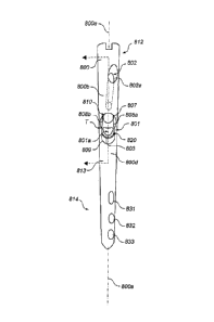
Referring to FIGS. 45-47, a universal femoral nail 800 defines a
reconstruction
aperture 801 for treating fractures or other injury to the femoral head and
neck in a
reconstruction mode and targets the femoral head and neck, as described above,
and an
antegrade aperture 802 for treating fractures of the femoral shaft in an
antegrade mode
and targets the lesser trochanter. The nail 800 includes a central long axis
800a, a head
800b formed at a proximal portion 812 of the nail 800 and a shaft 813
extending from the
head 800b to a distal portion 814 of the nail 800. The cross-sectional shape
of the head
800b in a plane perpendicular to the long axis 800a is generally non-circular.
As
illustrated in FIG. 50, the cross-sectional shape of the head 800b is
generally trapezoidal
and includes rounded portions. For example, at least a portion of a lateral
side 800d is
22
CA 02765376 2011-12-13
WO 2011/002903
PCT/US2010/040631
flat. However, the medial side 800e is generally rounded. As also illustrated
in FIG 50, a
medial to lateral plane M-L bisects the head 800b, includes a central through
axis 801a of
the reconstruction aperture, and includes the central long axis 800a of the
nail 800. Thus,
as illustrated, the medial to lateral plane M-L is coplanar with a corona]
plane of the nail
800 that separates the front of the nail from the back of the nail. It should
be noted that
this plane is not necessarily related to a coronal plane of a patient's body
or even a
coronal plane of a patient's femur. Additionally, in some implementations, the
reconstruction aperture 801 is not centrally disposed in the head 800b such
that the medial
to lateral plane M-L does not include the central through axis 801a, but the
medial to
to lateral plane M-L is then parallel to a central through axis 801a
of the reconstruction
aperture 801, and parallel to the coronal plane of the nail 800.
The reconstruction transverse aperture 801 is "light bulb" shaped and oriented
off
the long axis 800a of the nail 800, and is configured to receive a lag member
and a
compression member, such as the lag screw 202 and the compression screw 204
described
above. To target the femoral head and neck in the reconstruction mode, the
central
through axis 801a of the reconstruction aperture 801 lies in the medial to
lateral plane M-
L, and is oriented at an angle A of about 122 degrees relative to the central
long axis 800a.
The antegrade transverse aperture 802 is also oriented off the long axis 800a
by an angle
B, which is about 35 degrees. The antegrade aperture 802 is oriented such that
a central
through axis 802a of the antegrade aperture 802 lies in an antegrade plane AP,
which is
parallel to the long axis 800a and is radially offset from the medial to
lateral plane M-L
by an angle C of approximately 12 degrees. As illustrated in FIG. 50, the
antegrade
aperture 802 is not centered in the head 800b, such that the central through
axis 802a of
the antegrade aperture 802 does not intersect the long axis 800a of the nail
800. In some
implementations, and as illustrated in FIG. 50, the central through axis 802a
intersects the
medial to lateral plane M-L proximate the medial side 800e.
As shown in FIGS. 48 and 49, the nail 800 also includes three holes 831, 832,
and
833 located in a distal section 814 of the nail that, in use, can receive pins
or screws to
stabilize the distal section of the nail 800. The most proximal hole 831 is
formed as a
N slot, and the central and most distal holes 832 and 833 are
formed as circular holes.
When treating fractures of the femoral shaft, as discussed above, bone pins or
other
23
CA 02765376 2011-12-13
WO 2011/002903
PCT/US2010/040631
fasteners (not shown) are disposed within one or more of the three holes 831,
832, and
833 and secured to healthy bone.
The most proximal hole 831 and the most distal hole 833 are formed such that
respective central through axes 831a and 833a of the most proximal hole 831
and the
most distal hole 833 lie in planes that are parallel to the antegrade plane
AP. In other
words, the central through axes 831a and 833a are radially offset from the
medial to
lateral plane M-L by the same angle as the central through axis 802c of the
antegrade
aperture 802. Thus, the antegrade aperture 802, the most proximal hole 831 and
the most
distal hole 833 can be said to be parallel, or lie in parallel planes, even
though they may
be oriented differently with respect to the central axis 800a of the nail 800.
For example,
as discussed above, the central through axis 802a of the antegrade aperture
802 is oriented
at 35 degrees with respect to the central long axis 800a of the nail 800.
However, the
most proximal hole 831 and the most distal hole 833 may be formed at
approximately 90
degrees to the central long axis 800a, or at other angular orientations. In
some
implementations, the central though axis 831a of the most proximal hole 831
lies in the
same plane as the central through axis 833a of the most distal hole 833.
Additionally, the
central through axes 831a and 833a of the most proximal hole 831 and the most
distal
hole 833 can lie in the antegrade plane AP such that the central through axes
802a, 831a,
and 833a are coplanar.
A central through axis 832a of the central hole 832 is also radially offset
from the
medial to lateral plane M-L. However, the central through axis 832a is offset
from the
medial to lateral plane M-L by a different amount than the central through
axes 831a and
833a. For example, the central through axis 832a of the central hole 832 is
offset from
the medial to lateral plane M-L by 37 degrees, and is radially offset from the
antegrade
plane AP by 25 degrees.
When treating fractures of the neck, head, and intertrochanteric regions of
the
femur, the nail 800 is used in conjunction with first and second members, such
as the lag
screw 202 and the compression screw 204, received in the reconstruction
aperture 801.
When treating only a fracture in the femoral shaft, the nail 800 is used in
conjunction with
a bone pin received in the antegrade aperture 802. Running along the long axis
800a of
the nail 800 is a bore 816. A set screw (not shown) can be disposed in the
bore 816 for
locking the first and second members or the bone pin.
24
CA 02765376 2011-12-13
WO 2011/002903
PCT/US2010/040631
The reconstruction aperture 801 has a first semi-cylindrical aperture 810
associated with a first portion 811 (FIG. 46) of the reconstruction aperture
801, and a
second U-shaped aperture 820 associated with a second portion 821 (FIG. 46) of
the
reconstruction aperture 801. The nail 800 includes an inner wall 805 (FIG. 47)
that
defines the reconstruction aperture 801. The inner wall 805 includes a first,
semi-
cylindrical section 807 that defines the semi-cylindrical aperture 810 and a
second, U-
shaped section 809 that defines the U-shaped aperture 820. As shown, except
for a
shoulder 803, the reconstruction aperture 801 has a constant cross-sectional
shape along a
length dimension, L, of the reconstruction aperture 801. Shoulder 803 is
defined by an
outward step 818 in the U-shaped section 809.
The semi-cylindrical section 807 of the inner wall 805 comprises an arc
segment
that extends more than 180 degrees, for example, 270 degrees, and terminates
in two
opposing edges 808a and 808b. The plane between the opposing edges 808a and
808b
defines a face 841 of the semi-cylindrical section 807. The opposing edges
808a and
is 808b are located at a transition, T, between the semi-cylindrical
section 807 and the U-
shaped section 809 of the inner wall 805. Thus, the semi-cylindrical section
807 and the
U-shaped section 809 define a continuous surface of the reconstruction
aperture 801.
The U-shaped section 809 of the inner wall 805 includes a semi-cylindrical arc
segment 809a opposite the face 841 of the semi-cylindrical section 807 and two
mutually-
opposing walls 809b and 809c extending from the semi-cylindrical arc segment
809a.
The U-shaped section 809 of the inner wall 805 also includes a face 845
defined by the
plane between edges 809e and 809f of the walls 809b and 809c. As illustrated,
the face
845 of the U-shaped section 809 is coplanar with the face 841 of the semi-
cylindrical
section 807. The semi-cylindrical arc segment 809a includes a face 843 that
opposes the
face 841of the semi-cylindrical section 807 of the inner wall 805 (and the
face 845 of the
U-shaped section 809 of the inner wall 805), and is spaced therefrom by the
opposing
walls 809b and 809c.
In some implementations, the face 843 of the semi-cylindrical arc segment 809a
is
spaced from the first open face 841of the first semi-cylindrical aperture 810
by a distance
D such that a cylindrical member having a circular cross section of
substantially the same
diameter as the diameter of the semi-cylindrical arc segment 809a extends into
the first
portion 811 of the reconstruction aperture 801 when disposed in and abutting
the semi-
CA 0 2 7 6 5 3 7 6 2 0 1 1-1 2 ¨1 3
WO 2011/002903
PCT/US2010/040631
cylindrical arc segment 809a. For example, where the semi-cylindrical arc
segment 809a
is a 180 degree arc segment, the parallel walls 809b and 809c extend from the
semi-
cylindrical arc segment 809a (that is to say, from the face 843 of the semi-
cylindrical arc
segment 809a) the distance, D, which is less than the radius of the semi-
cylindrical arc
segment 809a. In some implementations, the diameter of the semi-cylindrical
arc
segment 809a is between about 5 millimeters and about 15 millimeters, and the
amount of
overlap of such a cylindrical member with a cylindrical member received within
the semi-
cylindrical section 807 is between about 1 millimeter and 5 millimeters.
As illustrated, the opposing walls 809b and 809c are parallel and the semi-
cylindrical arc segment 809a is a 180 degree arc segment. Alternatively,
however, the
opposing walls 809b and 809c can be divergent, and/or the semi-cylindrical arc
segment
809a can be an arc segment less than 180 degrees. Thus, when a member that is
sized to
fit within the semi-cylindrical arc segment 809a is disposed in the U-shaped
aperture 820,
the member is not constrained by a narrowing of the U-shaped aperture 820. As
such, a
member that is sized to fit within the semi-cylindrical arc segment 809a is
constrained
from moving into the semi-cylindrical aperture 810 only when a second member
is
disposed in the semi-cylindrical aperture 810. For example, when a compression
screw
204 is disposed within the U-shaped section 809 of the inner wall 805 and a
lag screw
202 is disposed within the semi-cylindrical section 807 of the inner wall 805,
the
compression screw 204 is constrained to remain in the U-shaped section 809,
and the lag
screw 202 and the compression screw 204 cooperate to resist a force moment
applied to
one or both of the lag screw 202 and the compression screw 204. However, if
the lag
screw 202 is not present within the semi-cylindrical section 807 of the inner
wall 805,
then the compression screw 204 can move in response to forces applied to the
compression screw 204, such that occurrence of bending or breaking of the
compression
screw 204 is reduced.
Referring additionally to FIGS. 48-52, the antegrade aperture 802 includes a
first
opening (or entry) 802a formed in a lateral side 800d of the nail 800 that is
proximal to a
first opening (or entry) 801a of the reconstruction aperture 801 formed in the
lateral side
800d of the nail 800. The first opening 801a of the reconstruction aperture
801 is
generally centered on the lateral side 800d of the nail 800 and the first
opening 802a of
the antegrade aperture 802 is not centered on the lateral side 800d of the
nail 800. The
26
CA 02765376 2011-12-13
WO 2011/002903
PCT/US2010/040631
non-circular cross-sectional shape of the head 800b with a larger lateral side
800d than
medial side 800e provides additional surface area for locating the first
opening 802a of
the antegrade aperture 802 off-center within the head 800b, and can provide
increased
strength compared to a head having a circular cross-sectional shape when the
antegrade
aperture 802 is oriented off the medial to lateral plane M-L.
Additionally, while the reconstruction aperture 801 is oriented in the
direction of a
femoral neck such that the second opening (or exit) 80 lb of the
reconstruction aperture
801 formed in a medial side 800e of the nail 800 is proximal to the first
opening 801a of
the reconstruction aperture 801, the antegrade aperture 802 is oriented
distally towards a
second opening (or exit) 802b that is formed in the inner wall 805 at a
location proximate
to the second opening 801b of the reconstruction aperture 801 formed in the
medial side
800e of the nail 800. However, in some implementations, the second opening
802b of the
antegrade aperture 802 can be formed in the medial side 800e of the nail 800,
and the
second opening 802b can be located proximally or distally of the second
opening 801b of
the reconstruction aperture 801.
As shown in FIGS. 51 and 52, the exit opening 802b is formed on the medial
side
800e of the nail 800 such that the exit opening 802b is contained entirely
within the exit
opening 80 lb of the reconstruction aperture 801. For given dimensions of the
antegrade
aperture 802 and the reconstruction aperture 801, the co-location of the exit
openings
802b and 801b reduces an amount of material that is removed from the medial
side 800e
of the nail 800. As mentioned above, the non-circular cross-sectional shape of
the head
800b allows for the co-location of the exit openings 802b and 801b in
conjunction with
the radial offset of the antegrade plane AP and the medial to lateral plane M-
L while
maintaining structural strength of the head 800b.
In some implementations, as illustrated in FIGS. 58 and 59, the head 800b of
the
nail 800 is angled from the shaft 813 by an angle D, such as a 5 degree angle.
The bend is
formed in the antegrade plane AP such that a tangent of the long axis 800a at
a location
831 in the head 800b makes an angle of approximately 5 degrees relative to a
tangent of
the long axis 800a at a location 833 in the shaft 813. As illustrated, the
head 800b and the
shaft 813 are both generally straight in the antegrade plane AP. As
illustrated in FIGS. 59
and 60, the nail 800 is curved perpendicular to the antegrade plane AP such
that the
27
CA 02765376 2011-12-13
WO 2011/002903
PCT/US2010/040631
antegrade plane AP is also curved. The curve illustrated in FIG. 60 is
compound, having
more than one radius of curvature perpendicular to the antegrade plane AP.
As those skilled in the art will appreciate, the particular implementations
described above and illustrated in the figures are provided for illustration,
and various
alterations may be made in the structure and materials of the illustrated
implementations.
For example, while the non-circular aperture of FIGS. 45-47 is illustrated
with circular
semi-cylindrical portions, the non-circular aperture can have semi-cylindrical
portions
having other cross-sectional shapes, such as oval or rectangular. Accordingly,
fastening
members with corresponding shapes, i.e., cylindrical fasteners having square,
rectangular,
oval, crescent, or other cross-sectional shapes can be used. Furthermore, the
non-circular
aperture may have additional portions, which may or may not be cylindrical.
Additionally, one or more of the apertures 831, 832, 833 located near the
distal end 800c
can be angled other than perpendicularly to the axis 800a having a first
opening located
proximally or distally of a second opening thereof. Furthermore, in general,
the cross-
sectional shape of the shaft 813 in a plane perpendicular to the long axis
800a is
substantially circular, although the diameter of the shaft 813 can be varied
along the long
axis 800a. For example, all or a portion of the shaft 813 can be tapered.
Also, the head
800b can be formed in other cross-sectional shapes, including circular, oval,
or polygonal,
for example. However, where other shapes are selected for the head 800b, the
medial to
lateral plane M-L still includes the central long axis 800a and is parallel to
or includes the
central through axis 801a of the reconstruction aperture 801. Additionally,
the transverse
aperture 801 can be oriented such that the angle A can be from about 110
degrees to about
150 degrees, or from about 120 degrees to about 130 degrees. The central
through axis
832a of the central hole 832 can be offset from the medial to lateral plane M-
L and the
antegrade plane AP by other amounts, such as by an angle from about 20 to
about 75
degrees, or from about 30 degrees to about 60 degrees. The angle C can be from
about 0
degrees to about 30 degrees, from about 0 degrees to about 20 degrees, or from
about 10
degrees to about 15 degrees. The angle D can be from about 0 to about 20
degrees, or
from about 0 to about 10 degrees. The central through axis 832a of the central
hole 832
can be radially offset from the antegrade plane by an angle from about 0 to
about 90
degrees, or from about 0 to about 45 degrees.
28