Note: Descriptions are shown in the official language in which they were submitted.
CA 02765457 2011-12-14
WO 2011/003182 PCT/CA2010/001023
METHODS FOR EARLY DETECTION OF BLOOD DISORDERS
CROSS REFERENCE TO RELATED APPLICATION
This application claims priority to and the benefit of U. S. Provisional
Application no. 61/223,573 filed on July 7, 2009, which is incorporated herein
in its
entirety.
FIELD
This disclosure relates to diagnostic screening and monitoring assays. More
particularly, this disclosure relates to diagnostic screening and monitoring
assays for
detecting blood disorders.
BACKGROUND
Hemoglobin is the iron-containing oxygen-transport metalloprotein in the red
blood cells of vertebrates, and the tissues of some invertebrates. The
chemical
formulae of hemoglobin vary- widely across species, and even slightly among
subgroups of humans. In adult humans, the most common hemoglobin type is a
tetramer called hemoglobin A. Hemoglobin A consist of non-covalently bound a
and
0 subunits. Mutations in the genes for the hemoglobin protein in a species
result in
hemoglobin variants. Hemoglobin variants are a part of the normal embryonic
and
fetal development, but mutant forms of hemoglobin in a population, may also be
caused by variations in genetics. Some well-known genetic hemoglobin variants
are
responsible for diseases such as sickle-cell anemia. A separate class of
diseases
known as thalassernias are caused by underproduction of normal and abnormal
hemoglobin and also, through problems with and mutations in globin gene
regulation.
To a small extent, hemoglobin A slowly combines with glucose at the
terminal valine of each 0 chain and the resulting molecule is often referred
to as
HbAlc. As the concentration of glucose in the blood increases, the percentage
of
hemoglobin A that turns into HbAlc increases. In diabetics whose glucose
usually
1
CA 02765457 2011-12-14
WO 2011/003182 PCT/CA2010/001023
runs high, the percent HbAlc also runs high. Long-term control of blood sugar
concentration can be measured by the concentration of HbAlc. A higher glucose
concentration results in more HbAlc. Because the reaction is slow, the HbAlc
proportion represents glucose level in blood averaged over the half-life of
red blood
cells, is typically 50-55 days.
Diabetes mellitus commonly known as diabetes, is a group of metabolic
diseases resulting in abnormally high blood sugar levels referred to as
hyperglycemia. Blood glucose levels are controlled by a complex interaction of
multiple chemicals and hormones in the body, including the hormone insulin.
More
specifically, Diabetes mellitus refers to a group of diseases that lead to
high blood
glucose levels due to defects in one of insulin secretion or insulin action.
Type 1
diabetes is a consequence of a diminished production of insulin while Type II
and
gestational diabetes are resistant to the effects of insulin. Type II diabetes
is the
most prevalent form of diabetes. Type II diabetes is often asymptomatic in its
early
stages and can remain undiagnosed for many years. Diabetes and its treatments
can
cause many complications. Acute complications exemplified by hypoglycemia,
ketoacidosis, or nonketotic hyperosmolar coma, may occur if the disease is not
adequately controlled. Serious long-term complications due to diabetes may
include
cardiovascular disease, chronic renal failure, retinal damage which can lead
to
blindness, several kinds of nerve damage, and micro-vascular damage, which may
cause erectile dysfunction and poor wound healing. In the developed world,
diabetes
is the most significant cause of adult blindness in the non-elderly and the
leading
cause of non-traumatic amputation in adults.
There is often a long, latent, asymptomatic period during which people with
Type II diabetes are undiagnosed. Most people are unaware they have Type II
diabetes, but experience physiological complications from the disease. Many
newly
diagnosed Type II diabetes cases already show evidence of micro-vascular
complications and serious effects and long term complications from the
disease.
Early detection of diabetes is essential and may help improve the outcome for
people
with Type II diabetes. Regular screening for diabetes will enhance quality and
2
CA 02765457 2011-12-14
WO 2011/003182 PCT/CA2010/001023
length of life for a diabetic person from reducing the severity and frequency
of
immediate effects or prevention and/or delay of long term complications.
Glycated substances are eliminated from the body slowly. Red blood cells,
which have a consistent lifespan of 120 days, are easily accessible for
measurement
of recent increased presence of glycating product. This fact is used in
monitoring
blood sugar control in diabetes by monitoring the glycated hemoglobin level,
also
known as HbAlc. Measurements of HbAlc in the 4-6% range are considered
normal, less than 7% is a well controlled diabetic, 7-8% is an average
diabetic and
greater than 8% is a poorly controlled diabetic. There are many known methods
to
screen for diabetes. The fast plasma glucose and oral glucose-tolerance tests
are
standard clinical tests. The fast plasma glucose test is measured in a blood
sample
taken after eight hours of complete fasting. The blood glucose tolerance test
is
measured in several blood samples taken at a series of intervals following the
administration of a specific glucose load. A current screening method referred
to as
the plasma glucose test does not require fasting and includes a blood and/or
urine
test that measures plasma glucose levels with enzymatic assay. Another common
screening method is to screen the blood for glycated hemoglobin (HbAlc) which
is
either based on charge difference between non-glycated and glycated hemoglobin
using ion-exchange chromatography, electrophoresis, or isoelectric focusing or
immunological methods employing antibodies against glycated N-terminal of (--
chain of the hemoglobin. Recently, the first molecular assay for glycated
hemoglobin was disclosed. The (3-chain of hemoglobin was digested with Glu-C,
providing an N-terminal hexapeptide which was measured using ElectroSpray
Ionization Liquid Chromatography Mass Spectrometry (ESI-LC/MS). The current
methods to screen and monitor for diabetes are expensive, laborious, and time-
consuming, require highly skilled operators, unreliable, and often require
repeat
testing.
There are many types of known hemoglobin (Hb) molecules and many are
associated with inherited blood disorders such as sickle cell, hemoglobin C, S-
C,
and E, thalassemia and analbuminaemia. The most common hemoglobin molecules
are HbA, HbA2, HbF, HbS, HbC, Hgb H, and Hgb M. Healthy adults only have
3
CA 02765457 2011-12-14
WO 2011/003182 PCT/CA2010/001023
significant levels of HbA and HbA2. Some people may also have small amounts of
HbF, which is the main type of hemoglobin in an unborn baby's body and certain
diseases are associated with high HbF levels. HbS is an abnormal form of
hemoglobin associated with sickle cell anemia. In adults, these hemoglobin
molecules make up the following percentages of total hemoglobin. Hgb Al: 95%
to
98%, Hgb A2: 2% to 3%, Hgb F: 0.8% to 2%, Hgb S: 0%, Hgb C: 0%. In infants
and children, these hemoglobin molecules make up the following percentages of
total hemoglobin, Hgb F (newborn): 50% to 80%, Hgb F (6 months): 8%, Hgb F
(over 6 months): 1% to 2%. The presence of significant levels of abnormal
hemoglobins may indicate hemoglobin C disease, rare hemoglobinopathy, sickle
cell
anemia, and thalassemia.
In general, people with these inherited blood disorders are physiologically
vulnerable and are at higher risk of infection, stroke, heart failure, liver
and acute
chest syndrome. The current method to screen and monitor for blood disorders
is
the hemoglobin electrophoresis diagnostic test. This test is a widely used
screening
test and if the presence of the blood disorder is indicated, a second
hemoglobin
electrophoresis diagnostic test is preformed to confirm the first diagnosis.
The
current test that is used to screen and monitor for blood disorders is
expensive,
laborious, and time-consuming, require highly skilled operators, unreliable,
and
requires repeat testing.
BRIEF DESCRIPTION OF THE DRAWINGS
The present disclosure will be described in conjunction with reference to the
following drawings in which:
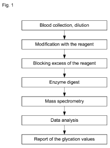
Fig. 1 is a schematic flowchart illustrating an exemplary method of the
present disclosure,
Fig. 2 is a flowchart illustrating an exemplary method of the present
disclosure for screening and monitoring for glycated and non-glycated
hemoglobin
and/or albumin, with an accompanying chart aligned with their mass spectra;
4
CA 02765457 2011-12-14
WO 2011/003182 PCT/CA2010/001023
Fig. 3 is a schematic illustrating amine reagent modification of the amino
acid sequences of albumin and/or hemoglobin N-terminal and/or lysine side
chains;
Fig. 4(a) is a chart illustrating an exemplary mass spectrum of in vitro, non-
glycated peaks of human serum albumin in phosphate buffered saline, 4(b) is a
chart
illustrating an exemplary mass spectrum of in vitro, glycated peaks of human
serum
albumin in phosphate buffered saline, 4(c) is a chart illustrating an
exemplary mass
spectrum of in vitro, non-glycated peaks of blood in phosphate buffered
saline, and
4(d) is a chart illustrating an exemplary mass spectrum of in vitro, glycated
peaks of
blood in phosphate buffered saline;
Fig. 5 is a chart illustrating the standard curve using liquid whole blood
samples;
Fig. 6 is a chart comparing % glycation results of an exemplary method of
the present disclosure with % glycation results of a prior art assay;
Fig. 7 is a mass spectrum of a blood sample produced with an exemplary
method of the present disclosure to screen and monitor for non-glycated and
glycated peaks representative of Type I and Type II diabetes,
Fig. 8 is a chart illustrating the standard curve using whole blood dried
blood
spot samples;
Fig. 9 is a chart comparing isotopic cluster area ratio with height ratio
results
of an exemplary method of the present disclosure;
Fig. 10(a) is a mass spectrum of a whole blood sample produced with
mutations in both S genes of hemoglobin with no modification reagent, 10(b) is
a
mass spectrum of the 906m/z to 988 m/z region,
Fig. 11(a) is a mass spectrum of a whole blood sample produced with
mutations in both S genes of hemoglobin with an exemplary method of the
present
disclosure to screen and monitor for variants, 11(b) is a mass spectrum of the
3188m/z to 3680 m/z region,
5
CA 02765457 2011-12-14
WO 2011/003182 PCT/CA2010/001023
Fig. 12(a) is a mass spectrum of a whole blood sample produced with a
mutation in one C gene of hemoglobin with no modification reagent, 12(b) is a
mass
spectrum of the 853m/z to 983 m/z region,
Fig. 13(a) is a mass spectrum of a whole blood sample produced with a
mutation in one C gene of hemoglobin with an exemplary method of the present
disclosure to screen and monitor for variants, 13(b) is a mass spectrum of the
2940m/z to 3726 m/z region,
Fig. 14(a) is a mass spectrum of a whole blood double mutant gene sample
produced with no modification reagent as in the exemplary method of the
present
disclosure to screen and monitor for variants, 14(b) is a mass spectrum of the
879m/z to 977m/z region, and
Fig. 15(a) is a mass spectrum of a whole blood double mutant gene sample
produced with an exemplary method of the present disclosure to screen and
monitor
for variants, 15(b) is a mass spectrum of the 3316m/z to 3803m/z region.
SEQUENCE LISTING
SEQ ID NO: 1 is a mutant hemoglobin S peptide identified using the
methods provided herein.
SEQ ID NOS: 2-3 are mutant and normal hemoglobin C peptide fragments,
respectively.
SEQ ID NOS: 4-5 are normal and mutant hemoglobin C peptides,
respectively.
SEQ ID NO: 6 is a mutant hemoglobin C peptide fragment.
DETAILED DESCRIPTION
The exemplary embodiments of the present disclosure relate to methods for
screening and monitoring blood samples for detection of disorders.
6
CA 02765457 2011-12-14
WO 2011/003182 PCT/CA2010/001023
As used herein, "MALDI MS" refers to Matrix-Assisted Laser Desorption /
Ionization Mass Spectrometry.
As used herein, "HbA1c" refers to glycated hemoglobin in a subject's blood;
As used herein, "N-terminal and/or N-terminus" refers to the end of a protein
or polypeptide terminated by an amino acid with a free amine group;
As used herein, "Calibration Curve" refers to a plot showing an instrument's
responses, i.e., analytical signal, with the concentration of the analyte
(i.e., the
substance to be measured). An operator prepares a series of standards across a
range
of concentrations approximate the anticipated concentration of analyte in
subjects'
samples. The concentrations of the standards must lie within the working range
of
the technique (i.e., instrumentation) being used. Analyzing each of these
standards
using the chosen technique will produce a series of measurements. For most
analyses, a plot of instrument response vs. analyte concentration will show a
linear
relationship. An operator can assess the data generated from the samples and
in
reference to the calibration curve, perform an interpolation to determine the
analyte
concentrations in the samples.
The present disclosure relates to methods for screening and monitoring for
Type I and Type II diabetes and blood disorders. An exemplary method for
monitoring and screening for blood disorders according to one embodiment of
the
present disclosure is illustrated in Fig. 1. A subject's blood is collected in
small
quantities via blood spots and/or liquid blood samples, then diluted and
modified
with an amine reagent wherein the available amino groups of albumin or
hemoglobin proteins are reacted with the amine reactive reagent thereby
modifying
the N-terminal and/or lysine side chains of the albumin and/or hemoglobin
moieties.
A blocking agent is then applied to the sample which is subsequently digested
with
an proteolytic enzyme. Suitable proteolytic enzymes that may be used when it
is
desirable to have proteolysis hindered by lysine groups modified by the amine
agent,
are exemplified by trypsin and Lys-C endoproteinase. Suitable proteolytic
enzymes
that may be used when it is desirable to have proteolysis unhindered by
modified
7
CA 02765457 2011-12-14
WO 2011/003182 PCT/CA2010/001023
IN-sine groups. are exemplified by trypsin and Arg-C endoproteinase, Asp-N
endoproteinase, pepsin, chymotrypsin, papain, and elastase. Ion signals
corresponding to specific pairs of glycated and non-glycated N-terminal
peptides are
detected and measured using MALDI MS.
The disclosure described herein demonstrates that changes in glycation can
be used to screen and monitor the population for Type I and Type II diabetes
and
other blood disorders.
Sample Preparation
It is known in the prior art that MALDI MS analyses can be conducted with
biological fluids exemplified by blood, without pre-processing the biological
fluid
samples. Suitable biological fluids are exemplified by blood. One exemplary
method of the present disclosure relates to processing a blood sample drawn
from a
subject. The blood sample may be diluted prior to processing. Alternatively,
the
blood sample may be placed onto a piece of paper and allowed to dry. The blood
sample is then modified with an exemplary amine reagent wherein the amines
preferentially bind to the alpha amino groups of N-terminals and/or epsilon
amino
groups of lysine side chains. The next step is application of a blocking
reagent
followed by digestion with an enzyme whereby the proteins are cleaved.
Suitable
proteolytic enzymes that may be used when it is desirable to have proteolysis
hindered by lysine groups modified by the amine agent, are exemplified by
trypsin
and Lys-C endoproteinase. Suitable proteolytic enzymes that may be used when
it is
desirable to have proteolysis unhindered by modified lysine groups. are
exemplified
by trypsin and Arg-C endoproteinase, Asp-N endoproteinase, pepsin,
chymotrypsin,
papain, and elastase. The cleaved proteins, now referred to as peptides, are
analysed
with mass spectrometry wherein the ion signals corresponding to the specific
pairs
of glycated and non-glycated N-terminal peptides are measured.
Mass Spectrometric Analysis
The use of MALDI MS allows the extent of glycation to be rapidly obtained
and further allows individual proteins of interest to be identified and
quantified
8
CA 02765457 2011-12-14
WO 2011/003182 PCT/CA2010/001023
within a biological sample. The proteins of interest can be converted to
peptides,
and it is the peptides that are analyzed to give a corresponding ion signal.
The
matrix used during MALDI MS analysis can include many types of suitable
organic
molecules exemplified by a-cyanocinnamic acid and other like materials
suitable for
absorbing energy from a laser. The laser may be a standard nitrogen laser or
any
laser known in the art. As known in the prior art, mass spectrometry peaks are
analyzed by determining ion signals attributes such as peak heights and/or
area
defined by the peak (relative to baselines). When two peaks are compared,
typically
a ratio is determined. A peak ratio can be determined from independent sets of
reactions from one or more samples for comparison and can be used over time to
screen and/or monitor the individual for Type I or Type II diabetes and
various
blood disorders. The extent of glycation can be analyzed and compared through
a
variety of calculations that are readily used to those skilled in the art. For
example,
the extent of glycation can be analyzed by comparing the peak height of a
glycated
peptide to that of a non-glycated peptide as shown in the formula,
% Glycation = Glycated Peptide Intensity X
100 (2)
(Non-Glycated Peptide Intensity + Glycated Peptide Intensity)
According to another exemplary embodiment, a peptide profile can be
analyzed by comparing the peak area of an individual peak to the peak areas of
other
individual peaks. In other examples, the peak area or height of individual
peaks or
combination of peaks may be compared to the peak height or area of an
individual
peak or a combination of peaks in a peptide profile. Accordingly, the
exemplary
methods of the present disclosure include any combination of calculations
performed on one or more peaks within a peptide profile that enable
comparisons of
one or more peaks to another peak or peaks in the same peptide profile or
different
peptide profiles.
The exemplary disclosure illustrates certain mass spectrometric peaks
observed in blood samples after being modified are indicative of the presence
or
absence of variants, including but not inclusive of sickle cell anemia, and
diabetes.
9
CA 02765457 2011-12-14
WO 2011/003182 PCT/CA2010/001023
Examples of peak ratios in a peptide profile that can screen for Type I and
Type II
diabetes that originate from human blood and are analyzed by MALDI MS include
but are not limited to, peaks at m/z values from about 3000 for glycated peaks
to
about 4000 for non-glycated peaks.
Screening and Monitoring for Type I and Type II Diabetes and Blood Disorders
Certain embodiments of the present disclosure relate to the screening and
monitoring methods of diabetes and blood disorders. Certain methods of the
disclosure are useful for screening, monitoring, evaluating, and controlling
the
presence, absence or severity of diabetes and blood disorders. The peptide
profile
may also be used to detect a change in health status. Comparison of an
individual
peptide profile to a predetermined baseline is useful as a predictor of a
change on the
health status of an individual. For example, an exemplary diabetes screening
method may comprise monitoring a peptide profile over a selected time period
to
enable assessment of therapy efficacy. For example, the progress of a patient
being
treated for diabetes could be monitored using the methods described herein to
determine if a therapeutic scheme was able to decrease the change in the
patient's
peptide profile.
While the technology can be applied to measure the extent of glycation of
glycopeptides, glycoproteins and glycolipids, it is especially useful in the
field of
screening, and monitoring where it can be applied to diabetes and blood
disorder
testing. The exemplary technology relates to a specific, robust, rapid, low
cost,
automated, direct molecular method for simultaneous quantification of
hemoglobin
and other blood protein glycation in whole blood by MALDI MS.
Kits
Certain exemplary embodiments of the present disclosure relate to kits
comprising elements configured to facilitate certain assay steps. Such kits
may relate
to the collection, storage, or shipping of biological samples to screen and
monitor for
Type I and Type II diabetes and blood disorders. Generally, an exemplary kit
can
include a container, a matrix, matrix solution and/or pre-spotted MALDI
targets and
CA 02765457 2011-12-14
WO 2011/003182 PCT/CA2010/001023
one amine reagent. An exemplary kit can also comprise packaging material,
instructions, a storage buffer, and/or materials on which the sample can be
dried. An
exemplary patient kit can include a container, packaging material,
instructions, a
storage buffer or material on which the sample can be dried. Exemplary kits
can
also be used to collect the biological samples. Suitable samples are
exemplified by
blood and the like.
MALDI-MS techniques and instrumentation
Those skilled in the art will understand that similar useful results are
obtainable with methods adapted for use with mass spectrometry instruments
exemplified by MALDI-QQQ (triple quadrupole), MALDI-Q-TOF (quadrupole
time-of-flight), MALDI-iontrap-TOF (time-of-flight), MALDI-FTICR-MS (Fourier
transform ion cyclotron resonance) and the like, using mass spectrometric
techniques exemplified by single reaction monitoring, multiple reaction
monitoring
and the like.
Example 1
An exemplary method according to the present disclosure shown in Fig. 2
illustrates the steps of modifying proteins in a biological sample with an
exemplary
amine reagent and then analyzing the modified proteins with mass spectrometry.
The first step comprises collecting a blood sample either via a dry blood spot
and/or
a liquid blood sample. The sample is then modified with an exemplary amine
reagent wherein the amines preferentially bind to the alpha amino groups of N-
terminals and/or epsilon amino groups of lysine side chains. The next step is
application of a blocking reagent followed by digestion with trypsin whereby
the
proteins are cleaved. The cleaved proteins, now referred to as peptides, are
analysed
with MALDI MS wherein the ion signals corresponding to the specific pairs of
glycated and non-glycated N-terminal peptides are measured. The mass spectrum
of
a non-glycated peptide is shown on the left side of the chart shown in Fig. 2,
and is
not indicative of diabetes. The ratio of glycated peptide sample indicative of
diabetes is illustrated in the mass spectra in the right hand side of the
chart in Fig. 2.
11
CA 02765457 2011-12-14
WO 2011/003182 PCT/CA2010/001023
The chart in Fig. 2 shows that the mass of the glucose modified peptide is
different
than the non-glucose modified peptide.
Example 2
Blood is drawn from a subject and is diluted or placed directly on a paper
sheet exemplified by tissue paper. Blood from a subject is obtained by a
finger prick
and a drop is collected on a small piece of paper. The blood sample is then
dried on
the paper piece and is subsequently stored at room temperature. Suitable dry
blood
spot samples can be as small as and less than 1 l. Biological fluids that are
analyzed according to this method of the disclosure preferably include whole
blood.
Example 3
For the purpose of the examples discussed herein, an exemplary amine
reagent was prepared and incubated with whole blood as follows. About 0.2 l
of
0.3M solution of pyridinecarboxylic acid N-hydroxysuccinimide ester in
dimethylsulfoxide and 0.2 pl 1M tri ethyl carbonate buffer, pH 8.0 was added
to 10 l
of whole blood diluted 1:100 with distilled water. The reaction mixture was
incubated at room temperature (25 C) for 30 minutes. 1 pl of 1M ammonium
bicarbonate was added and mixture was further incubated for 30 min at room
temperature.
Amine reagents for the modification of amino groups having the general
formula R1-R2, wherein RI is a reactive group specific for modification of
primary-
amino groups, and R2 is a modifying group which is conjugated to the amino
groups
through the reactive group as a result of the reaction.
Suitable, exemplary R1 groups include among others, N-
hvdroxvsuccinimidvl ester; N-hvdroxvsulfosuccinimidvl ester; isothiocvanate;
pentafluorophenyl ester; sulfotetrafluorophenyl ester; sulfonyl chloride; p-
nitrophenyl, and aldehyde.
R2 groups are selected for the specific carbohydrate to be measured and can
be any group which, following conjugation will produce mass increment
differing
12
CA 02765457 2011-12-14
WO 2011/003182 PCT/CA2010/001023
from the mass of the carbohydrate of interest. R2 groups preferably bear a
partial or
full positive charge. In the case of pyridinecarboxylic acid N-
hydroxysuccinimide
(PCAS), R2 differs from 105 Daltons, as this is the mass increment of PCAS.
Suitable, exemplary R2 groups include among others, pyridinyl, piperidinyl,
N-ally lpiperidinyl; piperazinyl, N-ally lpiperazinyl; imidazolyl; N-
alkylimidazolyl;
dialky lamine; and trialkvlamine.
0.5 pl of 1mg/nil trypsin solution in 0.1% acetic acid was added to the
mixture and mixture was incubated overnight at room temperature. 0.1 l of the
reaction mixture was spotted onto a MALDI plate and allowed to dry. The spot
was
overlaid with 0.3 pl of 5mg/nil a-cyanocinnamic acid matrix solution in 0.1%
trifluoroacetic acid 50% acetonitrile. The dried spot was analyzed by MALDI
MS.
Following the modification reaction, blocking of excessive reagent, trypsin
digestions and MALDI MS, the following N-terminal peptides were obtained, with
the small captions signifying the modification of N-terminal or lysine
residues:
dAHkSEVAHR N-terminal peptide for albumin,
vLSPADkTNVkAAWGkVGAHAGEYGAEALER N-terminal peptide for a-
chain of hemoglobin, and,
vHLTPEEkSAVTALWGkVNVDEVGGEALGR N-terminal peptides for 13-
chain of haemoglobin.
Example 4
An exemplary example of the present disclosure relates to the exemplary
amine modification method of the present disclosure. Glucose can be non-
enzymatically covalently bound to proteins including hemoglobin. The levels of
glycated hemoglobin are used as a diagnostic parameter and can screen and/or
monitor blood disorders. HbAlc refers to hemoglobin that is glycated at the (3-
chain
N-terminus. An amine reagent modifies the free N-terminal and/or lysine side
chain
amine groups. Fig. 3 shows the free N-terminal and lysine side chains that
have
been modified with an amine reagent to produce a weight of 3533 Daltons for
the
13
CA 02765457 2011-12-14
WO 2011/003182 PCT/CA2010/001023
glycated (3 -hemoglobin peptide with lysine modifications and 3476 Daltons for
the
(3-hemoglobin peptide that is modified at the N-terminal and the lysine side
chains.
Those skilled in these arts will understand how to configure suitable amine
reagents
for producing glycated and non-glycated moieties that are separated by at
least 10
Daltons by MALDI MS.
Example 5
An exemplary method for screening and/or monitoring an individual for
Type I and Type II diabetes and various blood disorders relates to detecting
glycated
peptides using MALDI MS. The peptide profiles in Fig. 4 were extremely
reproducible and the results relate to the use of the present disclosure as a
screening
and monitoring method for Type I and Type II diabetes and blood disorders. 20
l
of 3 mg/ml solutions of purified human serum albumin in PBS (Fig. 4(a) without
glucose and Fig. 4(b) with glucose) and 1:200 diluted using H2O of blood in
PBS
(Fig. 4(c) without glucose and Fig. 4(d) with glucose) with and without 0.5M
glucose were heated at 100 C 10 minutes in water bath. After cooling on ice,
the
samples were modified with NHS-pyridinecarboxylic acid for 30 minutes at 25 C
after which, excess reagent was blocked with 0.1M ammonium bicarbonate for 30
minutes at 25 C. Samples were digested overnight at 25 C with 1 pg of trypsin
and
then measured by MALDI MS. MALDI MS showed in vitro glycation of the
hemoglobin and albumin proteins resulting in appearance of the peaks
corresponding to the glycated forms of the proteins (Fig. 4(b) and Fig. 4(d)).
The
glycated peaks corresponding to masses of 1416.70 m/z for human serum albumin
and 3533.04 m/z for blood illustrated peptides with mass of 57 Dalton higher
than
mass of the peaks corresponding to the non-glycated form of the peptides.
Peaks are
well resolved and the intensities ratio is easily quantifiable. There are no
interfering
peaks in the mass range of glycated form of the N-terminal hemoglobin peptides
for
trypsin digest of blood sample, Fig. 4(d).
Example 6
14
CA 02765457 2011-12-14
WO 2011/003182 PCT/CA2010/001023
An exemplary example of the present disclosure relates to a method to
calibrate a standard curve that can be used to analyze unknown samples. Six
liquid
standard samples, were used to formulate the standard calibration curve
illustrated in
Fig. 5. The calibration curve has known 4% and 11% glycated samples. The known
4% and 11% glycated samples are mixed to produce the standard calibration
samples. The intensities of the non-glycated and glycated peptides were
measures
and the ratio was determined according to formula (3).
Peptide Ratio = Ig
(3)
(Ig + In)
where Ig and In are intensities or cluster peak areas of the glycated and non-
glycated
peaks respectively.
Example 7
An exemplary example of the present disclosure relates to the correlation
between the % glycation determined by a prior art assay and the % glycation of
the
hemoglobin determined by the exemplary MADLI MS assay as illustrated in Fig.
6.
The prior art assay is a immune assay which treats the sampled blood with
antibodies against glycated hemoglobin and measures the amount of % glycated
hemoglobin through changes in optical absorbance. During the MALDI MS assay
the liquid and/or dried blood samples are treated with an amine reagent,
blocking
reagent and trypsin digest and then analyzed using MADLI MS to detect
glycation
of hemoglobin. Measurements of HbAlc in the 4-6% range are considered normal,
less than 7% is a well-controlled diabetic, 7-8% is an average diabetic and
greater
than 8% is a poorly controlled diabetic. Fig. 6 illustrates a linear curve
with a line
equation being y=1.1713x - 0.8276, with R2=0.8976. The R squared indicates how
accurate the regression line is. The closer R squared is to 1, the more
accurate the
regression line is to sample data.
Example 8
CA 02765457 2011-12-14
WO 2011/003182 PCT/CA2010/001023
Peptide peaks were identified from the observed m/z values and the known
masses of the N-terminal (3-chain of hemoglobin using the exemplary method
described in this disclosure to screen and/or monitor for diabetes and blood
disorders. Fig. 7 illustrates that the individual in question has non-glycated
HbAlc
peaks at 3476 Daltons and glycated HbAlc peaks at 3533 Daltons from which
ratio
of intensities can be calculated using formula (4).
Peptide Ratio = Hg
(4)
(Hg + Hn)
where Hg and Hn are intensities or cluster peak heights of the glycated and
non-
glycated peaks respectively.
Example 9
An exemplary example of the present disclosure relates to the method to
calibrate a standard curve that can be used to analyze unknown samples. Six
liquid
blood standard samples, were used to formulate the standard calibration curve
illustrated in Fig. 8. The calibration curve has known 4% and 11% glycated
samples. The known 4% and 11% glycated samples are mixed to produce the
standard calibration samples and then dried on tissue paper. The intensities
of the
dried blood spots of the non-glycated and glycated peptides were measures
using
MALDI MS and the ratio was determined according to formula (4).
Example 10
An exemplary example of the present disclosure relates to the estimation of
the reproducibility of the assay for the Isotopic Cluster Area Ratios or the
Height
Ratios determined by the exemplary method using dried blood spots. The same
sample of standard mixture containing 5.4% HbAlc was equally applied to eight
spots on a MALDI plate and each spot was analysed using an exemplary method of
the present disclosure. From this type of analysis, statistical parameters of
the
measurements can be estimated.
Example 11
16
CA 02765457 2011-12-14
WO 2011/003182 PCT/CA2010/001023
Peptide peaks of a whole dried blood sample i ere analyzed for a known
sample that had mutations in both hemoglobin S genes. The whole blood sample
was diluted in distilled water (1:100) and not modified using the exemplary
disclosure. The mutant peptide that was not identified using MALDI MS has the
amino acid sequence VHLTPVEK (SEQ ID NO: 6), while it is known in the prior
art that the normal peptide sequence is VHLTPEEK (SEQ ID NO: 3). Fig. 10 (a)
and (b) illustrates that no peptide is present at 922.54 m/z thus indicating
that the
mutated peptide is not detected using the standard MALDI procedure known in
the
prior art.
Example 12
Peptide peaks of a whole dried blood sample i ere analyzed for a known
sample that had mutations in both hemoglobin S genes. The whole blood sample
was diluted in distilled water (1:100) and modified using the amine reagent,
blocking agent and trypsin digest as explained in the exemplary method of the
present disclosure. The peptide that was identified using the exemplary method
has
the amino acid sequence
VHLTPVEKSAVTALWGKVNVDEVGGEALGR (SEQ ID NO: 1).
The PCAS amine reagent bound to the IN-sine and N-terminus, thus illustrating
that a
peptide at 3446.63 m/z is present, as shown in Figs. 11 (a) and (b).
Example 13
Peptide peaks of a whole dried blood sample i ere analyzed for a known
sample that had a normal gene and a mutation in the hemoglobin C gene. The
whole
blood sample was diluted in distilled water (1:100) and not modified using the
exemplary method. The peptide that was not identified using MALDI MS has the
amino acid sequence VHLTPKEK (SEQ ID NO: 2), while it is known in the prior
art that the normal peptide sequence is VHLTPEEK (SEQ ID NO: 3). Fig. 12 (a)
and (b) illustrates that no peptide is present at 951.56 m/z indicating that
the
17
CA 02765457 2011-12-14
WO 2011/003182 PCT/CA2010/001023
mutated peptide is not detected using the standard MALDI procedure known in
the
prior art.
Example 14
Peptide peaks of a whole dried blood sample were analyzed for a known
sample that had a normal gene and a mutation in the hemoglobin C gene. The
whole
blood sample was diluted in distilled water (1:100) and modified using the
amine
reagent, blocking agent and trypsin digest as explained in the exemplary
method of
the present disclosure. The normal peptide and the mutated peptide were
identified
using the exemplary method and has the amino acid sequence
VHLTPEEKSAVTALWGKVNVDEVGGEALGR (SEQ ID NO: 4) and
VHLTPKEKSAVTALWGKVNVDEVGGEALGR (SEQ ID NO: 5) respectively.
The PCAS amine reagent bound to the lysine and N-terminus, illustrating the
normal
heterozygous peptide at 3476.90 m/z and the mutated modified peptide at
3580.99
m/z, as shown in Fig. 13 (a) and (b).
Example 15
Peptide peaks of a whole dried blood sample were analyzed for a known
sample that had a double mutant, one mutation in the S gene and one mutation
in the
C gene of hemoglobin. The whole blood sample was diluted in distilled water
(1: 100) and not modified using the exemplary method of the present
disclosure. The
peptides that were not identified using MALDI MS had the amino acid sequence
VHLTPVEK (SEQ ID NO: 6) and VHLTPKEK (SEQ ID NO: 2), while it is known
in the prior art that the normal peptide sequence is VHLTPEEK (SEQ ID NO: 3).
Fig. 14 (a) and (b) illustrates that no peptides are present at 922.54 m/z and
951.56
m/z indicating the mutated S and C peptides respectively, are not detected
using the
standard MALDI procedure known in the prior art.
Example 16
Peptide peaks of a whole dried blood sample were analyzed for a known
sample that had a double mutantation, one mutation in the S gene and one
mutation
18
CA 02765457 2011-12-14
WO 2011/003182 PCT/CA2010/001023
in the C gene of hemoglobin. The whole blood sample was diluted in distilled
water
(1:100) and modified using the amine reagent, blocking agent and trypsin
digest as
explained in the exemplary method of the present disclosure. The S and C
mutant
peptides were identified using the exemplary method and have the amino acid
sequences VHLTPVEKSAVTALWGKVNVDEVGGEALGR (SEQ ID NO: 1) and
VHLTPKEKSAVTALWGKVNVDEVGGEALGR (SEQ ID NO: 5) respectively.
The PCAS amine reagent bound to the lysine and N-terminus, illustrating the
mutant
S peptide at 3446.61 m/z and the mutant C peptide at 3580.70 m/z, as shown in
Fig.
(a) and (b).
19