Note: Descriptions are shown in the official language in which they were submitted.
CA 02765732 2011-12-15
WO 2010/149292 PCT/EP2010/003579
1
A BIOLOGICAL MICROFLUIDICS CHIP AND RELATED METHODS
This invention relates to a biological microfluidics chip, and methods of
using such a
biological microfluidics chip. For convenience, a biological microfluidic chip
may be
referred to as a "biochip".
In biological and biomedical scientific research it is commonplace to employ
cultivation
receptacles in which researchers cultivate cell cultures or embryos intended
for study.
One very common form of cultivation receptacle is a so-called microtitre plate
that
1o typically contains straight-sided cylindrical wells formed in a plate, the
latter being of
standardized shaped and dimensions for locking retention in an analytical
apparatus or
robotic handler.
In practice, a microtitre plate typically includes an array of microtitre
wells set in a grid-
like pattern. One well known arrangement includes 96 microtitre wells defining
an array
of 8 rows containing 12 microtitre wells each. The design of the 96-well plate
has
become an industry standard format, specified by the Society for Biomolecular
Screening.
A microtitre plate is typically manufactured from a transparent polymer such
as
acrylonitrile-butadiene-styrene ('ABS'). The transparency permits researchers
to perform
various optical tests on cells, embryos or larvae cultivated in the microtitre
wells. In
addition microtitre wells are suitable for carrying out numerous tests and
investigations
that do not involve cell material.
The microtitre wells are open-ended at their in-use upper ends. Electronically-
controlled
dosing apparatuses may be employed to inject each of the wells of a microtitre
plate with
a culture solution and e.g. reagents, enzymes or other additives the effect of
which on
cells in the microtitre wells it is desired to study.
As used herein terms such as "upper', "above", "lower", "vertical',
"horizontal', "upwardly'
and "downwardly' for convenience are construed with reference to a microtitre
well or
biological microfluidics chip in its operating orientation, as would arise
when a biochip is
placed flat on a horizontal surface such as a laboratory bench and the opening
of the
well is directed upwards. It is however recognised that in use of a biochip or
other cell
cultivation receptacle its orientation may change e.g. as a result of being
centrifuged or
otherwise agitated, or by reason of being tilted or inverted as part of an
experimental or
CONFIRMATION COPY
CA 02765732 2011-12-15
WO 2010/149292 PCT/EP2010/003579
2
observational procedure. The terms mentioned, and related terms, are not to be
construed as limiting the scope of the invention to any particular orientation
of the
cultivation receptacles, or to any particular mode of use.
The paper entitled "Microfluidic system for on-chip high-throughput whole-
animal sorting
and screening at subcellular resolution" by Christopher B. Rohde, et al
published in
PNAS, August 28, 2007, vol. 104, no. 35, 13891-13895 discloses microfluidic
devices
consisting of flow and control layers made from flexible polymers. The flow
layers contain
microchannels for manipulating C. elegans, immobilizing them for imaging, and
1o delivering media and reagents. The flow layers also contain microchambers
for
incubating the animals. The control layers consist of microchannels that when
pressurized, flex a membrane into the flow channels, blocking or redirecting
the flow.
Animals in the flow lines can be imaged through a transparent glass substrate
using
highresolution microscopy.
The listing or discussion of a prior-published document or any background in
this
specification should not necessarily be taken as an acknowledgement that the
document
or background is part of the state of the art or is common general knowledge.
One or
more aspects/embodiments of the present disclosure may or may not address one
or
more of the background issues.
According to first aspect of the invention, there is provided a biological
microfluidics chip
comprising:
a substrate;
a microfluidic inlet port defining an opening in a surface of the substrate;
a microfluidic outlet port defining an opening in a surface of the substrate;
and
a plurality of wells extending from a top surface of the substrate, wherein
each
well is bounded by one or more walls, and an inlet opening and an outlet
opening are
provided in a wall of each of the plurality of wells;
one or more microfluidic inlet channels in the substrate that connect the
microfluidic inlet port to each of the inlet openings in the walls of the
wells; and
one or more microfluidic outlet channels in the substrate that connect the
outlet
openings in the walls of the wells to the microfluidic outlet port.
In conventional use, the inlet channel will convey fluid towards the well, and
the outlet
channel will convey fluid away from the well. However, it is possible to
reverse this flow if
required
CA 02765732 2011-12-15
WO 2010/149292 PCT/EP2010/003579
3
The biological microfluidics chip (biochip) can be used for the culture and
study of cells,
embryos and larvae located in the wells, and the microfluidic channels can be
used to
provide the wells with one or more microfluids, and also to remove one or more
microfluids from the wells. In one example, drugs or other compounds and/or
nutrients
can be provided into a well for use in an experiment. In some embodiments,
biological
waste, metabolites and/or bacteria that would otherwise influence experiments
performed in the wells can be removed from the wells through a microfluidics
outlet
channel.
Biochips according to embodiments of the invention can enable improved
scientific
experiments to be performed in the wells of the biochip, and more accurate and
reliable
results to be achieved at lower cost and higher speed.
The walls of the plurality of wells may comprise lateral boundaries, which may
be
considered as side walls, and may also comprise a bottom wall that may be
considered
as a floor of the well. The walls may be flat/planar or curved.
It will be appreciated that the term "microfluidics" is associated with the
behavior, precise
control and manipulation of fluids that are geometrically constrained to a
small, typically
sub-millimeter, scale.
The length of a microfluidic channel between the microfluidic inlet port and
an inlet
opening in a wall of a well may be substantially the same for each well, or a
subset of
wells. That is, although some of the wells may be further from the inlet port,
the channel
length between the inlet port and the opening for each well can be
substantially the
same.
In this way, the pressure, and hence the flow-rate, of fluids that are
provided to the wells
by a microfluidic channel, may be substantially the same for each of the
individual wells.
This can reduce the chances of cross-contamination of the contents of
different wells and
ensure that the same environmental conditions can be provided for each of the
wells.
One or more biological microfluidics chips disclosed herein can enable
reliable,
reproducible experiments to be performed in a plurality of different wells at
the same time
under the same conditions.
CA 02765732 2011-12-15
WO 2010/149292 PCT/EP2010/003579
4
Similarly, the length of the microfluidic channels between each of the outlet
openings in
the walls of the wells and the microfluidic outlet port may be substantially
the same for
each well. In the same way as described above, this can ensure that fluids are
removed
from the wells at the same flow-rate for each well.
In some examples, the microfluidic channels may be of variable length so that
the wells
furthest from the port receive fluid at the same time, and under the same flow
rate, as the
wells nearest the inlet port. In each case, the same variable length can be
applied to the
corresponding microfluidic channel and each of the inlet/outlet openings in a
well.
The biological microfluidics chip may further comprise:
a temperature control inlet port and a temperature control outlet port, and
a temperature control channel configured to transport temperature control
fluid
from the temperature control inlet port to the temperature control outlet port
along a path
that is in proximity to one or more of the plurality of wells such that, in
use, heat is
exchangeable between the temperature control liquid and the contents of the
wells.
Providing such an in-built temperature control system can enable the biochip
to be
moved during an experiment, and the biochip is not restricted to a specific
location
because of a separate temperature control device.
Heat may be exchangeable in order to either heat or cool the contents of the
well in use,
and this may provide a stable and constant temperature among all wells.
The temperature control liquid may be an aqueous medium, an oil, or any other
fluid that
is suitable for maintaining a desired temperature as it travels along the
temperature
control channel.
One or more of the temperature control inlet port, temperature control outlet
port and
temperature control channels may be provided for a subset of the plurality of
wells, and
this can enable the temperature of individual wells, or subsets of wells to be
independently controlled.
In some examples, a single temperature control outlet port may be shared by
wells that
are associated with different temperature control inlet ports and/or
temperature control
channels. The temperature control channels from a plurality of inlet ports may
join
before opening at the outlet port such that the temperature control fluids,
which have
CA 02765732 2011-12-15
WO 2010/149292 PCT/EP2010/003579
already served their purpose of heating or cooling the wells, of each
individual channel
are mixed together. This may be advantageous as it reduces the number of ports
that
are required for a biochip, and may be considered acceptable because the
temperature
control fluids from each temperature control inlet port will have served its
purpose before
5 they are mixed together for exiting the biochip.
The plurality of wells may comprise an array of wells. A separate temperature
control
inlet port and temperature control channel may be provided for each row of the
array of
wells. In this way, the temperature of the contents of rows of wells can be
controlled
io independently of the temperature of wells in other rows.
The location of the inlet opening in a wall of a well may be lower than the
outlet opening.
This can enable microfluids that pass through the microfluidic channels into,
and out of,
the wells to be efficiently used in the wells. For example, having the first
opening (inlet)
lower than the second opening (outlet) can maintain a desired depth of fluid
in the well,
and can also ensure that the microfluids are efficiently passed through the
wells.
The biological microchip may further comprise a second microfluidic inlet port
and a
second inlet opening in a wall of each of the wells. The second inlet opening
may be in
fluid communication with the second microfluidic inlet port by a second
microfluidic inlet
channel. In this way, more sophisticated experiments can be performed in the
wells, for
example by combining different fluids from the different inlet ports in the
wells. In one
embodiment, different compounds may be provided to the same well via different
inlet
openings, for example on different sides of the wells. It may also be possible
to develop
in a well a gradient effect between different fluids/compounds received from
the first and
second microfluid inlet ports.
The location of the second inlet openings in the wall of the wells may be
lower than the
outlet opening in the wall of the wells. This can ensure that fluids received
at the first
3o and second inlet openings can be effectively flushed through the well to
the outlet
opening.
It will be appreciated that biological microfluidics chips according to
embodiments of the
invention can have wells with multiple inlet and/or outlet openings and
multiple inlet
and/or outlet ports. It will also be appreciated that in some embodiments an
inlet port
and inlet opening can be used as an outlet port and outlet opening, and vice
versa.
CA 02765732 2011-12-15
WO 2010/149292 PCT/EP2010/003579
6
In cases where there are more than one inlet opening and more than one outlet
opening,
an advantage is that if one opening becomes blocked, fluid can still flow in
that well
through the other openings.
The biological microfluidics chip may further comprise a lid which acts to
seal the well.
The lid may be sliding, self-sealing, removable, and/or heated. It may consist
of a
plastic, rubber, silicone or other such polymer film, bonded to the glass with
heat or
adhesives. Such lids may also be bonded to the chip by vacuum applied through
dedicated microchannels in the chip. Such lids can be used to control the
pressure in the
1o wells, protect the upper openings of the wells from foreign bodies and
fluid loss by
evaporation, and/or uncover the upper openings of the wells when it is desired
to insert
something (for example a product, a cell or group of cells, a further
microfluid) into a well.
A heated lid can reduce the likelihood of condensation forming on the lid,
which can
enable more accurate imaging operations to be performed through the lid.
The biological microfluidics chip may be located in a holder.
When the lid is in place, each well is sealed from the other wells. Therefore
there is no
risk of cross-contamination (e.g. bacteria or other pathogens, drugs and other
compounds) from one well to another via the upper opening of the well. The
risk of cross-
contamination between two or more wells via the microfluidics channels is
reduced/prevented because the channels are relatively long and contain moving
fluid.
Therefore, contaminants would have to travel against the fluid flow to pass
from one well
to another.
It will be appreciated that a lid is not an essential feature of biological
microfluidics chips
according to embodiments of the invention, and that examples described herein
can
operate both in an open mode (that is without a lid in place) or a closed mode
(that is,
with a lid covering the upper openings in the wells).
The biological microfluidics chip may be reusable, for example for a number of
the same
or different experiments. In some examples it may be possible to clean the
biochip after
use by flushing cleaning fluids through the microfluidic channels and the
wells. Cleaning
the biochip may be an automated, or semi-automated, process that can clean the
biochip
to a reproducible standard. The chip can also be sterilised with fluids,
irradiation or
ultraviolet light. This can provide a more economical biochip as biochips
according to
CA 02765732 2011-12-15
WO 2010/149292 PCT/EP2010/003579
7
embodiments of the invention may need to be replaced less frequently than
prior art
biochips.
The substrate may be manufactured, at least in part, out of D263 glass. This
type of
glass has been found to reduce autofluorescence compared to known polystyrene
products, and may be used in at least parts of the biochip through which
imaging
operations will be performed.
The plurality of wells may have a shape in vertical cross-section comprising
two
1o frustoconical shapes end to end, wherein the narrower ends of the
frustoconical shapes
are collocated. The wells may have an "hourglass" shape with a circular or
square
horizontal cross-section. The frustoconical shapes may be truncated cones or
pyramids,
for example square-based pyramids.
The substrate of the biochip may comprise a top and a bottom layer, and the
two
frustoconical shapes may be collocated at the boundary between the top and
bottom
layer. Also, the first and/or second microfluidic channels may be provided
between the
top and bottom layer of the substrate. These features can provide convenient
manufacture of the biochip.
One or more of the microfluidic channels may comprise a microfluidic valve.
The
microfluidic valves can be used to control the flow of fluids to and from the
wells in
accordance with the requirements of an experiment to be performed in the
wells. For
example, fluids can be delivered to the wells at desired times, and in desired
amounts in
accordance with a particular experiment. The microfluidic valves may also be
used to
reduce the chances of cross-contamination between the contents of different
wells.
According to a further aspect of the invention, there is provided a method of
using a
biological microfluidics chip, the biological microfluidics chip comprising:
a plurality of wells;
a microfluidic inlet port; and
a microfluidic outlet port;
the method comprising:
providing cells to one or more of the plurality of wells, wherein the cells
are for
use in an experiment;
providing a fluid to the microfluidic inlet port such that the fluid enters
the one or
more wells;
CA 02765732 2011-12-15
WO 2010/149292 PCT/EP2010/003579
8
removing a microfluid from the one or more wells via the microfluidic outlet
port;
and
imaging the contents of the one or more wells in order to obtain results of
the
experiment in the one or more wells.
Examples of an experiment can include:
= Experiments on zebrafish embryos/larvae, for example for the development of
the
embryos/larvae over a period of days. Other embryos used can include those of
other animals and plants.
= In other embodiments, experiments can be performed on a monolayer of cells,
for
example heart stem cells.
= Tissues and cells can be grown on matrices or membranes placed inside the
wells to allow cell and or tissue growth in 2 or 3 dimensions
The method may further comprise providing a temperature control fluid to a
temperature
control inlet port of the biological microchip in order to control the
temperature of the
contents of the one or more wells.
Providing cells to the one or more of the plurality of wells may comprise
injecting the cells
into an upper opening of the one or more wells, or introducing the cells via
the
microfluidics channels and ports.
There now follows a description of preferred embodiments of the invention, by
way of
non-limiting example, with reference to the accompanying drawings in which:
Figure 1 illustrates a biological microfluidics chip according to an
embodiment of
the invention;
Figure 2 illustrates a vertical cross-sectional view of a well of a biological
microfluidics chip according to an embodiment of the invention;
Figure 3 illustrates a biological microfluidics chip according to another
embodiment of the invention;
Figure 4 illustrates further detail of the biological microfluidics chip of
Figure 3;
Figure 5 illustrates further detail of the biological microfluidics chip of
Figure 3;
and
Figure 6 illustrates a biological microfluidics chip according to an
embodiment of
the invention, in use.
CA 02765732 2011-12-15
WO 2010/149292 PCT/EP2010/003579
9
Figure 7 illustrates a biological microfluidics chip in a holder according
to the invention.
One or more embodiments described herein relate to a biological microfluidics
chip
having a plurality of wells/recesses, and wherein each of the wells is in
fluid
communication with an inlet microfluidic channel and an outlet microfluidic
channel. In
this way, a fluid can be pumped through the wells in order to remove any
bacteria or
biological waste that may accumulate over time. Also, drugs or nutrients can
be pumped
into the wells via the microfluidic channels for use in the culture and study
of embryos,
larvae and adults of multi-cellular organisms, single/complex layered
tissues/organs,
cells or cell lines. Alternatively, or additionally, drugs or other compounds
can be
introduced into each well through an upper opening of the well.
In some embodiments, the microfluidic inlet channel between a microfluidic
inlet port and
an opening into a well may be of substantially the same length for each well,
or a subset
of wells. This can equalise pressure and retain flow rates between wells, to
each well, or
a subset of wells. In some examples, this can provide advantages as there is a
reduced
likelihood of cross-contamination between the contents of the separate wells.
Each of
the wells can be provided with clean fluid.
In other embodiments, the lengths of the microfluidic channels may be of
varying lengths
in order to equalise pressure and retain flow rates between an inlet port and
one or more
wells. It will be appreciated that the physical characteristics of the
microfluidic inlet
channel and microfluidic inlet port can be designed in any way that enables
fluid to be
provided to a plurality of wells at the same pressure with the same flow rate.
Examples
of the physical characteristics of the microfluidic inlet channel and/or
microfluidic inlet
port can include length, diameter, cross-sectional shape and surface
characteristics that
can affect fluid flow.
In some embodiments, the microfluidic outlet channel and microfluidic outlet
port can be
provided in a similar way to the microfluidic inlet channel and microfluidic
inlet port in
order to remove fluid from one or more of the wells.
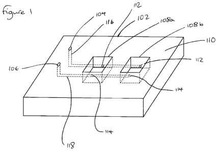
Figure 1 illustrates a biological microfluidics chip (biochip) 100 according
to an
embodiment of the invention.
CA 02765732 2011-12-15
WO 2010/149292 PCT/EP2010/003579
The biochip 100 comprises a substrate 102 having a microfluidic inlet port 104
and a
microfluidic outlet port 106 defining openings in the top surface 110 of the
substrate 102.
The microfluidic inlet port 104 and microfluidic outlet port 106 may be
considered as
entrances of the microfluidic channels to an external environment. A plurality
of
5 recesses/wells 108 extend downwardly from the top surface 110 of the
substrate 102.
In the embodiment shown in Figure 1, only two wells 108a, 108b are illustrated
in order
to clearly be able to show the features of the wells 108. It will be
appreciated that in
practice, a biochip 100 may comprise 32 wells, 96 wells, 869 wells, or any
number of
10 wells that are required. An advantage of embodiments of the biochip 100 is
that a large
number of wells, and their associated microfluidic channels, can be provided
in a small
area. For example, a biochip according to an embodiment of the invention can
accommodate 869 wells in the same area that a conventional microtitre plate
would
accommodate 96 wells.
Each of the wells 108 has a first opening 112 into a side wall of the well 108
and a
second opening 114 into the opposite side wall of the well 108. The first
opening 112 is
an example of an inlet opening, and the second opening 114 is an example of an
outlet
opening. In this example, the wells 108 have a square horizontal cross
section. In other
embodiments, wells 108 having cross sections of any shape can be used, and the
side
walls may take any configuration of lateral boundaries, and may be flat/planar
or curved,
for example.
An inlet microfluidic channel/conduit 116 connects the microfluidic inlet port
104 to each
of the inlet openings 112 in the wells 108. Similarly, an outlet microfluidic
channel 118
connects the microfluidic outlet port 106 to each of the outlet openings 114
in the side
walls of the wells 108.
In this example, the outlet openings 114 in the side walls of the wells 108
are higher (that
is, closer to the upper surface from which the wells 108 extend) than the
first openings
112. This configuration of inlet and outlet openings 112, 114 can be
advantageous for
causing a liquid that enters the well 108 from the inlet opening 112 to
subsequently exit
the well 108 through the outlet opening 114 without stagnating in the well
108. The
configuration of openings 112, 114 can provide for an efficient and economical
throughput of fluid through the well 108.
CA 02765732 2011-12-15
WO 2010/149292 PCT/EP2010/003579
11
Figure 2 illustrates a cross sectional side view through a well 202 of a
biochip 200
according to an embodiment of the invention.
The biochip 200 comprises a first/top layer of substrate 204, a second/middle
layer of
substrate 206, and a third/bottom layer of substrate 218. Constructing the
substrate as
three layers can enable the microfluidic channels to be conveniently located
within the
body of the substrate, between the layers, as will be appreciated from the
description
below.
The well 202 in this example has a circular horizontal cross-section when
viewed from
above, although the structure illustrated in Figure 2 is equally applicable to
wells 202
having different cross sectional shapes. The well 202 illustrated in Figure 2
may be
considered to have an "hourglass" shape in vertical cross-section.
The upper portion of the well 202 (that is the portion of the well 202 that
passes through
the upper layer 204) extends downwardly from the upper surface 214 of the
first layer of
substrate 204. The upper portion of the well 202 is, in this example,
frustoconical in
shape such that a cone associated with the upper portion of the well is
pointing
downwards (away from the upper surface 214 of the first layer of substrate
204).
The well 202 has a bottom portion that extends into the second layer of the
substrate
206. Again, the horizontal cross section of the lower portion of the well 202
is circular. In
this example, the shape of the bottom portion of the well 202 is also
frustoconical, but
this time a cone associated with the frustoconical shape of the bottom portion
of the well
202 points upwards (towards the top surface 214 of the first layer of
substrate 204).
The horizontal cross section of the well 202 at the boundary between the first
layer 204
and second layer 206 of substrate is substantially the same in each of the two
layers
204, 206 such that a continuous well 202 is provided. It will be appreciated
that the two
point-to-point frustoconical parts of the well 202 can be seen to provide an
"hourglass"
shape in vertical cross-section when the well 202 is considered as a whole.
Providing a well 202 having an hourglass shape can be advantageous in terms of
imaging the contents of the well 202. For example, a well 202 having this
shape can
enable the contents of the well 202 to be viewed/analysed/measured from either
the top
or bottom of the biochip 200 without having to look through unnecessary
regions of the
substrate 204, 206. The imaging may typically be performed by a microscope and
the
CA 02765732 2011-12-15
WO 2010/149292 PCT/EP2010/003579
12
microfluidics chip may be made out of glass layers that are graded for
microscopy. The
boundary between the first, second and third layers 204, 206, 218 may not
influence the
imaging of the contents of the wells. In other embodiments, the well walls may
be
flat/planar such that the wells have a constant cross-sectional shape and size
along their
length.
The well 202 has a first opening 208 that is in fluid communication with a
microfluidic
channel 207. In this example, the microfluidic channel 207 is an inlet for a
fluid. The
opening 208 is adjacent to a lower surface 216 of the well 202.
In addition, a second opening 210 is provided in the side wall of the well 202
between the
first and second layers 204, 206 of substrate. The second opening 210 is in
fluid
communication with a second microfluidic channel 212, which in this example is
a
microfluidic outlet channel. In use, an embryo or larvae, such as a zebra fish
embryo or
larva, can be located in the well 202, and nutrients, drugs or other
compounds, can be
pumped into the well 202 from the mircofluidic inlet channel 207 and the first
opening
208. Alternatively, these substances can be introduced via pipetting through
the upper
opening of the well.
One or more fluids can be removed from the well 202 through the second opening
210 in
the side wall of the well 202, which opens into the microfluidic outlet
channel 212. It will
be appreciated that fluids that are extracted from the well 202 can include
any waste
products that may be formed in the well 202 over time, and can include
products
generated by the embryo/larvae and any bacteria or other pathogens, or
biological
waste, or drugs or other compounds, or shed tissues or substrates.
Figure 3 illustrates a further embodiment of a biochip according to an
embodiment of the
invention.
3o The biochip 300 of Figure 3 has an array of wells/recesses 302, which in
this
embodiment is a 4x8 array of 32 wells. In addition, 8 inlet/outlet ports 304
are provided
as a 4x2 array at one end of the biochip 300. It will be appreciated that
other
configurations of ports and wells are possible. The microfluidic channels
extending from
the inlet/outlet ports 304 are not shown in Figure 3 to aid clarity. The
fluidic channels
extending from the inlet/outlet ports 304, and their interaction with the
wells 302, are
shown in more detail in Figures 4 and 5. It will be appreciated that the
microfluidic
channels illustrated in Figure 4 and Figure 5 separately, are in fact all
present in the
CA 02765732 2011-12-15
WO 2010/149292 PCT/EP2010/003579
13
same biochip 300 illustrated in Figure 3, but are illustrated separately for
clarity. In this
example, the wells are square in section, when viewed from above, but other
shapes are
possible.
Figure 4 shows the biochip 300 of Figure 3, and the associated microfluidic
inlet
channels extending from a microfluidic inlet port 410 to the wells 302, and
the
microfluidic outlet channels extending from a microfluidic outlet port 412 to
the wells 302.
It will be appreciated that the microfluidic inlet port 410 is suitable for
connecting to any
1o microfluidic source, such as a source of drugs or nutrient medium that are
to be provided
to the wells 302. As illustrated in Figure 4, microfluidic channels 414 extend
in parallel
from the microfluidic inlet port 410 to each row of wells 302. In this
example, the wells
are provided as a 4x8 array, and therefore four microfluidic channel branches
414 (one
for each row) extend from the inlet port 410.
Each of the microfluidic channels 414 for a given row of wells 302 further
branches off to
be in fluid communication with a first opening 418 of each of the wells 302.
Again, by
branching off in this way, the microfluidic channels 414 may be seen to
provide the fluid
to each of the wells 302 in parallel.
In this example, the microfluidic channel 414 does not extend directly from
the inlet port
410 to each of the first openings 418 in the wells 302, but is configured such
that the
channel length between the microfluidic inlet port 410 and each of the first
openings 418
are of substantially the same length for each well to retain similar fluid
flow rates for each
well. This is achieved by providing a different channel length between a main
artery of
the microfluidic channel 414 and the first openings 418 in the different wells
302. For
example, the microfluidic channel may follow a path that doubles back on
itself a number
of times in order to provide an overall required channel length between the
inlet port 410
and the first openings 418. This is shown in Figure 4 as reference 416. It
will be
appreciated that the path length between a main artery of the microfluidic
channel 414
and the first opening 418 should be shorter for wells 302 that are further
from the inlet
port 410 in order to provide an overall channel length between the inlet port
410 and the
first openings 418 that provides substantially consistent fluid flow for each
well. This can
mean that resistance that is experienced when supplying a fluid to each well
is
substantially the same, regardless of the distance of the well from the port,
and therefore
the fluid is supplied evenly to each of the wells.
CA 02765732 2011-12-15
WO 2010/149292 PCT/EP2010/003579
14
Providing microfluidic channels 414, 416 in this way can enable the physical
characteristics experienced by the fluid when it is en route to a well 302 to
be the same
for each of the wells 302. This can provide a consistent pressure of fluid to
each well
302, thereby providing a consistent flow rate, and therefore can reduce the
chances of
the contents of the well 302 being forced back into the microfluidic inlet
channel 414,
416. In turn, this can reduce the chance of cross-contamination between the
contents of
the individual wells 302.
The same structure is applied to the microfluidic output channel 424, 422 that
connects
1o the microfluidic outlet port 412 to second openings 420 in the wells 302.
Figure 5 illustrates ports and channels that are used to control the
temperature within the
wells 302 of the biochip 300. In this example, the wells 302 are square in
cross-section
when viewed from above, but other shapes are possible.
In this example, there are four temperature control inlet ports 510a, 510b,
510c, 510d:
one for each row of the array of wells 302. In this way, the temperature of
the wells 302
in each row can be independently controlled. The biochip 300 comprises a
single
temperature control outlet port 512. Other possible configurations include
having a
single inlet and a single outlet temperature control port, and in such
embodiments the
whole biochip can be maintained at a uniform temperature.
Extending from each of the temperature control inlet ports 510 is a
temperature control
channel 506. The temperature control channel 506 can transport a temperature
control
liquid from the inlet port 510 to the outlet port 512 along a path that is in
proximity to the
wells 302 in the row that is associated with the temperature inlet port 510.
It will be
appreciated that "in proximity" means that the temperature control channel 506
is located
close enough to the wells 302 such that heat can be exchanged between the
contents of
the well 304 and the temperature control fluid in the temperature control
channel 506.
Heat may be exchanged either to or from the temperature control fluid in order
to either
cool or heat the contents of the well 302.
In this example, the temperature control channel 506 follows a path that is
adjacent to
three of the four sides of the horizontal square cross section of the wells
302 in order to
evenly heat or cool the contents of the wells. It will be appreciated that the
temperature
control channel 506 may take any path in relation to the wells 302 as long as
heat can be
exchanged between temperature control fluid in the temperature control channel
506 and
CA 02765732 2011-12-15
WO 2010/149292 PCT/EP2010/003579
the contents of the wells 302. In examples where the wells 302 have a circular
horizontal
cross section, the temperature control channel may follow some, or
substantially all, of
the circumference of the circular well.
5 The temperature control channels 506 for each of the rows of wells all join
together after
they have passed by the wells in order to form a common temperature control
return
channel 508 that is in fluid communication with the temperature control outlet
port 512.
Prior art products are known to use external heating modules to maintain
steady
10 experimental temperatures for wells in a biochip. In the prior art systems,
the microtitre
plate must remain fixed on the heating module at all times, and this can make
it
impossible to relocate the plate during use. Therefore, prior art microtitre
plates cannot
easily be used in automated robotic handling systems for high throughput
screening if a
uniform temperature is required per well. In contrast, embodiments of a
biochip
15 described herein can have built-in heating/cooling channels and therefore
do not have to
be fixed on a thermostatic module, but can be moved freely in robotic systems.
This
provides a more flexible, in terms of usage, biochip than microtitre plates of
the prior art.
Embodiments described herein can be used to perform experiments on zebrafish
embryos/larvae in the wells, and the development of the embryos/larvae over a
period of
days can be monitored for example with or without exposure to drugs or other
compounds. In other embodiments, experiments can be performed on a monolayer
of
cells, on a membrane, matrix or other substratum within the well.
In this embodiment, the biochip uses glass and/or (fused) silica as base
compound(s).
This can reduce autofluorescence significantly when compared with known
polystyrene
products that are used for 96 well microtitre plates. Glass can be considered
a less
expensive material, whereas a combination of glass and polystyrene can be more
expensive due to the coating technology required. Furthermore, glass can be
more
resistant to scratching and to repeated cleaning cycles, than plastics.
In this example, the surface of the biochip is glass, which enables imaging
operations to
refocus on a specimen within 0.1 seconds. In contrast, known biochips having a
polystyrene surface generate drift due to a relatively rough surface of the
biochip and
deflection of infrared wavelengths that are used as part of an imaging
operation. This
can mean that refocusing for a prior art biochip can take more than 1 second
per well,
and this adds up logarithmically in time for large batch embryo screenings.
CA 02765732 2011-12-15
WO 2010/149292 PCT/EP2010/003579
16
Figure 6 illustrates a vertical cross-sectional view of a well 602 of a
biochip 300
according to an embodiment of the invention, in use, performing an experiment
on a
zebrafish embryo 604.
In this example, the embryo 604 is surrounded by its chorion 606 and embedded
in low
melting point-agarose 608 (injected into the well by a robotic handler) to
prevent the
embryo from moving around in the well. The agarose 608 solidifies into a gel
but does
not damage the specimen or prevent gas/nutrient exchange, and can be
convenient for
injecting test drugs through the extra-embryonic membrane (chorion 606). The
gel 608
can also limit the spread of a potential infection. Each well 602 in the
biochip 600 will
have a steady or constant supply of defined buffer through its microfluidic
inlet channels
610 that run in parallel, thereby reducing the risk of microbial cross-
contamination and
leakage of drug to neighbouring wells. In addition, drugs can be administered
by robotic
pipette handlers through a sliding lid, which may be plastic or glass, and can
be retracted
to expose the opening in the well for injection. Alternatively, the lid may be
a self-sealing
lid, for example, a rubber or polymer plug, laminate film or adhesive tape. It
should be
appreciated that the presence of agarose 608 is not essential, and in other
embodiments, the embryos can lie free in the fluid in the well or in any other
substance.
Some embodiments of a biochip described herein can comprise a lid that is
configured to
cover the openings of the wells in the top surface of the biochip substrate.
The lid may
be a sliding lid that is integrated as part of the biochip. This can enable
the lid to be slid
to one side to an open position to expose the openings of the wells for
embryos to be
introduced prior to the start of the experiment, and/or to enable drugs to be
introduced
during the experiment. The lid can then be slid back into a covered position
during the
experiment.
In some examples, the lid can seal the well gas- and/or fluid-tight to enable
efficient
microfluidic flow in the well. The removable lid can also enable recovery of
an embryo
after the experiment, and further detailed analyzes (for example, polymerase
chain
reaction (PCR), extraction of mRNA, etc.) can be performed after the end of
the
experiment in the biochip. Removal of the lid can also allow for convenient
cleaning of
the wells after the end of an experiment so that the biochip can be re-used.
In some embodiments, the lid can be heated so as to reduce the likelihood of
condensation building up on the lid. A reduction in condensation can enable
more
CA 02765732 2011-12-15
WO 2010/149292 PCT/EP2010/003579
17
accurate imaging operations to be performed through the lid in use.
Alternatively, the lid
may consist of a plastic film, applied under heat, or a perforated membrane
whose
perforations are aligned with the upper openings of the wells, and with a
glass cover
applied over the membrane. Where the lid is a film, it can be self-sealing
after a needle
has been passed through it into the well. In some cases the lid is optically
clear/transparent to permit microscopic analysis of the well contents; in
other cases it
may have a mirrored upper or lower surface.
Figure 7 illustrates a vertical cross sectional view of a well 202 of a
biochip 200
1o according to an embodiment of the invention (as in Figure 2), when placed
in a holder
according to the present invention. The holder comprises several layers of
materials that,
when connected, e.g. by screws, can fixate the biochip in a particular
position. The top lid
1 is made of metal/plastic or other material. The plate 2 is a glass plate or
polymer seal,
that can be mounted on top seal layer 3. Top seal layer 3 is for example made
of
silicone. Layer 4 is a bottom (silicone) seal. Layer 5 constitutes the bottom
lid, which can
be made of metal and/or plastic or other material. The angle between dotted
lines 6
indicates the bottom side field of view for imaging. Dotted line 7 indicates
the location of
the hole in layer 3 for imaging purposes. The angle between dotted lines 8
indicates the
top side field of view for imaging. The holder of the present invention is not
limited to this
particular configuration.
There will now be described specific implementation details of a biological
microfluidics
chip according to an embodiment of the invention. This embodiment of a
biological
microfluidics chip can be made from D263 glass, as this type of glass has been
found to
reduce autofluorescence compared to known polystyrene products.
The cross-sectional area of a well in the biochip is 2 - 4 mm2. This is in
contrast to
known 96-well microtitre plates where the cross-sectional area of a well is
significantly
larger, namely 33.18 mm2 (surface area (rrr2) r = 3.25 mm; h = 10 mm).
Therefore, this
embodiment of the biochip can reduce automated "find & mark" time when used in
automated systems.
The volume of a single well in the biochip of this embodiment is 8 mm3 = 8 pl
(2mm x
2mm x 2mm). This is in contrast to the well volume of known 96-well plates,
which is
typically around 250 - 331 mm3 (33.18 mm2 * (7.5mm or 10mm)). Therefore, this
embodiment of the biochip can provide a cost reduction in compound use, and is
estimates to be a reduction of 31 - 41% (250/8 to 331/8).
CA 02765732 2011-12-15
WO 2010/149292 PCT/EP2010/003579
18
In this example, the biochip is able to accommodate 869 wells in the same area
that a
conventional microtitre plate would hold 96 wells per plate. This is because
the surface
area of a 96-well plate (total area 7823 mm2; width x depth; 72.3mm x 108.2mm)
is
approximately 0.012 well/mm2. By contrast, a biochip according to an
embodiment of the
invention can be accommodate ? 0.11 wells/mm2 (surface area of each well plus
its
associated microfluidic channels, is 3mm x 3mm).
Embodiments described herein can be used to perform experiments in wells of a
1o biological microfluidics chip. The subject of the experiment, such as an
embryo, can be
inserted into an upper opening in the substrate that defines the well.
Optionally the
upper opening in the substrate can be covered with a lid as described above.
Examples described herein can immobilise an embryo or other subject in the
well before
subsequently exposing the subject to one or more fluids received from a
microfluidic
channel that opens into an opening in a wall of the well. As an example, the
fluids may
provide nutrients for the subject. The subject may not be moved to or from the
well
through a microfluidic channel.
The wells of one or more biological microfluidics chips described herein may
be
considered as a holding chamber for growth of a subject, and may be considered
to
relate to long-term culture experiments/systems. "Long-term" may be considered
to be
of the order of a few days, for example five days.
Biological microfluidics chips may be considered to be in a different
technical field to
microfluidic worm-sorters, wherein a worm is captured in an enclosed chamber
by
suction. Different technical considerations may be necessary for such
microfluidic
sorters compared with biological microfluidics chips having wells as described
herein.