Note: Descriptions are shown in the official language in which they were submitted.
CA 02765995 2011-12-19
WO 2011/004151 PCT/GB2010/001298
TELEMETIC ORTHOPAEDIC IMPLANT
CROSS REFERENCE TO RELATED APPLICATIONS
This application claims the benefit of UK provisional application No.
0911697.1, filed on
6 July 2009. The disclosure is herein incorporated, in its entirety, by
reference.
FIELD OF THE INVENTION
The invention relates generally to orthopaedic implants, and more particularly
to
orthopaedic implants having data acquisition capabilities and their use in
monitoring and
diagnosing fracture healing.
BACKGROUND TO THE INVENTION
Fractures of long bones are a prevalent problem, accounting for 10% of non-
fatal injuries
in the USA (Kanakaris 2007). Of these, the most common are fractures of the
tibial
shaft, approximated to result in 77,000 hospitalisations a year in the USA
(Schmidt et al
2003). The epidemiology and aetiology of tibial shaft fractures indicates a
relation with
risk behaviour. This type of fracture appears to be most prevalent in young
men (Grutter
2000). A study by Court-Brown, 1995 found the mean age of patients witfh
tibial shaft
fractures to be 37 years, with the highest incidence occurring amongst teenage
males.
The two most common causes being; sports related injuries and road traffic
accidents.
1
CA 02765995 2011-12-19
WO 2011/004151 PCT/GB2010/001298
There are several classifications described for fractures of the tibia,
perhaps the most
widely accepted of long bone fracture classifications in the AO/OTA
classification
(Arbeitsgemeinschaft for Osteosynthesefragen/ Orthopaedic Trauma Association).
This
classification system looks solely at the pattern of fracture, not taking into
consideration
the local soft tissue damage (Fig1). Associated soft tissue injury may be
classified
according to the Tscherne and Gotzen classification (Schmidt et al 2003) for
closed tibial
fractures, and according to the Gustilo Anderson classification (Gustilo &
Anderson
1976) for open fractures.
For an in-vitro biomechanical study of an instrumented nail, used for strain
telemetry, the
most useful of these classifications is the AO classification. This is an
alphanumeric
classification system for all long bone fractures. An example of a fracture
classified in
this .way is 42-C2. "4" represents the tibia, whilst the "2" tells us this is
a fracture of the
diaphysis. Having described the location, the letters A, B or C are assigned
to indicate
the fracture type and increasing complexity. Subgroups of these, in increasing
severity,
are assigned by the addition of the numbers 1,2 or 3 (Grutter 2000). Further
subdivisions
of these groups may be made, to indicate the number of fragments.
Of the various fracture, 42-A3 appears to be the most common, accounting for
23.9% of
tibial diaphyseal fractures (Court-Brown 1995).
Treatment of these fractures is broadly divided into two categories,
conservative and
surgical. Conservative therapy involved the use of a plaster-cast or
functional bracing.
Surgical treatment can involve either open-reduction and internal fixation
(ORIF) of
intramedullary (IM) nailing. A META-analysis comparing conservative treatment
to ORIF
found that despite significantly decreased risk of superficial wound
infection, casting
2
CA 02765995 2011-12-19
WO 2011/004151 PCT/GB2010/001298
resulted in a lower rate of union at 20 weeks (p=0.008) (Littenburg et al.
1998).
Additionally casting is limited by the severity of the fracture and deformity,
with initial
moderate or severe displacement increasing the rate of delayed of non-union
from 9% to
as much as 27% (Schmidt et al 2003).
IM nailing appears to be the preferred method of treatment for the majority of
tibial
fractures (Schmidt et al 2003). This suggestion is supported by a Randomised
Control
Trial (RCT) which shows IM nailing to result in faster union and a decrease in
the rates
of malunion, in comparison to conservative treatment (Hooper GJ 1991).
Delayed or non-union are a major concern with tibial fractures. On a "best
case
scenario" calculation the cost of one tibial non-union is estimated to be
16,330, with
20% being direct costs of treatment and 80% due to indirect costs (Kanakaris
2007). The
reported incidence of delayed union shows a great degree of variability due to
the
arbitrary definitions used. Generally delayed union of the tibia is recognised
at 20 weeks,
however, earlier detection may be possible. One could think of delayed union
as the
point at which altering the treatment may be considered, in order to achieve
union
(Phieffer & Goulet 2006). The definition of non-union is broadly accepted as
the
presence of no radiographic evidence of healing for three consecutive months,
in a
fracture of at least 9 months of age. The prevalence of delayed and non-union
is
reported to be 4.4% and 2.5% respectively. However, in open fractures, delayed
union
may be as high as 41 %, requiring further treatment before union is achieved
(Phieffer &
Goulet 2006).
Treatment for delayed union varies in light of the cause. This can, broadly
speaking,
involve stabilisation, re-nailing, bone-grafts, adjunct therapy such as
electrical
3
CA 02765995 2011-12-19
WO 2011/004151 PCT/GB2010/001298
stimulation, ultrasound or biological adjuncts such as Bone Morphogenic
Protein (BMP).
However timing is key to success as early diagnosis and treatment of delayed
union can
save the patient from considerable periods of disability and pain (Phieffer &
Goulet
2006), whilst also having an impact on health economics due to a reduction in
morbidity.
Various methods have been used to ascertain the end point of healing of
fractures. This
is fundamental knowledge to clinicians so as to advise patients on appropriate
load
bearing in the injured limb or to diagnose the risk of delayed or non-unions.
Currently there is a lack of a gold standard method which supplies sensitive
data, good
repeatability as well as ease of use. Serial radiographs and manual
manipulation, often
used in conjunction, are subjective and show inter-clinician variability. The
inaccuracy
and complexity of using dexa-scans, vibrational analysis, scintigraphy and
ultrasound
has also eliminated them as potential measurement tools.
TELEMETRY
An IM nail acts to provide stability, whilst transmitting rotational, bending
and
compressive forces across the fracture site and maintaining anatomical
alignment of the
bone. The IM nail also acts as a load sharing device, gradually shifting the
load to the
bone, as it heals.
Telemetry enables the direct measurement of strain and load carried by an
appropriately
instrumented IM and hence gives an indication of the progress of fracture
repair without
disrupting fracture healing. An example of a telemetric orthopaedic system is
disclosed
in WO 2007/025191, which is herein incorporated in its entirety. In addition
to its clinical
4
CA 02765995 2011-12-19
WO 2011/004151 PCT/GB2010/001298
use, such methodology proves to be of great benefit toward increasing our
understanding of fracture healing and its biomechanics. It allows optimisation
of post-
operative patient care, assessing the role of different activities on skeletal
loads to
identify which are most appropriate for providing the desired mechanical
environment
(Schneider E, 2001).
Strain gauges, which enable the direct measurement of the load applied to the
nail, are
conventionally located in multiple recesses in the outer wall of the nail and
hence have
the potential to cause changes in the biomechanical properties of the nail.
This in turn
could lead to local weakening or stress concentration.
We have identified redundancy associated with the provision of strain gauges
at multiple
locations on a nail and have identified: firstly an optimal position for a
recess comprising
a plurality of strain gauges and secondly an optimal orientation of the strain
gauges
relative to the longitudinal axis of the nail. The strain gauges are capable
of monitoring
the strain in a nail when it experiences either off-set axial compression,
torsional forces
or three/four point bending forces.
The identification of the optimal positioning and orientation of the strain
gauges will
facilitate the generation of a single commercial design of an IM nail which
can be used
with varying fracture patterns.
CA 02765995 2011-12-19
WO 2011/004151 PCT/GB2010/001298
RADIOSTEREOMETRIC ANALYSIS (RSA)
In vivo measurement of three-dimensional (3D) displacement of prosthetics or
body
parts was pioneered by Goran Selvik in 1974 (Bragdon et al 2002). RSA is also
referred
to as radiostereometry or roentgen stereophotogrammic analysis.
RSA measurements can be obtained using pairs of simultaneous radiographs taken
repeatedly over time. Tantalum bead markers are implanted into the body part
or implant
segment under study with at least three non- colinear beads needed to define
each rigid
body subject to scrutiny (Valstar et al. 2005). A 3D coordinate system is
achieved by
way of a calibration cage embedded with tantalum beads in well defined,
immoveable
positions. Two radiographs placed side-by-side, in a uniplanar arrangement or
at a 90
degree angle to each other, in the case of a bi-planar arrangement (Valstar et
al. 2005)
are used to establish the 3D coordinates of the markers, and displacement
between the
rigid bodies can be calculated (Madanat et al. 2006) using commercially
available RSA
software systems.
Whilst RSA is a "gold standard" technique for assessing fixation and migration
of joint
replacements and determining micromotion of the bone, this technique has not
be
considered for measuring inter-fragmentary movement in long bone fractures
fixated
with an orthopaedic fixation device.
We have identified that RSA can be used accurately and precisely to measure
inter-
fragmentary movement in a long bone, such as a tibia, fixated with an IM nail
before and
after reduction of the fracture.
6
CA 02765995 2011-12-19
WO 2011/004151 PCT/GB2010/001298
SUMMARY OF THE INVENTION
According to a first aspect of the invention there is provided a telemetric
orthopaedic
implant system, the system comprising:
(a) an orthopaedic implant, the orthopaedic implant having a longitudinal axis
and
comprising
(i) a strain gauge orientated at about +45 and/or at about -45 relative to
the longitudinal axis of the implant;
(ii) a recess adapted to receive said strain gauge(s);
(iii) an electronic component electrically connected to at least a power
supply, a first transmitter, a first receiver, and a first microprocessor;
(iv) a recess adapted to receive said electronic components;
(v) potting material to seal said recess;
(vi) a power source electrically connected to said electronic component.
(b) a control unit, the control unit comprising;
(i) a second microprocessor
(ii) a second transmitter electrically connected to said second
microprocessor, the second transmitter adapted to send a signal to
said first receiver of said electronic component; and
7
CA 02765995 2011-12-19
WO 2011/004151 PCT/GB2010/001298
(iii) a second receiver electrically connected to said second
microprocessor, the second receiver adapted to receive data from
said transmitter of said electronic component.
The gauges orientated at about +45 or at about -45 relative to the
longitudinal axis of
the orthopaedic implant have been identified as being optimally positioned to
measure
strain associated with either torque and also three- or four-point bending.
The relative
location of the gauges to the fracture site has been found to be unimportant
when
measuring strain upon application of torque.
In embodiments of the invention further strain gauges are provided which are
orientated
either at about 0 or about 90 relative to the longitudinal axis of the
orthopaedic implant.
Such orientation has been identified as being optimal for measuring strain
associated
with offset-axial loading. However, the relative location of the gauges to the
fracture site
has been found to be important and there is a significant diminishment in
sensitivity in
strain measurement when the fracture site is distal to the gauge.
It is therefore desirable in a commercial embodiment of a nail to provide
gauges which
are capable of measuring strain regardless of fracture type and location and
to provide
healthcare personnel with options relating to the mechanical loading regime to
be
utilised. For example, off-set axial compression loading requires the patient
to be
ambulatory.
Whilst a commercial IM nail could therefore be provided with gauges orientated
at about
+45 and'or about -45 relative to the longitudinal axis of the orthopaedic
implant this
would limit the loading regime to torque, which may not be satisfactory or
possible with
some patients. The potential for an IM nail to offer an alternative to torque
loading ie.
8
CA 02765995 2011-12-19
WO 2011/004151 PCT/GB2010/001298
off-set axial compression or three- or four point bending by the provision of
differently
orientated gauges in one recess is therefore viewed as an attractive
commercial offering
that will not prejudice the mechanical integrity of the IM nail.
Commercial embodiments of the nail have a recess which comprises a strain
gauge
orientated at about +45 and a strain gauge orientated at about 00, or a
strain gauge
orientated at about +45 and a strain gauge orientated at about 90 , or a
strain gauge
orientated at about -45 and a strain gauge orientated at about 00, or strain
gauge
orientated at about -45 and a strain gauge orientated at about 90 .
In embodiments of the invention the recess comprises a strain gauge orientated
at +45 ,
a strain gauge orientated at about -45 and a strain gauge located at about 0
, or a strain
gauge orientated at about +45 , a strain gauge orientated at about -45 and a
strain
gauge orientated at about 900, or a strain gauge orientated at about +45 , a
strain gauge
orientated at about 0 and a strain gauge orientated at about 90 , or a strain
gauge
orientated at about -45 , a strain gauge orientated at about 0 and a strain
gauge
orientated at about 90 .
In embodiments of the invention the recess comprises a strain gauge orientated
at about
+45 , a strain gauge orientated at about -45 , a strain gauge orientated at
about 0 and a
strain gauge orientated at about 90 .
Examples of suitable mechanical strain gauges include foil, thin film, or
semiconductor
strain gauges. Alternatively, the sensors may be load cells used to directly
measure
mechanical load.
In embodiments of the invention a lid is optionally associated with the recess
to provide
electrical shielding for the circuitry therein.
9
CA 02765995 2011-12-19
WO 2011/004151 PCT/GB2010/001298
According to a second aspect of the invention there is provided a telemetric
orthopaedic
implant comprising;
(i) a strain gauge orientated at about +45 and/or -45 relative to a
longitudinal axis of the implant;
(ii) a recess adapted to receive said strain gauge(s);
(iii) an electronic component electrically connected to at least a power
supply, a first transmitter, a first receiver, and a first microprocessor;
(iv) a recess adapted to receive said electronic components;
(v) potting material to seal said recesses;
(vi) a power source electrically connected to said electronic component.
In embodiments of the second aspect of the invention at least one further
strain gauge is
orientated at about 0 and/or at about 90 relative to the longitudinal axis
of the implant.
In embodiments of the invention a lid is optionally associated with the recess
to provide
electrical shielding for the circuitry therein.
In embodiments according to the first and second aspects of the invention the
orthopaedic implant is an IM nail.
A telemetric IM nail has been previously disclosed in WO 2007/025191 which is
herein
incorporated, by reference, in its entirety. Suitable materials and
methodology for the
instrumentation of a nail and examples of suitable peripheral components for
use in
communication and for storing information received from the nail are disclosed
in WO
20071025191.
CA 02765995 2011-12-19
WO 2011/004151 PCT/GB2010/001298
In embodiments of the invention the telemetric orthopaedic implant, more
specifically an
IM nail is provided with a single recess for receiving the strain gauges.
In specific embodiments of the invention this single recess is located in the
proximal
portion of the nail.
In specific embodiments of the invention this single recess comprises or
consists of
strain gauges orientated about +45 and about 00 or about -45 and about 0
relative to
the longitudinal axis of the nail.
In alternative embodiments of the invention the recess in which the strain
gauges are
provided is located substantially mid-way along the length of the longitudinal
axis of the
IM nail.
In an alternative embodiment of the invention the strain gauge recess is
located
substantially mid-way along the length of the longitudinal axis and extending
into the
tapered proximal region of the nail. The wall thickness of the proximal region
in some
designs of an IM nail is slightly thicker and the provision of a recess which
retains the
strain gauges and the associated electronic components has less effect on the
mechanical integrity of the nail than if the recess was located in other
regions of the nail.
In embodiments of the invention the recess is dimensioned such that the pocket
extends
along the longitudinal axis of the nail and has a length greater than its
width.
In embodiments of the invention the recess has a length of between about 10
and
150mm, or between about 10 and 130 mm, or between about 100mm and 150mm, or
between about 100mm and 140mm, or between about 100mm and 130mm, or between
about 120mm and 140mm.
11
CA 02765995 2011-12-19
WO 2011/004151 PCT/GB2010/001298
In embodiments of the invention the recess has a length of about 130mm.
The recess has a mid-way point along its length.
In embodiments of the invention the mid-way point along the length of the
recess is
located approximately mid-way along the longitudinal axis of the IM nail.
In embodiments of the invention the mid-way point along the length of the
recess is
offset from the mid-way point of the longitudinal axis of the nail, by up to
the length of the
pocket. For example, the length of the recess can be defined as having a first
end and
a second end, and either of these ends can be located at the mid-way point
along the
longitudinal axis of the nail.
An example of an IM nail is the TRIGEN META NAIL (Smith & Nephew). Due to the
design constraints of the TRIGEN META NAIL , the recess is located in the
proximal
region of the nail.
In embodiments of the invention the IM nail comprises or consists of the
design of the 8
or 9 pocket nail disclosed in Table 1
In embodiments of the invention the IM nail is for use in repairing fractures
of the long
bones, for example tibial or femoral fractures.
Alternative embodiments include incorporation of the strain gauges and the
other
electronic components within other implantable trauma products, such as a
plate, a bone
screw, a cannulated screw, a pin, a rod, a staple, and a cable. Further, the
12
CA 02765995 2011-12-19
WO 2011/004151 PCT/GB2010/001298
instrumentation described herein is extendable to joint replacement implant,
such as
total knee replacements (TKR) and total hip replacements (THR), dental
implants, and
craniomaxillofacial implants.
According to a,third aspect of the invention there is provided the use of a
telemetric
orthopaedic implant according to the second aspect of the invention in the
system
according to the first aspect of the invention.
While immobilization and surgery may facilitate bone healing, the healing of a
fracture
still requires adequate physiological healing which can be achieved through
continuously
monitoring changes in the in situ load distribution between the implant and
the
surrounding bone using sensors and a biotelemetry system. The mass and
architecture
of bone are known to be influenced by mechanical loading applied to them. In
the
absence of appropriate loading due to stress shielding caused by poor
management of
internal orthopaedic fixation systems, bone mass is reduced, resulting in
compromised
healing of the fracture. The primary function of a telemetric orthopaedic
implant is to
carry load immediately after surgical placement. For example, the telemetric
orthopaedic
nail carries the load immediately after surgical placement in the
intrameduallary canal.
With progression of fracture healing, the load sharing between the implant and
the bone
changes. This can be tracked by using strain gauges which are optimally
positioned
within the orthopaedic implant regardless of the location of the fracture is.
This has the
advantage that a single design of nail can be used for a range of fracture
types and
fracture locations. The strain gauges are used to monitor the progress of
union in the
case of fracture by either continuously or intermittently monitoring the load
component of
the healing bone in all spatial components, which is unobtainable from X-rays.
Periodic
13
CA 02765995 2011-12-19
WO 2011/004151 PCT/GB2010/001298
follow-up will provide a graph that shows the gradual decrease of relative
motion of the
fragments until union occurs.
Each fracture patient generates his or her own healing curve; however the
general
shape of the healing curve indicates whether the fracture will progress to
either a union
condition, delayed union condition or a non-union condition. The healing curve
generated from a patient is dependent on a number of factors including the
type and
location of the fracture, health status (underlying disease), age, activity,
rehabilitation,
and time to reach weight bearing.
According to a fourth aspect of the present invention there is provided a
method of
measuring applied mechanical load across an orthopaedic implant, said method
comprising the steps of;
(i) positioning a subject having a telemetric orthopaedic implant according to
the second aspect of the invention in a position suitable for applying a
desired mechanical load;
(ii) applying the mechanical load to the implant; and
(iii) interrogating at least one strain gauge provided within the implant.
The load measured by the strain gauge can then by compared with hypothetical
load vs.
healing time curves showing the load distribution between an instrumented nail
and the
surrounding bone for (i) fractures that progress to a union condition, (iii)
fractures that
are a delayed non-union and (iii) fractures that maintain a non-union
condition. Although
fracture healing results in a reduction in implant load, the remaining load of
the nail can
be significant and are expected to increase with patient activity. It has been
suggested
that loading of the bone may increase up to 50% after implant removal. The
load
14
CA 02765995 2011-12-19
WO 2011/004151 PCT/GB2010/001298
measured in the adjacent bone can be determined by subtracting the implant
load from
the load exerted through the limb, which is determined using either a force
plate or
balance. The clinician can also measure the load acting through the contra-
lateral limb in
order to provide a reference measurement for a fully functional limb.
If the surgeon observes that the strain on the implant is decreasing over
time, this
implies that the surrounding hard tissue (for example the callus) is accepting
some of the
load and thus, the fracture is healing. However, if the strain on the implant
is unchanged
with time and at the approximate level as when the patient was discharged from
hospital
or other health care facility, this implies that the surrounding hard tissue
is not bearing
the load and, therefore the fracture is not healing.
In embodiments of the method according to the fourth aspect of the invention
there is
provided a method of measuring the mechanical load across an implanted
telemetric
orthopaedic implant upon application of a torsional force, said method
comprising the
steps of,
(i) positioning a subject having the telemetric orthopaedic implant either in
a
stance or supine position;
(ii) applying a torsional force on the telemetric orthopaedic implant; and
(iii) interrogating a strain gauge in the about +450 and/or about -45
orientation.
In embodiments of the method according to the fourth aspect of the invention
there is
provided a method of measuring the mechanical load across an orthopaedic
implant
upon application of an off-set axial compressive force, said method comprising
the steps
of;
CA 02765995 2011-12-19
WO 2011/004151 PCT/GB2010/001298
(i) positioning a subject having the telemetric orthopaedic implant in a
stance
position;
(ii) applying an off-set axial compressive force on the telemetric orthopaedic
implant; and
(iii) interrogating a strain gauge in the about 0 and/or about 900
orientation.
In embodiments of the method according to the fourth aspect of the invention
there is
provided a method of measuring the mechanical load across an orthopaedic
implant
upon application of a three or four point bending force, said method
comprising the steps
of;
(i) positioning a subject having the telemetric orthopaedic implant in a
stance
or supine position;
(ii) applying a three or four point bending force on the telemetric
orthopaedic
implant; and
(iii) interrogating a strain gauge in the about +45 , about -45 , about 0
and/or
about 900 orientation.
According to a fifth aspect of the present invention there is provided a
method of
monitoring fracture healing in a subject, said method comprising the steps of;
(i) positioning a subject having a telemetric orthopaedic implant according to
the second aspect of the invention in a position suitable for applying a
desired mechanical load;
(ii) applying the mechanical load;
(iii) interrogating at least one strain gauge provided within the implant;
(iv) correlating the strain with a reference fracture healing curve.
16
CA 02765995 2011-12-19
WO 2011/004151 PCT/GB2010/001298
In embodiments according to the fifth aspect of the invention the mechanical
load is
selected from the group consisting of; off-set axial compression, torque,
three point
bending or four point bending, wherein the subjecting is optionally positioned
in the
stance or supine phase.
The IM nail can be used to detect changes in fracture callus stiffness and
determine
healing status of the patient. The IM nail can detect changes of at least 4.1
Nm/ in callus
stiffness. It is therefore envisaged that the nail can detect delayed or non-
union fracture
within one month of tibial fracture fixation based on callus stiffness
measurements.
According to a sixth aspect of the invention there is provided the use of
radiostereometric analysis for the measurement of inter-fragmentary movement
within a
bone fracture, wherein the bone fracture is fixed with an orthopaedic fixation
device.
In embodiments of the invention RSA can be used to differentiate between
intact,
reduced and non-reduced fractures.
According to a seventh aspect of the invention there is provided the use of
RSA to
differentiate between intact, reduced and non-reduced fractures.
According to an eighth aspect of the invention there is provided a method of
measuring
inter-fragmentary movement within a bone fracture, wherein the bone fracture
is fixed
with a fracture fixation device, said method comprising;
i) associating of a plurality of radio-opaque markers with the fractured bone
and/or the fracture fixation device;
17
CA 02765995 2011-12-19
WO 2011/004151 PCT/GB2010/001298
ii) positioning a calibration cage comprising a plurality of radio-opaque
markers at known locations in relation to the fracture site;
iii) undertaking a radiographic examination of the fracture site, wherein the
fracture site and the calibration cage are simultaneously x-rayed from at
least two angles;
iv) generating a three-dimensional co-ordinate system based upon the
location of the radio-opaque markers in the calibration cage;
v) comparing the three-dimensional location of the radio-opaque markers
associated with the fractured bone and/or the fracture fixation device with
the three-dimensional co-ordinate system.
In embodiments of the invention the fracture is of the long bones, for example
the tibia or
femur.
The orthopaedic device can be for example, an intrameduallary nail, bone plate
or
external fixator, such as an Ilazorov frame.
In a specific embodiment of the invention RSA is used to accurately and
precisely
monitor inter-fragmentary movement in a tibial shaft fracture fixed with an IM
nail.
An example of a suitable radio-opaque marker is a tantalum bead, although
alternative
radio-opaque makers which are suitable for use in RSA are envisaged.
Alternatively, the solder joints associated with the electronic components can
be utilised
as reference points for monitoring inter-fragmentary bone movement.
18
CA 02765995 2011-12-19
WO 2011/004151 PCT/GB2010/001298
The radio-opaque markers are preferably associated with the proximal and
distal
segments of the fracture, thereby defining the rigid body segments.
At least 3 radio-opaque markers are associated with the proximal and distal
segments of
the fracture.
The radio-opaque markers are preferably associated with the bone and/or
implant in a
scattered pattern.
The orthopaedic device can be selected from, for example, an IM nail, bone
plate or
external fixator, such as an llazorov frame.
RSA is capable of measuring micromotion of the bone as a result of positional
change of
the implant (through loosening or dynamization of the screws), variations of
the forces
acting on the implant (inducible displacements) and is also capable of
indirectly
measuring callus stiffness. Thus, RSA can be used post-operatively to assess
both
implant stability and fracture reduction.
It is further envisaged that RSA can be used as an intra-operative tool for
trauma
fixation. The utilisation of this technique will enable the surgeon to correct
implant
malposition or malalignment and to ensure that the fracture is adequately
reduced.
It is envisaged in further embodiments of the invention that the inventions
according to
one or more aspects of the invention can be combined. For example, a fracture
can be
fixed with an appropriately instrumented IM nail, allowing both the telemetric
and
radiostereometric assessment of fracture healing. Advantageously the
instrumented IM
19
CA 02765995 2011-12-19
WO 2011/004151 PCT/GB2010/001298
nail used and the system comprising the IM nail is as defined according to the
first and
second aspects of the invention.
According to a ninth aspect of the invention there is provided the use of a
system
according to the first aspect of the invention or a telemetric orthopeadic
implant
according to the second aspect of the invention in the in vitro analysis of
fracture
healing, for example biomechanical models of fracture healing, including
animal models.
According to a tenth aspect of the invention there is provided a methods,
devices and
systems as substantially herein described with reference to the accompanying
Examples, Tables and Figures.
Further features, aspects, and advantages of the present invention, as well as
the
structure and operation of various embodiments of the present invention, are
described
in detail below with reference to the accompanying drawings.
Brief description of the drawings
The accompanying drawings, which are incorporated in and form part of the
specification, illustrate the embodiments of the present invention and
together with the
description serve to explain the principle of the invention. In the drawings:
Fig 1: Aetiology of fractures
Fig. 2 CC spiral pocket arrangement in an anti-clockwise direction
CA 02765995 2011-12-19
WO 2011/004151 PCT/GB2010/001298
Fig 3: Pocket locations on the TRIGEN META nail
Fig 4: Loading rig for strain measurements as a result of (i) off-set axial
compression (0-
1000N) and (ii) torque ( 2.5 N.m)
Fig 5: Loading rig for strain measurements as a result of 3pt and 4pt bending
(0-1000N)
Fig 6: Positioning of X-ray tubes in the loading rig
Fig 7: A high precision rotation stage used for y axis rotation measurements
Fig 8: Three point loading rig positioned in front of an RSA calibration cage
Fig 9: Callus simulation using reinforced polyethylene
Fig 10: Regression of strain counts in relation to axial load
Fig 11: Regression of strain counts in relation to torque
Fig 12: Principal strain magnitude in relation to axial force and torque
Fig 13: Principal strain direction in relation to axial force and torque
Fig 14: Principal strain magnitude in relation to axial force and torque
Fig 15: Principal strain direction in relation to axial force and torque
21
CA 02765995 2011-12-19
WO 2011/004151 PCT/GB2010/001298
Fig 16: Illustrates the Y axis translation (micromotion) of a distal segment
relative to a
proximal segment in intact bone
Fig 17: Illustrates the Y axis rotation of a distal segment relative to a
proximal segment in
intact bone
Fig 18: illustrates the Y axis translation (micromotion) of a distal segment
relative to a
proximal segment in an AO 43-A3 fracture.
Fig19: illustrates the Y axis rotation of a distal segment relative to a
proximal segment in
an AO 43-A3 fracture.
Fig 20 illustrates the Y axis translation (micromotion) of a distal segment
relative to a
proximal segment in an AO 42-C2 fracture.
Fig 21 illustrates the Y axis rotation of a distal segment relative to a
proximal segment in
an AO 42-C2 fracture.
Fig 22: sensitivity (microstrain/kg) plotted against strain gauge pocket
number,
illustrating the relationship in a segmental fracture with the loading moment
being
applied halfway at 18cm between the two tibial suspension points
Fig 23: illustrates the trace sensitivity against strain pocket
Fig 24: illustrates an AO 42-A3 fracture which overlies pocket 6
22
CA 02765995 2011-12-19
WO 2011/004151 PCT/GB2010/001298
Fig 25: illustrates an AO 43-Al fracture which overlies pockets 7-8.
Fig 26: illustrates an AO 42-C2 fracture which overlies pockets 4, 5 and 6
with the
proximal and distal fracture lines overlying pockets 4 and 6 respectively
Fig 27:RSA trace illustrating increasing Z rotation with increasing load
Fig 28: RSA trace of sensitivity against strain pocket with incrementally
increasing
stiffness of the nail-bone composite
Fig 29: RSA trace of rotation in the Z plane against load for 4 and 8 loop
reinforced
polyethylene tape "callus"
Fig 30: Synthetic callus formation - circumferential application
Fig 31: Synthetic callus formation - segmental application
Fig 32: Fracture model: 42-A2-AO; pocket/gauge: 4A; 5B. stance- Off-axis
compression
loading
Fig 33: Fracture model: 42-A2-AO; pocket/gauge: 1 B; 9B. stance- Off-axis
compression
loading
Fig 34: Fracture model: 42-A2-A0; pocket/gauge: 1 B; 98. stance- 4 point
bending
Fig 35: Fracture model: 42-A2-A0; pocket/gauge: 4B; 5C. stance- 4 point
bending
23
CA 02765995 2011-12-19
WO 2011/004151 PCT/GB2010/001298
Fig 36: Fracture model: 42-A2-AO; pocket/gauge: 1A; 4A; 5A; 9A. stance: torque
loading
Fig 37: Fracture model: 42-A2-AO; pocket/gauge: 1A; 4A; 5A; 9A. supine: torque
loading
Fig 38: Fracture model: 42-A2-AO; pocket/gauge: 1113; 4B; 5B; 9B. supine:
torque
loading
DETAILED DESCRIPTION OF THE INVENTION
A "smart implant" is an implant that is able to sense its environment, apply
intelligence to
determine what action is required, and act on the sense information to change
something in a controlled, beneficial manner. One attractive application of
smart implant
technology is to measure loads on an orthopaedic implant. For example, an IM
nail is
subjected to three types of loading: bending, torsional, and compression.
These loads
may be measured indirectly by measuring sensor output of a series of strain
gauges
mounted on the orthopaedic implant. In the case of an IM nail, diametrically
apposed
strain gauges mounted on the outer surfaces of the nail are subjected to
tensile and
compressive forces, respectively. Typically, the strain measured from the
sensors is
higher when the implant is loaded in bending rather than in compression.
A fundamental parameter of the strain gauge is its sensitivity to strain,
expressed
quantitatively as the gauge factor G, as defined in WO 2007/025191.
24
CA 02765995 2011-12-19
WO 2011/004151 PCT/GB2010/001298
Incorporation of sensors and other electronic components within an implantable
medical
device, such as an IM nail, alters its primary function from a passive load-
supporting
device to a smart "intelligent" system with the ability to record and monitor
patient activity
and compliance.
MATERIALS AND METHODS
SAWBONE
A4 th generation composite sawbone was obtained from Sawbones Europe AB,
Malmo,
Sweden. This is representative of a left tibial bone.
AETIOLOGY OF FRACTURE PATTERNS
As illustrated in Figure 1:
(i) non-reduced mid-shaft comminuted segmental fracture (42-C2);
(ii) non-reduced distal extra-articular comminuted fracture (43-A3)
(iii) reduced simple spiral fracture (42-Al)
(iv) reduced simple transverse fracture (42-A3)
IM NAIL
38cm long (10mm outer diameter) tibial IM TRIGEN META NAIL (Smith & Nephew,
Inc).
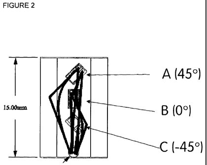
INSTRUMENTED IM NAIL
CA 02765995 2011-12-19
WO 2011/004151 PCT/GB2010/001298
A standard use 38cm long, 10 mm outer diameter, tibial IMTRIGEN META NAIL
(Smith
& Nephew, Inc) was used. Recesses were grooved into the surface of the nail,
with
dimensions 15mm long x 6mm wide, with a 34mm pitch. The pockets followed a
spiral
pattern, in an anti-clockwise direction, running down the shaft of the nail.
Table 1: Gauge co-ordinates for the Instrumented TRIGEN META NAIL
Gauge co-ordinates for 8 pocket nail
distance from centre of inferior proximal screw hole
(mm)
A (+450) B (0 ) C (-45 )
Pocket l 40 42.5 47
Pocket 2 74 76.5 81
Pocket 3 108 110.5 115
Pocket 4 142 144.5 149
Pocket 5 176 178.5 183
Pocket6 210 212.5 217
Pocket 7 244 246.5 251
Pocket 8 278 280.5 285
Table 1 cont......
Gauge co-ordinates for 9 pocket nail
distance from centre of inferior proximal screw hole
(mm)
A (+45 ) B (0 ) C (-45 )
Pocket l 40 42.5 47
Pocket 2 72 74.5 79
Pocket 3 104 106.5 111
Pocket 4 136 138.5 143
26
CA 02765995 2011-12-19
WO 2011/004151 PCT/GB2010/001298
Pocket 5 168 170.5 175
Pocket 6 200 202.5 207
Pocket 7 232 234.5 239
Pocket 8 264 266.5 271
Pocket 9 296 298.5 303
There are three anterior pockets (1,4,7), two medial pockets (3,6) and three
lateral
pockets (2,5,8). Each pocket had a hole at its base to pass 4 wires from 3
gauges
through to the cannulation. The wires ran down to the distal end of the nail,
inside the
canal. The wires exited through the most proximal of the distal screw holes
and ran
along an angled channel machined to avoid the two alternative screw holes, and
down to
the end of the nail. Three foil strain gauges (N3K-XX-S022H-50C/DP), Vishay
Ltd, were
bonded to the base of each pocket using MBond-600 adhesive as per Figure 2 and
3.
The gauges are orientated relative to the longitudinal axis. Gauge A is
oriented at 45 ,
Gauge B in line with, and Gauge C at -45 respectively. The 0 gauge detects
flexion
and extension, whilst the +45 and -45 gauges detect lateral bend and
torsion. Gauges
were conformably protected with MBond-43B coating. A free length of 500mm of
wire
was provided for attachment to the amplifiers.
Gauges were wired as quarter bridges with a single return wire in each pocket.
The
wires were attached to 8mm diameter connectors which were able to easily pass
through the reamed canal. These were attached to an amplifier, which sent the
data to
Labview v8 . The data from the 8 gauges in the load cell was also recorded
simultaneously through the same system. Labview v8 collects 512 measurements
for
each of the eight strain gauges in a 5 second window. The average value is
used for
27
CA 02765995 2011-12-19
WO 2011/004151 PCT/GB2010/001298
data analysis. The strain count can be converted to microstrain by dividing by
a factor of
6.8. Electrical noise was reduced by covering all wires with grounded tin foil
and
grounding components such as the loading rig. Additionally, removing the mains
supply
to the laptop was also found to be beneficial.
A. TELEMETRY METHODOLOGIES
1. Measurement of strain in an instrumented IM nail under axial and torsional
loads
1.1 Fracture patterns
Two fracture patterns were tested; (i) 42-C2 and (ii) 43-A3
For the mid-shaft 42-C2 fracture a pair of transverse cuts were made 5mm
apart, 24 cm
from the proximal end of the tibia. Another pair of cuts needed to be made
60mm further.
Removing the two 5mm fragments allowed 3 segments to be created, the middle
segment would be stabilised by the nail, with the distal and proximal segments
secured
by the cross screws. The fracture gaps represented the multiple fragments of
the
comminuted fracture. The fractures were aligned with pocket 6 at the distal
end and
pocket 4 at the proximal end of the instrumented IM nail.
The distal fracture was created by cutting across the bone 30mm and 40mm above
the
proximal distal screw hole. The segment produced was removed. The fracture
intersected pocket 8 of the instrumented IM nail.
28
CA 02765995 2011-12-19
WO 2011/004151 PCT/GB2010/001298
The IM nail was inserted into the Sawbones which were over-reamed by 2mm to a
diameter of 12mm, with the canal extending to the end of the bone.
1.2 Loading Rig
The loading rig for the nail was constructed using an aluminium frame with an
adjustable
top platform, as illustrated in Figure 4. A load cell was placed on the
inferior surface, this
was then mounted with an anti-torque jig, to house the distal end of the nail.
It was
important that the inferior surface of the nail was accessible as the hole for
exit of the
wires was located here.
The load cell used was designed to measure bending, internal and external
torque and
axial load.
The Sawbones was loaded via two balls, at either end. An offset of 9mm and
23mm
medial to the anatomical midline was used for load application at the distal
and proximal
end respectively. This offset is more representative of the mechanical line of
action of
the tibia (Hutson et al 1995). The proximal loading point was constructed by
marking out
the desired location (23mm medial to midline), and then placing a washer over
this area.
The metal washer was secured using Araldite adhesive. The central hole in the
metal
washer was able to house the proximal ball and form the point of load
transfer. Distally a
small metal cap, made to size, was fitted onto the bone by fitting into two
small holes
created in the bone. This cap had a hole overlapping.with the reamed canal of
the bone,
for exit of the wires, and also has a small socket for housing the distal
ball, 9mm medial
to the midline. The ball communicated with the top of the load cell, with the
anti-torque
jig securing the distal end of the tibia in place.
29
CA 02765995 2011-12-19
WO 2011/004151 PCT/GB2010/001298
1.3 Axial load application
Axial load application was performed via an adjustable screw fixed to the top
platform.
The screw was positioned to align with the loading washer on the tibia. The
screw was
connected to a spring, which communicated with the ball. A metal cap located
around
the spring guided its movement whilst providing minimal restraint. The cap was
large
enough to allow the ball to slide up, when the spring was under compression.
1.4 Torque application
Torque application was performed via a pulley system. A 100mm cross bar was
passed
horizontally through the proximal end of the bone, running in the medio-
lateral (ML)
direction. A pair of wires, able to withstand at least 10kg of weight, were
attached at the
ends of the bar and passed over pulleys in the same horizontal plane. Torque
was
applied by hanging weights to the end of the wires. Swinging the wires over to
the other
side allowed for torque application in the opposite direction.
A set of plates which held both sides of the medial malleolus in place
provided a method
of applying anti-torque. A cross bar was inserted through the malleolus in the
antero-
posterior (AP) direction and running through both plates, allowed the plates
to be tight
enough to prevent twisting, and also prevented the plates from dislocating the
bone.
Torques of up to 5Nm were applied, in both clockwise and anti-clockwise
directions by
applying weights, in increments of 500g, up to a maximum of 5kg.
CA 02765995 2011-12-19
WO 2011/004151 PCT/GB2010/001298
1.5 Step-by-step method
1 Insert nail
2 Place bone-nail construct in the loading rig
3 Connect wires to amplifier and start running Labview v8.
4 Begin loading. Torque measurement carried out at 0, 250, 500, 750 and 1000N
of axial load, with increasing increments of 500g weights being added up to
5Kg,
in both the clockwise and anti-clockwise directions. Axial load increased from
0 to
1000N in increments of 50N. When unloading the construct, only axial
measurements are needed. At each loading setup a new set of data needs to be
collected.
Remove bone and extract nail.
6 Repeated for each loading cycle.
1.6 Data analysis
Data analysis was performed via calculation of the average strain count for
each load
application and then performing multiple regression with two variables.
Regression was
carried out with respect to axial load and torque, in order to observe the
pattern of strain
at the various gauges in response to these two types of loading.
2. Measurement of strain in an instrumented IM nail under three-point bending
2.1 Fracture patterns
31
CA 02765995 2011-12-19
WO 2011/004151 PCT/GB2010/001298
Four fracture patterns were tested; (i) non-reduced mid-shaft comminuted
segmental
fracture (42-C2), (ii) non-reduced distal extra-articular comminuted fracture
(43-A3), (iii)
reduced simple transverse fracture (42-A3) and (iv) reduced simple spiral
fracture (42-
Al).
2.3 The Loading Rig
The loading rig is illustrated in Fig 5.
The nail-bone composite was suspended horizontally by means of two strings
attached
to two test tube clamps. Using a spirit level and a drill bit fixed on the
bone vertically,
care was taken that the nail was both perfectly horizontal and importantly
that the nail's
anterior surface was facing upwards and was perpendicular to the horizontal.
This
arrangement ensured that the 0 in-line anterior pocket gauges were
perpendicular to
the direction of force application and would thus be optimally positioned to
detect
longitudinal compression and extension of the nail. This jig enabled movement
of the
nail-bone composite with applied load in the X, Y and Z planes. This is
because the two
bone ends were not fixed rigidly. Proximally, the suspension points consisted
of two
screws in the medial and lateral tibial condyles. Distally, the strings
attached to the
protruding distal locking screws.
To apply strain to the nail-bone composite, weights were suspended from the
horizontally suspended bone by means of a string and a hook. The weights were
placed
in five distinct positions along the length of the nail. The proximal tibial
condyle screws
from which the bone was suspended were the zero reference value. From this
reference,
the weight suspension positions in centimetres were 9, 13.5, 18, 22.5, and 27.
This
32
CA 02765995 2011-12-19
WO 2011/004151 PCT/GB2010/001298
experimental set-up simulates rotational freedom afforded to the tibia by the
collateral
and cruciate ligaments at the knee joint. At each of these positions weights
were added
in one Kg increments starting at 0kg, with the maximum being 10kg. 10kg is
representative of physiological loads experienced in the tibia during the gait
cycle on the
basis of Wehner 2009. Therefore at each of the five positions, 11 strain count
versus
load measurements were taken. For each of the four fracture patterns 55 (11 x
5) strain
count Vs load measurements were taken.
In order to assess the repeatability of the measurements, loads were applied
in 1 kg
increments from 0-10kg at the mid-position (18cm from the proximal attachment
point).
Strain count Vs load measurements were taken. This same process was repeated
three
times.
33
CA 02765995 2011-12-19
WO 2011/004151 PCT/GB2010/001298
B. RSA METHODOLOGIES
3. Measurement of inter-fragmentary movement in an IM nail fixated tibial
fracture
under axial and torsional loads
Accuracy and Precision experiments
3.1 X-ray energy level setting
A rig to load the Sawbones was constructed from bars of aluminium, a metal
which is
substantially radiolucent (see Figure 4). RSA images which were taken with the
loading
rig in place were carried out at 90kV.
3.2 Radiographic Technique
The RSA set-up consisted of a calibration cage (cage 43, RSA BioMedical, Umea,
Sweden) which contained tantalum beads used to create a 3D coordinate system.
The
x-rays were taken on 2 AGFA CRMD4.0 General Cassettes (350mm by 420mm),
processed in AGFA format and then sent to DICOM Link. The images were imported
to
UmRSA Digital Measure 6.0 where the reference and bone markers were labelled.
Bone
markers were always numbered as 201, 202... for the proximal segment, and 301,
302... for the distal segment. Data regarding kinematics were obtained from
UmRSA
Analysis 6.0 (RSA BioMedical, Umea, Sweden). The kinematic data indicated
migration
of the distal fragment of the Sawbones tibia relative to the proximal
fragment using the
"segment motion" method (displacement of segment 30 relative to segment 20).
34
CA 02765995 2011-12-19
WO 2011/004151 PCT/GB2010/001298
3.3 Tantalum beads
Spherical tantalum beads with a diameter of 0.8mm (RSA BioMedical, Umea,
Sweden)
were used as bone markers.
3. 4 Accuracy and Precision Protocol for Linear Displacement
A Sawbones with a distal third fracture gap of 1 cm was used to determine the
accuracy
and precision of RSA for linear displacement in the x, y, and z axes. 8
tantalum beads
were inserted in the proximal and distal portions of the Sawbones , in the
areas closest
to the osteotomy, using a drill and spring-loaded piston (RSA BioMedical,
Umea,
Sweden). The x-ray tubes were positioned above the set-up facing downwards, as
illustrated in Figure 6. The calibration cage was placed under the radiolucent
table on
which the phantom model was located.
The proximal segment of the Sawbones was attached to a high precision
translation
stage (M-460A-xyz, Newport, Irvine, CA, USA), via 2 plastic pegs, in order to
measure
translation in the x, y and z axes. Three Vernier micrometers (model SM 13,
Newport,
Irvine, CA, USA) were attached to the translation stage. This set-up,
according to the
Newport company, has accuracy of 1 pm for translation. The translation stage
was
attached to the Plexiglas base with screws. The distal segment of the Sawbones
tibia
was fixed with a plastic peg to the base.
The proximal segment of the synthetic tibia was moved towards the distal
segment by
increments of 100pm, with a simultaneous film pair taken at each point. The x-
ray beams
intersected directly over the fracture in the phantom model. This was repeated
10 times,
CA 02765995 2011-12-19
WO 2011/004151 PCT/GB2010/001298
until the proximal segment was 1 mm closer to the distal segment. An identical
protocol
was followed for carrying out measurements during y axis displacement, and
lastly for
movement in the z plane. Furthermore, five radio-pairs were taken with zero
displacement. Another five were also done, each time moving the micrometer
from Opm
to 10pm and then back to Opm.
3.5 Accuracy and Precision Protocol for y rotation and angulation (z axis
rotation)
To measure accuracy and precision in y axis rotation, a high precision
rotation stage
was used (M-UTR-80, Newport, Irvine, CA, USA) which was screwed to a wooden
baseboard and connected to the distal segment of the Sawbones via a wooden
block
and a plastic peg (Figure 7; upper image). The accuracy of the rotation stage
was 1/60 .
The distal segment was moved 5 clockwise, then 5 anticlockwise, with x-rays
taken
after every 1 of rotational movement. The proximal segment of the synthetic
tibia was
firmly attached to a wooden backboard with 2 plastic pegs.
For measurement of angular movement the same translation stage (Figure 7;
lower
image) was used as before, but this time with only 1 Vernier micrometer. It
was screwed
to a wooden backboard, and it was attached to a wooden block which pushed on
both
segments of the Sawbones . To simulate angular movement of a fractured tibia,
both
segments of the Sawbones . were mounted on a backboard, using plastic pegs,
with z
axis rotation possible. A micrometer pushed the distal end of the proximal
segment, and
the proximal end of the distal segment, for 5mm in the negative x direction.
This was
done in increments of 500pm, up to 5mm, and caused the segments to become more
aligned in the y axis. This was done in order to establish the accuracy and
precision of
36
CA 02765995 2011-12-19
WO 2011/004151 PCT/GB2010/001298
RSA when measuring angular motion (z axis rotation) resulting from a load
applied in the
medio-lateral direction.
3.6 Accuracy and Precision Calculations
Accuracy and precision were determined for overall linear displacement, as
well as in
each of the three planes of linear movement. Similar measurements were made
for
angular motion and y axis rotation.
Accuracy, the nearness of measured values to true reference values (Bragdon et
al.
2002), can be determined by comparing the measured RSA displacement results
with
the true micrometer values using linear regression analysis, and calculating
the 95%
prediction interval, using SPSS (version 14.0 for Windows, Chicago, IL). The
maximum
and minimum bounds for the prediction interval can be determined, and the mean
of the
interval can be presented as the accuracy (Onsten et al. 2001).
Precision is the potential for the same result to be achieved on repeated
occasions
(Valstar et al. 2005). Precision in this study was calculated as p= (y)(SE)
(Altman 2000).
The y value was determined for a 95% confidence level, with the degree of
freedom=no.
of error values-1 (Bragdon et al. 2002). Error values were established by
taking the true
value and subtracting the measured value. This gives a total average error
from which
standard deviation and standard error can be determined.
37
CA 02765995 2011-12-19
WO 2011/004151 PCT/GB2010/001298
Loading experiments
3.7 Axial and Torsional Load
1000N of axial load, which was offset from the centre of the Sawbones by 23mm
medially at the proximal end and 9mm at the distal end, mimicked the resultant
force
experienced by the tibia during peak loading in the single leg stance interval
of the gait
cycle (Hutson et al. 1995).
5Nm of torque was applied.
3.9 Loading protocol
Bone A
An intact Sawbones was positioned in the loading rig. This particular
Sawbones was
reamed to 12mm, but an IM nail had been inserted and removed from it
approximately
15 times previously. The tantalum beads were implanted using a drill and a
spring
loaded piston (RSA BioMedical, Umea, Sweden) but the Sawbones was so hard
that
only 3 markers made it into the distal segment. It was possible to apply load
to the
synthetic tibia, and identify how much load the bone was taking, because
information
from the load cell in the rig, which was positioned under the distal end of
the Sawbones
passed to an amplifier, and the output was interpreted using LabVIEW v8. Axial
load
was applied in increments of 250N, up to and including 1000N. At every level a
simultaneous film pair was taken with the bone under axial loading
exclusively, then
axial loading with +5Nm of torque, and finally axial loading with -5Nm of
torque. The
procedure was repeated using the same nail with all 4 locking screws in place.
38
CA 02765995 2011-12-19
WO 2011/004151 PCT/GB2010/001298
Bone B
An intact Sawbones was positioned in the loading rig, with the IM nail
inserted. This
Sawbones had been reamed to 12mm with an IM nail inserted and removed once
before. Tantalum beads were applied with Araldite adhesive. Eight markers
were stuck
to the proximal segment but only 7 remained attached to the distal portion.
The same
loading protocol was followed as outlined above. The IM nail was left in
place, 4 locking
screws were inserted, and the procedure repeated again. The nail was then
removed.
An extra-articular metaphyseal complex fracture (43-A3 AO classification) was
simulated
by making 2 transverse cuts, 3cm and 4cm above the AP distal screw hole,
creating a
1cm gap. 43-A3 fractures are sub-grouped according to the number of
intermediate
fragments separating the distal and proximal tibial segments (AO Surgery
Reference
2009). These were not replicated, however, and instead they were represented
by the
1cm gap between the two tibial pieces. The instrumented IM nail and locking
screws
were then re-inserted in the bone, and the same loading procedure was
repeated.
Bone C
An intact Sawbones was positioned in the loading rig. This Sawbones had been
reamed to 12mm with an IM nail inserted and removed once before. Nine tantalum
beads were glued to the proximal and distal segments of the Sawbones with
Araldite ,
although two markers had to be discarded during the analysis. An IM nail was
inserted
with the 4 locking screws in position. Measurements were carried out as
before. The
screws and nail were removed, and a mid-shaft complex segmental fracture (42-
C2 AO
classification) was simulated by making 4 transverse cuts 20.75cm, 21.25cm,
27.25cm
and 27.75cm below the proximal end of the synthetic tibia. This created an
intermediate
segment, 6cm in length, which was separated from both the proximal and distal
segments by gaps of 5mm. The presence of wedge fragments calls for further sub-
39
CA 02765995 2011-12-19
WO 2011/004151 PCT/GB2010/001298
grouping of 42-C2 fractures. In this particular scenario, however, the aim was
to recreate
42-C2.1 which has no wedge fragments (AO Surgery Reference 2009). The nail and
screws were re-inserted and measurements were taken under the same conditions
previously outlined.
4. Measurement of inter-fragmentary movement in an IM nail under 3-point
loading
RSA was used to determine the extent of movement in the six degrees of freedom
(linear displacement and rotation in the X, Y and Z planes) at the fracture
site with
applied load.
The 3-point loading rig was positioned in front of an RSA calibration cage
(Figure 8). A
minimum of 5 tantalum beads were placed on either side of the fracture locus
at a
spacing consistent between all Sawbones .
The nail-bone composite was loaded in three positions relative to the fixation
point of the
proximal condylar screws. The loading positions were 9, 18 and 27 cm. A 500mg
hook
weight was used as the 0kg starting point. Incremental weights of 2 kg were
added until
a maximum of 10kg. At each position and for each incremental weight one
digital film
was taken from each of the two X-ray machines.
For each of the Sawbones a total of 18 (3 positions x 6 weights) digital
films were
taken, which were used to derive measurements of load versus linear and
rotational
displacement at the fracture site in the X, Y, Z planes.
CA 02765995 2011-12-19
WO 2011/004151 PCT/GB2010/001298
The X-rays were processed digitally using an Agfa processor and sent
electronically to
the UmRSA processing software. By transposing the two X-rays of each
measurement
condition, and calibrating the tantalum beads at each fracture end against the
calibration
cage in 3D- space, we were able to quantify the relative displacement of the
fractured
bone ends with increasing load. This data was also used to calculate the
stiffness of the
nail-bone composite of each fracture configuration and our reinforced
polyethylene tape
simulated "callus".
5. Detecting changes in stiffness of the Sawbone with the instrumented nail
(callus simulation)
The chosen method to achieve increase in stiffness across the fracture, and
hence
simulate callus was to apply loops of reinforced polyethylene tape across the
fracture.
Four different stiffnesses were simulated by applying the reinforced
polyethylene tape in
incremental multiples of four loops, up to a maximum of 16 loops. Figure 9
illustrates
using four loops - of reinforced polyethylene tape. The width of the
reinforced
polyethylene tape applied was equal to the separation of the tantalum beads
across the
fracture. Both of those parameters were arbitrary.
The reduced 42-A3 fracture was chosen to test the nail's ability to detect
changes in
stifness. The reduced fracture afforded less movement at the fracture gap and
thus
represented a harder challenge to the nail's detection capabilities, lending
greater
validity to the results.
41
CA 02765995 2011-12-19
WO 2011/004151 PCT/GB2010/001298
RSA was used to determine the stiffness of the simulated "callus". Stiffness
is defined
as the resistance of a body to deformation (bending, stretching or
compression).
Mathematically it is represented as:
stiffness = F
b
F is the applied force or moment in Newtons and b is the displacement produced
by the
force. The SI units are Nm-'.
C CORRELATION OF STRAIN WITH FRACTURE TYPE, FRACTURE LOCATION,
CALLUS MATURATION, APPLIED LOAD AND POSITION OF STRAIN GAUGES.
(i) Formation of a synthetic callus
As illustrated in Figure 30, callus is composed of layers of tissue, each
having a
characteristic compression modulus (Lacroix et al, 2001)
El = Granulation tissue (E=0.36 MPa)
E2 = Fibrous tissue (E=1.52 MPa)
E3 = Cartilage (E= 11.4 MPa)
E4 = Immature bone (E=1.24 GPa)
The following synthetic analogues, designed to mimic the layers of callus
tissue, were
layered around the bone to simulate the early stages of fracture healing:
42
CA 02765995 2011-12-19
WO 2011/004151 PCT/GB2010/001298
El = 15% stainless steel (SS) + polyurethane potting compound
E2 = 15% hydroxyapatite (HA) + polyurethane potting compound
E3 = 15% tin (Sn) + polyurethane potting compound
E4 = 10% Beech wood shavings (BW) and Araldite 2014.
The layering was either circumferentially (C) (Figure 30) or (ii) segmentally
(S) in
discrete quadrants (Figure 31).
The sequence of the application of the discrete quadrants of callus growth
was:
1. External callus bridging - posterior plane; applied on day 1: layers E1-4
2. External callus bridging - medial plane; applied on day 2: layers E1-4
3. External callus bridging - lateral plane; applied on day 3: layers E1-4
4. External callus bridging - anterior plane; applied on day 4: layers E1-4
(ii) Measurement of strain/load applied to instrumented nail versus callus
growth/stiffness
The fracture model used was a reduced 42-A2-AO fracture.
The instrumented nail was provided with strain gauges orientated at (A) 450,
(B) 00 and/
or (C) -450 either adjacent to the fracture site or remote from the fracture
site.
The nail was subjected to the following loading patterns during the first 4-6
weeks of
healing:
- stance "off axis axial compression loading"
43
CA 02765995 2011-12-19
WO 2011/004151 PCT/GB2010/001298
- stance "4 point bending loading"
- torque loading 0-25 N.m at 1000N compression
RESULTS
1. Measurement of strain in relation to axial and torsional loads in an IM
nail in a
tibial fracture by the use of strain gauges recessed into the nail
Axial load
Gauge B is in line with (i.e 0 ) the longitudinal axis of the nail and is
designed to be most
sensitive to strain in the axial direction. A regression of strain counts in
relation to the
axial load applied was carried out for each strain gauge and is shown in
Figure 10. This
graph demonstrates several key concepts:
(i) the strain count was minimal in gauges on the anterior aspect of the nail
(pockets
1,4&7), higher on the postero-medial gauges (pockets 2,5,8) and lower on the
posterior-
lateral gauges (pockets 3,6). There is a difference in strain around the
circumference of
the nail.
(ii) both the pattern and magnitude of strain is fairly consistent between
three groups of
data; intact bone with no screws, intact bone with screws and intact bone 2.
There is one
outlier, gauges in pocket 7B in the intact bone with screws, however this has
been
traced back to a faulty connection, which was repaired before any further data
collection.
44
CA 02765995 2011-12-19
WO 2011/004151 PCT/GB2010/001298
(iii) the two fractures showed a large deviation from the patterns observed in
all three
intact scenarios, with the magnitude of difference in R2 being greater closer
to the
fracture sites.
Torque load
Gauges A and C (orientated at +45 and -45) respectively to the longitudinal
axis of the
nail) were placed perpendicular to one another and were both designed to be
sensitive
to torque. Theoretically, their relative positioning should result in equal
and opposite
strain counts.
Figure 11 demonstrates several key concepts:
(i) minimal strain response with non-intact, non-fixated nail
(ii) equal sharing of the strain along the length of the nail for the intact,
fixated nail
(iii) increase in strain magnitude as the gauge location moves more distally
with
fractured bones peaking at pocket 6 for the distal mid shaft fracture
(iv) nail strain are higher in torsion than in axial compression.
CA 02765995 2011-12-19
WO 2011/004151 PCT/GB2010/001298
Principal strain magnitude and direction
Combining strain counts obtained for all 3 gauges at any given location allows
the
magnitude of strain and the principal direction of strain to be determined in
relation to a
specific gauge.
Figure 12 and 13 demonstrates the strain magnitude and direction of principal
strain for
a mid-shaft fracture (42-C2) under axial force and torque. Several key
concepts:
(i) there is a large change in magnitude of strain for both torque and axial
force. This
change is larger nearer the fracture site detected by gauges in pockets 5 and
6.
(ii) fracture effects torsion to a greater extent than axial strain.
(iii) the principal direction of strain does not appear to change very much,
except for the
axial load detected by gauges in pockets 6 and 7.
Figure 14 and 15 demonstrates the strain magnitude and direction of principal
strain for
a distal fracture (AO 42-A3) under axial force and torque. Several key
concepts:
(i) the magnitude of strain increased with distal compared to midshaft
fracture
(ii) the direction of principal strain is relatively unaffected with respect
to torsional stress
compared to axial force application.
(iii) the direction of strain is more in line with gauge B, in the fractured
bone; indicating a
change in strain direction to be in line with the longitudinal axis of the
nail.
46
CA 02765995 2011-12-19
WO 2011/004151 PCT/GB2010/001298
Measurement of inter-fragmentary movement in an IM nail under axial and
torsional loads using RSA
Precision and accuracy measurements of inter-fragmentary movements.
When analyzing the radiographs it was possible to view all the tantalum
markers
inserted into the Sawbones . Accuracy and precision calculations were
performed
because the difference between the migration measured with UmRSA Software and
the
"gold standard" micrometer was not zero.
The tantalum beads were inserted into each segment of the Sawbones in a
random
manner, and their relative placement within each segment was quantified by a
measurement known as the "condition number". The condition number changes
according to the arrangement of tantalum beads, with a low number indicating
good
marker scatter, and a high number suggesting that the markers are arranged in
a more
linear fashion.
The mean error of rigid body fitting (ME) values for linear displacement
varied between
2pm to 21 pm for the proximal fragment and from 4pm to 18pm for the distal
segment.
These values were similar to those seen in angular movement, which ranged from
2pm
to 12pm for the proximal segment and from 5pm to 20pm for the distal segment.
For y
axis rotation, the ME values for the proximal segment were spread between 2pm
and
15pm, and those in the distal segment varied between 6pm and 15pm.
In the situations of angular motion and y axis rotation, the condition number
increased as
the number of tantalum beads used to calculate precision was reduced from
eight to
three. For y rotation, the precision decreased by 1.46 fold when measured with
three
47
CA 02765995 2011-12-19
WO 2011/004151 PCT/GB2010/001298
markers (0.145 ) than with eight markers (0.099 ). In angular movement,
precision was
9.5-fold worse when calculated using three markers (0.095 ) rather than eight
(0.01 ).
The precision of linear movement in the x, y and z planes also presented
increasing
condition numbers as the marker quantity decreased. The best precision for
linear
displacement was for y axis movement calculated with eight markers ( 10,7pm),
and the
worst was for z axis motion measured using eight markers ( 144.7pm). For x and
y axis
linear displacement, the number of markers and the condition number seemed to
have
little impact on the precision.
For y axis rotation, the mean of the prediction interval, that is to say the
accuracy of the
RSA measurements, varied between from 0.04 to 0.136 (R2> 0.99851,
p<0.0005).
Angular motion had accuracy ranging between 0.036 degrees and 0.04 (R2:51,
p<0.0005). In both types of movement, the worst accuracy was measured when
only
three tantalum beads were being used for the calculations. In the case of y
axis rotation,
decreasing the number of markers from 8 to 3, or increasing the condition
number,
decreased the accuracy by 3.3-fold.
The accuracy for linear displacement ranged from 4.46pm to 60.3pm
(R2>_0.962_<1,
p<0.0005). The best accuracy for translational movement was in the y axis and
the worst
was in the z-axis. In the x and y axes, the quantity of tantalum markers and
their degree
of scatter seemed to have little effect on the accuracy value.
Inter-fragmentary translations/ Micromotion Measurements
48
CA 02765995 2011-12-19
WO 2011/004151 PCT/GB2010/001298
Figure 16 illustrates the Y axis translation (micromotion) of a distal segment
relative to a
proximal segment in an intact bone.
Figure 17 illustrates the Y axis rotation of a distal segment relative to a
proximal
segment in an intact bone.
Figure 18 illustrates the Y axis translation (micromotion) of a distal segment
relative to a
proximal segment in an AO 43-A3 fracture.
Figure 19 illustrates the Y axis rotation of a distal segment relative to a
proximal
segment in an AO 43-A3 fracture.
Figure 20 illustrates the Y axis translation (micromotion) of a distal segment
relative to a
proximal segment in an AO 42-C2 fracture.
Figure 21 illustrates the Y axis rotation of a distal segment relative to a
proximal
segment in an AO 42-C2 fracture.
3. Measurement of strain in relation to 3-point loading in an IM nail in a
tibial
fracture
49
CA 02765995 2011-12-19
WO 2011/004151 PCT/GB2010/001298
Testing the effect of different fracture configurations (AO: 42-A3, 42-C2, 43-
Al,
non-fractured sawbone ) on the sensitivity of the strain gauges
When interpreting the results and graphs it is imperative to note the position
of the
fracture relative to the strain gauge pocket. This relationship is as follows:
1. Reduced AO 42-A3 simple transverse fracture - overlies pocket 6 (pockets 5
and 7
are adjacent)
2. Non-reduced transverse fracture AO 43-Al- overlies pocket 8
3. Non-reduced segmental fracture AO 42-C2- overlies pockets 4, 5 and 6 with
the
proximal and distal fracture lines overlying pockets 4 and 6 respectively
Figure 22 showing sensitivity (microstrain/kg) plotted against strain gauge
pocket
number, illustrates the relationship in a segmental fracture with the loading
moment
being applied halfway at 18cm between the two tibial suspension points (the
distance
between the proximal and distal tibial suspension points = 36cm). In this
unreduced
fracture configuration the proximal and distal fracture lines overly pockets 4
and 6
respectively with the fracture segment overlying pocket 5. The greatest
changes in
sensitivity (microstrain /kg) from baseline are seen in the 00 gauges. In
these gauges,
the greatest deflections occur over pockets 4 - the proximal fracture line,
pocket 5 which
is under the segment and pocket 6 which lies under the distal fracture line.
The moment
in this case was applied at 18cm which is between pockets 4 and 5. We can thus
justify
choosing the 00 gauges in each of the 8 pockets for measurements because in 3-
point
loading they show the greatest deflection from baseline.
CA 02765995 2011-12-19
WO 2011/004151 PCT/GB2010/001298
Figures 23-26 plot sensitivity (microstrain/kg) against the location of the
strain gauge
pocket for the intact Sawbone and three simulated sawbone fractures (AO 42-
A3,
43-Al, 42-C1 respectively). On each of the plots representing a specific
fracture, the five
applied moments are also represented.
Figure 23 shows the trace of sensitivity against strain gauge pocket for an
intact
Sawbone 3 point-loaded at five separate positions. It demonstrates peak
positive
sensitivities occurring in the gauges under tension (pockets 2, 3, 5, 6 and
8). These are
the medial and lateral pockets on the convex side of the nail. Negative peak
sensitivities
occur in pockets under compression (1, 4 and 7). These are the anterior
pockets on the
concave side of the nail. In the intact 38 cm tibia Sawbone there appears to
be a tri-
modal distribution of positive sensitivity peaks at pockets 2, 5 and 8. These
correspond
to the lateral pockets. The lesser sensitivity in the medial pockets could as
a result of
rotation of the nail onto its lateral surface during loading. We can also
observe that the
sensitivity of a pocket is increased if the load is applied over it. Since no
fracture is
present, we would have expected all 8 strain gauge pockets to be equally
sensitive, their
sensitivity only being affected by position of moment application. Therefore
we would
have expected a symmetrical graph with similar peak sensitivities. The
discrepancies in
our graph are explained by the fact that the more sensitive gauges are closer
to the point
of moment application.
Figure 24 illustrates an AO 42-A3 fracture which overlies pocket 6 (pockets 5
and 7 are
adjacent) The pockets closest to the fracture site are also the most sensitive
in this
fracture configuration. Sensitivity is maximised by applying the moment over
the pocket
under the fracture site, in this case at 22.5 cm.
51
CA 02765995 2011-12-19
WO 2011/004151 PCT/GB2010/001298
Figure 25 illustrates an AO 43-Al fracture which overlies between pockets 7 &
8. Pocket
7 and 8 are the most sensitive with the moment at 27cm maximising strain gauge
sensitivity.
Figure 26 illustrates an AO 42-C2 fracture which overlies pockets 4, 5 and 6
with the
proximal and distal fracture lines overlying pockets 4 and 6 respectively. In
this
segmental fracture the pattern repeats. Pockets 4 and 5 show the greatest
deflections.
We would have however expected pocket 6 to show a greater deflection then
pocket 7.
From Figures 23 - 26 the following is observed:
1) In the case of the three fractures configurations subjected to 3- point
loading, the
greatest sensitivity is achieved in the strain gauges closest to the fracture
site.
2) Applying the bending moment over the fracture site maximises the
sensitivity of
the underlying strain gauges by up to 300 strain counts/kg (in fracture AO 42-
C2)
3) The strain gauges become less sensitive as the nail-bone composite
stiffens, as
demonstrated by the non-fractured and reduced Sawbones .
4) In the non-fractured sawbone , consistently with the findings in the
fractured
Sawbones , applying the bending moment over the strain gauge pocket
increases its sensitivity. All 8 strain gauge pockets appear approximately
equally sensitive.
Inter-fragmentary movement in response to three-point loading
There were two purposes for utilising the RSA methodology. The first purpose
of the
RSA was to act as a control to the strain count versus load (Kg) measurements.
The
52
CA 02765995 2011-12-19
WO 2011/004151 PCT/GB2010/001298
second purpose was to enable a relationship to be established between applied
bending
moment and bending at the fracture site.
These results to showed that the simulated "callus" stiffness was
physiologically
representative
Rotation in the Z plane was chosen to measure bending of the bone.
The results (Figure 27) show that there is increasing Z rotation with
increasing load. This
corroborates the strain count readings which also increase (if under tension,
and
decrease when under compression) with increasing load.
Detecting changes in stiffness of the Sawbones with the instrumented nail
(Callus
simulation)
Figure 28 is a trace of sensitivity against strain gauge pocket with
incrementally
increasing stiffness of the nail-bone composite. The stiffer the composite the
smaller the
peak sensitivity. (Key: DT = reinforced polyethylene tape, the number refers
to the
number of loops of reinforced polyethylene tape).
This graph prooves the concept that the instrumented nail is able to detect
changes in
stiffness. This ability is an imperative property of the instrumented nail if
it is to fulfil its
ultimate role of being able to detect delayed or non-union on the basis of
changing nail-
bone composite stiffness over time.
In order to quantify the stiffness of the reinforced polyethylene tape
"callus" simulation,
RSA was performed on DT - 4 loops and DT - 8 loops models.
53
CA 02765995 2011-12-19
WO 2011/004151 PCT/GB2010/001298
RSA traces (Figure 29) of rotation in the Z plane against load for the 4 and 8
loop
reinforced polyethylene tape "callus" models enabled the quantification of the
stiffness
by calculating the reciprocal value of the gradient.
The stiffness of the simulated "callus" nail-bone composite was calculated as
follows:
For DT - 4 loops Z rotation gradient = -0.076 degrees/kg, therefore stiffness
= 1/gradient
= 13.15 kg/ =131.5 N/ . At moment 18cm = 23.6 Nm/ .
For DT - 8 loops Z rotation gradient = -0.065 degrees/kg, therefore stiffness
=
1/gradient = 15.4 kg/ = 154N/ . At moment moment 18cm, stiffness = 27.7 Nm/ .
The gradient of the fracture without any reinforced polyethylene tape
indicates a greater
stiffness than for DT - 4 and DT -8. However the correlation between the two
variables is
also lower in this case. This may suggest that this is an abherent result.
The stiffness in the sagittal plane of the "callus" simulated by four and
eight loops of
reinforced polyethylene tape equalled 23.6 Nm/ and 27.7Nm/ respectively. The
eight
loop "callus" was stiffer as one would expect, requiring a greater moment to
produce one
degree of rotation. Both simulations were approximately within the
physiological range of
human bone callus stiffness. From the work of Richardson et al. the callus at
the point of
tibial union can be considered to have a stiffness of 15 Nm/ in the sagittal
plane.
The instrumented nail is able to detect differences in stiffness of at least
27.7 - 23.6 =
4.1 Nm/ . Thus the nail has sufficient resolution to be able to detect
physiologically
54
CA 02765995 2011-12-19
WO 2011/004151 PCT/GB2010/001298
relevant stiffness changes. Ultimately, the nail has to be sensitive enough to
be able to
detect stiffness changes in the first one month of callus formation.
4. Correlation of strain with fracture type, fracture location, callus
maturation,
applied load and position of strain gauges.
Figure 32
Fracture model:
Reduced fracture 42-A2-AO
Loading pattern: stance "off-axis axial compression loading" during 4-6 weeks
of
simulated healing
Gauges located either side of the fracture site in pocket 4 (gauge orientation
A = 45 )
and pocket 5 (gauge orientation B = 0 )
S = Segmental application of the callus; C = Circumferential application of
the callus
Conclusion:
Gauge B in pocket 5, located distal of the fracture site, is capable of
sensing callus
growth/stiffness increase over time in both models of callus formation.
Gauge A in pocket 4, located proximal to the fracture site, is not sensitive
enough to
sense any callus growth/stifness increase over time in either model of callus
formation
under off-axis axial compression loading.
Figure 33
CA 02765995 2011-12-19
WO 2011/004151 PCT/GB2010/001298
Fracture model:
Reduced fracture 42-A2-AO
Loading pattern: stance "off-axis axial compression loading" during 4-6 weeks
of
simulated healing
Gauges located at distal and proximal ends of nail in pocket 1 (gauge
orientation B = 0 )
and pocket 9 (gauge orientation B = 0 )
S = Segmental application of the callus; C = Circumferential application of
the callus
Conclusion:
Although Gauge B in pocket 9, located at the distal end of the nail, is
capable of sensing
callus growth/stifness increase over time in the model of callus formation in
which the
callus was circumferentially applied, this gauge is not as sensitive as the
gauge in
pocket 5 (see Figure 32). Additionally, this gauge is not capable of sensing
callus
growth/stifness increase over time in the model of callus formation in which
the callus
was segmentally applied and hence this gauge is not considered as clinically
useful for
determining fracture healing for this type of fracture pattern.
Gauge B in pocket 1 is not sensitive enough to sense any callus
growth/stifness
increase over time in either model of callus formation.
Figure 34
Fracture model:
Reduced fracture 42-A2-AO
Loading pattern: supine "4 point bending loading" during 4-6 weeks of
simulated healing
56
CA 02765995 2011-12-19
WO 2011/004151 PCT/GB2010/001298
Gauges located at distal and proximal ends of nail in pocket 1 (gauge
orientation B = 0 )
and pocket 9 (gauge orientation B = 0 )
S = Segmental application of the callus; C = Circumferential application of
the callus
Conclusion:
Gauge B in pocket 1 is capable of sensing callus growth/stifness increase over
time in
the model of callus formation in which the callus was circumferentially
applied.
Gauge B in pocket 1 is capable of sensing callus growth/stifness increase over
time in
the models of callus formation in which the callus was segmentally applied,
although
there is significant amount of "noise".
Gauge B in pocket 9 is not sensitive enough to sense any callus
growth/stifness
increase over time in either model of callus formation, this could be as a
result of contact
of the distal section of the nail with the walls of the intramedullary canal,
which can lead
to interference in strain measurements.
Figure 35
Fracture model:
Reduced fracture 42-A2-AO
Loading pattern: supine "4 point bending loading" during 4-6 weeks of
simulated healing
Gauges located either side of the fracture site in pocket 4 (gauge orientation
B = 0 ) and
pocket 5 (gauge orientation C = -45 )
S = Segmental application of the callus; C = Circumferential application of
the callus
Conclusion:
57
CA 02765995 2011-12-19
WO 2011/004151 PCT/GB2010/001298
Gauge B in pocket 4 is capable of sensing callus growth/stifness increase over
time in
both models of callus formation.
Gauge B in pocket 5 and gauge C in pocket 4 is not sensitive enough to sense
any
callus growth/stifness increase over time in either model of callus formation.
Figure 36
Fracture model:
Reduced fracture 42-A2-AO
Loading pattern: stance "torque loading of 0-2.5N.m at 1000N compresion"
during 4-6
weeks of simulated healing
Gauges located either at proximal and distal ends in pockets 1, 4, 5 and 9
(gauge
orientation A = 45 );
S = Segmental application of the callus; C = Circumferential application of
the callus
Conclusion:
All of the A gauges, independent of location on the nail relative to the
fracture site, are
capable of satisfactorily sensing callus growth/stifness increase over time in
either model
when torque loading is applied in the supine position.
Figure 37
Fracture model:
Reduced fracture 42-A2-AO
58
CA 02765995 2011-12-19
WO 2011/004151 PCT/GB2010/001298
Loading pattern: supine "torque loading of 0-2.5N.m at zero compresion load"
during 4-6
weeks of simulated healing
Gauges located either at proximal and distal ends in pockets 1, 4, 5 and 9
(gauge
orientation A = 45 );
S = Segmental application of the callus; C = Circumferential application of
the callus
Conclusion:
Likeiwse all of the A gauges, independent of location on the nail relative to
the fracture
site, are capable of satisfactorily sensing callus growth/stifness increase
over time in
either model when torque loading is applied in the supine position.
Figure 38
Fracture model:
Reduced fracture 42-A2-AO
Loading pattern: supine "torque loading of 0-2.5N.m at zero compresion load"
during 4-6
weeks of simulated healing
Gauges located either at proximal and distal ends in pockets 1, 4, 5 and 9
(gauge
orientation B = 0 );
S = Segmental application of the callus; C = Circumferential application of
the callus
Conclusion:
None of the B gauges, are considered as satisfactory for sensing callus
growth/stifness
increase over time in either model when torque loading is applied in the
supine position.
59
CA 02765995 2011-12-19
WO 2011/004151 PCT/GB2010/001298
Overall Conclusion:
Torque gauges especially at 45 (i.e gauge A and C) are able to detect a
decrease in
strain away from the fracture site but appear to be unable to determine site
location.
Offset axial loading can detect site location from multiple gauge sites, but
especially at 0
(i.e gauge B), but cannot detect changes in strain at fracture sites at distal
location.
4 point bending can also detect changes in strain at remote locations but not
as well as
torque bending.
CA 02765995 2011-12-19
WO 2011/004151 PCT/GB2010/001298
REFERENCES
Bragdon, C. R., Malchau, H., Yuan, X., Perinchief, R., =Karrholm, J., Borlin,
N., Estok, D.
M., & Harris, W. H. 2002, "Experimental assessment of precision and accuracy
of
radiostereometric analysis for the determination of polyethylene wear in a
total hip
replacement model", J.Orthop.Res., vol. 20, no. 4, pp. 688-695.
Court-Brown 1995, "The epidemiology of tibial fractures", Journal of Bone and
Joint
Surgery.British Volume, vol. 77, no. 3, p. 417.
Grutter 2000, "The epidemiology of diaphyseal fractures of the tibia", Injury,
vol. 31, p.
64.
Gustilo, R. B. & Anderson, J. T. 1976, "Prevention of infection in the
treatment of one
thousand and twenty-five open fractures of long bones: retrospective and
prospective
analyses", Journal of Bone and Joint Surgery, vol. 58, no. 4, pp. 453-458.
Hooper GJ 1991, "Conservative management or closed nailing for tibial shaft
fractures.
A randomised prospective trial", Journal of Bone and Joint Surgery.British
Volume, vol.
73, no. 1, p. 83.
Kanakaris, N. 2007, "The health economics of the treatment of long-bone non-
unions",
Injury, vol. 38, p. 77.
Lacroix, D and Prendergast P.J 2001, !A mechano-regulation model for tissue
differentiation during fracture healing: analysis of gap size and loading".
Journal of
Biomechanics Vol.35, Issue 9, Pages 1163-1171 (September 2002)
61
CA 02765995 2011-12-19
WO 2011/004151 PCT/GB2010/001298
Littenberg, B. E. N. J., Weinstein, L. P., McCarren, M. A. D. E., Mead, T. H.
O. M.,
Swiontkowski M. F., Rudicel, S. A., & Heck, D. A. V. I. 1998, "Closed
Fractures of the
Tibial Shaft. A META-analysis of Three Methods of Treatment", Journal of Bone
and
Joint Surgery, vol. 80, no. 2, pp. 174-183.
Madanat, R., Moritz, N., Larsson, S., & Aro, H. T. 2006, "RSA applications in
monitoring
of fracture healing in clinical trials", Scand.J.Surg., vol. 95, no. 2, pp.
119-127.
Phieffer, L. S. & Goulet, J. A. 2006, "Delayed Unions of the Tibia", Journal
of Bone and
Joint Surgery, vol. 88, no. 1, pp. 205-216.
Schmidt et at 2003, "Treatment of Closed Tibial Fractures", Journal of Bone
and Joint
Surgery, vol. 85, no. 2, pp. 352-368.
Schneider, E., Michel, M. C., Genge, M., Zuber, K, Ganz, R., & Perren, S. M.
2001,
"Loads acting in an intramedullary nail during fracture healing in the human
femur",
J.Biomech., vol. 34, no. 7, pp. 849-857.
Valstar, E. R., Gill, R., Ryd, L., Flivik, G., Borlin, N., & Karrholm, J.
2005, "Guidelines for
standardization of radiostereometry (RSA) of implants", Acta Orthop., vol. 76,
no. 4, pp.
563-572.
62