Note: Descriptions are shown in the official language in which they were submitted.
DEMANDE OU BREVET VOLUMINEUX
LA PRRSENTE PARTIE DE CETTE DEMANDE OU CE BREVET COMPREND
PLUS D'UN TOME.
CECI EST LE TOME 1 DE 2
CONTENANT LES PAGES 1 A 165
NOTE : Pour les tomes additionels, veuillez contacter le Bureau canadien des
brevets
JUMBO APPLICATIONS/PATENTS
THIS SECTION OF THE APPLICATION/PATENT CONTAINS MORE THAN ONE
VOLUME
THIS IS VOLUME 1 OF 2
CONTAINING PAGES 1 TO 165
NOTE: For additional volumes, please contact the Canadian Patent Office
NOM DU FICHIER / FILE NAME:
NOTE POUR LE TOME / VOLUME NOTE:
CA 02766403 2011-12-21
WO 2011/008696 PCT/US2010/041706
DIAGNOSTIC METHODS AND COMPOSITIONS FOR
TREATMENT OF CANCER
RELATED APPLICATIONS
[0001] The present application claims the benefit of U.S. Provisional
Patent Applications Nos. 61/225120 filed July 13, 2009 and 61/351733 filed
June 4,
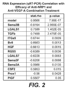
2010, the disclosures of which are hereby incorporated by reference in their
entirety
for all purposes.
FIELD OF THE INVENTION
[0002] The present invention relates to diagnostic methods and
compositions useful in the treatment of angiogenic disorders including, e.g.,
cancer.
BACKGROUND OF THE INVENTION
[0003] Angiogenic disorders such as cancer are one of the most deadly
threats to human health. In the U.S. alone, cancer affects nearly 1.3 million
new
patients each year, and is the second leading cause of death after
cardiovascular
disease, accounting for approximately 1 in 4 deaths. Solid tumors are
responsible for
most of those deaths. Although there have been significant advances in the
medical
treatment of certain cancers, the overall 5-year survival rate for all cancers
has
improved only by about 10% in the past 20 years. Cancers, or malignant tumors,
metastasize and grow rapidly in an uncontrolled manner, making timely
detection and
treatment extremely difficult.
[0004] Depending on the cancer type, patients typically have several
treatment options available to them including chemotherapy, radiation and
antibody-
based drugs. Diagnostic methods useful for predicting clinical outcome from
the
different treatment regimens would greatly benefit clinical management of
these
patients. Several studies have explored the correlation of gene expression
with the
identification of specific cancer types, e.g., by mutation-specific assays,
microarray
analysis, qPCR, etc. Such methods may be useful for the identification and
classification of cancer presented by a patient. However, much less is known
about
the predictive or prognostic value of gene expression with clinical outcome.
1
CA 02766403 2011-12-21
WO 2011/008696 PCT/US2010/041706
[0005] Thus, there is a need for objective, reproducible methods for the
optimal treatment regimen for each patient.
SUMMARY OF THE INVENTION
[0006] The methods of the present invention can be utilized in a variety of
settings, including, for example, in selecting the optimal treatment course
for a
patient, in predicting the likelihood of success when treating an individual
patient with
a particular treatment regimen, in assessing disease progression, in
monitoring
treatment efficacy, in determining prognosis for individual patients and in
assessing
predisposition of an individual to benefit from a particular therapy, e.g., an
anti-
angiogenic therapy including, for example, an anti-cancer therapy).
[0007] The present invention is based, in part, on the use of biomarkers
indicative for efficacy of therapy (e.g., anti-angiogenic therapy including,
for
example, an anti-cancer therapy). More particularly, the invention is based on
measuring an increase or decrease in the expression level(s) of at least one
gene
selected from: 18S rRNA, ACTB, RPS13, VEGFA, VEGFC, VEGFD, Bv8, P1GF,
VEGFR1/Fltl, VEGFR2, VEGFR3, NRP1, sNRP1, Podoplanin, Proxl, VE-Cadherin
(CD144, CDH5), robo4, FGF2, IL8/CXCL8, HGF, THBS1/TSP1, Egfl7, NG3/Egfl8,
ANG1, GM-CSF/CSF2, G-CSF/CSF3, FGF9, CXCL12/SDF1, TGF(31, TNFa, Alkl,
BMP9, BMP10, HSPG2/perlecan, ESM1, Sema3a, Sema3b, Sema3c, Sema3e,
Sema3f, NG2, ITGa5, ICAM1, CXCR4, LGALSI/Galectinl, LGALS7B/Galectin7,
Fibronectin, TMEM 100, PECAM/CD3 1, PDGF(3, PDGFR(3, RGS5, CXCL 1, CXCL2,
robo4, LyPD6, VCAM1, collagen IV, Spred-1, Hhex, ITGa5, LGALSI/Galectinl,
LGALS7/Galectin7, TMEM100, MFAP5, Fibronectin, fibulin2, fibulin4/Efemp2,
HMBS,SDHA, UBC, NRP2, CD34, DLL4, CLECSF5/CLEC5a, CCL2/MCP1, CCL5,
CXCL5/ENA-78, ANG2, FGF8, FGF8b, PDGFC, cMet, JAG1, CD105/Endoglin,
Notchl, EphB4, EphA3, EFNB2, TIE2/TEK, LAMA4, NID2, Map4k4, BC12A1,
IGFBP4, VIM/vimentin, FGFR4, FRAS1, ANTXR2, CLECSF5/CLEC5a, and
Mincle/CLEC4E/CLECSF9 to predict the efficacy of therapy (e.g., anti-
angiogenic
therapy including, for example, an anti-cancer therapy).
[0008] One embodiment of the invention provides methods of identifying a
patient who may benefit from treatment with an anti-cancer therapy other than
or in
addition to a VEGF antagonist. The methods comprise determining expression
levels
2
CA 02766403 2011-12-21
WO 2011/008696 PCT/US2010/041706
of at least one gene set forth in Table 1 in a sample obtained from the
patient, wherein
an increased expression level of the at least one gene in the sample as
compared to a
reference sample indicates that the patient may benefit from treatment with
the anti-
cancer therapy other than or in addition to a VEGF antagonist.
[0009] Another embodiment of the invention provides methods of identifying
a patient who may benefit from treatment with an anti-cancer therapy other
than or in
addition to a VEGF antagonist. The methods comprise: determining expression
levels
of at least one gene set forth in Table 1 in a sample obtained from the
patient, wherein
a decreased expression level of the at least one gene in the sample as
compared to a
reference sample indicates that the patient may benefit from treatment with
the anti-
cancer therapy other than or in addition to a VEGF antagonist.
[0010] A further embodiment of the invention provides methods of predicting
responsiveness of a patient suffering from cancer to treatment with an anti-
cancer
therapy other than or in addition to a VEGF antagonist. The methods comprise
determining expression levels of at least one gene set forth in Table 1 in a
sample
obtained from the patient, wherein an increased expression level of the at
least one
gene in the sample as compared to a reference sample indicates that the
patient is
more likely to be responsive to treatment with the anti-cancer therapy other
than or in
addition to a VEGF antagonist.
[0011] Yet another embodiment of the invention provides methods of
predicting responsiveness of a patient suffering from cancer to treatment with
an anti-
cancer therapy other than or in addition to a VEGF antagonist. The methods
comprise: determining expression levels of at least one gene set forth in
Table 1 in a
sample obtained from the patient, wherein a decreased expression level of the
at least
one gene in the sample as compared to a reference sample indicates that the
patient is
more likely to be responsive to treatment with the anti-cancer therapy other
than or in
addition to a VEGF antagonist.
[0012] Even another embodiment of the invention provides methods for
determining the likelihood that a patient with cancer will exhibit benefit
from anti-
cancer therapy other than or in addition to a VEGF antagonist. The methods
comprise: determining expression levels of at least one gene set forth in
Table 1 in a
sample obtained from the patient, wherein an increased expression level of the
at least
one gene in the sample as compared to a reference sample indicates that the
patient
3
CA 02766403 2011-12-21
WO 2011/008696 PCT/US2010/041706
has increased likelihood of benefit from the anti-cancer therapy other than or
in
addition to a VEGF antagonist.
[0013] Another embodiment of the invention provides methods for
determining the likelihood that a patient with cancer will exhibit benefit
from anti-
cancer therapy other than or in addition to a VEGF antagonist. The methods
comprise: determining expression levels of at least one gene set forth in
Table 1 in a
sample obtained from the patient, wherein a decreased expression level of the
at least
one gene in the sample as compared to a reference sample indicates that the
patient
has increased likelihood of benefit from the anti-cancer therapy other than or
in
addition to a VEGF antagonist.
[0014] A further embodiment of the invention provides methods for treating
cancer in a patient. The methods comprise: determining that a sample obtained
from
the patient has increased expression levels, as compared to a reference
sample, of at
least one gene set forth in Table 1, and administering an effective amount of
an anti-
cancer therapy other than or in addition to a VEGF antagonist to the patient,
whereby
the cancer is treated.
[0015] Another embodiment of the invention provides methods for treating
cancer in a patient. The methods comprise determining that a sample obtained
from
the patient has decreased expression levels, as compared to a reference
sample, of at
least one gene set forth in Table 1, and administering an effective amount of
an anti-
cancer therapy other than or in addition to a VEGF antagonist to the patient,
whereby
the cancer is treated.
[0016] In some embodiments of the invention, the sample obtained from the
patient is selected from: tissue, whole blood, blood-derived cells, plasma,
serum, and
combinations thereof. In some embodiments of the invention, the expression
level is
mRNA expression level. In some embodiments of the invention, the expression
level
is protein expression level.
[0017] In some embodiments of the invention, the methods further comprise
detecting the expression of at least a second, third, fourth, fifth, sixth,
seventh, eighth,
ninth, tenth, eleventh, twelfth, thirteenth, fourteenth, fifteenth, sixteenth,
seventeenth,
eighteenth, nineteenth, or twentieth gene set forth in Table 1.
[0018] In some embodiments of the invention, the methods further comprising
administering the anti-cancer therapy other than a VEGF antagonist to the
patient. In
some embodiments of the invention, the anti-cancer therapy is selected from:
an
4
CA 02766403 2011-12-21
WO 2011/008696 PCT/US2010/041706
antibody, a small molecule, and an siRNA. In some embodiments of the
invention,
the anti-cancer therapy is a member selected from: an EGFL7 antagonist, a NRP1
antagonist, and a VEGF-C antagonist. In some embodiments of the invention, the
EGFL7 antagonist is an antibody. In some embodiments of the invention, the
NRP1
antagonist is an antibody. In some embodiments of the invention, the VEGF-C
antagonist is an antibody.
[0019] In some embodiments of the invention, the methods further comprise
administering the VEGF antagonist to the patient. In some embodiments of the
invention, the VEGF antagonist is an anti-VEGF antibody. In some embodiments
of
the invention, the anti-VEGF antibody is bevacizumab. In some embodiments of
the
invention, the anti-cancer therapy and the VEGF antagonist are administered
concurrently. In some embodiments of the invention, the anti-cancer therapy
and the
VEGF antagonist are administered sequentially.
[0020] Even another embodiment of the invention provides kits for
determining whether a patient may benefit from treatment with an anti-cancer
therapy
other than or in addition to a VEGF antagonist. The kits comprise an array
comprising polynucleotides capable of specifically hybridizing to at least one
gene set
forth in Table 1 and instructions for using said array to determine the
expression
levels of the at least one gene to predict responsiveness of a patient to
treatment with
an anti-cancer therapy in addition to a VEGF antagonist, wherein an increase
in the
expression level of the at least one gene as compared to the expression level
of the at
least one gene in a reference sample indicates that the patient may benefit
from
treatment with the anti-cancer therapy in addition to a VEGF antagonist.
[0021] A further embodiment of the invention provides kits for determining
whether a patient may benefit from treatment with an anti-cancer therapy other
than or
in addition to a VEGF antagonist. The kits comprise an array comprising
polynucleotides capable of specifically hybridizing to at least one gene set
forth in
Table 1 and instructions for using said array to determine the expression
levels of the
at least one gene to predict responsiveness of a patient to treatment with an
anti-
cancer therapy in addition to a VEGF antagonist, wherein a decrease in the
expression
level of the at least one gene as compared to the expression level of the at
least one
gene in a reference sample indicates that the patient may benefit from
treatment with
an anti-cancer therapy in addition to a VEGF antagonist.
CA 02766403 2011-12-21
WO 2011/008696 PCT/US2010/041706
[0022] Another embodiment of the invention provides sets of compounds for
detecting expression levels of at least one gene set forth in Table 1 to
determine the
expression levels of the at least one gene in a sample obtained from a cancer
patient.
The sets comprise at least one compound capable of specifically hybridizing to
at least
one gene set forth in Table 1, wherein an increase in the expression level of
the at
least one gene as compared to the expression level of the at least one gene in
a
reference sample indicates that the patient may benefit from treatment with an
anti-
cancer therapy in addition to a VEGF antagonist. In some embodiments of the
invention, the compounds are polynucleotides. In some embodiments of the
invention, the polynucleotides comprise three sequences set forth in Table 2.
In some
embodiments of the invention, the compounds are proteins, such as, for
example,
antibodies.
[0023] Yet another embodiment of the invention provides sets of compounds
for detecting expression levels of at least one gene set forth in Table 1 to
determine
the expression levels of the at least one gene in a sample obtained from a
cancer
patient. The sets comprise at least one compound capable of specifically
hybridizing
to at least one gene set forth in Table 1, wherein a decrease in the
expression level of
the at least one gene as compared to the expression level of the at least one
gene in a
reference sample indicates that the patient may benefit from treatment with an
anti-
cancer therapy in addition to a VEGF antagonist. In some embodiments of the
invention, the compounds are polynucleotides. In some embodiments of the
invention, the polynucleotides comprise three sequences set forth in Table 2.
In some
embodiments of the invention, the compounds are proteins, such as, for
example,
antibodies.
[0024] One embodiment of the invention provides methods of identifying a
patient suffering from cancer who may benefit from treatment with a neuropilin-
1
(NRP1) antagonist. The methods comprise determining expression levels of at
least
one gene selected from: TGF(31, Bv8, Sema3A, P1GF, LGALSI, ITGa5, CSF2,
Vimentin, CXCL5, CCL2, CXCL2, Alkl, and FGF8 in a sample obtained from the
patient, wherein increased expression levels of the at least one gene in the
sample as
compared to a reference sample indicates that the patient may benefit from
treatment
with the NRP 1 antagonist.
[0025] Another embodiment of the invention provides methods of identifying
a patient suffering from cancer who may benefit from treatment with a
neuropilin-1
6
CA 02766403 2011-12-21
WO 2011/008696 PCT/US2010/041706
(NRP1) antagonist. The methods comprise determining expression levels of at
least
one gene selected from: Proxl, RGS5, HGF, Sema3B, Sema3F, LGALS7, FGRF4,
PLC, IGFB4, and TSP1 in a sample obtained from the patient, wherein decreased
expression levels of the at least one gene in the sample as compared to a
reference
sample indicates that the patient may benefit from treatment with the NRP 1
antagonist.
[0026] A further embodiment of the invention provides methods of predicting
responsiveness of a patient suffering from cancer to treatment with a NRP1
antagonist. The methods comprise determining expression levels of at least one
gene
selected from: TGF(31, Bv8, Sema3A, P1GF, LGALSI, ITGa5, CSF2, Vimentin,
CXCL5, CCL2, CXCL2, Alkl, and FGF8 in a sample obtained from the patient,
wherein increased expression levels of the at least one gene in the sample as
compared to a reference sample indicates that the patient is more likely to be
responsive to treatment with the NRP 1 antagonist.
[0027] Even another further embodiment of the invention provides methods of
predicting responsiveness of a patient suffering from cancer to treatment with
a NRP1
antagonist. The methods comprise determining expression levels of at least one
gene
selected from: Proxl, RGS5, HGF, Sema3B, Sema3F, LGALS7, FGRF4, PLC,
IGFB4, and TSP1 in a sample obtained from the patient, wherein decreased
expression levels of the at least one gene in the sample as compared to a
reference
sample indicates that the patient is more likely to be responsive to treatment
with the
NRP 1 antagonist.
[0028] Yet another embodiment of the invention provides methods of
determining the likelihood that a patient will exhibit a benefit from
treatment with a
NRP1 antagonist. The methods comprise determining expression levels of at
least
one gene selected from: TGF(31, Bv8, Sema3A, P1GF, LGALSI, ITGa5, CSF2,
Vimentin, CXCL5, CCL2, CXCL2, Alkl, and FGF8 in a sample obtained from the
patient, wherein increased expression levels of the at least one gene in the
sample as
compared to a reference sample indicates that the patient has increased
likelihood of
benefit from treatment with the NRP1 antagonist.
[0029] Another embodiment of the invention provide methods of determining
the likelihood that a patient will exhibit a benefit from treatment with a
NRP1
antagonist. The methods comprise determining expression levels of at least one
gene
selected from: Proxl, RGS5, HGF, Sema3B, Sema3F, LGALS7, FGRF4, PLC,
7
CA 02766403 2011-12-21
WO 2011/008696 PCT/US2010/041706
IGFB4, and TSP1 in a sample obtained from the patient, wherein decreased
expression levels of the at least one gene in the sample as compared to a
reference
sample indicates that the patient has increased likelihood of benefit of
treatment with
the NRP 1 antagonist.
[0030] Yet another embodiment of the invention provides methods of
optimizing therapeutic efficacy of a NRP1 antagonist. The methods comprise
determining expression levels of at least one gene selected from: TGF(31, Bv8,
Sema3A, P1GF, LGALSI, ITGa5, CSF2, Vimentin, CXCL5, CCL2, CXCL2, Alkl,
and FGF8 in a sample obtained from the patient, wherein increased expression
levels
of the at least one gene in the sample as compared to a reference sample
indicates that
the patient has increased likelihood of benefit from treatment with the NRP1
antagonist.
[0031] Another embodiment of the invention provide methods of optimizing
therapeutic efficacy of a NRP1 antagonist. The methods comprise determining
expression levels of at least one gene selected from: Proxl, RGS5, HGF,
Sema3B,
Sema3F, LGALS7, FGRF4, PLC, IGFB4, and TSP1 in a sample obtained from the
patient, wherein decreased expression levels of the at least one gene in the
sample as
compared to a reference sample indicates that the patient has increased
likelihood of
benefit of treatment with the NRP1 antagonist.
[0032] A further embodiment of the invention provides methods for treating a
cell proliferative disorder in a patient. The methods comprise determining
that a
sample obtained from the patient has increased expression levels, as compared
to a
reference sample, of at least one gene selected from: TGF(31, Bv8, Sema3A,
P1GF,
LGALSI, ITGa5, CSF2, Vimentin, CXCL5, CCL2, CXCL2, Alkl, and FGF8, and
administering to the patient an effective amount of a NRP1 antagonist, whereby
the
cell proliferative disorder is treated.
[0033] Yet another embodiment of the invention provides methods for treating
a cell proliferative disorder in a patient. The methods comprise determining
that a
sample obtained from the patient has decreased expression levels, as compared
to a
reference sample, of at least one gene selected from: Proxl, RGS5, HGF,
Sema3B,
Sema3F, LGALS7, FGRF4, PLC, IGFB4, and TSP1, and administering to the patient
an effective amount of a NRP 1 antagonist, whereby the cell proliferative
disorder is
treated.
8
CA 02766403 2011-12-21
WO 2011/008696 PCT/US2010/041706
[0034] In some embodiments of the invention, the sample obtained from the
patient is a member selected from: tissue, whole blood, blood-derived cells,
plasma,
serum, and combinations thereof. In some embodiments of the invention, the
expression level is mRNA expression level. In some embodiments of the
invention,
the expression level is protein expression level. In some embodiments of the
invention, the NRP1 antagonist is an anti-NRP1 antibody.
[0035] In some embodiments of the invention, the methods further comprise
administering a VEGF antagonist to the patient. In some embodiments of the
invention, the VEGF antagonist and the NRP1 antagonist are administered
concurrently. In some embodiments of the invention, the VEGF antagonist and
the
NRP1 antagonist are administered sequentially. In some embodiments of the
invention, the VEGF antagonist is an anti-VEGF antibody. In some embodiments
of
the invention, the anti-VEGF antibody is bevacizumab.
[0036] Another embodiment of the invention provides methods of identifying
a patient suffering from cancer who may benefit from treatment with a NRP1
antagonist. The methods comprise determining expression levels of P1GF in a
sample
obtained from the patient, wherein increased expression levels of P1GF in the
sample
as compared to a reference sample indicates that the patient may benefit from
treatment with the NRP 1 antagonist.
[0037] Even another embodiment of the invention provides methods of
predicting responsiveness of a patient suffering from cancer to treatment with
a NRP1
antagonist. The methods comprise determining expression levels of P1GF in a
sample
obtained from the patient, wherein increased expression levels of P1GF in the
sample
as compared to a reference sample indicates that the patient is more likely to
be
responsive to treatment with the NRP 1 antagonist.
[0038] Yet another embodiment of the invention provides methods of
determining the likelihood that a patient will exhibit a benefit from
treatment with a
NRP1 antagonist. The methods comprise determining expression levels of P1GF in
a
sample obtained from the patient, wherein increased expression levels of P1GF
in the
sample as compared to a reference sample indicates that the patient has
increased
likelihood of benefit from treatment with the NRP1 antagonist.
[0039] Even another embodiment of the invention provides methods of
optimizing therapeutic efficacy of a NRP1 antagonist. The methods comprise
determining expression levels of P1GF in a sample obtained from the patient,
wherein
9
CA 02766403 2011-12-21
WO 2011/008696 PCT/US2010/041706
increased expression levels of P1GF in the sample as compared to a reference
sample
indicates that the patient has increased likelihood of benefit from treatment
with the
NRP 1 antagonist.
[0040] A further embodiment of the invention provides methods for treating a
cell proliferative disorder in a patient. The methods comprise determining
that a
sample obtained from the patient has increased expression levels of P1GF as
compared
to a reference sample, and administering to the patient an effective amount of
a NRP 1
antagonist, whereby the cell proliferative disorder is treated.
[0041] Even a further embodiment of the invention provides methods of
identifying a patient suffering from cancer who may benefit from treatment
with a
neuropilin-1 (NRP1) antagonist. The methods comprise determining expression
levels of Sema3A in a sample obtained from the patient, wherein increased
expression
levels of Sema3A in the sample as compared to a reference sample indicates
that the
patient may benefit from treatment with the NRP 1 antagonist.
[0042] Yet a further embodiment of the invention provides methods of
predicting responsiveness of a patient suffering from cancer to treatment with
a NRP1
antagonist. The methods comprise determining expression levels of Sema3A in a
sample obtained from the patient, wherein increased expression levels of
Sema3A in
the sample as compared to a reference sample indicates that the patient is
more likely
to be responsive to treatment with the NRP1 antagonist.
[0043] Another embodiment of the invention provides methods of determining
the likelihood that a patient will exhibit a benefit from treatment with a NRP
1
antagonist. The methods comprise determining expression levels of Sema3A in a
sample obtained from the patient, wherein increased expression levels of
Sema3A in
the sample as compared to a reference sample indicates that the patient has
increased
likelihood of benefit from treatment with the NRP 1 antagonist.
[0044] Another embodiment of the invention provides methods of optimizing
therapeutic efficacy of a NRP 1 antagonist. The methods comprise determining
expression levels of Sema3A in a sample obtained from the patient, wherein
increased
expression levels of Sema3A in the sample as compared to a reference sample
indicates that the patient has increased likelihood of benefit from treatment
with the
NRP 1 antagonist.
[0045] Even another embodiment of the invention provides methods for
treating a cell proliferative disorder in a patient. The methods comprise
determining
CA 02766403 2011-12-21
WO 2011/008696 PCT/US2010/041706
that a sample obtained from the patient has increased expression levels of
Sema3A as
compared to a reference sample, and administering to the patient an effective
amount
of a NRP 1 antagonist, whereby the cell proliferative disorder is treated
[0046] Yet another embodiment of the invention provides methods of
identifying a patient suffering from cancer who may benefit from treatment
with a
neuropilin-1 (NRP1) antagonist. The methods comprise determining expression
levels of TGF(31 in a sample obtained from the patient, wherein increased
expression
levels of TGF(31 in the sample as compared to a reference sample indicates
that the
patient may benefit from treatment with the NRP 1 antagonist.
[0047] A further embodiment of the invention provides methods of predicting
responsiveness of a patient suffering from cancer to treatment with a NR-P1
antagonist. The methods comprise determining expression levels of TGF(31 in a
sample obtained from the patient, wherein increased expression levels of
TGF(31 in
the sample as compared to a reference sample indicates that the patient is
more likely
to be responsive to treatment with the NRP1 antagonist. In some embodiments of
the
invention, the methods further comprise administering an effective amount of a
NRP1
antagonist to the patient.
[0048] Even a further embodiment of the invention provides methods of
determining the likelihood that a patient will exhibit a benefit from
treatment with a
NRP1 antagonist. The methods comprise determining expression levels of TGF(31
in
a sample obtained from the patient, wherein increased expression levels of
TGF(31 in
the sample as compared to a reference sample indicates that the patient has
increased
likelihood of benefit from treatment with the NRP1 antagonist.
[0049] Even a further embodiment of the invention provides methods of
optimizing therapeutic efficacy of a NRP1 antagonist. The methods comprise
determining expression levels of TGF(31 in a sample obtained from the patient,
wherein increased expression levels of TGF(31 in the sample as compared to a
reference sample indicates that the patient has increased likelihood of
benefit from
treatment with the NRP 1 antagonist.
[0050] Yet a further embodiment of the invention provides methods for
treating a cell proliferative disorder in a patient. The methods comprise
determining
that a sample obtained from the patient has increased expression levels of
TGF(31 as
11
CA 02766403 2011-12-21
WO 2011/008696 PCT/US2010/041706
compared to a reference sample, and administering to the patient an effective
amount
of a NRP 1 antagonist, whereby the cell proliferative disorder is treated
[0051] In some embodiments of the invention, the NRP1 antagonist is an anti-
NRP1 antibody. In some embodiments of the invention, the methods further
comprises administering a VEGF-A antagonist to the patient. In some
embodiments
of the invention, the VEGF-A antagonist and the NRP1 antagonist are
administered
concurrently. In some embodiments of the invention, the VEGF-A antagonist and
the
NRP1 antagonist are administered sequentially. In some embodiments of the
invention, the VEGF-A antagonist is an anti-VEGF-A antibody. In some
embodiments of the invention, the anti-VEGF-A antibody is bevacizumab.
[0052] Another embodiment of the invention provides kits for determining the
expression levels of at least one gene selected from: TGF(31, Bv8, Sema3A,
P1GF,
LGALSI, ITGa5, CSF2, Vimentin, CXCL5, CCL2, CXCL2, Alkl, and FGF8. The
kits comprise an array comprising polynucleotides capable of specifically
hybridizing
to at least one gene selected from: TGF(31, Bv8, Sema3A, P1GF, LGALSI, ITGa5,
CSF2, Vimentin, CXCL5, CCL2, CXCL2, Alkl, and FGF8 and instructions for using
the array to determine the expression levels of the at least one gene to
predict
responsiveness of a patient to treatment with a NRP1 antagonist, wherein an
increase
in the expression level of the at least one gene as compared to the expression
level of
the at least one gene in a reference sample indicates that the patient may
benefit from
treatment with the NRP 1 antagonist.
[0053] Even another embodiment of the invention provides kits for
determining the expression levels of at least one gene selected from: Proxl,
RGS5,
HGF, Sema3B, Sema3F, LGALS7, FGRF4, PLC, IGFB4, and TSP1. The kits
comprise an array comprising polynucleotides capable of specifically
hybridizing to at
least one gene selected from: Proxl, RGS5, HGF, Sema3B, Sema3F, LGALS7,
FGRF4, PLC, IGFB4, and TSP1 and instructions for using the array to determine
the
expression levels of the at least one gene to predict responsiveness of a
patient to
treatment with a NRP1 antagonist, wherein a decrease in the expression level
of the at
least one gene as compared to the expression level of the at least one gene in
a
reference sample indicates that the patient may benefit from treatment with
the NRP 1
antagonist.
[0054] Yet another embodiment of the invention provides sets of compounds
capable of detecting expression levels of at least one gene selected from:
TGF(31,
12
CA 02766403 2011-12-21
WO 2011/008696 PCT/US2010/041706
Bv8, Sema3A, P1GF, LGALS1, ITGa5, CSF2, Vimentin, CXCL5, CCL2, CXCL2,
Alkl, and FGF8 to determine the expression levels of the at least one gene in
a sample
obtained from a cancer patient. The sets comprise at least one compound
capable of
specifically hybridizing to at least one gene selected from: TGF(31, Bv8,
Sema3A,
P1GF, LGALS1, ITGa5, CSF2, Vimentin, CXCL5, CCL2, CXCL2, Alkl, and FGF8,
wherein an increase in the expression level of the at least one gene as
compared to the
expression level of the at least one gene in a reference sample indicates that
the
patient may benefit from treatment with a NRP 1 antagonist. In some
embodiments of
the invention, the compounds are polynucleotides. In some embodiments of the
invention, the polynucleotides comprise three sequences set forth in Table 2.
In some
embodiments of the invention, the compounds are proteins, including, for
example,
antibodies.
[0055] A further embodiment of the invention provides sets of compounds
capable of detecting expression levels of at least one gene selected from:
Prox1,
RGS5, HGF, Sema3B, Sema3F, LGALS7, FGRF4, PLC, IGFB4, and TSPI to
determine the expression levels of the at least one gene in a sample obtained
from a
cancer patient. The sets comprise at least one compound capable of
specifically
hybridizing to at least one gene selected from: Proxl, RGS5, HGF, Sema3B,
Sema3F, LGALS7, FGRF4, PLC, IGFB4, and TSPI, wherein a decrease in the
expression level of said at least gene as compared to the expression level of
the at
least one gene in a reference sample indicates that the patient may benefit
from
treatment with a NRP1 antagonist. In some embodiments of the invention, the
compounds are polynucleotides. In some embodiments of the invention, the
polynucleotides comprise three sequences set forth in Table 2. In some
embodiments
of the invention, the compounds are proteins, including, for example,
antibodies.
[0056] Another embodiment of the invention provides methods of identifying
a patient suffering from cancer who may benefit from treatment with a Vascular
Endothelial Growth Factor C (VEGF-C) antagonist. The methods comprise
determining expression levels of at least one gene selected from: VEGF-C, VEGF-
D,
VEGFR3, FGF2, RGS5/CDH5, IL-8, CXCL1, and CXCL2 in a sample obtained from
the patient, wherein increased expression levels of the at least one gene in
the sample
as compared to a reference sample indicates that the patient may benefit from
treatment with the VEGF-C antagonist.
13
CA 02766403 2011-12-21
WO 2011/008696 PCT/US2010/041706
[0057] Even another embodiment of the invention provides methods of
identifying a patient suffering from cancer who may benefit from treatment
with a
VEGF-C antagonist. The methods comprise determining expression levels of at
least
one gene selected from: VEGF-A, CSF2, Proxl, ICAM1, ESM1, P1GF, ITGa5,
TGF(3, Hhex, Col4al, Co14a2, and Alkl in a sample obtained from the patient,
wherein decreased expression levels of the at least one gene in the sample as
compared to a reference sample indicates that the patient may benefit from
treatment
with the VEGF-C antagonist.
[0058] Yet another embodiment of the invention provides methods of
predicting responsiveness of a patient suffering from cancer to treatment with
a
VEGF-C antagonist. The methods comprise determining expression levels of at
least
one gene selected from: VEGF-C, VEGF-D, VEGFR3, FGF2, RGS5/CDH5, IL-8,
CXCL 1, and CXCL2 in a sample obtained from the patient, wherein increased
expression levels of the at least one gene in the sample as compared to a
reference
sample indicates that the patient is more likely to be responsive to treatment
with the
VEGF-C antagonist.
[0059] A further embodiment of the invention provides methods of predicting
responsiveness of a patient suffering from cancer to treatment with a VEGF-C
antagonist. The methods comprise determining expression levels of at least one
gene
selected from: VEGF-A, CSF2, Proxl, ICAM1, ESM1, P1GF, ITGa5, TGF(3, Hhex,
Col4al, Co14a2, and Alkl in a sample obtained from the patient, wherein
decreased
expression levels of the at least one gene in the sample as compared to a
reference
sample indicates that the patient is more likely to be responsive to treatment
with the
VEGF-C antagonist.
[0060] Even a further embodiment of the invention provides methods of
determining the likelihood that a patient will exhibit a benefit from
treatment with a
VEGF-C antagonist. The methods comprise determining expression levels of at
least
one gene selected from: VEGF-C, VEGF-D, VEGFR3, FGF2, RGS5/CDH5, IL-8,
CXCL 1, and CXCL2 in a sample obtained from the patient, wherein increased
expression levels of the at least one gene in the sample as compared to a
reference
sample indicates that the patient has increased likelihood of benefit from
treatment
with the VEGF-C antagonist.
[0061] Yet a further embodiment of the invention provides methods of
determining the likelihood that a patient will exhibit a benefit from
treatment with a
14
CA 02766403 2011-12-21
WO 2011/008696 PCT/US2010/041706
VEGF-C antagonist. The methods comprise determining expression levels of at
least
one gene selected from: VEGF-A, CSF2, Proxl, ICAM1, ESM1, P1GF, ITGa5,
TGF(3, Hhex, Col4al, Co14a2, and Alkl in a sample obtained from the patient,
wherein decreased expression levels of the at least one gene in the sample as
compared to a reference sample indicates that the patient has increased
likelihood of
benefit of treatment with the VEGF-C antagonist.
[0062] Even a further embodiment of the invention provides methods of
optimizing therapeutic efficacy of a VEGF-C antagonist. The methods comprise
determining expression levels of at least one gene selected from: VEGF-C, VEGF-
D,
VEGFR3, FGF2, RGS5/CDH5, IL-8, CXCL1, and CXCL2 in a sample obtained from
the patient, wherein increased expression levels of the at least one gene in
the sample
as compared to a reference sample indicates that the patient has increased
likelihood
of benefit from treatment with the VEGF-C antagonist.
[0063] Yet a further embodiment of the invention provides methods of
optimizing therapeutic efficacy of a VEGF-C antagonist. The methods comprise
determining expression levels of at least one gene selected from: VEGF-A,
CSF2,
Proxl, ICAM1, ESM1, P1GF, ITGa5, TGF(3, Hhex, Col4al, Co14a2, and Alkl in a
sample obtained from the patient, wherein decreased expression levels of the
at least
one gene in the sample as compared to a reference sample indicates that the
patient
has increased likelihood of benefit of treatment with the VEGF-C antagonist.
[0064] Another embodiment of the invention provides methods for treating a
cell proliferative disorder in a patient. The methods comprise determining
that a
sample obtained from the patient has increased expression levels, as compared
to a
reference sample, of at least one gene selected from: VEGF-C, VEGF-D, VEGFR3,
FGF2, RGS5/CDH5, IL-8, CXCL1, and CXCL2, and administering to the patient an
effective amount of a VEGF-C antagonist, whereby the cell proliferative
disorder is
treated.
[0065] Even another embodiment of the invention provides methods for
treating a cell proliferative disorder in a patient. The methods comprise
determining
that a sample obtained from the patient has decreased expression levels, as
compared
to a reference sample, of at least one gene selected from: VEGF-A, CSF2,
Proxl,
ICAM1, ESM1, P1GF, ITGa5, TGF(3, Hhex, Col4al, Co14a2, and Alkl, and
administering to the patient an effective amount of a VEGF-C antagonist,
whereby the
cell proliferative disorder is treated.
CA 02766403 2011-12-21
WO 2011/008696 PCT/US2010/041706
[0066] In some embodiments of the invention, the sample obtained from
the patient is selected from: tissue, whole blood, blood-derived cells,
plasma, serum,
and combinations thereof. In some embodiments of the invention, the expression
level is mRNA expression level. In some embodiments of the invention, the
expression level is protein expression level. In some embodiments of the
invention,
the VEGF-C antagonist is an anti-VEGF-C antibody.
[0067] In some embodiments of the invention, the methods further
comprise administering a VEGF-A antagonist to the patient. In some embodiments
of
the invention, the VEGF-A antagonist and the VEGF-C antagonist are
administered
concurrently. In some embodiments of the invention, the VEGF-A antagonist and
the
VEGF-C antagonist are administered sequentially. In some embodiments of the
invention, the VEGF-A antagonist is an anti-VEGF-A antibody. In some
embodiments of the invention, the anti-VEGF-A antibody is bevacizumab.
[0068] Another embodiment of the invention provides methods of
identifying a patient suffering from cancer who may benefit from treatment
with a
VEGF-C antagonist. The methods comprise determining expression levels of VEGF-
C in a sample obtained from the patient, wherein increased expression levels
of
VEGF-C in the sample as compared to a reference sample indicates that the
patient
may benefit from treatment with the VEGF-C antagonist.
[0069] Even another embodiment of the invention provides methods of
predicting responsiveness of a patient suffering from cancer to treatment with
a
VEGF-C antagonist. The methods comprise determining expression levels of VEGF-
C in a sample obtained from the patient, wherein increased expression levels
of
VEGF-C in the sample as compared to a reference sample indicates that the
patient is
more likely to be responsive to treatment with the VEGF-C antagonist.
[0070] Yet another embodiment of the invention provides methods of
determining the likelihood that a patient will exhibit a benefit from
treatment with a
VEGF-C antagonist. The methods comprise determining expression levels of VEGF-
C in a sample obtained from the patient, wherein increased expression levels
of
VEGF-C in the sample as compared to a reference sample indicates that the
patient
has an increased likelihood of benefit from treatment with the VEGF-C
antagonist.
[0071] Yet another embodiment of the invention provides methods of
optimizing therapeutic efficacy of a VEGF-C antagonist. The methods comprise
determining expression levels of VEGF-C in a sample obtained from the patient,
16
CA 02766403 2011-12-21
WO 2011/008696 PCT/US2010/041706
wherein increased expression levels of VEGF-C in the sample as compared to a
reference sample indicates that the patient has an increased likelihood of
benefit from
treatment with the VEGF-C antagonist.
[0072] A further embodiment of the invention provides methods for
treating a cell proliferative disorder in a patient. The methods comprise
determining
that a sample obtained from the patient has increased expression levels of
VEGF-C as
compared to a reference sample, and administering to the patient an effective
amount
of a VEGF-C antagonist, whereby the cell proliferative disorder is treated.
[0073] Even a further embodiment of the invention provides methods of
identifying a patient suffering from cancer who may benefit from treatment
with a
VEGF-C antagonist. The methods comprise determining expression levels of VEGF-
D in a sample obtained from the patient, wherein increased expression levels
of
VEGF-D in the sample as compared to a reference sample indicates that the
patient
may benefit from treatment with the VEGF-C antagonist.
[0074] Yet a further embodiment of the invention provides methods of
predicting responsiveness of a patient suffering from cancer to treatment with
a
VEGF-C antagonist. The methods comprise determining expression levels of VEGF-
D in a sample obtained from the patient, wherein increased expression levels
of
VEGF-D in the sample as compared to a reference sample indicates that the
patient is
more likely to be responsive to treatment with the VEGF-C antagonist.
[0075] Another embodiment of the invention provides methods of
determining the likelihood that a patient will exhibit a benefit from
treatment with a
VEGF-C antagonist. The methods comprise determining expression levels of VEGF-
D in a sample obtained from the patient, wherein increased expression levels
of
VEGF-D in the sample as compared to a reference sample indicates that the
patient
has an increased likelihood of benefit from treatment with the VEGF-C
antagonist.
[0076] Another embodiment of the invention provides methods of
optimizing therapeutic efficacy of a VEGF-C antagonist. The methods comprise
determining expression levels of VEGF-D in a sample obtained from the patient,
wherein increased expression levels of VEGF-D in the sample as compared to a
reference sample indicates that the patient has an increased likelihood of
benefit from
treatment with the VEGF-C antagonist.
[0077] Even another embodiment of the invention provides methods for
treating a cell proliferative disorder in a patient. The methods comprise
determining
17
CA 02766403 2011-12-21
WO 2011/008696 PCT/US2010/041706
that a sample obtained from the patient has increased expression levels of
VEGF-D as
compared to a reference sample, and administering to the patient an effective
amount
of a VEGF-C antagonist, whereby the cell proliferative disorder is treated
[0078] Yet another embodiment of the invention provides methods of
identifying a patient suffering from cancer who may benefit from treatment
with a
VEGF-C antagonist. The methods comprise determining expression levels of
VEGFR3 in a sample obtained from the patient, wherein increased expression
levels
of VEGFR3 in the sample as compared to a reference sample indicates that the
patient
may benefit from treatment with the VEGF-C antagonist.
[0079] A further embodiment of the invention provides methods of
predicting responsiveness of a patient suffering from cancer to treatment with
a
VEGF-C antagonist. The methods comprise determining expression levels of
VEGFR3 in a sample obtained from the patient, wherein increased expression
levels
of VEGFR3 in the sample as compared to a reference sample indicates that the
patient
is more likely to be responsive to treatment with the VEGF-C antagonist.
[0080] Even a further embodiment of the invention provides methods of
determining the likelihood that a patient will exhibit a benefit from
treatment with a
VEGF-C antagonist. The methods comprise determining expression levels of
VEGFR3 in a sample obtained from the patient, wherein increased expression
levels
of VEGFR3 in the sample as compared to a reference sample indicates that the
patient
has an increased likelihood of benefit from treatment with the VEGF-C
antagonist.
[0081] A further embodiment of the invention provides methods of
optimizing therapeutic efficacy of a VEGF-C antagonist. The methods comprise
determining expression levels of VEGFR3 in a sample obtained from the patient,
wherein increased expression levels of VEGFR3 in the sample as compared to a
reference sample indicates that the patient has an increased likelihood of
benefit from
treatment with the VEGF-C antagonist.
[0082] Yet a further embodiment of the invention provides methods for
treating a cell proliferative disorder in a patient. The methods comprise
determining
that a sample obtained from the patient has increased expression levels of
VEGFR3 as
compared to a reference sample, and administering to the patient an effective
amount
of a VEGF-C antagonist, whereby the cell proliferative disorder is treated
[0083] Another embodiment of the invention provides methods of
identifying a patient suffering from cancer who may benefit from treatment
with a
18
CA 02766403 2011-12-21
WO 2011/008696 PCT/US2010/041706
VEGF-C antagonist. The methods comprise determining expression levels of FGF2
in a sample obtained from the patient, wherein increased expression levels of
FGF2 in
the sample as compared to a reference sample indicates that the patient may
benefit
from treatment with the VEGF-C antagonist.
[0084] Even another embodiment of the invention provides methods of
predicting responsiveness of a patient suffering from cancer to treatment with
a
VEGF-C antagonist. The methods comprise determining expression levels of FGF2
in a sample obtained from the patient, wherein increased expression levels of
FGF2 in
the sample as compared to a reference sample indicates that the patient is
more likely
to be responsive to treatment with the VEGF-C antagonist.
[0085] Yet another embodiment of the invention provides methods of
determining the likelihood that a patient will exhibit a benefit from
treatment with a
VEGF-C antagonist. The methods comprise determining expression levels of FGF2
in a sample obtained from the patient, wherein increased expression levels of
FGF2 in
the sample as compared to a reference sample indicates that the patient has an
increased likelihood of benefit from treatment with the VEGF-C antagonist.
[0086] Even another embodiment of the invention provides methods of
optimizing therapeutic efficacy of a VEGF-C antagonist. The methods comprise
determining expression levels of FGF2 in a sample obtained from the patient,
wherein
increased expression levels of FGF2 in the sample as compared to a reference
sample
indicates that the patientt has an increased likelihood of benefit from
treatment with
the VEGF-C antagonist.
[0087] A further embodiment of the invention provides methods for
treating a cell proliferative disorder in a patient. The methods comprise
determining
that a sample obtained from the patient has increased expression levels of
FGF2 as
compared to a reference sample, and administering to the patient an effective
amount
of a VEGF-C antagonist, whereby the cell proliferative disorder is treated
[0088] Even a further embodiment of the invention provides methods of
identifying a patient suffering from cancer who may benefit from treatment
with a
VEGF-C antagonist. The methods comprise determining expression levels of VEGF-
A in a sample obtained from the patient, wherein decreased expression levels
of
VEGF-A in the sample as compared to a reference sample indicates that the
patient
may benefit from treatment with the VEGF-C antagonist.
19
CA 02766403 2011-12-21
WO 2011/008696 PCT/US2010/041706
[0089] Yet a further embodiment of the invention provides methods of
predicting responsiveness of a patient suffering from cancer to treatment with
a
VEGF-C antagonist. The methods comprise determining expression levels of VEGF-
A in a sample obtained from the patient, wherein decreased expression levels
of
VEGF-A in the sample as compared to a reference sample indicates that the
patient is
more likely to be responsive to treatment with the VEGF-C antagonist.
[0090] Another embodiment of the invention provides methods of
determining the likelihood that a patient will exhibit a benefit from
treatment with a
VEGF-C antagonist. The methods comprise determining expression levels of VEGF-
A in a sample obtained from the patient, wherein decreased expression levels
of
VEGF-A in the sample as compared to a reference sample indicates that the
patient
has an increased likelihood of benefit from treatment with the VEGF-C
antagonist.
[0091] Another embodiment of the invention provides methods of
optimizing therapeutic efficacy of a VEGF-C antagonist. The methods comprise
determining expression levels of VEGF-A in a sample obtained from the patient,
wherein decreased expression levels of VEGF-A in the sample as compared to a
reference sample indicates that the patient has an increased likelihood of
benefit from
treatment with the VEGF-C antagonist.
[0092] Even another embodiment of the invention provides methods for
treating a cell proliferative disorder in a patient. The methods comprise
determining
that a sample obtained from the patient has decreased expression levels of
VEGF-A as
compared to a reference sample, and administering to the patient an effective
amount
of a VEGF-C antagonist, whereby the cell proliferative disorder is treated.
[0093] Yet another embodiment of the invention provides methods of
identifying a patient suffering from cancer who may benefit from treatment
with a
VEGF-C antagonist. The methods comprise determining expression levels of P1GF
in
a sample obtained from the patient, wherein decreased expression levels of
P1GF in
the sample as compared to a reference sample indicates that the patient may
benefit
from treatment with the VEGF-C antagonist.
[0094] A further embodiment of the invention provides methods of
predicting responsiveness of a patient suffering from cancer to treatment with
a
VEGF-C antagonist. The methods comprise determining expression levels of P1GF
in
a sample obtained from the patient, wherein decreased expression levels of
P1GF in
CA 02766403 2011-12-21
WO 2011/008696 PCT/US2010/041706
the sample as compared to a reference sample indicates that the patient is
more likely
to be responsive to treatment with the VEGF-C antagonist.
[0095] Even a further embodiment of the invention provides methods of
determining the likelihood that a patient will exhibit a benefit from
treatment with a
VEGF-C antagonist. The methods comprise determining expression levels of P1GF
in
a sample obtained from the patient, wherein decreased expression levels of
P1GF in
the sample as compared to a reference sample indicates that the patient has an
increased likelihood of benefit from treatment with the VEGF-C antagonist.
[0096] Even a further embodiment of the invention provides methods of
optimizing therapeutic efficacy of a VEGF-C antagonist. The methods comprise
determining expression levels of P1GF in a sample obtained from the patient,
wherein
decreased expression levels of P1GF in the sample as compared to a reference
sample
indicates that the patient has an increased likelihood of benefit from
treatment with
the VEGF-C antagonist.
[0097] Yet a further embodiment of the invention provides methods for
treating a cell proliferative disorder in a patient. The methods comprise
determining
that a sample obtained from the patient has decreased expression levels of
P1GF as
compared to a reference sample, and administering to the patient an effective
amount
of a VEGF-C antagonist, whereby the cell proliferative disorder is treated.
[0098] In some embodiments of the invention, the VEGF-C antagonist is
an anti-VEGF-C antibody. In some embodiments of the invention, the methods
further comprise administering a VEGF-A antagonist to the patient. In some
embodiments of the invention, the VEGF-A antagonist and the VEGF-C antagonist
are administered concurrently. In some embodiments of the invention, the VEGF-
A
antagonist and the VEGF-C antagonist are administered sequentially. In some
embodiments of the invention, the VEGF-A antagonist is an anti-VEGF-A
antibody.
In some embodiments of the invention, the anti-VEGF-A antibody is bevacizumab.
[0099] Another embodiment of the invention provides kits for determining
the expression levels of at least one gene selected from: VEGF-C, VEGF-D,
VEGFR3, FGF2, RGS5/CDH5, IL-8, CXCL1, and CXCL2. The kits comprise an
array comprising polynucleotides capable of specifically hybridizing to at
least one
gene selected from: VEGF-C, VEGF-D, VEGFR3, FGF2, RGS5/CDH5, IL-8,
CXCL 1, and CXCL2, and instructions for using the array to determine the
expression
levels of the at least one gene to predict responsiveness of a patient to
treatment with a
21
CA 02766403 2011-12-21
WO 2011/008696 PCT/US2010/041706
VEGF-C antagonist, wherein an increase in the expression level of the at least
one
gene as compared to the expression level of the at least one gene in a
reference sample
indicates that the patient may benefit from treatment with the VEGF-C
antagonist.
[0100] Another embodiment of the invention provides kits for determining
the expression levels of at least one gene selected from: VEGF-A, CSF2, Proxl,
ICAM1, ESM1, P1GF, ITGa5, TGF(3, Hhex, Col4al, Co14a2, and Alkl. The kits
comprise an array comprising polynucleotides capable of specifically
hybridizing to at
least one gene selected from: VEGF-A, CSF2, Proxl, ICAM1, ESM1, P1GF, ITGa5,
TGF(3, Hhex, Col4al, Co14a2, and Alkl and instructions for using the array to
determine the expression levels of the at least one gene to predict
responsiveness of a
patient to treatment with a VEGF-C antagonist, wherein a decrease in the
expression
level of the at least one gene as compared to the expression level of the at
least one
gene in a reference sample indicates that the patient may benefit from
treatment with
the VEGF-C antagonist.
[0101] A further embodiment of the invention provides sets of compounds
capable of detecting expression levels of at least one gene selected from:
VEGF-C,
VEGF-D, VEGFR3, FGF2, RGS5/CDH5, IL-8, CXCL1, and CXCL2 to determine
the expression levels of the at least one gene in a sample obtained from a
cancer
patient. The sets comprise at least one compound capable of specifically
hybridizing
to at least one gene selected from: VEGF-C, VEGF-D, VEGFR3, FGF2,
RGS5/CDH5, IL-8, CXCL1, and CXCL2, wherein an increase in the expression level
of the at least one gene as compared to the expression level of the at least
one gene in
a reference sample indicates that the patient may benefit from treatment with
a VEGF-
C antagonist. In some embodiments of the invention, the compounds are
polynucleotides. In some embodiments of the invention, the compounds are
proteins,
such as, for example, antibodies.
[0102] Even another embodiment of the invention provides sets of
compounds capable of detecting expression levels of at least one gene selected
from:
VEGF-A, CSF2, Proxl, ICAM1, ESM1, P1GF, ITGa5, TGF(3, Hhex, Col4al, Co14a2,
and Alkl to determine the expression levels of the at least one gene in a
sample
obtained from a cancer patient. The sets comprise at least one compound
capable of
specifically hybridizing to at least one gene selected from: VEGF-A, CSF2,
Proxl,
ICAM1, ESM1, P1GF, ITGa5, TGF(3, Hhex, Col4al, Co14a2, and Alkl, wherein a
decrease in the expression level of the at least gene as compared to the
expression
22
CA 02766403 2011-12-21
WO 2011/008696 PCT/US2010/041706
level of the at least one gene in a reference sample indicates that the
patient may
benefit from treatment with a VEGF-C antagonist. In some embodiments of the
invention, the compounds are polynucleotides. In some embodiments of the
invention, the compounds are proteins, such as, for example, antibodies.
[0103] One embodiment of the invention provides methods of identifying
a patient suffering from cancer who may benefit from treatment with an EGF-
like-
domain, multiple 7 (EGFL7) antagonist. The methods comprise determining
expression levels of at least one gene selected from: VEGF-C, BV8, CSF2, TNFa,
CXCL2, PDGF-C, and Mincle in a sample obtained from the patient, wherein
increased expression levels of the at least one gene in the sample as compared
to a
reference sample indicates that the patient may benefit from treatment with
the
EGFL7 antagonist.
[0104] Another embodiment of the invention provides methods of
identifying a patient suffering from cancer who may benefit from treatment
with an
EGFL7 antagonist. The methods comprise determining expression levels of at
least
one gene selected from: Sema3B, FGF9, HGF, RGS5, NRP1, FGF2, CXCR4, cMet,
FNl, Fibulin 2, Fibulin4/EFEMP2, MFAP5, PDGF-C, Sema3F, and FNl in a sample
obtained from the patient, wherein decreased expression levels of the at least
one gene
in the sample as compared to a reference sample indicates that the patient may
benefit
from treatment with the EGFL7 antagonist.
[0105] A further embodiment of the invention provides methods of
predicting responsiveness of a patient suffering from cancer to treatment with
an
EGFL7 antagonist. The methods comprise determining expression levels of at
least
one gene selected from: VEGF-C, BV8, CSF2, TNFa, CXCL2, PDGF-C, and Mincle
in a sample obtained from the patient, wherein increased expression levels of
the at
least one gene in the sample as compared to a reference sample indicates that
the
patient is more likely to be responsive to treatment with the EGFL7
antagonist.
[0106] Yet another embodiment of the invention provides methods of
predicting responsiveness of a patient suffering from cancer to treatment with
an
EGFL7 antagonist. The methods comprise determining expression levels of at
least
one gene selected from: Sema3B, FGF9, HGF, RGS5, NRP1, FGF2, CXCR4, cMet,
FNl, Fibulin 2, Fibulin4/EFEMP2, MFAP5, PDGF-C, Sema3F, and FNl in a sample
obtained from the patient, wherein decreased expression levels of the at least
one gene
23
CA 02766403 2011-12-21
WO 2011/008696 PCT/US2010/041706
in the sample as compared to a reference sample indicates that the patient is
more
likely to be responsive to treatment with the EGFL7 antagonist.
[0107] Even another embodiment of the invention provides methods of
determining the likelihood that a patient will exhibit a benefit from
treatment with an
EGFL7 antagonist. The methods comprise determining expression levels of at
least
one gene selected from: VEGF-C, BV8, CSF2, TNFa, CXCL2, PDGF-C, and Mincle
in a sample obtained from the patient, wherein increased expression levels of
the at
least one gene in the sample as compared to a reference sample indicates that
the
patient has increased likelihood of benefit from treatment with the EGFL7
antagonist.
[0108] Yet a further embodiment of the invention provides methods of
determining the likelihood that a patient will exhibit a benefit from
treatment with an
EGFL7 antagonist. The methods comprise determining expression levels of at
least
one gene selected from: Sema3B, FGF9, HGF, RGS5, NRP1, FGF2, CXCR4, cMet,
FNl, Fibulin 2, Fibulin4/EFEMP2, MFAP5, PDGF-C, Sema3F, and FNl in a sample
obtained from the patient, wherein decreased expression levels of the at least
one gene
in the sample as compared to a reference sample indicates that the patient has
increased likelihood of benefit of treatment with the EGFL7 antagonist.
[0109] Even another embodiment of the invention provides methods of
optimizing therapeutic efficacy of an EGFL7 antagonist. The methods comprise
determining expression levels of at least one gene selected from: VEGF-C, BV8,
CSF2, TNFa, CXCL2, PDGF-C, and Mincle in a sample obtained from the patient,
wherein increased expression levels of the at least one gene in the sample as
compared to a reference sample indicates that the patient has increased
likelihood of
benefit from treatment with the EGFL7 antagonist.
[0110] Yet a further embodiment of the invention provides methods of
optimizing therapeutic efficacy of an EGFL7 antagonist. The methods comprise
determining expression levels of at least one gene selected from: Sema3B,
FGF9,
HGF, RGS5, NRP1, FGF2, CXCR4, cMet, FN I, Fibulin 2, Fibulin4/EFEMP2,
MFAP5, PDGF-C, Sema3F, and FNl in a sample obtained from the patient, wherein
decreased expression levels of the at least one gene in the sample as compared
to a
reference sample indicates that the patient has increased likelihood of
benefit of
treatment with the EGFL7 antagonist.
[0111] Another embodiment of the invention provides methods for
treating a cell proliferative disorder in a patient. The methods comprise
determining
24
CA 02766403 2011-12-21
WO 2011/008696 PCT/US2010/041706
that a sample obtained from the patient has increased expression levels, as
compared
to a reference sample, of at least one gene selected from: VEGF-C, BV8, CSF2,
TNFa, CXCL2, PDGF-C, and Mincle, and administering to the patient an effective
amount of an EGFL7 antagonist, whereby the cell proliferative disorder is
treated.
[0112] A further embodiment of the invention provides methods for
treating a cell proliferative disorder in a patient. The methods comprise
determining
that a sample obtained from the patient has decreased expression levels, as
compared
to a reference sample, of at least one gene selected from: Sema3B, FGF9, HGF,
RGS5, NRP1, FGF2, CXCR4, cMet, FN I, Fibulin 2, Fibulin4/EFEMP2, MFAP5,
PDGF-C, Sema3F, and FNl, and administering to the patient an effective amount
of
an EGFL7 antagonist, whereby the cell proliferative disorder is treated.
[0113] In some embodiments of the invention, the sample obtained from
the patient is selected from: tissue, whole blood, blood-derived cells,
plasma, serum,
and combinations thereof. In some embodiments of the invention, the expression
level is mRNA expression level. In some embodiments of the invention, the
expression level is protein expression level. In some embodiments of the
invention,
the EGFL7 antagonist is an anti-EGFL7 antibody.
[0114] In some embodiments of the invention, the methods further
comprises administering a VEGF-A antagonist to the patient. In some
embodiments
of the invention, the VEGF-A antagonist and the EGFL7 antagonist are
administered
concurrently. In some embodiments of the invention, the VEGF-A antagonist and
the
EGFL7 antagonist are administered sequentially. In some embodiments of the
invention, the VEGF-A antagonist is an anti-VEGF-A antibody. In some
embodiments of the invention, the anti-VEGF-A antibody is bevacizumab.
[0115] A further embodiment of the invention provides methods of
identifying a patient suffering from cancer who may benefit from treatment
with an
EGFL7 antagonist. The methods comprise determining expression levels of VEGF-C
in a sample obtained from the patient, wherein increased expression levels of
VEGF-
C in the sample as compared to a reference sample indicates that the patient
may
benefit from treatment with the EGFL7 antagonist.
[0116] Another embodiment of the invention provides methods of
predicting responsiveness of a patient suffering from cancer to treatment with
an
EGFL7 antagonist. The methods comprise determining expression levels of VEGF-C
in a sample obtained from the patient, wherein increased expression levels of
VEGF-
CA 02766403 2011-12-21
WO 2011/008696 PCT/US2010/041706
C in the sample as compared to a reference sample indicates that the patient
is more
likely to be responsive to treatment with the EGFL7 antagonist.
[0117] Even another embodiment of the invention provides methods of
determining the likelihood that a patient will exhibit a benefit from
treatment with an
EGFL7 antagonist. The methods comprise determining expression levels of VEGF-C
in a sample obtained from the patient, wherein increased expression levels of
VEGF-
C in the sample as compared to a reference sample indicates that the patient
has an
increased likelihood of benefit from treatment with the EGFL7 antagonist.
[0118] Even another embodiment of the invention provides methods of
optimizing therapeutic efficacy of an EGFL7 antagonist. The methods comprise
determining expression levels of VEGF-C in a sample obtained from the patient,
wherein increased expression levels of VEGF-C in the sample as compared to a
reference sample indicates that the patient has an increased likelihood of
benefit from
treatment with the EGFL7 antagonist.
[0119] A further embodiment of the invention provides methods for
treating a cell proliferative disorder in a patient. The methods comprise
determining
that a sample obtained from the patient has increased expression levels of
VEGF-C as
compared to a reference sample, and administering to the patient an effective
amount
of an EGFL7 antagonist, whereby the cell proliferative disorder is treated.
[0120] Yet another embodiment of the invention provides methods of
identifying a patient suffering from cancer who may benefit from treatment
with an
EGFL7 antagonist. The methods comprise determining expression levels of BV8 in
a
sample obtained from the patient, wherein increased expression levels of BV8
in the
sample as compared to a reference sample indicates that the patient may
benefit from
treatment with the EGFL7 antagonist.
[0121] Another embodiment of the invention provides methods of
predicting responsiveness of a patient suffering from cancer to treatment with
an
EGFL7 antagonist. The methods comprise determining expression levels of BV8 in
a
sample obtained from the patient, wherein increased expression levels of BV8
in the
sample as compared to a reference sample indicates that the patient is more
likely to
be responsive to treatment with the EGFL7 antagonist.
[0122] Even another embodiment of the invention provides methods of
determining the likelihood that a patient will exhibit a benefit from
treatment with an
EGFL7 antagonist. The methods comprise determining expression levels of BV8 in
a
26
CA 02766403 2011-12-21
WO 2011/008696 PCT/US2010/041706
sample obtained from the patient, wherein increased expression levels of BV8
in the
sample as compared to a reference sample indicates that the patient has an
increased
likelihood of benefit from treatment with the EGFL7 antagonist.
[0123] Even another embodiment of the invention provides methods of
optimizing therapeutic efficacy of an EGFL7 antagonist. The methods comprise
determining expression levels of BV8 in a sample obtained from the patient,
wherein
increased expression levels of BV8 in the sample as compared to a reference
sample
indicates that the patient has an increased likelihood of benefit from
treatment with
the EGFL7 antagonist.
[0124] Yet a further embodiment of the invention provides methods for
treating a cell proliferative disorder in a patient. The methods comprise
determining
that a sample obtained from the patient has increased expression levels of BV8
as
compared to a reference sample, and administering to the patient an effective
amount
of an EGFL7 antagonist, whereby the cell proliferative disorder is treated
[0125] Another embodiment of the invention provides methods of
identifying a patient suffering from cancer who may benefit from treatment
with an
EGFL7 antagonist. The methods comprise determining expression levels of CSF2
in
a sample obtained from the patient, wherein increased expression levels of
CSF2 in
the sample as compared to a reference sample indicates that the patient may
benefit
from treatment with the EGFL7 antagonist.
[0126] Even another embodiment of the invention provides methods of
predicting responsiveness of a patient suffering from cancer to treatment with
an
EGFL7 antagonist. The methods comprise determining expression levels of CSF2
in a
sample obtained from the patient, wherein increased expression levels of CSF2
in the
sample as compared to a reference sample indicates that the patient is more
likely to
be responsive to treatment with the EGFL7 antagonist.
[0127] A further embodiment of the invention provides methods of
determining the likelihood that a patient will exhibit a benefit from
treatment with an
EGFL7 antagonist. The methods comprise determining expression levels of CSF2
in
a sample obtained from the patient, wherein increased expression levels of
CSF2 in
the sample as compared to a reference sample indicates that the patient has an
increased likelihood of benefit from treatment with the EGFL7 antagonist.
[0128] A further embodiment of the invention provides methods of
optimizing therapeutic efficacy of an EGFL7 antagonist. The methods comprise
27
CA 02766403 2011-12-21
WO 2011/008696 PCT/US2010/041706
determining expression levels of CSF2 in a sample obtained from the patient,
wherein
increased expression levels of CSF2 in the sample as compared to a reference
sample
indicates that the patient has an increased likelihood of benefit from
treatment with
the EGFL7 antagonist.
[0129] A yet further embodiment provides methods for treating a cell
proliferative disorder in a patient. The methods comprise determining that a
sample
obtained from the patient has increased expression levels of CSF2 as compared
to a
reference sample, and administering to the patient an effective amount of an
EGFL7
antagonist, whereby the cell proliferative disorder is treated
[0130] Another embodiment of the invention provides methods of
identifying a patient suffering from cancer who may benefit from treatment
with an
EGFL7 antagonist. The methods comprise determining expression levels of TNFa
in
a sample obtained from the patient, wherein increased expression levels of
TNFa in
the sample as compared to a reference sample indicates that the patient may
benefit
from treatment with the EGFL7 antagonist.
[0131] Even another embodiment of the invention provides methods of
predicting responsiveness of a patient suffering from cancer to treatment with
an
EGFL7 antagonist. The methods comprise determining expression levels of TNFa
in
a sample obtained from the patient, wherein increased expression levels of
TNFa in
the sample as compared to a reference sample indicates that the patient is
more likely
to be responsive to treatment with the EGFL7 antagonist.
[0132] Yet another embodiment of the invention provides methods of
determining the likelihood that a patient will exhibit a benefit from
treatment with an
EGFL7 antagonist. The methods comprise determining expression levels of TNFa
in
a sample obtained from the patient, wherein increased expression levels of
TNFa in
the sample as compared to a reference sample indicates that the patient has an
increased likelihood of benefit from treatment with the EGFL7 antagonist.
[0133] Yet another embodiment of the invention provides methods of
optimizing therapeutic efficacy of an EGFL7 antagonist. The methods comprise
determining expression levels of TNFa in a sample obtained from the patient,
wherein
increased expression levels of TNFa in the sample as compared to a reference
sample
indicates that the patient has an increased likelihood of benefit from
treatment with
the EGFL7 antagonist.
28
CA 02766403 2011-12-21
WO 2011/008696 PCT/US2010/041706
[0134] A further embodiment of the invention provides methods for
treating a cell proliferative disorder in a patient. The methods comprise
determining
that a sample obtained from the patient has increased expression levels of
TNFa as
compared to a reference sample, and administering to the patient an effective
amount
of an EGFL7 antagonist, whereby the cell proliferative disorder is treated.
[0135] Even a further embodiment of the invention provides methods of
identifying a patient suffering from cancer who may benefit from treatment
with an
EGFL7 antagonist. The methods comprise determining expression levels of Sema3B
in a sample obtained from the patient, wherein decreased expression levels of
Sema3B
in the sample as compared to a reference sample indicates that the patient may
benefit
from treatment with the EGFL7 antagonist.
[0136] Yet a further embodiment of the invention provides methods of
predicting responsiveness of a patient suffering from cancer to treatment with
an
EGFL7 antagonist. The methods comprise determining expression levels of Sema3B
in a sample obtained from the patient, wherein decreased expression levels of
Sema3B
in the sample as compared to a reference sample indicates that the patient is
more
likely to be responsive to treatment with the EGFL7 antagonist.
[0137] Another embodiment of the invention provides methods of
determining the likelihood that a patient will exhibit a benefit from
treatment with an
EGFL7 antagonist. The methods comprise determining expression levels of Sema3B
in a sample obtained from the patient, wherein decreased expression levels of
Sema3B
in the sample as compared to a reference sample indicates that the patient has
an
increased likelihood of benefit from treatment with the EGFL7 antagonist.
[0138] Another embodiment of the invention provides methods of
optimizing therapeutic efficacy of an EGFL7 antagonist. The methods comprise
determining expression levels of Sema3B in a sample obtained from the patient,
wherein decreased expression levels of Sema3B in the sample as compared to a
reference sample indicates that the patient has an increased likelihood of
benefit from
treatment with the EGFL7 antagonist.
[0139] Even another embodiment of the invention provides methods for
treating a cell proliferative disorder in a patient. The methods comprise
determining
that a sample obtained from the patient has decreased expression levels of
Sema3B as
compared to a reference sample, and administering to the patient an effective
amount
of an EGFL7 antagonist, whereby the cell proliferative disorder is treated.
29
CA 02766403 2011-12-21
WO 2011/008696 PCT/US2010/041706
[0140] Yet another embodiment of the invention provides methods of
identifying a patient suffering from cancer who may benefit from treatment
with an
EGFL7 antagonist. The methods comprise determining expression levels of FGF9
in
a sample obtained from the patient, wherein decreased expression levels of
FGF9 in
the sample as compared to a reference sample indicates that the patient may
benefit
from treatment with the EGFL7 antagonist.
[0141] A further embodiment of the invention provides methods of
predicting responsiveness of a patient suffering from cancer to treatment with
an
EGFL7 antagonist. The methods comprise determining expression levels of FGF9
in
a sample obtained from the patient, wherein decreased expression levels of
FGF9 in
the sample as compared to a reference sample indicates that the patient is
more likely
to be responsive to treatment with the EGFL7 antagonist.
[0142] Even a further embodiment of the invention provides methods of
determining the likelihood that a patient will exhibit a benefit from
treatment with an
EGFL7 antagonist. The methods comprise determining expression levels of FGF9
in
a sample obtained from the patient, wherein decreased expression levels of
FGF9 in
the sample as compared to a reference sample indicates that the patient has an
increased likelihood of benefit from treatment with the EGFL7 antagonist.
[0143] Even a further embodiment of the invention provides methods of
optimizing therapeutic efficacy of an EGFL7 antagonist. The methods comprise
determining expression levels of FGF9 in a sample obtained from the patient,
wherein
decreased expression levels of FGF9 in the sample as compared to a reference
sample
indicates that the patient has an increased likelihood of benefit from
treatment with
the EGFL7 antagonist.
[0144] Another embodiment of the invention provides methods for
treating a cell proliferative disorder in a patient. The methods comprise
determining
that a sample obtained from the patient has decreased expression levels of
FGF9 as
compared to a reference sample, and administering to the patient an effective
amount
of an EGFL7 antagonist, whereby the cell proliferative disorder is treated.
[0145] Even another embodiment of the invention provides methods of
identifying a patient suffering from cancer who may benefit from treatment
with an
EGFL7 antagonist. The methods comprise determining expression levels of HGF in
a
sample obtained from the patient, wherein decreased expression levels of HGF
in the
CA 02766403 2011-12-21
WO 2011/008696 PCT/US2010/041706
sample as compared to a reference sample indicates that the patient may
benefit from
treatment with the EGFL7 antagonist.
[0146] Yet another embodiment of the invention provides methods of
predicting responsiveness of a patient suffering from cancer to treatment with
an
EGFL7 antagonist. The methods comprise determining expression levels of HGF in
a
sample obtained from the patient, wherein decreased expression levels of HGF
in the
sample as compared to a reference sample indicates that the patient is more
likely to
be responsive to treatment with the EGFL7 antagonist.
[0147] A further embodiment of the invention provides methods of
determining the likelihood that a patient will exhibit a benefit from
treatment with an
EGFL7 antagonist. The methods comprise determining expression levels of HGF in
a
sample obtained from the patient, wherein decreased expression levels of HGF
in the
sample as compared to a reference sample indicates that the patient has an
increased
likelihood of benefit from treatment with the EGFL7 antagonist.
[0148] A further embodiment of the invention provides methods of
optimizing therapeutic efficacy of an EGFL7 antagonist. The methods comprise
determining expression levels of HGF in a sample obtained from the patient,
wherein
decreased expression levels of HGF in the sample as compared to a reference
sample
indicates that the patient has an increased likelihood of benefit from
treatment with
the EGFL7 antagonist.
[0149] Even a further embodiment of the invention provides methods for
treating a cell proliferative disorder in a patient. The methods comprise
determining
that a sample obtained from the patient has decreased expression levels of HGF
as
compared to a reference sample, and administering to the patient an effective
amount
of an EGFL7 antagonist, whereby the cell proliferative disorder is treated.
[0150] Another embodiment of the invention provides methods of
identifying a patient suffering from cancer who may benefit from treatment
with an
EGFL7 antagonist. The methods comprise determining expression levels of RGS5
in
a sample obtained from the patient, wherein decreased expression levels of
RGS5 in
the sample as compared to a reference sample indicates that the patient may
benefit
from treatment with the EGFL7 antagonist.
[0151] Yet another embodiment of the invention provides methods of
predicting responsiveness of a patient suffering from cancer to treatment with
an
EGFL7 antagonist. The methods comprise determining expression levels of RGS5
in
31
CA 02766403 2011-12-21
WO 2011/008696 PCT/US2010/041706
a sample obtained from the patient, wherein decreased expression levels of
RGS5 in
the sample as compared to a reference sample indicates that the patient is
more likely
to be responsive to treatment with the EGFL7 antagonist.
[0152] A further embodiment of the invention provides methods of
determining the likelihood that a patient will exhibit a benefit from
treatment with an
EGFL7 antagonist. The methods comprise determining expression levels of RGS5
in
a sample obtained from the patient, wherein decreased expression levels of
RGS5 in
the sample as compared to a reference sample indicates that the patient has an
increased likelihood of benefit from treatment with the EGFL7 antagonist.
[0153] A further embodiment of the invention provides methods of
optimizing therapeutic efficacy of an EGFL7 antagonist. The methods comprise
determining expression levels of RGS5 in a sample obtained from the patient,
wherein
decreased expression levels of RGS5 in the sample as compared to a reference
sample
indicates that the patient has an increased likelihood of benefit from
treatment with
the EGFL7 antagonist.
[0154] Yet a further embodiment of the invention provides methods for
treating a cell proliferative disorder in a patient. The methods comprise
determining
that a sample obtained from the patient has decreased expression levels of
RGS5 as
compared to a reference sample, and administering to the patient an effective
amount
of an EGFL7 antagonist, whereby the cell proliferative disorder is treated.
[0155] Even a further embodiment of the invention provides methods of
identifying a patient suffering from cancer who may benefit from treatment
with an
EGFL7 antagonist. The methods comprise determining expression levels of NRP 1
in
a sample obtained from the patient, wherein decreased expression levels of
NRP1 in
the sample as compared to a reference sample indicates that the patient may
benefit
from treatment with the EGFL7 antagonist.
[0156] Another embodiment of the invention provides methods of
predicting responsiveness of a patient suffering from cancer to treatment with
an
EGFL7 antagonist. The methods comprise determining expression levels of NRP 1
in
a sample obtained from the patient, wherein decreased expression levels of
NRP1 in
the sample as compared to a reference sample indicates that the patient is
more likely
to be responsive to treatment with the EGFL7 antagonist.
[0157] Yet another embodiment of the invention provides methods of
determining the likelihood that a patient will exhibit a benefit from
treatment with an
32
CA 02766403 2011-12-21
WO 2011/008696 PCT/US2010/041706
EGFL7 antagonist. The methods comprise determining expression levels of NRP 1
in
a sample obtained from the patient, wherein decreased expression levels of
NRP1 in
the sample as compared to a reference sample indicates that the patient has an
increased likelihood of benefit from treatment with the EGFL7 antagonist.
[0158] Yet another embodiment of the invention provides methods of
optimizing therapeutic efficacy of an EGFL7 antagonist. The methods comprise
determining expression levels of NRP1 in a sample obtained from the patient,
wherein
decreased expression levels of NRP 1 in the sample as compared to a reference
sample
indicates that the patient has an increased likelihood of benefit from
treatment with
the EGFL7 antagonist.
[0159] Even another embodiment of the invention provides methods for
treating a cell proliferative disorder in a patient. The methods comprise
determining
that a sample obtained from the patient has decreased expression levels of
NRP1 as
compared to a reference sample, and administering to the patient an effective
amount
of an EGFL7 antagonist, whereby the cell proliferative disorder is treated.
[0160] Even another embodiment of the invention provides methods of
identifying a patient suffering from cancer who may benefit from treatment
with an
EGFL7 antagonist. The methods comprise determining expression levels of FGF2
in
a sample obtained from the patient, wherein decreased expression levels of
FGF2 in
the sample as compared to a reference sample indicates that the patient may
benefit
from treatment with the EGFL7 antagonist.
[0161] Yet another embodiment of the invention provides methods of
predicting responsiveness of a patient suffering from cancer to treatment with
an
EGFL7 antagonist. The methods comprise determining expression levels of FGF2
in
a sample obtained from the patient, wherein decreased expression levels of
FGF2 in
the sample as compared to a reference sample indicates that the patient is
more likely
to be responsive to treatment with the EGFL7 antagonist.
[0162] A further embodiment of the invention provides methods of
determining the likelihood that a patient will exhibit a benefit from
treatment with an
EGFL7 antagonist. The methods comprise determining expression levels of FGF2
in
a sample obtained from the patient, wherein decreased expression levels of
FGF2 in
the sample as compared to a reference sample indicates that the patient has an
increased likelihood of benefit from treatment with the EGFL7 antagonist.
33
CA 02766403 2011-12-21
WO 2011/008696 PCT/US2010/041706
[0163] A further embodiment of the invention provides methods of
optimizing therapeutic efficacy of an EGFL7 antagonist. The methods comprise
determining expression levels of FGF2 in a sample obtained from the patient,
wherein
decreased expression levels of FGF2 in the sample as compared to a reference
sample
indicates that the patient has an increased likelihood of benefit from
treatment with
the EGFL7 antagonist.
[0164] Yet a further embodiment of the invention provides methods for
treating a cell proliferative disorder in a patient. The methods comprise
determining
that a sample obtained from the patient has decreased expression levels of
FGF2 as
compared to a reference sample, and administering to the patient an effective
amount
of an EGFL7 antagonist, whereby the cell proliferative disorder is treated.
[0165] Even a further embodiment of the invention provides methods of
identifying a patient suffering from cancer who may benefit from treatment
with an
EGFL7 antagonist. The methods comprise determining expression levels of CXCR4
in a sample obtained from the patient, wherein decreased expression levels of
CXCR4
in the sample as compared to a reference sample indicates that the patient may
benefit
from treatment with the EGFL7 antagonist.
[0166] Another embodiment of the invention provides methods of
predicting responsiveness of a patient suffering from cancer to treatment with
an
EGFL7 antagonist. The methods comprise determining expression levels of CXCR4
in a sample obtained from the patient, wherein decreased expression levels of
CXCR4
in the sample as compared to a reference sample indicates that the patient is
more
likely to be responsive to treatment with the EGFL7 antagonist.
[0167] Even another embodiment of the invention provides methods of
determining the likelihood that a patient will exhibit a benefit from
treatment with an
EGFL7 antagonist. The methods comprise determining expression levels of CXCR4
in a sample obtained from the patient, wherein decreased expression levels of
CXCR4
in the sample as compared to a reference sample indicates that the patient has
an
increased likelihood of benefit from treatment with the EGFL7 antagonist.
[0168] Even another embodiment of the invention provides methods of
optimizing therapeutic efficacy of an EGFL7 antagonist. The methods comprise
determining expression levels of CXCR4 in a sample obtained from the patient,
wherein decreased expression levels of CXCR4 in the sample as compared to a
34
CA 02766403 2011-12-21
WO 2011/008696 PCT/US2010/041706
reference sample indicates that the patient has an increased likelihood of
benefit from
treatment with the EGFL7 antagonist.
[0169] Yet another embodiment of the invention provides methods for
treating a cell proliferative disorder in a patient. The methods comprise
determining
that a sample obtained from the patient has decreased expression levels of
CXCR4 as
compared to a reference sample, and administering to the patient an effective
amount
of an EGFL7 antagonist, whereby the cell proliferative disorder is treated.
[0170] Another embodiment of the invention provides methods of
identifying a patient suffering from cancer who may benefit from treatment
with an
EGFL7 antagonist. The methods comprise determining expression levels of cMet
in a
sample obtained from the patient, wherein decreased expression levels of cMet
in the
sample as compared to a reference sample indicates that the patient may
benefit from
treatment with the EGFL7 antagonist.
[0171] Yet another embodiment of the invention provides methods of
predicting responsiveness of a patient suffering from cancer to treatment with
an
EGFL7 antagonist. The methods comprise determining expression levels of cMet
in a
sample obtained from the patient, wherein decreased expression levels of cMet
in the
sample as compared to a reference sample indicates that the patient is more
likely to
be responsive to treatment with the EGFL7 antagonist.
[0172] Even another embodiment of the invention provides methods of
determining the likelihood that a patient will exhibit a benefit from
treatment with an
EGFL7 antagonist. The methods comprise determining expression levels of cMet
in a
sample obtained from the patient, wherein decreased expression levels of cMet
in the
sample as compared to a reference sample indicates that the patient has an
increased
likelihood of benefit from treatment with the EGFL7 antagonist.
[0173] Even another embodiment of the invention provides methods of
optimizing therapeutic efficacy of an EGFL7 antagonist. The methods comprise
determining expression levels of cMet in a sample obtained from the patient,
wherein
decreased expression levels of cMet in the sample as compared to a reference
sample
indicates that the patient has an increased likelihood of benefit from
treatment with
the EGFL7 antagonist.
[0174] A further embodiment of the invention provides methods for
treating a cell proliferative disorder in a patient. The methods comprise
determining
that a sample obtained from the patient has decreased expression levels of
cMet as
CA 02766403 2011-12-21
WO 2011/008696 PCT/US2010/041706
compared to a reference sample, and administering to the patient an effective
amount
of an EGFL7 antagonist, whereby the cell proliferative disorder is treated.
[0175] Yet a further embodiment of the invention provides methods of
identifying a patient suffering from cancer who may benefit from treatment
with an
EGFL7 antagonist. The methods comprise determining expression levels of FNl in
a
sample obtained from the patient, wherein decreased expression levels of FNl
in the
sample as compared to a reference sample indicates that the patient may
benefit from
treatment with the EGFL7 antagonist.
[0176] Even a further embodiment of the invention provides methods of
predicting responsiveness of a patient suffering from cancer to treatment with
an
EGFL7 antagonist. The methods comprise determining expression levels of FNl in
a
sample obtained from the patient, wherein decreased expression levels of FNl
in the
sample as compared to a reference sample indicates that the patient is more
likely to
be responsive to treatment with the EGFL7 antagonist.
[0177] Another embodiment of the invention provides methods of
determining the likelihood that a patient will exhibit a benefit from
treatment with an
EGFL7 antagonist. The methods comprise determining expression levels of FNl in
a
sample obtained from the patient, wherein decreased expression levels of FNl
in the
sample as compared to a reference sample indicates that the patient has an
increased
likelihood of benefit from treatment with the EGFL7 antagonist.
[0178] Another embodiment of the invention provides methods of
optimizing therapeutic efficacy of an EGFL7 antagonist. The methods comprise
determining expression levels of FN 1 in a sample obtained from the patient,
wherein
decreased expression levels of FNl in the sample as compared to a reference
sample
indicates that the patient has an increased likelihood of benefit from
treatment with
the EGFL7 antagonist.
[0179] Even another embodiment of the invention provides methods for
treating a cell proliferative disorder in a patient. The methods comprise
determining
that a sample obtained from the patient has decreased expression levels of FNl
as
compared to a reference sample, and administering to the patient an effective
amount
of an EGFL7 antagonist, whereby the cell proliferative disorder is treated.
[0180] Yet another embodiment of the invention provides methods of
identifying a patient suffering from cancer who may benefit from treatment
with an
EGFL7 antagonist. The methods comprise determining expression levels of
Fibulin 2
36
CA 02766403 2011-12-21
WO 2011/008696 PCT/US2010/041706
in a sample obtained from the patient, wherein decreased expression levels of
Fibulin
2 in the sample as compared to a reference sample indicates that the patient
may
benefit from treatment with the EGFL7 antagonist.
[0181] A further embodiment of the invention provides methods of
predicting responsiveness of a patient suffering from cancer to treatment with
an
EGFL7 antagonist. The methods comprise determining expression levels of
Fibulin 2
in a sample obtained from the patient, wherein decreased expression levels of
Fibulin
2 in the sample as compared to a reference sample indicates that the patient
is more
likely to be responsive to treatment with the EGFL7 antagonist.
[0182] Even a further embodiment of the invention provides methods of
determining the likelihood that a patient will exhibit a benefit from
treatment with an
EGFL7 antagonist. The methods comprise determining expression levels of
Fibulin 2
in a sample obtained from the patient, wherein decreased expression levels of
Fibulin
2 in the sample as compared to a reference sample indicates that the patient
has an
increased likelihood of benefit from treatment with the EGFL7 antagonist.
[0183] Even a further embodiment of the invention provides methods of
optimizing therapeutic efficacy of an EGFL7 antagonist. The methods comprise
determining expression levels of Fibulin 2 in a sample obtained from the
patient,
wherein decreased expression levels of Fibulin 2 in the sample as compared to
a
reference sample indicates that the patient has an increased likelihood of
benefit from
treatment with the EGFL7 antagonist.
[0184] Yet a further embodiment of the invention provides methods for
treating a cell proliferative disorder in a patient. The methods comprise
determining
that a sample obtained from the patient has decreased expression levels of
Fibulin 2 as
compared to a reference sample, and administering to the patient an effective
amount
of an EGFL7 antagonist, whereby the cell proliferative disorder is treated.
[0185] Another embodiment of the invention provides methods of
identifying a patient suffering from cancer who may benefit from treatment
with an
EGFL7 antagonist. The methods comprise determining expression levels of
Fibulin4
in a sample obtained from the patient, wherein decreased expression levels of
Fibulin4
in the sample as compared to a reference sample indicates that the patient may
benefit
from treatment with the EGFL7 antagonist.
[0186] Even another embodiment of the invention provides methods of
predicting responsiveness of a patient suffering from cancer to treatment with
an
37
CA 02766403 2011-12-21
WO 2011/008696 PCT/US2010/041706
EGFL7 antagonist. The methods comprise determining expression levels of
Fibulin4
in a sample obtained from the patient, wherein decreased expression levels of
Fibulin4
in the sample as compared to a reference sample indicates that the patient is
more
likely to be responsive to treatment with the EGFL7 antagonist.
[0187] A further embodiment of the invention provides methods of
determining the likelihood that a patient will exhibit a benefit from
treatment with an
EGFL7 antagonist. The methods comprise determining expression levels of
Fibulin4
in a sample obtained from the patient, wherein decreased expression levels of
Fibulin4
in the sample as compared to a reference sample indicates that the patient has
an
increased likelihood of benefit from treatment with the EGFL7 antagonist.
[0188] A further embodiment of the invention provides methods of
optimizing therapeutic efficacy of an EGFL7 antagonist. The methods comprise
determining expression levels of Fibulin4 in a sample obtained from the
patient,
wherein decreased expression levels of Fibulin4 in the sample as compared to a
reference sample indicates that the patient has an increased likelihood of
benefit from
treatment with the EGFL7 antagonist.
[0189] Even a further embodiment of the invention provides methods for
treating a cell proliferative disorder in a patient. The methods comprise
determining
that a sample obtained from the patient has decreased expression levels of
Fibulin4 as
compared to a reference sample, and administering to the patient an effective
amount
of an EGFL7 antagonist, whereby the cell proliferative disorder is treated.
[0190] Yet a further embodiment of the invention provides methods of
identifying a patient suffering from cancer who may benefit from treatment
with an
EGFL7 antagonist. The methods comprise determining expression levels of MFAP5
in a sample obtained from the patient, wherein decreased expression levels of
MFAP5
in the sample as compared to a reference sample indicates that the patient may
benefit
from treatment with the EGFL7 antagonist.
[0191] Another embodiment of the invention provides methods of
predicting responsiveness of a patient suffering from cancer to treatment with
an
EGFL7 antagonist. The methods comprise determining expression levels of MFAP5
in a sample obtained from the patient, wherein decreased expression levels of
MFAP5
in the sample as compared to a reference sample indicates that the patient is
more
likely to be responsive to treatment with the EGFL7 antagonist.
38
CA 02766403 2011-12-21
WO 2011/008696 PCT/US2010/041706
[0192] Even another embodiment of the invention provides methods of
determining the likelihood that a patient will exhibit a benefit from
treatment with an
EGFL7 antagonist. The methods comprise determining expression levels of MFAP5
in a sample obtained from the patient, wherein decreased expression levels of
MFAP5
in the sample as compared to a reference sample indicates that the patient has
an
increased likelihood of benefit from treatment with the EGFL7 antagonist.
[0193] Even another embodiment of the invention provides methods of
optimizing therapeutic efficacy of an EGFL7 antagonist. The methods comprise
determining expression levels of MFAP5 in a sample obtained from the patient,
wherein decreased expression levels of MFAP5 in the sample as compared to a
reference sample indicates that the patient has an increased likelihood of
benefit from
treatment with the EGFL7 antagonist.
[0194] Yet another embodiment of the invention provides methods for
treating a cell proliferative disorder in a patient. The methods comprise
determining
that a sample obtained from the patient has decreased expression levels of
MFAP5 as
compared to a reference sample, and administering to the patient an effective
amount
of an EGFL7 antagonist, whereby the cell proliferative disorder is treated.
[0195] A further embodiment of the invention provides methods of
identifying a patient suffering from cancer who may benefit from treatment
with an
EGFL7 antagonist. The methods comprise determining expression levels of PDGF-C
in a sample obtained from the patient, wherein decreased expression levels of
PDGF-
C in the sample as compared to a reference sample indicates that the patient
may
benefit from treatment with the EGFL7 antagonist.
[0196] Even a further embodiment of the invention provides methods of
predicting responsiveness of a patient suffering from cancer to treatment with
an
EGFL7 antagonist. The methods comprise determining expression levels of PDGF-C
in a sample obtained from the patient, wherein decreased expression levels of
PDGF-
C in the sample as compared to a reference sample indicates that the patient
is more
likely to be responsive to treatment with the EGFL7 antagonist.
[0197] Yet a further embodiment of the invention provides methods of
determining the likelihood that a patient will exhibit a benefit from
treatment with an
EGFL7 antagonist. The methods comprise determining expression levels of PDGF-C
in a sample obtained from the patient, wherein decreased expression levels of
PDGF-
39
CA 02766403 2011-12-21
WO 2011/008696 PCT/US2010/041706
C in the sample as compared to a reference sample indicates that the patient
has an
increased likelihood of benefit from treatment with the EGFL7 antagonist.
[0198] Yet a further embodiment of the invention provides methods of
optimizing therapeutic efficacy of an EGFL7 antagonist. The methods comprise
determining expression levels of PDGF-C in a sample obtained from the patient,
wherein decreased expression levels of PDGF-C in the sample as compared to a
reference sample indicates that the patient has an increased likelihood of
benefit from
treatment with the EGFL7 antagonist.
[0199] Another embodiment of the invention provides methods for
treating a cell proliferative disorder in a patient. The methods comprise
determining
that a sample obtained from the patient has decreased expression levels of
PDGF-C as
compared to a reference sample, and administering to the patient an effective
amount
of an EGFL7 antagonist, whereby the cell proliferative disorder is treated.
[0200] Even another embodiment of the invention provides methods of
identifying a patient suffering from cancer who may benefit from treatment
with an
EGFL7 antagonist. The methods comprise determining expression levels of Sema3F
in a sample obtained from the patient, wherein decreased expression levels of
Sema3F
in the sample as compared to a reference sample indicates that the patient may
benefit
from treatment with the EGFL7 antagonist.
[0201] Yet another embodiment of the invention provides methods of
predicting responsiveness of a patient suffering from cancer to treatment with
an
EGFL7 antagonist. The methods comprise determining expression levels of Sema3F
in a sample obtained from the patient, wherein decreased expression levels of
Sema3F
in the sample as compared to a reference sample indicates that the patient is
more
likely to be responsive to treatment with the EGFL7 antagonist.
[0202] A further embodiment of the invention provides methods of
determining the likelihood that a patient will exhibit a benefit from
treatment with an
EGFL7 antagonist. The methods comprise determining expression levels of Sema3F
in a sample obtained from the patient, wherein decreased expression levels of
Sema3F
in the sample as compared to a reference sample indicates that the patient has
an
increased likelihood of benefit from treatment with the EGFL7 antagonist.
[0203] A further embodiment of the invention provides methods of
optimizing therapeutic efficacy of an EGFL7 antagonist. The methods comprise
determining expression levels of Sema3F in a sample obtained from the patient,
CA 02766403 2011-12-21
WO 2011/008696 PCT/US2010/041706
wherein decreased expression levels of Sema3F in the sample as compared to a
reference sample indicates that the patient has an increased likelihood of
benefit from
treatment with the EGFL7 antagonist.
[0204] Even a further embodiment of the invention provides methods for
treating a cell proliferative disorder in a patient. The methods comprise
determining
that a sample obtained from the patient has decreased expression levels of
Sema3F as
compared to a reference sample, and administering to the patient an effective
amount
of an EGFL7 antagonist, whereby the cell proliferative disorder is treated.
[0205] In some embodiments of the invention, the EGFL7 antagonist is an
anti- EGFL7 antibody. In some embodiments of the invention, the methods
further
comprises administering a VEGF-A antagonist to the patient. In some
embodiments
of the invention, the VEGF-A antagonist and the EGFL7 antagonist are
administered
concurrently. In some embodiments of the invention, the VEGF-A antagonist and
the
EGFL7 antagonist are administered sequentially. In some embodiments of the
invention, the VEGF-A antagonist is an anti-VEGF-A antibody. In some
embodiments of the invention, the anti-VEGF-A antibody is bevacizumab.
[0206] Another embodiment of the invention provides kits for determining
the expression levels of at least one gene selected from: VEGF-C, BV8, CSF2,
TNFa, CXCL2, PDGF-C, and Mincle. The kits comprise an array comprising
polynucleotides capable of specifically hybridizing to at least one gene
selected from:
VEGF-C, BV8, CSF2, TNFa, CXCL2, PDGF-C, and Mincle, and instructions for
using the array to determine the expression levels of the at least one gene to
predict
responsiveness of a patient to treatment with an EGFL7 antagonist, wherein an
increase in the expression level of the at least one gene as compared to the
expression
level of the at least one gene in a reference sample indicates that the
patient may
benefit from treatment with the EGFL7 antagonist.
[0207] Even another embodiment of the invention provides kits for
determining the expression levels of at least one gene selected from: Sema3B,
FGF9,
HGF, RGS5, NRP1, FGF2, CXCR4, cMet, FN I, Fibulin 2, Fibulin4/EFEMP2,
MFAP5, PDGF-C, Sema3F, and FNl. The kits comprise an array comprising
polynucleotides capable of specifically hybridizing to at least one gene
selected from:
Sema3B, FGF9, HGF, RGS5, NRP1, FGF2, CXCR4, cMet, FNl, Fibulin 2,
Fibulin4/EFEMP2, MFAP5, PDGF-C, Sema3F, and FNl and instructions for using
the array to determine the expression levels of the at least one gene to
predict
41
CA 02766403 2011-12-21
WO 2011/008696 PCT/US2010/041706
responsiveness of a patient to treatment with an EGFL7 antagonist, wherein a
decrease in the expression level of the at least one gene as compared to the
expression
level of the at least one gene in a reference sample indicates that the
patient may
benefit from treatment with the EGFL7 antagonist.
[0208] Yet another embodiment of the invention provides sets of
compounds capable of detecting expression levels of at least one gene selected
from:
VEGF-C, BV8, CSF2, TNFa, CXCL2, PDGF-C, and Mincle. The sets comprise at
least one compound capable of specifically hybridizing to at least one gene
selected
from: VEGF-C, BV8, CSF2, TNFa, CXCL2, PDGF-C, and Mincle, wherein an
increase in the expression level of the at least one gene as compared to the
expression
level of the at least one gene in a reference sample indicates that the
patient may
benefit from treatment with an EGFL7 antagonist. In some embodiments of the
invention, the compounds are polynucleotides. In some embodiments of the
invention, the polynucleotides comprise three sequences set forth in Table 2.
In some
embodiments of the invention, the compounds are proteins, such as, for
example,
antibodies.
[0209] A further embodiment of the invention provides sets of compounds
capable of detecting expression levels of at least one gene selected from:
Sema3B,
FGF9, HGF, RGS5, NRP1, FGF2, CXCR4, cMet, FN I, Fibulin 2, Fibulin4/EFEMP2,
MFAP5, PDGF-C, Sema3F, and FNl. The sets comprise at least one compound that
specifically hybridizes to at least one gene selected from: Sema3B, FGF9, HGF,
RGS5, NRP1, FGF2, CXCR4, cMet, FN I, Fibulin 2, Fibulin4/EFEMP2, MFAP5,
PDGF-C, Sema3F, and FNl, wherein a decrease in the expression level of the at
least
gene as compared to the expression level of the at least one gene in a
reference sample
indicates that the patient may benefit from treatment with an EGFL7
antagonist. In
some embodiments of the invention, the compounds are polynucleotides. In some
embodiments of the invention, the polynucleotides comprise three sequences set
forth
in Table 2. In some embodiments of the invention, the compounds are proteins,
such
as, for example, antibodies.
[0210] These and other embodiments of the invention are further described
in the detailed description that follows.
42
CA 02766403 2011-12-21
WO 2011/008696 PCT/US2010/041706
BRIEF DESCRIPTION OF THE DRAWINGS
[0211] Figure 1 is a table showing the efficacy of combination treatment
with anti-VEGF antibody and anti-NRP1 antibody in inhibiting tumor growth in
various tumor xenograft models.
[0212] Figure 2 is a table showing p- and r-values for the correlation of
marker RNA expression (qPCR) and efficacy of combination treatment with anti-
VEGF antibody and anti-NRP1 antibody.
[0213] Figure 3 is a graph showing improved efficacy of combination
treatment with anti-VEGF antibody and anti-NRP1 antibody versus relative
expression of TGF(31 (transforming growth factor (31).
[0214] Figure 4 is a graph showing improved efficacy of combination
treatment with anti-VEGF antibody and anti-NRP1 antibody versus relative
expression of Bv8/Prokineticin 2.
[0215] Figure 5 is a graph showing improved efficacy of combination
treatment with anti-VEGF antibody and anti-NRP1 antibody versus relative
expression of Sema3A (semaphorin3A).
[0216] Figure 6 is a graph showing improved efficacy of combination
treatment with anti-VEGF antibody and anti-NRP1 antibody versus relative
expression of P1GF (placental growth factor).
[0217] Figure 7 is a graph showing improved efficacy of combination
treatment with anti-VEGF antibody and anti-NRP1 antibody versus relative
expression of LGALSI (Galectin-1).
[0218] Figure 8 is a graph showing improved efficacy of combination
treatment with anti-VEGF antibody and anti-NRP1 antibody versus relative
expression of ITGa5 (integrin alpha 5).
[0219] Figure 9 is a graph showing improved efficacy of combination
treatment with anti-VEGF antibody and anti-NRP1 antibody versus relative
expression of CSF2/GM-CSF (colony stimulating factor 2/ granulocyte macrophage
colony-stimulating factor).
[0220] Figure 10 is a graph showing improved efficacy of combination
treatment with anti-VEGF antibody and anti-NRP1 antibody versus relative
expression of Proxl (prospero-related homeobox 1).
43
CA 02766403 2011-12-21
WO 2011/008696 PCT/US2010/041706
[0221] Figure 11 is a graph showing improved efficacy of combination
treatment with anti-VEGF antibody and anti-NRP1 antibody versus relative
expression of RGS5 (regulator of G-protein signaling 5).
[0222] Figure 12 is a graph showing improved efficacy of combination
treatment with anti-VEGF antibody and anti-NRP1 antibody versus relative
expression of HGF (hepatocyte growth factor).
[0223] Figure 13 is a graph showing improved efficacy of combination
treatment with anti-VEGF antibody and anti-NRP1 antibody versus relative
expression of Sema3B (semaphorin 3B).
[0224] Figure 14 is a graph showing improved efficacy of combination
treatment with anti-VEGF antibody and anti-NRP1 antibody versus relative
expression of Sema3F (semaphorin 3F).
[0225] Figure 15 is a graph showing improved efficacy of combination
treatment with anti-VEGF antibody and anti-NRP1 antibody versus relative
expression of LGALS7 (Galectin-7).
[0226] Figure 16 is a table showing the efficacy of combination treatment
with anti-VEGF-A antibody and anti-VEGF-C antibody in inhibiting tumor growth
in
various tumor xenograft models.
[0227] Figure 17 is a table showing p- and r-values for the correlation of
marker RNA expression (qPCR) and efficacy of combination treatment with anti-
VEGF-A antibody and anti-VEGF-C antibody.
[0228] Figure 18 is a graph showing improved efficacy of the combination
treatment with anti-VEGF-A antibody and anti-VEGF-C antibody versus relative
expression of VEGF-A.
[0229] Figure 19 is a graph showing improved efficacy of the combination
treatment with anti-VEGF-A antibody and anti-VEGF-C antibody versus relative
expression of VEGF-C.
[0230] Figure 20 is a graph showing improved efficacy of the combination
treatment with anti-VEGF-A antibody and anti- VEGF-C antibody versus relative
expression of VEGF-D.
[0231] Figure 21 is a graph showing improved efficacy of the combination
treatment with anti-VEGF-A antibody and anti-VEGF-C antibody versus relative
expression of VEGFR3.
44
CA 02766403 2011-12-21
WO 2011/008696 PCT/US2010/041706
[0232] Figure 22 is a graph showing improved efficacy of the combination
treatment with anti-VEGF-A antibody and anti-VEGF-C antibody versus relative
expression of FGF2.
[0233] Figure 23 is a graph showing improved efficacy of the combination
treatment with anti-VEGF-A antibody and anti-VEGF-C antibody versus relative
expression of CSF2 (colony stimulating factor 2).
[0234] Figure 24 is a graph showing improved efficacy of the combination
treatment with anti-VEGF-A antibody and anti-VEGF-C antibody versus relative
expression of ICAM1.
[0235] Figure 25 is a graph showing improved efficacy of the combination
treatment with anti-VEGF-A antibody and anti-VEGF-C antibody versus relative
expression of RGS5 (regulator of G-protein signaling 5).
[0236] Figure 26 is a graph showing improved efficacy of the combination
treatment with anti-VEGF-A antibody and anti-VEGF-C antibody versus relative
expression of ESM1.
[0237] Figure 27 is a graph showing improved efficacy of the combination
treatment with anti-VEGF-A antibody and anti-VEGF-C antibody with anti-VEGF-A
antibody and anti- VEGF-C antibody versus relative expression of Proxl
(prospero-
related homeobox 1).
[0238] Figure 28 is a graph showing improved efficacy of the combination
treatment with anti-VEGF-A antibody and anti-VEGF-C antibody versus relative
expression of P1GF.
[0239] Figure 29 is a graph showing improved efficacy of the combination
treatment with anti-VEGF-A antibody and anti-VEGF-C antibody versus relative
expression of ITGa5.
[0240] Figure 30 is a graph showing improved efficacy of the combination
treatment with anti-VEGF-A antibody and anti-VEGF-C antibody versus relative
expression of TGF-(3.
[0241] Figure 31 is a table showing the efficacy of combination treatment
with anti-VEGF-A antibody and anti-EGFL7 antibody in inhibiting tumor growth
in
various tumor xenograft models.
[0242] Figure 32 is a table showing p- and r-values for the correlation of
marker RNA expression (qPCR) and efficacy of combination treatment with anti-
VEGF-A antibody and anti-EGFL7 antibody.
CA 02766403 2011-12-21
WO 2011/008696 PCT/US2010/041706
[0243] Figure 33 is a graph showing improved efficacy of the combination
treatment with anti-VEGF-A antibody and anti-EGFL7 antibody versus relative
expression of Sema3B.
[0244] Figure 34 is a graph showing improved efficacy of the combination
treatment with anti-VEGF-A antibody and anti-EGFL7 antibody versus relative
expression of FGF9.
[0245] Figure 35 is a graph showing improved efficacy of the combination
treatment with anti-VEGF-A antibody and anti-EGFL7 antibody versus relative
expression of HGF.
[0246] Figure 36 is a graph showing improved efficacy of the combination
treatment with anti-VEGF-A antibody and anti-EGFL7 antibody versus relative
expression of VEGF-C.
[0247] Figure 37 is a graph showing improved efficacy of the combination
treatment with anti-VEGF-A antibody and anti-EGFL7 antibody versus relative
expression of RGS5.
[0248] Figure 38 is a graph showing improved efficacy of the combination
treatment with anti-VEGF-A antibody and anti-EGFL7 antibody versus relative
expression ofNRPI.
[0249] Figure 39 is a graph showing improved efficacy of the combination
treatment with anti-VEGF-A antibody and anti-EGFL7 antibody versus relative
expression of FGF2.
[0250] Figure 40 is a graph showing improved efficacy of the combination
treatment with anti-VEGF-A antibody and anti-EGFL7 antibody versus relative
expression of CSF2.
[0251] Figure 41 is a graph showing improved efficacy of the combination
treatment with anti-VEGF-A antibody and anti-EGFL7 antibody versus relative
expression of Bv8.
[0252] Figure 42 is a graph showing improved efficacy of the combination
treatment with anti-VEGF-A antibody and anti-EGFL7 antibody versus relative
expression of CXCR4.
[0253] Figure 43 is a graph showing improved efficacy of the combination
treatment with anti-VEGF-A antibody and anti-EGFL7 antibody versus relative
expression of TNFa.
46
CA 02766403 2011-12-21
WO 2011/008696 PCT/US2010/041706
[0254] Figure 44 is a graph showing improved efficacy of the combination
treatment with anti-VEGF-A antibody and anti-EGFL7 antibody versus relative
expression of cMet.
[0255] Figure 45 is a graph showing improved efficacy of the combination
treatment with anti-VEGF-A antibody and anti-EGFL7 antibody versus relative
expression of FN1.
[0256] Figure 46 is a graph showing improved efficacy of the combination
treatment with anti-VEGF-A antibody and anti-EGFL7 antibody versus relative
expression of Fibulin2.
[0257] Figure 47 is a graph showing improved efficacy of the combination
treatment with anti-VEGF-A antibody and anti-EGFL7 antibody versus relative
expression of Fibulin4.
[0258] Figure 48 is a graph showing improved efficacy of the combination
treatment with anti-VEGF-A antibody and anti-EGFL7 antibody versus relative
expression of MFAP5.
[0259] Figure 49 is a graph showing improved efficacy of the combination
treatment with anti-VEGF-A antibody and anti-EGFL7 antibody versus relative
expression of PDGF-C.
[0260] Figure 50 is a table showing the efficacy of combination treatment
with anti-VEGF antibody and anti-NRP1 antibody in inhibiting tumor growth in
various tumor xenograft models.
[0261] Figure 51 is a table showing p- and r-values for the correlation of
marker RNA expression (qPCR) and efficacy of combination treatment with anti-
VEGF antibody and anti-NRP1 antibody.
[0262] Figure 52 is a graph showing improved efficacy of combination
treatment with anti-VEGF antibody and anti-NRP1 antibody versus relative
expression of Sema3B.
[0263] Figure 53 is a graph showing improved efficacy of combination
treatment with anti-VEGF antibody and anti-NRP1 antibody versus relative
expression of TGF(3.
[0264] Figure 54 is a graph showing improved efficacy of combination
treatment with anti-VEGF antibody and anti-NRP1 antibody versus relative
expression of FGFR4.
47
CA 02766403 2011-12-21
WO 2011/008696 PCT/US2010/041706
[0265] Figure 55 is a graph showing improved efficacy of combination
treatment with anti-VEGF antibody and anti-NRP 1 antibody versus relative
expression of Vimectin.
[0266] Figure 56 is a graph showing improved efficacy of combination
treatment with anti-VEGF antibody and anti-NRP 1 antibody versus relative
expression of Sema3A.
[0267] Figure 57 is a graph showing improved efficacy of combination
treatment with anti-VEGF antibody and anti-NRP 1 antibody versus relative
expression of PLC.
[0268] Figure 58 is a graph showing improved efficacy of combination
treatment with anti-VEGF antibody and anti-NRP 1 antibody versus relative
expression of CXCL5.
[0269] Figure 59 is a graph showing improved efficacy of combination
treatment with anti-VEGF antibody and anti-NRP 1 antibody versus relative
expression of ITGa5.
[0270] Figure 60 is a graph showing improved efficacy of combination
treatment with anti-VEGF antibody and anti-NRP 1 antibody versus relative
expression of P1GF.
[0271] Figure 61 is a graph showing improved efficacy of combination
treatment with anti-VEGF antibody and anti-NRP 1 antibody versus relative
expression of CCL2.
[0272] Figure 62 is a graph showing improved efficacy of combination
treatment with anti-VEGF antibody and anti-NRP 1 antibody versus relative
expression of IGFB4.
[0273] Figure 63 is a graph showing improved efficacy of combination
treatment with anti-VEGF antibody and anti-NRP 1 antibody versus relative
expression of LGALS 1.
[0274] Figure 64 is a graph showing improved efficacy of combination
treatment with anti-VEGF antibody and anti-NRP 1 antibody versus relative
expression of HGF.
[0275] Figure 65 is a graph showing improved efficacy of combination
treatment with anti-VEGF antibody and anti-NRP 1 antibody versus relative
expression of TSP I.
48
CA 02766403 2011-12-21
WO 2011/008696 PCT/US2010/041706
[0276] Figure 66 is a graph showing improved efficacy of combination
treatment with anti-VEGF antibody and anti-NRP1 antibody versus relative
expression of CXCL1.
[0277] Figure 67 is a graph showing improved efficacy of combination
treatment with anti-VEGF antibody and anti-NRP1 antibody versus relative
expression of CXCL2.
[0278] Figure 68 is a graph showing improved efficacy of combination
treatment with anti-VEGF antibody and anti-NRP1 antibody versus relative
expression of Alkl.
[0279] Figure 69 is a graph showing improved efficacy of combination
treatment with anti-VEGF antibody and anti-NRP1 antibody versus relative
expression of FGF8.
[0280] Figure 70 is a table showing the efficacy of combination treatment
with anti-VEGF-A antibody and anti-VEGF-C antibody in inhibiting tumor growth
in
various tumor xenograft models.
[0281] Figure 71 is a table showing values for the correlation of marker
RNA expression (qPCR) and efficacy of combination treatment with anti-VEGF-A
antibody and anti-VEGF-C antibody.
[0282] Figure 72 is a graph showing improved efficacy of the combination
treatment with anti-VEGF-A antibody and anti-VEGF-C antibody versus relative
expression of VEGF-A.
[0283] Figure 73 is a graph showing improved efficacy of the combination
treatment with anti-VEGF-A antibody and anti-VEGF-C antibody versus relative
expression of VEGF-C.
[0284] Figure 74 is a graph showing improved efficacy of the combination
treatment with anti-VEGF-A antibody and anti-VEGF-C antibody versus relative
expression of VEGF-C.
[0285] Figure 75 is a graph showing improved efficacy of the combination
treatment with anti-VEGF-A antibody and anti-VEGF-C antibody versus relative
expression of VEGF-D.
[0286] Figure 76 is a graph showing improved efficacy of the combination
treatment with anti-VEGF-A antibody and anti-VEGF-C antibody versus relative
expression of VEGFR3.
49
CA 02766403 2011-12-21
WO 2011/008696 PCT/US2010/041706
[0287] Figure 77 is a graph showing improved efficacy of the combination
treatment with anti-VEGF-A antibody and anti-VEGF-C antibody versus relative
expression of ESM I.
[0288] Figure 78 is a graph showing improved efficacy of the combination
treatment with anti-VEGF-A antibody and anti-VEGF-C antibody versus relative
expression of ESM I.
[0289] Figure 79 is a graph showing improved efficacy of the combination
treatment with anti-VEGF-A antibody and anti-VEGF-C antibody versus relative
expression of P1GF.
[0290] Figure 80 is a graph showing improved efficacy of the combination
treatment with anti-VEGF-A antibody and anti-VEGF-C antibody versus relative
expression of IL-8.
[0291] Figure 81 is a graph showing improved efficacy of the combination
treatment with anti-VEGF-A antibody and anti-VEGF-C antibody versus relative
expression of IL-8.
[0292] Figure 82 is a graph showing improved efficacy of the combination
treatment with anti-VEGF-A antibody and anti-VEGF-C antibody versus relative
expression of CXCL1.
[0293] Figure 83 is a graph showing improved efficacy of the combination
treatment with anti-VEGF-A antibody and anti-VEGF-C antibody versus relative
expression of CXCL1.
[0294] Figure 84 is a graph showing improved efficacy of the combination
treatment with anti-VEGF-A antibody and anti-VEGF-C antibody versus relative
expression of CXCL2.
[0295] Figure 85 is a graph showing improved efficacy of the combination
treatment with anti-VEGF-A antibody and anti-VEGF-C antibody versus relative
expression of CXCL2.
[0296] Figure 86 is a graph showing improved efficacy of the combination
treatment with anti-VEGF-A antibody and anti-VEGF-C antibody versus relative
expression of Hhex.
[0297] Figure 87 is a graph showing improved efficacy of the combination
treatment with anti-VEGF-A antibody and anti-VEGF-C antibody versus relative
expression of Hhex.
CA 02766403 2011-12-21
WO 2011/008696 PCT/US2010/041706
[0298] Figure 88 is a graph showing improved efficacy of the combination
treatment with anti-VEGF-A antibody and anti-VEGF-C antibody versus relative
expression of Col4al and Co14a2.
[0299] Figure 89 is a graph showing improved efficacy of the combination
treatment with anti-VEGF-A antibody and anti-VEGF-C antibody versus relative
expression of Col4al and Co14a2.
[0300] Figure 90 is a graph showing improved efficacy of the combination
treatment with anti-VEGF-A antibody and anti-VEGF-C antibody versus relative
expression of Alkl.
[0301] Figure 91 is a graph showing improved efficacy of the combination
treatment with anti-VEGF-A antibody and anti-VEGF-C antibody versus relative
expression of Alkl.
[0302] Figure 92 is a graph showing improved efficacy of the combination
treatment with anti-VEGF-A antibody and anti-VEGF-C antibody versus relative
expression of Mincle.
[0303] Figure 93 is a table showing the efficacy of combination treatment
with anti-VEGF-A antibody and anti-EGFL7 antibody in inhibiting tumor growth
in
various tumor xenograft models.
[0304] Figure 94 is a table showing p- and r-values for the correlation of
marker RNA expression (qPCR) and efficacy of combination treatment with anti-
VEGF-A antibody and anti-EGFL7 antibody.
[0305] Figure 95 is a graph showing improved efficacy of the combination
treatment with anti-VEGF-A antibody and anti-EGFL7 antibody versus relative
expression of Sema3B.
[0306] Figure 96 is a graph showing improved efficacy of the combination
treatment with anti-VEGF-A antibody and anti-EGFL7 antibody versus relative
expression of FGF9.
[0307] Figure 97 is a graph showing improved efficacy of the combination
treatment with anti-VEGF-A antibody and anti-EGFL7 antibody versus relative
expression of HGF.
[0308] Figure 98 is a graph showing improved efficacy of the combination
treatment with anti-VEGF-A antibody and anti-EGFL7 antibody versus relative
expression of VEGF-C.
51
CA 02766403 2011-12-21
WO 2011/008696 PCT/US2010/041706
[0309] Figure 99 is a graph showing improved efficacy of the combination
treatment with anti-VEGF-A antibody and anti-EGFL7 antibody versus relative
expression of FGF2.
[0310] Figure 100 is a graph showing improved efficacy of the
combination treatment with anti-VEGF-A antibody and anti-EGFL7 antibody versus
relative expression of Bv8.
[0311] Figure 101 is a graph showing improved efficacy of the
combination treatment with anti-VEGF-A antibody and anti-EGFL7 antibody versus
relative expression of TNFa.
[0312] Figure 102 is a graph showing improved efficacy of the
combination treatment with anti-VEGF-A antibody and anti-EGFL7 antibody versus
relative expression of cMet.
[0313] Figure 103 is a graph showing improved efficacy of the
combination treatment with anti-VEGF-A antibody and anti-EGFL7 antibody versus
relative expression of FN I.
[0314] Figure 104 is a graph showing improved efficacy of the
combination treatment with anti-VEGF-A antibody and anti-EGFL7 antibody versus
relative expression of Fibulin 2.
[0315] Figure 105 is a graph showing improved efficacy of the
combination treatment with anti-VEGF-A antibody and anti-EGFL7 antibody versus
relative expression of EFEMP2/fibulin 4.
[0316] Figure 106 is a graph showing improved efficacy of the
combination treatment with anti-VEGF-A antibody and anti-EGFL7 antibody versus
relative expression of MFAP5.
[0317] Figure 107 is a graph showing improved efficacy of the
combination treatment with anti-VEGF-A antibody and anti-EGFL7 antibody versus
relative expression of PDGF-C.
[0318] Figure 108 is a graph showing improved efficacy of the
combination treatment with anti-VEGF-A antibody and anti-EGFL7 antibody versus
relative expression of Fras I.
[0319] Figure 109 is a graph showing improved efficacy of the
combination treatment with anti-VEGF-A antibody and anti-EGFL7 antibody versus
relative expression of CXCL2.
52
CA 02766403 2011-12-21
WO 2011/008696 PCT/US2010/041706
[0320] Figure 110 is a graph showing improved efficacy of the
combination treatment with anti-VEGF-A antibody and anti-EGFL7 antibody versus
relative expression of Mincle.
DETAILED DESCRIPTION OF THE INVENTION
1. Introduction
[0321] The present invention provides methods and compositions for
identifying patients who may benefit from treatment with an anti-angiogenic
therapy
including, for example, anti-cancer therapy, other than or in addition to a
VEGF
antagonist. The invention is based on the discovery that measuring an an
increase or
decrease in expression of at least one gene selected from 18S rRNA, ACTB,
RPS13,
VEGFA, VEGFC, VEGFD, Bv8, P1GF, VEGFRI/Fltl, VEGFR2, VEGFR3, NRP1,
sNRPI, Podoplanin, ProxI, VE-Cadherin (CD144, CDH5), robo4, FGF2,
IL8/CXCL8, HGF, THBSI/TSP1, Egfl7, NG3/Egfl8, ANG1, GM-CSF/CSF2, G-
CSF/CSF3, FGF9, CXCL12/SDF1, TGF(31, TNFa, Alkl, BMP9, BMP10,
HSPG2/perlecan, ESM1, Sema3a, Sema3b, Sema3c, Sema3e, Sema3f, NG2, ITGa5,
ICAM1, CXCR4, LGALSI/Galectinl, LGALS7B/Galectin7, Fibronectin, TMEM100,
PECAM/CD31, PDGF(3, PDGFR(3, RGS5, CXCL1, CXCL2, robo4, LyPD6,
VCAM1, collagen IV (al), collagen IV (a2), collagen IV (a3), Spred-1, Hhex,
ITGa5,
LGALSI/Galectinl, LGALS7/Galectin7, TMEM100, MFAP5, Fibronectin, fibulin2,
and fibulin4/Efemp2 is useful for monitoring a patient's responsiveness or
sensitivity
to treatment with an anti-angiogenic therapy other than or in addition to a
VEGF
antagonist or for determining the likelihood that a patient will benefit or
exhibit
benefit from treatment with an anti-angiogenic therapy other than or in
addition to a
VEGF antagonist. Suitable anti-angiogenic therapies include treatment with,
e.g., a
NRP1 antagonist, a VEGF-C antagonist, or an EGFL7 antagonist.
II. Definitions
[0322] The techniques and procedures described or referenced herein are
generally well understood and commonly employed using conventional methodology
by those skilled in the art, such as, for example, the widely utilized
methodologies
described in Sambrook et al., Molecular Cloning: A Laboratory Manual 3rd.
edition
(2001) Cold Spring Harbor Laboratory Press, Cold Spring Harbor, N.Y. CURRENT
PROTOCOLS IN MOLECULAR BIOLOGY (F. M. Ausubel, et al. eds., (2003)); the
53
CA 02766403 2011-12-21
WO 2011/008696 PCT/US2010/041706
series METHODS IN ENZYMOLOGY (Academic Press, Inc.): PCR 2: A
PRACTICAL APPROACH (M. J. MacPherson, B. D. Haines and G. R. Taylor eds.
(1995)), Harlow and Lane, eds. (1988) ANTIBODIES, A LABORATORY
MANUAL, and ANIMAL CELL CULTURE (R. I. Freshney, ed. (1987));
Oligonucleotide Synthesis (M. J. Gait, ed., 1984); Methods in Molecular
Biology,
Humana Press; Cell Biology: A Laboratory Notebook (J. E. Cellis, ed., 1998)
Academic Press; Animal Cell Culture (R. I. Freshney), ed., 1987); Introduction
to Cell
and Tissue Culture Q. P. Mather and P. E. Roberts, 1998) Plenum Press; Cell
and
Tissue Culture: Laboratory Procedures (A. Doyle, J. B. Griffiths, and D. G.
Newell,
eds., 1993-8) J. Wiley and Sons; Handbook of Experimental Immunology (D. M.
Weir
and C. C. Blackwell, eds.); Gene Transfer Vectors for Mammalian Cells (J. M.
Miller
and M. P. Calos, eds., 1987); PCR: The Polymerase Chain Reaction, (Mullis et
al.,
eds., 1994); Current Protocols in Immunology (J. E. Coligan et al., eds.,
1991); Short
Protocols in Molecular Biology (Wiley and Sons, 1999); Immunobiology (C. A.
Janeway and P. Travers, 1997); Antibodies (P. Finch, 1997); Antibodies: A
Practical
Approach (D. Catty., ed., IRL Press, 1988-1989); Monoclonal Antibodies: A
Practical
Approach (P. Shepherd and C. Dean, eds., Oxford University Press, 2000); Using
Antibodies: A Laboratory Manual (E. Harlow and D. Lane (Cold Spring Harbor
Laboratory Press, 1999); The Antibodies (M. Zanetti and J. D. Capra, eds.,
Harwood
Academic Publishers, 1995); and Cancer: Principles and Practice of Oncology
(V. T.
DeVita et al., eds., J.B. Lippincott Company, 1993).
[0323] Unless defined otherwise, technical and scientific terms used herein
have the same meaning as commonly understood by one of ordinary skill in the
art to
which this invention belongs. Singleton et al., Dictionary of Microbiology and
Molecular Biology 2nd ed., J. Wiley & Sons (New York, N.Y. 1994), and March,
Advanced Organic Chemistry Reactions, Mechanisms and Structure 4th ed., John
Wiley & Sons (New York, N.Y. 1992), provide one skilled in the art with a
general
guide to many of the terms used in the present application. All references
cited
herein, including patent applications, patent publications, and Genbank
Accession
numbers are herein incorporated by reference, as if each individual reference
were
specifically and individually indicated to be incorporated by reference.
[0324] For purposes of interpreting this specification, the following
definitions will apply and whenever appropriate, terms used in the singular
will also
include the plural and vice versa. In the event that any definition set forth
below
54
CA 02766403 2011-12-21
WO 2011/008696 PCT/US2010/041706
conflicts with any document incorporated herein by reference, the definition
set forth
below shall control.
[0325] An "individual," "subject," or "patient" is a vertebrate. In certain
embodiments, the vertebrate is a mammal. Mammals include, but are not limited
to,
farm animals (such as cows), sport animals, pets (such as cats, dogs, and
horses),
primates, mice and rats. In certain embodiments, a mammal is a human.
[0326] The term "sample," or "test sample" as used herein, refers to a
composition that is obtained or derived from a subject of interest that
contains a
cellular and/or other molecular entity that is to be characterized and/or
identified, for
example based on physical, biochemical, chemical and/or physiological
characteristics. In one embodiment, the definition encompasses blood and other
liquid samples of biological origin and tissue samples such as a biopsy
specimen or
tissue cultures or cells derived therefrom. The source of the tissue sample
may be
solid tissue as from a fresh, frozen and/or preserved organ or tissue sample
or biopsy
or aspirate; blood or any blood constituents; bodily fluids; and cells from
any time in
gestation or development of the subject or plasma.
[0327] The term "sample," or "test sample" includes biological samples
that have been manipulated in any way after their procurement, such as by
treatment
with reagents, solubilization, or enrichment for certain components, such as
proteins
or polynucleotides, or embedding in a semi-solid or solid matrix for
sectioning
purposes. For the purposes herein a "section" of a tissue sample is meant a
single part
or piece of a tissue sample, e.g. a thin slice of tissue or cells cut from a
tissue sample.
[0328] Samples include, but not limited to, primary or cultured cells or cell
lines, cell supernatants, cell lysates, platelets, serum, plasma, vitreous
fluid, lymph
fluid, synovial fluid, follicular fluid, seminal fluid, amniotic fluid, milk,
whole blood,
blood-derived cells, urine, cerebro-spinal fluid, saliva, sputum, tears,
perspiration,
mucus, tumor lysates, and tissue culture medium, tissue extracts such as
homogenized
tissue, tumor tissue, cellular extracts, and combinations thereof.
[0329] In one embodiment, the sample is a clinical sample. In another
embodiment, the sample is used in a diagnostic assay. In some embodiments, the
sample is obtained from a primary or metastatic tumor. Tissue biopsy is often
used to
obtain a representative piece of tumor tissue. Alternatively, tumor cells can
be
obtained indirectly in the form of tissues or fluids that are known or thought
to contain
the tumor cells of interest. For instance, samples of lung cancer lesions may
be
CA 02766403 2011-12-21
WO 2011/008696 PCT/US2010/041706
obtained by resection, bronchoscopy, fine needle aspiration, bronchial
brushings, or
from sputum, pleural fluid or blood.
[0330] In one embodiment, a sample is obtained from a subject or patient
prior to anti-angiogenic therapy. In another embodiment, a sample is obtained
from a
subject or patient prior to VEGF antagonist therapy. In yet another
embodiment, a
sample is obtained from a subject or patient prior to anti-VEGF antibody
therapy. In
even another embodiment, a sample is obtained from a subject or patient
following at
least one treatment with VEGF antagonist therapy.
[0331] In one embodiment, a sample is obtained from a subject or patient
after at least one treatment with an anti-angiogenic therapy. In yet another
embodiment, a sample is obtained from a subject or patient following at least
one
treatment with an anti-VEGF antibody. In some embodiments, a sample is
obtained
from a patient before cancer has metastasized. In certain embodiments, a
sample is
obtained from a patient after cancer has metastasized.
[0332] A "reference sample," as used herein, refers to any sample,
standard, or level that is used for comparison purposes. In one embodiment, a
reference sample is obtained from a healthy and/or non-diseased part of the
body
(e.g., tissue or cells) of the same subject or patient. In another embodiment,
a
reference sample is obtained from an untreated tissue and/or cell of the body
of the
same subject or patient. In yet another embodiment, a reference sample is
obtained
from a healthy and/or non-diseased part of the body (e.g., tissues or cells)
of an
individual who is not the subject or patient. In even another embodiment, a
reference
sample is obtained from an untreated tissue and/or cell part of the body of an
individual who is not the subject or patient.
[0333] In certain embodiments, a reference sample is a single sample or
combined multiple samples from the same subject or patient that are obtained
at one
or more different time points than when the test sample is obtained. For
example, a
reference sample is obtained at an earlier time point from the same subject or
patient
than when the test sample is obtained. Such reference sample may be useful if
the
reference sample is obtained during initial diagnosis of cancer and the test
sample is
later obtained when the cancer becomes metastatic.
[0334] In certain embodiments, a reference sample includes all types of
biological samples as defined above under the term "sample" that is obtained
from
one or more individuals who is not the subject or patient. In certain
embodiments, a
56
CA 02766403 2011-12-21
WO 2011/008696 PCT/US2010/041706
reference sample is obtained from one or more individuals with an angiogenic
disorder (e.g., cancer) who is not the subject or patient.
[0335] In certain embodiments, a reference sample is a combined multiple
samples from one or more healthy individuals who are not the subject or
patient. In
certain embodiments, a reference sample is a combined multiple samples from
one or
more individuals with a disease or disorder (e.g., an angiogenic disorder such
as, for
example, cancer) who are not the subject or patient. In certain embodiments, a
reference sample is pooled RNA samples from normal tissues or pooled plasma or
serum samples from one or more individuals who are not the subject or patient.
In
certain embodiments, a reference sample is pooled RNA samples from tumor
tissues
or pooled plasma or serum samples from one or more individuals with a disease
or
disorder (e.g., an angiogenic disorder such as, for example, cancer) who are
not the
subject or patient.
[0336] Expression levels/amount of a gene or biomarker can be
determined qualitatively and/or quantitatively based on any suitable criterion
known
in the art, including but not limited to mRNA, cDNA, proteins, protein
fragments
and/or gene copy number. In certain embodiments, expression/amount of a gene
or
biomarker in a first sample is increased as compared to expression/amount in a
second
sample. In certain embodiments, expression/amount of a gene or biomarker in a
first
sample is decreased as compared to expression/amount in a second sample. In
certain
embodiments, the second sample is reference sample. Additional disclosures for
determining expression level/amount of a gene are described hereinbelow under
Methods of the Invention and in Examples 1 and 2.
[0337] In certain embodiments, the term "increase" refers to an overall
increase of 5%, 10%, 20%, 25%, 30%, 40%, 50%, 60%, 70%, 80%, 85%, 90%, 95%,
96%, 97%, 98%, 99% or greater, in the level of protein or nucleic acid,
detected by
standard art known methods such as those described herein, as compared to a
reference sample. In certain embodiments, the term increase refers to the
increase in
expression level/amount of a gene or biomarker in the sample wherein the
increase is
at least about 1.5X, 1.75X, 2X, 3X, 4X, 5X, 6X, 7X, 8X, 9X, I OX, 25X, 50X,
75X, or
I OOX the expression level/amount of the respective gene or biomarker in the
reference
sample.
[0338] In certain embodiments, the term "decrease" herein refers to an
overall reduction of 5%, 10%, 20%, 25%, 30%, 40%, 50%, 60%, 70%, 80%, 85%,
57
CA 02766403 2011-12-21
WO 2011/008696 PCT/US2010/041706
90%, 95%, 96%, 97%, 98%, 99% or greater, in the level of protein or nucleic
acid,
detected by standard art known methods such as those described herein, as
compared
to a reference sample. In certain embodiments, the term decrease refers to the
decrease in expression level/amount of a gene or biomarker in the sample
wherein the
decrease is at least about 0.9X, 0.8X, 0.7X, 0.6X, 0.5X, 0.4X, 0.3X, 0.2X,
O.1X,
0.05X, or 0.01X the expression level/amount of the respective gene or
biomarker in
the reference sample.
[0339] "Detection" includes any means of detecting, including direct and
indirect detection.
[0340] In certain embodiments, by "correlate" or "correlating" is meant
comparing, in any way, the performance and/or results of a first analysis or
protocol
with the performance and/or results of a second analysis or protocol. For
example,
one may use the results of a first analysis or protocol in carrying out a
second
protocols and/or one may use the results of a first analysis or protocol to
determine
whether a second analysis or protocol should be performed. With respect to the
embodiment of gene expression analysis or protocol, one may use the results of
the
gene expression analysis or protocol to determine whether a specific
therapeutic
regimen should be performed.
[0341] "Neuropilin" or "NRP" refers collectively to neuropilin-1 (NRP1),
neuropilin-2 (NRP2) and their isoforms and variants, as described in Rossignol
et al.
(2000) Genomics 70:211-222. Neuropilins are 120 to 130 kDa non-tyrosine kinase
receptors. There are multiple NRP-1 and NRP-2 splice variants and soluble
isoforms.
The basic structure of neuropilins comprises five domains: three extracellular
domains
(ala2, blb2 and c), a transmembrane domain, and a cytoplasmic domain. The ala2
domain is homologous to complement components C lr and Cis (CUB), which
generally contains four cysteine residues that form two disculfid bridges. The
blb2
domain is homologous to coagulation factors V and VIII. The central portion of
the c
domain is designated as MAM due to its homology to meprin, AS and receptor
tyrosine phosphotase proteins. The ala2 and blb2 domains are responsible for
ligand binding, whereas the c domain is critical for homodimerization or
heterodimerization. Gu et al. (2002) J. Biol. Chem. 277:18069-76; He and
Tessier-
Lavigne (1997) Cell 90:739-51.
58
CA 02766403 2011-12-21
WO 2011/008696 PCT/US2010/041706
[0342] "Neuropilin mediated biological activity" or "NRP mediated
biological activity" refers in general to physiological or pathological events
in which
neuropilin-1 and/or neuropilin-2 plays a substantial role. Non-limiting
examples of
such activities are axon guidance during embryonic nervous system development
or
neuron-regeneration, angiogenesis (including vascular modeling), tumorgenesis
and
tumor metastasis.
[0343] A "NRP 1 antagonist" or "NRP 1-specific antagonist" refers to a
molecule capable of neutralizing, blocking, inhibiting, abrogating, reducing
or
interfering with NRP mediated biological activities including, but not limited
to, its
binding to one or more NRP ligands, e.g., VEGF, P1GF, VEGF-B, VEGF-C, VEGF-
D, Sema3A, Sema3B, Sema3C, HGF, FGF1, FGF2, Galectin-1. NRP1 antagonists
include, without limitation, anti-NRP1 antibodies and antigen-binding
fragments
thereof and small molecule inhibitors of NRP 1. The term "NRP 1 antagonist,"
as used
herein, specifically includes molecules, including antibodies, antibody
fragments,
other binding polypeptides, peptides, and non-peptide small molecules, that
bind to
NRP1 and are capable of neutralizing, blocking, inhibiting, abrogating,
reducing or
interfering with NRP1 activities. Thus, the term "NRP1 activities"
specifically
includes NRP 1 mediated biological activities of NRP 1. In certain
embodiments, the
NRP1 antagonist reduces or inhibits, by at least 10%, 20%, 30%, 40%, 50%, 60%,
70%, 80%, 90% or more, the expression level or biological activity of NRP 1.
[0344] An "anti-NRP1 antibody" is an antibody that binds to NRP1 with
sufficient affinity and specificity. An "anti-NRP1B antibody" is an antibody
that
binds to the coagulation factor V/VIII domains (blb2) of NRP1. In certain
embodiments, the antibody selected will normally have a sufficiently binding
affinity
for NRP 1, for example, the antibody may bind human NRP1 with a Kd value of
between 100 nM-1 pM. Antibody affinities may be determined by a surface
plasmon
resonance based assay (such as the BlAcore assay as described in PCT
Application
Publication No. W02005/012359); enzyme-linked immunoabsorbent assay (ELISA);
and competition assays (e.g. RIA's), for example. In certain embodiment, the
anti-
NRP1 antibody can be used as a therapeutic agent in targeting and interfering
with
diseases or conditions wherein the NRP1 activity is involved. Also, the
antibody may
be subjected to other biological activity assays, e.g., in order to evaluate
its
effectiveness as a therapeutic. Such assays are known in the art and depend on
the
target antigen and intended use for the antibody. Examples include the HUVEC
59
CA 02766403 2011-12-21
WO 2011/008696 PCT/US2010/041706
inhibition assay; tumor cell growth inhibition assays (as described in WO
89/06692,
for example); antibody-dependent cellular cytotoxicity (ADCC) and complement-
mediated cytotoxicity (CDC) assays (US Patent 5,500,362); and agonistic
activity or
hematopoiesis assays (see WO 95/27062). An anti-NRP1 antibody will usually not
bind to other neuropilins such as NRP2. In one embodiment the anti- NRP 1 B
antibody of the invention preferably comprises a light chain variable domain
comprising the following CDR amino acid sequences: CDRL1 (RASQYFSSYLA),
CDRL2 (GASSRAS) and CDRL3 (QQYLGSPPT). For example, the anti- NRP1B
antibody comprises a light chain variable domain sequence of SEQ ID NO:5 of
PCT
publication No. W02007/056470. The anti- NRP1B antibody of the invention
preferably comprises a heavy chain variable domain comprising the following
CDR
amino acid sequences: CDRH1 (GFTFSSYAMS), CDRH2
(SQISPAGGYTNYADSVKG) and CDRH3 (ELPYYRMSKVMDV). For example,
the anti- NRP1B antibody comprises a heavy chain variable domain sequence of
SEQ
ID NO:6 of PCT publication No. W02007/056470. In another embodiment the anti-
NRP1B antibody is generated according to PCT publication No. W02007/056470 or
US publication No. US2008/213268.
[0345] The terms "EGFL7" or "EGF-like-domain, multiple 7" are used
interchangeably herein to refers to any native or variant (whether native or
synthetic)
EGFL7 polypeptide. The term "native sequence" specifically encompasses
naturally
occurring truncated or secreted forms (e.g., an extracellular domain
sequence),
naturally occurring variant forms (e.g., alternatively spliced forms) and
naturally-
occurring allelic variants. The term "wild type EGFL7" generally refers to a
polypeptide comprising the amino acid sequence of a naturally occurring EGFL7
protein. The term "wild type EGFL7 sequence" generally refers to an amino acid
sequence found in a naturally occurring EGFL7.
[0346] An "EGFL7 antagonist" or "EGFL7-specific antagonist" refers to a
molecule capable of neutralizing, blocking, inhibiting, abrogating, reducing
or
interfering with EGFL7-mediated biological activities including, but not
limited
to, EGFL7-mediated HUVEC cell adhesion or HUVEC cell migration. EGFL7
antagonists include, without limitation, anti-EGFL7 antibodies and antigen-
binding
fragments thereof and small molecule inhibitors of EGFL7. The term "EGFL7
antagonist," as used herein, specifically includes molecules, including
antibodies,
antibody fragments, other binding polypeptides, peptides, and non-peptide
small
CA 02766403 2011-12-21
WO 2011/008696 PCT/US2010/041706
molecules, that bind to EGFL7 and are capable of neutralizing, blocking,
inhibiting,
abrogating, reducing or interfering with EGFL7 activities. Thus, the term
"EGFL7
activities" specifically includes EGFL7-mediated biological activities of
EGFL7. In
certain embodiments, the EGFL7 antagonist reduces or inhibits, by at least
10%, 20%,
30%, 40%, 50%, 60%, 70%, 80%, 90% or more, the expression level or biological
activity of EGFL7.
[0347] An "anti-EGFL7 antibody" is an antibody that binds to EGFL7
with sufficient affinity and specificity. In certain embodiments, the antibody
selected
will normally have a sufficiently binding affinity for EGFL7, for example, the
antibody may bind human EGFL7 with a Kd value of between 100 nM-1 pM.
Antibody affinities may be determined by a surface plasmon resonance based
assay
(such as the BlAcore assay as described in PCT Application Publication No.
W02005/012359); enzyme-linked immunoabsorbent assay (ELISA); and competition
assays (e.g. RIA's), for example. In certain embodiment, the anti-EGFL7
antibody
can be used as a therapeutic agent in targeting and interfering with diseases
or
conditions wherein the EGFL7 activity is involved. Also, the antibody may be
subjected to other biological activity assays, e.g., in order to evaluate its
effectiveness
as a therapeutic. Such assays are known in the art and depend on the target
antigen
and intended use for the antibody. Examples include inhibition of HUVEC cell
adhesion and/or migration; tumor cell growth inhibition assays (as described
in WO
89/06692, for example); antibody-dependent cellular cytotoxicity (ADCC) and
complement-mediated cytotoxicity (CDC) assays (US Patent 5,500,362); and
agonistic activity or hematopoiesis assays (see WO 95/27062). In some
embodiments,
the anti-EGFL7 antibody of the invention comprises a light chain variable
domain
comprising the following CDR amino acid sequences: CDRL1
(KASQSVDYSGDSYMS), CDRL2 (GASYRES) and CDRL3 (QQNNEEPYT). In
some embodiments, the anti-EGFL7 antibody of the invention comprises a light
chain
variable domain comprising the following CDR amino acid sequences: CDRL1
(RTSQSLVHINAITYLH), CDRL2 (RVSNRFS) and CDRL3 (GQSTHVPLT). In
some embodiments, the anti- EGFL7 antibody of the invention preferably
comprises a
heavy chain variable domain comprising the following CDR amino acid sequences:
CDRH1 (GHTFTTYGMS), CDRH2 (GWINTHSGVPTYADDFKG) and CDRH3
(LGSYAVDY). In some embodiments, the anti- EGFL7 antibody of the invention
preferably comprises a heavy chain variable domain comprising the following
CDR
61
CA 02766403 2011-12-21
WO 2011/008696 PCT/US2010/041706
amino acid sequences: CDRH1 (GYTFIDYYMN), CDRH2
(GDINLDNSGTHYNQKFKG) and CDRH3 (AREGVYHDYDDYAMDY).
[0348] The terms "vascular endothelial growth factor-C", "VEGF-C",
"VEGFC", "VEGF-related protein", "VRP", "VEGF2" and "VEGF-2" are used
interchangeably, and refer to a member of the VEGF family, is known to bind at
least
two cell surface receptor families, the tyrosine kinase VEGF receptors and the
neuropilin (Nrp) receptors. Of the three VEGF receptors, VEGF-C can bind
VEGFR2
(KDR receptor) and VEGFR3 (Flt-4 receptor) leading to receptor dimerization
(Shinkai et al., JBiol Chem 273, 31283-31288 (1998)), kinase activation and
autophosphorylation (Heldin, Cell 80, 213-223 (1995); Waltenberger et al., J.
Biol
Chem 269, 26988-26995 (1994)). The phosphorylated receptor induces the
activation
of multiple substrates leading to angiogenesis and lymphangiogenesis (Ferrara
et al.,
Nat Med 9, 669-676 (2003)). Overexpression of VEGF-C in tumor cells was shown
to promote tumor-associated lymphangiogenesis, resulting in enhanced
metastasis to
regional lymph nodes (Karpanen et al., Faseb J20, 1462-1472 (2001); Mandriota
et
al., EMBO J20, 672-682 (2001); Skobe et al., Nat Med 7, 192-198 (2001);
Stacker et
al., Nat Rev Cancer 2, 573-583 (2002); Stacker et al., Faseb J 16, 922-934
(2002)).
VEGF-C expression has also been correlated with tumor-associated
lymphangiogenesis and lymph node metastasis for a number of human cancers
(reviewed in Achen et al., 2006, supra. In addition, blockade of VEGF-C-
mediated
signaling has been shown to suppress tumor lymphangiogenesis and lymph node
metastases in mice (Chen et al., Cancer Res 65, 9004-9011 (2005); He et al.,
J. Natl
Cancer Inst 94, 8190825 (2002); Krishnan et al., Cancer Res 63, 713-722
(2003); Lin
et al., Cancer Res 65, 6901-6909 (2005)).
[0349] "Vascular endothelial growth factor-C", "VEGF-C", "VEGFC",
"VEGF-related protein", "VRP", "VEGF2" and "VEGF-2" refer to the full-length
polypeptide and/or the active fragments of the full-length polypeptide. In one
embodiment, active fragments include any portions of the full-length amino
acid
sequence which have less than the full 419 amino acids of the full-length
amino acid
sequence as shown in SEQ ID NO:3 of US Patent No. 6,451,764, the entire
disclosure
of which is expressly incorporated herein by reference. Such active fragments
contain VEGF-C biological activity and include, but not limited to, mature
VEGF-
C. In one embodiment, the full-length VEGF-C polypeptide is proteolytically
processed produce a mature form of VEGF-C polypeptide, also referred to as
mature
62
CA 02766403 2011-12-21
WO 2011/008696 PCT/US2010/041706
VEGF-C. Such processing includes cleavage of a signal peptide and cleavage of
an
amino-terminal peptide and cleavage of a carboxyl-terminal peptide to produce
a
fully-processed mature form. Experimental evidence demonstrates that the full-
length
VEGF-C, partially-processed forms of VEGF-C and fully processed mature forms
of
VEGF-C are able to bind VEGFR3 (Flt-4 receptor). However, high affinity
binding
to VEGFR2 occurs only with the fully processed mature forms of VEGF-C.
[0350] The term "biological activity" and "biologically active" with
regard to a VEGF-C polypeptide refer to physical/chemical properties and
biological
functions associated with full-length and/or mature VEGF-C. In some
embodiments, VEGF-C "biological activity" means having the ability to bind to,
and
stimulate the phosphorylation of, the Flt-4 receptor (VEGFR3). Generally, VEGF-
C
will bind to the extracellular domain of the Flt-4 receptor and thereby
activate or
inhibit the intracellular tyrosine kinase domain thereof. Consequently,
binding of
VEGF-C to the receptor may result in enhancement or inhibition of
proliferation
and/or differentiation and/or activation of cells having the Flt-4 receptor
for the
VEGF-C in vivo or in vitro. Binding of VEGF-C to the Flt-4 receptor can be
determined using conventional techniques, including competitive binding
methods,
such as RIAs, ELISAs, and other competitive binding assays. Ligand/receptor
complexes can be identified using such separation methods as filtration,
centrifugation, flow cytometry (see, e.g., Lyman et at., Cell, 75:1157-1167
[1993];
Urdal et at., J. Biol. Chem., 263:2870-2877 [1988]; and Gearing et at., EMBO
J.,
8:3667-3676 [1989]), and the like. Results from binding studies can be
analyzed
using any conventional graphical representation of the binding data, such as
Scatchard
analysis (Scatchard, Ann. NY Acad. Sci.,51:660-672 [1949]; Goodwin et at.,
Cell,
73:447-456 [1993]), and the like. Since VEGF-C induces phosphorylation of the
Flt-
4 receptor, conventional tyrosine phosphorylation assays can also be used as
an
indication of the formation of a Flt-4 receptor/VEGF-C complex. In another
embodiment, VEGF-C "biological activity" means having the ability to bind to
KDR
receptor (VEGFR2). vascular permeability, as well as the migration and
proliferation
of endothelial cells. In certain embodiments, binding of VEGF-C to the KDR
receptor may result in enhancement or inhibition of vascular permeability as
well as
migration and/or proliferation and/or differentiation and/or activation of
endothelial
cells having the KDR receptor for the VEGF-C in vivo or in vitro.
63
CA 02766403 2011-12-21
WO 2011/008696 PCT/US2010/041706
[0351] The term "VEGF-C antagonist" is used herein to refer to a
molecule capable of neutralizing, blocking, inhibiting, abrogating, reducing
or
interfering with VEGF-C activities. In certain embodiments, VEGF-C antagonist
refers to a molecule capable of neutralizing, blocking, inhibiting,
abrogating, reducing
or interfering with the ability of VEGF-C to modulate angiogenesis, lymphatic
endothelial cell (EC) migration, proliferation or adult lymphangiogenesis,
especially
tumoral lymphangiogenesis and tumor metastasis. VEGF-C antagonists include,
without limitation, anti-VEGF-C antibodies and antigen-binding fragments
thereof,
receptor molecules and derivatives which bind specifically to VEGF-C thereby
sequestering its binding to one or more receptors, anti-VEGF-C receptor
antibodies
and VEGF-C receptor antagonists such as small molecule inhibitors of the
VEGFR2
and VEGFR3. The term "VEGF-C antagonist," as used herein, specifically
includes
molecules, including antibodies, antibody fragments, other binding
polypeptides,
peptides, and non-peptide small molecules, that bind to VEGF-C and are capable
of
neutralizing, blocking, inhibiting, abrogating, reducing or interfering with
VEGF-C
activities. Thus, the term "VEGF-C activities" specifically includes VEGF-C
mediated biological activities (as hereinabove defined) of VEGF-C.
[0352] The term "anti-VEGF-C antibody" or "an antibody that binds to
VEGF-C" refers to an antibody that is capable of binding VEGF-C with
sufficient
affinity such that the antibody is useful as a diagnostic and/or therapeutic
agent in
targeting VEGF-C. Anti-VEGF-C antibodies are described, for example, in
Attorney
Docket PR429 1, the entire content of the patent application is expressly
incorporated
herein by reference. In one embodiment, the extent of binding of an anti-VEGF-
C
antibody to an unrelated, non-VEGF-C protein is less than about 10% of the
binding
of the antibody to VEGF-C as measured, e.g., by a radioimmunoassay (RIA). In
certain embodiments, an antibody that binds to VEGF-C has a dissociation
constant
(Kd) of < 1 M, < 100 nM, < 10 nM, < 1 nM, or < 0.1 nM. In certain embodiments,
an anti-VEGF-C antibody binds to an epitope of VEGF-C that is conserved among
VEGF-C from different species.
[0353] The term "VEGF" or "VEGF-A" as used herein refers to the 165-
amino acid human vascular endothelial cell growth factor and related 121-, 189-
, and
206- amino acid human vascular endothelial cell growth factors, as described
by
Leung et al. (1989) Science 246:1306, and Houck et al. (1991) Mol. Endocrin,
5:1806,
together with the naturally occurring allelic and processed forms thereof. The
term
64
CA 02766403 2011-12-21
WO 2011/008696 PCT/US2010/041706
"VEGF" also refers to VEGFs from non-human species such as mouse, rat or
primate.
Sometimes the VEGF from a specific species are indicated by terms such as
hVEGF
for human VEGF, mVEGF for murine VEGF, and etc. The term "VEGF" is also used
to refer to truncated forms of the polypeptide comprising amino acids 8 to 109
or 1 to
109 of the 165-amino acid human vascular endothelial cell growth factor.
Reference
to any such forms of VEGF may be identified in the present application, e.g.,
by
"VEGF (8-109)," "VEGF (1-109)" or "VEGF165." The amino acid positions for a
"truncated" native VEGF are numbered as indicated in the native VEGF sequence.
For example, amino acid position 17 (methionine) in truncated native VEGF is
also
position 17 (methionine) in native VEGF. The truncated native VEGF has binding
affinity for the KDR and Flt-1 receptors comparable to native VEGF.
[0354] "VEGF biological activity" includes binding to any VEGF
receptor or any VEGF signaling activity such as regulation of both normal and
abnormal angiogenesis and vasculogenesis (Ferrara and Davis-Smyth (1997)
Endocrine Rev. 18:4-25; Ferrara (1999) J. Mol. Med. 77:527-543); promoting
embryonic vasculogenesis and angiogenesis (Carmeliet et al. (1996) Nature
380:435-439; Ferrara et al. (1996) Nature 380:439-442); and modulating the
cyclical blood vessel proliferation in the female reproductive tract and for
bone
growth and cartilage formation (Ferrara et al. (1998) Nature Med. 4:336-340;
Gerber et al. (1999) Nature Med. 5:623-628). In addition to being an
angiogenic
factor in angiogenesis and vasculogenesis, VEGF, as a pleiotropic growth
factor,
exhibits multiple biological effects in other physiological processes, such as
endothelial cell survival, vessel permeability and vasodilation, monocyte
chemotaxis
and calcium influx (Ferrara and Davis-Smyth (1997), supra and Cebe-Suarez et
al.
Cell. Mol. Life Sci. 63:601-615 (2006)). Moreover, recent studies have
reported
mitogenic effects of VEGF on a few non-endothelial cell types, such as retinal
pigment epithelial cells, pancreatic duct cells, and Schwann cells. Guerrin et
al.
(1995) J. Cell Physiol. 164:385-394; Oberg-Welsh et al. (1997) Mol. Cell.
Endocrinol. 126:125-132; Sondell et al. (1999) J. Neurosci. 19:5731-5740.
[0355] A "VEGF antagonist" or "VEGF-specific antagonist" refers to a
molecule capable of binding to VEGF, reducing VEGF expression levels, or
neutralizing, blocking, inhibiting, abrogating, reducing, or interfering with
VEGF
biological activities, including, but not limited to, VEGF binding to one or
more
VEGF receptors and VEGF mediated angiogenesis and endothelial cell survival or
CA 02766403 2011-12-21
WO 2011/008696 PCT/US2010/041706
proliferation. Included as VEGF-specific antagonists useful in the methods of
the
invention are polypeptides that specifically bind to VEGF, anti-VEGF
antibodies and
antigen-binding fragments thereof, receptor molecules and derivatives which
bind
specifically to VEGF thereby sequestering its binding to one or more
receptors,
fusions proteins (e.g., VEGF-Trap (Regeneron)), and VEGF121-gelonin
(Peregrine).
VEGF-specific antagonists also include antagonist variants of VEGF
polypeptides,
antisense nucleobase oligomers directed to VEGF, small RNA molecules directed
to
VEGF, RNA aptamers, peptibodies, and ribozymes against VEGF. VEGF-specific
antagonists also include nonpeptide small molecules that bind to VEGF and are
capable of blocking, inhibiting, abrogating, reducing, or interfering with
VEGF
biological activities. Thus, the term "VEGF activities" specifically includes
VEGF
mediated biological activities of VEGF. In certain embodiments, the VEGF
antagonist reduces or inhibits, by at least 10%, 20%, 30%, 40%, 50%, 60%, 70%,
80%, 90% or more, the expression level or biological activity of VEGF.
[0356] An "anti-VEGF antibody" is an antibody that binds to VEGF with
sufficient affinity and specificity. In certain embodiments, the antibody
selected will
normally have a sufficiently binding affinity for VEGF, for example, the
antibody
may bind hVEGF with a Kd value of between 100 nM-1 pM. Antibody affinities may
be determined by a surface plasmon resonance based assay (such as the BlAcore
assay as described in PCT Application Publication No. W02005/012359); enzyme-
linked immunoabsorbent assay (ELISA); and competition assays (e.g. RIA's), for
example.
[0357] In certain embodiment, the anti-VEGF antibody can be used as a
therapeutic agent in targeting and interfering with diseases or conditions
wherein the
VEGF activity is involved. Also, the antibody may be subjected to other
biological
activity assays, e.g., in order to evaluate its effectiveness as a
therapeutic. Such
assays are known in the art and depend on the target antigen and intended use
for the
antibody. Examples include the HUVEC inhibition assay; tumor cell growth
inhibition assays (as described in WO 89/06692, for example); antibody-
dependent
cellular cytotoxicity (ADCC) and complement-mediated cytotoxicity (CDC) assays
(US Patent 5,500,362); and agonistic activity or hematopoiesis assays (see WO
95/27062). An anti-VEGF antibody will usually not bind to other VEGF
homologues
such as VEGF-B or VEGF-C, nor other growth factors such as P1GF, PDGF or bFGF.
In one embodiment, anti-VEGF antibody is a monoclonal antibody that binds to
the
66
CA 02766403 2011-12-21
WO 2011/008696 PCT/US2010/041706
same epitope as the monoclonal anti-VEGF antibody A4.6.1 produced by hybridoma
ATCC HB 10709. In another embodiment, the anti-VEGF antibody is a recombinant
humanized anti-VEGF monoclonal antibody generated according to Presta et al.
(1997) Cancer Res. 57:4593-4599, including but not limited to the antibody
known as
bevacizumab (BV; AVASTIN ).
[0358] The anti-VEGF antibody "Bevacizumab (BV)," also known as
"rhuMAb VEGF" or "AVASTIN ," is a recombinant humanized anti-VEGF
monoclonal antibody generated according to Presta et al. (1997) Cancer Res.
57:4593-4599. It comprises mutated human IgGi framework regions and antigen-
binding complementarity-determining regions from the murine anti-hVEGF
monoclonal antibody A.4.6.1 that blocks binding of human VEGF to its
receptors.
Approximately 93% of the amino acid sequence of Bevacizumab, including most of
the framework regions, is derived from human IgGi, and about 7% of the
sequence
is derived from the murine antibody A4.6. 1. Bevacizumab has a molecular mass
of
about 149,000 daltons and is glycosylated. Bevacizumab and other humanized
anti-
VEGF antibodies are further described in U.S. Pat. No. 6,884,879 issued Feb.
26,
2005, the entire disclosure of which is expressly incorporated herein by
reference.
[0359] The two best characterized VEGF receptors are VEGFR1 (also
known as Flt-1) and VEGFR2 (also known as KDR and FLK-1 for the murine
homolog). The specificity of each receptor for each VEGF family member varies
but
VEGF-A binds to both Flt-1 and KDR. The full length Flt-1 receptor includes an
extracellular domain that has seven Ig domains, a transmembrane domain, and an
intracellular domain with tyrosine kinase activity. The extracellular domain
is
involved in the binding of VEGF and the intracellular domain is involved in
signal
transduction.
[0360] VEGF receptor molecules, or fragments thereof, that specifically
bind to VEGF can be used as VEGF inhibitors that bind to and sequester the
VEGF
protein, thereby preventing it from signaling. In certain embodiments, the
VEGF
receptor molecule, or VEGF binding fragment thereof, is a soluble form, such
as sFlt-
1. A soluble form of the receptor exerts an inhibitory effect on the
biological activity
of the VEGF protein by binding to VEGF, thereby preventing it from binding to
its
natural receptors present on the surface of target cells. Also included are
VEGF
receptor fusion proteins, examples of which are described below.
67
CA 02766403 2011-12-21
WO 2011/008696 PCT/US2010/041706
[0361] A chimeric VEGF receptor protein is a receptor molecule having
amino acid sequences derived from at least two different proteins, at least
one of
which is a VEGF receptor protein (e.g., the flt-1 or KDR receptor), that is
capable of
binding to and inhibiting the biological activity of VEGF. In certain
embodiments,
the chimeric VEGF receptor proteins of the present invention consist of amino
acid
sequences derived from only two different VEGF receptor molecules; however,
amino
acid sequences comprising one, two, three, four, five, six, or all seven Ig-
like domains
from the extracellular ligand-binding region of the flt-1 and/or KDR receptor
can be
linked to amino acid sequences from other unrelated proteins, for example,
immunoglobulin sequences. Other amino acid sequences to which Ig-like domains
are combined will be readily apparent to those of ordinary skill in the art.
Examples
of chimeric VEGF receptor proteins include, but not limited to, soluble Flt-
1/Fc,
KDR/Fc, or Flt-1/KDR/Fc (also known as VEGF Trap). (See for example PCT
Application Publication No. W097/44453).
[0362] A soluble VEGF receptor protein or chimeric VEGF receptor
proteins includes VEGF receptor proteins which are not fixed to the surface of
cells
via a transmembrane domain. As such, soluble forms of the VEGF receptor,
including chimeric receptor proteins, while capable of binding to and
inactivating
VEGF, do not comprise a transmembrane domain and thus generally do not become
associated with the cell membrane of cells in which the molecule is expressed.
[0363] Additional VEGF inhibitors are described in, for example in WO
99/24440, PCT International Application PCT/IB99/00797, in WO 95/21613, WO
99/61422, U.S. Pat. No. 6,534,524, U.S. Pat. No. 5,834,504, WO 98/50356, U.S.
Pat.
No. 5,883,113, U.S. Pat. No. 5,886,020, U.S. Pat. No. 5,792,783, U.S. Pat. No.
6,653,308, WO 99/10349, WO 97/32856, WO 97/22596, WO 98/54093, WO
98/02438, WO 99/16755, and WO 98/02437, all of which are herein incorporated
by
reference in their entirety.
[0364] The term "B20 series polypeptide" as used herein refers to a
polypeptide, including an antibody that binds to VEGF. B20 series polypeptides
includes, but not limited to, antibodies derived from a sequence of the B20
antibody
or a B20-derived antibody described in US Publication No. 20060280747, US
Publication No. 20070141065 and/or US Publication No. 20070020267, the content
of
these patent applications are expressly incorporated herein by reference. In
one
embodiment, B20 series polypeptide is B20-4.1 as described in US Publication
No.
68
CA 02766403 2011-12-21
WO 2011/008696 PCT/US2010/041706
20060280747, US Publication No. 20070141065 and/or US Publication No.
20070020267. In another embodiment, B20 series polypeptide is B20-4.1.1
described
in US Patent Application 60/991,302, the entire disclosure of which is
expressly
incorporated herein by reference.
[0365] The term "G6 series polypeptide" as used herein refers to a
polypeptide, including an antibody that binds to VEGF. G6 series polypeptides
includes, but not limited to, antibodies derived from a sequence of the G6
antibody or
a G6-derived antibody described in US Publication No. 20060280747, US
Publication
No. 20070141065 and/or US Publication No. 20070020267. G6 series polypeptides,
as described in US Publication No. 20060280747, US Publication No. 20070141065
and/or US Publication No. 20070020267 include, but not limited to, G6-8, G6-23
and
G6-3 1.
[0366] For additional antibodies see U.S. Pat. Nos. 7,060,269, 6,582,959,
6,703,020; 6,054,297; W098/45332; WO 96/30046; W094/10202; EP 0666868B1;
U.S. Patent Application Publication Nos. 2006009360, 20050186208, 20030206899,
20030190317, 20030203409, and 20050112126; and Popkov et at., Journal of
Immunological Methods 288:149-164 (2004). In certain embodiments, other
antibodies include those that bind to a functional epitope on human VEGF
comprising
of residues F17, M18, D19, Y21, Y25, Q89,191, K101, E103, and C104 or,
alternatively, comprising residues F17, Y21, Q22, Y25, D63, 183 and Q89.
[0367] Other anti-VEGF antibodies and anti-NRP1 antibodies are also
known, and described, for example, in Liang et al., JMo1 Biol 366, 815-829
(2007)
and Liang et al., JBiol Chem 281, 951-961 (2006), PCT publication number
W02007/056470 and PCT Application No. PCT/US2007/069179, the content of these
patent applications is expressly incorporated herein by reference.
[0368] The word "label" when used herein refers to a compound or
composition which is conjugated or fused directly or indirectly to a reagent
such as a
nucleic acid probe or an antibody and facilitates detection of the reagent to
which it is
conjugated or fused. The label may itself be detectable (e.g., radioisotope
labels or
fluorescent labels) or, in the case of an enzymatic label, may catalyze
chemical
alteration of a substrate compound or composition which is detectable.
[0369] A "small molecule" is defined herein to have a molecular weight
below about 500 Daltons.
69
CA 02766403 2011-12-21
WO 2011/008696 PCT/US2010/041706
[0370] "Polynucleotide," or "nucleic acid," as used interchangeably
herein, refer to polymers of nucleotides of any length, and include DNA and
RNA.
The nucleotides can be deoxyribonucleotides, ribonucleotides, modified
nucleotides
or bases, and/or their analogs, or any substrate that can be incorporated into
a polymer
by DNA or RNA polymerase or by a synthetic reaction. A polynucleotide may
comprise modified nucleotides, such as methylated nucleotides and their
analogs.
[0371] "Oligonucleotide," as used herein, generally refers to short,
generally single-stranded, generally synthetic polynucleotides that are
generally, but
not necessarily, less than about 200 nucleotides in length. The terms
"oligonucleotide" and "polynucleotide" are not mutually exclusive. The
description
above for polynucleotides is equally and fully applicable to oligonucleotides.
[0372] In certain embodiments, polynucleotides are capable of specifically
hybridizing to a gene under various stringency conditions. "Stringency" of
hybridization reactions is readily determinable by one of ordinary skill in
the art, and
generally is an empirical calculation dependent upon probe length, washing
temperature, and salt concentration. In general, longer probes require higher
temperatures for proper annealing, while shorter probes need lower
temperatures.
Hybridization generally depends on the ability of denatured DNA to reanneal
when
complementary strands are present in an environment below their melting
temperature. The higher the degree of desired homology between the probe and
hybridizable sequence, the higher the relative temperature which can be used.
As a
result, it follows that higher relative temperatures would tend to make the
reaction
conditions more stringent, while lower temperatures less so. For additional
details
and explanation of stringency of hybridization reactions, see Ausubel et at.,
Current
Protocols in Molecular Biology, Wiley Interscience Publishers, (1995).
[0373] Stringent conditions or high stringency conditions may be
identified by those that: (1) employ low ionic strength and high temperature
for
washing, for example 0.015 M sodium chloride/0.0015 M sodium citrate/0.1%
sodium dodecyl sulfate at 50 C; (2) employ during hybridization a denaturing
agent,
such as formamide, for example, 50% (v/v) formamide with 0.1% bovine serum
albumin/0.1 % Ficoll/0.1 % polyvinylpyrrolidone/50mM sodium phosphate buffer
at
pH 6.5 with 750 mM sodium chloride, 75 mM sodium citrate at 42 C; or (3)
employ
50% formamide, 5 x SSC (0.75 M NaCl, 0.075 M sodium citrate), 50 mM sodium
CA 02766403 2011-12-21
WO 2011/008696 PCT/US2010/041706
phosphate (pH 6.8), 0.1% sodium pyrophosphate, 5 x Denhardt's solution,
sonicated
salmon sperm DNA (50 g/ml), 0.1% SDS, and 10% dextran sulfate at 42 C, with
washes at 42 C in 0.2 x SSC (sodium chloride/sodium citrate) and 50% formamide
at
55 C, followed by a high-stringency wash consisting of 0.1 x SSC containing
EDTA
at 55 C.
[0374] Moderately stringent conditions may be identified as described by
Sambrook et al., Molecular Cloning: A Laboratory Manual, New York: Cold Spring
Harbor Press, 1989, and include the use of washing solution and hybridization
conditions (e.g., temperature, ionic strength and %SDS) less stringent that
those
described above. An example of moderately stringent conditions is overnight
incubation at 37 C in a solution comprising: 20% formamide, 5 x SSC (150 mM
NaCl, 15 mM trisodium citrate), 50 mM sodium phosphate (pH 7.6), 5 x
Denhardt's
solution, 10% dextran sulfate, and 20 mg/ml denatured sheared salmon sperm
DNA,
followed by washing the filters in 1 x SSC at about 37-50 C. The skilled
artisan will
recognize how to adjust the temperature, ionic strength, etc. as necessary to
accommodate factors such as probe length and the like.
[0375] An "isolated" nucleic acid molecule is a nucleic acid molecule that
is identified and separated from at least one contaminant nucleic acid
molecule with
which it is ordinarily associated in the natural source of the polypeptide
nucleic acid.
An isolated nucleic acid molecule is other than in the form or setting in
which it is
found in nature. Isolated nucleic acid molecules therefore are distinguished
from the
nucleic acid molecule as it exists in natural cells. However, an isolated
nucleic acid
molecule includes a nucleic acid molecule contained in cells that ordinarily
express
the polypeptide where, for example, the nucleic acid molecule is in a
chromosomal
location different from that of natural cells.
[0376] A "primer" is generally a short single stranded polynucleotide,
generally with a free 3'-OH group, that binds to a target potentially present
in a
sample of interest by hybridizing with a target sequence, and thereafter
promotes
polymerization of a polynucleotide complementary to the target.
[0377] The term "housekeeping gene" refers to a group of genes that codes
for proteins whose activities are essential for the maintenance of cell
function. These
genes are typically similarly expressed in all cell types.
71
CA 02766403 2011-12-21
WO 2011/008696 PCT/US2010/041706
[0378] The term "biomarker" as used herein refers generally to a
molecule, including a gene, protein, carbohydrate structure, or glycolipid,
the
expression of which in or on a mammalian tissue or cell can be detected by
standard
methods (or methods disclosed herein) and is predictive, diagnostic and/or
prognostic
for a mammalian cell's or tissue's sensitivity to treatment regimes based on
inhibition
of angiogenesis e.g. an anti-angiogenic agent such as a VEGF-specific
inhibitor. In
certain embodiments, the expression of such a biomarker is determined to be
higher or
lower than that observed for a reference sample. Expression of such biomarkers
can
be determined using a high-throughput multiplexed immunoassay such as those
commercially available from Rules Based Medicine, Inc. or Meso Scale
Discovery.
Expression of the biomarkers may also be determined using, e.g., PCR or FACS
assay, an immunohistochemical assay or a gene chip-based assay.
[0379] The term "array" or "microarray," as used herein refers to an
ordered arrangement of hybridizable array elements, preferably polynucleotide
probes
(e.g., oligonucleotides), on a substrate. The substrate can be a solid
substrate, such as
a glass slide, or a semi-solid substrate, such as nitrocellulose membrane. The
nucleotide sequences can be DNA, RNA, or any permutations thereof.
[0380] A "gene," "target gene," "target biomarker," "target sequence,"
"target nucleic acid" or "target protein," as used herein, is a polynucleotide
or protein
of interest, the detection of which is desired. Generally, a "template," as
used herein,
is a polynucleotide that contains the target nucleotide sequence. In some
instances,
the terms "target sequence," "template DNA," "template polynucleotide,"
"target
nucleic acid," "target polynucleotide," and variations thereof, are used
interchangeably.
[0381] "Amplification," as used herein, generally refers to the process of
producing multiple copies of a desired sequence. "Multiple copies" mean at
least 2
copies. A "copy" does not necessarily mean perfect sequence complementarity or
identity to the template sequence. For example, copies can include nucleotide
analogs
such as deoxyinosine, intentional sequence alterations (such as sequence
alterations
introduced through a primer comprising a sequence that is hybridizable, but
not
complementary, to the template), and/or sequence errors that occur during
amplification.
[0382] A "native sequence" polypeptide comprises a polypeptide having
the same amino acid sequence as a polypeptide derived from nature. Thus, a
native
72
CA 02766403 2011-12-21
WO 2011/008696 PCT/US2010/041706
sequence polypeptide can have the amino acid sequence of naturally occurring
polypeptide from any mammal. Such native sequence polypeptide can be isolated
from nature or can be produced by recombinant or synthetic means. The term
"native
sequence" polypeptide specifically encompasses naturally occurring truncated
or
secreted forms of the polypeptide (e.g., an extracellular domain sequence),
naturally
occurring variant forms (e.g., alternatively spliced forms) and naturally
occurring
allelic variants of the polypeptide.
[0383] An "isolated" polypeptide or "isolated" antibody is one that has
been identified and separated and/or recovered from a component of its natural
environment. Contaminant components of its natural environment are materials
that
would interfere with diagnostic or therapeutic uses for the polypeptide, and
may
include enzymes, hormones, and other proteinaceous or nonproteinaceous
solutes. In
certain embodiments, the polypeptide will be purified (1) to greater than 95%
by
weight of polypeptide as determined by the Lowry method, or more than 99% by
weight, (2) to a degree sufficient to obtain at least 15 residues of N-
terminal or
internal amino acid sequence by use of a spinning cup sequenator, or (3) to
homogeneity by SDS-PAGE under reducing or nonreducing conditions using
Coomassie blue, or silver stain. Isolated polypeptide includes the polypeptide
in situ
within recombinant cells since at least one component of the polypeptide's
natural
environment will not be present. Ordinarily, however, isolated polypeptide
will be
prepared by at least one purification step.
[0384] A polypeptide "variant" means a biologically active polypeptide
having at least about 80% amino acid sequence identity with the native
sequence
polypeptide. Such variants include, for instance, polypeptides wherein one or
more
amino acid residues are added, or deleted, at the N- or C-terminus of the
polypeptide.
Ordinarily, a variant will have at least about 80% amino acid sequence
identity, more
preferably at least about 90% amino acid sequence identity, and even more
preferably
at least about 95% amino acid sequence identity with the native sequence
polypeptide.
[0385] The term "antibody" is used in the broadest sense and specifically
covers monoclonal antibodies (including full length monoclonal antibodies),
polyclonal antibodies, multispecific antibodies (e.g., bispecific antibodies),
and
antibody fragments so long as they exhibit the desired biological activity.
[0386] The term "monoclonal antibody" as used herein refers to an
antibody obtained from a population of substantially homogeneous antibodies,
i.e., the
73
CA 02766403 2011-12-21
WO 2011/008696 PCT/US2010/041706
individual antibodies comprising the population are identical except for
possible
mutations, e.g., naturally occurring mutations, that may be present in minor
amounts.
Thus, the modifier "monoclonal" indicates the character of the antibody as not
being a
mixture of discrete antibodies. In certain embodiments, such a monoclonal
antibody
typically includes an antibody comprising a polypeptide sequence that binds a
target,
wherein the target-binding polypeptide sequence was obtained by a process that
includes the selection of a single target binding polypeptide sequence from a
plurality
of polypeptide sequences. For example, the selection process can be the
selection of a
unique clone from a plurality of clones, such as a pool of hybridoma clones,
phage
clones, or recombinant DNA clones. It should be understood that a selected
target
binding sequence can be further altered, for example, to improve affinity for
the
target, to humanize the target binding sequence, to improve its production in
cell
culture, to reduce its immunogenicity in vivo, to create a multispecific
antibody, etc.,
and that an antibody comprising the altered target binding sequence is also a
monoclonal antibody of this invention. In contrast to polyclonal antibody
preparations, which typically include different antibodies directed against
different
determinants (epitopes), each monoclonal antibody of a monoclonal antibody
preparation is directed against a single determinant on an antigen. In
addition to their
specificity, monoclonal antibody preparations are advantageous in that they
are
typically uncontaminated by other immunoglobulins.
[0387] The modifier "monoclonal" indicates the character of the antibody
as being obtained from a substantially homogeneous population of antibodies,
and is
not to be construed as requiring production of the antibody by any particular
method. For example, the monoclonal antibodies to be used in accordance with
the
present invention may be made by a variety of techniques, including, for
example, the
hybridoma method (e.g., Kohler and Milstein, Nature, 256:495-97 (1975); Hongo
et
at., Hybridoma, 14 (3): 253-260 (1995), Harlow et at., Antibodies: A
Laboratory
Manual, (Cold Spring Harbor Laboratory Press, 2nd ed. 1988); Hammerling et
at., in:
Monoclonal Antibodies and T-Cell Hybridomas 563-681 (Elsevier, N.Y., 1981)),
recombinant DNA methods (see, e.g., U.S. Patent No. 4,816,567), phage-display
technologies (see, e.g., Clackson et at., Nature, 352: 624-628 (1991); Marks
et at., J.
Mol. Biol. 222: 581-597 (1992); Sidhu et at., J. Mol. Biol. 338(2): 299-310
(2004);
Lee et at., J. Mol. Biol. 340(5): 1073-1093 (2004); Fellouse, Proc. Natl.
Acad. Sci.
USA 101(34): 12467-12472 (2004); and Lee et at., J. Immunol. Methods 284(1-2):
74
CA 02766403 2011-12-21
WO 2011/008696 PCT/US2010/041706
119-132(2004), and technologies for producing human or human-like antibodies
in
animals that have parts or all of the human immunoglobulin loci or genes
encoding
human immunoglobulin sequences (see, e.g., WO 1998/24893; WO 1996/34096; WO
1996/33735; WO 1991/10741; Jakobovits et al., Proc. Natl. Acad. Sci. USA 90:
2551
(1993); Jakobovits et at., Nature 362: 255-258 (1993); Bruggemann et at., Year
in
Immunol. 7:33 (1993); U.S. Patent Nos. 5,545,807; 5,545,806; 5,569,825;
5,625,126;
5,633,425; and 5,661,016; Marks et at., Bio/Technology 10: 779-783 (1992);
Lonberg
et al., Nature 368: 856-859 (1994); Morrison, Nature 368: 812-813 (1994);
Fishwild
et at., Nature Biotechnol. 14: 845-851 (1996); Neuberger, Nature Biotechnol.
14: 826
(1996); and Lonberg and Huszar, Intern. Rev. Immunol. 13: 65-93 (1995).
[0388] The monoclonal antibodies herein specifically include "chimeric"
antibodies in which a portion of the heavy and/or light chain is identical
with or
homologous to corresponding sequences in antibodies derived from a particular
species or belonging to a particular antibody class or subclass, while the
remainder of
the chain(s) is identical with or homologous to corresponding sequences in
antibodies
derived from another species or belonging to another antibody class or
subclass, as
well as fragments of such antibodies, so long as they exhibit the desired
biological
activity (see, e.g., U.S. Patent No. 4,816,567; and Morrison et at., Proc.
Natl. Acad.
Sci. USA 81:6851-6855 (1984)). Chimeric antibodies include PRIMATIZED
antibodies wherein the antigen-binding region of the antibody is derived from
an
antibody produced by, e.g., immunizing macaque monkeys with the antigen of
interest.
[0389] Unless indicated otherwise, the expression "multivalent antibody"
denotes an antibody comprising three or more antigen binding sites. In certain
embodiment, the multivalent antibody is engineered to have the three or more
antigen
binding sites and is generally not a native sequence IgM or IgA antibody.
[0390] "Humanized" forms of non-human (e.g., murine) antibodies are
chimeric antibodies that contain minimal sequence derived from non-human
immunoglobulin. In one embodiment, a humanized antibody is a human
immunoglobulin (recipient antibody) in which residues from a HVR of the
recipient
are replaced by residues from a HVR of a non-human species (donor antibody)
such
as mouse, rat, rabbit, or nonhuman primate having the desired specificity,
affinity,
and/or capacity. In some instances, FR residues of the human immunoglobulin
are
replaced by corresponding non-human residues. Furthermore, humanized
antibodies
CA 02766403 2011-12-21
WO 2011/008696 PCT/US2010/041706
may comprise residues that are not found in the recipient antibody or in the
donor
antibody. These modifications may be made to further refine antibody
performance.
In general, a humanized antibody will comprise substantially all of at least
one, and
typically two, variable domains, in which all or substantially all of the
hypervariable
loops correspond to those of a non-human immunoglobulin, and all or
substantially all
of the FRs are those of a human immunoglobulin sequence. The humanized
antibody
optionally will also comprise at least a portion of an immunoglobulin constant
region
(Fc), typically that of a human immunoglobulin. For further details, see,
e.g., Jones et
at., Nature 321:522-525 (1986); Riechmann et at., Nature 332:323-329 (1988);
and
Presta, Curr. Op. Struct. Biol. 2:593-596 (1992). See also, e.g., Vaswani and
Hamilton, Ann. Allergy, Asthma & Immunol. 1:105-115 (1998); Harris, Biochem.
Soc.
Transactions 23:1035-1038 (1995); Hurle and Gross, Curr. Op. Biotech. 5:428-
433
(1994); and U.S. Pat. Nos. 6,982,321 and 7,087,409.
[0391] A "human antibody" is one which possesses an amino acid
sequence which corresponds to that of an antibody produced by a human and/or
has
been made using any of the techniques for making human antibodies as disclosed
herein. This definition of a human antibody specifically excludes a humanized
antibody comprising non-human antigen-binding residues. Human antibodies can
be
produced using various techniques known in the art, including phage-display
libraries.
Hoogenboom and Winter, J. Mol. Biol., 227:381 (1991); Marks et at., J. Mol.
Biol.,
222:581 (1991). Also available for the preparation of human monoclonal
antibodies
are methods described in Cole et at., Monoclonal Antibodies and Cancer
Therapy,
Alan R. Liss, p. 77 (1985); Boerner et at., J. Immunol., 147(1):86-95 (1991).
See also
van Dijk and van de Winkel, Curr. Opin. Pharmacol., 5: 368-74 (2001). Human
antibodies can be prepared by administering the antigen to a transgenic animal
that
has been modified to produce such antibodies in response to antigenic
challenge, but
whose endogenous loci have been disabled, e.g., immunized xenomice (see, e.g.,
U.S.
Pat. Nos. 6,075,181 and 6,150,584 regarding XENOMOUSETM technology). See
also, for example, Li et at., Proc. Natl. Acad. Sci. USA, 103:3557-3562 (2006)
regarding human antibodies generated via a human B-cell hybridoma technology.
[0392] The "variable region" or "variable domain" of an antibody refers to
the amino-terminal domains of the heavy or light chain of the antibody. The
variable
domain of the heavy chain may be referred to as "VH." The variable domain of
the
76
CA 02766403 2011-12-21
WO 2011/008696 PCT/US2010/041706
light chain may be referred to as "VL." These domains are generally the most
variable parts of an antibody and contain the antigen-binding sites.
[0393] The term "variable" refers to the fact that certain portions of the
variable domains differ extensively in sequence among antibodies and are used
in the
binding and specificity of each particular antibody for its particular
antigen.
However, the variability is not evenly distributed throughout the variable
domains of
antibodies. It is concentrated in three segments called hypervariable regions
(HVRs)
both in the light-chain and the heavy-chain variable domains. The more highly
conserved portions of variable domains are called the framework regions (FR).
The
variable domains of native heavy and light chains each comprise four FR
regions,
largely adopting a beta-sheet configuration, connected by three HVRs, which
form
loops connecting, and in some cases forming part of, the beta-sheet structure.
The
HVRs in each chain are held together in close proximity by the FR regions and,
with
the HVRs from the other chain, contribute to the formation of the antigen-
binding site
of antibodies (see Kabat et al., Sequences of Proteins of Immunological
Interest, Fifth
Edition, National Institute of Health, Bethesda, MD (1991)). The constant
domains
are not involved directly in the binding of an antibody to an antigen, but
exhibit
various effector functions, such as participation of the antibody in antibody-
dependent
cellular toxicity.
[0394] "Antibody fragments" comprise a portion of an intact antibody,
preferably comprising the antigen binding region thereof. Examples of antibody
fragments include Fab, Fab', F(ab')2, and Fv fragments; diabodies; linear
antibodies;
single-chain antibody molecules; and multispecific antibodies formed from
antibody
fragments.
[0395] "Fv" is the minimum antibody fragment which contains a complete
antigen-binding site. In one embodiment, a two-chain Fv species consists of a
dimer
of one heavy- and one light-chain variable domain in tight, non-covalent
association.
In a single-chain Fv (scFv) species, one heavy- and one light-chain variable
domain
can be covalently linked by a flexible peptide linker such that the light and
heavy
chains can associate in a "dimeric" structure analogous to that in a two-chain
Fv
species. It is in this configuration that the three HVRs of each variable
domain
interact to define an antigen-binding site on the surface of the VH-VL dimer.
Collectively, the six HVRs confer antigen-binding specificity to the antibody.
However, even a single variable domain (or half of an Fv comprising only three
77
CA 02766403 2011-12-21
WO 2011/008696 PCT/US2010/041706
HVRs specific for an antigen) has the ability to recognize and bind antigen,
although
at a lower affinity than the entire binding site.
[0396] The Fab fragment contains the heavy- and light-chain variable
domains and also contains the constant domain of the light chain and the first
constant
domain (CH1) of the heavy chain. Fab' fragments differ from Fab fragments by
the
addition of a few residues at the carboxy terminus of the heavy chain CH1
domain
including one or more cysteines from the antibody hinge region. Fab'-SH is the
designation herein for Fab' in which the cysteine residue(s) of the constant
domains
bear a free thiol group. F(ab')z antibody fragments originally were produced
as pairs
of Fab' fragments which have hinge cysteines between them. Other chemical
couplings of antibody fragments are also known.
[0397] The term "hypervariable region," "HVR," or "HV," when used
herein refers to the regions of an antibody variable domain which are
hypervariable in
sequence and/or form structurally defined loops. Generally, antibodies
comprise six
HVRs; three in the VH (H1, H2, H3), and three in the VL (L1, L2, L3). In
native
antibodies, H3 and L3 display the most diversity of the six HVRs, and H3 in
particular is believed to play a unique role in conferring fine specificity to
antibodies.
See, e.g., Xu et al., Immunity 13:37-45 (2000); Johnson and Wu, in Methods in
Molecular Biology 248:1-25 (Lo, ed., Human Press, Totowa, NJ, 2003). Indeed,
naturally occurring camelid antibodies consisting of a heavy chain only are
functional
and stable in the absence of light chain. See, e.g., Hamers-Casterman et al.,
Nature
363:446-448 (1993); Sheriff et al., Nature Struct. Biol. 3:733-736 (1996).
[0398] "Framework" or "FR" residues are those variable domain residues
other than the HVR residues as herein defined.
[0399] An "affinity matured" antibody is one with one or more alterations
in one or more HVRs thereof which result in an improvement in the affinity of
the
antibody for antigen, compared to a parent antibody which does not possess
those
alteration(s). In one embodiment, an affinity matured antibody has nanomolar
or even
picomolar affinities for the target antigen. Affinity matured antibodies may
be
produced using certain procedures known in the art. For example, Marks et at.
Bio/Technology 10:779-783 (1992) describes affinity maturation by VH and VL
domain shuffling. Random mutagenesis of HVR and/or framework residues is
described by, for example, Barbas et at. Proc Nat. Acad. Sci. USA 91:3809-3813
(1994); Schier et at. Gene 169:147-155 (1995); Yelton et at. J. Immunol.
155:1994-
78
CA 02766403 2011-12-21
WO 2011/008696 PCT/US2010/041706
2004 (1995); Jackson et at., J. Immunol. 154(7):3310-9 (1995); and Hawkins et
at, J.
Mol. Biol. 226:889-896 (1992).
[0400] The term "Fc region" herein is used to define a C-terminal region
of an immunoglobulin heavy chain, including native sequence Fc regions and
variant
Fc regions. Although the boundaries of the Fc region of an immunoglobulin
heavy
chain might vary, the human IgG heavy chain Fc region is usually defined to
stretch
from an amino acid residue at position Cys226, or from Pro230, to the carboxyl-
terminus thereof. The C-terminal lysine (residue 447 according to the EU
numbering
system) of the Fc region may be removed, for example, during production or
purification of the antibody, or by recombinantly engineering the nucleic acid
encoding a heavy chain of the antibody. Accordingly, a composition of intact
antibodies may comprise antibody populations with all K447 residues removed,
antibody populations with no K447 residues removed, and antibody populations
having a mixture of antibodies with and without the K447 residue.
[0401] A "functional Fc region" possesses an "effector function" of a
native sequence Fc region. Exemplary "effector functions" include C l q
binding;
CDC; Fc receptor binding; ADCC; phagocytosis; down regulation of cell surface
receptors (e.g. B cell receptor; BCR), etc. Such effector functions generally
require
the Fc region to be combined with a binding domain (e.g., an antibody variable
domain) and can be assessed using various assays as disclosed, for example, in
definitions herein.
[0402] A "native sequence Fc region" comprises an amino acid sequence
identical to the amino acid sequence of an Fc region found in nature. Native
sequence
human Fc regions include a native sequence human IgGI Fc region (non-A and A
allotypes); native sequence human IgG2 Fc region; native sequence human IgG3
Fc
region; and native sequence human IgG4 Fc region as well as naturally
occurring
variants thereof.
[0403] A "variant Fc region" comprises an amino acid sequence which
differs from that of a native sequence Fc region by virtue of at least one
amino acid
modification, preferably one or more amino acid substitution(s). Preferably,
the
variant Fc region has at least one amino acid substitution compared to a
native
sequence Fc region or to the Fc region of a parent polypeptide, e.g. from
about one to
about ten amino acid substitutions, and preferably from about one to about
five amino
acid substitutions in a native sequence Fc region or in the Fc region of the
parent
79
CA 02766403 2011-12-21
WO 2011/008696 PCT/US2010/041706
polypeptide. The variant Fc region herein will preferably possess at least
about 80%
homology with a native sequence Fc region and/or with an Fc region of a parent
polypeptide, and most preferably at least about 90% homology therewith, more
preferably at least about 95% homology therewith.
[0404] "Fc receptor" or "FcR" describes a receptor that binds to the Fc
region of an antibody. In some embodiments, an FcR is a native human FcR. In
some embodiments, an FcR is one which binds an IgG antibody (a gamma receptor)
and includes receptors of the FcyRI, FcyRII, and FcyRIII subclasses, including
allelic
variants and alternatively spliced forms of those receptors. FcyRII receptors
include
FcyRIIA (an "activating receptor") and FcyRIIB (an "inhibiting receptor"),
which
have similar amino acid sequences that differ primarily in the cytoplasmic
domains
thereof. Activating receptor FcyRIIA contains an immunoreceptor tyrosine-based
activation motif (ITAM) in its cytoplasmic domain. Inhibiting receptor FcyRIIB
contains an immunoreceptor tyrosine-based inhibition motif (ITIM) in its
cytoplasmic
domain. (see, e.g., Daeron, Annu. Rev. Immunol. 15:203-234 (1997)). FcRs are
reviewed, for example, in Ravetch and Kinet, Annu. Rev. Immunol 9:457-92
(1991);
Capel et at., Immunomethods 4:25-34 (1994); and de Haas et at., J. Lab. Clin.
Med.
126:330-41 (1995). Other FcRs, including those to be identified in the future,
are
encompassed by the term "FcR" herein.
[0405] The term "Fc receptor" or "FcR" also includes the neonatal
receptor, FcRn, which is responsible for the transfer of maternal IgGs to the
fetus
(Guyer et at., J. Immunol. 117:587 (1976) and Kim et at., J. Immunol. 24:249
(1994))
and regulation of homeostasis of immunoglobulins. Methods of measuring binding
to
FcRn are known (see, e.g., Ghetie and Ward., Immunol. Today 18(12):592-598
(1997); Ghetie et at., Nature Biotechnology, 15(7):637-640 (1997); Hinton et
at., J.
Biol. Chem. 279(8):6213-6216 (2004); WO 2004/92219 (Hinton et al.).
[0406] Binding to human FcRn in vivo and serum half life of human FcRn
high affinity binding polypeptides can be assayed, e.g., in transgenic mice or
transfected human cell lines expressing human FcRn, or in primates to which
the
polypeptides with a variant Fc region are administered. WO 2000/42072 (Presta)
describes antibody variants with improved or diminished binding to FcRs. See
also,
e.g., Shields et at. J. Biol. Chem. 9(2):6591-6604 (2001).
CA 02766403 2011-12-21
WO 2011/008696 PCT/US2010/041706
[0407] "Human effector cells" are leukocytes which express one or more
FcRs and perform effector functions. In certain embodiments, the cells express
at
least FcyRIII and perform ADCC effector function(s). Examples of human
leukocytes
which mediate ADCC include peripheral blood mononuclear cells (PBMC), natural
killer (NK) cells, monocytes, cytotoxic T cells, and neutrophils. The effector
cells
may be isolated from a native source, e.g., from blood.
[0408] "Antibody-dependent cell-mediated cytotoxicity" or "ADCC"
refers to a form of cytotoxicity in which secreted Ig bound onto Fc receptors
(FcRs)
present on certain cytotoxic cells (e.g. NK cells, neutrophils, and
macrophages) enable
these cytotoxic effector cells to bind specifically to an antigen-bearing
target cell and
subsequently kill the target cell with cytotoxins. The primary cells for
mediating
ADCC, NK cells, express FcyRIII only, whereas monocytes express FcyRI, FcyRII,
and FcyRIII. FcR expression on hematopoietic cells is summarized in Table 3 on
page 464 of Ravetch and Kinet, Annu. Rev. Immunol 9:457-92 (1991). To assess
ADCC activity of a molecule of interest, an in vitro ADCC assay, such as that
described in US Patent No. 5,500,362 or 5,821,337 or U.S. Patent No. 6,737,056
(Presta), may be performed. Useful effector cells for such assays include PBMC
and
NK cells. Alternatively, or additionally, ADCC activity of the molecule of
interest
may be assessed in vivo, e.g., in an animal model such as that disclosed in
Clynes et
at. PNAS (USA) 95:652-656 (1998).
[0409] "Complement dependent cytotoxicity" or "CDC" refers to the lysis
of a target cell in the presence of complement. Activation of the classical
complement
pathway is initiated by the binding of the first component of the complement
system
(C l q) to antibodies (of the appropriate subclass), which are bound to their
cognate
antigen. To assess complement activation, a CDC assay, e.g., as described in
Gazzano-Santoro et at., J. Immunol. Methods 202:163 (1996), may be performed.
Polypeptide variants with altered Fc region amino acid sequences (polypeptides
with a
variant Fc region) and increased or decreased Clq binding capability are
described,
e.g., in US Patent No. 6,194,551 B1 and WO 1999/51642. See also, e.g.,
Idusogie et
at. J. Immunol. 164: 4178-4184 (2000).
[0410] The term "Fc region-comprising antibody" refers to an antibody
that comprises an Fc region. The C-terminal lysine (residue 447 according to
the EU
numbering system) of the Fc region may be removed, for example, during
purification
81
CA 02766403 2011-12-21
WO 2011/008696 PCT/US2010/041706
of the antibody or by recombinant engineering of the nucleic acid encoding the
antibody. Accordingly, a composition comprising an antibody having an Fc
region
according to this invention can comprise an antibody with K447, with all K447
removed, or a mixture of antibodies with and without the K447 residue.
[0411] A "blocking" antibody or an "antagonist" antibody is one which
inhibits or reduces biological activity of the antigen it binds. For example,
a VEGF-
specific antagonist antibody binds VEGF and inhibits the ability of VEGF to
induce
vascular endothelial cell proliferation or vascular permeability. Certain
blocking
antibodies or antagonist antibodies substantially or completely inhibit the
biological
activity of the antigen.
[0412] As used herein, "treatment" (and variations such as "treat" or
"treating") refers to clinical intervention in an attempt to alter the natural
course of the
individual or cell being treated, and can be performed either for prophylaxis
or during
the course of clinical pathology. Desirable effects of treatment include
preventing
occurrence or recurrence of disease, alleviation of symptoms, diminishment of
any
direct or indirect pathological consequences of the disease, preventing
metastasis,
decreasing the rate of disease progression, amelioration or palliation of the
disease
state, and remission or improved prognosis. In some embodiments, methods and
compositions of the invention are used to delay development of a disease or
disorder
or to slow the progression of a disease or disorder.
[0413] An "effective amount" refers to an amount effective, at dosages
and for periods of time necessary, to achieve the desired therapeutic or
prophylactic
result.
[0414] A "therapeutically effective amount" of a substance/molecule of
the invention may vary according to factors such as the disease state, age,
sex, and
weight of the individual, and the ability of the substance/molecule, to elicit
a desired
response in the individual. A therapeutically effective amount encompasses an
amount in which any toxic or detrimental effects of the substance/molecule are
outweighed by the therapeutically beneficial effects. A therapeutically
effective
amount also encompasses an amount sufficient to confer benefit, e.g., clinical
benefit.
[0415] A "prophylactically effective amount" refers to an amount
effective, at dosages and for periods of time necessary, to achieve the
desired
prophylactic result. Typically, but not necessarily, since a prophylactic dose
is used
in subjects prior to or at an earlier stage of disease, the prophylactically
effective
82
CA 02766403 2011-12-21
WO 2011/008696 PCT/US2010/041706
amount would be less than the therapeutically effective amount. A
prophylactically
effective amount encompasses an amount sufficient to confer benefit, e.g.,
clinical
benefit.
[0416] In the case of pre-cancerous, benign, early or late-stage tumors, the
therapeutically effective amount of the angiogenic inhibitor may reduce the
number of
cancer cells; reduce the primary tumor size; inhibit (i.e., slow to some
extent and
preferably stop) cancer cell infiltration into peripheral organs; inhibit
(i.e., slow to
some extent and preferably stop) tumor metastasis; inhibit or delay, to some
extent,
tumor growth or tumor progression; and/or relieve to some extent one or more
of the
symptoms associated with the disorder. To the extent the drug may prevent
growth
and/or kill existing cancer cells, it may be cytostatic and/or cytotoxic. For
cancer
therapy, efficacy in vivo can, for example, be measured by assessing the
duration of
survival, time to disease progression (TTP), the response rates (RR), duration
of
response, and/or quality of life.
[0417] To "reduce" or "inhibit" is to decrease or reduce an activity,
function, and/or amount as compared to a reference. In certain embodiments, by
"reduce" or "inhibit" is meant the ability to cause an overall decrease of 20%
or
greater. In another embodiment, by "reduce" or "inhibit" is meant the ability
to cause
an overall decrease of 50% or greater. In yet another embodiment, by "reduce"
or
"inhibit" is meant the ability to cause an overall decrease of 75%, 85%, 90%,
95%, or
greater. Reduce or inhibit can refer to the symptoms of the disorder being
treated, the
presence or size of metastases, the size of the primary tumor, or the size or
number of
the blood vessels in angiogenic disorders.
[0418] A "disorder" is any condition that would benefit from treatment
including, but not limited to, chronic and acute disorders or diseases
including those
pathological conditions which predispose the mammal to the disorder in
question.
Disorders include angiogenic disorders. "Angiogenic disorder" as used herein
refers
to any condition involving abnormal angiogenesis or abnormal vascular
permeability
or leakage. Non-limiting examples of angiogenic disorders to be treated herein
include malignant and benign tumors; non-leukemias and lymphoid malignancies;
and, in particular, tumor (cancer) metastasis.
[0419] "Abnormal angiogenesis" occurs when new blood vessels grow
either excessively or otherwise inappropriately (e.g., the location, timing,
degree, or
onset of the angiogenesis being undesired from a medical standpoint) in a
diseased
83
CA 02766403 2011-12-21
WO 2011/008696 PCT/US2010/041706
state or such that it causes a diseased state. In some cases, excessive,
uncontrolled, or
otherwise inappropriate angiogenesis occurs when there is new blood vessel
growth
that contributes to the worsening of the diseased state or cause of a diseased
state.
The new blood vessels can feed the diseased tissues, destroy normal tissues,
and in the
case of cancer, the new vessels can allow tumor cells to escape into the
circulation
and lodge in other organs (tumor metastases). Examples of disorders involving
abnormal angiogenesis include, but are not limited to cancer, especially
vascularized
solid tumors and metastatic tumors (including colon, lung cancer (especially
small-
cell lung cancer), or prostate cancer), diseases caused by ocular
neovascularisation,
especially diabetic blindness, retinopathies, primarily diabetic retinopathy
or age-
related macular degeneration, choroidal neovascularization (CNV), diabetic
macular
edema, pathological myopia, von Hippel-Lindau disease, histoplasmosis of the
eye,
Central Retinal Vein Occlusion (CRVO), corneal neovascularization, retinal
neovascularization and rubeosis; psoriasis, psoriatic arthritis,
haemangioblastoma
such as haemangioma; inflammatory renal diseases, such as glomerulonephritis,
especially mesangioproliferative glomerulonephritis, haemolytic uremic
syndrome,
diabetic nephropathy or hypertensive nephrosclerosis; various imflammatory
diseases,
such as arthritis, especially rheumatoid arthritis, inflammatory bowel
disease,
psorsasis, sarcoidosis, arterial arteriosclerosis and diseases occurring after
transplants,
endometriosis or chronic asthma and other conditions.
[0420] "Abnormal vascular permeability" occurs when the flow of fluids,
molecules (e.g., ions and nutrients) and cells (e.g., lymphocytes) between the
vascular
and extravascular compartments is excessive or otherwise inappropriate (e.g.,
the
location, timing, degree, or onset of the vascular permeability being
undesired from a
medical standpoint) in a diseased state or such that it causes a diseased
state.
Abnormal vascular permeability may lead to excessive or otherwise
inappropriate
"leakage" of ions, water, nutrients, or cells through the vasculature. In some
cases,
excessive, uncontrolled, or otherwise inappropriate vascular permeability or
vascular
leakage exacerbates or induces disease states including, e.g., edema
associated with
tumors including, e.g., brain tumors; ascites associated with malignancies;
Meigs'
syndrome; lung inflammation; nephrotic syndrome; pericardial effusion; pleural
effusion,; permeability associated with cardiovascular diseases such as the
condition
following myocardial infarctions and strokes and the like. The present
invention
84
CA 02766403 2011-12-21
WO 2011/008696 PCT/US2010/041706
contemplates treating those patients that have developed or are at risk of
developing
the diseases and disorders associated with abnormal vascular permeability or
leakage.
[0421] The terms "cell proliferative disorder" and "proliferative disorder"
refer to disorders that are associated with some degree of abnormal cell
proliferation.
In one embodiment, the cell proliferative disorder is cancer. In one
embodiment, the
cell proliferative disorder is a tumor.
[0422] "Tumor," as used herein, refers to all neoplastic cell growth and
proliferation, whether malignant or benign, and all pre-cancerous and
cancerous cells
and tissues. The terms "cancer", "cancerous", "cell proliferative disorder",
"proliferative disorder" and "tumor" are not mutually exclusive as referred to
herein.
[0423] The terms "cancer" and "cancerous" refer to or describe the
physiological condition in mammals that is typically characterized by
unregulated cell
growth. Examples of cancer include but are not limited to, carcinoma,
lymphoma,
blastoma, sarcoma, and leukemia or lymphoid malignancies. More particular
examples of such cancers include, but not limited to, squamous cell cancer
(e.g.,
epithelial squamous cell cancer), lung cancer including small-cell lung
cancer, non-
small cell lung cancer, adenocarcinoma of the lung and squamous carcinoma of
the
lung, cancer of the peritoneum, hepatocellular cancer, gastric or stomach
cancer
including gastrointestinal cancer and gastrointestinal stromal cancer,
pancreatic
cancer, glioblastoma, cervical cancer, ovarian cancer, liver cancer, bladder
cancer,
cancer of the urinary tract, hepatoma, breast cancer, colon cancer, rectal
cancer,
colorectal cancer, endometrial or uterine carcinoma, salivary gland carcinoma,
kidney
or renal cancer, prostate cancer, vulval cancer, thyroid cancer, hepatic
carcinoma, anal
carcinoma, penile carcinoma, melanoma, superficial spreading melanoma, lentigo
maligna melanoma, acral lentiginous melanomas, nodular melanomas, multiple
myeloma and B-cell lymphoma (including low grade/follicular non-Hodgkin's
lymphoma (NHL); small lymphocytic (SL) NHL; intermediate grade/follicular NHL;
intermediate grade diffuse NHL; high grade immunoblastic NHL; high grade
lymphoblastic NHL; high grade small non-cleaved cell NHL; bulky disease NHL;
mantle cell lymphoma; AIDS-related lymphoma; and Waldenstrom's
Macroglobulinemia); chronic lymphocytic leukemia (CLL); acute lymphoblastic
leukemia (ALL); hairy cell leukemia; chronic myeloblastic leukemia; and post-
transplant lymphoproliferative disorder (PTLD), as well as abnormal vascular
proliferation associated with phakomatoses, edema (such as that associated
with brain
CA 02766403 2011-12-21
WO 2011/008696 PCT/US2010/041706
tumors), Meigs' syndrome, brain, as well as head and neck cancer, and
associated
metastases. In certain embodiments, cancers that are amenable to treatment by
the
antibodies of the invention include breast cancer, colorectal cancer, rectal
cancer, non-
small cell lung cancer, glioblastoma, non-Hodgkins lymphoma (NHL), renal cell
cancer, prostate cancer, liver cancer, pancreatic cancer, soft-tissue sarcoma,
kaposi's
sarcoma, carcinoid carcinoma, head and neck cancer, ovarian cancer,
mesothelioma,
and multiple myeloma. In some embodiments, the cancer is selected from: small
cell
lung cancer, gliblastoma, neuroblastomas, melanoma, breast carcinoma, gastric
cancer, colorectal cancer (CRC), and hepatocellular carcinoma. Yet, in some
embodiments, the cancer is selected from: non-small cell lung cancer,
colorectal
cancer, glioblastoma and breast carcinoma, including metastatic forms of those
cancers.
[0424] The term "anti-cancer therapy" refers to a therapy useful in treating
cancer. Examples of anti-cancer therapeutic agents include, but are limited
to, e.g.,
chemotherapeutic agents, growth inhibitory agents, cytotoxic agents, agents
used in
radiation therapy, anti-angiogenic agents, apoptotic agents, anti-tubulin
agents, and
other agents to treat cancer, such as anti-HER-2 antibodies, anti-CD20
antibodies, an
epidermal growth factor receptor (EGFR) antagonist (e.g., a tyrosine kinase
inhibitor),
HERl/EGFR inhibitor (e.g., erlotinib (TarcevaTM), platelet derived growth
factor
inhibitors (e.g., GleevecTM (Imatinib Mesylate)), a COX-2 inhibitor (e.g.,
celecoxib),
interferons, cytokines, antagonists (e.g., neutralizing antibodies) that bind
to one or
more of the following targets ErbB2, ErbB3, ErbB4, PDGFR-beta, B1yS, APRIL,
BCMA or VEGF receptor(s), TRAIL/Apo2, and other bioactive and organic chemical
agents, etc. Combinations thereof are also included in the invention.
[0425] An "angiogenic factor or agent" is a growth factor or its receptor
which is involved in stimulating the development of blood vessels, e.g.,
promote
angiogenesis, endothelial cell growth, stabiliy of blood vessels, and/or
vasculogenesis,
etc. For example, angiogenic factors, include, but are not limited to, e.g.,
VEGF and
members of the VEGF family and their receptors (VEGF-B, VEGF-C, VEGF-D,
VEGFR1, VEGFR2 and VEGFR3), P1GF, PDGF family, fibroblast growth factor
family (FGFs), TIE ligands (Angiopoietins, ANGPT1, ANGPT2), TIE 1, TIE2,
ephrins, Bv8, Delta-like ligand 4 (DLL4), Del-l, fibroblast growth factors:
acidic
(aFGF) and basic (bFGF), FGF4, FGF9, BMP9, BMP10, Follistatin, Granulocyte
colony-stimulating factor (G-CSF), GM-CSF, Hepatocyte growth factor (HGF)
86
CA 02766403 2011-12-21
WO 2011/008696 PCT/US2010/041706
/scatter factor (SF), Interleukin-8 (IL-8), CXCL12, Leptin, Midkine,
neuropilins,
NRP1, NRP2, Placental growth factor, Platelet-derived endothelial cell growth
factor
(PD-ECGF), Platelet-derived growth factor, especially PDGF-BB, PDGFR-alpha, or
PDGFR-beta, Pleiotrophin (PTN), Progranulin, Proliferin, Transforming growth
factor-alpha (TGF-alpha), Transforming growth factor-beta (TGF-beta), Tumor
necrosis factor-alpha (TNF-alpha), Alkl, CXCR4, Notchl, Notch4, Sema3A,
Sema3C, Sema3F, Robo4, etc. It would further include factors that promote
angiogenesis, such as ESM1 and Perlecan. It would also include factors that
accelerate wound healing, such as growth hormone, insulin-like growth factor-I
(IGF-
I), VIGF, epidermal growth factor (EGF), EGF-like domain, multiple 7 (EGFL7),
CTGF and members of its family, and TGF-alpha and TGF-beta. See, e.g.,
Klagsbrun
and D'Amore (1991) Annu. Rev. Physiol. 53:217-39; Streit and Detmar (2003)
Oncogene 22:3172-3179; Ferrara & Alitalo (1999) Nature Medicine 5(12):1359-
1364;
Tonini et al. (2003) Oncogene 22:6549-6556 (e.g., Table 1 listing known
angiogenic
factors); and, Sato (2003) Int. J. Clin. Oncol. 8:200-206.
[0426] An "anti-angiogenic agent" or "angiogenic inhibitor" refers to a
small molecular weight substance, a polynucleotide (including, e.g., an
inhibitory
RNA (RNAi or siRNA)), a polypeptide, an isolated protein, a recombinant
protein, an
antibody, or conjugates or fusion proteins thereof, that inhibits
angiogenesis,
vasculogenesis, or undesirable vascular permeability, either directly or
indirectly. It
should be understood that the anti-angiogenic agent includes those agents that
bind
and block the angiogenic activity of the angiogenic factor or its receptor.
For
example, an anti-angiogenic agent is an antibody or other antagonist to an
angiogenic
agent as defined above, e.g., antibodies to VEGF-A or to the VEGF-A receptor
(e.g.,
KDR receptor or Flt-1 receptor), anti-PDGFR inhibitors, small molecules that
block
VEGF receptor signaling (e.g., PTK787/ZK2284, SU6668, SUTENT /SU11248
(sunitinib malate), AMG706, or those described in, e.g., international patent
application WO 2004/113304). Anti-angiogenic agents include, but are not
limited to,
the following agents: VEGF inhibitors such as a VEGF-specific antagonist, EGF
inhibitor, EGFR inhibitors, Erbitux (cetuximab, ImClone Systems, Inc.,
Branchburg, N.J.), Vectibix (panitumumab, Amgen, Thousand Oaks, CA), TIE2
inhibitors, IGFIR inhibitors, COX-II (cyclooxygenase II) inhibitors, MMP-2
(matrix-
metalloproteinase 2) inhibitors, and MMP-9 (matrix-metalloproteinase 9)
inhibitors,
CP-547,632 (Pfizer Inc., NY, USA), Axitinib (Pfizer Inc.; AG-013736), ZD-6474
87
CA 02766403 2011-12-21
WO 2011/008696 PCT/US2010/041706
(AstraZeneca), AEE788 (Novartis), AZD-2171), VEGF Trap (Regeneron/Aventis),
Vatalanib (also known as PTK-787, ZK-222584: Novartis & Schering A G), Macugen
(pegaptanib octasodium, NX-1838, EYE-001, Pfizer Inc./Gilead/Eyetech), IM862
(Cytran Inc. of Kirkland, Wash., USA); and angiozyme, a synthetic ribozyme
from
Ribozyme (Boulder, Colo.) and Chiron (Emeryville, Calif.) and combinations
thereof.
Other angiogenesis inhibitors include thrombospondinl, thrombospondin2,
collagen
IV and collagen XVIII. VEGF inhibitors are disclosed in U.S. Pat. Nos.
6,534,524
and 6,235,764, both of which are incorporated in their entirety for all
purposes. Anti-
angiogenic agents also include native angiogenesis inhibitors , e.g.,
angiostatin,
endostatin, etc. See, e.g., Klagsbrun and D'Amore (1991) Annu. Rev. Physiol.
53:217-39; Streit and Detmar (2003) Oncogene 22:3172-3179 (e.g., Table 3
listing
anti-angiogenic therapy in malignant melanoma); Ferrara & Alitalo (1999)
Nature
Medicine 5(12):1359-1364; Tonini et al. (2003) Oncogene 22:6549-6556 (e.g.,
Table
2 listing known antiangiogenic factors); and, Sato (2003) Int. J. Clin. Oncol.
8:200-
206 (e.g., Table 1 listing anti-angiogenic agents used in clinical trials).
[0427] The term "anti-angiogenic therapy" refers to a therapy useful for
inhibiting angiogenesis which comprises the administration of an anti-
angiogenic
agent.
[0428] The term "cytotoxic agent" as used herein refers to a substance that
inhibits or prevents a cellular function and/or causes cell death or
destruction. The
term is intended to include radioactive isotopes (e.g., At2ll 1131 1125 Y90
Re'86 Re'88
Smis3 Bi212, P32 Pb212 and radioactive isotopes of Lu), chemotherapeutic
agents (e.
g=,
methotrexate, adriamicin, vinca alkaloids (vincristine, vinblastine,
etoposide),
doxorubicin, melphalan, mitomycin C, chlorambucil, daunorubicin or other
intercalating agents, enzymes and fragments thereof such as nucleolytic
enzymes,
antibiotics, and toxins such as small molecule toxins or enzymatically active
toxins of
bacterial, fungal, plant or animal origin, including fragments and/or variants
thereof,
and the various antitumor or anticancer agents disclosed below. Other
cytotoxic
agents are described below. A tumoricidal agent causes destruction of tumor
cells.
[0429] A "toxin" is any substance capable of having a detrimental effect
on the growth or proliferation of a cell.
[0430] A "chemotherapeutic agent" is a chemical compound useful in the
treatment of cancer. Examples of chemotherapeutic agents include alkylating
agents
such as thiotepa and cyclosphosphamide (CYTOXAN ); alkyl sulfonates such as
88
CA 02766403 2011-12-21
WO 2011/008696 PCT/US2010/041706
busulfan, improsulfan and piposulfan; aziridines such as benzodopa,
carboquone,
meturedopa, and uredopa; ethylenimines and methylamelamines including
altretamine, triethylenemelamine, triethylenephosphoramide,
triethylenethiophosphoramide and trimethylomelamine; acetogenins (especially
bullatacin and bullatacinone); delta-9-tetrahydrocannabinol (dronabinol,
MARINOL ); beta-lapachone; lapachol; colchicines; betulinic acid; a
camptothecin
(including the synthetic analogue topotecan (HYCAMTIN ), CPT-11 (irinotecan,
CAMPTOSAR ), acetylcamptothecin, scopolectin, and 9-aminocamptothecin);
bryostatin; callystatin; CC-1065 (including its adozelesin, carzelesin and
bizelesin
synthetic analogues); podophyllotoxin; podophyllinic acid; teniposide;
cryptophycins
(particularly cryptophycin 1 and cryptophycin 8); dolastatin; duocarmycin
(including
the synthetic analogues, KW-2189 and CB1-TM1); eleutherobin; pancratistatin; a
sarcodictyin; spongistatin; nitrogen mustards such as chlorambucil,
chlornaphazine,
chlorophosphamide, estramustine, ifosfamide, mechlorethamine, mechlorethamine
oxide hydrochloride, melphalan, novembichin, phenesterine, prednimustine,
trofosfamide, uracil mustard; nitrosoureas such as carmustine, chlorozotocin,
fotemustine, lomustine, nimustine, and ranimnustine; antibiotics such as the
enediyne
antibiotics (e. g., calicheamicin, especially calicheamicin gammall and
calicheamicin
omegall (see, e.g., Nicolaou et at., Angew. Chem Intl. Ed. Engl., 33: 183-186
(1994));
CDP323, an oral alpha-4 integrin inhibitor; dynemicin, including dynemicin A;
an
esperamicin; as well as neocarzinostatin chromophore and related chromoprotein
enediyne antibiotic chromophores), aclacinomysins, actinomycin, authramycin,
azaserine, bleomycins, cactinomycin, carabicin, carminomycin, carzinophilin,
chromomycins, dactinomycin, daunorubicin, detorubicin, 6-diazo-5-oxo-L-
norleucine,
doxorubicin (including ADRIAMYCIN , morpholino-doxorubicin,
cyanomorpholino-doxorubicin, 2-pyrrolino-doxorubicin, doxorubicin HC1 liposome
injection (DOXIL ), liposomal doxorubicin TLC D-99 (MYOCET ), peglylated
liposomal doxorubicin (CAELYX ), and deoxydoxorubicin), epirubicin,
esorubicin,
idarubicin, marcellomycin, mitomycins such as mitomycin C, mycophenolic acid,
nogalamycin, olivomycins, peplomycin, porfiromycin, puromycin, quelamycin,
rodorubicin, streptonigrin, streptozocin, tubercidin, ubenimex, zinostatin,
zorubicin;
anti-metabolites such as methotrexate, gemcitabine (GEMZAR ), tegafur
(UFTORAL ), capecitabine (XELODA ), an epothilone, and 5-fluorouracil (5-FU);
combretastatin; folic acid analogues such as denopterin, methotrexate,
pteropterin,
89
CA 02766403 2011-12-21
WO 2011/008696 PCT/US2010/041706
trimetrexate; purine analogs such as fludarabine, 6-mercaptopurine,
thiamiprine,
thioguanine; pyrimidine analogs such as ancitabine, azacitidine, 6-azauridine,
carmofur, cytarabine, dideoxyuridine, doxifluridine, enocitabine, floxuridine;
androgens such as calusterone, dromostanolone propionate, epitiostanol,
mepitiostane,
testolactone; anti-adrenals such as aminoglutethimide, mitotane, trilostane;
folic acid
replenisher such as frolinic acid; aceglatone; aldophosphamide glycoside;
aminolevulinic acid; eniluracil; amsacrine; bestrabucil; bisantrene;
edatraxate;
defofamine; demecolcine; diaziquone; elfornithine; elliptinium acetate; an
epothilone;
etoglucid; gallium nitrate; hydroxyurea; lentinan; lonidainine; maytansinoids
such as
maytansine and ansamitocins; mitoguazone; mitoxantrone; mopidanmol;
nitraerine;
pentostatin; phenamet; pirarubicin; losoxantrone; 2-ethylhydrazide;
procarbazine;
PSK polysaccharide complex (JHS Natural Products, Eugene, OR); razoxane;
rhizoxin; sizofiran; spirogermanium; tenuazonic acid; triaziquone; 2,2',2'-
trichlorotriethylamine; trichothecenes (especially T-2 toxin, verracurin A,
roridin A
and anguidine); urethan; vindesine (ELDISINE , FILDESIN ); dacarbazine;
mannomustine; mitobronitol; mitolactol; pipobroman; gacytosine; arabinoside
("Ara-
C"); thiotepa; taxoid, e.g., paclitaxel (TAXOL , Bristol-Myers Squibb
Oncology,
Princeton, N.J.), albumin-engineered nanoparticle formulation of paclitaxel
(ABRAXANETM), and docetaxel (TAXOTERE , Rhome-Poulene Rorer, Antony,
France); chloranbucil; 6-thioguanine; mercaptopurine; methotrexate; platinum
agents
such as cisplatin, oxaliplatin (e.g., ELOXATIN ), and carboplatin; vincas,
which
prevent tubulin polymerization from forming microtubules, including
vinblastine
(VELBAN ), vincristine (ONCOVIN ), vindesine (ELDISINE , FILDESIN ),
and vinorelbine (NAVELBINE ); etoposide (VP- 16); ifosfamide; mitoxantrone;
leucovorin; novantrone; edatrexate; daunomycin; aminopterin; ibandronate;
topoisomerase inhibitor RFS 2000; difluoromethylornithine (DMFO); retinoids
such
as retinoic acid, including bexarotene (TARGRETIN ); bisphosphonates such as
clodronate (for example, BONEFOS or OSTAC ), etidronate (DIDROCAL ), NE-
58095, zoledronic acid/zoledronate (ZOMETA ), alendronate (FOSAMAX ),
pamidronate (AREDIA ), tiludronate (SKELID ), or risedronate (ACTONEL );
troxacitabine (a 1,3-dioxolane nucleoside cytosine analog); antisense
oligonucleotides, particularly those that inhibit expression of genes in
signaling
pathways implicated in aberrant cell proliferation, such as, for example, PKC-
alpha,
Raf, H-Ras, and epidermal growth factor receptor (EGF-R) (e.g., erlotinib
CA 02766403 2011-12-21
WO 2011/008696 PCT/US2010/041706
(TarcevaTM)); and VEGF-A that reduce cell proliferation; vaccines such as
THERATOPE vaccine and gene therapy vaccines, for example, ALLOVECTIN
vaccine, LEUVECTIN vaccine, and VAXID vaccine; topoisomerase 1 inhibitor
(e.g., LURTOTECAN ); rmRH (e.g., ABARELIX ); BAY439006 (sorafenib;
Bayer); SU-1 1248 (sunitinib, SUTENT , Pfizer); perifosine, COX-2 inhibitor
(e.g.
celecoxib or etoricoxib), proteosome inhibitor (e.g. PS341); bortezomib
(VELCADE ); CCI-779; tipifarnib (RI 1577); orafenib, ABT5 10; Bcl-2 inhibitor
such as oblimersen sodium (GENASENSE ); pixantrone; EGFR inhibitors; tyrosine
kinase inhibitors; serine-threonine kinase inhibitors such as rapamycin
(sirolimus,
RAPAMUNE ); famesyltransferase inhibitors such as lonafarnib (SCH 6636,
SARASARTM); and pharmaceutically acceptable salts, acids or derivatives of any
of
the above; as well as combinations of two or more of the above such as CHOP,
an
abbreviation for a combined therapy of cyclophosphamide, doxorubicin,
vincristine,
and prednisolone; and FOLFOX, an abbreviation for a treatment regimen with
oxaliplatin (ELOXATINTM) combined with 5-FU and leucovorin, and
pharmaceutically acceptable salts, acids or derivatives of any of the above;
as well as
combinations of two or more of the above.
[0431] Chemotherapeutic agents as defined herein include "anti-hormonal
agents" or "endocrine therapeutics" which act to regulate, reduce, block, or
inhibit the
effects of hormones that can promote the growth of cancer. They may be
hormones
themselves, including, but not limited to: anti-estrogens and selective
estrogen
receptor modulators (SERMs), including, for example, tamoxifen (including
NOLVADEX tamoxifen), raloxifene, droloxifene, 4-hydroxytamoxifen, trioxifene,
keoxifene, LY117018, onapristone, and FARESTON= toremifene; aromatase
inhibitors that inhibit the enzyme aromatase, which regulates estrogen
production in
the adrenal glands, such as, for example, 4(5)-imidazoles, aminoglutethimide,
MEGASE megestrol acetate, AROMASIN exemestane, formestanie, fadrozole,
RIVISOR vorozole, FEMARA letrozole, and ARIMIDEX anastrozole; and anti-
androgens such as flutamide, nilutamide, bicalutamide, leuprolide, and
goserelin; as
well as troxacitabine (a 1,3-dioxolane nucleoside cytosine analog); antisense
oligonucleotides, particularly those which inhibit expression of genes in
signaling
pathways implicated in abherant cell proliferation, such as, for example, PKC-
alpha,
Raf and H-Ras; ribozymes such as a VEGF expression inhibitor (e.g.,
ANGIOZYME ribozyme) and a HER2 expression inhibitor; vaccines such as gene
91
CA 02766403 2011-12-21
WO 2011/008696 PCT/US2010/041706
therapy vaccines, for example, ALLOVECTIN vaccine, LEUVECTIN vaccine,
and VAXID vaccine; PROLEUKIN rIL-2; LURTOTECAN topoisomerase 1
inhibitor; ABARELIX rmRH; Vinorelbine and Esperamicins (see U.S. Pat. No.
4,675,187), and pharmaceutically acceptable salts, acids or derivatives of any
of the
above; as well as combinations of two or more of the above.
[0432] A "growth inhibitory agent" when used herein refers to a
compound or composition which inhibits growth of a cell either in vitro or in
vivo. In
one embodiment, growth inhibitory agent is growth inhibitory antibody that
prevents
or reduces proliferation of a cell expressing an antigen to which the antibody
binds.
In another embodiment, the growth inhibitory agent may be one which
significantly
reduces the percentage of cells in S phase. Examples of growth inhibitory
agents
include agents that block cell cycle progression (at a place other than S
phase), such
as agents that induce G1 arrest and M-phase arrest. Classical M-phase blockers
include the vincas (vincristine and vinblastine), taxanes, and topoisomerase
II
inhibitors such as doxorubicin, epirubicin, daunorubicin, etoposide, and
bleomycin.
Those agents that arrest G1 also spill over into S-phase arrest, for example,
DNA
alkylating agents such as tamoxifen, prednisone, dacarbazine, mechlorethamine,
cisplatin, methotrexate, 5-fluorouracil, and ara-C. Further information can be
found
in Mendelsohn and Israel, eds., The Molecular Basis of Cancer, Chapter 1,
entitled
"Cell cycle regulation, oncogenes, and antineoplastic drugs" by Murakami et
al.
(W.B. Saunders, Philadelphia, 1995), e.g., p. 13. The taxanes (paclitaxel and
docetaxel) are anticancer drugs both derived from the yew tree. Docetaxel
(TAXOTERE , Rhone-Poulenc Rorer), derived from the European yew, is a
semisynthetic analogue of paclitaxel (TAXOL , Bristol-Myers Squibb).
Paclitaxel
and docetaxel promote the assembly of microtubules from tubulin dimers and
stabilize
microtubules by preventing depolymerization, which results in the inhibition
of
mitosis in cells.
[0433] By "radiation therapy" is meant the use of directed gamma rays or
beta rays to induce sufficient damage to a cell so as to limit its ability to
function
normally or to destroy the cell altogether. It will be appreciated that there
will be
many ways known in the art to determine the dosage and duration of treatment.
Typical treatments are given as a one time administration and typical dosages
range
from 10 to 200 units (Grays) per day.
92
CA 02766403 2011-12-21
WO 2011/008696 PCT/US2010/041706
[0434] The term "pharmaceutical formulation" refers to a preparation
which is in such form as to permit the biological activity of the active
ingredient to be
effective, and which contains no additional components which are unacceptably
toxic
to a subject to which the formulation would be administered. Such formulations
may
be sterile.
[0435] A "sterile" formulation is aseptic or free from all living
microorganisms and their spores.
[0436] Administration "in combination with" one or more further
therapeutic agents includes simultaneous (concurrent) and consecutive or
sequential
administration in any order.
[0437] The term "concurrently" is used herein to refer to administration of
two or more therapeutic agents, where at least part of the administration
overlaps in
time. Accordingly, concurrent administration includes a dosing regimen when
the
administration of one or more agent(s) continues after discontinuing the
administration of one or more other agent(s).
[0438] "Chronic" administration refers to administration of the agent(s) in
a continuous mode as opposed to an acute mode, so as to maintain the initial
therapeutic effect (activity) for an extended period of time. "Intermittent"
administration is treatment that is not consecutively done without
interruption, but
rather is cyclic in nature.
[0439] "Carriers" as used herein include pharmaceutically acceptable
carriers, excipients, or stabilizers which are nontoxic to the cell or mammal
being
exposed thereto at the dosages and concentrations employed. Often the
physiologically acceptable carrier is an aqueous pH buffered solution.
Examples of
physiologically acceptable carriers include buffers such as phosphate,
citrate, and
other organic acids; antioxidants including ascorbic acid; low molecular
weight (less
than about 10 residues) polypeptide; proteins, such as serum albumin, gelatin,
or
immunoglobulins; hydrophilic polymers such as polyvinylpyrrolidone; amino
acids
such as glycine, glutamine, asparagine, arginine or lysine; monosaccharides,
disaccharides, and other carbohydrates including glucose, mannose, or
dextrins;
chelating agents such as EDTA; sugar alcohols such as mannitol or sorbitol;
salt-
forming counterions such as sodium; and/or nonionic surfactants such as
TWEENTM,
polyethylene glycol (PEG), and PLURONICSTM.
93
CA 02766403 2011-12-21
WO 2011/008696 PCT/US2010/041706
[0440] A "liposome" is a small vesicle composed of various types of
lipids, phospholipids and/or surfactant which is useful for delivery of a drug
(such as a
anti-VEGF antibody or anti-NRP1 antibody) to a mammal. The components of the
liposome are commonly arranged in a bilayer formation, similar to the lipid
arrangement of biological membranes.
[0441] The term "diagnosis" is used herein to refer to the identification of
a molecular or pathological state, disease or condition, such as the
identification of
cancer or to refer to identification of a cancer patient who may benefit from
a
particular treatment regimen.
[0442] The term "prognosis" is used herein to refer to the prediction of the
likelihood of benefit from anti-cancer therapy.
[0443] The term "prediction" or "predicting" is used herein to refer to the
likelihood that a patient will respond either favorably or unfavorably to a
particular
anti-cancer therapy. In one embodiment, prediction or predicting relates to
the extent
of those responses. In one embodiment, the prediction or predicting relates to
whether
and/or the probability that a patient will survive or improve following
treatment, for
example treatment with a particular therapeutic agent, and for a certain
period of time
without disease recurrence. The predictive methods of the invention can be
used
clinically to make treatment decisions by choosing the most appropriate
treatment
modalities for any particular patient. The predictive methods of the present
invention
are valuable tools in predicting if a patient is likely to respond favorably
to a treatment
regimen, such as a given therapeutic regimen, including for example,
administration
of a given therapeutic agent or combination, surgical intervention, steroid
treatment,
etc., or whether long-term survival of the patient, following a therapeutic
regimen is
likely.
[0444] Responsiveness of a patient can be assessed using any endpoint
indicating a benefit to the patient, including, without limitation, (1)
inhibition, to some
extent, of disease progression, including slowing down and complete arrest;
(2)
reduction in lesion size; (3) inhibition (i.e., reduction, slowing down or
complete
stopping) of disease cell infiltration into adjacent peripheral organs and/or
tissues; (4)
inhibition (i.e. reduction, slowing down or complete stopping) of disease
spread; (5)
relief, to some extent, of one or more symptoms associated with the disorder;
(6)
increase in the length of disease-free presentation following treatment;
and/or (8)
decreased mortality at a given point of time following treatment.
94
CA 02766403 2011-12-21
WO 2011/008696 PCT/US2010/041706
[0445] The term "benefit" is used in the broadest sense and refers to any
desirable effect and specifically includes clinical benefit as defined herein.
[0446] Clinical benefit can be measured by assessing various endpoints,
e.g., inhibition, to some extent, of disease progression, including slowing
down and
complete arrest; reduction in the number of disease episodes and/or symptoms;
reduction in lesion size; inhibition (i.e., reduction, slowing down or
complete
stopping) of disease cell infiltration into adjacent peripheral organs and/or
tissues;
inhibition (i.e. reduction, slowing down or complete stopping) of disease
spread;
decrease of auto-immune response, which may, but does not have to, result in
the
regression or ablation of the disease lesion; relief, to some extent, of one
or more
symptoms associated with the disorder; increase in the length of disease-free
presentation following treatment, e.g., progression-free survival; increased
overall
survival; higher response rate; and/or decreased mortality at a given point of
time
following treatment.
[0447] The term "resistant cancer or "resistant tumor" refers to cancer,
cancerous cells, or a tumor that does not respond completely, or loses or
shows a
reduced response over the course of cancer therapy to a cancer therapy
comprising at
least a VEGF antagonist. In certain embodiments, resistant tumor is a tumor
that is
resistant to anti-VEGF antibody therapy. In one embodiment, the anti-VEGF
antibody is bevacizumab. In certain embodiments, a resistant tumor is a tumor
that is
unlikely to respond to a cancer therapy comprising at least a VEGF antagonist.
[0448] "Relapsed" refers to the regression of the patient's illness back to
its former diseased state, especially the return of symptoms following an
apparent
recovery or partial recovery. Unless otherwise indicted, relapsed state refers
to the
process of returning to or the return to illness before the previous treatment
including,
but not limited to, VEGF antagonist and chemotherapy treatments. In certain
embodiments, VEGF antagonist is an anti-VEGF antibody.
III. Methods of the Invention
[0449] The present invention is based partly on the use of specific genes or
biomarkers that correlate with efficacy of anti-angiogenic therapy or
treatment other
than or in addition to a VEGF antagonist. Suitable therapy or treatment other
than or
in addition to a VEGF antagonist include, but are not limited to a NRP1
antagonist, an
EGFL7 antagonist, or a VEGF-C antagonist. Thus, the disclosed methods provide
CA 02766403 2011-12-21
WO 2011/008696 PCT/US2010/041706
convenient, efficient, and potentially cost-effective means to obtain data and
information useful in assessing appropriate or effective therapies for
treating patients.
For example, a cancer patient could have a biopsy performed to obtain a tissue
or cell
sample, and the sample could be examined by various in vitro assays to
determine
whether the expression level of one or more biomarkers has increased or
decreased as
compared to the expression level in a reference sample. If expression levels
of at least
1, 2, 3, 4, 5, 6, 7, 8, 9, 10, 11, 12, 13, 14, 15, 16, 17, 18, 19, 20, 21, 22,
23, 24, 25, 26,
27, 28, 29, 30, 31, 32, 33, 34, 35, 36, 37, 38, 39, 40, 41, 42, 43, 44, 45,
46, 47, 48, 49,
50,51,52,53,54,55,56,57,58,59,60,61,62,63,64,65,66,67,68,69,70,71,72,
73, 74, 75, 76, 77, 78, 79, 80, 81, 82, 83, 84, 85, 86, 87, 88, 89, 90, 91,
92, 93, or 94
of the genes listed in Table 1 is increased or decreased, then the patient is
likely to
benefit from treatment with a therapy or treatment other than or in addition
to a VEGF
antagonist.
[0450] Expression levels/amount of a gene or a biomarker can be
determined based on any suitable criterion known in the art, including but not
limited
to mRNA, cDNA, proteins, protein fragments and/or gene copy number.
[0451] Expression of various genes or biomarkers in a sample can be
analyzed by a number of methodologies, many of which are known in the art and
understood by the skilled artisan, including but not limited to,
immunohistochemical
and/or Western blot analysis, immunoprecipitation, molecular binding assays,
ELISA,
ELIFA, fluorescence activated cell sorting (FACS) and the like, quantitative
blood
based assays (as for example Serum ELISA) (to examine, for example, levels of
protein expression), biochemical enzymatic activity assays, in situ
hybridization,
Northern analysis and/or PCR analysis of mRNAs, as well as any one of the wide
variety of assays that can be performed by gene and/or tissue array analysis.
Typical
protocols for evaluating the status of genes and gene products are found, for
example in
Ausubel et al. eds., 1995, Current Protocols In Molecular Biology, Units 2
(Northern
Blotting), 4 (Southern Blotting), 15 (Immunoblotting) and 18 (PCR Analysis).
Multiplexed immunoassays such as those available from Rules Based Medicine or
Meso
Scale Discovery (MSD) may also be used.
[0452] In certain embodiments, expression/amount of a gene or biomarker
in a sample is increased as compared to expression/amount in a reference
sample if
the expression level/amount of the gene or biomarker in the sample is greater
than the
expression level/amount of the gene or biomarker in reference sample.
Similarly,
96
CA 02766403 2011-12-21
WO 2011/008696 PCT/US2010/041706
expression/amount of a gene or biomarker in a sample is decreased as compared
to
expression/amount in a reference sample if the expression level/amount of the
gene or
biomarker in the ample is less than the expression level/amount of the gene or
biomarker in the reference sample.
[0453] In certain embodiments, the samples are normalized for both
differences in the amount of RNA or protein assayed and variability in the
quality of
the RNA or protein samples used, and variability between assay runs. Such
normalization may be accomplished by measuring and incorporating the
expression of
certain normalizing genes, including well known housekeeping genes, such as
ACTB.
Alternatively, normalization can be based on the mean or median signal of all
of the
assayed genes or a large subset thereof (global normalization approach). On a
gene-
by-gene basis, measured normalized amount of a patient tumor mRNA or protein
is
compared to the amount found in a reference set. Normalized expression levels
for
each mRNA or protein per tested tumor per patient can be expressed as a
percentage
of the expression level measured in the reference set. The expression level
measured
in a particular patient sample to be analyzed will fall at some percentile
within this
range, which can be determined by methods well known in the art.
[0454] In certain embodiments, relative expression level of a gene is
determined as follows:
Relative expression genet samplel = 2 exp (Ct housekeeping gene - Ct genet)
with Ct
determined in a sample.
Relative expression genet reference RNA = 2 exp (Ct housekeeping gene - Ct
genet) with
Ct determined in the reference sample.
Normalized relative expression genet samplel = (relative expression genet
samplel
/ relative expression genet reference RNA) x 100
[0455] Ct is the threshold cycle. The Ct is the cycle number at which the
fluorescence generated within a reaction crosses the threshold line.
[0456] All experiments are normalized to a reference RNA, which is a
comprehensive mix of RNA from various tissue sources (e.g., reference RNA
#636538 from Clontech, Mountain View, CA). Identical reference RNA is included
in each qRT-PCR run, allowing comparison of results between different
experimental
runs.
[0457] A sample comprising a target gene or biomarker can be obtained by
methods well known in the art, and that are appropriate for the particular
type and
97
CA 02766403 2011-12-21
WO 2011/008696 PCT/US2010/041706
location of the cancer of interest. See under Definitions. For instance,
samples of
cancerous lesions may be obtained by resection, bronchoscopy, fine needle
aspiration,
bronchial brushings, or from sputum, pleural fluid or blood. Genes or gene
products
can be detected from cancer or tumor tissue or from other body samples such as
urine,
sputum, serum or plasma. The same techniques discussed above for detection of
target genes or gene products in cancerous samples can be applied to other
body
samples. Cancer cells may be sloughed off from cancer lesions and appear in
such
body samples. By screening such body samples, a simple early diagnosis can be
achieved for these cancers. In addition, the progress of therapy can be
monitored
more easily by testing such body samples for target genes or gene products.
[0458] Means for enriching a tissue preparation for cancer cells are known
in the art. For example, the tissue may be isolated from paraffin or cryostat
sections.
Cancer cells may also be separated from normal cells by flow cytometry or
laser
capture microdissection. These, as well as other techniques for separating
cancerous
from normal cells, are well known in the art. If the cancer tissue is highly
contaminated with normal cells, detection of signature gene or protein
expression
profile may be more difficult, although techniques for minimizing
contamination
and/or false positive/negative results are known, some of which are described
herein
below. For example, a sample may also be assessed for the presence of a
biomarker
known to be associated with a cancer cell of interest but not a corresponding
normal
cell, or vice versa.
[0459] In certain embodiments, the expression of proteins in a sample is
examined using immunohistochemistry ("IHC") and staining protocols.
Immunohistochemical staining of tissue sections has been shown to be a
reliable
method of assessing or detecting presence of proteins in a sample.
Immunohistochemistry techniques utilize an antibody to probe and visualize
cellular
antigens in situ, generally by chromogenic or fluorescent methods.
[0460] The tissue sample may be fixed (i.e. preserved) by conventional
methodology (See e.g., "Manual of Histological Staining Method of the Armed
Forces Institute of Pathology," 3rd edition (1960) Lee G. Luna, HT (ASCP)
Editor,
The Blakston Division McGraw-Hill Book Company, New York; The Armed Forces
Institute of Pathology Advanced Laboratory Methods in Histology and Pathology
(1994) Ulreka V. Mikel, Editor, Armed Forces Institute of Pathology, American
Registry of Pathology, Washington, D.C.). One of skill in the art will
appreciate that
98
CA 02766403 2011-12-21
WO 2011/008696 PCT/US2010/041706
the choice of a fixative is determined by the purpose for which the sample is
to be
histologically stained or otherwise analyzed. One of skill in the art will
also
appreciate that the length of fixation depends upon the size of the tissue
sample and
the fixative used. By way of example, neutral buffered formalin, Bouin's or
paraformaldehyde, may be used to fix a sample.
[0461] Generally, the sample is first fixed and is then dehydrated through
an ascending series of alcohols, infiltrated and embedded with paraffin or
other
sectioning media so that the tissue sample may be sectioned. Alternatively,
one may
section the tissue and fix the sections obtained. By way of example, the
tissue sample
may be embedded and processed in paraffin by conventional methodology (See
e.g.,
"Manual of Histological Staining Method of the Armed Forces Institute of
Pathology", supra). Examples of paraffin that may be used include, but are not
limited to, Paraplast, Broloid, and Tissuemay. Once the tissue sample is
embedded,
the sample may be sectioned by a microtome or the like (See e.g., "Manual of
Histological Staining Method of the Armed Forces Institute of Pathology",
supra).
By way of example for this procedure, sections may range from about three
microns
to about five microns in thickness. Once sectioned, the sections may be
attached to
slides by several standard methods. Examples of slide adhesives include, but
are not
limited to, silane, gelatin, poly-L-lysine and the like. By way of example,
the paraffin
embedded sections may be attached to positively charged slides and/or slides
coated
with poly-L-lysine.
[0462] If paraffin has been used as the embedding material, the tissue
sections are generally deparaffinized and rehydrated to water. The tissue
sections
may be deparaffinized by several conventional standard methodologies. For
example,
xylenes and a gradually descending series of alcohols may be used (See e.g.,
"Manual
of Histological Staining Method of the Armed Forces Institute of Pathology",
supra).
Alternatively, commercially available deparaffinizing non-organic agents such
as
Hemo-De7 (CMS, Houston, Texas) may be used.
[0463] In certain embodiments, subsequent to the sample preparation, a
tissue section may be analyzed using IHC. IHC may be performed in combination
with additional techniques such as morphological staining and/or fluorescence
in-situ
hybridization. Two general methods of IHC are available; direct and indirect
assays.
According to the first assay, binding of antibody to the target antigen is
determined
directly. This direct assay uses a labeled reagent, such as a fluorescent tag
or an
99
CA 02766403 2011-12-21
WO 2011/008696 PCT/US2010/041706
enzyme-labeled primary antibody, which can be visualized without further
antibody
interaction. In a typical indirect assay, unconjugated primary antibody binds
to the
antigen and then a labeled secondary antibody binds to the primary antibody.
Where
the secondary antibody is conjugated to an enzymatic label, a chromogenic or
fluorogenic substrate is added to provide visualization of the antigen. Signal
amplification occurs because several secondary antibodies may react with
different
epitopes on the primary antibody.
[0464] The primary and/or secondary antibody used for
immunohistochemistry typically will be labeled with a detectable moiety.
Numerous
labels are available which can be generally grouped into the following
categories:
(a) Radioisotopes, such as 355, 14C, 1251, 3H, and 1311. The antibody can be
labeled with the radioisotope using the techniques described in Current
Protocols in
Immunology, Volumes 1 and 2, Coligen et al., Ed. Wiley-Interscience, New York,
New York, Pubs. (1991) for example and radioactivity can be measured using
scintillation counting.
(b) Colloidal gold particles.
(c) Fluorescent labels including, but are not limited to, rare earth chelates
(europium chelates), Texas Red, rhodamine, fluorescein, dansyl, Lissamine,
umbelliferone, phycocrytherin, phycocyanin, or commercially available
fluorophores
such SPECTRUM ORANGE7 and SPECTRUM GREEN7 and/or derivatives of any
one or more of the above. The fluorescent labels can be conjugated to the
antibody
using the techniques disclosed in Current Protocols in Immunology, supra, for
example. Fluorescence can be quantified using a fluorimeter.
(d) Various enzyme-substrate labels are available and U.S. Patent No.
4,275,149 provides a review of some of these. The enzyme generally catalyzes a
chemical alteration of the chromogenic substrate that can be measured using
various
techniques. For example, the enzyme may catalyze a color change in a
substrate,
which can be measured spectrophotometrically. Alternatively, the enzyme may
alter
the fluorescence or chemiluminescence of the substrate. Techniques for
quantifying a
change in fluorescence are described above. The chemiluminescent substrate
becomes electronically excited by a chemical reaction and may then emit light
which
can be measured (using a chemiluminometer, for example) or donates energy to a
fluorescent acceptor. Examples of enzymatic labels include luciferases (e.g.,
firefly
luciferase and bacterial luciferase; U.S. Patent No. 4,737,456), luciferin,
2,3-
100
CA 02766403 2011-12-21
WO 2011/008696 PCT/US2010/041706
dihydrophthalazinediones, malate dehydrogenase, urease, peroxidase such as
horseradish peroxidase (HRPO), alkaline phosphatase, (3-galactosidase,
glucoamylase,
lysozyme, saccharide oxidases (e.g., glucose oxidase, galactose oxidase, and
glucose-
6-phosphate dehydrogenase), heterocyclic oxidases (such as uricase and
xanthine
oxidase), lactoperoxidase, microperoxidase, and the like. Techniques for
conjugating
enzymes to antibodies are described in O'Sullivan et at., Methods for the
Preparation
of Enzyme-Antibody Conjugates for use in Enzyme Immunoassay, in Methods in
Enzym. (ed. J. Langone & H. Van Vunakis), Academic press, New York, 73:147-166
(1981).
[0465] Examples of enzyme-substrate combinations include, for example:
(i) Horseradish peroxidase (HRPO) with hydrogen peroxidase as a
substrate, wherein the hydrogen peroxidase oxidizes a dye precursor (e.g.,
orthophenylene diamine (OPD) or 3,3',5,5'-tetramethyl benzidine hydrochloride
(TMB));
(ii) alkaline phosphatase (AP) with para-Nitrophenyl phosphate as
chromogenic substrate; and
(iii) (3-D-galactosidase ((3-D-Gal) with a chromogenic substrate (e.g., p-
nitrophenyl-(3-D-galactosidase) or fluorogenic substrate (e.g., 4-
methylumbelliferyl-(3-
D-galactosidase).
[0466] Numerous other enzyme-substrate combinations are available to
those skilled in the art. For a general review of these, see U.S. Patent Nos.
4,275,149
and 4,318,980. Sometimes, the label is indirectly conjugated with the
antibody. The
skilled artisan will be aware of various techniques for achieving this. For
example,
the antibody can be conjugated with biotin and any of the four broad
categories of
labels mentioned above can be conjugated with avidin, or vice versa. Biotin
binds
selectively to avidin and thus, the label can be conjugated with the antibody
in this
indirect manner. Alternatively, to achieve indirect conjugation of the label
with the
antibody, the antibody is conjugated with a small hapten and one of the
different types
of labels mentioned above is conjugated with an anti-hapten antibody. Thus,
indirect
conjugation of the label with the antibody can be achieved.
[0467] Aside from the sample preparation procedures discussed above,
further treatment of the tissue section prior to, during or following IHC may
be
desired. For example, epitope retrieval methods, such as heating the tissue
sample in
101
CA 02766403 2011-12-21
WO 2011/008696 PCT/US2010/041706
citrate buffer may be carried out (see, e.g., Leong et at. Appl.
Immunohistochem.
4(3):201 (1996)).
[0468] Following an optional blocking step, the tissue section is exposed
to primary antibody for a sufficient period of time and under suitable
conditions such
that the primary antibody binds to the target protein antigen in the tissue
sample.
Appropriate conditions for achieving this can be determined by routine
experimentation. The extent of binding of antibody to the sample is determined
by
using any one of the detectable labels discussed above. In certain
embodiments, the
label is an enzymatic label (e.g. HRPO) which catalyzes a chemical alteration
of the
chromogenic substrate such as 3,3'-diaminobenzidine chromogen. In one
embodiment, the enzymatic label is conjugated to antibody which binds
specifically to
the primary antibody (e.g. the primary antibody is rabbit polyclonal antibody
and
secondary antibody is goat anti-rabbit antibody).
Specimens thus prepared may be mounted and coverslipped. Slide evaluation
is then determined, e.g., using a microscope, and staining intensity criteria,
routinely used in the art, may be employed. Staining intensity criteria may be
evaluated as follows:
Staining Pattern Score
No staining is observed in cells. 0
Faint/barely perceptible staining is detected in more l+
than 10% of the cells.
Weak to moderate staining is observed in more than 2+
10% of the cells.
Moderate to strong staining is observed in more than 3+
10% of the cells.
[0469] In some embodiments, a staining pattern score of about l+ or
higher is diagnostic and/or prognostic. In certain embodiments, a staining
pattern
score of about 2+ or higher in an IHC assay is diagnostic and/or prognostic.
In other
embodiments, a staining pattern score of about 3 or higher is diagnostic
and/or
prognostic. In one embodiment, it is understood that when cells and/or tissue
from a
102
CA 02766403 2011-12-21
WO 2011/008696 PCT/US2010/041706
tumor or colon adenoma are examined using IHC, staining is generally
determined or
assessed in tumor cell and/or tissue (as opposed to stromal or surrounding
tissue that
may be present in the sample).
[0470] In alternative methods, the sample may be contacted with an
antibody specific for said biomarker under conditions sufficient for an
antibody-
biomarker complex to form, and then detecting said complex. The presence of
the
biomarker may be detected in a number of ways, such as by Western blotting and
ELISA procedures for assaying a wide variety of tissues and samples, including
plasma or serum. A wide range of immunoassay techniques using such an assay
format are available, see, e.g., U.S. Pat. Nos. 4,016,043, 4,424,279 and
4,018,653.
These include both single-site and two-site or "sandwich" assays of the non-
competitive types, as well as in the traditional competitive binding assays.
These
assays also include direct binding of a labelled antibody to a target
biomarker.
[0471] Sandwich assays are among the most useful and commonly used
assays. A number of variations of the sandwich assay technique exist, and all
are
intended to be encompassed by the present invention. Briefly, in a typical
forward
assay, an unlabelled antibody is immobilized on a solid substrate, and the
sample to
be tested brought into contact with the bound molecule. After a suitable
period of
incubation, for a period of time sufficient to allow formation of an antibody-
antigen
complex, a second antibody specific to the antigen, labelled with a reporter
molecule
capable of producing a detectable signal is then added and incubated, allowing
time
sufficient for the formation of another complex of antibody-antigen-labelled
antibody.
Any unreacted material is washed away, and the presence of the antigen is
determined
by observation of a signal produced by the reporter molecule. The results may
either
be qualitative, by simple observation of the visible signal, or may be
quantitated by
comparing with a control sample containing known amounts of biomarker.
[0472] Variations on the forward assay include a simultaneous assay, in
which both sample and labelled antibody are added simultaneously to the bound
antibody. These techniques are well known to those skilled in the art,
including any
minor variations as will be readily apparent. In a typical forward sandwich
assay, a
first antibody having specificity for the biomarker is either covalently or
passively
bound to a solid surface. The solid surface is typically glass or a polymer,
the most
commonly used polymers being cellulose, polyacrylamide, nylon, polystyrene,
polyvinyl chloride or polypropylene. The solid supports may be in the form of
tubes,
103
CA 02766403 2011-12-21
WO 2011/008696 PCT/US2010/041706
beads, discs of microplates, or any other surface suitable for conducting an
immunoassay. The binding processes are well-known in the art and generally
consist
of cross-linking covalently binding or physically adsorbing, the polymer-
antibody
complex is washed in preparation for the test sample. An aliquot of the sample
to be
tested is then added to the solid phase complex and incubated for a period of
time
sufficient (e.g. 2-40 minutes or overnight if more convenient) and under
suitable
conditions (e.g. from room temperature to 40 C such as between 25 C and 32 C
inclusive) to allow binding of any subunit present in the antibody. Following
the
incubation period, the antibody subunit solid phase is washed and dried and
incubated
with a second antibody specific for a portion of the biomarker. The second
antibody
is linked to a reporter molecule which is used to indicate the binding of the
second
antibody to the molecular marker.
[0473] An alternative method involves immobilizing the target biomarkers
in the sample and then exposing the immobilized target to specific antibody
which
may or may not be labelled with a reporter molecule. Depending on the amount
of
target and the strength of the reporter molecule signal, a bound target may be
detectable by direct labelling with the antibody. Alternatively, a second
labelled
antibody, specific to the first antibody is exposed to the target-first
antibody complex
to form a target-first antibody-second antibody tertiary complex. The complex
is
detected by the signal emitted by the reporter molecule. By "reporter
molecule", as
used in the present specification, is meant a molecule which, by its chemical
nature,
provides an analytically identifiable signal which allows the detection of
antigen-
bound antibody. The most commonly used reporter molecules in this type of
assay
are either enzymes, fluorophores or radionuclide containing molecules (i.e.
radioisotopes) and chemiluminescent molecules.
[0474] In the case of an enzyme immunoassay, an enzyme is conjugated to
the second antibody, generally by means of glutaraldehyde or periodate. As
will be
readily recognized, however, a wide variety of different conjugation
techniques exist,
which are readily available to the skilled artisan. Commonly used enzymes
include
horseradish peroxidase, glucose oxidase, -galactosidase and alkaline
phosphatase,
amongst others. The substrates to be used with the specific enzymes are
generally
chosen for the production, upon hydrolysis by the corresponding enzyme, of a
detectable color change. Examples of suitable enzymes include alkaline
phosphatase
and peroxidase. It is also possible to employ fluorogenic substrates, which
yield a
104
CA 02766403 2011-12-21
WO 2011/008696 PCT/US2010/041706
fluorescent product rather than the chromogenic substrates noted above. In all
cases,
the enzyme-labelled antibody is added to the first antibody-molecular marker
complex, allowed to bind, and then the excess reagent is washed away. A
solution
containing the appropriate substrate is then added to the complex of antibody-
antigen-
antibody. The substrate will react with the enzyme linked to the second
antibody,
giving a qualitative visual signal, which may be further quantitated, usually
spectrophotometrically, to give an indication of the amount of biomarker which
was
present in the sample. Alternately, fluorescent compounds, such as fluorescein
and
rhodamine, may be chemically coupled to antibodies without altering their
binding
capacity. When activated by illumination with light of a particular
wavelength, the
fluorochrome-labelled antibody adsorbs the light energy, inducing a state to
excitability in the molecule, followed by emission of the light at a
characteristic color
visually detectable with a light microscope. As in the EIA, the fluorescent
labelled
antibody is allowed to bind to the first antibody-molecular marker complex.
After
washing off the unbound reagent, the remaining tertiary complex is then
exposed to
the light of the appropriate wavelength, the fluorescence observed indicates
the
presence of the molecular marker of interest. Immunofluorescence and EIA
techniques are both very well established in the art. However, other reporter
molecules, such as radioisotope, chemiluminescent or bioluminescent molecules,
may
also be employed.
[0475] It is contemplated that the above described techniques may also be
employed to detect expression of at least 1, 2, 3, 4, 5, 6, 7, 8, 9, 10, 11,
12, 13, 14, 15,
16, 17, 18, 19, 20, 21, 22, 23, 24, 25, 26, 27, 28, 29, 30, 31, 32, 33, 34,
35, 36, 37, 38,
39, 40, 41, 42, 43, 44, 45, 46, 47, 48, 49, 50, 51, 52, 53, 54, 55, 56, 57,
58, 59, 60, 61,
62, 63, 64, 65, 66, 67, 68, 69, 70, 71, 72, 73, 74, 75, 76, 77, 78, 79, 80,
81, 82, 83, 84,
85, 86, 87, 88, 89, 90, 91, 92, 93, or 94 of the target genes wherein the
target genes
are the genes set forth in Table 1.
[0476] Methods of the invention further include protocols which examine
the presence and/or expression of mRNAs of the at least 1, 2, 3, 4, 5, 6, 7,
8, 9, 10, 11,
12, 13, 14, 15, 16, 17, 18, 19, 20, 21, 22, 23, 24, 25, 26, 27, 28, 29, 30,
31, 32, 33, 34,
35, 36, 37, 38, 39, 40, 41, 42, 43, 44, 45, 46, 47, 48, 49, 50, 51, 52, 53,
54, 55, 56, 57,
58, 59, 60, 61, 62, 63, 64, 65, 66, 67, 68, 69, 70, 71, 72, 73, 74, 75, 76,
77, 78, 79, 80,
81, 82, 83, 84, 85, 86, 87, 88, 89, 90, 91, 92, 93, or 94 of the target genes
set forth in
Table 1, in a tissue or cell sample. Methods for the evaluation of mRNAs in
cells are
105
CA 02766403 2011-12-21
WO 2011/008696 PCT/US2010/041706
well known and include, for example, hybridization assays using complementary
DNA
probes (such as in situ hybridization using labeled riboprobes specific for
the one or
more genes, Northern blot and related techniques) and various nucleic acid
amplification
assays (such as RT-PCR using complementary primers specific for one or more of
the
genes, and other amplification type detection methods, such as, for example,
branched
DNA, SISBA, TMA and the like).
[0477] Tissue or cell samples from mammals can be conveniently assayed
for mRNAs using Northern, dot blot or PCR analysis. For example, RT-PCR assays
such as quantitative PCR assays are well known in the art. In an illustrative
embodiment of the invention, a method for detecting a target mRNA in a
biological
sample comprises producing cDNA from the sample by reverse transcription using
at
least one primer; amplifying the cDNA so produced using a target
polynucleotide as
sense and antisense primers to amplify target cDNAs therein; and detecting the
presence of the amplified target cDNA using polynucleotide probes. In some
embodiments, primers and probes comprising the sequences set forth in Table 2
are
used to detect expression of at least 1, 2, 3, 4, 5, 6, 7, 8, 9, 10, 11, 12,
13, 14, 15, 16,
17, 18, 19, 20, 21, 22, 23, 24, 25, 26, 27, 28, 29, 30, 31, 32, 33, 34, 35,
36, 37, 38, 39,
40, 41, 42, 43, 44, 45, 46, 47, 48, 49, 50, 51, 52, 53, 54, 55, 56, 57, 58,
59, 60, 61, 62,
63, 64, 65, 66, 67, 68, 69, 70, 71, 72, 73, 74, 75, 76, 77, 78, 79, 80, 81,
82, 83, 84, 85,
86, 87, 88, 89, 90, 91, 92, 93, or 94 of the target genes set forth in Table
1. In
addition, such methods can include one or more steps that allow one to
determine the
levels of target mRNA in a biological sample (e.g., by simultaneously
examining the
levels a comparative control mRNA sequence of a "housekeeping" gene such as an
actin family member). Optionally, the sequence of the amplified target cDNA
can be
determined.
[0478] Optional methods of the invention include protocols which examine
or detect mRNAs, such as target mRNAs, in a tissue or cell sample by
microarray
technologies. Using nucleic acid microarrays, test and control mRNA samples
from
test and control tissue samples are reverse transcribed and labeled to
generate cDNA
probes. The probes are then hybridized to an array of nucleic acids
immobilized on a
solid support. The array is configured such that the sequence and position of
each
member of the array is known. For example, a selection of genes whose
expression
correlate with increased or reduced clinical benefit of anti-angiogenic
therapy may be
arrayed on a solid support. Hybridization of a labeled probe with a particular
array
106
CA 02766403 2011-12-21
WO 2011/008696 PCT/US2010/041706
member indicates that the sample from which the probe was derived expresses
that
gene. Differential gene expression analysis of disease tissue can provide
valuable
information. Microarray technology utilizes nucleic acid hybridization
techniques
and computing technology to evaluate the mRNA expression profile of thousands
of
genes within a single experiment. (see, e.g., WO 01/75166 published October
11,
2001; (see, for example, U.S. 5,700,637, U.S. Patent 5,445,934, and U.S.
Patent
5,807,522, Lockart, Nature Biotechnology, 14:1675-1680 (1996); Cheung, V.G. et
at.,
Nature Genetics 21(Suppl):15-19 (1999) for a discussion of array fabrication).
DNA
microarrays are miniature arrays containing gene fragments that are either
synthesized
directly onto or spotted onto glass or other substrates. Thousands of genes
are usually
represented in a single array. A typical microarray experiment involves the
following
steps: 1) preparation of fluorescently labeled target from RNA isolated from
the
sample, 2) hybridization of the labeled target to the microarray, 3) washing,
staining,
and scanning of the array, 4) analysis of the scanned image and 5) generation
of gene
expression profiles. Currently two main types of DNA microarrays are being
used: oligonucleotide (usually 25 to 70 mers) arrays and gene expression
arrays
containing PCR products prepared from cDNAs. In forming an array,
oligonucleotides can be either prefabricated and spotted to the surface or
directly
synthesized on to the surface (in situ).
[0479] The Affymetrix GeneChip system is a commercially available
microarray system which comprises arrays fabricated by direct synthesis of
oligonucleotides on a glass surface. Probe/Gene Arrays: Oligonucleotides,
usually 25
mers, are directly synthesized onto a glass wafer by a combination of
semiconductor-
based photolithography and solid phase chemical synthesis technologies. Each
array
contains up to 400,000 different oligos and each oligo is present in millions
of copies.
Since oligonucleotide probes are synthesized in known locations on the array,
the
hybridization patterns and signal intensities can be interpreted in terms of
gene
identity and relative expression levels by the Affymetrix Microarray Suite
software.
Each gene is represented on the array by a series of different oligonucleotide
probes. Each probe pair consists of a perfect match oligonucleotide and a
mismatch
oligonucleotide. The perfect match probe has a sequence exactly complimentary
to
the particular gene and thus measures the expression of the gene. The mismatch
probe differs from the perfect match probe by a single base substitution at
the center
base position, disturbing the binding of the target gene transcript. This
helps to
107
CA 02766403 2011-12-21
WO 2011/008696 PCT/US2010/041706
determine the background and nonspecific hybridization that contributes to the
signal
measured for the perfect match oligo. The Microarray Suite software subtracts
the
hybridization intensities of the mismatch probes from those of the perfect
match
probes to determine the absolute or specific intensity value for each probe
set. Probes
are chosen based on current information from Genbank and other nucleotide
repositories. The sequences are believed to recognize unique regions of the 3'
end of
the gene. A GeneChip Hybridization Oven ("rotisserie" oven) is used to carry
out the
hybridization of up to 64 arrays at one time. The fluidics station performs
washing
and staining of the probe arrays. It is completely automated and contains four
modules, with each module holding one probe array. Each module is controlled
independently through Microarray Suite software using preprogrammed fluidics
protocols. The scanner is a confocal laser fluorescence scanner which measures
fluorescence intensity emitted by the labeled cRNA bound to the probe arrays.
The
computer workstation with Microarray Suite software controls the fluidics
station and
the scanner. Microarray Suite software can control up to eight fluidics
stations using
preprogrammed hybridization, wash, and stain protocols for the probe array.
The
software also acquires and converts hybridization intensity data into a
presence/absence call for each gene using appropriate algorithms. Finally, the
software detects changes in gene expression between experiments by comparison
analysis and formats the output into .txt files, which can be used with other
software
programs for further data analysis.
[0480] Expression of a selected gene or biomarker in a tissue or cell
sample may also be examined by way of functional or activity-based assays. For
instance, if the biomarker is an enzyme, one may conduct assays known in the
art to
determine or detect the presence of the given enzymatic activity in the tissue
or cell
sample.
[0481] The kits of the invention have a number of embodiments. In
certain embodiments, a kit comprises a container, a label on said container,
and a
composition contained within said container; wherein the composition includes
one or
more primary antibodies that bind to one or more target polypeptide sequences
corresponding to at least 1, 2,3, 4, 5, 6, 7, 8, 9, 10, 11, 12, 13, 14, 15,
16, 17, 18, 19,
20, 21, 22, 23 , 24, 25, 26, 27, 28, 29, 30, 31, 32, 33, 34, 35, 36, 37, 38,
39, 40, 41, 42,
43, 44, 45, 46, 47, 48, 49, 50, 51, 52, 53, 54, 55, 56, 57, 58, 59, 60, 61,
62, 63, 64, 65,
66, 67, 68, 69, 70, 71, 72, 73, 74, 75, 76, 77, 78, 79, 80, 81, 82, 83, 84,
85, 86, 87, 88,
108
CA 02766403 2011-12-21
WO 2011/008696 PCT/US2010/041706
89, 90, 91, 92, 93, or 94 genes set forth in Table 1, the label on the
container
indicating that the composition can be used to evaluate the presence of one or
more
target proteins in at least one type of mammalian cell, and instructions for
using the
antibodies for evaluating the presence of one or more target proteins in at
least one
type of mammalian cell. The kit can further comprise a set of instructions and
materials for preparing a tissue sample and applying antibody and probe to the
same
section of a tissue sample. The kit may include both a primary and secondary
antibody, wherein the secondary antibody is conjugated to a label, e.g., an
enzymatic
label.
[0482] Another embodiment is a kit comprising a container, a label on said
container, and a composition contained within said container; wherein the
composition includes one or more polynucleotides that hybridize to the
polynucleotide sequence of the at least 1, 2,3, 4, 5, 6, 7, 8, 9, 10, 11, 12,
13, 14, 15,
16, 17, 18, 19, 20, 21, 22, 23 , 24, 25, 26, 27, 28, 29, 30, 31, 32, 33, 34,
35, 36, 37, 38,
39, 40, 41, 42, 43, 44, 45, 46, 47, 48, 49, 50, 51, 52, 53, 54, 55, 56, 57,
58, 59, 60, 61,
62, 63, 64, 65, 66, 67, 68, 69, 70, 71, 72, 73, 74, 75, 76, 77, 78, 79, 80,
81, 82, 83, 84,
85, 86, 87, 88, 89, 90, 91, 92, 93, or 94 genes set forth in Table 1, under
stringent
conditions, the label on said container indicates that the composition can be
used to
evaluate the presence of and/or expression levels of the one or more target
genes in at
least one type of mammalian cell, and instructions for using the
polynucleotide for
evaluating the presence of and/or expression levels of one or more target RNAs
or
DNAs in at least one type of mammalian cell. In some embodiments, the kits
comprise polynucleotide primers and probes comprising the sequences set forth
in
Table 2
[0483] Other optional components in the kit include one or more buffers
(e.g., block buffer, wash buffer, substrate buffer, etc), other reagents such
as substrate
(e.g., chromogen) which is chemically altered by an enzymatic label, epitope
retrieval
solution, control samples (positive and/or negative controls), control
slide(s) etc.
IV. Pharmaceutical Formulations
[0484] For the methods of the invention, therapeutic formulations of the
anti-NRP1, anti-EGFL7antibody, anti-VEGF-C antibody, or anti-VEGF antibody are
prepared for storage by mixing the antibody having the desired degree of
purity with
optional physiologically acceptable carriers, excipients or stabilizers
(Remington's
109
CA 02766403 2011-12-21
WO 2011/008696 PCT/US2010/041706
Pharmaceutical Sciences 16th edition, Osol, A. Ed. (1980)), in the form of
lyophilized
formulations or aqueous solutions. Acceptable carriers, excipients, or
stabilizers are
nontoxic to recipients at the dosages and concentrations employed, and include
buffers such as phosphate, citrate, and other organic acids; antioxidants
including
ascorbic acid and methionine; preservatives (such as octadecyldimethylbenzyl
ammonium chloride; hexamethonium chloride; benzalkonium chloride, benzethonium
chloride; phenol, butyl or benzyl alcohol; alkyl parabens such as methyl or
propyl
paraben; catechol; resorcinol; cyclohexanol; 3-pentanol; and m-cresol); low
molecular
weight (less than about 10 residues) polypeptide; proteins, such as serum
albumin,
gelatin, or immunoglobulins; hydrophilic polymers such as
polyvinylpyrrolidone;
amino acids such as glycine, glutamine, asparagine, histidine, arginine, or
lysine;
monosaccharides, disaccharides, and other carbohydrates including glucose,
mannose,
or dextrins; chelating agents such as EDTA; sugars such as sucrose, mannitol,
trehalose or sorbitol; salt-forming counter-ions such as sodium; metal
complexes (e.g.,
Zn-protein complexes); and/or non-ionic surfactants such as TWEENTM,
PLURONICSTM or polyethylene glycol (PEG).
[0485] The formulation herein may also contain more than one active
compound as necessary for the particular indication being treated, preferably
those
with complementary activities that do not adversely affect each other. For
example, it
may be desirable to further provide an immunosuppressive agent. Such molecules
are
suitably present in combination in amounts that are effective for the purpose
intended.
[0486] The active ingredients may also be entrapped in microcapsule
prepared, for example, by coacervation techniques or by interfacial
polymerization,
for example, hydroxymethylcellulose or gelatin-microcapsule and poly-
(methylmethacylate) microcapsule, respectively, in colloidal drug delivery
systems
(for example, liposomes, albumin microspheres, microemulsions, nano-particles
and
nanocapsules) or in macroemulsions. Such techniques are disclosed in
Remington's
Pharmaceutical Sciences 16th edition, Osol, A. Ed. (1980).
[0487] Sustained-release preparations may be prepared. Suitable
examples of sustained-release preparations include semipermeable matrices of
solid
hydrophobic polymers containing the antibody, which matrices are in the form
of
shaped articles, e.g., films, or microcapsule. Examples of sustained-release
matrices
include polyesters, hydrogels (for example, poly(2-hydroxyethyl-methacrylate),
or
poly(vinylalcohol)), polylactides (U.S. Pat. No. 3,773,919), copolymers of L-
glutamic
110
CA 02766403 2011-12-21
WO 2011/008696 PCT/US2010/041706
acid and y ethyl-L-glutamate, non-degradable ethylene-vinyl acetate,
degradable lactic
acid-glycolic acid copolymers such as the LUPRON DEPOTTM (injectable
microspheres composed of lactic acid-glycolic acid copolymer and leuprolide
acetate), and poly-D-(-)-3-hydroxybutyric acid. While polymers such as
ethylene-
vinyl acetate and lactic acid-glycolic acid enable release of molecules for
over 100
days, certain hydrogels release proteins for shorter time periods. When
encapsulated
antibodies remain in the body for a long time, they may denature or aggregate
as a
result of exposure to moisture at 37 C, resulting in a loss of biological
activity and
possible changes in immunogenicity. Rational strategies can be devised for
stabilization depending on the mechanism involved. For example, if the
aggregation
mechanism is discovered to be intermolecular S-S bond formation through thio-
disulfide interchange, stabilization may be achieved by modifying sulfhydryl
residues,
lyophilizing from acidic solutions, controlling moisture content, using
appropriate
additives, and developing specific polymer matrix compositions.
V. Therapeutic Uses
[0488] The present invention contemplates a method for treating an
angiogenic disorder (e.g., a disorder characterized by abnormal angiogenesis
or
abnormal vascular leakage) in a patient comprising the steps of determining
that a
sample obtained from the patient has increased or decreased expression levels
of at
least 1, 2, 3, 4, 5, 6, 7, 8, 9, 10, 11, 12, 13, 14, 15, 16, 17, 18, 19, 20,
21, 22, 23, 24,
25, 26, 27, 28, 29, 30, 31, 32, 33, 34, 35, 36, 37, 38, 39, 40, 41, 42, 43,
44, 45, 46, 47,
48, 49, 50, 51, 52, 53, 54, 55, 56, 57, 58, 59, 60, 61, 62, 63, 64, 65, 66,
67, 68, 69, 70,
71, 72, 73, 74, 75, 76, 77, 78, 79, 80, 81, 82, 83, 84, 85, 86, 87, 88, 89,
90, 91, 92, 93,
or 94 genes set forth in Table 1, and administering to the patient an
effective amount
of an anti-cancer therapy whereby the tumor, cancer or cell proliferative
disorder is
treated. The anticancer therapy may be, e.g., a NRP1 antagonist, an EGFL7
antagonist, or a VEGF-C antagonist.
[0489] Examples of angiogenic disorders to be treated herein include, but
are not limited to cancer, especially vascularized solid tumors and metastatic
tumors
(including colon, lung cancer (especially small-cell lung cancer), or prostate
cancer),
diseases caused by ocular neovascularisation, especially diabetic blindness,
retinopathies, primarily diabetic retinopathy or age-related macular
degeneration,
choroidal neovascularization (CNV), diabetic macular edema, pathological
myopia,
111
CA 02766403 2011-12-21
WO 2011/008696 PCT/US2010/041706
von Hippel-Lindau disease, histoplasmosis of the eye, Central Retinal Vein
Occlusion
(CRVO), corneal neovascularization, retinal neovascularization and rubeosis;
psoriasis, psoriatic arthritis, haemangioblastoma such as haemangioma;
inflammatory
renal diseases, such as glomerulonephritis, especially mesangioproliferative
glomerulonephritis, haemolytic uremic syndrome, diabetic nephropathy or
hypertensive nephrosclerosis; various imflammatory diseases, such as
arthritis,
especially rheumatoid arthritis, inflammatory bowel disease, psorsasis,
sarcoidosis,
arterial arteriosclerosis and diseases occurring after transplants,
endometriosis or
chronic asthma and other conditions; disease states including, e.g., edema
associated
with tumors including, e.g., brain tumors; ascites associated with
malignancies;
Meigs' syndrome; lung inflammation; nephrotic syndrome; pericardial effusion;
pleural effusion,; permeability associated with cardiovascular diseases such
as the
condition following myocardial infarctions and strokes and the like.
[0490] Examples of cancer to be treated herein include, but are not limited
to, carcinoma, lymphoma, blastoma, sarcoma, and leukemia. More particular
examples of such cancers include squamous cell cancer, lung cancer (including
small-
cell lung cancer, non-small cell lung cancer, adenocarcinoma of the lung, and
squamous carcinoma of the lung), cancer of the peritoneum, hepatocellular
cancer,
gastric or stomach cancer (including gastrointestinal cancer), pancreatic
cancer,
glioblastoma, cervical cancer, ovarian cancer, liver cancer, bladder cancer,
hepatoma,
breast cancer, colon cancer, colorectal cancer, endometrial or uterine
carcinoma,
salivary gland carcinoma, kidney or renal cancer, liver cancer, prostate
cancer, vulval
cancer, thyroid cancer, hepatic carcinoma and various types of head and neck
cancer,
as well as B-cell lymphoma (including low grade/follicular non-Hodgkin's
lymphoma
(NHL); small lymphocytic (SL) NHL; intermediate grade/follicular NHL;
intermediate grade diffuse NHL; high grade immunoblastic NHL; high grade
lymphoblastic NHL; high grade small non-cleaved cell NHL; bulky disease NHL;
mantle cell lymphoma; AIDS-related lymphoma; and Waldenstrom's
Macroglobulinemia); chronic lymphocytic leukemia (CLL); acute lymphoblastic
leukemia (ALL); Hairy cell leukemia; chronic myeloblastic leukemia; and post-
transplant lymphoproliferative disorder (PTLD), as well as abnormal vascular
proliferation associated with phakomatoses, edema (such as that associated
with brain
tumors), and Meigs' syndrome. More particularly, cancers that are amenable to
treatment by the antibodies of the invention include breast cancer, colorectal
cancer,
112
CA 02766403 2011-12-21
WO 2011/008696 PCT/US2010/041706
rectal cancer, non-small cell lung cancer, non-Hodgkins lymphoma (NHL), renal
cell
cancer, prostate cancer, liver cancer, pancreatic cancer, soft-tissue sarcoma,
Kaposi's
sarcoma, carcinoid carcinoma, head and neck cancer, melanoma, ovarian cancer,
mesothelioma, and multiple myeloma. In some embodiments, the cancer may be a
resistant cancer. In some embodiments, the cancer may be a relapsed cancer.
[0491] It is contemplated that when used to treat various diseases such as
tumors, the NRP1 antagonist, EGFL7 antagonist, or VEGF-C antagonist can be
combined with one or more other therapeutic agents suitable for the same or
similar
diseases. For example, when used for treating cancer, the NRP1 antagonist,
EGFL7
antagonist, or VEGF-C antagonist may be used in combination with conventional
anti-cancer therapies, such as surgery, radiotherapy, chemotherapy or
combinations
thereof.
[0492] In certain aspects, other therapeutic agents useful for combination
cancer therapy with the NRP1 antagonist, EGFL7 antagonist, or VEGF-C
antagonist
include other anti-angiogenic agents. Many anti-angiogenic agents have been
identified and are known in the arts, including those listed by Carmeliet and
Jain
(2000) Nature 407(6801):249-57.
[0493] In one aspect, the NRP1 antagonist, EGFL7 antagonist, or VEGF-C
antagonist is used in combination with a VEGF antagonist or a VEGF receptor
antagonist such as anti-VEGF antibodies, VEGF variants, soluble VEGF receptor
fragments, aptamers capable of blocking VEGF or VEGFR, neutralizing anti-VEGFR
antibodies, inhibitors of VEGFR tyrosine kinases and any combinations thereof.
Alternatively, or in addition, two or more NRP1 antagonists, EGFL7
antagonists, or
VEGF-C antagonists may be co-administered to the patient. In a preferred
embodiment, an anti-NRP1 antibody is used in combination with an anti-VEGF
antibody to generate additive or synergistic effects. In another preferred
embodiment,
an anti-EGFL7 antibody is used in combination with an anti-VEGF antibody to
generate additive or synergistic effects. In a further preferred embodiment,
an anti-
VEGF-C antibody is used in combination with an anti-VEGF antibody to generate
additive or synergistic effects. Preferred anti-VEGF antibodies include those
that
bind to the same epitope as the anti-hVEGF antibody A4.6. 1. More preferably
the
anti-VEGF antibody is bevacizumab or ranibizumab.
[0494] In some other aspects of the methods of the invention, other
therapeutic agents useful for combination tumor therapy with the NRP1
antagonist,
113
CA 02766403 2011-12-21
WO 2011/008696 PCT/US2010/041706
EGFL7 antagonist, or VEGF-C antagonist, include antagonists of other factors
that
are involved in tumor growth, such as EGFR, ErbB2 (also known as Her2) ErbB3,
ErbB4, or TNF. Preferably, the anti-NRP1 antibody, anti-EGFL7 antibody, or
VEGF-
C antibody of the invention can be used in combination with small molecule
receptor
tyrosine kinase inhibitors (RTKIs) that target one or more tyrosine kinase
receptors
such as VEGF receptors, FGF receptors, EGF receptors and PDGF receptors. Many
therapeutic small molecule RTKIs are known in the art, including, but are not
limited
to, vatalanib (PTK787), erlotinib (TARCEVA ), OSI-7904, ZD6474 (ZACTIMA ),
ZD6126 (ANG453), ZD1839, sunitinib (SUTENT ), semaxanib (SU5416), AMG706,
AG013736, Imatinib (GLEEVEC ), MLN-518, CEP-701, PKC- 412, Lapatinib
(GSK572016), VELCADE , AZD2171, sorafenib (NEXAVAR ), XL880, and
CHIR-265.
[0495] The methods of the invention can also include use of the NRP1
antagonist, EGFL7 antagonist, or VEGF-C antagonist, either alone or in
combination
with a second therapeutic agent (such as an anti-VEGF antibody) and further in
combination with one or more chemotherapeutic agents. A variety of
chemotherapeutic agents may be used in the combined treatment methods of the
invention. An exemplary and non-limiting list of chemotherapeutic agents
contemplated is provided herein above.
[0496] For the methods of the invention, when the NRP1 antagonist,
EGFL7 antagonist, or VEGF-C antagonist is co-administered with a second
therapeutic agent, the second therapeutic agent may be administered first,
followed by
the NRP1 antagonist, EGFL7 antagonist, or VEGF-C antagonist. However,
simultaneous administration or administration of the NRP1 antagonist, EGFL7
antagonist, or VEGF-C antagonist first is also contemplated. Suitable dosages
for the
second therapeutic agent are those presently used and may be lowered due to
the
combined action (synergy) of the agent and NRP1 antagonist, EGFL7 antagonist,
or
VEGF-C antagonist.
[0497] Where the method of the invention contemplates administration of
an antibody to a patient, depending on the type and severity of the disease,
about 1
g/kg to 50 mg/kg (e.g. 0.1-20mg/kg) of antibody is an initial candidate dosage
for
administration to the patient, whether, for example, by one or more separate
administrations, or by continuous infusion. A typical daily dosage might range
from
about 1 g/kg to about 100 mg/kg or more, depending on the factors mentioned
114
CA 02766403 2011-12-21
WO 2011/008696 PCT/US2010/041706
above. For repeated administrations over several days or longer, depending on
the
condition, the treatment is sustained until a desired suppression of disease
symptoms
occurs. However, other dosage regimens may be useful. In a preferred aspect,
the
antibody is administered every two to three weeks, at a dose ranged from about
5mg/kg to about 15 mg/kg. In one aspect the antibody is administered every two
to
three weeks at a dose of about 5mg/kg, 7.5 mg/kg, 10mg/kg or 15 mg/kg. Such
dosing regimen may be used in combination with a chemotherapy regimen. In some
aspects, the chemotherapy regimen involves the traditional high-dose
intermittent
administration. In some other aspects, the chemotherapeutic agents are
administered
using smaller and more frequent doses without scheduled breaks ("metronomic
chemotherapy"). The progress of the therapy of the invention is easily
monitored by
conventional techniques and assays.
[0498] The antibody composition will be formulated, dosed, and
administered in a fashion consistent with good medical practice. Factors for
consideration in this context include the particular disorder being treated,
the
particular mammal being treated, the clinical condition of the individual
patient, the
cause of the disorder, the site of delivery of the agent, the method of
administration,
the scheduling of administration, and other factors known to medical
practitioners.
The "therapeutically effective amount" of the antibody to be administered will
be
governed by such considerations, and is the minimum amount necessary to
prevent,
ameliorate, or treat a disease or disorder. The antibody need not be, but is
optionally
formulated with one or more agents currently used to prevent or treat the
disorder in
question. The effective amount of such other agents depends on the amount of
antibody present in the formulation, the type of disorder or treatment, and
other
factors discussed above. These are generally used in the same dosages and with
administration routes as used hereinbefore or about from 1 to 99% of the
heretofore
employed dosages. Generally, alleviation or treatment of a disease or disorder
involves the lessening of one or more symptoms or medical problems associated
with
the disease or disorder. In the case of cancer, the therapeutically effective
amount of
the drug can accomplish one or a combination of the following: reduce the
number of
cancer cells; reduce the tumor size; inhibit (i.e., to decrease to some extent
and/or
stop) cancer cell infiltration into peripheral organs; inhibit tumor
metastasis; inhibit,
to some extent, tumor growth; and/or relieve to some extent one or more of the
symptoms associated with the cancer. To the extent the drug may prevent growth
115
CA 02766403 2011-12-21
WO 2011/008696 PCT/US2010/041706
and/or kill existing cancer cells, it may be cytostatic and/or cytotoxic. In
some
embodiments, a composition of this invention can be used to prevent the onset
or
reoccurrence of the disease or disorder in a subject or mammal.
[0499] Although in the foregoing description the invention is illustrated
with reference to certain embodiments, it is not so limited. Indeed, various
modifications of the invention in addition to those shown and described herein
will be
apparent to those skilled in the art from the foregoing description and fall
within the
scope of the appended claims. All references cited throughout the
specification, and
the references cited therein, are hereby expressly incorporated by reference
in their
entirety for all purposes.
EXAMPLES
Example 1 Identification of Agents with Tumor Inhibitory Activities
[0500] All studies are conducted in accordance with the Guide for the Care
and Use of Laboratory Animals, published by the NIH (NIH Publication 85-23,
revised 1985). An Institutional Animal Care and Use Committee (IACUC) approved
all animal protocols.
[0501] Studies are conducted with a suitable tumor models including, for
example, breast cancer models such as, e.g., MDA-MB231, MX1, BT474, MCF7,
KPL-4, 66c14, Fo5, and MAXF583; colon cancer models such as, e.g., LS174t, DLD-
1, HT29, SW620, SW480, HCT116, colo205, HM7, LoVo, LS180, CXF243, and
CXF260; lung cancer models such as, e.g., A549, H460, SKMES, H1299, MV522,
Calu-6, Lewis Lung carcinoma, H520, NCI-H2122, LXFE409, LXFL1674,
LXFA629, LXFA737, LXFA1335, and 1050489; ovarian cancer models such as, e.g.,
OVCAR3, A2780, SKOV3, and IGROV-1; pancreatic cancer models such as, e.g.,
BxPC3, PANC1, MiaPaCa-2, KP4, and SU8686; prostate cancer models such as,
e.g.,
PC3, DU145; brain cancer models such as, e.g., U87MG (glioblastoma), SF295
(glioblastoma), and SKNAS (neuroblastoma); liver cancer models such as, e.g.,
Hep3B, Huh-7, and JHH-7; melanoma models such as, e.g., A2058, A375, SKMEL-5,
A2058, and MEXF989; renal cancer models such as, e.g., Caki-1, Caki-2, and 786-
0;
Ewing's sarcoma and bone cancer such as, e.g., MHH-ES-1; gastric cancer models
such as, e.g., SNU5; rhabdomyosarcoma models such as, e.g., A673 and SXF463;
myeloma models such as, e.g., OPM2-FcRH5; and B cell lymphoma such as, e.g.,
116
CA 02766403 2011-12-21
WO 2011/008696 PCT/US2010/041706
WSU-DLCL2; and urinary cancer bladder models such as, e.g., BXF1218 and
BXF1352 using standardized techniques. Briefly, human tumor cells are
implanted
subcutaneously in the right flank of each test mouse. On the day of tumor
implant,
tumor cells are harvested and resuspended in PBS at a concentration of 5 x 10⁷
cells/mL. Each test mouse receives 1 x 10⁷ tumor cells implanted
subcutaneously in
the right flank, and tumor growth is monitored.
[0502] Tumor growth is monitored as the average size approached 120-
180 mm3. On study day 1, the mice are sorted by tumor size into three test
groups
(one control group and two treatment groups). Tumor volume is calculated using
the
formula:
Tumor volume (mm) (w2 x 1)/2
where w = width and 1= length in mm of the tumor.
[0503] All treatments are administered intra-peritoneally. Mice are treated
twice weekly for up to 10-20 weeks with 5-10 mg/kg each of control antibody,
an
agent blocking VEGF activity, or the combination of an agent blocking VEGF
activity
and an test agent. For the combination treatment group, the anti-angiogenic
agent is
administered concurrently with the anti-VEGF antibody or sequentially with the
anti-
VEGF antibody. If the test agent and the anti-VEGF antibody are administered
sequentially, the test agent is administered no earlier than 30 minutes prior
to
administration of the anti-VEGF antibody or no later than thirty minutes after
administration of the anti-VEGF antibody. Each dose is delivered in a volume
of 0.2
mL per 20 grams body weight (l OmL/kg), and is scaled to the body weight of
the
animal.
[0504] Tumor volume is recorded twice weekly using calipers. Each
animal was euthanized when its tumor reached the endpoint size (generally 1000
mm3) or at the conclusion of the study, whichever occurs first. Tumor are
harvested
and either fixated overnight in 10% NBF, followed by 70% ethanol and
subsequent
embedding in paraffin, or within two minutes frozen in liquid nitrogen for
subsequent
storage at -80 C.
[0505] The time to endpoint (TTE) is calculated from the following
equation:
TTE (days) = (login (endpoint volume, mm3 - b) / in
where b is the intercept and in is the slope of the line obtained by linear
regression of a log-transformed tumor growth data set.
117
CA 02766403 2011-12-21
WO 2011/008696 PCT/US2010/041706
[0506] Animals that reach the endpoint are assigned a TTE value equal to
the last day of the study. Animals classified as NTR (non-treatment-related)
deaths
due to accident (NTRa) or due unknown causes (NTRu) are excluded from TTE
calculations (and all further analyses). Animals classified as TR (treatment-
related)
deaths or NTRm (non-treatment-related death due to metastasis) are assigned a
TTE
value equal to the day of death.
[0507] Treatment outcome is evaluated by tumor growth delay (TGD),
which is defined as the increase in the median time to endpoint (TTE) in a
treatment
group compared to the control group, which is calculated as follows:
TGD = T - C, expressed in days, or as a percentage of the median TTE of the
control group, which is calculated as follows:
%TGD = [(T - C) i C] x 100,
where T = median TTE for a treatment group and C = median TTE for the
control group.
[0508] The A%TGD is calculated as above, with C= control group being
the group receiving anti-VEGF-A treatment alone, and T=treatment group being
the
group receiving the combination of anti-VEGF and a test agent. The logrank
test is
employed to analyze the significance of the difference between the TTE values
of two
groups. Two-tailed statistical analyses are conducted at significance level p=
0.05. A
value of "1" indicates that treatment resulted in an additional delay in tumor
progression. A value of "0" indicates that the treatment did not result in an
additional
delay in tumor progression.
Example 2 Identification of Biomarkers for Efficacy of Treatment
[0509] Gene expression analysis of at least one gene set forth in Table 1
below is performed using qRT-PCR on tumor samples obtained from the tumor
model
experiments described above in Example 1.
Table 1
Gene
18S rRNA
ACTB
RPS 13
VEGFA
VEGFC
118
CA 02766403 2011-12-21
WO 2011/008696 PCT/US2010/041706
VEGFD
Bv8
PIGF
VEGFR1/Fltl
VEGFR2
VEGFR3
NRP 1 (transmembrane and
soluble)
Podoplanin
Proxl
VE-Cadherin (CD144,
CDHS
FGF2
IL8/CXCL8
HGF
THBS 1 /TSP 1
E fl7
NG3/Egfl8
ANG 1
GM-CSF/CSF2
G-CSF/CSF3
FGF9
CXCL12/SDF1
TGFb l
TNFa
Alkl
BMP9
BMP 10
HSPG2/perlecan
ESM1
Sema3a
Sema3b
Sema3c
Sema3e
Sema3f
NG2
ICAM 1
CXCR4
TMEM 100
PECAM/CD31
PDGFb
PDGFRb
RGS5
CXCL1
CXCL2
Robo4
LyPD6
VCAM 1
119
CA 02766403 2011-12-21
WO 2011/008696 PCT/US2010/041706
collagen IV (al, a2, or a3)
S red-1
Hhex
ITGa5
LGALS 1/Galectinl
LGALS7/Galectin7
MFAP5
Fibronectin
fibulin2
fibulin4/Efemp2
HMBS
SDHA
UBC
NRP2
CD34
DLL4
CLECSF5/CLEC5a
CCL2/MCP1
CCL5
CXCL5/ENA-78
ANG2
FGF8
FGF8b
PDGFC
cMet
JAG I
CD 105/Endo lin
Notchl
EphB4
EphA3
EFNB2
TIE2/TEK
LAMA4
NID2
Ma 4k4
Bc12Al
IGFBP4
VIM/vimentin
FGFR4
FRAS 1
ANTXR2
CLECSF5/CLEC5a
Mincle/CLEC4E/CLECSF9
PTGS2
PDGFA
[0510] From frozen material, small cubes of maximal 3 mm side length
are solubilized using commercially available reagents and equipment (RNeasy ,
120
CA 02766403 2011-12-21
WO 2011/008696 PCT/US2010/041706
Tissuelyzer, both Qiagen Inc, Germany). After column purification RNA is
eluated
with H20, precipitated with ethanol after the addition of glycogen and Sodium
acetate. RNA is pelleted by centrifugation for at least 30 min, washed twice
with 80%
ethanol, and the pellet resuspended in H2O after drying. RNA concentrations
are
assessed using a spectrophotometer or a bioanalyzer (Agilent, Foster City,
CA), and
50 ng of total RNA is used per reaction in the subsequent gene expression
analysis.
Gene specific primer and probe sets were designed for qRT-PCR expression
analysis.
The primer and probe set sequences are set forth in Table 2 below.
Table 2
SEQ ID
NO:
human 18S rRNA
Forward primer AGT CCC TGC CCT TTG TAC ACA 1
Reverse Primer CCG AGG GCC TCA CTA AAC C 2
Probe CGC CCG TCG CTA CTA CCG ATT GG 3
human ACTS
Forward primer GAAGGCTTTTGGTCTCCCTG 4
Reverse Primer GGTGTGCACTTTTATTCAACTGG 5
Probe AGGGCTTACCTGTACACTG 6
murine ACTS
Forward primer CCA TGA AAT AAG TGG TTA CAG GAA GTC 7
Reverse Primer CAT GGA CGC GAC CAT CCT 8
Probe TCC CAA AAG CCA CCC CCA CTC CTA AG 9
human RPS13
Forward primer CACCGTTTGGCTCGATATTA 10
Reverse Primer GGCAGAGGCTGTAGATGATTC 11
Probe ACCAAGCGAGTCCTCCCTCCC 12
murine RPS13
Forward primer CACCGATTGGCTCGATACTA 13
Reverse Primer TAGAGCAGAGGCTGTGGATG 14
Probe CGGGTGCTCCCACCTAATTGGA 15
human VEGF-A
Forward primer ATC ACC ATG CAG ATT ATG CG 16
Reverse Primer TGC ATT CAC ATT TGT TGT GC 17
Probe TCA AAC CTC ACC AAG GCC AGC A 18
murine VEGF-A
Forward primer GCAGAAGTCCCATGAAGTGA 19
121
CA 02766403 2011-12-21
WO 2011/008696 PCT/US2010/041706
Reverse Primer CTCAATCGGACGGCAGTAG 20
Probe TCAAGTTCATGGATGTCTACCAGCGAA 21
human VEGF-C
Forward primer CAGTGTCAGGCAGCGAACAA 22
Reverse Primer CTTCCTGAGCCAGGCATCTG 23
Probe CTGCCCCACCAATTACATGTGGAATAATCA 24
murine VEGF-C
Forward primer AAAGGGAAGAAGTTCCACCA 25
Reverse Primer CAGTCCTGGATCACAATGCT 26
Probe TCAGTCGATTCGCACACGGTCTT 27
human VEGF-D
Forward primer CTGCCAGAAGCACAAGCTAT 28
Reverse Primer ACATGGTCTGGTATGAAAGGG 29
Probe CACCCAGACACCTGCAGCTGTG 30
murine VEGF-D
Forward primer TTG ACC TAG TGT CAT GGT AAA GC 31
Reverse Primer TCA GTG AAC TGG GGA ATC AC 32
Probe ACA TTT CCA TGC AAT GGC GGC T 33
human Bv8
Forward primer ATG GCA CGG AAG CTA GGA 34
Reverse Primer GCA GAG CTG AAG TCC TCT TGA 35
Probe TGC TGC TGG ACC CTT CCT AAA CCT 36
murine Bv8
Forward primer CGG AGG ATG CAC CAC ACC 37
Reverse Primer CCG GTT GAA AGA AGT CCT TAA ACA 38
Probe CCC CTG CCT GCC AGG CTT GG 39
human P1GF all
isoforms
Forward primer CAGCAGTGGGCCTTGTCT 40
Reverse Primer AAGGGTACCACTTCCACCTC 41
Probe TGACGAGCCGTTCCCAGC 42
human P1GF,
isoforms 1 and 2
Forward primer GAGCTGACGTTCTCTCAGCA 43
Reverse Primer CTTTCCGGCTTCATCTTCTC 44
Probe CTGCGAATGCCGGCCTCTG 45
murine P1GF
Forward primer TGCTTCTTACAGGTCCTAGCTG 46
Reverse Primer AAAGGCACCACTTCCACTTC 47
122
CA 02766403 2011-12-21
WO 2011/008696 PCT/US2010/041706
Probe CCCTGGGAATGCACAGCCAA 48
human
VEGFR1/Fltl
Forward primer CCGGCTTTCAGGAAGATAAA 49
Reverse Primer TCCATAGTGATGGGCTCCTT 50
Probe AACCGTCAGAATCCTCCTCTTCCTCA 51
murine VEGFR1/Flt
(ECD)
Forward primer GGCACCTGTACCAGACAAACTAT 52
Reverse Primer GGCGTATTTGGACATCTAGGA 53
Probe TGACCCATCGGCAGACCAATACA 54
murine
VEGFR1/Fltl (IC
Kinase Domain)
Forward primer CGGAAACCTGTCCAACTACC 55
Reverse Primer TGGTTCCAGGCTCTCTTTCT 56
Probe CAACAAGGACGCAGCCTTGCA 57
human VEGFR2
Forward primer GGTCAGGCAGCTCACAGTCC 58
Reverse Primer ACTTGTCGTCTGATTCTCCAGGTT 59
Probe AGCGTGTGGCACCCACGATCAC 60
murine VEGFR2
Forward primer TCATTATCCTCGTCGGCACTG 61
Reverse Primer CCTTCATTGGCCCGCTTAA 62
Probe TTCTGGCTCCTTCTTGTCATTGTCCTACGG 63
human VEGFR3
Forward primer ACAGACAGTGGGATGGTGCTGGCC 64
Reverse Primer CAAAGGCTCTGTGGACAACCA 65
Probe TCTCTATCTGCTCAAACTCCTCCG 66
murine VEGFR3
Forward primer AGGAGCTAGAAAGCAGGCAT 67
Reverse Primer CTGGGAATATCCATGTGCTG 68
Probe CAGCTTCAGCTGTAAAGGTCCTGGC 69
human NRP 1
(transmembrane and
soluble)
Forward primer CGGACCCATACCAGAGAATTA 70
Reverse Primer CCATCGAAGACTTCCACGTA 71
Probe TCAACCCTCACTTCGATTTGGAGGA 72
123
CA 02766403 2011-12-21
WO 2011/008696 PCT/US2010/041706
human NRP 1
(transmembrane)
Forward primer AAACCAGCAGACCTGGATAAA 73
Reverse Primer CACCTTCTCCTTCACCTTCG 74
Probe TCCTGGCGTGCTCCCTGTTTC 75
murine NRP 1
(transmembrane and
soluble)
Forward primer TTTCTCAGGAAGACTGTGCAA 76
Reverse Primer TGGCTTCCTGGAGATGTTCT 77
Probe CCTGGAGTGCTCCCTGTTTCATCA 78
murine NRP 1
(transmembrane)
Forward primer CTGGAGATCTGGGATGGATT 79
Reverse Primer TTTCTGCCCACAATAACGC 80
Probe CCTGAAGTTGGCCCTCACATTGG 81
human NRP 1
(soluble, isoform 12)
Forward primer CCACAGTGGAACAGGTGATG 82
Reverse Primer CTGTCACATTTCGTATTTTATTTGA 83
Probe GAAAAGCCCACGGTCATAGA 84
human NRP 1
(soluble, isoform 11)
Forward primer CCACAGTGGAACAGGTGATG 85
Reverse Primer ATGGTACAGCAATGGGATGA 86
Probe CCAGCTCACAGGTGCAGAAACCA 87
human NRP 1
(soluble, isoform IV)
Forward primer GACTGGGGCTCAGAATGG 88
Reverse Primer CTATGACCGTGGGCTTTTCT 89
Probe TGAAGTGGAAGGTGGCACCAC 90
human Podoplanin
Forward primer CCGCTATAAGTCTGGCTTGA 91
Reverse Primer GATGCGAATGCCTGTTACAC 92
Probe AACTCTGGTGGCAACAAGTGTCAACA 93
murine Podoplanin
Forward primer GGATGAAACGCAGACAACAG 94
Reverse Primer GACGCCAACTATGATTCCAA 95
Probe TGGCTTGCCAGTAGTCACCCTGG 96
human Proxl
Forward primer ACAAAAATGGTGGCACGGA 97
124
CA 02766403 2011-12-21
WO 2011/008696 PCT/US2010/041706
Reverse Primer CCT GAT GTA CTT CGG AGC CTG 98
Probe CCCAGTTTCCAAGCCAGCGGTCTCT 99
murine Proxl
Forward primer GCTGAAGACCTACTTCTCGGA 100
Reverse Primer ACGGAAATTGCTGAACCACT 101
Probe TTCAACAGATGCATTACCTCGCAGC 102
human VE-Cadherin
(CD144, CDH5)
Forward primer GAACAACTTTACCCTCACGGA 103
Reverse Primer GGTCAAACTGCCCATACTTG 104
Probe CACGATAACACGGCCAACATCACA 105
murine VE-Cadherin
(CD144, CDH5)
Forward primer TGAAGAACGAGGACAGCAAC 106
Reverse Primer CCCGATTAAACTGCCCATAC 107
Probe CACCGCCAACATCACGGTCA 108
human robo4
Forward primer GGGACCCACTAGACTGTCG 109
Reverse Primer AGTGCTGGTGTCTGGAAGC 110
Probe TCGCTCCTTGCTCTCCTGGGA 111
human ICAM1
Forward primer AACCAGAGCCAGGAGACACT 112
Reverse Primer CGTCAGAATCACGTTGGG 113
Probe TGACCATCTACAGCTTTCCGGCG 114
murine ICAM1
Forward primer CACGCTACCTCTGCTCCTG 115
Reverse Primer CTTCTCTGGGATGGATGGAT 116
Probe CACCAGGCCCAGGGATCACA 117
human ESM1
Forward primer TTCAGTAACCAAGTCTTCCAACA 118
Reverse Primer TCACAATATTGCCATCTCCAG 119
Probe TCTCACGGAGCATGACATGGCA 120
murine ESM1
Forward primer CAGTATGCAGCAGCCAAATC 121
Reverse Primer CTCTTCTCTCACAGCGTTGC 122
Probe TGCCTCCCACACAGAGCGTG 123
human NG2
Forward primer AGGCAGCTGAGATCAGAAGG 124
Reverse Primer GATGTCTGCAGGTGGCACT 125
125
CA 02766403 2011-12-21
WO 2011/008696 PCT/US2010/041706
Probe CTCCTGGGCTGCCTCCAGCT 126
murine NG2
Forward primer ACAGTGGGCTTGTGCTGTT 127
Reverse Primer AGAGAGGTCGAAGTGGAAGC 128
Probe TCCTTCCAGGGCTCCTCTGTGTG 129
human FGF2
Forward primer ACCCCGACGGCCGA 130
Reverse Primer TCTTCTGCTTGAAGTTGTAGCTTGA 131
Probe TCCGGGAGAAGAGCGACCCTCAC 132
murine FGF2
Forward primer ACCTTGCTATGAAGGAAGATGG 133
Reverse Primer TTCCAGTCGTTCAAAGAAGAAA 134
Probe AACACACTTAGAAGCCAGCAGCCGT 135
human IL8/CXCL8
Forward primer GGCAGCCTTCCTGATTTCT 136
Reverse Primer TTCTTTAGCACTCCTTGGCA 137
Probe AAACTGCACCTTCACACAGAGCTGC 138
human HGF
Forward primer TGGGACAAGAACATGGAAGA 139
Reverse Primer GCATCATCATCTGGATTTCG 140
Probe TCAGCTTACTTGCATCTGGTTCCCA 141
murine HGF
Forward primer GGACCAGCAGACACCACA 142
Reverse Primer TATCATCAAAGCCCTTGTCG 143
Probe CCGGCACAAGTTCTTGCCAGAA 144
human THBS1/TSP1
Forward primer TTTGGAACCACACCAGAAGA 145
Reverse Primer GTCAAGGGTGAGGAGGACAC 146
Probe CCTCAGGAACAAAGGCTGCTCCA 147
murine
THBS1/TSP1
Forward primer CGATGACAACGACAAGATCC 148
Reverse Primer TCTCCCACATCATCTCTGTCA 149
Probe CCATTCCATTACAACCCAGCCCA 150
human ANG1
Forward primer AGTTAATGGACTGGGAAGGG 151
Reverse Primer GCTGTCCCAGTGTGACCTTT 152
Probe ACCGAGCCTATTCACAGTATGACAGA 153
126
CA 02766403 2011-12-21
WO 2011/008696 PCT/US2010/041706
human GM-
CSF/CSF2
Forward primer TGCTGCTGAGATGAATGAAA 154
Reverse Primer CCCTGCTTGTACAGCTCCA 155
Probe CTCCAGGAGCCGACCTGCCT 156
murine GM-
CSF/CSF2
Forward primer AGCCAGCTACTACCAGACATACTG 157
Reverse Primer GAAATCCGCATAGGTGGTAAC 158
Probe AACTCCGGAAACGGACTGTGAAACAC 159
human G-CSF/CSF3
Forward primer GTCCCACCTTGGACACACT 160
Reverse Primer TCCCAGTTCTTCCATCTGCT 161
Probe CTGGACGTCGCCGACTTTGC 162
murine G-CSF/CSF3
Forward primer GAGTGGCTGCTCTAGCCAG 163
Reverse Primer GACCTTGGTAGAGGCAGAGC 164
Probe TGCAGCAGACACAGTGCCTAAGCC 165
human FGF9
Forward primer TATCCAGGGAACCAGGAAAG 166
Reverse Primer CAGGCCCACTGCTATACTGA 167
Probe CACAGCCGATTTGGCATTCTGG 168
human
CXCL12/SDF1
Forward primer ACACTCCAAACTGTGCCCTT 169
Reverse Primer GGGTCAATGCACACTTGTCT 170
Probe TGTAGCCCGGCTGAAGAACAACA 171
murine
CXCL12/SDF1
Forward primer CCAACGTCAAGCATCTGAAA 172
Reverse Primer GGGTCAATGCACACTTGTCT 173
Probe TGCCCTTCAGATTGTTGCACGG 174
human TGFb 1
Forward primer CGTCTGCTGAGGCTCAAGT 175
Reverse Primer GGAATTGTTGCTGTATTTCTGG 176
Probe CAGCTCCACGTGCTGCTCCA 177
murine TGFb1
Forward primer CCCTATATTTGGAGCCTGGA 178
Reverse Primer CGGGTTGTGTTGGTTGTAGA 179
Probe CACAGTACAGCAAGGTCCTTGCCC 180
127
CA 02766403 2011-12-21
WO 2011/008696 PCT/US2010/041706
human TNFa
Forward primer TCAGATCATCTTCTCGAACCC 181
Reverse Primer CAGCTTGAGGGTTTGCTACA 182
Probe CGAGTGACAAGCCTGTAGCCCATG 183
murine TNFa
Forward primer AGTTCTATGGCCCAGACCCT 184
Reverse Primer TCCACTTGGTGGTTTGCTAC 185
Probe TCGAGTGACAAGCCTGTAGCCCA 186
human BMP9
Forward primer CAACATTGTGCGGAGCTT 187
Reverse Primer GAGCAAGATGTGCTTCTGGA 188
Probe CAGCATGGAAGATGCCATCTCCA 189
human BMP10
Forward primer CCTTGGTCCACCTCAAGAAT 190
Reverse Primer GGAGATGGGCTCTAGCTTTG 191
Probe CCAAAGCCTGCTGTGTGCCC 192
human Sema3a
Forward primer GAGGTTCTGCTGGAAGAAATG 193
Reverse Primer CTGCTTAGTGGAAAGCTCCAT 194
Probe CGGGAACCGACTGCTATTTCAGC 195
murine Sema3a
Forward primer TCCTCATGCTCACGCTATTT 196
Reverse Primer AGTCAGTGGGTCTCCATTCC 197
Probe CGTCTTGTGCGCCTCTTTGCA 198
human Sema3b
Forward primer ACCTGGACAACATCAGCAAG 199
Reverse Primer GCCCAGTTGCACTCCTCT 200
Probe CCGGCCAGGCCAGCTTCTT 201
murine Sema3b
Forward primer AGCTGCCGATGGACACTAC 202
Reverse Primer GGGACTGAGATCACTTTCAGC 203
Probe TGTGCCCACATCTGTACCAATGAAGA 204
human Sema3c
Forward primer CAGGGCAGAATTCCATATCC 205
Reverse Primer CGCATATTGGGTGTAAATGC 206
Probe CGCCCTGGAACTTGTCCAGGA 207
murine Sema3c
Forward primer ATGTGAGACATGGAAACCCA 208
128
CA 02766403 2011-12-21
WO 2011/008696 PCT/US2010/041706
Reverse Primer TTCAGCTGCATTTCTGTATGC 209
Probe TTGAACCCTCGGCATTGTGTCA 210
human Sema3e
Forward primer GCTCACGCAATTTACACCAG 211
Reverse Primer TTCTCTGCCCTCCTACATCA 212
Probe TTCACACAGAGTCGCCCGACC 213
murine Sema3e
Forward primer CCACTGGTCACTATATGAAGGAA 214
Reverse Primer CTTGCCTCCGTTTACTTTGC 215
Probe CAAGGCCTGGTTCCTGTGCCA 216
human Sema3f
Forward primer GGAACCCTGTCATTTACGCT 217
Reverse Primer GTAGACACACACGGCAGAGC 218
Probe CCTCTGGCTCCGTGTTCCGA 219
murine Sema3f
Forward primer CGTCAGGAACCCAGTCATTT 220
Reverse Primer AGACACACACTGCAGACCCT 221
Probe CTTTACCTCTTCAGGCTCTGTGTTCCG 222
human
LGALS 1 /Galectinl
Forward primer CTCAAACCTGGAGAGTGCCT 223
Reverse Primer GGTTCAGCACGAAGCTCTTA 224
Probe CGTCAGGAGCCACCTCGCCT 225
murine
LGALS 1 /Galectinl
Forward primer AATCATGGCCTGTGGTCTG 226
Reverse Primer CCCGAACTTTGAGACATTCC 227
Probe TCGCCAGCAACCTGAATCTCA 228
human
LGALS7B/Galectin7
Forward primer CCTTCGAGGTGCTCATCATC 229
Reverse Primer GGCGGAAGTGGTGGTACT 230
Probe ACCACGGCCTTGAAGCCGTC 231
murine
LGALS7B/Galectin7
Forward primer GAGAATTCGAGGCATGGTC 232
Reverse Primer ATCTGCTCCTTGCTCCTCAC 233
Probe CATGGAACCTGCCAGCCTGG 234
human TMEM 100
129
CA 02766403 2011-12-21
WO 2011/008696 PCT/US2010/041706
Forward primer TGGTAATGGATTGCCTCTCTC 235
Reverse Primer CAGTGCTTCTAAGCTGGGTTT 236
Probe CGAGCTTTCACCCTGGTGAGACTG 237
murine TMEM100
Forward primer AGTCAAGTGGCCTCTCTGGT 238
Reverse Primer CGCTTCACAGGCTAGATTTG 239
Probe TGAGCTTGCATCCTGACCAGGC 240
human AlkI
Forward primer AGGTGGTGTGTGTGGATCAG 241
Reverse Primer CCGCATCATCTGAGCTAGG 242
Probe CTGGCTGCAGACCCGGTCCT 243
murine Alk I
Forward primer CTTTGGCCTAGTGCTATGGG 244
Reverse Primer GAAAGGTGGCCTGTAATCCT 245
Probe CGGCGGACCATCATCAATGG 246
human ITGa5
Forward primer GCCTCAATGCTTCTGGAAA 247
Reverse Primer CAGTCCAGCTGAAGTTCCAC 248
Probe CGTTGCTGACTCCATTGGTTTCACA 249
murine ITGa5
Forward primer ACCGTCCTTAATGGCTCAGA 250
Reverse Primer CCACAGCATAGCCGAAGTAG 251
Probe CAACGTCTCAGGAGAACAGATGGCC 252
human CXCR4
Forward primer CTTCCTGCCCACCATCTACT 253
Reverse Primer CATGACCAGGATGACCAATC 254
Probe CATCTTCTTAACTGGCATTGTGGGCA 255
human E fl7
Forward primer GTGTACCAGCCCTTCCTCAC 256
Reverse Primer CGGTCCTATAGATGGTTCGG 257
Probe ACCGGGCCTGCAGCACCTA 258
murine E fl7
Forward primer GGCAGCAGATGGTACTACTGAG 259
Reverse Primer GATGGAACCTCCGGAAATC 260
Probe CCCACAGTACACACTCTACGGCTGG 261
human NG3/Egfl8
Forward primer AAGCCCTACCTGACCTTGTG 262
Reverse Primer ATAACGCGGTACATGGTCCT 263
Probe AGTGCTGCAGATGCGCCTCC 264
130
CA 02766403 2011-12-21
WO 2011/008696 PCT/US2010/041706
murine NG3/Egfl8
Forward primer CTGTCAGGGCTGGAAGAAG 265
Reverse Primer CACCTCCATTAAGACAAGGCT 266
Probe TCACCTGTGATGCCATCTGCTCC 267
human
HSPG2/perlecan
Forward primer CGGCCATGAGTCCTTCTACT 268
Reverse Primer GGAGAGGGTGTATCGCAACT 269
Probe CCGTAGGCCGCCACCTTGTC 270
human Fibronectin
Forward primer GGTTCGGGAAGAGGTTGTTA 271
Reverse Primer TCATCCGTAGGTTGGTTCAA 272
Probe CCGTGGGCAACTCTGTCAACG 273
murine Fibronectin
Forward primer AGAACCAGAGGAGGCACAAG 272
Reverse Primer CATCTGTAGGCTGGTTCAGG 275
Probe CCTTCGCTGACAGCGTTGCC 276
murine LyPD6
Forward primer CTCAGTCCCGAGACTTCACA 277
Reverse Primer AAACACTTAAACCCACCAGGA 278
Probe CCTCCACCCTTCAACCACTCCG 279
murine S red-1
Forward primer CGAGGCATTCGAAGAGCTA 280
Reverse Primer TCCTCCTTCAGCCTCAGTTT 281
Probe TCTCTAGGGTGCCCAGCGTCAA 282
murine MFAP5
Forward primer CATCGGCCAGTCAGACAGT 283
Reverse Primer AGTCGGGAACAGATCTCATTATT 284
Probe CTGCTTCACCAGTTTACGGCGC 285
murine MFAP5
Forward primer GACACACTCAGCAGCCAGAG 286
Reverse Primer CCAAGAACAGCATATTGTCTACAG 287
Probe CCGGCAGACAGATCGCAGCT 288
murine fibulin2
Forward primer AGAATGGTGCCCAGAGTGA 289
Reverse Primer TTCTCTTTCAAGTAGGAGATGCAG 290
Probe CATTGCCTCTGGGCTATCCTACAGATG 291
murine
131
CA 02766403 2011-12-21
WO 2011/008696 PCT/US2010/041706
fibulin4/Efemp2
Forward primer CACCTGCCCTGATGGTTAC 292
Reverse Primer CAATAGCGGTAACGACACTCA 293
Probe TGTCCACACATTCGGGTCCAATTT 294
murine collagen IV
al
Forward primer CGGCAGAGATGGTCTTGAA 295
Reverse Primer TCTCTCCAGGCTCTCCCTTA 296
Probe CCTTGTGGACCCGGCAATCC 297
murine collagen IV
(a2)
Forward primer TTCATTCCTCATGCACACTG 298
Reverse Primer GCACGGAAGTCCTCTAGACA 299
Probe ACTGGCCACCGCCTTCATCC 300
murine collagen IV
a3
Forward primer TTACCCTGCTGCTACTCCTG 301
Reverse Primer GCATTGTCCTTTGCCTTTG 302
Probe CACAGCCCTTGCTAGCCACAGG 303
murine Hhex
Forward primer GGCCAAGATGTTACAGCTCA 304
Reverse Primer TTGCTTTGAGGATTCTCCTG 305
Probe CCTGGTTTCAGAATCGCCGAGC 306
murine robo4
Forward primer CCTTTCTCTTCGTGGAGCTT 307
Reverse Primer GTCAGAGGAGGGAGCTTGG 308
Probe TCCACACACTGGCTCTGTGGGTC 309
murine PDGFb
Forward primer CATCTCGAGGGAGGAGGAG 310
Reverse Primer CACTCGGCGATTACAGCA 311
Probe TGCTGCTGCCAGGGACCCTA 312
murine PDGFRb
Forward primer CTTATGATAACTATGTCCCATCTGC 313
Reverse Primer CTGGTGAGTCGTTGATTAAGGT 314
Probe CCCTGAAAGGACCTATCGCGCC 315
murine RGS5
Forward primer GAGGAGGTCCTGCAGTGG 316
Reverse Primer TGAAGCTGGCAAATCCATAG 317
Probe CGCCAGTCCCTGGACAAGCTT 318
132
CA 02766403 2011-12-21
WO 2011/008696 PCT/US2010/041706
murine CXCL 1
Forward primer CCGAAGTCATAGCCACACTC 319
Reverse Primer TTTCTGAACCAAGGGAGCTT 320
Probe AAGGCAAGCCTCGCGACCAT 321
murine CXCL2
Forward primer AAAGGCAAGGCTAACTGACC 322
Reverse Primer CTTTGGTTCTTCCGTTGAGG 323
Probe CAGCAGCCCAGGCTCCTCCT 324
murine
PECAM/CD31
Forward primer TCC CCG AAG CAG CAC TCT T 325
Reverse Primer ACC GCA ATG AGC CCT TTC T 326
Probe CAG TCA GAG TCT TCC TTG CCC CAT GG 327
murine VCAM1
Forward primer AACCCAAACAGAGGCAGAGT 328
Reverse Primer CAGATGGTGGTTTCCTTGG 329
Probe CAGCCTCTTTATGTCAACGTTGCCC 330
Human HMBS
forward primer CTTGATGACTGCCTTGCCTC 331
reverse primer GGTTACATTCAAAGGCTGTTGCT 332
probe TCTTTAGAGAAGTCC 333
Human SDHA
forward primer GGGAGCGTGGCACTTACCT 334
reverse primer TGCCCAGTTTTATCATCTCACAA 335
probe TGTCCCTTGCTTCATT 336
Human UBC
forward primer TGCACTTGGTCCTGCGCTT 337
reverse primer GGGAATGCAACAACTTTATTGAAA 338
probe TGTCTAAGTTTCCCCTTTTA 339
Human VEGFD
forward primer ATTGACATGCTATGGGATAGCAACA 340
reverse primer CTGGAGATGAGAGTGGTCTTCT 341
probe TGTGTTTTGCAGGAGGAAAATCCACTTGCTGGA 342
Human VEGFRI
forward primer CTGGCAAGCGGTCTTACC 343
reverse primer GCAGGTAACCCATCTTTTAACCATAC 344
probe AAGTGAAGGCATTTCCCTCGCCGGAA 345
Human VEGFR2
forward primer AGG GAG TCT GTG GCA TCT G 346
133
CA 02766403 2011-12-21
WO 2011/008696 PCT/US2010/041706
reverse primer GGA GTG ATA TCC GGA CTG GTA 347
probe AGG CTC AAA CCA GAC AAG CGG C 348
Human NRP2
forward primer AGGACTGGATGGTGTACCG 350
reverse primer TTCAGAACCACCTCAGTTGC 351
probe CCACAAGGTATTTCAAGCCAACAACG 352
Human Proxl
forward primer TCAGATCACATTACGGGAGTTT 352
reverse primer CAGCTTGCAGATGACCTTGT 353
probe TCAATGCCATTATCGCAGGCAAA 354
Human VE-Cadherin
(CD144, CDH5)
forward primer ACA ATG TCC AAA CCC ACT CAT G 355
reverse primer GAT GTG ACA ACA GCG AGG TGT AA 356
probe TGC ATG ACG GAG CCG AGC CAT 357
Human CD31/Pecam
forward primer AGAAGCAAAATACTGACAGTCAGAG 358
reverse primer GAG CAA TGA TCA CTC CGA TG 359
probe CTGCAATAAGTCCTTTCTTCCATGG 360
Human Col4al
forward primer CTGGAGGACAGGGACCAC 361
reverse primer GGGAAACCCTTCTCTCCTTT 362
probe CCAGGAGGGCCTGACAACCC 363
Human Col4a2
forward primer GCTACCCTGAGAAAGGTGGA 364
reverse primer GGGAATCCTTGTAATCCTGGT 365
probe CACTGGCCCAGGCTGACCAC 366
Human Col4a3
forward primer AGGAATCCCAGGAGTTGATG 367
reverse primer CCTGGGATATAAGGGCACTG 368
probe CCCAAAGGAGAACCAGGCCTCC 369
Human Hhex
forward primer CTCAGCGAGAGACAGGTCAA 370
reverse primer TTTATTGCTTTGAGGGTTCTCC 371
probe TCTCCTCCATTTAGCGCGTCGA 372
Human DLL4
forward primer AGGCCTGTTTTGTGACCAAGA 373
reverse primer GAGCACGTTGCCCCATTCT 374
probe ACTGCACCCACCACT 375
134
CA 02766403 2011-12-21
WO 2011/008696 PCT/US2010/041706
Human PDGFRb
forward primer CGGAAACGGCTCTACATCTT 376
reverse primer AGTTCCTCGGCATCATTAGG 377
probe CCAGATCCCACCGTGGGCTT 378
Human RGS5
forward primer ACCAGCCAAGACCCAGAAA 379
reverse primer GCAAGTCCATAGTTGTTCTGC 380
probe CACTGCAGGGCCTCGTCCAG 381
Human CCL2/MCP 1
forward primer GAAGATCTCAGTGCAGAGGCT 382
reverse primer TGAAGATCACAGCTTCTTTGG 383
probe CGCGAGCTATAGAAGAATCACCAGCA 384
Human CCL5
forward primer TACACCAGTGGCAAGTGCTC 385
reverse primer CACACTTGGCGGTTCTTTC 386
probe CCCAGCAGTCGTCTTTGTCACCC 387
Human
CXCL5/ENA-78
forward primer GACGGTGGAAACAAGGAAA 388
reverse primer TCTCTGCTGAAGACTGGGAA 389
probe TCCATGCGTGCTCATTTCTCTTAATCA 390
Human FGF8
forward primer GGCCAACAAGCGCATCA 391
reverse primer AAGGTGTCCGTCTCCACGAT 392
probe CCTTCGCAAAGCT 393
Human FGF8b
forward primer GCTGGTCCTCTGCCTCCAA 394
reverse primer TCCCTCACATGCTGTGTAAAATTAG 395
probe CCCAGGTAACTGTTCAGT 396
Human
CXCL12/SDF1
forward primer TCTCAACACTCCAAACTGTGC 397
reverse primer GGGTCAATGCACACTTGTCT 170
probe CCTTCAGATTGTAGCCCGGCTGA 398
Human TGFb 1
forward primer TTTGATGTCACCGGAGTTGT 399
reverse primer GCGAAAGCCCTCAATTTC 400
probe TCCACGGCTCAACCACTGCC 401
135
CA 02766403 2011-12-21
WO 2011/008696 PCT/US2010/041706
Human BMP9
forward primer GGAGTAGAGGGAAGGAGCAG 402
reverse primer CTGGGTTGTGGGAAATAACA 403
probe CCGCGTGTCACACCCATCATT 404
Human Sema3c
forward primer GCCATTCCTGTTCCAGATTC 405
reverse primer TCAGTGGGTTTCCATGTCTC 406
probe TCGGCTCCTCCGTTTCCCAG 407
Human cMet
forward primer CACCATAGCTAATCTTGGGACAT 408
reverse primer TGATGGTCCTGATCGAGAAA 409
probe CCACAACCTGCATGAAGCGACC 410
Human JAG 1
forward primer CGGGAACATACTGCCATGAA 411
reverse primer GCAAGTGCCACCGTTTCTACA 412
probe ATGACTGTGAGAGCAAC 413
Human Notchl
forward primer CACCTGCCTGGACCAGAT 414
reverse primer GTCTGTGTTGACCTCGCAGT 415
probe TCTGCATGCCCGGCTACGAG 416
Human EphB4
forward primer TCTGAAGTGGGTGACATTCC 417
reverse primer CTGTGCTGTTCCTCATCCAG 418
probe CTCCCACTGCCCGTCCACCT 419
Human EFNB2
forward primer ATCCAGGTTCTAGCACAGACG 420
reverse primer TGAAGCAATCCCTGCAAATA 421
probe TCCTCGGTTCCGAAGTGGCC 422
Human FN 1 EIIIA
forward primer GAATCCAAGCGGAGAGAGTC 423
reverse primer ACATCAGTGAATGCCAGTCC 424
probe TGCAGTAACCAACATTGATCGCCC 425
Human EFEMP2
forward primer GATCAGCTTCTCCTCAGGATTC 426
reverse primer TGTCTGGGTCCCACTCATAG 427
probe CCCGACAGCTACACGGAATGCA 428
Human FBLN2
forward primer GAGCCAAGGAGGGTGAGAC 429
reverse primer CCACAGCAGTCACAGCATT 430
136
CA 02766403 2011-12-21
WO 2011/008696 PCT/US2010/041706
probe ACGACAGCTGCGGCATCTCC 431
Human MFAP5
forward primer AGGAGATCTGCTCTCGTCTTG 432
reverse primer AGCCATCTGACGGCAAAG 433
probe CTCATCTTTCATAGCTTCGTGTTCCTT 434
Human LyPD6
forward primer AGAGACTCCGAGCATGAAGG 435
reverse primer GGGCAGTGGCAAGTTACAG 436
probe CCACAAGGTCTGCACTTCTTGTTGTG 437
Human Ma 4k4
forward primer TTCTCCATCTAGCGGAACAACA 438
reverse primer GGTCTCATCCCATCACAGGAA 439
probe TGACATCTGTGGTGGGAT 440
Human FRAS 1
forward primer TACTTGGAGAGCACTGGCAT 441
reverse primer CTGTGCAGTTATGTGGGCTT 442
probe TGTGAAGCTTGCCACCAGTCCTG 443
Murine ACTS
forward primer GCAAGCAGGAGTACGATGAG 444
reverse primer TAACAGTCCGCCTAGAAGCA 445
probe CCTCCATCGTGCACCGCAAG 446
Murine HMBS
forward primer CTCCCACTCAGAACCTCCTT 447
reverse primer AGCAGCAACAGGACACTGAG 448
probe CCCAAAGCCCAGCCTGGC 449
Murine SDHA
forward primer CTACAAGGGACAGGTGCTGA 450
reverse primer GAGAGAATTTGCTCCAAGCC 451
probe CCTGCGCCTCAGTGCATGGT 452
Murine VEGFD
forward primer ATG CTG TGG GAT AAC ACC AA 453
reverse primer GTG GGT TCC TGG AGG TAA GA 454
probe CGA GAC TCC ACT GCC TGG GAC A 455
Murine Bv8
forward primer AAAGTCATGTTGCAAATGGAAG 456
reverse primer AATGGAACCTCCTTCTTCCTC 457
probe TCTTCGCCCTTCTTCTTTCCTGC 458
Murine NRP 1
137
CA 02766403 2011-12-21
WO 2011/008696 PCT/US2010/041706
forward primer CTCAGGTGGAGTGTGCTGAC 459
reverse primer TTGCCATCTCCTGTATGGTC 460
probe CTGAATCGGCCCTGTCTTGCTG 461
Murine NRP 1
forward primer CTACTGGGCTGTGAAGTGGA 462
reverse primer CACACTCATCCACTGGGTTC 463
probe CAGCTGGACCAACCACACCCA 464
Murine NRP2
forward primer GCATTATCCTGCCCAGCTAT 465
reverse primer GATCGTCCCTTCCCTATCAC 466
probe TCCCTCGAACACGATCTGATACTCCA 467
Murine Proxl
forward primer CGGACGTGAAGTTCAACAGA 468
reverse primer ACGCGCATACTTCTCCATCT 469
probe CGCAGCTCATCAAGTGGTTCAGC 470
Murine Murine
CD34
forward primer CCTGGAAGTACCAGCCACTAC 471
reverse primer GGGTAGCTGTAAAGTTGACCGT 472
probe ACCACACCAGCCATCTCAGAGACC 473
Murine FGF8
forward primer CAGGTCTCTACATCTGCATGAAC 474
reverse primer AATACGCAGTCCTTGCCTTT 475
probe AAGCTAATTGCCAAGAGCAACGGC 476
Murine FGF8b CTGCCTGCTGTTGCACTT 477
forward primer TTAGGTGAGGACTGAACAGTTACC 478
reverse primer CTGGTTCTCTGCCTCCAAGCCC 479
probe
Murine CXCL2
forward primer ACATCCAGAGCTTGAGTGTGA 480
reverse primer GCCCTTGAGAGTGGCTATG 481
probe CCCACTGCGCCCAGACAGAA 482
Murine CCL5
forward primer GCCCACGTCAAGGAGTATTT 483
reverse primer TCGAGTGACAAACACGACTG 484
probe CACCAGCAGCAAGTGCTCCAATC 485
Murine TNFa
forward primer CAGACCCTCACACTCAGATCA 486
138
CA 02766403 2011-12-21
WO 2011/008696 PCT/US2010/041706
reverse primer TCCACTTGGTGGTTTGCTAC 185
probe TCGAGTGACAAGCCTGTAGCCCA 186
Murine Sema3b
forward primer AGTACCTGGAGTTGAGGGTGA 487
reverse primer GTCTCGGGAGGACAGAAGG 488
probe CACCCACTTTGACCAACTTCAGGATG 489
Murine PDGFC
forward primer CCATGAGGTCCTTCAGTTGAG 490
reverse primer TCCTGCGTTTCCTCTACACA 491
probe CCTCGTGGTGTTCCAGAGCCA 492
Murine Ang 1
forward primer CACGAAGGATGCTGATAACG 493
reverse primer ACCACCAACCTCCTGTTAGC 494
probe CAACTGTATGTGCAAATGCGCTCTCA 495
Murine An g2
forward primer CACAAAGGATTCGGACAATG 496
reverse primer AAGTTGGAAGGACCACATGC 497
probe CAAACCACCAGCCTCCTGAGAGC 498
Murine BMP9
forward primer CTTCAGCGTGGAAGATGCTA 499
reverse primer TGGCAGGAGACATAGAGTCG 500
probe CGACAGCTGCCACGGAGGAC 501
Murine BMP 10
forward primer CCATGCCGTCTGCTAACAT 502
reverse primer GATATTTCCGGAGCCCATTA 503
probe CAGATCTTCGTTCTTGAAGCTCCGG 504
Murine cMet
forward primer ACGTCAGAAGGTCGCTTCA 505
reverse primer ACATGAGGAGTGAGGTGTGC 506
probe TGTTCGAGAGAGCACCACCTGCA 507
Murine CXCR4
forward primer TGTAGAGCGAGTGTTGCCA 508
reverse primer CCAGAACCCACTTCTTCAGAG 509
probe TGTATATACTCACACTGATCGGTTCCA 510
Murine DLL4
forward primer ATGCCTGGGAAGTATCCTCA 511
reverse primer GGCTTCTCACTGTGTAACCG 512
probe TGGCACCTTCTCTCCTAAGCTCTTGTC 513
139
CA 02766403 2011-12-21
WO 2011/008696 PCT/US2010/041706
Murine JAG1
forward primer ACATAGCCTGTGAGCCTTCC 514
reverse primer CTTGACAGGGTTCCCATCAT 515
probe CGTGGCCATCTCTGCAGAAGACA 516
Murine EFNB2
forward primer GTCCAACAAGACGTCCAGAG 517
reverse primer CGGTGCTAGAACCTGGATTT 518
probe TCAACAACAAGTCCCTTTGTGAAGCC 519
Murine EFNB2
forward primer TTGGACAAGATGCAAGTTCTG 520
reverse primer TCTCCCATTTGTACCAGCTTC 521
probe TCAGCCAGGAATCACGGTCCA 522
Murine NotchI
forward primer CACTGCATGGACAAGATCAA 523
reverse primer TCATCCACATCATACTGGCA 524
probe CCCAAAGGCTTCAACGGGCA 525
Murine TIE2
forward primer CACGAAGGATGCTGATAACG 526
reverse primer ACCACCAACCTCCTGTTAGC 527
probe CAACTGTATGTGCAAATGCGCTCTCA 528
Murine EphA3
forward primer TTGCAATGCTGGGTATGAAG 529
reverse primer AGCCTTGTAGAAGCCTGGTC 530
probe AACGAGGTTTCATATGCCAAGCTTGTC 531
Murine Bc12A1
forward primer CAGAATTCATAATGAATAACACAGGA 532
reverse primer CAGCCAGCCAGATTTGG 533
probe GAATGGAGGTTGGGAAGATGGCTTC 534
Murine Ma 4k4
forward primer TTGCCACGTACTATGGTGCT 535
reverse primer CCATAACAAGCCAGAGTTGG 536
probe TCATCATGTCCTGGAGGGCTCTTCT 537
Murine ANTXR2
forward primer TGGGAAGTCTGCTGTCTCAA 538
reverse primer AATAGCTACGATGGCTGCAA 539
probe CACAGCCACAGAATGTACCAATGGG 540
Murine IGFBP4
forward primer CCCTGCGTACATTGATGC 541
reverse primer GCTCTCATCCTTGTCAGAGGT 542
140
CA 02766403 2011-12-21
WO 2011/008696 PCT/US2010/041706
probe ACAGCTCCGTGCACACGCCT 543
Murine FGFR4
forward primer GAGGCATGCAGTATCTGGAG 544
reverse primer CTCGGTCACCAGCACATTT 545
probe CTCGGAAGTGCATCCACCGG 546
Murine
CLECSF5/CLEC5a
forward primer GTACGTCAGCCTGGAGAGAA 547
reverse primer ATTGGTAACATTGCCATTGAAC 548
probe AAAGTGGCGCTGGATCAACAACTCT 549
Murine
Mincle/CLECSF9
forward primer GAATGAATTCAACCAAATCGC 550
reverse primer CAGGAGAGCACTTGGGAGTT 551
probe TCCCACCACACAGAGAGAGGATGC 552
Murine
FBLN2/fibulin2
forward primer TTGTCCACCCAACTATGTCC 553
reverse primer CGTGATATCCTGGCATGTG 554
probe TGCGCTCGCACTTCGTTTCTG 555
Murine E fl7
forward primer AGCCTTACCTCACCACTTGC 556
reverse primer ATAGGCAGTCCGGTAGATGG 557
probe CGGACACAGAGCCTGCAGCA 558
Murine LAMA4
forward primer ATTCCCATGAGTGCTTGGAT 559
reverse primer CACAGTGCTCTCCTGTTGTGT 560
probe CTGTCTGCACTGCCAGCGGA 561
Murine NID2
forward primer GCAGATCACTTCTACCACACG 562
reverse primer CTGGCCACTGTCCTTATTCA 563
probe TGATATAACACCATCCCTCCGCCA 564
Murine FRAS 1
forward primer GGC AAT AAA CCG AGG ACT TC 565
reverse primer TCA AGT GCT GCT CTG TGA TG 566
probe CGT GCT ACG GAC CCT GCT GAA A 567
Murine PLC/HSPG2
forward primer GAGACAAGGTGGCAGCCTAT 568
reverse primer TGTTATTGCCCGTAATCTGG 569
141
CA 02766403 2011-12-21
WO 2011/008696 PCT/US2010/041706
probe CGGGAAGCTGCGGTACACCC 570
Human hPTGS2
forward primer GCTGGAACATGGAATTACCC 571
reverse primer GTACTGCGGGTGGAACATT 572
probe ACCAGCAACCCTGCCAGCAA 573
Human PDGFA
forward primer GTCCATGCCACTAAGCATGT 574
reverse primer ACAGCTTCCTCGATGCTTCT 575
probe CCCTGCCCATTCGGAGGAAG 576
Example 3 Tumor Inhibitory Activities of Anti-NRP1 Antibodies
[0511] All studies were conducted in accordance with the Guide for the
Care and Use of Laboratory Animals, published by the NIH (NIH Publication 85-
23,
revised 1985). An Institutional Animal Care and Use Committee (IACUC) approved
all animal protocols.
[0512] Studies were conducted with the following human tumor models
using standardized techniques: LS174t, A549, H1299, MV522, MDA-MB231, HT29,
SKMES. Human tumor cells were implanted subcutaneously in the right flank of
each test mouse. For example, for H1299, xenografts were initiated from
cultured
H1299 human non-small cell lung carcinoma cells (grown to mid-log phase in
RPMI-
1640 medium containing 10% heat-inactivated fetal bovine serum, 100 units/mL
penicillin G, 100 g/mL streptomycin sulfate, 0.25 g/mL amphotericin B, 1 mM
sodium pyruvate, 2 mM glutamine, 10 mM HEPES, 0.075% sodium bicarbonate, and
25 g/mL gentamicin) or from A549 human lung adenocarcinoma cells (cultured in
Kaighn's modified Ham's F12 medium containing 10% heat-inactivated fetal
bovine
serum, 100 units/mL penicillin G, 100 g/mL streptomycin sulfate, 0.25 g/mL
amphotericin B, 2 mM glutamine, 1 mM sodium pyruvate, and 25 g/mL
gentamicin). On the day of tumor implant, H 1299 cells were harvested and
resuspended in PBS at a concentration of 5 x 10⁷ cells/mL. Each test mouse
received
1 x 10⁷ H1299 tumor cells implanted subcutaneously in the right flank. For
A549
tumors, A549 cells were resuspended in 100% MatrigelTM matrix (BD Biosciences,
San Jose, CA) at a concentration of 5 x 10⁷ cells/mL. A549 cells (1 x 10⁷ in
0.2 mL)
were implanted subcutaneously in the right flank of each test mouse, and tumor
growth was monitored. As an alternate example, a fragment of a LXFA629 tumor
142
CA 02766403 2011-12-21
WO 2011/008696 PCT/US2010/041706
was implanted into the right flank of each test mouse and tumor growth was
monitored.
[0513] Tumor growth was monitored as the average size approached 120-
180 mm3. On study day 1, individual tumors sizes ranged from 126 to 196 mm3
and
the animals were sorted by tumor size into three test groups (one control
group and
two treatment groups). Tumor volume was calculated using the formula:
Tumor volume (mm) _ = (w2 x 1)/2
where w = width and 1= length in mm of the tumor.
[0514] All treatments were administered intra-peritoneally. Tumors were
treated twice weekly for up to 10-20 weeks with 5-10 mg/kg each of control
antibody,
an agent blocking VEGF-A activity (anti-VEGF-A antibody B20-4.1 at 5 mg/kg),
or
the combination of an agent blocking VEGF-A activity and an agent blocking
NRP1
activity (anti-NRP1 antibody at 10 mg/kg). For the combination treatment
group,
anti-NRP1 antibody was administered no later than thirty minutes after
administration
of the anti-VEGF-A antibody. Each dose was delivered in a volume of 0.2 mL per
20
grams body weight (l OmL/kg), and was scaled to the body weight of the animal.
[0515] Tumor volume was recorded twice weekly using calipers. Each
animal was euthanized when its tumor reached the endpoint size (generally 1000
mm3) or at the conclusion of the study, whichever occurred first.
[0516] The time to endpoint (TTE) was calculated from the following
equation:
TTE (days) = (login (endpoint volume, mm3 - b) / m
where b is the intercept and m is the slope of the line obtained by linear
regression of a log-transformed tumor growth data set.
[0517] Animals that did reach the endpoint were assigned a TTE value
equal to the last day of the study. Animals classified as NTR (non-treatment-
related)
deaths due to accident (NTRa) or due unknown causes (NTRu) were excluded from
TTE calculations (and all further analyses). Animals classified as TR
(treatment-
related) deaths or NTRm (non-treatment-related death due to metastasis) were
assigned a TTE value equal to the day of death. Tumor were harvested and
either
fixated overnight in 10% NBF, followed by 70% ethanol and subsequent embedding
in paraffin, or within two minutes frozen in liquid nitrogen for subsequent
storage at -
80 C.
143
CA 02766403 2011-12-21
WO 2011/008696 PCT/US2010/041706
[0518] Treatment outcome was evaluated by tumor growth delay (TGD),
which is defined as the increase in the median time to endpoint (TTE) in a
treatment
group compared to the control group, which was calculated as follows:
TGD = T - C, expressed in days, or as a percentage of the median TTE of the
control group, which was calculated as follows:
%TGD = [(T - C) i C] x 100,
where T = median TTE for a treatment group and C = median TTE for the
control group.
[0519] The A%TGD was calculated as above, with C= control group being
the group receiving anti-VEGF-A treatment alone, and T=treatment group being
the
group receiving the combination of anti-VEGF-A and anti-NRP1 treatment. The
logrank test was employed to analyze the significance of the difference
between the
TTE values of two groups. Two-tailed statistical analyses were conducted at
significance level p= 0.05. A value of "1" indicates that treatment resulted
in an
additional delay in tumor progression. A value of "0" indicates that the
treatment did
not result in an additional delay in tumor progression.
[0520] Treatment with the combination of anti-NRP1 antibody and anti-
VEGF-A antibody resulted in additional delay in tumor progression in MDA-MB23
1,
HT29, SKMES and H 1299 tumors, compared to anti-VEGF treatment alone (Figure
1).
Example 4 Identification of biomarkers for efficacy of anti-NRP1 antibody
treatment
[0521] Gene expression analysis was performed using qRT-PCR on frozen
tumor samples obtained from the tumor model experiments described above in
Example 3. From frozen material, small cubes of maximal 3 mm side length were
solubilized using commercially available reagents and equipment (RNeasy ,
Tissuelyzer, both Qiagen Inc, Germany). After column purification RNA was
eluated
with H20, precipitated with ethanol after the addition of glycogen and Sodium
acetate. RNA was pelleted by centrifugation for at least 30 min, washed twice
with
80% ethanol, and the pellet resuspended in H2O after drying. RNA
concentrations
were assessed using a spectrophotometer or a bioanalyzer (Agilent, Foster
City, CA),
and 50 ng of total RNA used per reaction in the subsequent gene expression
analysis.
144
CA 02766403 2011-12-21
WO 2011/008696 PCT/US2010/041706
[0522] Gene specific primer and probe sets set forth in Example 1 above
were used for qRT-PCR expression analysis of 18SrRNA, human and mouse RPS13
(housekeeping gene), NRP1 (transmembrane form only, and transmembrane and
soluble form), Sema3A, Sema3B, Sema3F, P1GF, TGF(31, HGF, Bv8, RGS5, Proxl,
CSF2, LGALSI, LGALS7, and ITGa5.
[0523] Relative expression levels ofNRPI, Sema3A, Sema3B, Sema3F,
P1GF, TGF(31, HGF, Bv8, RGS5, Proxl, CSF2, LGALSI, LGALS7 and ITGa5 was
determined. For example, relative expression level of NRP 1 was calculated as
follows:
Relative expression NRP1 sample = 2 exp (Ct [(18SrRNA+RPS13)/2] - Ct NRP1)
with
Ct determined in the sample, where Ct is the threshold cycle. The Ct is the
cycle
number at which the fluorescence generated within a reaction crosses the
threshold
line.
[0524] To allow comparison of results from different reaction plates,
relative expression was then calculated as a fraction to the relative
expression to an
internal reference RNA that was identical in all experimental runs, multiplied
by 100:
Normalized relative expression NRP 1 sample = (relative expression NRP 1
sample /
relative expression NRP 1 reference RNA) x 100, where relative expression NRP
1 reference
RNA = 2 exp (Ct [(18SrRNA+RPS13)/2] - CtNRri) with Ct determined in the
reference RNA
[0525] Using this calculation, samples that had any signal in the qRT-PCR
reaction had values above `1', samples with values below `1' were classed as
`negative' for the particular analyte.
[0526] The p- and r-values for the correlation of marker RNA expression
(qPCR) and combination treatment efficacy are shown in Figure 2.
[0527] Results from the gene expression analysis are shown in Figures 3-
15. In each of Figures 3-15, the relative expression of the gene assayed is
compared
to the percent change in tumor growth delay (A%TGD) exhibited by the seven
different tumor models examined.
[0528] Tumor models that responded to treatment with anti-NRP 1
antibody in combination with anti-VEGF-A antibody expressed higher levels of
TGF(31, Bv8, Sema3A, P1GF, LGALSI, ITGa5 and CSF2 compared to tumor models
that did not respond to the combination treatment (see Figures 3-9).
145
CA 02766403 2011-12-21
WO 2011/008696 PCT/US2010/041706
[0529] Tumor models responsive to the combination treatment with anti-
NRP1 antibody and anti-VEGF-A antibody also expressed lower levels of Prox 1,
RGS5, HGF, Sema3B, Sema3F and LGALS7 as compared to the tumor models that
did not respond to the combination treatment (see Figures 10-15).
Example 5 Tumor Inhibitory Activities of Anti-VEGF-C Antibodies
[0530] All studies were conducted in accordance with the Guide for the
Care and Use of Laboratory Animals, published by the NIH (NIH Publication 85-
23,
revised 1985). An Institutional Animal Care and Use Committee (IACUC) approved
all animal protocols.
[0531] Studies were conducted with the following human tumor models
using standardized techniques: A549, MDA-MB23 1, H460, BxPC3, DLD- 1, HT29,
SKMES, MV522 and PC3. Human tumor cells were implanted subcutaneously in the
right flank of each test mouse. For example, for A549, xenografts were
initiated from
cultured A549 human non-small cell lung carcinoma cells (grown to mid-log
phase in
RPMI- 1640 medium containing 10% heat-inactivated fetal bovine serum, 100
units/mL penicillin G, 100 g/mL streptomycin sulfate, 0.25 g/mL amphotericin
B, 1
mM sodium pyruvate, 2 mM glutamine, 10 mM HEPES, 0.075% sodium bicarbonate,
and 25 g/mL gentamicin) or from A549 human lung adenocarcinoma cells
(cultured
in Kaighn's modified Ham's F12 medium containing 10% heat-inactivated fetal
bovine serum, 100 units/mL penicillin G, 100 g/mL streptomycin sulfate, 0.25
g/mL amphotericin B, 2 mM glutamine, 1 mM sodium pyruvate, and 25 g/mL
gentamicin). On the day of tumor implant, A549 cells were harvested and
resuspended in PBS at a concentration of 5 x 10⁷ cells/mL. Each test mouse
received
1 x 10⁷ A549 tumor cells implanted subcutaneously in the right flank. For A549
tumors, A549 cells were resuspended in 100% MatrigelTM matrix (BD Biosciences,
San Jose, CA) at a concentration of 5 x 10⁷ cells/mL. A549 cells (1 x 10⁷ in
0.2 mL)
were implanted subcutaneously in the right flank of each test mouse, and tumor
growth was monitored.
[0532] Tumor growth was monitored as the average size approached 120-
180 mm3. On study day 1, individual tumors sizes ranged from 126 to 196 mm3
and
the animals were sorted by tumor size into three test groups (one control
group and
two treatment groups). Tumor volume was calculated using the formula:
Tumor volume (mm) (w2 x 1)/2
146
CA 02766403 2011-12-21
WO 2011/008696 PCT/US2010/041706
where w = width and 1= length in mm of the tumor.
[0533] All treatments were administered intra-peritoneally. Tumors were
treated twice weekly for up to 10-20 weeks with 5-10 mg/kg each of control
antibody,
an agent blocking VEGF-A activity (anti-VEGF-A antibody B20-4.1 at 5 mg/kg),
or
the combination of an agent blocking VEGF-A activity and an agent blocking
VEGF-
C activity (anti-VEGF-C antibody at 10 mg/kg. For the combination treatment
group,
anti-VEGF-C antibody was administered no later than thirty minutes after
administration of the anti-VEGF-A antibody. Each dose was delivered in a
volume of
0.2 mL per 20 grams body weight (l OmL/kg), and was scaled to the body weight
of
the animal.
[0534] Tumor volume was recorded twice weekly using calipers. Each
animal was euthanized when its tumor reached the endpoint size (generally 1000
mm3) or at the conclusion of the study, whichever came first. Tumors were
harvested
and either fixated overnight in 10% NBF, followed by 70% ethanol and
subsequent
embedding in paraffin, or within two minutes frozen in liquid nitrogen for
subsequent
storage at -80 C.
[0535] The time to endpoint (TTE) was calculated from the following
equation:
TTE (days) = (login (endpoint volume, mm3 - b) / in
where b is the intercept and in is the slope of the line obtained by linear
regression of a log-transformed tumor growth data set.
[0536] Animals that did reach the endpoint were assigned a TTE value
equal to the last day of the study. Animals classified as NTR (non-treatment-
related)
deaths due to accident (NTRa) or due unknown causes (NTRu) were excluded from
TTE calculations (and all further analyses). Animals classified as TR
(treatment-
related) deaths or NTRm (non-treatment-related death due to metastasis) were
assigned a TTE value equal to the day of death.
[0537] Treatment outcome was evaluated by tumor growth delay (TGD),
which is defined as the increase in the median time to endpoint (TTE) in a
treatment
group compared to the control group, which was calculated as follows:
TGD = T - C, expressed in days, or as a percentage of the median TTE of the
control group, which was calculated as follows:
%TGD = [(T - C) / C] x 100,
where T = median TTE for a treatment group and C = median TTE for the
147
CA 02766403 2011-12-21
WO 2011/008696 PCT/US2010/041706
control group.
[0538] The A%TGD was calculated as above, with C = control group
being the group receiving anti-VEGF-A antibody treatment alone, and T =
treatment
group being the group receiving the combination of anti-VEGF-A antibody and
anti-
VEGF-C antibody treatment. The logrank test was employed to analyze the
significance of the difference between the TTE values of two groups. Two-
tailed
statistical analyses were conducted at significance level p= 0.05. A value of
"1"
indicates that treatment resulted in an additional delay in tumor progression.
A value
of "0" indicates that the treatment did not result in an additional delay in
tumor
progression.
[0539] Treatment with the combination of anti-VEGF-C antibody and
anti-VEGF-A antibody resulted in additional delay in tumor progression in A549
and
H460 tumors, compared to anti-VEGF-A antibody treatment alone (Figure 16).
Example 6 Identification of biomarkers for efficacy of anti-VEGF-C antibody
treatment
[0540] Gene expression analysis was performed using qRT-PCR on frozen
tumor samples obtained from the tumor model experiments described above in
Example 5. From frozen material, small cubes of maximal 3 mm side length were
solubilized using commercially available reagents and equipment (RNeasy ,
Tissuelyzer, both Qiagen Inc., Germany). After column purification RNA was
eluted
with H20, precipitated with ethanol after the addition of glycogen and Sodium
acetate. RNA was pelleted by centrifugation for at least 30 min, washed twice
with
80% ethanol, and the pellet resuspended in H2O after drying. RNA
concentrations
were assessed using a spectrophotometer or a bioanalyzer (Agilent, Foster
City, CA),
and 50 ng of total RNA used per reaction in the subsequent gene expression
analysis.
[0541] Gene specific primer and probe sets were designed for qRT-PCR
expression analysis of 18SrRNA, human and mouse RPS13 (housekeeping gene),
VEGF-C, VEGF-A, VEGF-D, VEGFR3, FGF2, CSF2, ICAM1, RGS5/CDH5, ESM1,
Proxl, P1GF, ITGa5 and TGF-(3. The primer and probe set sequences are listed
in
Table 2.
[0542] Relative expression levels of VEGF-C, VEGF-A, VEGF-D,
VEGFR3, FGF2, CSF2, ICAM1, RGS5/CDH5, ESM1, Proxl, P1GF, ITGa5 and
148
CA 02766403 2011-12-21
WO 2011/008696 PCT/US2010/041706
TGF-(3 were determined. For example, relative expression level of VEGF-C was
calculated as follows:
Relative expression VEGF-C sample = 2 exp (Ct [(18SrRNA+RPS13)/2] - Ct VEGF-C)
with Ct determined in the sample, where Ct is the threshold cycle. The Ct is
the cycle
number at which the fluorescence generated within a reaction crosses the
threshold
line.
[0543] To allow comparison of results from different reaction plates,
relative expression was then calculated as a fraction to the relative
expression to an
internal reference RNA that was identical in all experimental runs, multiplied
by 100:
Normalized relative expression VEGF-C sample = (relative expression VEGF-C
sample / relative expression VEGF-C reference RNA) x 100, where relative
expression
VEGF-C reference RNA = 2 exp (Ct [(18SrRNA+RPS13)/2] - Ct VEGF-C) with Ct
determined in the
reference RNA
[0544] Using this calculation, samples that had any signal in the qRT-PCR
reaction had values above `1', samples with values below `1' were classed as
`negative' for the particular analyte.
[0545] The p- and r-values for the correlation of marker RNA expression
(qPCR) and combination treatment efficacy are shown in Figure 17.
[0546] Results from the gene expression analysis are shown in Figures 18-
30. In each of Figures 18-30, the relative expression of the gene assayed is
compared
to the percent change in tumor growth delay (A%TGD) exhibited by the seven
different tumor models examined. Tumor models that responded to treatment with
anti-VEGF-C antibody in combination with anti-VEGF-A antibody expressed higher
levels of VEGF-C, VEGF-D, VEGFR3, FGF2 and RGS5/CDH5 compared to tumor
models that did not respond to the combination treatment (see Figures 19-22
and 25).
[0547] Tumor models responsive to the combination treatment with anti-
VEGF-C antibody and anti-VEGF-A antibody also expressed lower levels of VEGF-
A, CSF2, Proxl, ICAM1, ESM1, P1GF, ITGa5 and TGF(3 as compared to the tumor
models that did not respond to the combination treatment (see Figures 18, 23-
24, and
26-30).
[0548] Example 7 Tumor Inhibitory Activities of Anti-EGFL7
Antibodies
149
CA 02766403 2011-12-21
WO 2011/008696 PCT/US2010/041706
[0549] All studies were conducted in accordance with the Guide for the
Care and Use of Laboratory Animals, published by the NIH (NIH Publication 85-
23,
revised 1985). An Institutional Animal Care and Use Committee (IACUC) approved
all animal protocols.
[0550] Studies were conducted with the following human tumor models
using standardized techniques: A549, MDA-MB23 1, H460, BxPC3, SKMES,
SW620, H1299, MV522 and PC3. Human tumor cells were implanted
subcutaneously in the right flank of each test mouse. For example, for A549,
xenografts were initiated from cultured A549 human non-small cell lung
carcinoma
cells (grown to mid-log phase in RPMI- 1640 medium containing 10% heat-
inactivated fetal bovine serum, 100 units/mL penicillin G, 100 g/mL
streptomycin
sulfate, 0.25 g/mL amphotericin B, 1 mM sodium pyruvate, 2 MM glutamine, 10
mM HEPES, 0.075% sodium bicarbonate, and 25 g/mL gentamicin) or from A549
human lung adenocarcinoma cells (cultured in Kaighn's modified Ham's F12
medium
containing 10% heat-inactivated fetal bovine serum, 100 units/mL penicillin G,
100
g/mL streptomycin sulfate, 0.25 g/mL amphotericin B, 2 mM glutamine, 1 mM
sodium pyruvate, and 25 g/mL gentamicin). On the day of tumor implant, A549
cells were harvested and resuspended in PBS at a concentration of 5 x 10⁷
cells/mL.
Each test mouse received 1 x 10⁷ A549 tumor cells implanted subcutaneously in
the
right flank. For A549 tumors, A549 cells were resuspended in 100% MatrigelTM
matrix (BD Biosciences, San Jose, CA) at a concentration of 5 x 10⁷ cells/mL.
A549
cells (1 x 10⁷ in 0.2 mL) were implanted subcutaneously in the right flank of
each test
mouse, and tumor growth was monitored.
[0551] Tumor growth was monitored as the average size approached 120-
180 mm3. On study day 1, individual tumors sizes ranged from 126 to 196 mm3
and
the animals were sorted by tumor size into three test groups (one control
group and
two treatment groups). Tumor volume was calculated using the formula:
Tumor volume (mm) _ (w2 x 1)/2
where w = width and 1= length in mm of the tumor.
[0552] All treatments were administered intra-peritoneally. Tumors were
treated twice weekly for up to 10-20 weeks with 5-10 mg/kg each of control
antibody,
an agent blocking VEGF-A activity (anti-VEGF-A antibody B20-4.1 at 5 mg/kg),
or
the combination of an agent blocking VEGF-A activity and an agent blocking
EGFL7
activity (anti-EGFL7 antibody at 10 mg/kg). For the combination treatment
group,
150
CA 02766403 2011-12-21
WO 2011/008696 PCT/US2010/041706
anti-EGFL7 antibody was administered no later than thirty minutes after
administration of the anti-VEGF-A antibody. Each dose was delivered in a
volume of
0.2 mL per 20 grams body weight (l OmL/kg), and was scaled to the body weight
of
the animal.
[0553] Tumor volume was recorded twice weekly using calipers. Each
animal was euthanized when its tumor reached the endpoint size (generally 1000
mm3) or at the conclusion of the study, whichever came first. Tumors were
harvested
and either fixated overnight in 10% NBF, followed by 70% ethanol and
subsequent
embedding in paraffin, or within two minutes frozen in liquid nitrogen for
subsequent
storage at -80 C.
[0554] The time to endpoint (TTE) was calculated from the following
equation:
TTE (days) = (login (endpoint volume, mm3 - b) / in
where b is the intercept and in is the slope of the line obtained by linear
regression of a log-transformed tumor growth data set.
[0555] Animals that did reach the endpoint were assigned a TTE value
equal to the last day of the study. Animals classified as NTR (non-treatment-
related)
deaths due to accident (NTRa) or due unknown causes (NTRu) were excluded from
TTE calculations (and all further analyses). Animals classified as TR
(treatment-
related) deaths or NTRm (non-treatment-related death due to metastasis) were
assigned a TTE value equal to the day of death.
[0556] Treatment outcome was evaluated by tumor growth delay (TGD),
which is defined as the increase in the median time to endpoint (TTE) in a
treatment
group compared to the control group, which was calculated as follows:
TGD = T - C, expressed in days, or as a percentage of the median TTE of the
control group, which was calculated as follows:
%TGD = [(T - C) / C] x 100,
where T = median TTE for a treatment group and C = median TTE for the
control group.
[0557] The A%TGD was calculated as above, with C = control group
being the group receiving anti-VEGF-A antibody treatment alone, and T =
treatment
group being the group receiving the combination of anti-VEGF-A antibody and
anti-
VEGF-C antibody treatment. The logrank test was employed to analyze the
significance of the
151
CA 02766403 2011-12-21
WO 2011/008696 PCT/US2010/041706
difference between the TTE values of two groups. Two-tailed statistical
analyses were
conducted at significance level p= 0.05. A value of "1" indicates that
treatment
resulted in an additional delay in tumor progression. A value of "0" indicates
that the
treatment did not result in an additional delay in tumor progression.
[0558] Treatment with the combination of anti-EGFL7 antibody and anti-
VEGF-A antibody resulted in additional delay in tumor progression in MDA-MB23
1,
H460, and H 1299 tumors, compared to anti-VEGF-A antibody treatment alone
(Figure 31).
Example 8 Identification of biomarkers for efficacy of anti-EGFL7 antibody
treatment
[0559] Gene expression analysis was performed using qRT-PCR on frozen
tumor samples obtained from the tumor model experiments described above in
Example 7. From frozen material, small cubes of maximal 3 mm side length were
solubilized using commercially available reagents and equipment (RNeasy ,
TissueLyzer, both Qiagen Inc., Germany). After column purification RNA was
eluted
with H20, precipitated with ethanol after the addition of glycogen and sodium
acetate.
RNA was pelleted by centrifugation for at least 30 min, washed twice with 80%
ethanol, and the pellet resuspended in H2O after drying. RNA concentrations
were
assessed using a spectrophotometer or a bioanalyzer (Agilent, Foster City,
CA), and
50 ng of total RNA used per reaction in the subsequent gene expression
analysis.
[0560] Gene specific primer and probe sets were designed for qRT-PCR
expression analysis of 18SrRNA, human and mouse RPS13 (housekeeping gene),
cMet, Sema3B, FGF9, FN1, HGF, MFAP5, EFEMP2/fibulin4, VEGF-C, RGS5,
NRP1, FBLN2, FGF2, CSF2, PDGF-C, BV8, CXCR4, and TNFa. The primer and
probe set sequences are listed in Table 2.
[0561] Relative expression levels of cMet, Sema3B, FGF9, FN1, HGF,
MFAP5, EFEMP2/fibulin4, VEGF-C, RGS5, NRP1, FBLN2, FGF2, CSF2, PDGF-C,
BV8, CXCR4, and TNFa were determined. For example, relative expression level
of
VEGF-C was calculated as follows:
Relative expression VEGF-C sample = 2 exp (Ct [(18SrRNA+RPS13)/2] - Ct VEGF-C)
with Ct determined in the sample, where Ct is the threshold cycle. The Ct is
the cycle
152
CA 02766403 2011-12-21
WO 2011/008696 PCT/US2010/041706
number at which the fluorescence generated within a reaction crosses the
threshold
line.
[0562] To allow comparison of results from different reaction plates,
relative expression was then calculated as a fraction to the relative
expression to an
internal reference RNA that was identical in all experimental runs, multiplied
by 100:
Normalized relative expression VEGF-C sample = (relative expression VEGF-C
sample / relative expression VEGF-C reference RNA) x 100, where relative
expression
VEGF-C reference RNA = 2 exp (Ct [(18SrRNA+RPS13)/2] - Ct VEGF-C) with Ct
determined in the
reference RNA
[0563] Using this calculation, samples that had any signal in the qRT-PCR
reaction had values above `1', samples with values below `1' were classed as
`negative' for the particular analyte.
[0564] The p- and r-values for the correlation of marker RNA expression
(qPCR) and combination treatment efficacy are shown in Figure 32.
[0565] Results from the gene expression analysis are shown in Figures 33-
49. In each of Figures 33-49, the relative expression of the gene assayed is
compared
to the percent change in tumor growth delay (A%TGD ) exhibited by the nine
different tumor models examined. Tumor models that responded to treatment with
anti-EGFL7 antibody in combination with anti-VEGF-A antibody expressed higher
levels of VEGF-C, BV8, CSF2 and TNFa compared to tumor models that did not
respond to the combination treatment (see Figures 36, 40, 41, and 43).
[0566] Tumor models responsive to the combination treatment with anti-
VEGF-C antibody and anti-EGFL7 antibody also expressed lower levels of Sema3B,
FGF9, HGF, RGS5, NRP1, FGF2, CXCR4, cMet, FN I, Fibulin 2, Fibulin4, MFAP5,
PDGF-C and Sema3F as compared to the tumor models that did not respond to the
combination treatment (see Figures 33-35, 37-39, 42, and 44-49).
153
CA 02766403 2011-12-21
WO 2011/008696 PCT/US2010/041706
Example 9 Tumor Inhibitory Activities of Anti-NRP1 Antibodies
[0567] All studies were conducted in accordance with the Guide for the
Care and Use of Laboratory Animals, published by the NIH (NIH Publication 85-
23,
revised 1985). An Institutional Animal Care and Use Committee (IACUC) approved
all animal protocols.
[0568] Studies were conducted with the following human tumor models
using standardized techniques: MDA-MB231, H1299, SKMES, HT29, 1050489,
A2780, U87MG, MV522, LS174t, A549, and Caki-2. Human tumor cells were
implanted subcutaneously in the right flank of each test mouse. For example,
for
H1299, xenografts were initiated from cultured H1299 human non-small cell lung
carcinoma cells (grown to mid-log phase in RPMI-1640 medium containing 10%
heat-inactivated fetal bovine serum, 100 units/mL penicillin G, 100 g/mL
streptomycin sulfate, 0.25 g/mL amphotericin B, 1 mM sodium pyruvate, 2 MM
glutamine, 10 mM HEPES, 0.075% sodium bicarbonate, and 25 g/mL gentamicin)
or from A549 human lung adenocarcinoma cells (cultured in Kaighn's modified
Ham's F12 medium containing 10% heat-inactivated fetal bovine serum, 100
units/mL penicillin G, 100 g/mL streptomycin sulfate, 0.25 g/mL amphotericin
B, 2
mM glutamine, 1 mM sodium pyruvate, and 25 g/mL gentamicin). On the day of
tumor implant, H1299 cells were harvested and resuspended in PBS at a
concentration
of 5 x 10⁷ cells/mL. Each test mouse received 1 x 10⁷ H1299 tumor cells
implanted
subcutaneously in the right flank. For A549 tumors, A549 cells were
resuspended in
100% MatrigelTM matrix (BD Biosciences, San Jose, CA) at a concentration of 5
x 107
cells/mL. A549 cells (1 x 10⁷ in 0.2 mL) were implanted subcutaneously in the
right
flank of each test mouse, and tumor growth was monitored. As another example,
a
fragment of a 1050489 tumor was implanted into the right flank of each test
mouse
and tumor growth was monitored.
[0569] Tumor growth was monitored as the average size approached 120-
180 mm3. On study day 1, individual tumors sizes ranged from 126 to 196 mm3
and
the animals were sorted by tumor size into three test groups (one control
group and
two treatment groups). Tumor volume was calculated using the formula:
Tumor volume (mm) _ = (w2 x 1)/2
where w = width and 1= length in mm of the tumor.
154
CA 02766403 2011-12-21
WO 2011/008696 PCT/US2010/041706
[0570] All treatments were administered intra-peritoneally. Tumors were
treated twice weekly for up to 10-20 weeks with 5-10 mg/kg each of control
antibody,
an agent blocking VEGF-A activity (anti-VEGF-A antibody B20-4.1 at 5 mg/kg),
or
the combination of an agent blocking VEGF-A activity and an agent blocking
NRP1
activity (anti-NRP1 antibody at 10 mg/kg). For the combination treatment
group,
anti-NRP1 antibody was administered no later than thirty minutes after
administration
of the anti-VEGF-A antibody. Each dose was delivered in a volume of 0.2 mL per
20
grams body weight (l OmL/kg), and was scaled to the body weight of the animal.
[0571] Tumor volume was recorded twice weekly using calipers. Each
animal was euthanized when its tumor reached the endpoint size (generally 1000
mm3) or at the conclusion of the study, whichever occurred first.
[0572] The time to endpoint (TTE) was calculated from the following
equation:
TTE (days) = (login (endpoint volume, mm3 - b) / m
where b is the intercept and m is the slope of the line obtained by linear
regression of a log-transformed tumor growth data set.
[0573] Animals that did reach the endpoint were assigned a TTE value
equal to the last day of the study. Animals classified as NTR (non-treatment-
related)
deaths due to accident (NTRa) or due unknown causes (NTRu) were excluded from
TTE calculations (and all further analyses). Animals classified as TR
(treatment-
related) deaths or NTRm (non-treatment-related death due to metastasis) were
assigned a TTE value equal to the day of death. Tumor were harvested and
either
fixated overnight in 10% NBF, followed by 70% ethanol and subsequent embedding
in paraffin, or within two minutes frozen in liquid nitrogen for subsequent
storage at -
80 C.
[0574] Treatment outcome was evaluated by tumor growth delay (TGD),
which is defined as the increase in the median time to endpoint (TTE) in a
treatment
group compared to the control group, which was calculated as follows:
TGD = T - C, expressed in days, or as a percentage of the median TTE of the
control group, which was calculated as follows:
%TGD = [(T - C) / C] x 100,
where T = median TTE for a treatment group and C = median TTE for the
control group.
155
CA 02766403 2011-12-21
WO 2011/008696 PCT/US2010/041706
[0575] The A%TGD was calculated as above, with C= control group being
the group receiving anti-VEGF-A treatment alone, and T=treatment group being
the
group receiving the combination of anti-VEGF-A and anti-NRP1 treatment. The
logrank test was employed to analyze the significance of the difference
between the
TTE values of two groups. Two-tailed statistical analyses were conducted at
significance level p= 0.05. A value of "1" indicates that treatment resulted
in an
additional delay in tumor progression. A value of "0" indicates that the
treatment did
not result in an additional delay in tumor progression.
[0576] Treatment with the combination of anti-NRP1 antibody and anti-
VEGF-A antibody resulted in additional delay in tumor progression in MDA-MB23
1,
H1299, SKMES, HT29, 1050489, A2780, and U87MG tumors, compared to anti-
VEGF treatment alone (Figure 50).
Example l0 Identification of biomarkers for efficacy of anti-NRP1 antibody
treatment
[0577] Gene expression analysis was performed using qRT-PCR on frozen
tumor samples obtained from the tumor model experiments described above in
Example 9. From frozen material, small cubes of maximal 3 mm side length were
solubilized using commercially available reagents and equipment (RNeasy ,
Tissuelyzer, both Qiagen Inc, Germany). After column purification RNA was
eluted
with H20, precipitated with ethanol after the addition of glycogen and Sodium
acetate. RNA was pelleted by centrifugation for at least 30 min, washed twice
with
80% ethanol, and the pellet resuspended in H2O after drying. RNA
concentrations
were assessed using a spectrophotometer or a bioanalyzer (Agilent, Foster
City, CA),
and 50 ng of total RNA used per reaction in the subsequent gene expression
analysis.
[0578] Gene specific primer and probe sets set forth in Example 1 above
were used for qRT-PCR expression analysis of 18SrRNA, RPS13, HMBS, ACTB,
and SDHA (housekeeping genes) and SEMA3B, TGFB1, FGFR4, Vimentin,
SEMA3A, PLC, CXCL5, ITGa5, PLGF, CCL2, IGFBP4, LGALSI, HGF, TSP1,
CXCL1, CXCL2, Alkl, and FGF8.
[0579] Relative expression levels of SEMA3B, TGFB1, FGFR4,
Vimentin, SEMA3A, PLC, CXCL5, ITGa5, PLGF, CCL2, IGFBP4, LGALS1, HGF,
156
CA 02766403 2011-12-21
WO 2011/008696 PCT/US2010/041706
TSP1, CXCL1, CXCL2, Alkl, and FGF8 was determined. For example, relative
expression level of SEMA3B was calculated as follows:
Relative expression SEMA3Bsample = 2 exp (Ct [(HK1 + HK2 +HKx)/x] - Ct
SEMA3B);
where HK is a housekeeping gene (e.g., 18sRNA, ACTB, RPS13, HMBS, SDHA, OR
UBC), and x is the total number of housekeeping genes used to normalize the
data
with Ct determined in the sample, where Ct is the threshold cycle. The Ct is
the cycle
number at which the fluorescence generated within a reaction crosses the
threshold
line.
[0580] To allow comparison of results from different reaction plates,
relative expression was then calculated as a fraction to the relative
expression to an
internal reference RNA that was identical in all experimental runs:
Normalized relative expression SEMA3B sample = (relative expression
SEMA3B sample / relative expression SEMA3B reference RNA ), where relative
expression
SEMA3B sample = 2 exp (Ct [(HK1+HK2 +HKx)/x] - Ct SEMA3B) with Ct determined
in the
reference RNA.
[0581] The p- and r-values for the correlation of marker RNA expression
(qPCR) and combination treatment efficacy are shown in Figure 51.
[0582] Results from the gene expression analysis are shown in Figures 52-
69. In each of Figures 52-69, the relative expression of the gene assayed is
compared
to the percent change in tumor growth delay (A%TGD) exhibited by the seven
different tumor models examined.
[0583] Tumor models that responded to treatment with anti-NRP 1
antibody in combination with anti-VEGF-A antibody expressed higher levels of
TGF(31, Vimentin, Sema3A, CXCL5, ITGa5, P1GF, CCL2, LGALSI, CXCL2, Alkl,
and FGF8 compared to tumor models that did not respond to the combination
treatment (see Figures 53, 55-56, 58-61, 63, and 66-69).
[0584] Tumor models responsive to the combination treatment with anti-
NRP1 antibody and anti-VEGF-A antibody also expressed lower levels of Sema3B,
FGRF4, PLC, IGFB4, HGF, and TSP 1 as compared to the tumor models that did not
respond to the combination treatment (see Figures 52, 54, 57, 62, and 64-65 ).
Example 11 Tumor Inhibitory Activities of Anti-VEGF-C Antibodies
157
CA 02766403 2011-12-21
WO 2011/008696 PCT/US2010/041706
[0585] All studies were conducted in accordance with the Guide for the
Care and Use of Laboratory Animals, published by the NIH (NIH Publication 85-
23,
revised 1985). An Institutional Animal Care and Use Committee (IACUC) approved
all animal protocols.
[0586] Studies were conducted with the following human tumor models
using standardized techniques: A549, MDA-MB23 1, H460, BxPC3, DLD- 1, HT29,
SKMES, MV522, PC3, LXFE409, LXFL1674, LXFA629, LXFA737, LXFA1335,
CXF243, CXF260, MAXF583, MEXF989, BXF1218, BXF1352, and SXF463.
Human tumor cells were implanted subcutaneously in the right flank of each
test
mouse. For example, for A549, xenografts were initiated from cultured A549
human
non-small cell lung carcinoma cells (grown to mid-log phase in RPMI- 1640
medium
containing 10% heat-inactivated fetal bovine serum, 100 units/mL penicillin G,
100
g/mL streptomycin sulfate, 0.25 g/mL amphotericin B, 1 mM sodium pyruvate, 2
mM glutamine, 10 mM HEPES, 0.075% sodium bicarbonate, and 25 g/mL
gentamicin) or from A549 human lung adenocarcinoma cells (cultured in Kaighn's
modified Ham's F12 medium containing 10% heat-inactivated fetal bovine serum,
100 units/mL penicillin G, 100 g/mL streptomycin sulfate, 0.25 g/mL
amphotericin
B, 2 mM glutamine, 1 mM sodium pyruvate, and 25 g/mL gentamicin). On the day
of tumor implant, A549 cells were harvested and resuspended in PBS at a
concentration of 5 x 10⁷ cells/mL. Each test mouse received 1 x 10⁷ A549 tumor
cells
implanted subcutaneously in the right flank. For A549 tumors, A549 cells were
resuspended in 100% MatrigelTM matrix (BD Biosciences, San Jose, CA) at a
concentration of 5 x 10⁷ cells/mL. A549 cells (1 x 10⁷ in 0.2 mL) were
implanted
subcutaneously in the right flank of each test mouse, and tumor growth was
monitored. As another example, a fragment of a LXFA629 tumor was implanted
into
the right flank of each test mouse and tumor growth was monitored.
[0587] Tumor growth was monitored as the average size approached 120-
180 mm3. On study day 1, individual tumors sizes ranged from 126 to 196 mm3
and
the animals were sorted by tumor size into three test groups (one control
group and
two treatment groups). Tumor volume was calculated using the formula:
Tumor volume (mm) _ = (w2 x 1)/2
where w = width and 1= length in mm of the tumor.
[0588] All treatments were administered intra-peritoneally. Tumors were
treated twice weekly for up to 10-20 weeks with 5-10 mg/kg each of control
antibody,
158
CA 02766403 2011-12-21
WO 2011/008696 PCT/US2010/041706
an agent blocking VEGF-A activity (anti-VEGF-A antibody B20-4.1 at 5 mg/kg),
or
the combination of an agent blocking VEGF-A activity and an agent blocking
VEGF-
C activity (anti-VEGF-C antibody at 10 mg/kg. For the combination treatment
group,
anti-VEGF-C antibody was administered no later than thirty minutes after
administration of the anti-VEGF-A antibody. Each dose was delivered in a
volume of
0.2 mL per 20 grams body weight (l OmL/kg), and was scaled to the body weight
of
the animal.
[0589] Tumor volume was recorded twice weekly using calipers. Each
animal was euthanized when its tumor reached the endpoint size (generally 1000
mm3) or at the conclusion of the study, whichever came first. Tumors were
harvested
and either fixated overnight in 10% NBF, followed by 70% ethanol and
subsequent
embedding in paraffin, or within two minutes frozen in liquid nitrogen for
subsequent
storage at -80 C.
[0590] The time to endpoint (TTE) was calculated from the following
equation:
TTE (days) = (login (endpoint volume, mm3 - b) / in
where b is the intercept and in is the slope of the line obtained by linear
regression of a log-transformed tumor growth data set.
[0591] Animals that did reach the endpoint were assigned a TTE value
equal to the last day of the study. Animals classified as NTR (non-treatment-
related)
deaths due to accident (NTRa) or due unknown causes (NTRu) were excluded from
TTE calculations (and all further analyses). Animals classified as TR
(treatment-
related) deaths or NTRm (non-treatment-related death due to metastasis) were
assigned a TTE value equal to the day of death.
[0592] Treatment outcome was evaluated by tumor growth delay (TGD),
which is defined as the increase in the median time to endpoint (TTE) in a
treatment
group compared to the control group, which was calculated as follows:
TGD = T - C, expressed in days, or as a percentage of the median TTE of the
control group, which was calculated as follows:
%TGD = [(T - C) / C] x 100,
where T = median TTE for a treatment group and C = median TTE for the
control group.
[0593] The A%TGD was calculated as above, with C = control group
being the group receiving anti-VEGF-A antibody treatment alone, and T =
treatment
159
CA 02766403 2011-12-21
WO 2011/008696 PCT/US2010/041706
group being the group receiving the combination of anti-VEGF-A antibody and
anti-
VEGF-C antibody treatment. The logrank test was employed to analyze the
significance of the difference between the TTE values of two groups. Two-
tailed
statistical analyses were conducted at significance level p= 0.05. A value of
"1"
indicates that treatment resulted in an additional delay in tumor progression.
A value
of "0" indicates that the treatment did not result in an additional delay in
tumor
progression.
[0594] Treatment with the combination of anti-VEGF-C antibody and
anti-VEGF-A antibody resulted in additional delay in tumor progression in
A549,
H460, LXFA629, CXF243, BXF1218, and BXF1352 tumors, compared to anti-
VEGF-A antibody treatment alone (Figure 70).
Example 12 Identification of biomarkers for efficacy of anti-VEGF-C antibody
treatment
[0595] Gene expression analysis was performed using qRT-PCR on frozen
tumor samples obtained from the tumor model experiments described above in
Example 11. From frozen material, small cubes of maximal 3 mm side length were
solubilized using commercially available reagents and equipment (RNeasy ,
Tissuelyzer, both Qiagen Inc., Germany). After column purification RNA was
eluted
with H20, precipitated with ethanol after the addition of glycogen and Sodium
acetate. RNA was pelleted by centrifugation for at least 30 min, washed twice
with
80% ethanol, and the pellet resuspended in H2O after drying. RNA
concentrations
were assessed using a spectrophotometer or a bioanalyzer (Agilent, Foster
City, CA),
and 50 ng of total RNA used per reaction in the subsequent gene expression
analysis.
[0596] Gene specific primer and probe sets were designed for qRT-PCR
expression analysis of 18SrRNA, RPS13, HMBS, ACTB, and SDHA (housekeeping
genes) and VEGF-A, PLGF, VEGF-C, VEGF-D, VEGFR3, IL-8, CXCL1, CXCL2,
Hhex, Col4al, Co14a2, Alkl, ESM1, and Mincle. The primer and probe set
sequences
are listed in Table 2.
[0597] Relative expression levels of VEGF-A, PLGF, VEGF-C, VEGF-D,
VEGFR3, IL-8, CXCL1, CXCL2, Hhex, Col4al, Co14a2, Alkl, ESM1, and Mincle
were determined. For example, relative expression level of VEGF-C was
calculated
as follows:
160
CA 02766403 2011-12-21
WO 2011/008696 PCT/US2010/041706
Relative expression VEGF-C sample = 2 exp (Ct [(HKI+HK2+HKx)/x] - Ct VEGF-C) ,
where HK is a housekeeping gene (e.g., 18SrRNA, RPS13, HMBS, ACTB, and
SDHA) and x is the total number of housekeeping genes used to normalize the
data,
with Ct determined in the sample, where Ct is the threshold cycle. The Ct is
the cycle
number at which the fluorescence generated within a reaction crosses the
threshold
line.
[0598] To allow comparison of results from different reaction plates,
relative expression was then calculated as a fraction to the relative
expression to an
internal reference RNA that was identical in all experimental runs:
Normalized relative expression VEGF-C sample = (relative expression VEGF-C
sample / relative expression VEGF-C reference RNA ), where relative expression
VEGF-C
sample = 2 exp (Ct [(HK1+HK2+HKX)/X]] - Ct VEGF-C) with Ct determined in the
reference
RNA
[0599] The values for the correlation of marker RNA expression (qPCR)
and combination treatment efficacy are shown in Figure 71.
[0600] Results from the gene expression analysis are shown in Figures 72-
92. In each of Figures 72-92, the relative expression of the gene assayed is
compared
to the percent change in tumor growth delay (A%TGD) exhibited by the seven
different tumor models examined. Tumor models that responded to treatment with
anti-VEGF-C antibody in combination with anti-VEGF-A antibody expressed higher
levels of VEGF-C, VEGF-D, VEGFR3, IL-8, CXCL1, and CXCL2 compared to
tumor models that did not respond to the combination treatment (see Figures 73-
76
and 80-85).
[0601] Tumor models responsive to the combination treatment with anti-
VEGF-C antibody and anti-VEGF-A antibody also expressed lower levels of VEGF-
A, P1GF, Hhex, Col4al, Co14a2, Alkl, and ESM1 as compared to the tumor models
that did not respond to the combination treatment (see Figures 72, 77-79, and
86-92).
[0602] Example 13 Tumor Inhibitory Activities of Anti-EGFL7
Antibodies
161
CA 02766403 2011-12-21
WO 2011/008696 PCT/US2010/041706
[0603] All studies were conducted in accordance with the Guide for the
Care and Use of Laboratory Animals, published by the NIH (NIH Publication 85-
23,
revised 1985). An Institutional Animal Care and Use Committee (IACUC) approved
all animal protocols.
[0604] Studies were conducted with the following human tumor models
using standardized techniques: A549, MDA-MB23 1, H460, BxPC3, SKMES,
SW620, H1299, MV522 and PC3. Human tumor cells were implanted
subcutaneously in the right flank of each test mouse. For example, for A549,
xenografts were initiated from cultured A549 human non-small cell lung
carcinoma
cells (grown to mid-log phase in RPMI- 1640 medium containing 10% heat-
inactivated fetal bovine serum, 100 units/mL penicillin G, 100 g/mL
streptomycin
sulfate, 0.25 g/mL amphotericin B, 1 mM sodium pyruvate, 2 MM glutamine, 10
mM HEPES, 0.075% sodium bicarbonate, and 25 g/mL gentamicin) or from A549
human lung adenocarcinoma cells (cultured in Kaighn's modified Ham's F12
medium
containing 10% heat-inactivated fetal bovine serum, 100 units/mL penicillin G,
100
g/mL streptomycin sulfate, 0.25 g/mL amphotericin B, 2 mM glutamine, 1 mM
sodium pyruvate, and 25 g/mL gentamicin). On the day of tumor implant, A549
cells were harvested and resuspended in PBS at a concentration of 5 x 10⁷
cells/mL.
Each test mouse received 1 x 10⁷ A549 tumor cells implanted subcutaneously in
the
right flank. For A549 tumors, A549 cells were resuspended in 100% MatrigelTM
matrix (BD Biosciences, San Jose, CA) at a concentration of 5 x 10⁷ cells/mL.
A549
cells (1 x 10⁷ in 0.2 mL) were implanted subcutaneously in the right flank of
each test
mouse, and tumor growth was monitored.
[0605] Tumor growth was monitored as the average size approached 120-
180 mm3. On study day 1, individual tumors sizes ranged from 126 to 196 mm3
and
the animals were sorted by tumor size into three test groups (one control
group and
two treatment groups). Tumor volume was calculated using the formula:
Tumor volume (mm) _ (w2 x 1)/2
where w = width and 1= length in mm of the tumor.
[0606] All treatments were administered intra-peritoneally. Tumors were
treated twice weekly for up to 10-20 weeks with 5-10 mg/kg each of control
antibody,
an agent blocking VEGF-A activity (anti-VEGF-A antibody B20-4.1 at 5 mg/kg),
or
the combination of an agent blocking VEGF-A activity and an agent blocking
EGFL7
activity (anti-EGFL7 antibody at 10 mg/kg). For the combination treatment
group,
162
CA 02766403 2011-12-21
WO 2011/008696 PCT/US2010/041706
anti-EGFL7 antibody was administered no later than thirty minutes after
administration of the anti-VEGF-A antibody. Each dose was delivered in a
volume of
0.2 mL per 20 grams body weight (l OmL/kg), and was scaled to the body weight
of
the animal.
[0607] Tumor volume was recorded twice weekly using calipers. Each
animal was euthanized when its tumor reached the endpoint size (generally 1000
mm3) or at the conclusion of the study, whichever came first. Tumors were
harvested
and either fixated overnight in 10% NBF, followed by 70% ethanol and
subsequent
embedding in paraffin, or within two minutes frozen in liquid nitrogen for
subsequent
storage at -80 C.
[0608] The time to endpoint (TTE) was calculated from the following
equation:
TTE (days) = (login (endpoint volume, mm3 - b) / in
where b is the intercept and in is the slope of the line obtained by linear
regression of a log-transformed tumor growth data set.
[0609] Animals that did reach the endpoint were assigned a TTE value
equal to the last day of the study. Animals classified as NTR (non-treatment-
related)
deaths due to accident (NTRa) or due unknown causes (NTRu) were excluded from
TTE calculations (and all further analyses). Animals classified as TR
(treatment-
related) deaths or NTRm (non-treatment-related death due to metastasis) were
assigned a TTE value equal to the day of death.
[0610] Treatment outcome was evaluated by tumor growth delay (TGD),
which is defined as the increase in the median time to endpoint (TTE) in a
treatment
group compared to the control group, which was calculated as follows:
TGD = T - C, expressed in days, or as a percentage of the median TTE of the
control group, which was calculated as follows:
%TGD = [(T - C) / C] x 100,
where T = median TTE for a treatment group and C = median TTE for the
control group.
[0611] The A%TGD was calculated as above, with C = control group
being the group receiving anti-VEGF-A antibody treatment alone, and T =
treatment
group being the group receiving the combination of anti-VEGF-A antibody and
anti-
VEGF-C antibody treatment. The logrank test was employed to analyze the
significance of the difference between the TTE values of two groups. Two-
tailed
163
CA 02766403 2011-12-21
WO 2011/008696 PCT/US2010/041706
statistical analyses were conducted at significance level p= 0.05. A value of
"1"
indicates that treatment resulted in an additional delay in tumor progression.
A value
of "0" indicates that the treatment did not result in an additional delay in
tumor
progression.
[0612] Treatment with the combination of anti-EGFL7 antibody and anti-
VEGF-A antibody resulted in additional delay in tumor progression in MDA-MB23
1,
H460, and H 1299 tumors, compared to anti-VEGF-A antibody treatment alone
(Figure 93).
Example 14 Identification of biomarkers for efficacy of anti-EGFL7 antibody
treatment
[0613] Gene expression analysis was performed using qRT-PCR on frozen
tumor samples obtained from the tumor model experiments described above in
Example 13. From frozen material, small cubes of maximal 3 mm side length were
solubilized using commercially available reagents and equipment (RNeasy ,
TissueLyzer, both Qiagen Inc., Germany). After column purification RNA was
eluted
with H20, precipitated with ethanol after the addition of glycogen and sodium
acetate.
RNA was pelleted by centrifugation for at least 30 min, washed twice with 80%
ethanol, and the pellet resuspended in H2O after drying. RNA concentrations
were
assessed using a spectrophotometer or a bioanalyzer (Agilent, Foster City,
CA), and
50 ng of total RNA used per reaction in the subsequent gene expression
analysis.
[0614] Gene specific primer and probe sets were designed for qRT-PCR
expression analysis of 18SrRNA, RPS13, ACTB, HNBS, and SDHA (housekeeping
genes) and FRAS1, cMet, Sema3B, FGF9, FN1, HGF, MFAP5, EFEMP2/fibulin4,
VEGF-C, CXCL2, FBLN2, FGF2, PDGF-C, BV8, TNFa, and Mincle. The primer
and probe set sequences are listed in Table 2.
[0615] Relative expression levels of FRAS1, cMet, Sema3B, FGF9, FN I,
HGF, MFAP5, EFEMP2/fibulin4, VEGF-C, CXCL2, FBLN2, FGF2, PDGF-C, BV8,
TNFa, and Mincle were determined. For example, relative expression level of
VEGF-
C was calculated as follows:
Relative expression VEGF-C sample = 2 exp (Ct [(HK1+HK2+ HKx)/x] - Ct VEGF-C),
where HK is a housekeeping gene (e.g., 18SrRNA, RPS13, HMBS, ACTB, and
SDHA) and x is the total number of housekeeping genes used to normalize the
data,
164
CA 02766403 2011-12-21
WO 2011/008696 PCT/US2010/041706
with Ct determined in the sample, where Ct is the threshold cycle. The Ct is
the cycle
number at which the fluorescence generated within a reaction crosses the
threshold
line.
[0616] To allow comparison of results from different reaction plates,
relative expression was then calculated as a fraction to the relative
expression to an
internal reference RNA that was identical in all experimental runs, multiplied
by 100:
Normalized relative expression VEGF-C sample = (relative expression VEGF-C
sample / relative expression VEGF-C reference RNA) x 100, where relative
expression
VEGF-C sample = 2 exp (Ct [(HK1+HK2+HKx)/x] - Ct VEGF-C) with Ct determined in
the
reference RNA
[0617] The p- and r-values for the correlation of marker RNA expression
(qPCR) and combination treatment efficacy are shown in Figure 94.
[0618] Results from the gene expression analysis are shown in Figures 95-
110. In each of Figures 95-110, the relative expression of the gene assayed is
compared to the percent change in tumor growth delay (A%TGD ) exhibited by the
nine different tumor models examined. Tumor models that responded to treatment
with anti-EGFL7 antibody in combination with anti-VEGF-A antibody expressed
higher levels of VEGF-C, CXCL2, PDGF-C, BV8, TNFa, and Mincle compared to
tumor models that did not respond to the combination treatment (see Figures
98, 100,
101, 107, 109-110)
[0619] Tumor models responsive to the combination treatment with anti-
VEGF-A antibody and anti-EGFL7 antibody also expressed lower levels of FRAS1,
cMet, Sema3B, FGF9, FN1, HGF, MFAP5, EFEMP2/fibulin4, Fibulin 2, and FGF2
as compared to the tumor models that did not respond to the combination
treatment
(see Figures 95-97, 99, 102-106, and 108)
165
DEMANDE OU BREVET VOLUMINEUX
LA PRRSENTE PARTIE DE CETTE DEMANDE OU CE BREVET COMPREND
PLUS D'UN TOME.
CECI EST LE TOME 1 DE 2
CONTENANT LES PAGES 1 A 165
NOTE : Pour les tomes additionels, veuillez contacter le Bureau canadien des
brevets
JUMBO APPLICATIONS/PATENTS
THIS SECTION OF THE APPLICATION/PATENT CONTAINS MORE THAN ONE
VOLUME
THIS IS VOLUME 1 OF 2
CONTAINING PAGES 1 TO 165
NOTE: For additional volumes, please contact the Canadian Patent Office
NOM DU FICHIER / FILE NAME:
NOTE POUR LE TOME / VOLUME NOTE: