Note: Descriptions are shown in the official language in which they were submitted.
CA 02766837 2011-12-28
WO 2010/145547 PCT/CN2010/074012
TISSUE REPAIR METHOD AND KIT
FIELD OF THE INVENTION
The present invention generally relates to a kit and method for the deployment
and
placement of a mesh-sheet in a body, such as for hernia repair in a
laparoscopic procedure.
BACKGROUND OF THE INVENTION
Hernias are abnormal protrusions of an organ (or organs) through a defect or
natural opening in a covering membrane, muscle or bone. Most hernias protrude
in the
inguinal region as inguinal (direct or indirect) or femoral hernias and in the
anterior
abdominal wall region, as incisional, umbilical, epigastric or Spigelian
hernias.
Hernia repair may require surgery. A small defective gap may be closed by
sutures, and in cases of a larger defective gap, a mesh-sheet (or mesh, for
short) may be
applied over the gap. In some cases a mesh-sheet may be used for reinforcing a
primary
sutured defect in the abdominal wall. The mesh-sheet is fixed with sutures at
peripheral
edges thereof to the abdominal wall.
Tools have been developed to assist deploying and placing mesh-sheets in a
laparoscopic procedure. However, although mesh sheets are used in most hernia
repair
procedures today, in the prior art there has been no tool or accepted standard
for
deploying and placing the mesh laparoscopically.
SUMMARY OF THE INVENTION
The present invention seeks to provide a kit and method for the deployment and
placement of a mesh-sheet in a body, e.g., the abdominal cavity or the
inguinal space,
such as for covering a hernial defect of a patient during a hernia repair in a
laparoscopic
procedure.
The term hernia is used throughout the specification and claims to encompass
any
type of hernia, such as but not limited to, abdominal hernia (incisional,
umbilical,
epigastric or Spigelian), inguinal hernia (inguinal or femoral) and others. It
is noted that
the invention is not limited to repair of hernias and may be used for any
medical
procedure that requires some kind of deployment of a mesh.
The present invention may be used for abdominal hernia repairs of any of the
aforementioned types, e.g., incisional, umbilical, Spigelian and epigastric
hernias.
Moreover, it is appreciated that the invention is applicable in a variety of
similar
operations, such as, for example inguinal hernias, etc (direct and indirect)
and femoral
hernias.
1
CA 02766837 2011-12-28
WO 2010/145547 PCT/CN2010/074012
There is thus provided in accordance with an embodiment of the present
invention
a kit for tissue repair including:
(A) a mesh placer including an application member that articulates with
respect to
a deployment rod, a mesh being attached to the application member,
(B) a mesh stitcher for stitching the mesh to tissue, including a first
puncture
element including a sharp distal end for puncturing tissue, a second puncture
element
including a sharp distal end for puncturing tissue, the distal ends of the
first and second
puncture elements being spaced from each other by a gap, suture thread
disposed along a
portion of the first puncture element, wherein the suture thread is arranged
to be grabbed
at the distal end of the first puncture element, and a suture grabber
positioned at the distal
end of the second puncture element, the suture assembly having a mode of
operation
wherein:
a. the first puncture element punctures through a tissue wall so that the
suture
thread passes from a near side of the tissue wall to a far side of the tissue
wall,
b. the second puncture element punctures through the tissue wall from the near
side to the far side of the tissue wall, and
c. the suture grabber grabs the suture thread at the distal end of the first
puncture
element at the far side of the tissue wall, brings the suture thread across
the gap and
moves the suture thread proximally away from the distal end of the second
puncture
element back through to the near side of the tissue wall, and
(C) a tacker for applying a rotary tack for tacking the mesh to tissue
including a
handle with a first trigger assembly and a second trigger assembly, the
trigger assemblies
being coupled to an articulated applicator arm which is disposed through a
drive shaft
connected to the handle, the first trigger assembly operative to apply a
rotary tack from a
distal end of the applicator arm and the second trigger assembly operative to
bend the
distal end of the applicator arm, wherein a longitudinal axis of the handle is
tilted with
respect to the drive shaft.
The suture grabber may be manipulated by a manipulator operable by one hand.
The kit may further include a rotary tack disposed on the applicator arm. The
rotary tack
may include a helical body constructed of a resorbable material.
There is also provided in accordance with an embodiment of the present
invention
a method for tissue repair including providing a kit as described above,
placing the mesh
at a tissue repair site with the mesh placer, holding the mesh in place with
the mesh placer,
and fastening the mesh to tissue with at least one of the mesh tacker and mesh
stitcher.
2
CA 02766837 2011-12-28
WO 2010/145547 PCT/CN2010/074012
BRIEF DESCRIPTION OF THE DRAWINGS
The present invention will be understood and appreciated more fully from the
following detailed description taken in conjunction with the drawings in
which:
Fig. 1 is a simplified illustration of a mesh placer, constructed and
operative in
accordance with an embodiment of the present invention, which is part of the
kit and
method for mesh deployment of the present invention;
Fig. 2 is a simplified illustration of a mesh attached to the mesh placer, in
accordance with an embodiment of the present invention;
Fig. 3 is a simplified illustration of articulate the application member of
the mesh
placer, which curves to hold the mesh anatomically in place at the hernia
site, in
accordance with an embodiment of the present invention;
Fig. 4 is a simplified illustration showing flexibility of the application
member of
the mesh placer to center the mesh beneath the hernia site, in accordance with
an
embodiment of the present invention;
Fig. 5 is a simplified illustration showing that the flexibility of the
application
member enables adjustment to the abdominal wall curve, in accordance with an
embodiment of the present invention;
Fig. 6 is a simplified illustration of a mesh stitcher, constructed and
operative in
accordance with an embodiment of the present invention, which is part of the
kit and
method for mesh deployment of the present invention;
Fig. 7 is a simplified illustration of the mesh stitcher inserted to the
abdominal
wall, with stitcher needles (puncture elements) simultaneously penetrating the
abdominal
wall, in accordance with an embodiment of the present invention;
Fig. 8 is a simplified illustration of a suture passing from one needle to the
other,
with a one-handed manipulation of the stitcher, in accordance with an
embodiment of the
present invention;
Fig. 9 is a simplified illustration of making the stitch, in accordance with
an
embodiment of the present invention;
Fig. 10 is a simplified illustration of removing the mesh stitcher from the
suture
site;
Figs. 11A-11D are simplified pictorial illustrations of a mesh tacker,
constructed
and operative in accordance with an embodiment of the present invention, which
is part of
the kit and method for mesh deployment of the present invention; and
3
CA 02766837 2015-07-27
Fig. 12 is a simplified pictorial illustration of a rotary tack for use with
the mesh tacker,
constructed and operative in accordance with an embodiment of the present
invention.
DETAILED DESCRIPTION OF EMBODIMENTS
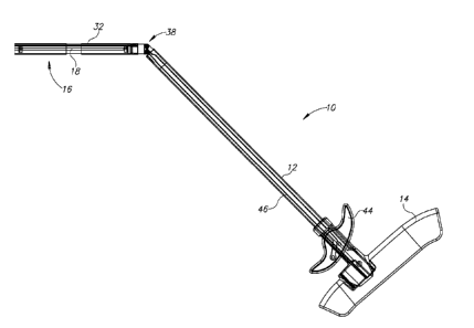
Reference is now made to Figs. 1-5, which illustrate a mesh placer 10,
constructed and
operative in accordance with an embodiment of the present invention, which is
part of the kit and
method for mesh deployment of the present invention.
Mesh placer 10 may be similar in construction to the mesh deployment apparatus
described in PCT Patent Application PCT/IL2008/000149 (and copending US Patent
Application
11/674683).
Mesh placer 10 may include a deployment rod 12 having a handle 14 at a
proximal
portion thereof and an application member 16 at a distal portion thereof (Fig.
1). Application
member 16 may include a shaft 18, which may be a rotating shaft as seen in
Fig. 1, but not
necessarily rotating, as seen in Figs. 2-5. A mesh 22, made of a biocompatible
material as is well
known in the art, is detachably attached to shaft 18 of application member 16
(Fig. 2). Shaft 18 is
thus a mesh attachment member for attaching mesh 22 thereto.
As seen in Fig. 1, application member 16 may optionally be disposed in a
cannula 32.
Application member 16 may be articulated with respect to deployment rod 12 by
means of a joint
38. In the non-limiting illustrated embodiment, the joint 38 is made up of two
pinned
connections between deployment rod 12 and roller portion 16.
A manipulating member 44 may be mounted on deployment rod 12. For example,
manipulating member 44 may include a lever arm pivotedly mounted on deployment
rod 12 and
operatively connected to application member 16 by a linking member 46 (or
alternatively,
pulleys, gears or other mechanisms) that runs through a lumen formed in
deployment rod 12. By
pivoting manipulating member 44 (pulling either side of manipulating member 44
towards
handle 14), the manipulating member 44 moves application member 16 about joint
38 to a
position wherein application member 16 is not collinear with deployment rod
12.
The jointed connection of mesh placer 10 may enable placing mesh 22 in a
patient with
significantly greater dexterity and possibilities of motion that heretofore
were not possible.
Fig. 2 illustrates mesh 22 attached to mesh placer 10 and held in place, such
as by means
of resilient fingers 17. Fig. 3 illustrates articulating application member 16
of the mesh placer,
wherein shaft 18 curves to hold mesh 22 anatomically in place at the hernia
site. Fig. 4 illustrates
4
CA 02766837 2015-07-27
the flexibility of the shaft 18 of application member 16 to center mesh 22
beneath the hernia site.
Fig. 5 illustrates that the flexibility of application member 16 enables
adjustment to the
abdominal wall curve.
The mesh placer 10 facilitates laparoscopic introduction of the mesh 22 to the
repair site
and allows for optimal placement. Although mesh is used in most hernia repair
procedures today,
in the prior art there has been no tool or accepted standard for deploying and
placing the mesh
laparoscopically. The mesh placer 10 of the present invention can be used as a
standard tool,
enabling broader adoption of laparoscopic hernia repair by shortening the
learning curve,
reducing procedure time, optimizing techniques and minimizing complications.
Proper centering of mesh over an abdominal wall defect is challenging in
laparoscopic
procedures because maneuverability is limited. The mesh placer 10 gives the
surgeon the
flexibility needed to optimally center, position, and deploy the mesh.
Reference is now made to Figs. 6-10, which illustrate a mesh stitcher 30,
constructed and
operative in accordance with an embodiment of the present invention, which is
part of the kit and
method for mesh deployment of the present invention.
Mesh stitcher 30 may be similar in construction to the suture assembly
described in PCT
Patent Application PCT/IL2008/001518 (and copending US Patent Application
11/947798).
Mesh stitcher 30 includes a first puncture element 32 including a sharp distal
end 34 for
puncturing tissue, and a second puncture element 36 including a sharp distal
end 38 for
puncturing tissue. The distal ends 34 and 38 of first and second puncture
elements 32 and 34 are
spaced from each other by a gap 40. i'referably, but not necessarily, first
and second puncture
elements 32 and 34 are parallel to each other.
First and second puncture elements 32 and 36 are hollow. A suture manipulating
assembly 42 that includes a suture thread receiving member 44 and a suture
grabber 46, which
can be passed into the hollow portions of first and second puncture elements
32 and 36,
respectively. First and second puncture elements 32 and 36 are provided with
distal funnel cups
48 and 50, respectively, for guiding insertion of suture thread receiving
member 44 and suture
grabber 46. The funnel cups 48 and 50 also serve as stops to limit movement of
suture thread
receiving member 44 and suture grabber 46 into first and second puncture
elements 32 and 36.
Proximal ends of suture thread receiving member 44 and suture grabber 46 are
mounted on a
handle assembly 52.
CA 02766837 2015-07-27
In the non-limiting illustrated embodiment, suture thread receiving member 44
is
mounted to a one-handed manipulator 60 of handle assembly 52. Manipulator 60
is arranged for
moving with respect to a block 62 of handle assembly 52 by means of a latch,
for example.
Fig. 7 illustrates mesh stitcher 30 inserted to the abdominal wall, with
puncture elements
(needles) 32 and 36 simultaneously penetrating the abdominal wall. In the non-
limiting
illustrated embodiment, as seen in Fig. 8, the distal end of suture thread
receiving member 44 is
split or forked into two distal portions 44A and 44B. The rest of suture
thread receiving member
44 is a rod that passes through first puncture element 32. Both portions 44A
and 44B have a
groove for receiving therein the suture 64. Suture thread receiving member 44
(or at least
portions 44A and 44B) is made of a flexible resilient material, such as but
not limited to,
NITINOL. In this manner, when suture thread receiving member 44 is moved
distally out of first
puncture element 32, the spring energy (and/or shape memory) of the resilient
body urges distal
portions 44A and 44B to protrude out of first puncture element 32 and bend
towards the distal
end of suture grabber 46. The distal portions 44A and 44B are arranged so that
they straddle the
distal end of suture grabber 46. In other words, the distal end of suture
grabber 46 is between
distal portions 44A and 44B. In this manner, suture 64 easily passes from one
needle to the other,
with a one-handed manipulation of the stitcher.
Fig. 9 illustrates making the stitch, wherein distal portions 44A and 44B have
been
retracted back into first puncture element 32. Fig. 10 illustrates removing
the mesh stitcher from
the suture site.
Accordingly, the mesh stitcher 30 is a dual-action stitching device that makes
precise
laparoscopic mesh transfacial fixation faster and easier. Using one hand, the
surgeon can pass the
suture from one arm of the mesh stitcher 30 to the other, creating a stitch in
three quick moves.
The device allows the surgeon to quickly secure even a large mesh. It can be
used to close trocar
sites as well.
Reference is now made to Figs. HA-11D, which illustrate a mesh tacker 70,
constructed
and operative in accordance with an embodiment of the present invention, which
is part of the kit
and method for mesh deployment of the present invention.
Mesh tacker 70 may be similar in construction to the suture assembly described
in
copending US Patent Application 12/427778.
Tacker 70 may include a handle 72 with a first trigger assembly 74 and a
second trigger
assembly 76. Both trigger assemblies 74 and 76 are coupled to an articulated
applicator arm 78
6
CA 02766837 2015-07-27
which is disposed through a drive shaft 80. The first trigger assembly 74 is
used to apply rotary
tacks (not shown in these figures) from a distal end 82 of applicator arm 78.
This is
accomplished by squeezing a trigger 84 towards the body of handle 72 (as shown
by comparing
Figs. 11C and 11D). The second trigger assembly 76 is used to bend the distal
end 82 of
applicator arm 78 up (Fig. 11A) or down (Fig. 11B).
The central (longitudinal) axis C of handle 72 is tilted at an angle A in the
range of about
7-25 , preferably about 77 , with respect to drive shaft 80 (that is, with
respect to the proximal
portion of applicator arm 78 which remains unbent), as seen in Fig. 11A. The
tilted configuration
of handle 72 is an important ergonomic feature of tacker 70. Prior art tackers
have a pistol grip
handle wherein the longitudinal axis of the handle is aligned or parallel with
the drive shaft; there
is no tilt. The prior art tacker is more cumbersome to use and can cause
fatigue to the user. With
the tilt of the present invention, tacker 70 is significantly more comfortable
to use than prior art
tackers. Another ergonomic feature is that trigger 84 is tilted at an angle B
in the range of about
7-25 , preferably about 76 , with respect to drive shaft 80.
The angling tip and in-line handle of mesh tacker 70 enable a secure tack
fixation angle
through fewer trocars, leading to improved outcomes and optimal patient care.
The angulation of
mesh tacker 70 (the articulating tip) allows fixation of the mesh 360 from
one side, and enables
tacking from both the lateral and contralateral sides, and reaching difficult
positions. This
reduces or eliminates the need for placing additional trocars on the
contralateral side. The in-line
handle provides a more ergonomic design that reduces stress and increases
surgeon comfort
throughout the entire fixation process.
Mesh tacker 70 can be loaded in an angled position for intraoperative
efficiency. The
tacks are absorbable (see below) and longer than other available tacks. The
additional length of
the tack and the angulating tip of the tacker help provide the laparoscopic
surgeon more
consistent and secure mesh fixation.
Reference is now made to Fig. 12, which illustrates a rotary tack 110,
constructed and
operative in accordance with an embodiment of the present invention, which is
part of the kit and
method for mesh deployment of the present invention.
Rotary tack 110 may be similar in construction to the suture assembly
described in
copending US Patent Application 12/427780.
7
CA 02766837 2011-12-28
WO 2010/145547 PCT/CN2010/074012
Rotary tack 110 is preferably constructed of a resorbable material. Tack 110
may
be constructed, without limitation, from a biodegradable polymer or copolymer
of a type
selected in accordance with the desired degradation time. A common
biodegradable
polymer used in absorbable sutures and the like is poly(L-lactide) which has a
degradation time of about twelve to eighteen months. Without limitation, the
tack may be
constructed from an absorbable copolymer derived from glycolic and lactic
acids, such as
a synthetic polyester chemically similar to other commercial available
glycolide and
lactide copolymers. Glycolide and lactide, in vivo, degrade and absorb by
hydrolysis into
lactic acid and glycolic acid which are then metabolized by the body.
In accordance with a non-limiting embodiment of the present invention, tack
110
includes a helical body 112 having a closed-loop base 114 and a helix of
spiral coils 116
that extend from base 114 and which terminate in a tip 118 for piercing tissue
(not shown).
In prior art helical fasteners, the base is open, that is, the lowermost coil
is not a
continuous closed coil or loop. In contrast, in the present invention, base
114 is closed.
This geometry provides tack 110 with superior strength, especially important
for use with
the resorbable material. Tip 118 has a sharp edge at a wide angle W aimed
upwards
relative to the helix. Angle W is much greater than the helix angle of the
coils.
The invention thus provides a kit for tissue repair, including mesh placer 10,
mesh
stitcher 30 and mesh tacker 70 (and may also include rotary tacks 110). The
kit makes
laparoscopic hernia repair (or other tissue repair) more consistent and
efficient than the
prior art.
A preferred method of using the kit includes first placing the mesh at the
hernia
repair site with the mesh placer, as described above. The surgeon can operate
the mesh
placer with just one hand to place the mesh at the desired position and
angular orientation.
Afterwards, the surgeon may use his/her other hand to grasp the mesh placer
and hold the
mesh in place. This frees the dexterous hand of the surgeon for the next step
(of course, if
the surgeon is ambidextrous, there is no need to switch hands). In the next
step, the
surgeon fastens the mesh to the tissue at the repair site. Fastening the mesh
may be done
by tacking the mesh to tissue with the mesh tacker using rotary tacks and/or
by stitching
the mesh to tissue with the mesh stitcher. Depending on the situation, the
mesh may be
fastened only with tacks, only with stitches, or any combination thereof.
Although in most
procedures, the mesh is first tacked and then stitched, the stitching and
tacking may be
done in any order. The stitching and tacking are done with the mesh stitcher
and mesh
tacker, respectively, as described above.
8
CA 02766837 2011-12-28
WO 2010/145547 PCT/CN2010/074012
It will be appreciated by persons skilled in the art that the present
invention is not
limited by what has been particularly shown and described hereinabove. Rather
the scope
of the present invention includes both combinations and subcombinations of the
features
described hereinabove as well as modifications and variations thereof which
would occur
to a person of skill in the art upon reading the foregoing description and
which are not in
the prior art.
9