Note: Descriptions are shown in the official language in which they were submitted.
CA 02766945 2011-12-28
WO 2011/097469 PCT/US2011/023731
LAPAROSCOPIC RADIOFREQUENCY SURGICAL DEVICE
CROSS-REFERENCE TO RELATED APPLICATIONS
[001] This application claims priority to U.S. Provisional Patent Application
No. 61/301,295
of Walberg, entitled "Laparoscopic radiofrequency surgical device", as filed
on February 4,
2010. This application is also a continuation-in-part of U.S. Patent
application No. 11/743,579 of
Eder et al., entitled "Surgical Tool", as filed on May 2, 2007, that
application being a
continuation-in-part of U.S. Patent Application No. 11/382,652, of Eder et
al., entitled
"Apparatus for tissue cauterization, as filed May 10, 2006, and claiming
priority to U. S.
Provisional Patent Application No. 60/746,256 of Eder et al., entitled
"Surgical Tool", as filed
on May 2, 2006.
INCORPORATION BY REFERENCE
[002] All publications and patent applications mentioned in this specification
are herein
incorporated by reference to the same extent as if each such individual
publication or patent
application were specifically and individually indicated to be so incorporated
by reference.
TECHNICAL FIELD
[003] The disclosed technology relates to systems and methods for
electrosurgery. More
particularly, the technology relates to an electrosurgical device appropriate
for laparoscopic
surgery through a trocar with a 5mm port.
BACKGROUND
[004] Biopolar electrosurgical instruments apply radiofrequency (RF) energy to
a surgical site
to cut, ablate, or coagulate tissue. A particular application of these
electrosurgical effects is to
seal blood vessels or tissue sheets. A typical instrument takes the form of a
set of forceps or pair
of jaws, with one or more electrodes on each jaw tip. In an electrosurgical
procedure, the
electrodes are placed in close proximity to each other as the jaws are closed
on a target site such
that the path of alternating current between the two electrodes passes through
tissue within the
target site. The mechanical force exerted by the jaws and the electrical
current combine to create
the desired surgical effect. By controlling the level of mechanical and
electrical parameters, such
as the pressure applied by the jaws, the gap distance between electrodes, and
the voltage, current,
frequency, and duration of the electrosurgical energy applied to the tissue,
the surgeon can
coagulate, cauterize, or seal tissue toward a therapeutic end.
[005] Electrosurgical procedures can be performed in an open environment,
through
conventional incisions, or they may be performed laparoscopically, through
small incisions,
1
CA 02766945 2011-12-28
WO 2011/097469 PCT/US2011/023731
typically 0.5 cm - 1.5 cm in length. A laparoscopic procedure may include the
use of a telescopic
rod lens system that is connected to a video camera and to a fiber optic cable
system that conveys
light to illuminate the operative field. A laparoscope is typically inserted
into a port in the body
through a 5 mm or 10 mm cannula or trocar to view the operative field. Surgery
is performed
during a laparoscopic procedure with any of various tools that are typically
arranged at the distal
end of a shaft and are operable by manipulation of a handle or an actuator
positioned at the
proximal end of the shaft, and are dimensioned such that they can pass through
a port provided
by the 5 mm or 10 mm cannula.
[006] As electrosurgical tools are applied in laparoscopic procedures,
challenges to the
devices arise regarding dimensional constraints imposed by the operating
environment, including
the smallness of a typical port of entry, which includes the use of
conventional trocars with a
5mm inner diameter. The technology provided herein addresses the need for
improvements in
device technology, that permit downsizing of the device while maintaining
appropriate levels of
mechanical strength and electrosurgical capability. For example, it is
generally desirable to
extend the length of conventional forceps in order to allow the sealing of
greater lengths of
tissue. As forceps length increases, it becomes a challenge to exert an
appropriate level of force,
particularly from the distal end of the forceps. The present disclosure
provides technologies that
represent progress in addressing these challenges.
SUMMARY OF THE DISCLOSURE
[007] Embodiments of the technology relate to an electrosurgical device that
is particularly
suitable for laparoscopic procedures in that its distal insertable portion,
including a shaft and an
end effector, may have a diameter no wider than about 5 mm. This 5 mm
insertable profile
allows insertion of the device through a conventional 5 mm trocar.
Commercially available
trocars that are conventionally referred to as being "5 mm" generally have an
internal diameter
specification commonly expressed in inch units, and actually vary in range
between about 0.230
inch and about 0.260 inch, even though 5 mm actually is the equivalent of
0.197 inches. In the
present disclosure, therefore, "5 mm" or "about 5 mm", when referring to the
insertable profile
of the device, or to the diameter of the shaft or the jaws in a closed
configuration, refers to a
diameter that is accommodated by presently available "5 mm" trocars. More
particularly,
embodiments of the shaft and closed jaws disclosed herein typically have a
diameter in the range
of about 0.215 inch to about 0.222 inch.
[008] Embodiments of the electrosurgical device have an end effector such as a
set of two
opposing jaws or forceps that include one or more bipolar electrode pairs
disposed on tissue
engaging surfaces of the jaws, the device being adapted to effect tissue
sealing and cutting. In
2
CA 02766945 2011-12-28
WO 2011/097469 PCT/US2011/023731
some embodiments, the device includes a single bipolar electrode pair, one
electrode in each of
the jaws. In these embodiments, the electrodes are typically powered by a
generator operating
with a single radiofrequency channel. Other embodiments of the device may
include a plurality
of bipolar electrode pairs, and an operation by way of a plurality of
radiofrequency channels.
Some particular embodiments of the technology may take the form of non-
electrical surgical
device whose operation takes advantage of the mechanical and dimensional
aspects of the
technology.
[009] Embodiments of electrosurgical device may have jaws that self align with
respect to
their longitudinal axes when the jaws are approaching closure. Self-alignment,
as used herein,
may further be understood to include lateral alignment, such that when
longitudinally aligned
jaws converge on closure, they meet opposingly, their lateral or tissue
engaging faces meeting
each other fully, from respective proximal end to distal end. The mutual
alignment of the jaws
can be particularly challenged when the jaws are closing around a piece of
tissue, whose
presence can urge the jaws to skew laterally out of alignment such that they
do not meet
opposingly. Accordingly, in these jaw set embodiments, the tissue-engaging
surfaces of each of
the opposing jaws, respectively, have mutually complementary longitudinally
oriented self-
aligning features that are sufficiently robust to be effective when there is a
surgically appropriate
amount of target tissue within the space between the closing jaws. Aspects and
details of
embodiments of self-alignable jaws are described further below.
[0010] Embodiments of the jaws may be rotatable with respect to each other by
way of a
pinless rotation mechanism that operates by way of rotatably cooperative
features of the jaws
that connect the jaws together. The pinless rotatable mechanism, in addition
to securing the jaws
together, allows the jaws to pivot between the open position and the closed
position.
Embodiments of the jaw set may pivot as a whole between an open and a closed
position by
virtue of one jaw pivoting with respect to a shaft while the other jaw remains
fixed with respect
to the shaft. The center of rotation of this pinless rotation system is not
necessarily disposed at a
position on a line corresponding to a central longitudinal axis of the shaft.
Particular
embodiments of the pinless rotation mechanism are displaced from that line. An
advantage of
this pivotal or rotational mechanism is that force that is transferred to the
mechanism from an
actuator wire is increased by the angular moment provided by the distance of
displacement of the
center of rotation from the longitudinal axis of the shaft, or more
particularly by the distance
between the axis of the actuator wire within the shaft and the center of
rotation.
[0011] In some embodiments, the rotatably cooperative features of the pinless
rotation
mechanism of the jaws include a first jaw in which a proximal aspect of the
jaw has a first
3
CA 02766945 2011-12-28
WO 2011/097469 PCT/US2011/023731
arcuate track, and second jaw in which a proximal aspect has a second arcuate
track, the first and
second arcuate tracks being mutually complementary and slidably engageable
with each other. In
one arrangement of these rotatable components, the arcuate track aspect of
first jaw is generally
external or female with respect to the arcuate aspect of the second jaw. Thus,
the track of the first
jaw accommodates and generally encloses the track portion of the second jaw,
and the second
jaw is rotatable within the space provided by the first jaw. The complementary
rotatable portions
of the first and second jaws are dimensioned such that their facing surfaces
can be easily moved
slidably past each other. In some of these embodiments, the second arcuate
track substantially
resides within an enclosure formed by the first arcuate track. Although the
proximally positioned
arcuate tracks are rotatable with respect to each other, in some embodiments,
at least the
proximal portion of the first jaw is fixed with regard to the shaft, while the
second jaw is
pivotable with respect to the shaft.
[0012] In some embodiments of the pinless rotation mechanism, the first
arcuate track has two
concentric surfaces facing each other, one smaller and the other larger, and
the second arcuate
track has two concentric surfaces facing away each other, one smaller and the
other larger. The
concentric surfaces of the two tracks are mating surfaces between the tracks.
More particularly,
the smaller concentric surfaces of the first and second tracks, respectively,
are complementary to
each other. The larger concentric surfaces of the first track and second
track, respectively, are
complementary to each other. The second arcuate track substantially resides
within an enclosure
formed by the first arcuate track. In particular embodiments, the first jaw
includes a retaining
strap that backs the smaller concentric surface of the first arcuate track and
is positioned laterally
across a surface of a housing of first jaw within a portion of the housing
overlaying and securing
the smaller concentric surface of the second jaw. This strap is configured to
retain the proximal
aspect of the second jaw within the enclosure provided by the first arcuate
track.
[0013] From a general perspective, in embodiments of the device in which the
shaft and the
jaws are freely rotatable with respect to a handle portion, designating one
jaw as a lower jaw and
the other jaw as an upper jaw may not be particularly meaningful.
Nevertheless, in some
embodiments of the device, by virtue of a convention, or by some designation,
there may be a
default rotational position of the jaws that particularizes one jaw as a lower
jaw and the other as
an upper jaw. Thus, in particular embodiments of the device, and in the
examples of device
embodiments depicted herein, from the perspective of an operator of the device
and with the
jaws in a default operating position, a referenced first jaw is a lower jaw
and a referenced second
jaw is an upper jaw.
4
CA 02766945 2011-12-28
WO 2011/097469 PCT/US2011/023731
[0014] Typical embodiments of an electrosurgical device as described herein
may have one
jaw that is pivotable with respect to the shaft and a second jaw having at
least a base portion that
is fixed with respect to the shaft. Embodiments such as these are described in
detail herein, and
are depicted as examples in the figures. Alternative embodiments of the
device, however, may be
configured such that both jaws are pivotable with respect to the shaft.
[0015] Typical embodiments of the device as described herein are also
configured such that
one jaw is a two-piece jaw, including a proximal base piece and a distal piece
that is pivotable
with respect to the proximal base piece, and a second jaw that is unitary.
Embodiments such as
these are described in detail herein, and are depicted as examples in the
figures. Alternative
embodiments of the device, however, may be configured such that both jaws have
two pieces,
with a distal portion that is pivotable with respect to a proximal base
portion.
[0016] Embodiments of an electrosurgical device as provided herein may vary in
terms of the
distribution of features between a first jaw and a second jaw. Thus, in some
embodiments of the
device (Embodiment A), a first jaw (a lower jaw, for example) is a two-piece
jaw, having a
proximal piece that is fixed with respect to the shaft, a distal jaw piece
that is pivotable with
respect to the proximal piece, and a pivotable assembly connecting the
proximal piece and the
distal jaw piece; and a second jaw (an upper jaw, for example) is unitary and
pivotable with
respect to the shaft.
[0017] In alternative embodiments of the device (Embodiment B), a first jaw (a
lower jaw, for
example) is unitary and fixed with respect to the shaft; and a second jaw (an
upper jaw, for
example) is a 2-piece jaw, having a proximal jaw piece that is pivotable with
respect to the shaft,
a distal jaw piece that is pivotable with respect to the proximal piece, and a
pivotable assembly
connecting the proximal jaw piece and the distal jaw piece. Examples of both
embodiments A
and B are depicted as examples in the figures.
[0018] In another aspect, Embodiment A of the device may be described as
having two jaws, a
first jaw that is fixed with respect to the shaft and having pivotable
electrode tray positioned with
in it, and a second jaw being pivotable with respect the shaft and having a
fixed electrode tray
disposed within it. Embodiment B of the device may be described as having two
jaws, a first jaw
that is fixed with respect to the shaft and having a fixed electrode tray
disposed with in it, and a
second jaw that is pivotable with respect to the shaft, and having a pivotable
electrode tray with
in it. Other than the variation associated with the distribution of jaw
attributes between
Embodiments A and B, other features of the Embodiment A and Embodiment B
devices are
substantially similar. The majority of features shown in figures included
herein are consistent
5
CA 02766945 2011-12-28
WO 2011/097469 PCT/US2011/023731
with Embodiment A or common to both Embodiments A and B. Figs. 5A - 5C depict
Embodiment B in particular.
[0019] A further embodiment (Embodiment C) of the device may be described as
having two
jaws, a first jaw that is fixed with respect to the shaft and having a
pivotable electrode tray
positioned with in it, and a second jaw that is pivotable with respect to the
shaft, and having a
pivotable electrode tray with in it. Still further embodiments have both jaws
pivotable with
respect to the shaft. Thus Embodiment D has two jaws that are pivotable with
respect to the
shaft; it has a first jaw having a pivotable electrode tray positioned within
it and a second jaw
having a fixed electrode tray disposed with in it. Embodiment E has two jaws
that are pivotable
with respect to the shaft, both jaws having a pivotable electrode tray
positioned within it.
[0020] In some aspects of the disclosure, an embodiment of the device consists
of a set of jaws
as described herein, but which are specifically absent a shaft, absent a
handle, or absent both a
shaft and a handle. The set of jaws per these embodiments may be appropriately
fitted onto a
shaftless device, or alternatively, onto a robotic device. These embodiments
may or may not be
configured for electrosurgery. Some embodiments include bipolar electrodes;
some
embodiments may be configured for mechanical functions without being enabled
to deliver
radiofrequency energy. These embodiments may further include various aspects
of the
technology disclosed herein, such as having a diameter that is no greater than
about 5 mm,
having a pinless rotation mechanism to open and close the jaws, or the jaws
may include
longitudinally aligned self-aligning features.
[0021] Some embodiments of an electrosurgical device include a blade that is
capable of
separating radiofrequency-sealed tissue into two portions. Embodiments of the
blade maybe be
positioned on a longitudinally disposed blade track; the blade may be
positioned at home
position at a proximal end of the track, at a distal end of the track, or at
any point along the track
between the distal and proximal ends of the track. In various embodiments of
the device, when
the jaws are in the open position, the proximal home position of the blade is
configured such that
the movement of the blade in a distal direction is prevented. In some
embodiments, distal
movement may be physically blocked by an impeding structure distal to the
blade, in other
embodiments distal movement may be prevented by a locking mechanism proximal
to the blade.
[0022] On the other hand, when embodiments of the jaws are in the closed
position, the
proximal home position of the blade may be configured to allow distal movement
of the blade,
the first and second jaws collectively forming a clear through path to the
distal end of blade
track. The availability of the space for the through-path is, at least in
part, due to the pinless
aspect of the rotation mechanism in that the presence of a pin, for a pin-
based jaw rotation
6
CA 02766945 2011-12-28
WO 2011/097469 PCT/US2011/023731
mechanism, could otherwise occupy the space, and impede the path. The through
path of the
blade includes slots and clefts through various structures, as described below
in the context of
the figures. In typical embodiments of a blade of the configuration described,
the blade is distal
facing with a leading V-shaped notch, which cuts tissue as it is moved
distally. At its proximal
end, the blade is connected to a mechanical linkage in the handle that
maintains it in a proximally
biased position.
[0023] As noted above, dimensions of embodiments the electrosurgical device
are important
aspects of the technology, as embodiments of the device are intended to be
compatible with
trocars having an inner diameter of about 5 mm (in the conventional or
commercial sense as
described above). Thus, in particular embodiments, the set of jaws, when
closed, has a diameter
no greater than about 5 mm when the device is in an insertable configuration.
An insertable
configuration for a device with openable jaws is one, for example, in which
the set of jaws is in a
closed configuration, and wherein the jaws of the device are aligned with the
longitudinal axis of
the shaft. Thus, in particular embodiments of the technology described, the
shaft has a diameter
no greater than about 5 mm, and the set of jaws, when closed, provides a
maximum diameter of
about 5 mm.
[0024] Other dimensions and structural features of the technology are directed
toward features
and operational specifications of embodiments of the device that also need to
accommodate the
constraints imposed by the requirement for a 5 mm maximal diameter. For
example, in particular
embodiments, the jaws have a length of at least about 2.5 cm. Further, some
embodiments of a 5
mm diameter constrained device that has jaws with a length of at least about
2.5 cm are able to
exert a pressure in the range of about 14 lbs. to about 28 lbs. at the tip of
the jaws, and in
particular embodiments, the jaws are able to exert a pressure of at least
about 16 lbs. at their tip.
[0025] One of the approaches to delivering high surgical performance from an
electrosurgical
device with 5 mm diameter constraint is to minimize the cross sectional area
that is occupied by
components or materials that do not provide distally projecting or contiguous
structural support
to the jaws, and particularly to support their ability to deliver sufficient
closing force. Here are
some examples of a material or component that could located in this region
that do not lend
distally projected support, or which interrupt longitudinal structural
continuity in a portion of the
cross sectional area of a device. One could consider a pin positioned
orthogonally across a
portion of the proximal aspect of the jaws, to be used, for example, as a
structure upon which
other features might pivot or rotate. A pin of this nature, while performing
an operational role,
does not strengthen the ability of the jaws to exert a compressive force, nor
does it strengthen the
ability of the jaws to maintain their position when the jaws encounter
resistance provided by
7
CA 02766945 2011-12-28
WO 2011/097469 PCT/US2011/023731
body structures within the laparoscopic operating space. Typical embodiments
of provided
device do not have a pin. Another example of a component occupying cross
sectional area that
does not provide distally projected structural support to the jaws relates to
actuator members and
electrically conductive members. Some embodiments of the provided device have
connecting
members that serve both a physical actuating function and an electrically
conductive function,
thus conserving cross sectional structural area. By these various aspects of
embodiments of the
device, the cross sectional fraction of the device that does not provide
distally projecting
structural support may be minimized.
[0026] Accordingly, with regard to a cross sectional slice taken through a
portion of the device
that includes the pinless rotation mechanism, in some embodiments of the
device, a ratio of the
structural material that contributes to supporting the set of jaws to the
total cross sectional area of
the device is at least about 82%. A similar analysis of distally directed
structural support could
make use of a volume-based constraint. For example, the central portion of the
distal end of the
device, at least the proximal aspect of the set of jaws, can include a given
length of the shaft
and/or jaws within proximal and distal boundaries. If that given length is
multiplied by cross-
sectional area within the set of distal and proximal boundaries, it may be
understood that a
measure of structural material can report structural material in terms of its
volume and can be
expressed as a percent of the total volume of the device portion within the
boundaries.
[0027] As summarized above, some embodiments of the set jaws are configured in
a manner
such that the jaws self align with respect to their longitudinal axes when the
jaws are
approaching closure. Accordingly, in these jaw set embodiments, the tissue-
engaging surfaces of
each of the opposing jaws, respectively, have mutually complementary
longitudinally oriented
self-aligning features that prevent lateral slippage of jaws as they close
toward each other.
Inasmuch as these features prevent or correct incipient lateral slippage as
the jaws close, these
features may be characterized as longitudinally aligning and laterally
stabilizing aspects of the
tissue-engaging surfaces.
[0028] Embodiments of self-aligning jaw features may be disposed along the
substantial
entirety of the length of the jaws. In another aspect, embodiments of self-
aligning jaw features
may as occupy the substantial entirety of available tissue engaging surfaces
of the jaws. In
various embodiments, the self-aligning features may fully or substantially
occupy the length
tissue-engaging surfaces of the jaws; in other embodiments, the self-aligning
features may
occupy only a portion of the length of tissue-engaging surfaces of the jaws.
The structural
features associated with this approach to longitudinally aligning the jaws
generally conserves on
materials, costs, or dimensions, that would otherwise be associated with
achieving
8
CA 02766945 2011-12-28
WO 2011/097469 PCT/US2011/023731
manufacturing tolerances required to support a guarantee of collinear
alignment of the two jaws
when they close.
[0029] In particular embodiments, the self-aligning configuration of the
tissue-engaging
surfaces of the jaws includes a longitudinally aligned V-shaped projecting
surface on one jaw
and a complementary longitudinally aligned V-shaped receding surface or
recession on the other
jaw. In some embodiments, the V-shaped projection is on the lower jaw, and the
V-shaped
recession is on the upper jaw. The longitudinally aligned V-shaped projecting
surface on one jaw
and the complementary longitudinally aligned V-shaped receding surface on the
other jaw, when
the set of jaws is closed, form a V-shaped common interface with internal
angle in the range of
about 90 degrees to about 175 degrees. In particular embodiments, the V-shaped
common
interface has internal angle about 150 degrees.
[0030] In a more general aspect, the self-aligning configuration of
embodiments of the tissue-
engaging surfaces of the jaws, in a lateral cross section, form a zone or
interface of tissue contact
more complex than that of a single straight cross-sectional line. By virtue of
being non-linear, the
width of the contact zone between the closed jaws and the grasped tissue is
greater than would be
the tissue width of a linear tissue contact zone. Thus, the width of the
tissue seal created by the
V-shaped configuration of the tissue engaging surfaces of the jaws is greater
than would be the
width of a tissue seal created by flat tissue engaging surfaces. The
arrangement just described, of
complementary V-shaped projection and V-shaped recession forming a V-shaped
zone of tissue
being contacted by such jaws, is just one example of self-aligning tissue
engaging surfaces.
[0031] In some embodiments of the technology, the electrosurgical device has
an insulative
layer applied over aspects of at least one of the opposing jaws, the
insulative layer forming a
spatial gap between the upper jaw and the lower jaw that prevents any direct
electrical
connection therebetween. In various embodiments, each jaw tip each has an
electrically
conductive surface on or within its tissue-engaging surface, and an aspect of
the insulative layer
includes strips aligned across the electrically conductive surface of at least
one of the forcep tips.
The strips form a gap between the electrically conductive surfaces of the two
jaws when the jaws
are in a closed position. Such a gap is typically about 0.006 inch; more
generally, the gap has a
range of about 0.0045 inch to about 0.0075 inch. In various embodiments of the
insulative layer,
it may include a polymer, such as polyether ether ketone (PEEK), merely by way
of example. In
other embodiments, the insulative layer may include a ceramic material, such
as any of alumina
or alumina-titania, merely by way of example. Ceramic compositions can be
advantageous for
their relative hardness, incompressibility, and/or general durability. In some
embodiments, the
9
CA 02766945 2011-12-28
WO 2011/097469 PCT/US2011/023731
ceramic material is positioned at one or more sites on the surface of the
device that are
particularly subject to abrasive and/or compressive stress.
[0032] In some embodiments of the technology, the device includes a handle
portion proximal
to the shaft, a jaw actuator mechanism associated with the handle portion and
configured to
actuate a mechanical capability of the jaws, and a jaw actuator wire connected
proximally to the
actuator mechanism and connected distally to the set of jaws. In various
embodiments, the
mechanical capability of the jaws includes opening and closing the set of
jaws. In some
embodiments, the actuator wire is configured to actuate an opening and closing
of the jaws by
pivoting a second jaw with respect to at least a proximal piece of the first
jaw, the proximal piece
of the first jaw being fixed with respect to the shaft.
[0033] Further, in some embodiments, the same wire that serves as a mechanical
actuator force
transfer member is further configured to deliver RF energy to the jaws. From
another
perspective, embodiments of the device include an energy-delivery wire
extending distally from
the handle portion to the set of jaws. In some of these energy-delivery wire
embodiments, the
energy-delivery wire may be further configured to perform as an actuator of
mechanical
capability of the jaws, such as moving the jaws between an open and a closed
position.
[0034] Some embodiments of the jaw actuator wire include a single wire in
looped
configuration that, in effect, forms a paired or double wire connection
between the actuator
mechanism and an attachment site on at least one of the jaws. In these
embodiments, the looped
wire has a distal-most looped terminal or turn around portion that is looped
around its attachment
site to one of the jaws. In embodiments wherein at least the proximal piece of
the first jaw is
fixed with respect to the shaft and the second jaw is pivotable with respect
to the shaft, the
actuator wire is attached to a proximal aspect of the second jaw.
[0035] In some embodiments, the actuator wire is configured as a push and pull
mechanism,
such that a distally-directed push from the wire moves the jaws to their open
position, and a
proximally-directed pull from the wire moves the jaws to their closed
position. In some of these
embodiments, the actuator wire is biased so as to support the jaws in their
open position by
virtue of a proximally directed pull by a spring associated with the jaw
actuator.
[0036] In some embodiments, the jaw actuator includes a biasing member that
maintains a
push on the actuator wire, such push causing the jaws to have a default
position of being held in
the open position. Further, in some embodiments, the jaw actuator includes a
manual lever that
an operator may pull to effect a proximally-direct pull to close the jaws.
Further still, in some of
these embodiments, the actuator wire and connections associated with proximal
and distal
attachments of the wire are collectively configured to be able to
operationally withstand between
CA 02766945 2011-12-28
WO 2011/097469 PCT/US2011/023731
about 80 and about 120 lbs. of tension; in particular embodiments the actuator
wire and its
connections are configured to be able to withstand at least about 100 lbs. of
tension.
[0037] In some embodiments of the device, each of the upper jaw and the lower
jaw include a
metal portion, and the entirety of each of these metal portions form an
electrode. In other words,
in some embodiments, there is no metal portion in either jaw that is not part
of the electrode. In
some embodiments, the device includes a single bipolar electrode pair, one
electrode in each of
the jaws. In these single bipolar pair embodiments, the electrodes are powered
by a generator
operating on a single radiofrequency channel. Other embodiments of the device
may include a
plurality of bipolar electrode pairs, and such plurality of bipolar electrode
pairs may be
controlled by a plurality of operating radiofrequency channels.
[0038] Some embodiments of the electrosurgical device include a shaft
rotational actuator
positioned proximal to the shaft; embodiments of the shaft rotator are
typically associated with a
handle portion of the device. In some embodiments, the shaft rotational
actuator is configured to
be able to rotate freely in both clockwise and counter clockwise directions,
such rotation of the
actuator being directly translatable to rotation of the shaft, and in turn,
rotation of the set of jaws
about their longitudinal axis. Free rotation in this context, whether in
reference to a shaft rotator,
the shaft, or the jaws, per embodiments of the technology, refers to a
rotation that may occur
indefinitely in either direction, without a stop, and without a change of
direction. Further, per
embodiments of the technology, rotation may freely occur without consequence
or compromise
with regard to any mechanical or electrical capability of embodiments of the
electrosurgical
device.
[0039] In some embodiments of the electrosurgical device, the set of two
opposing jaws
(including a first jaw and a second jaw) is configured such that the jaws can
open to an angle in
the range of about 30 degrees to about 40 degrees. In some the set of two
opposing jaws is
configured such that when the set is moving from an open position toward the
closed position, a
first point of mutual contact between the two jaws occurs at a distal end of
each jaw. The set of
jaws may be further configured such that after the first point of mutual
contact has been made
and as the set moves further toward a closed position, a distal pivotable
piece of the first jaw
pivots within a plane of its longitudinal axis such that the proximal end of
the first jaw comes
into contact with the proximal end of the second jaw.
[0040] In some embodiments, the set of two opposing jaws is configured such
that when the set
of jaws is moving from an open position toward the closed position, a first
point of mutual
contact between the two jaws occurs at a distal end of each jaw. In some of
these embodiments,
after the first point of mutual contact has been made and the jaw set is then
moving further
11
CA 02766945 2011-12-28
WO 2011/097469 PCT/US2011/023731
toward a closed position, a distal pivotable piece of the first jaw pivots
within a plane of its
longitudinal axis such that the proximal end of the first jaw comes into
contact with the proximal
end of the second jaw.
[0041] Some embodiments of the device and its dynamics of closing may be
understood in terms
of the response of the jaws to the presence of target tissue within the grasp
of closing jaws. In
some embodiments, for example, the set of jaws may be configured such that
when the set is
moving toward the closed position and has made an initial contact with the
target tissue, a
pivotable piece of the first jaw then pivots in response to the presence of
the target tissue as the
jaws move further toward the closed position to grasp the tissue. The pivoting
of the pivotable
jaw piece may effect a substantially equivalent distribution of pressure along
the grasped piece of
the target tissue, particularly in comparison to the unequal distribution of
pressure that may occur
in the absence of such ultra-jaw pivotability. In a related aspect of the
device, the pivotable jaw
piece is configured to pivot toward a parallel relationship with the second
jaw.
[0042] In various embodiments, the pivotable jaw piece may be configured such
that it can pivot
around its pivotable connection within an arc having pivotable range that
varies between about 2
degrees to about 8 degrees. In particular embodiments, the pivotable jaw piece
may be
configured such that it can pivot around its pivotable connection within an
are having a pivotable
range of about 6 degrees. In another aspect, the pivotable jaw piece has an
arc of a given
pivotable range and is biased such that a distal tip of the first jaw is
canted toward the second jaw
within the arc of pivotable range.
[0043] In some embodiments, the first jaw includes a proximal jaw piece fixed
with respect to
the shaft, a pivotable distal jaw piece, and a pivotable assembly that
connects the proximal jaw
piece and distal jaw piece. In various of these embodiments, the pivotable
assembly may be
positioned longitudinally at a substantially central site on the distal piece.
In some of these
embodiments, a tissue engaging surfaces comprises the substantial entirety of
the distal and
pivotable piece of the first jaw. Accordingly, a central location on the
distal piece of the jaw also
represents a central location with respect to a tissue-engaging surface of the
jaw. In another
aspect of some embodiments, the substantial entirety of the tissue engaging
surface of the distal
piece of the first jaw comprises an electrode. Thus, a central site on the
distal piece of the first
jaw represents a central site on the electrode. The centrality of the site of
the pivotable assembly
on the distal and pivotable jaw piece may be related to ability of the distal
piece to pivot in such
a manner so as to evenly distribute pressure across the surface target tissue
as the jaws close on
the tissue. In some of these embodiments, the pivotable assembly may include a
laterally
projecting boss on each of both sides of the distal pivotal jaw piece and an
internally accessible
12
CA 02766945 2011-12-28
WO 2011/097469 PCT/US2011/023731
receptacle on each of both sides of the proximal fixed jaw piece, the
laterally projecting bosses
and the internally accessible receptacles being mutually compatible. Other
arrangements and
configurations that support a pivoting capability such as that described and
depicted herein are
known in the art, and are be considered to included in the scope of the
present technology.
[0044] In some embodiments of the device, with reference to a method of
fabrication, the
proximal and distal pieces of the two-piece jaw may be assembled in a snap fit
manner. More
particularly, in such embodiments, the fixed proximal jaw piece is
sufficiently flexible that it can
deflect to allow the insertion of the laterally projecting bosses of the
distal pivotable jaw piece in
a snap fit manner.
[0045] Another aspect to the pivoted bias of the distal and pivotable piece of
a two-piece jaw
relates to a biasing member that maintains the pivotable piece in a default
pivot position. In some
embodiments, for example, the distal pivotable piece of the first jaw includes
a biasing member
that is configured to press against a shelf of the proximal jaw piece, and by
such pressing bias the
distal pivotable piece of the first jaw such that the distal tip of the distal
pivotable piece is canted
toward the second jaw. With more particularity, in some of these embodiments,
the biasing
member takes the form a leaf spring positioned in a recess within the distal
pivotable piece on an
aspect of the distal pivotable piece that faces the fixed proximal piece of
the first jaw.
[0046] In another aspect, the technology provides a surgical device having a
set of opposing
jaws disposed distal to a shaft, the set of jaws having a first jaw and a
second jaw. Each of the
opposing jaws has a longitudinal axis and a tissue-engaging surface, and the
tissue-engaging
surface of each jaw may have a complementary self-aligning configuration with
respect to the
longitudinal axis of the other jaw. In some embodiments of the provided
surgical technology, the
set of jaws, when closed, has a diameter no greater than about 5 mm, and the
shaft has a diameter
no greater than about 5 mm. Embodiments of the surgical device may further
include a pinless
rotation mechanism formed from rotatably cooperative features of the first jaw
and the second
jaw. This pinless rotation mechanism enables the set of jaws to pivot between
an open position
and a closed position. The pinless rotation mechanism is configured such that
the pinless rotation
mechanism creates a common center of rotation that is not necessarily
positioned at a point on a
line corresponding to a central longitudinal axis of the shaft.
[0047] Embodiments of the technology further relate to a method of
electrosurgical sealing in
a laparoscopic environment. The method may include moving a set of jaws of an
electrosurgical
instrument into a proximity of the target tissue, the set of jaws comprising a
first jaw and a
second jaws. More particularly, moving toward an electrosurgical site may
include advancing a
distal portion of an electrosurgical device into a patient through an in-place
trocar having an
13
CA 02766945 2011-12-28
WO 2011/097469 PCT/US2011/023731
internal diameter of about 5 mm. The distal portion of the electrosurgical
device, in this
circumstance, includes a distal aspect of a shaft and the set of jaws,
including a first jaw and a
second jaw, that are positioned on a distal end of the shaft. Embodiments of
the method may
include moving the jaws between an open position and a closed position. Moving
the jaws
between an open position and a closed position opening may include rotating
cooperative
structures of the first jaw and the second jaw, the first and second jaws not
being connected by a
pin. Moving the jaws to a closing position may further include grasping the
target tissue with the
jaws. The method may still further include delivering radiofrequency energy to
the target tissue
from the jaws.
[0048] In some embodiments of the method, moving the set of jaws into a
proximity of the
target tissue further includes rotating the jaws around their central
longitudinal axis. Rotating the
jaws may occur by way of rotating the shaft of the device around its central
longitudinal axis.
Rotating the shaft of the device may occur by rotating a shaft rotation
actuator proximal to the
shaft. In various embodiments of the method, embodiments of the shaft rotation
actuator, the
shaft, and the jaws may all have the capability of rotating freely in both
clockwise and counter
clockwise directions without a stop, or a need to reverse direction.
[0049] In some embodiments of the method, moving the jaws between an open
position and a
closed position opening includes rotating cooperative structures of the first
jaw and the second
jaw at their respective proximal ends, the first and second jaws not being
connected by a pin.
Moving the jaws between an open position and a closed position opening may
include pivoting
the jaws with respect to each other around a center of rotation that is not
necessarily on a line
corresponding to a central longitudinal axis of the shaft. In some
embodiments, moving the jaws
between an open position and a closed position opening includes pivoting the
jaws around a
center of rotation that is not on a line corresponding to a central
longitudinal axis of the shaft,
and in some embodiments, the center of rotation may be displaced to a position
beyond the
diameter of the shaft.
[0050] In another aspect, moving the jaws between an open position and a
closed position
opening may include at least a proximal piece of a first jaw remaining fixed
with respect to the
shaft and a second jaw pivoting with respect to the shaft. In some of these
embodiments, moving
the jaws to the closed position may included a distal piece of the first jaw
pivoting with respect
to the proximal piece of the jaw, and thus pivoting with respect to the shaft.
In some
embodiments, the pivoting of the distal piece of the first jaw with respect to
the shaft includes
the distal end of the distal piece pivoting away from the second jaw and the
proximal end of the
distal piece pivoting toward the second jaw.
14
CA 02766945 2011-12-28
WO 2011/097469 PCT/US2011/023731
[0051] In some embodiments of the method, moving the jaws to a closed position
includes
pivoting a distal piece of the first jaw from a pivotable connection that is
positioned at a
substantially central portion of the distal piece. In some aspects of the
method, there is an
interaction between the jaws as they are closing and the target tissue that
the jaws are closing
around. Thus, in some embodiments, pivoting the distal piece of the first jaw
includes pivoting in
response to the presence of the target tissue between the jaws, in such a
manner that distributes
pressure with substantial equivalence along the grasped portion of the target
tissue. Further,
pivoting a distal piece of a first jaw from a connection positioned at a
substantially central
portion of the distal piece comprises pivoting in response to the presence of
the target tissue
between the jaws, thereby allowing the distal piece of the first jaw to pivot
toward a parallel
alignment with respect to the second jaw.
[0052] In some embodiments of the method, moving the jaws to a closed position
includes
mutually aligning the respective central longitudinal axes of the first and
second jaws. In
instances when the jaws are moving to a closed position so as to grasp tissue,
moving the jaws to
a closed position may include mutually aligning the respective central
longitudinal axes of the
first and second jaws comprises in such a manner so as to resist a misaligning
effect of target
tissue have on the jaws as they are closing.
[0053] In some embodiments of the method, moving the jaws to a closed position
comprises
grasping the target tissue with a force in a range of about 14 lbs. to about
28 pounds. Further, in
some embodiments, moving the jaws to a closed position includes grasping a
portion of target
tissue of up to about 2.5 cm in length.
[0054] In some embodiments of the method, opening and then closing the jaws
includes
transferring a force from a mechanical actuator to the jaws via an actuator
wire. In some of these
embodiments, closing the jaws includes pulling the actuator wire in a proximal
direction, and in
some embodiments, opening the jaws pushing the actuator wire in a distal
direction. In some
embodiments of the method, delivering radiofrequency energy to the target
tissue may include
delivering energy to the jaws via the actuator wire.
[0055] In some embodiments of the method, moving the jaws to a closed position
includes
moving the jaws toward a closed position in a manner such that a first point
of mutual contact
between the two jaws occurs at a distal end of each jaw. In some of these
embodiments, moving
the jaws to a closed position after the point of first mutual contact has
occurred includes pivoting
a distal pivotable piece of a first jaw within a plane of its longitudinal
axis such that the proximal
end of the first jaw comes into contact with the proximal end of the second
jaw.
CA 02766945 2011-12-28
WO 2011/097469 PCT/US2011/023731
[0056] In some embodiments of the method, delivering radiofrequency energy to
the target
tissue includes energy through a wire that is further enabled to perform a
mechanical function,
such as actuating the jaws between an open and closed position. In various
embodiments of the
method, electrosurgically treating tissue particularly includes sealing edges
of target tissue
together.
[0057] In some embodiments of the method, after delivering radiofrequency
energy to the
target tissue, the method further includes separating newly sealed target
tissue into two sealed
tissue segments. In various embodiments, separating newly sealed target tissue
into two sealed
tissue segments includes advancing a blade distally through sealed target
tissue.
[0058] Some embodiments of the method include electrosurgically treating more
than one site
during a single procedure, or treating a lengthy target site with a series of
sealing maneuvers.
Thus, some embodiments of the method further include identifying a second
target site and then
repeating the steps of grasping and delivering energy, the steps being
directed toward the second
target site.
BRIEF DESCRIPTION OF THE DRAWINGS
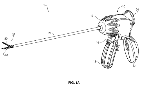
[0059] Fig. 1A is a perspective view of an embodiment of a laparoscopic
electrosurgical
device.
[0060] Fig. 1B is a side view of an embodiment of an electrosurgical device
with the jaws in
an open position.
[0061] Fig. 1C is a perspective view of an embodiment of an electrosurgical
device with the
jaws in a closed and locked position, and with the blade in a retracted in
proximal position.
[0062] Fig. 1D is a perspective view of an electrosurgical device with the
jaws in a closed and
locked position, and with the blade in a distally advanced position.
[0063] Fig. 2A is a transparent perspective view of an embodiment set of jaws
of an
electrosurgical device, with the jaws in an open position.
[0064] Fig. 2B is a transparent perspective view of an embodiment of a lower
jaw of a set of
jaws an electrosurgical device, with a blade moved distally to a position
about half way to its
distal stop point.
[0065] Fig. 3A is a side view through the longitudinal midline of an
embodiment of a set of
jaws of an electrosurgical device, with the jaws in an open position.
[00661 Fig. 3B is a side view through the longitudinal midline of an
embodiment of a set of
jaws of an electrosurgical device, with the jaws in a closed position.
16
CA 02766945 2011-12-28
WO 2011/097469 PCT/US2011/023731
[0067] Fig. 3C is a side view through the longitudinal midline of an
embodiment of a lower
jaw of a set of jaws an electrosurgical device.
[0068] Fig. 4A is a side view through the longitudinal midline of an
embodiment of a set of
jaws of an electrosurgical device, with the jaws in an open position, and
further showing a blade
in a proximal and raised holding position.
[0069] Fig. 4B is a side view through the longitudinal midline of an
embodiment of a set of
jaws of an electrosurgical device, with the jaws in a closed position, and
further showing a blade
in a proximal and lowered holding position, ready to be distally advanced.
[0070] Fig. 4C is a side view through the longitudinal midline of an
embodiment of a set of
jaws of an electrosurgical device, with the jaws in a closed position, and
further showing a blade
in a distally advanced position.
[0071] Fig. 4D is a perspective view of a blade isolated from the shaft and
jaws.
[0072] Fig. 5A is a perspective view of an alternative embodiment of an
electrosurgical device
with the jaws in an open position.
[0073] Fig. 5B is a side view of an embodiment of an alternative embodiment of
an
electrosurgical device with the jaws closed to a position where the distal
tips of the jaws are in
contact.
[0074] Fig. 5C is a side view of an embodiment of an alternative embodiment of
an
electrosurgical device with the jaws in a fully closed position.
[0075] Fig. 6 is a distal looking perspective view of an embodiment of a set
of jaws of an
electrosurgical device with the jaws in a closed position, a cross sectional
exposure showing a
passage through which a blade may be distally advanced.
[0076] Fig. 7A is a side view of an embodiment of set of jaws of an
electrosurgical device,
with the jaws in an open position.
[0077] Fig. 7B is a side view of an embodiment of set of jaws of an
electrosurgical device,
with the jaws at an initial point of closure, when the distal tips of the jaws
have first made
contact each other and a gap remains between the jaws at their proximal end.
[0078] Fig. 7C is a side view of an embodiment of set of jaws set of an
electrosurgical device,
with the jaws in a fully closed position, wherein the jaws are in full contact
with each other from
distal tip to proximal end.
[0079] Fig. 7D is a side view of a set of jaws of an embodiment of an
electrosurgical device in
a partially closed position, with the jaws as they would be positioned when
closing around a
17
CA 02766945 2011-12-28
WO 2011/097469 PCT/US2011/023731
portion of relatively thick target tissue, the jaws in a parallel alignment,
spaced relatively widely
apart by the presence of thick tissue therebetween.
[0080] Fig. 7E is a side view of a set of jaws of an embodiment of an
electrosurgical device in
a partially closed position, with the jaws as they would be when closing
around a portion of
relatively thin target tissue, the jaws in a parallel alignment, spaced apart
by a narrow gap,
reflecting the presence of thin tissue therebetween.
[0081] Fig. 8 is a perspective and upward looking view of a set of jaws of an
embodiment of
an electrosurgical device with the jaws in an open position, the view showing,
more specifically,
an isolated upper jaw, an isolated distal pivotable piece of a lower jaw, and
an actuator wire
looped around an attachment point at the proximal end of the upper jaw.
[0082] Fig. 9A is a side view of an embodiment of an isolated lower jaw of an
electrosurgical
device, the lower jaw including a proximal jaw piece that is fixed with
respect to the shaft and a
distal pivotable jaw piece mounted at a substantially central point of the
distal piece on the
proximal jaw piece.
[0083] Fig. 9B is a perspective and exploded view of an embodiment of a
isolated lower jaw
of a laparoscopic electrosurgical device, the lower jaw having a proximal jaw
piece fixed to a
shaft and distal pivotable jaw piece, the proximal and distal jaw pieces shown
in an exploded
relationship.
[0084] Fig. 9C is a bottom view of a lower jaw of an embodiment of an
electrosurgical device,
showing a connection between a proximal fixed jaw piece and distal pivotable
jaw piece.
[0085] Fig. 9D is an upward looking perspective view of an embodiment of a
distal piece of a
lower jaw of an electrosurgical device.
[0086] Fig. 10A is a semitransparent side view of an embodiment of a lower jaw
of an
electrosurgical device, showing a proximal jaw piece and pivotably connected
distal pivotable
jaw piece, the distal pivotable piece in its default biased position, the
distal end of the distal
pivotable jaw piece pivoted to its upper end point, toward an upper jaw (not
shown).
[0087] Fig. lOB is a semitransparent side view of an embodiment of a lower jaw
of an
electrosurgical device, showing a pivotably connected proximal jaw piece and
distal pivotable
jaw piece, the distal end of the distal pivotable jaw piece pivoted toward its
lower end point, the
proximal end of the distal pivotable jaw piece pivoted toward its upper end
point, such a position
putting the lower jaw in a substantially parallel relationship with the upper
jaw (not shown).
[0088] Fig. 11A is a side view of an embodiment of a lower jaw of an
electrosurgical device
similar to the view shown in Fig. 10A, showing a leaf spring attached an upper
aspect of the
18
CA 02766945 2011-12-28
WO 2011/097469 PCT/US2011/023731
proximal jaw piece, the spring pushing against the distal pivotable jaw piece
so as to maintain
the distal pivotable piece in its default biased position, the distal end of
the distal pivotable jaw
piece pivoted to its upper end point.
[0089] Fig. 11B is a side view of an embodiment of a lower jaw of an
electrosurgical device
similar to the view shown in Fig. IOB, showing a leaf spring attached an upper
aspect of the
proximal jaw piece, the spring collapsed by the pressure being exerted on the
distal end of the
distal pivotable piece of the jaw, as would occur during closure of the jaw.
[0090] Fig. 12A is a proximal-looking perspective view of an embodiment of
distal tips of a
closed set of jaws of an electrosurgical device, the distal tips aligned by
complementary
longitudinal aligning features, a V-shaped projection on the lower jaw, and a
V-shaped recession
on the upper jaw.
[0091] Fig. 12B is a proximal-looking front view of an embodiment of the
distal tips of a
closed set of jaws of a laparoscopic electrosurgical device, the distal tips
aligned by
complementary longitudinal aligning features, a V-shaped projection on the
lower jaw, and a V-
shaped recession on the upper jaw.
[0092] Fig. 12C is a proximal-looking perspective view of a distal aspect of
an electrosurgical
device, with a set of jaws in an open position showing complementary
longitudinal aligning
features, a V-shaped projection on the lower jaw, and a V-shaped recession on
the upper jaw, as
well as a central longitudinally-oriented gap in both V-shaped surfaces that
form a through
passage for a blade that is distally advanceable when the jaws are in a closed
position.
[0093] Fig. 13A is a proximal looking perspective view, partially exposed, of
an embodiment
of an electrosurgical device that shows aspects of the proximal portion of a
set of jaws through
which jaw actuator cables transit; the jaw actuator cables also serve as an
electrical conduit to the
upper jaw.
[0094] Fig. 13B is a proximal looking perspective view of an embodiment of an
electrosurgical device that shows aspects of the proximal portion of a set of
jaws through which
jaw actuator cables transit.
[0095] Fig. 13C is a distal looking transparent perspective view of an
embodiment of an
electrosurgical device that shows aspects of the proximal portion of a set of
jaws through which
jaw actuator cables transit.
[0096] Fig. 13D is a distal looking transparent perspective view of an
embodiment of an
electrosurgical device similar to Fig. 13C, that shows aspects of the proximal
portion of a set of
jaws through which jaw actuator cables transit, with the cables in place.
19
CA 02766945 2011-12-28
WO 2011/097469 PCT/US2011/023731
[0097] Fig. 13E is a longitudinal section view, slightly offset from midline,
showing the paths
of cables through the distal portion of the shaft and into the proximal aspect
of the jaws.
[0098] Fig. 13F is proximal looking perspective view of the proximal end of a
lower jaw that
is inserted into the distal end of a shaft, further showing engagement of the
proximal end of the
shaft with a cable isolator unit.
[0099] Fig. 14A is a bottom perspective view of an embodiment of an upper jaw
of an
electrosurgical device that shows plastic insulator layer overlaying the
electrode.
[00100] Fig. 14B is a top perspective view of an embodiment of an upper jaw of
an
electrosurgical device that shows polymer insulator layer overlaying the
electrode.
[00101] Fig. 14C is a top perspective view of an embodiment of an upper jaw of
an
electrosurgical device that shows polymer insulator layer overlaying the
electrode, with the
proximal portion of the jaw truncated to expose a cross section.
[00102] Fig. 15A is a top perspective view of an embodiment of an upper jaw of
an
electrosurgical device that shows points of ceramic overlaying the electrode
at abrasive stress
points.
[00103] Fig. 15B is a top perspective view of an embodiment of an upper jaw of
an
electrosurgical device that shows points of ceramic overlaying the electrode
at abrasive stress
points as they are embedded in a more extensive polymer layer.
[00104] Fig. 15C is a top perspective view of an embodiment of a pair of
closed jaws of an
electrosurgical device that shows points of ceramic overlaying the electrode
at abrasive stress
points as they are embedded in a more extensive polymer layer.
[00105] Fig. 16A is an exposed perspective view of a handle of an embodiment
of an
electrosurgical device that shows aspects of the proximal end of a rotatable
shaft.
[00106] Fig. 16B is a perspective view of an isolated proximal end of a
rotatable shaft.
[00107] Fig. 16C is a midline sectional view of an isolated proximal end of a
rotatable shaft.
[00108] Fig. 16D is a midline sectional view of a proximal portion of a
rotatable shaft.
DETAILED DESCRIPTION
[00109] Embodiments of the technology described herein provide various
improvements over
available electrosurgical devices, such improvements permitting a physical
downsizing of a
device to a dimension that permits practical use of an electrosurgical device
within the
constraints of a laparoscopic surgical environment. One of these constraints
to working
laparoscopically relates to the 5 mm inner diameter opening provided by a
commercially
CA 02766945 2011-12-28
WO 2011/097469 PCT/US2011/023731
standard trocar. A device compatible with the 5 mm opening constraint needs to
have an
insertable configuration with a maximal diameter that is insertable
therethrough. These
technological improvements are generally directed toward creating a high
degree of efficiency
with regard to performance of the device per unit volume or cross sectional
area. For example, a
jaw set of a disclosed device, in spite of small physical dimension, is able
to deliver an
appropriate level of force to tissue being clamped by the jaws, and the
structure and material of
the jaws have sufficient strength to maintain integrity during the delivery of
such force.
[00110] In one aspect, the technology includes maximizing the amount of
structural material in
particular areas as a percent of total amount of device material. The proximal
aspect of the jaw
set, for example, includes various components, some that contribute structural
support for the
jaws, and other components that perform other functions, such as mechanical or
electrical
functions. The technology, in this aspect, is directed toward minimizing cross
sectional area or
volume that does not directly support the jaws. Some components of
conventional electrosurgical
devices are typically dedicated to a single use, such as electrodes, power
lines, or actuator lines;
in contrast, various components of embodiments of the presently disclosed
device do double
duty both as structural and electrical components in embodiments of the
technology. In another
example of material and occupied volume efficiency, some structural
components, such as a pin
connecting two jaws at their base, are eliminated and replaced by a pinless
mechanism that links
upper and lower jaws of a jaw set together.
[00111] Aspects of the technology in the form of embodiments of the disclosed
electrosurgical
device and methods of using the device are illustrated in Figs. 1 - 16D. With
regard to
Embodiments A and B, as described above, the majority of the figures depict
examples of
Embodiment A, or they relate to aspects of the technology that are common to
both
Embodiments A and B. Figs. 5A - 5C particularly depict examples in accordance
with
Embodiment B. It should be understood that in any reference to a lower jaw or
an upper jaw
when describing the figures is for a convenient visual reference with respect
to a conventional
positioning of the rotatable jaws, and that the two jaws could be more
generally referred to as a
first jaw and a second jaw. Further, with respect to orientation of the
figures, in general a distal
end of a device is on the left, and a proximal end of a device is on the
right.
[00112] Figs. 1A -1D provide various views of embodiments of a laparoscopic
electrosurgical
device as a whole. Fig. 1A is a perspective view of an embodiment of an
electrosurgical device 1
as provided herein, with a set of jaws 30 in an open position. Fig. lB is a
side view of an
embodiment of an electrosurgical device 1 with the jaws 30 in the same open
position as in Fig.
1A. A handle 10 supports a jaw actuator grip 15 and blade actuator lever 16,
and a shaft rotator
21
CA 02766945 2011-12-28
WO 2011/097469 PCT/US2011/023731
12. A shaft 20 extends distally from the handle, and supports an end effector
such as a set of jaws
30 at its distal end. In the embodiments described and depicted herein, the
end effector takes the
form of a forceps or pair of jaws 30, with a first law or lower jaw 40 and a
second jaw or upper
jaw 80. A pinless rotation assembly or mechanism 101 operates pivoting of the
jaws between an
open position and a closed position.
[00113] The shaft rotator 12 is configured to move freely in both clockwise
and
counterclockwise directions, and in so moving, rotates the shaft around its
longitudinal axis.
Rotation of the shaft translates into rotation of the end effector 30 around
its longitudinal axis.
The jaw actuator grip 15 is operably connected to end effector 30 by an
actuation wire disposed
within the shaft, which is configured to open and close the jaws. The
actuation wire is configured
as a push and pull mechanism, where in a push of the wire opens the jaws and a
pull on the wire
closes them. A biasing mechanism within the handle at the proximal end of the
wire maintains a
distal-ward bias that pushes the wire, maintaining the jaws in a default open
position. A proximal
pull on the jaw actuator grip 15 pulls the actuator wire proximally, causing
the jaws to pull. The
jaw actuator grip is lockable in its proximally pulled position, thereby
locking the jaws in a
closed position. A second pull on the jaw actuator grip releases the lock,
thereby allowing the
jaws to open. The blade actuation lever 16, positioned in this embodiment
distal to the jaw
actuator grip, is connected by mechanical linkage to a blade disposed within
the shaft. A pull on
the blade actuation lever moves the blade forward distally, to effect a
separation of tissue after it
has been sealed by radiofrequency energy delivered to the tissue by bipolar
electrodes within the
set of jaws. A radiofrequency on/off button 24 is positioned at an upper
proximal site on the
handle.
[00114] Fig. 1C is a perspective view of an embodiment of an electrosurgical
device 1 with the
jaws 30 in a closed and locked position, and with the blade in a retracted in
proximal position.
Fig. 1D is a perspective view of an electrosurgical device 1 with the jaws 30
in a closed and
locked position, and with the blade in a distally advanced position. The blade
itself, is not visible
in these figures, but the forward position of the depicted blade actuator
lever 16 in Fig. 1C is
indicative of the blade being in a retracted or home position, and the pulled
back position of the
blade actuator lever in Fig. 1D is indicative of the blade being in a forward
position. Fig. 1C also
shows the jaw actuator grip in a pulled back position, locked into the main
handle piece 10. In
this position, and typically only in this position, is the blade actuator
lever free to be pulled back
so as to advance the blade distally.
[00115] Embodiments of electrosurgical devices, as described herein, may be
configured such
that the (1) provision of radiofrequency energy delivery to seal tissue
portions and (2) the
22
CA 02766945 2011-12-28
WO 2011/097469 PCT/US2011/023731
movement of the blade to sever or separate sealed tissue portions are separate
and independent
operations. Distal movement of the blade from its proximal home position is
typically allowed
only when the jaws are closed and in a locked position, the locking occurring
by way of
engagement between the jaw actuator grip and elements within the handle. (As
described further
below, in the context of describing Fig. 4A, a jaw-based blocking system also
operates to
prevent distal movement of the blade when the jaws are closed.) Once the jaws
are in such a
locked position, the blade is free to move through its full range of proximal
to distal movement.
Although the blade is free to move when the jaws are closed and locked, its
default and biased
position is its proximal home position; pressure from blade actuator lever 16
needs to be
maintained in order for the blade to remain at its most distal position.
Further detail related to the
distal movement of the blade is provided below in the context of Figs. 4A -
4D.
[00116] Figs. 2A and 2B provide similar transparent views of embodiments of a
set of jaws 30
in an open position; these figures show a pinless rotation mechanism or
assembly 101 that
comprises proximal aspects of both the lower jaw 40 and the upper jaw 80. Fig.
2A is a
transparent perspective view of a set of jaws of laparoscopic electrosurgical
device in an open
position, with a blade 105 disposed in a proximal or home position within a
proximal space in
the jaws, and extending further into a distal portion of the shaft. Fig. 2B is
a transparent
perspective view of a lower jaw of set of jaws of laparoscopic electrosurgical
device with a blade
moved distally to a position about half way to its distal stop point.
[00117] An embodiment of a pinless rotation assembly 101, as shown in Figs. 2A
and 2B
includes a first arcuate track portion 85 of upper jaw 80 and a second arcuate
track portion 45 of
lower jaw 40. Aside from the specific structures that comprise rotation
assembly, identifier 101
in figures generally designates a junctional region of the devise that
includes the proximal
aspects of both upper and lower jaws. Because of the transparency of the
drawing, arcuate track
45 of lower jaw 40 is difficult to see; it is shown in greater solid detail in
further figures. Arcuate
track 85 of upper jaw 80 is rendered as a solid. Further visible in these
figures is the surface of an
electrode tray or bipolar electrode 62, within the pivotable portion 60 of
lower jaw 40. Blade
track or passageway 108A is centrally disposed within electrode 62. A
companion facing half of
the full blade track is similarly disposed (not visible) within the electrode
portion of upper jaw
80.
[00118] Figs. 3A-3C provide a side views through the longitudinal midline of
an embodiment
of a set of jaws of a laparoscopic electrosurgical device; the blade is not
shown in these views.
Fig. 3A shows the jaws in an open position; Fig. 3B shows the jaws in a closed
position. Fig. 3C
shows the lower jaw 40 in isolation, without the upper jaw. Figs. 3A - 3C
collectively focus on
23
CA 02766945 2011-12-28
WO 2011/097469 PCT/US2011/023731
an embodiment of a pinless rotation assembly 101 that joins upper jaw 80 and
lower jaw 40
together, and allows the jaws to pivot with respect to each other. More
specifically, pinless
rotation assembly 101 allows the upper jaw to pivot with respect to the
proximal base portion 50
of lower jaw 40. Notably, the rotation assembly does not include a through
pin. More
particularly, these figures focus on arcuate track portions of both jaws that
cooperate to allow the
jaws to open and close. A first arcuate track 45 is formed on a proximal
aspect of a proximal
portion 50 of lower jaw 40. A second arcuate track 85 is formed on a proximal
aspect of upper
jaw 80. Fig. 3C shows the lower jaw 40 in isolation unimpeded by the
intervening appearance of
upper jaw, and provides the best view of a first arcuate track 45, with its
upper and smaller
concentric surface 47 and lower and larger concentric surface 46.
[00119] Both of the first and second arcuate tracks include concentric
surfaces, one surface
smaller and more central to the other, and the other surface larger and more
peripheral to the
other. First arcuate track 45 of lower jaw 40 (more particularly of proximal
portion 50 of lower
jaw 40) has a larger concentric engagement surface 46 on its lower aspect, and
it has a smaller
concentric surface 47 on its upper aspect. Second arcuate track 85 of upper
jaw 80 has a larger
concentric engagement surface 86 on its lower aspect, and it has a smaller
concentric surface 87
on its upper aspect. As a whole, second arcuate track 85 (of upper jaw 80) is
generally contained
within an enclosure provided by first arcuate track 45 (of lower jaw 40). The
first and second
arcuate tracks are dimensioned such that the second arcuate track can freely
rotate within first
arcuate track. The two larger concentric surfaces, i.e., the lower surface 46
of the lower jaw and
the lower surface 86 of the upper jaw are complementary. And the two smaller
concentric
surfaces, i.e., the upper surface 47 of the lower jaw and the upper surface 87
of the upper jaw are
complementary.
[00120] A detail of both first and second arcuate tracks, not seen in Figs. 3A
- 3C since they are
side views, is that they arcuate track includes a central slot to accommodate
through passage of a
blade 105. Aspects of the arcuate tracks and the blade through path may be
seen in Figs. 6 and
12 and will be described further below. The arrangement of complementary
surfaces, and the
enclosure of the second arcuate track within the first arcuate track permit
the pivoting of the
upper jaw 80 with respect to lower jaw 40. A retaining strap 42 of the
proximal portion 50 of
lower jaw 40 is arranged laterally across the top of the upper and smaller
concentric surface 87.
Retaining strap 42 securely retains the second arcuate track within the first
arcuate track such
that it cannot be lifted from within its enclosure.
[00121] Also shown in Figs. 3A - 3C is the site of a pivotable connection 75
between distal jaw
piece 60 and proximal jaw piece 50; aspects of pivotable connection 75 are
described below in
24
CA 02766945 2011-12-28
WO 2011/097469 PCT/US2011/023731
the context of Figs. 7A - 7C. Further shown in Figs. 3A - 3C is a biasing
member 74, which is
described below in the context of Fig. 9D and Figs. 11A -11B.
[00122] Figs. 4A - 4D provide side views through the longitudinal midline of
an embodiment
of set of jaws and various views of an embodiment of a tissue dissecting
blade, per the disclosed
technology. The focus of these figures relates to aspects of the blade and its
proximal holding
space that prevents distal movement of the blade when the jaws are in an open
position. Fig. 4A
shows the device embodiment in an open position with a blade 105 in a proximal
and raised
holding position. Fig. 4B shows the device embodiment in closed position, with
the blade 105 in
a proximal and lowered holding position, ready to be distally advanced. Fig.
4C shows the
device in closed position, with the blade in a distally advanced position.
When blade 105 is in a
proximal holding position, its bottom edge 105B rests on shelf 95, a feature
of second arcuate
track piece 85 of upper jaw 80. (Shelf 95 can also be seen in Figs. 3A and
3B.) In comparing the
views of Fig. 4A (jaws open) and Fig. 4B (jaws closed), it can be seen that
when the jaws are
open, shelf 95 is rotated to a raised position, and when the jaws are closed,
shelf 95 is rotated to a
lower position. The raised position of the shelf prevents distal movement of
the blade; the
lowered position of the shelf allows distal movement of the blade. Fig. 4D is
a perspective view
of a blade isolated from the shaft and jaws. At its proximal end, blade 105 is
connected to a site
109 in the handle that is supported by a mechanical linkage that maintains the
blade in a
withdrawn or proximally biased position.
[00123] The pivoting of upper jaw 80 pivots upward so as to move jaw set into
an open position
is driven by the rotation of second arcuate track 85 within the enclosure of
first arcuate track 45.
As seen in Fig. 4A, as arcuate track 85 rotates upward (clockwise, in this
view), its shelf 95 also
rotates upward, lifting blade 105 upward. As blade 105 is lifted, its upper
edge 105A is lifted
above the ceiling of distal ward opening of blade track or through passage
106. Blade track 106
is not visible in the side views of Figs. 4A and 4C, but it can be seen in
Figs. 5A and 5B. When
upper jaw 80 is closed with respect to lower jaw 40 (as in Fig. 4B), second
arcuate track 85 and
its blade shelf 95 is rotated downward, allowing blade 105 to drop into a
position such that it has
a clear path into blade track 106. This described and depicted relationship
among the blade, the
shelf of the rotatable second arcuate track (of upper jaw 80), and the blade
track, thus creates a
mechanism that prevents distal movement of the blade when the jaws are in an
open position,
allowing distal movement only when the jaws are in a closed position, as seen
in Fig. 4C.
[00124] Figs. 5A - 5C provide views of an alternative embodiment (Embodiment
B) of a
laparoscopic electrosurgical device in which a set of jaws 130 includes a
first jaw 140 that is
unitary and fixed with respect to the shaft and the second jaw 180 is a two-
piece jaw that is
CA 02766945 2011-12-28
WO 2011/097469 PCT/US2011/023731
pivotable with respect to the shaft. More particularly, the two-piece (second)
jaw of this
embodiment has a proximal piece 150 that is pivotable with respect to the
shaft, a distal jaw
piece 160 that is pivotable with respect to the proximal piece, and a
pivotable assembly 155
connecting the proximal jaw piece and the distal jaw piece. Fig. 5A provides a
perspective view
of this device embodiment with the jaws in an open position. Fig. 5B provides
a side view of the
embodiment with the jaws closed to a point where the distal tips of the jaws
are in contact. Fig.
5C provides a side view of the embodiment with the jaws in a fully closed
position. Fig. 5A
shows the jaws without a polymer coating; this affords a view of troughs 84
within the electrode
surface 142. Similar troughs are present in the upper jaw of embodiment A.
[00125] Other than the variation in the configuration of the jaws as just
described, other aspects
of embodiments A and B are substantially the same. In particular, the dynamics
of the closing of
the jaws of Embodiment B are the substantially the same as those of Embodiment
A, which are
described in detail below, in the context of Figs. 7A - 7E.
[00126] Fig. 6 provide distal looking perspective views of a set of jaws of an
embodiment of
laparoscopic electrosurgical device in closed position, more particularly, a
cross sectional
exposure shows a blade passage way or track 106 through which a blade may be
distally
advanced. The cross sectional slice on the right side of Fig. 6 reveals a
section through first
arcuate track 45 (of the proximal portion 50 of lower jaw 40) that
substantially encloses second
arcuate track 85 (of upper jaw 80). A proximal cross sectional slice through
of blade 105 can be
seen within slot 88 of second arcuate track 85. Slot 88 is contiguous with
blade track 106 of the
jaws, as seen best in Fig. 12C.
[00127] Fig. 6 also provides a view that allows a calculation of the
proportion of the total cross
sectional area of a critical portion of the device that provides forward
supporting structure to the
jaws. This portion of the device is a relevant site to consider for its
structural content in that it
includes the pinless rotational mechanism whereby the jaws pivot with respect
to each other. In
an otherwise more conventional structure, this area might include through pins
or other
structures that do not convey structural support to the jaws. In this area,
thus, embodiments of a
pinless rotation mechanism provide structural material content that might
otherwise be missing.
If a diameter of 0.218 inch is considered, which is consistent with the
contiguous circular aspect
of the base of the jaws is drawn, the cross sectional area included therein is
about 0.0373 square
inches. Through this section the cross sectional area of the upper jaw is
about 0.0151 square
inches, and that of the lower jaw is about 0.0155 square inches. The summed
area of the upper
and lower jaws is about 0.0306 square inches, or about 82% of the total cross
sectional area.
26
CA 02766945 2011-12-28
WO 2011/097469 PCT/US2011/023731
[00128] Figs. 7A - 7E provide side views of a set of jaws of an embodiment of
a laparoscopic
electrosurgical device in an open position, and in several states of partial
or initial closure and
full closure. These figures focus on the pivotable relationship between distal
pivotable piece or
portion 60 and fixed proximal or base piece 50 of lower jaw 40, as enabled by
pivotable rotation
assembly or mechanism 75. The pivotable relationship between pivotable portion
60 and base
portion 50 plays out in various ways that lower jaw 40 and upper jaw 80
approach each other as
they close, particularly as they close around a portion of target tissue to be
treated
electrosurgically.
[00129] Fig. 7A shows the jaw embodiments in an open position. Pivotable jaw
portion 60 of
first jaw or lower jaw 40 is pivotable within its longitudinal axis at
pivotable connection 75
through an arc with total rotational range of about 6 degrees. In various
embodiments, the
rotational range may be between about 2 degrees and about 8 degrees or more.
In the open
position as shown in Fig. 7A, pivotable jaw piece 60 is pivoted to its maximal
degree of
clockwise rotation, with the distal end of the pivotable jaw piece in a raised
position. (The terms
clockwise and counter clockwise are used in relative to the side view
depicted, with the distal
end of the jaw on the left hand side of the image.) This clockwise position is
a default or biased
position as shown in Fig. 11A, which show the lower jaw 40 isolated from upper
jaw 80. This
default position may be maintained by a push from a spring or biasing
mechanism disposed at
the proximal end of an actuator wire (not shown).
[00130] A clockwise rotation or pivoting of pivotable jaw piece 60 (of lower
jaw 40) results in
its distal end or tip 66 assuming a relatively high profile and its proximal
aspect assuming a
relatively low profile with respect to proximal jaw piece 50. The differences
in profile are
relatively subtle, but are apparent when the proximal aspect of the upper
profile of the surface of
electrode 62 is viewed in relationship to the upper surface of the proximal
aspect of the proximal
jaw piece 50. In Fig. 7A, for example, there is a relatively small linear
profile of electrode 62
visible over the base provided by proximal jaw piece 50. The height of this
profile, indicative of
the relative degree of pivoting of the pivotable jaw piece 60, will be pointed
out in the
descriptions associated with Figs. 7B - 7E, below. The relationship between
the pivoting of the
pivotable jaw piece 60 with respect to base jaw piece 50 is also apparent in
Figs. 10A and 10B.
[00131] Fig. 7B shows an embodiment of a set of jaws at a point when they are
moving toward
a closed position, when the distal tips of the jaws (distal tip 96 of upper
jaw 80 and distal tip 66
of lower jaw piece 60) first contact each other. Upon first contact of the
tips of the jaws, a gap
remains in the region between the jaws 111 at their proximal end. As in Fig.
7A, the pivotable
piece 60 is in its default biased position, pivoted to its maximal degree of
clockwise rotation. In
27
CA 02766945 2011-12-28
WO 2011/097469 PCT/US2011/023731
this position, upon first contact of the tips, no pressure has yet been
applied to the tips of the
jaws. As in Fig. 7A, there is a relatively small linear profile of electrode
62 visible over the base
provided by proximal jaw piece 50.
[00132] Fig. 7C shows the jaw embodiments in a fully closed position, with the
jaws, from
distal tip to proximal end, in full contact with each other. This relative
positioning of the jaws
may be understood as one that would occur when the jaws are being closed
without intervening
tissue between them, or when intervening tissue is very thin. Thus, this
relative configuration is
similar to that arrived at when the jaws are closed around a thin piece of
tissue, as seen in Fig.
7E (described below), but without the intervening space occupied by tissue.
This position is
arrived at by a counter clockwise pivoting of the pivotable piece 60 of lower
jaw 40 around
pivotable connection 75 such that the distal tip of the pivotable piece has
moved downward, and
the proximal end of the pivotable piece has moved upward. Consistent with this
raised aspect of
the proximal piece of pivotable jaw piece 60, and in contrast to the view seen
in Fig. 7A and 7B,
Fig. 7C shows there to be a relatively high linear profile of electrode 62
visible over the base
provided by proximal jaw piece 50. Details of pivotable connection 75, in its
components that
are associated with both the pivotable jaw piece 60 and the distal base jaw
piece 50 may be seen
in Figs. 9A - 9D.
[00133] Fig. 7D shows the jaw embodiments in a partially closed position, with
the jaws as they
would be when closing around a portion of relatively thick portion of target
tissue (not shown),
but of a thickness that does not exceed the effective capacity of the jaws.
The intra jaw
pivotability, as represented by first jaw 40, provides a capability for a set
of jaws to align in a
parallel or substantially parallel configuration as they close around a
portion of tissue, a
capability that provides an advantage over a set of conventional jaws without
such intra jaw
pivotability. The configuration of jaws as depicted in Fig. 7D is one in which
thickness of target
tissue would likely exceed the therapeutically acceptable limit of thickness
for a conventional set
of jaws, but which is well within the therapeutically effective capacity.
[00134] A non-parallel closure of jaws, as is typical of conventional jaws
that do not have intra-
jaw pivotability or another compensatory mechanism, can have therapeutically
unsatisfactory
consequences, such as uneven distribution of pressure on tissue along the line
of jaw contact, as
well as uneven distribution of radiofrequency energy when delivered by
electrodes.
Embodiments of a set of jaws as provided herein, however, can of course still
be confronted with
a portion of target of tissue that exceeds their capacity for parallel closure
of tissue engaging
surfaces of jaws. However, as noted, the thickness of tissue that would
account for the
28
CA 02766945 2011-12-28
WO 2011/097469 PCT/US2011/023731
configuration of the jaws as seen in Fig. 7D is one that demonstrates the
therapeutic advantage of
the intra jaw pivotability of lower jaw 40.
[00135] This relative positioning of the jaw embodiments as seen in Fig. 7D
comes about for at
least two reasons. First, the jaws are not completely closed at the level of
the rotational assembly
connecting the proximal aspects of the jaws. Second, as in Fig. 7C, this
position has been arrived
at by a counter clockwise pivoting of the pivotable piece 60 of lower jaw 40
around pivotable
connection 75 at least partially through its range of angular rotation. From
the default position of
pivotable piece 60, this clockwise rotation has moved the distal tip of jaw
piece 60 downward
and the proximal end of jaw piece 60 upward. Accordingly, and by virtue of
this parallel jaw
configuration, pressure being applied to the tissue from the jaws is
distributed with substantial
evenness across the length of contact between the jaws and the tissue, and
radiofrequency
energy, when delivered, is also distributed with substantial longitudinal
evenness or uniformity.
[00136] Fig. 7E shows the jaw embodiments in a partially closed position, with
the jaws, as
they would be when closing around a portion of relatively thin target tissue,
the jaws in a parallel
alignment, spaced apart by a narrow gap, reflecting the presence of thin
tissue therebetween.
This relative positioning of the jaws comes about at least for two reasons, as
similarly described
above in the context of Fig 7D. First, the jaws are nearly but not completely
closed at the level of
the rotational assembly connecting the proximal aspects of the jaws. Second,
this position has
been arrived at by a counter clockwise pivoting of the pivotable piece 60 of
lower jaw 40 around
pivotable connection 75 through, or nearly through its range of angular
rotation. This clockwise
rotation has moved the distal tip of jaw piece 60 slightly downward and the
proximal end of jaw
piece 60 slightly upward. As seen in Figs. 7A and 7B, there is a relatively
small linear profile of
electrode 62 visible over the base provided by proximal jaw piece 50.
[00137] Fig. 8 is a perspective and upward looking view of a set of jaws of an
embodiment of a
laparoscopic electrosurgical device in an open position. More specifically, it
shows an isolated
upper jaw 80 and an isolated distal pivotable jaw piece 60 of a lower jaw, and
an actuator wire or
cable 22 looped around an attachment point 99 at the proximal end of the upper
jaw. An
advantage provided by this arrangement relates to ease of manufacture and
assembly of this
aspect of the device in that a fixed soldering point is not needed. A further
structural advantage is
that tension within the actuator wire is distributed through a portion of the
length of the loop,
rather than being focused at an attachment point. It can be seen that a distal
push by actuator wire
22 would cause an upward pivoting of upper jaw 80 toward an open jaw position,
and a proximal
pull would cause a downward pivoting of upper jaw 80 toward a closed jaw
position. At its
proximal end, actuator wire 22 is connected to jaw actuator grip 15, shown in
Fig. 1.
29
CA 02766945 2011-12-28
WO 2011/097469 PCT/US2011/023731
[00138] Figs. 9A-9D provide various views of a lower jaw 40 of an embodiment
of a
laparoscopic electrosurgical device that includes proximal or base jaw piece
50 that is fixed with
respect to the shaft and distal pivotable jaw piece 60 that is pivotably
connected to the base
piece. The focus of Figs. 9A - 9D relates to embodiments of a pivotable
connection or assembly
75 that connects jaw pieces 50 and 60. The pivotable proximal jaw piece and
the distal jaw piece
are pivotably connected at pivotable joint located at a substantially central
site on the pivotable
piece and at a distal aspect of the proximal jaw piece.
[00139] Fig. 9A is a side view of an isolated lower jaw 40 of a laparoscopic
electrosurgical
device, the lower jaw including a proximal jaw piece 50, fixed with respect to
the shaft, and
distal pivotable jaw piece 60 mounted at a substantially central point on a
distal aspect of the
proximal jaw piece. It can be seen that pivotable assembly 75 includes a boss
71 of pivotable jaw
piece 60 rotatably disposed in a recess 48 of base jaw piece 50. This is a
bilateral arrangement,
bosses 71 projecting outward on both sides of pivotable jaw piece 60, and
mating recesses 48 on
both sides of base jaw piece 50. This arrangement thus represents a pivotable
mechanism that
does not include a through pin. This arrangement further provides advantage in
ease of assembly,
in that the component parts can be snap fitted together.
[00140] Fig. 9B is a perspective view of an embodiment of an isolated lower
jaw 40 of a
laparoscopic electrosurgical device that shows a lower jaw 40 having a
proximal jaw piece 50
and distal pivotable jaw piece 60 in an exploded relationship. Distal piece 60
is shown moved up
and moved distally with respect to its assembled position within proximal
piece 50. A boss 71 is
visible on one side of pivotable jaw piece 60, and both of receptacles or
recesses 48 of lower
base jaw piece 50 are visible. The proximal aspect of base jaw piece 50 is
sufficiently flexible
that it can expand to accommodate entry of pivotable jaw piece 60. After
engagement of both
bosses 71 into their respective receptacles 48, the expanded base piece snaps
back to its native
configuration, thus securing the pivotable jaw piece in place. Also visible in
this view is pivot
ridge 30, centrally disposed beneath bosses 71. When assembled, pivot ridge is
in contact with
an upper surface of base jaw piece 50, and provides the elevation that allows
pivoting to occur.
Fig. 9C provides a bottom view of a lower jaw 40 of a laparoscopic
electrosurgical device,
showing a view of the connection between a proximal jaw piece 50 and distal
pivotable jaw
piece 60 assembled together. Bosses 71 of pivotable jaw piece 60 are visible
within recesses 48
of lower base jaw piece 50.
[00141] Fig. 9D is an upward looking perspective view of an isolated distal
pivotable piece 60
of a lower jaw 40 of a laparoscopic electrosurgical device. Bosses 71 are
visible; as is pivot ridge
73. Also visible is a biasing member such as leaf spring 74 that is positioned
in a recess of the
CA 02766945 2011-12-28
WO 2011/097469 PCT/US2011/023731
lower aspect of pivotable jaw piece 60 of lower jaw piece 50. Embodiments of a
biasing member
disposed in this position serve to maintain a bias or default position of
pivotable piece 60 such
that its distal tip is pushed away from the distal end of companion fixed jaw
piece 50 of lower
jaw 40, and toward the distal tip of upper jaw 80, as seen, for example, in
Fig. 7B. The proximal
end 65 of pivotable piece 60 includes a centrally disposed longitudinal cleft,
which is a part of
and contiguous with blade track 108A in the lower law, as seen from a top view
perspective in
Figs. 2A and 12C.
[00142] Figs. 1OA and 10B provide semitransparent side views of a lower jaw 40
of an
embodiment of a laparoscopic electrosurgical device, showing a proximal base
jaw piece 50 and
pivotably connected to distal pivotable jaw piece 60. Fig. 1OA shows the
distal pivotable jaw
piece 60 in its default biased position, the distal end of the distal
pivotable jaw piece being
pivoted to its upper end point, toward the upper jaw (not shown). This default
position is
maintained as a bias by a spring, as seen best in Figs. 11A and 11B. This is
the pivoted position
of distal jaw piece when the jaws are open, and which is held as the jaws are
closed until a point
when the distal tips of the jaws first make mutual contact, such contact
representing a default tip-
first closure feature of the jaws.
[00143] In contrast, Fig. 10B shows the distal end of the distal pivotable jaw
piece 60 pivoted
toward its lower end point, the proximal end of the distal pivotable jaw piece
being pivoted
toward its upper end point, such a position would putting the lower jaw in a
generally parallel
relationship with the upper jaw (not shown). This is the pivoted position of
distal jaw piece when
the jaws when the jaws are closed, or generally the position when jaws are
closed around tissue,
particularly when they closed around thing tissue. A boss 71 and pivot ridge
73 on the pivotal
jaw piece 60 can be seen. Boss 71 is positioned within receptacle or recess 48
of base jaw piece
50. The boss and receptacle arrangement and the pivot ridge together form a
pivotable
connection or assembly 75.
[00144] As summarized above, embodiments of the pivotable connection or
assembly 75
provide a pivotable range of about 2 degrees to about 8 degrees; particular
embodiments are
configured to pivot within a range of about 6 degrees. The relationship
between the pivoting of
distal jaw piece 60 and the dynamics associated with opening and closing the
jaws, with and
without tissue being grasped between them, is described above in the context
of Figs. 7A - 7E.
Particularly clear in Figs. 1OA and 10B is the difference in elevation of the
proximal aspect of
pivotable jaw 60 and its electrode bearing and tissue engaging surface 62
above the upper edge
of the proximal portion of base jaw piece 50.
31
CA 02766945 2011-12-28
WO 2011/097469 PCT/US2011/023731
[00145] Figs. 11A and 11B provide side views of a lower jaw of a laparoscopic
electrosurgical
device that are similar to those shown in Figs. 1OA and 1OB, but which have a
greater degree of
transparency through the distal and pivotable piece 60 of lower jaw 40. These
figures focus on a
biasing member 74 in the form of a leaf spring attached to an upper aspect of
the distal piece of
proximal and fixed jaw piece 50. Embodiments of the technology include other
arrangements
that would serve the same biasing function. For example, the biasing member
may include other
types of springs, and it could be attached to the pivotable piece of the jaw
rather than the fixed
piece. In the depicted example, Fig. 11A shows leaf spring 74 attached an
upper aspect of the
proximal jaw piece; the spring is in an expanded configuration, pushing
against the distal
pivotable jaw piece so as to maintain the distal pivotable piece in its
default biased position
whereby the distal end of the distal pivotable jaw piece pivoted to its upper
end point. In
contrast, Fig. 11B the spring collapsed or compressed configuration, the
result of pressure being
exerted on the distal end of the distal pivotable piece of the jaw, as would
occur during closure of
the jaw.
[00146] Figs. 12A - 12C provide various proximal looking views of the distal
tips of the jaws
of an embodiment of laparoscopic electrosurgical device. These views focus on
mutually
complementary longitudinal aligning features that prevent lateral slippage or
misalignment when
the jaws close, particularly when they close around a portion of target
tissue. Complementary V-
shaped surfaces are used in the depicted examples of longitudinal features
that encourage the
self-alignment of jaws, but those familiar with the art will recognize that
other complementary
surfaces will serve the same purpose, and as functional equivalents, are
included as embodiments
of the disclosed technology.
[00147] Fig. 12A is a proximal-looking perspective view of the distal tips of
a closed set of
jaws, while Fig. 12B is a facing view. Upper jaw 80 shows a V-shaped recession
on distal tip 96;
distal piece 60 of lower jaw 40 shows a V-shaped projection on its distal tip
66. The mutually
complementary V-shaped profiles are represent a profile that extends
substantially through the
length of the respective electrode surfaces, i, e., electrode surface 82 of
upper jaw 80 and
electrode surface 62 of pivotable piece 60 of lower jaw 40, respectively. The
full length of the
respective electrode surfaces is best seen in Fig. 12C. Embodiments of the
technology include
configurations where the mutually complementary jaw surfaces do not extend the
full length of
the jaws, and the shape of the complementary surfaces need not necessarily be
of consistent
shape through the length of the jaws.
[00148] Fig. 12C is a proximal-looking perspective view of a distal aspect of
an open set of
jaws of laparoscopic electrosurgical device showing a V-shaped projection on
the lower jaw, and
32
CA 02766945 2011-12-28
WO 2011/097469 PCT/US2011/023731
a V-shaped recession on the upper jaw, as well as a central longitudinally-
oriented gap in both
V-shaped surfaces that form a through passage for a blade that is distally
advanceable when the
jaws are in a closed position. Fig. 12C further shows insulative strips 92
arranged across
electrode tray or bipolar electrode surface 82 of upper jaw 80. Additionally,
centrally disposed
longitudinal gaps are visible in both the upper jaw and lower jaw. Gap 108A in
lower jaw piece
60 and gap 108B in upper jaw 80 collectively form a through path for distal
passage 106 of for
blade 105 (not seen here, but shown in Fig. 2B).
[00149] Figs. 13A - 15C all relate to in various ways to aspects of the
junction between the
proximal end of a jaw set and the distal end of a shaft, and to the separate
and insulated electrical
pathways to the upper jaw and lower jaw, respectively, per embodiments of the
technology. Figs.
13A - 13F provide various views of an embodiment of an electrosurgical device
that show
aspects of the proximal portion of a set of jaws and the very distal portion
of the shaft through
which jaw actuator cables or wires transit. Fig. 13A provides an exposed
proximal looking
perspective view of a wire isolator or channelizing unit 210 disposed at the
bottom (in this view)
of the distal end of shaft 20. This isolator unit 210 guides the twinned
actuator wires (not shown)
from the center of the shaft to this cross-sectionally eccentric position such
that the wire is
positioned for its attachment to a proximal site of the arcuate track 85 of
upper jaw 80 (see Fig.
8). Twin wire channels 202 may be seen in the distal face of channelizing unit
210. As noted
above, embodiments of the actuator wire for upper jaw 80 also convey
electrical current to upper
jaw 80. Another function of wire isolator unit 210 is thus to insulate shaft
20 and proximal base
50 of the lower jaw from current being conveyed to the upper jaw.
[00150] Fig. 13B has the same perspective orientation as that of Fig. 13A, but
shows a cable
retaining plate 205 in place over an area where cables emerge from a central
transit through the
shaft and are diverted to an eccentric site, where they are attached to a
proximal aspect of the
pivotable upper jaw. Cable retaining plate 205 secures cables through this
portion of their path,
and also provides electrically insulates the wires within this space. Fig. 13C
is a distal looking
transparent view that shows a cable isolator unit with parallel cable
channels. Figs. 13C and 13D
both provide a view of blade 105 and its path through isolator unit 210, as
well as the distal
openings of wire channels 202. Fig. 13D provides a view similar to that of
Figs. 13C, but with
the cables 22 in place.
[00151] Fig. 13E is a longitudinal section side view, slightly offset from
midline, showing the
paths of cables 22 through the distal portion of the shaft and into the
proximal aspect of the jaws.
The closer of the twinned cables 22 can be seen being channeled from its
substantially central
position within the main body of the shaft to a peripheral position at the
very distal end of the
33
CA 02766945 2011-12-28
WO 2011/097469 PCT/US2011/023731
shaft. As cable 22 transitions into the proximal base of the jaws, it wraps
around attachment site
99 of the base of upper jaw 80. Polymer layer 90 can be seen as an outline
surrounding a major
portion of the arcuate track portion 85 of upper jaw 80, however cable
attachment site is not
covered with polymer. The bare aspect of cable attachment site 99 can also be
seen in Figs. 14A,
14B, and 15A, and 15B. Other aspects of the arcuate track portion of the upper
jaw that engage
surfaces of the base portion 50 of the lower jaw are coated with polymer 90
such that upper and
lower jaw surfaces are insulated from each other. Accordingly, twinned cable
22 makes direct
electrical contact with upper jaw 80 to the exclusion of contact with lower
jaw piece 50. Cable
retaining plate 205 (see Fig. 13B) is formed from plastic, and it thus also
serves an insulative
function.
[00152] Fig. 13F is proximal looking perspective view of the proximal end of a
lower jaw piece
50 that is inserted into the distal end of a shaft, further showing engagement
of the proximal end
of the shaft with a cable isolator unit. Fig. 13E and Fig. 13F also generally
depict a distal aspect
of the electrical path that provides radiofrequency energy to the upper jaw,
to the exclusion of
the lower jaw. The electrical path that provides radiofrequency to the lower
jaw is the shaft 20 as
a whole. Aspects of the proximal portions of the electrical paths to the upper
and lower jaws are
shown in Figs. 16A - 16D.
[00153] Figs. 14A - 14C provide various non-transparent views of an embodiment
of an
insulative layer 91 that covers aspects of an upper jaw 80 of an
electrosurgical device. Fig. 14A
is a bottom perspective view of an embodiment of an upper jaw of that shows
plastic insulator
layer overlaying aspects of an electrode. Fig. 14B is a top perspective view
of an embodiment of
an upper jaw of an electrosurgical device that shows polymer insulator layer
overlaying
peripheral and proximal aspects of the electrode. Fig. 14C is a top
perspective view of an
embodiment of an upper jaw that shows polymer insulator layer overlaying the
electrode, with
the proximal portion of an jaw truncated to expose a cross section. Figs. 14A -
14C show
polymer layer 90 (bolded indicator) in a relatively light rendering that
covers a major portion of
upper jaw 80; uncoated metal is shown in a darker rendering. These figures
also provide a good
view of aspects of the arcuate track 85 portion of upper jaw 80, including the
upper and smaller
arcuate track surface 87, the lower and greater arcuate track surface 86, and
a central slot 88,
which is contiguous with blade track 106 (as also seen in Fig. 12C).
[00154] In Fig. 14A, polymer coating 90 is seen around the periphery of the
exposed metal
electrode surface 82 and actuator attachment site 99 in Fig. 14A. The more
lightly rendered
polymer overlay also takes the form of insulative strips 92 that are arranged
across the surface of
electrode 82. The thickness of the polymer coating 90 is in the range of about
0.005 inch to about
34
CA 02766945 2011-12-28
WO 2011/097469 PCT/US2011/023731
0.015 inch. The polymer layer that takes the form of insulative strips 92
stands off from the
broader electrode surface 82 by about 0.004 inch to about 0.008 inch, but its
overall thickness is
greater because it is positioned in a trough, as seen in Fig. 5A (trough 84
within electrode surface
142).
[00155] Figs. 14B and 14C show exposed or uncoated metal on the upper surface
83 of upper
jaw 80. Fig. 14B shows that insulative layer 90 fully coats the proximal
aspect of upper jaw 80,
including the surfaces of arcuate track portion 85. Receptacles 89 on the
upper aspect of the jaw
are filled with polymer 90, as the polymer fills these receptacles such that
it is a continuous fill
from the lower electrode side of the jaw (as seen in Fig. 14A) through to a
top surface exposure.
[00156] Fig. 14C differs from Fig. 14B in that the proximal aspect of the jaw
is truncated with a
cross section exposure 85C just distal of smaller or upper concentric surface
of arcuate track 85.
Figs. 14B and 14C also show insulator strip anchoring receptacles 89 on the
top of jaw 80.
These receptacles penetrate the metal and fill with polymer during the coating
process, anchoring
the coating against the electrode surface. On the bottom surface of the
electrode, receptacles 89
are positioned within blade track 108B (see Fig. 14A). Peripheral anchoring
recesses 91 are
arranged around the edge of jaw 80, also serving to stabilize polymer layer 90
in place.
[00157] Figs. 15A - 15C provide various views of an embodiment of an
insulative layer 90 that
covers aspects of an upper jaw of an electrosurgical device and which includes
areas of ceramic
reinforcement 93 at particular sites that can be subject to abrasive stress or
erosion. These
abrasively stressed sites are on the upper surface of arcuate track 85 (more
particularly the
smaller concentric surface 86) of upper jaw 80. When the jaws pivot, these
sites rotate against
the upper concentric surface of the arcuate track of the lower jaw (see Figs.
3A - 3C and Fig. 8).
The stress applied to this area of rotational engagement of the upper and
lower jaws comes from
the tension that can be applied by the jaw actuator wire.
[00158] Fig. 15A is a top perspective view of an embodiment of an upper jaw
that shows
ceramic points 93 overlaying the electrode at abrasive stress points. This
view does not include
an overlaying polymer layer. Fig. 15B is a top perspective view of an
embodiment of an upper
jaw that shows points of ceramic 93 overlaying the electrode at abrasive
stress points as they are
embedded in a more extensive polymer layer 90. Fig. 15C is a top perspective
view of an
embodiment of a pair of closed jaws that shows ceramic points 93 overlaying
the electrode at
abrasive stress points as they are embedded or disposed within a more
extensive polymer layer
90.
[00159] Figs. 16A - 16D show various views of the proximal portion of an
embodiment of a
rotatable shaft 20 and electrical and mechanical components associated with
the shaft that are
CA 02766945 2011-12-28
WO 2011/097469 PCT/US2011/023731
housed in the handle 10 of an electrosurgical device. Fig. 16A is an exposed
distal looking
perspective view of a handle of an embodiment that shows aspects of the
proximal end of a
rotatable shaft. Fig. 16B is a proximal looking perspective view of an
isolated proximal end of a
rotatable shaft. Fig. 16C is a midline sectional side view of an isolated
proximal end of a
rotatable shaft. Fig. 16D is a midline exposed sectional view of a portion of
the rotatable shaft
that is housed in the handle.
[00160] As seen in these various views, the proximal end of shaft 20
terminates into a proximal
shaft-associated assembly that includes an actuation collar 307 around which
is slidably
wrapped within a power tube 313. Proximal to actuation collar 307 are a
control flange 303 and a
control post 301. A jaw actuator engagement groove 305 is disposed between
control flange 303
and control post 301. The actuation collar and its wrap around power tube are
disposed within
the partially enclosing U-shaped proximal electrical connector 311. The
actuation collar and
power tube are both rotatable and slidable within the proximal electrical
connector. Actuation of
the rotation of the shaft (and the actuation collar and power tube) is
controlled by rotation
actuator 12, as shown in Figs. IA -1D, but not shown in this view. Actuation
of the distal-
proximal slidability of the collar and power tube is controlled by a
mechanical linkage that is
ultimately connected to jaw actuator grip 15 as shown in Figs. 1B - 1D. The
jaw actuator linkage
engages the shaft-associated assembly within groove 305.
[00161] The proximal electrical connector 311 delivers radiofrequency
electrical energy to
power tube 313 through a secure but slidable contact that is maintained
regardless of the
rotational position of the power tube, and regardless of the distal to
proximal translational
position of the power tube. Electrical energy is conveyed by this path from a
generator that is
part of a larger electrosurgical system to cables 22 that terminate proximally
within actuation
collar 307 at a proximal cable attachment site 310. A collar plug 309 that
fills an asymmetric
space within a proximal aspect of actuation collar 307 serves in several
mechanical capacities,
one of them being to secure cables 22 in their attachment to attachment site
310. Cables 22
terminate distally in an attachment to an upper jaw, as shown in Fig. 8.
[00162] Electrical energy is also conveyed to distal electrical connector 315
from a system
generator, and electrical connector 315 delivers energy to the shaft 20, which
then conducts
energy to the lower jaw piece 50. By these approaches, electrical paths to the
upper jaw and
lower jaw, respectively are segregated within the handle. Separate paths are
maintained
throughout the main body of the shaft, where electrical energy to the upper
jaw travels through
the centrally disposed twin cables 22, and where electrical energy to the
lower jaw travels
36
CA 02766945 2011-12-28
WO 2011/097469 PCT/US2011/023731
through the columnar shaft 20. Segregation of these two paths at the junction
of the shaft and the
jaws is described above in the context of Figs. 13A - 13F.
[00163] Unless defined otherwise, all technical terms used herein have the
same meanings as
commonly understood by one of ordinary skill in the art of surgery, including
electrosurgery.
Specific methods, devices, and materials are described in this application,
but any methods and
materials similar or equivalent to those described herein can be used in the
practice of the present
invention. While embodiments of the invention have been described in some
detail and by way
of illustrations, such illustration is for purposes of clarity of
understanding only, and is not
intended to be limiting. Various terms have been used in the description to
convey an
understanding of the invention; it will be understood that the meaning of
these various terms
extends to common linguistic or grammatical variations or forms thereof. It
will also be
understood that when terminology referring to devices or equipment, that these
terms or names
are provided as contemporary examples, and the invention is not limited by
such literal scope.
Terminology that is introduced at a later date that may be reasonably
understood as a derivative
of a contemporary term or designating of a hierarchal subset embraced by a
contemporary term
will be understood as having been described by the now contemporary
terminology. Further,
while some theoretical considerations may have been advanced in furtherance of
providing an
understanding of the technology, the appended claims to the invention are not
bound by such
theory. Moreover, any one or more features of any embodiment of the invention
can be
combined with any one or more other features of any other embodiment of the
invention, without
departing from the scope of the invention. Still further, it should be
understood that the invention
is not limited to the embodiments that have been set forth for purposes of
exemplification, but is
to be defined only by a fair reading of claims appended to the patent
application, including the
full range of equivalency to which each element thereof is entitled.
37