Note: Descriptions are shown in the official language in which they were submitted.
CA 02766961 2011-12-29
WO 2011/015659 PCT/EP2010/061497
1
Delivery device with sensor and one or more cannulas
The invention concerns a base part for a medication delivery device. The base
part
is during use fastened to a patient's skin and connected to a separate cannula
part
which cannula part is positioned at least partly subcutaneous. The base part
is also
connected to a sensor unit which can detect one or more components e.g.
glucose
content in the patients blood.
Background of the invention
The document US 2009/0118592 discloses (fig. 28C, example 4, page 14) a
medical drug delivery device comprising a transcutaneous device unit and a
reservoir unit in combination with a Blood Glucose Meter (820), a Continuous
blood
Glucose Meter (816) and a wireless remote control unit (830) comprising an
infusion
calculator which parts together form a system (802). A transcutaneous sensor
(817)
can be formed as part of the transcutaneous device unit and the sensor
electronics
adapted to process and /or transmit the sensor data is formed as part of the
reservoir unit. The sensor can be replaced together with the transcutaneous
device
or independently thereof.
The document US 2008/0200897 discloses an infusion device an integrated
infusion
device and analyte monitoring system. This document provides several methods
and systems for modular combination of medication delivery and physiological
condition monitoring.
Neither of the devices allows for subcutaneously positioned units such as
cannulas
and sensors can be pointed in different directions when positioned on one
single
patch or mounting surface and neither of the devices allows for retraction of
a
cannula without removing the base part or patch which the cannula(s) are part
of.
US 2004/0162521 discloses a needle device comprising a housing, a base portion
having a mounting surface adapted for application to the skin of a patient and
a
plurality of needles. Each needle comprises a distal pointed end adapted to
penetrate the skin of a patient and each needle has a first position in which
the
distal end is retracted relative to the mounting surface and a second position
in
which the distal end projects from the mounting surface. A needle device
according
CA 02766961 2011-12-29
WO 2011/015659 PCT/EP2010/061497
2
to this document being mounted on the patients has to have a height at least
corresponding to the length of a needle as the needles before and after use
are
retracted in their full length perpendicular to the mounting surface, also the
cannulas
according to the shown embodiments have to be hard, self-penetrating cannulas
provided with a side inlet opening.
US 2008/0004515 discloses an integrated analyte monitoring system combined
with
an on-body patch pump provided with multiple cannulas and a sensor
combination.
In accordance with an embodiment of this document a first cannula can be
configured for transcutaneous delivery of a medication at a first infusion
site for an
initial time period of e.g. three to four days. Thereafter the first cannula
is retracted
from the infusion site under the control and operation of one or more
controller and
infusion management units. After retraction of the first cannula, a second
cannula
can be inserted at a second infusion site. The second cannula may be inserted
automatically by using an insertion device such as an insertion gun configured
to
couple to the second cannula e.g. including a spring bias driven insertion
mechanism. The second cannula (290) is mounted on a base part separate from
the
patch pump (210) in connection with which the first cannula is mounted.
Summary of the invention
The current invention provides an assembly comprising an insertion device for
subcutaneously introduction of a penetrating member, where a "penetrating
member" is understood to be a needle, a cannula, a sensor or the like. The
penetrating member is normally prior and during insertion kept in a position
where it
is not visible to the patient and where it can not get in contact with the
user or the
patient before it is actually inserted.
The object of the invention is to provide a base part to be combined with a
detachable reservoir/delivery part, the base part comprising fastening means
which
fastening means releasably attach the reservoir/delivery part to the base part
during
use and a first fluid path or means corresponding to a first fluid path from a
reservoir
permitting a flow of fluid between the reservoir/delivery part and the base
part when
the reservoir/delivery part is attached to the base part, the first fluid path
comprises
means for interrupting the fluid flow when the detachable reservoir/delivery
part is
CA 02766961 2011-12-29
WO 2011/015659 PCT/EP2010/061497
3
not attached to the base part and opening the fluid path when the delivery
part is
attached to the base part, the base part also comprises a lower mounting
surface
and one or more openings through which two or more subcutaneous units in the
form of at least one cannula and at least one sensor part or at least two
cannulas
extend, a second fluid path permitting a flow of fluid from the outlet of the
first fluid
path to an inlet of a subcutaneously positioned cannula during use, and a
signal
path is provided from the reservoir/delivery part to a sensor contact part,
wherein
the second fluid path is in fluid connection with an end opening of a
subcutaneously
positioned cannula during use.
The end opening connecting to the second flow path being an end opening which
is
placed above the patient's skin during use. The construction of the base part
according to claim 1 allows for the use of soft cannulas although it does not
exclude
the use of hard cannulas. In some of the illustrating embodiments hard
cannulas are
used and in some of the embodiments soft cannulas which normally are inserted
with an insertion needle are used. The flat base part with openings allows for
the
use of a separate inserter which can be removed from the base part after
mounting
of the subcutaneously position units. Secondly, it is difficult to provide a
fluid path by
a side opening in a soft unsupported cannula i.e. no rigid walls supports the
circumference of the soft cannula as the soft walls might move in a
longitudinal
direction or might give in to fluid pressure which might reduce inner diameter
of the
cannula.
Definition of end opening: a cannula consists of an elongated tube-shaped
piece
made by either a soft and flexible material such as elastomer or a hard and
rigid
material such as metal or hard plastic and this elongated tube-shaped piece
can
have two end openings: an inlet opening and an outlet opening. If the cannula
is of
the sprinkler type it might have one or more side outlet openings as well. If
the
cannula at one end is provided with a part having an extended diameter such as
a
hub which e.g. is normally used when fastening a moulded cannula inside a
holding
body of a hard material, this hub is not considered to the part of the
elongated tube-
shaped piece.
CA 02766961 2011-12-29
WO 2011/015659 PCT/EP2010/061497
4
According to an embodiment of the base part the first fluid path can be formed
by a
connector needle either being part of the base part or a connector needle
being part
of the reservoir/delivery part and a corresponding entrance for the connector
needle
on the other part which entrance is normally protected by a protective sealing
membrane.
The first fluid path is interrupted when the delivery part is detached and
moved away
from the base part as at least a sealing membrane covering the outlet of the
reservoir is self-closing and upon retraction of the connector needle from the
membrane, the membrane will prevent fluid from flowing from the reservoir to
the
second fluid path. This of cause is only the case when the same reservoir is
mounted several times. Often, the protective membranes are covering both the
outlet of the reservoir as well as the inlet of the second fluid path of the
base part.
According to an embodiment of the base part the second fluid path includes one
surface opening surrounded with a gasket having a central opening through
which
fluid can flow, and a second surface opening surrounded with a hard smooth
surface. The second fluid path can comprise a movable part which movable part
has at least two different positions each position providing a separate second
fluid
path connecting the first fluid path to a given cannula.
According to an embodiment of the base part the delivery part has more than
one
fastening position relative to the base part and each position forms a second
fluid
path different from all others.
According to an embodiment of the base part one or more of the cannula parts
comprise a body of a hard and rigid material having a fluid inlet and a fluid
outlet,
the fluid outlet from the body corresponding to the inlet end of a cannula.
The
cannula of the cannula part can e.g. be made of a soft and flexible material
such as
an elastomer and the hard body of the cannula part can be provided with a top
opening.
When the cannula is soft and flexible it is necessary to insert the cannula
with an
insertion needle which normally passes through a top opening in the hard body
i.e.
CA 02766961 2011-12-29
WO 2011/015659 PCT/EP2010/061497
an opening placed opposite and in extension of the tube-shaped cannula which
top
opening is protected by a septum i.e. a self-closing membrane. According to
this
embodiment the base part can comprise attachment means for an inserter in
connection with each opening and position and fastening means adapted for
5 fastening of each cannula or cannula part or sensor part which is inserted
after
mounting the base part on the patient's skin. E.g. one cannula and one sensor
is
inserted e.g. simultaneously through the opening(s) in and attached to the
base part
on day 0, normally at least one inserter is normally attached to the base part
during
the manufacturing procedure and when the user receives a device including the
base part, the device comprises both a base part comprising a mounting surface
and an inserter releasably attached to the base part in a ready-to-use
position. The
attachment means for the inserter allows for the user to remove the insertion
needle
e.g. together with remains of inserter after inserting the cannula and/or
sensor.
Therefore no penetrating needle is necessarily mounted during use, instead
only
soft cannulas or soft sensor parts are mounted subcutaneously during use, this
is
more comfortable for the patient.
The current invention might provide an assembly comprising an insertion device
for
subcutaneously introduction of a penetrating member, where a "penetrating
member" is understood to be a needle, a cannula, a sensor or the like. The
penetrating member is normally prior and during insertion kept in a position
where it
is not visible to the patient and where it can not get in contact with the
user or the
patient before it is actually inserted.
The object of the invention is to provide a base part comprising or being
connectable to at least one cannula to be placed subcutaneously which base
part
also comprises
- a contact or mounting surface for fastening the base part to a patients
skin,
- fastening means (4) connecting medication supply (6) or the like to the base
part
during use,
- a sensor and/or transmitter unit,
wherein at least one cannula is attached to or comprises retraction means
which
retraction means make it possible to remove the at least one cannula from its
subcutaneous use position and retract the cannula(s) to a position where it is
not
CA 02766961 2011-12-29
WO 2011/015659 PCT/EP2010/061497
6
engaged with the patients skin. The base part can comprise or be connectable
to at
least two cannulas.
According to one embodiment the at least two cannulas are placed with a
distance I1
of at least 10 mm between each other, normally with a distance I1 of at least
20 mm
between each other.
According to an embodiment the base part comprises a connection part being a
part
of the base part which connection part comprises a fluid connection having at
least
one inlet opening and at least one outlet opening where the inlet opening
forms a
fluid connection to a medication supply or the like and the second opening
forms a
fluid connection to a cannula part. The connection part is stationary relative
to the
mounting surface and the base part further comprises a movable part which
movable part can move relative to the connection part and the mounting surface
comprises at least two separated fluid paths where one fluid path guides fluid
to a
first cannula and the second fluid path guides fluid to a second cannula. The
base
part can comprise guiding means where each guiding means guides a
subcutaneously placed part to its fully forward i.e. subcutaneous position.
Further,
the guiding means can direct each subcutaneous positioned part to cut through
the
patients skin in a direction which deviates 15-85 from a direction parallel
to the
patients skin surface at the area where the mounting surface is attached
during use.
According to one embodiment the at least one of the cannula parts is a
separate
part which has to be inserted and fastened to the base part before the base
part can
transfer fluid to the patient.
According to one embodiment the sensor part is a separate unit which has to be
inserted and fastened to the base part before it is possible to establish a
measurement of the desired parameter.
According to one embodiment the sensor part measures glucose or an analyte
corresponding to glucose and the medication delivered through the at least one
cannula is insulin.
CA 02766961 2011-12-29
WO 2011/015659 PCT/EP2010/061497
7
According to another aspect of the invention, the invention relates to a
system
comprising a base part according any of the preceding claims and a delivery
device
which can be attached to and worn by the patient together with the base part
which
delivery device comprises a reservoir containing fluid, pumping means for
transferring fluid from the reservoir to the base part, and a power source
which
power source provides power to both the pumping means and to the sensor of the
base part.
Another object of the invention is to provide a base part being connectable to
a
separate cannula part and comprises means for receiving a separate cannula
part,
the base part comprising
- a contact surface for fastening the base part to a patients skin,
- fastening means connecting medication supply (6) or the like to the base
part
during use,
- a connection part being a part of the base part which connection part
comprises a
fluid connection having at least a first and a second opening, i.e. an inlet
and an
outlet, where the first opening forms a fluid connection to a medication
supply or the
like and the second opening forms a fluid connection to a separate cannula
part,
which base part is connectable to or comprise a sensor unit, and wherein the
connection part is rigid and each opening of the fluid connection is either
provided
with a sealing or adapted to fit with a corresponding sealing of an adjacent
part.
Definitions
"Parallel" or "essentially parallel" as used herein refers to a second
movement in a
direction, plane, item or the like defined in relation to a first or a
reference plane or
direction which reference plane or direction has a direction defined as the
angle a =
0 ; and the second plane or direction deviates at maximum 10 ; normally not
more
than 5 from the first or reference direction a.
In the context of the application "horizontal" or "essentially horizontal"
means that a
movement in a direction, a direction, plane, item or the like is horizontal or
es-
sentially horizontal is parallel or essentially parallel to the surface of the
skin of a
patient as defined above. For example, the base part to which the insertion
device is
CA 02766961 2011-12-29
WO 2011/015659 PCT/EP2010/061497
8
fastened can be horizontal, or essentially horizontal, parallel or essentially
parallel to
the skin.
"Perpendicular" or "essentially perpendicular" as used herein refers to a
second
movement in a direction, a direction, plane, item or the like defined in
relation to a
reference plane or direction which reference plane or direction has a position
or a
direction in the angle R = 0 ; and the second plane or direction deviates
between 80-
1000; normally between 85-95 from the first reference R.
In the context of the application "Transversal" or "essentially transversal"
can be
used interchangeably with perpendicular or essentially perpendicular as
defined
above.
"Means": As used herein, the expression "means" can comprise one or more
means. This is irrespective, if with respect to grammar, the verb relating to
said
means indicates singular or plural.
Brief description of the drawings
A detailed description of embodiments of the current invention will be made
with
reference to the accompanying figures, wherein like numerals designate corre-
sponding parts in different figures.
Fig. 1A shows an embodiment of a base part according to the invention which
base part is provided with an opening for a cannula part and an opening for a
sensor part.
Fig. 1 B shows an embodiment of a delivery part corresponding to the
embodiment of a base part in fig. 1A.
Fig. 2 shows the same embodiment of a base part as fig. 1 combined with an
inserter for inserting a sensor part.
Fig. 3 shows an embodiment of a sensor part which can be used together with
the embodiment of the base part shown in fig. 1 and 2.
Fig. 4A-B shows two views of the embodiment of the base part of fig. 1 which
has both a cannula part and a sensor part positioned in each there opening.
CA 02766961 2011-12-29
WO 2011/015659 PCT/EP2010/061497
9
Fig. 5A-C shows the embodiment of the base part of fig. 1, 2 and 4 where the
sensor part has been inserted subcutaneously and the inserter are still in
position on
the base part.
Fig. 6A-B show the embodiment the base part of fig. 1 together with a delivery
part. Fig. 6C shows a controller to be used with such a combined delivery
device.
Fig. 7 shows the same embodiment of the base part including a sensor as
shown in fig. 4A-B, the figure further includes an illustration of the
position of the
contacts of the delivery part during use.
Fig. 8 shows the same embodiment as fig. 7 including the contacts 71 a of the
delivery device where the delivery device is in a pre-use position.
Fig. 9 shows the same embodiment as fig. 7 including the contacts 71 a of the
delivery device where the delivery device is in a use position.
Fig. 10 shows an embodiment of a base part having two separate cannulas and
a sensor placed subcutaneously.
Fig. 11 illustrates how to use the device of fig. 10.
Fig. 12 shows an embodiment of a base part having two cannulas seen from
above.
Fig. 13 shows the same embodiment of a base part as fig. 12 seen from the
side.
Fig. 14 shows an embodiment of a base part provided with two separated
receiving positions for cannulas.
Fig. 15 shows a side view of an embodiment of base part having two separated
positions for cannulas and a delivery part mounted in each of these two
positions.
Fig. 16 shows a embodiment of a base part combined with a releasable site for
a
cannula.
Fig. 17 shows an embodiment of a base part where the fluid path is established
by pushing a common part.
Fig. 18A-B show an embodiment of a base part provided comprising a second
fluid path having a slidable unit.
Detailed description of the invention
Fig. 1A shows an embodiment of a base part 1 comprising one through going
opening 12A for a cannula part 7 and one through going opening 12C for a
sensor
part. The cannula part 7 is mounted in the through going opening in fig. 1 and
the
CA 02766961 2011-12-29
WO 2011/015659 PCT/EP2010/061497
top surface of the cannula part 7 provided with a centrally positioned septum
can be
seen. The base part 1 comprises a flat surface having a lower side, the lower
side
being in touch with the patients skin during use, is provided with a mounting
surface;
normally the mounting surface will consist of a pressure adhesive layer either
5 welded to the lower side of the base part 1 or adhered directly to the lower
side of
the base part 1. The upper side of the base part 1 comprises fastening means
15
having the form of two protruding parts which fastening means 15 in
combination
with longitudinal raised guiding means 4 keeps a delivery part 8 in position
during
use.
The base part can e.g. deliver insulin based on a measurement of glucose in
the
patient's blood.
The sensor part is not shown in fig. 1 i.e. the base part 1 is in a state (pre-
use)
where the sensor has not yet been positioned in the opening 12C. According to
the
embodiment of fig. 1 the sensor opening 12C is provided with attachment means
for
an inserter in the form of a cylindrical wall 12D standing upright relative to
the flat
surface of the base part 1. The upright or protruding cylindrical wall
functions as
attachment means for a sensor part inserter when positioning the sensor part
as exit
end of the sensor part inserter fits tightly around the cylindrical walls 12D.
The
attachment means 12D used to position the sensor part might have other shapes
e.g. the attachment means 12D can have the form of on or more upright stick(s)
or
bar(s), or one or more openings into the surface of the base plate 1.
The opening 12C for the sensor is placed at the opposite end of the surface
plate 1
relative to the opening 12A for the cannula part 7. This ensures that the
interference
between medication input and a measurement of a physiological effect of the
medication is as small as possible. A necessary minimum distance between the
two
points i.e. the point of inflow of medication and the point of measurement of
a
physiological effect relating to the decomposition of medication, will depend
both on
the kind of medication and concentration of the medication which is supplied
to the
patient and also of which subcutaneous depth each of the two points are
positioned
in. Often a distance of at least 20 mm between the two points will be
acceptable.
CA 02766961 2011-12-29
WO 2011/015659 PCT/EP2010/061497
11
Fig. 1 B shows a delivery part 8 corresponding to the base part 1 of fig. 1A.
From the
of the lower side it is possible to see how a reservoir 6 can be positioned in
the
delivery part 8 and to see how two opposite positioned release handles 9
corresponding to the fastening means 15 of the base part 1 are placed at the
edge
of the delivery part 8. Further a longitudinal track corresponding to
longitudinal
raised guiding means 4 on the base part can be seen. The corresponding means
of
the delivery part 8 can slide along a metal lining 5 of the guiding means 4
having the
form of a raised platform 4 in the longitudinal direction. The metal lining 5
can e.g.
be magnetic and provide easy "catching" of the delivery part 8 during mounting
of
the delivery part 8, if the delivery part 8 is provided with a corresponding
magnetic
part. When the delivery part 8 arrives at its working position, the two
release
handles 9 engage respectively with the fastening means 15 in the form of two
parts
protruding from the upper surface of the upper surface of the base part 1.
When the
delivery part 8 is in its working position it is locked in all horizontal
directions by the
release handles 9. The locking mechanisms make it possible to fasten and
release
the delivery device from the base part as often as needed i.e. a single-use
base part
can be combined with a multi-use delivery part.
The two release handles 9 are formed as s-shaped bands where one end is fas-
tened hinge-like to the housing of the delivery part 8 and the first curve in
the s-
shape is slightly extending the outer surface of the housing of the delivery
part
whereas the second curve is free i.e. not attached to the housing of the
delivery part
8 and is provided with a hook-like shape which can fold around the fastening
means
15 protruding from the distal surface of the base part 1. When the delivery
part 8 is
locked to the base part 1 both release handles 9 are folded round the
fastening
means 15, when the delivery part 8 is to be removed from the base part, the
two
opposite release handles 9 are pushed together whereby the hook-like parts of
the
release handles 9 are released from the protruding parts and the delivery part
8 can
be moved backwards i.e. in the direction away from the cannula part 7 and
removed
from the base part in this direction.
In fig. 1 B the delivery part is also shown from above.
CA 02766961 2011-12-29
WO 2011/015659 PCT/EP2010/061497
12
In fig. 1A and 1 B it is not possible to see the inlet opening in the
connection part 3
through which e.g. medication from the reservoir 6 can enter, normally the
inlet
opening is protected with a membrane to prevent contamination with
microorganisms. According to one embodiment the connection part 3 is provided
with both a connector needle (not shown as it is placed behind the bubble
shaped
membrane) and a bubble shaped self closing membrane 17 and the reservoir 6 can
be provided with a bubble shaped self closing membrane. Hereby a fluid path is
established providing transfer of medication e.g. insulin or nutrients from
the
reservoir to the connector part 3. As both parts are provided with self
closing
membranes it will be possible to separate the two units from each other and
rejoin
them at a later time without the connection part 3 and thereby the patient
being
contaminated.
The present device is especially directed towards use of a subgroup of
cannulas
known as soft needle cannulas and they have a wide range of applications, e.g.
in
automated drug delivery devices such as insulin delivery devices. The soft
needle
cannulas are in general more flexible and softer than other cannulas.
The soft needle cannulas are generally used together with an introducer needle
11,
where the needle is used to penetrate the barrier to the body e.g. the skin
and assist
the introduction of the cannula. The needle is removed after introduction of
the
cannula into a body cavity. The soft needle cannula is left in the body cavity
for a
desired period of time in which it functions as the means for drug delivery.
The soft
needle cannula is removed from the body cavity, by simple withdrawing after
end of
use.
A soft needle cannula often comprises a tube-shaped flexible part and a hub.
The
tube-shaped flexible part is adapted for insertion into a patient and it
facilitates the
fluid transport to or from a body cavity. The tube-shaped part must be
flexible in
order to allow the carrier of the cannula, e.g. a patient, to move without
serious
unpleasantness. However it must not be so flexible that it is capable of
forming kinks
which may stop the drug delivery. The hub is the connecting means on the tube
shaped part adapted for connecting the tube shaped part to either the drug
delivery
CA 02766961 2011-12-29
WO 2011/015659 PCT/EP2010/061497
13
devise, to the fluid collecting container or to another connecting means e.g.
a
second tube. Preferably soft needle cannulas are composed of a material which
are
sufficiently flexible to bend, when the carrier moves and sufficiently rigid
to avoid
kinking closing off the drug supply. Further the material must be compatible
with
medical use i.e. irritation of the skin must be kept at a minimum, being non-
toxic it
must not decompose in the body, etc. Thermoplastic elastomers (TPE) are a type
of
material which fulfils these requirements. Examples of such useful elastomers
are:
polyester ethers, ECDEL, styrene based TPE, olefin based TPE, urethane based
TPE, ester based TPE, amid based TPE, polyolefines and silicone rubbers. In a
preferred embodiment the material is selected from the group consisting of
polypropylene, C-FLEXTM, mixtures of C-FLEXTM and polypropylene, LUPOLENTM
1840H, LUPOLENTM 3020D, PELLETHANE TM 2363-75D, PELLETHANETM 2363-
55D, TECOTHANE TM and CARBOTHANETM.
According to one embodiment a cannula part can comprise a hard hub or body
provided with a cannula and with a protruding front having a flat surface
provided
with an opening. The protruding front of the cannula part need not be flat; it
can
actually have any desired shape as long as it is possible to create a
corresponding
surface on the connection part 3 facing the cannula part. The front can be
inclined in
such a way that the cross-section at the upper i.e. distal end of the cannula
part is
larger than the cross-section at the proximal end of the front, i.e. the end
closest to
the patient after insertion. The opening of the protruding front is an inlet
or outlet
through which liquid can enter or exit the cannula part. The body is further
provided
with a top opening which can be covered with a self closing membrane. The top
opening need some kind of entrance protection as it is facing an outer surface
which
is in contact with the surroundings. The top opening is primarily used when
inserting
the cannula part if the cannula 22 is a soft cannula. That the cannula is soft
means
that it is made of a relatively soft material which cannot by itself penetrate
the
patients skin, in this case it is necessary to use a pointy insertion needle
of a
relatively hard material when inserting the cannula and this pointy needle can
be
inserted through the top opening, pass through an inner hollow in the body of
the
cannula part and further pass through the full length of the cannula in such a
way
that the pointy end of the insertion needle stick out of the open end of the
hollow
CA 02766961 2011-12-29
WO 2011/015659 PCT/EP2010/061497
14
cannula. After insertion i.e. after the cannula has been placed sub- or
transcutaneous in the patient, then the insertion needle is retracted and the
cannula
is left inside the patient. The cannula part can also provided with fastening
means
which can have the form of a series of outward hooks being flexibly fastened
to the
body in such a way that the hooks can pivot inwards toward the centre of the
cannula part. When the cannula part is pressed toward the base part, the hooks
passes an edge which pushes them toward the centre as they passes the edge and
when the hooks have passed the edge they return to their original position and
as a
upward surface of one or more of the hooks touch a downward surface of the
edge
the cannula part is locked unreleasably against the edge.
The cannula part might also be provided with a guiding track on opposite sides
of
the body corresponding to protruding parts on the not shown connection part 3.
Further the opening to the top placed septum can be provided with an upright
edge
helping by providing an injection site if the user want to perform injections
of liquid
by a syringe.
The fastening means of the cannula part lock the cannula part to the base part
at
the time where it is fully inserted. The fastening means can comprise outward
hooks
that can pivot around an axe close to the body of the cannula part in such a
way that
the diameter formed by the outermost edge of the hooks can be reduced when the
hooks are pressed inward i.e. towards the centre of the cannula part. When the
pressure is removed the hooks will return to their original position due to
the
flexibility of the material. The hooks will be pushed inwards when they pass
an
opening such as e.g. the opening 12B or a corresponding opening in the surface
plate having a cross-section which at least in one dimension is smaller than
the
outer edge of the hooks and as the hooks return to their original position
after
having passed through the opening, the hooks will lock the cannula part in the
inserted position.
The body of the cannula part might also have the shape or profile of a
truncated
cone i.e. in each horizontal cross-section of the body it is round having
varying
diameters. The body might then be provided with two permanently attached
circular
sealings or gaskets. Between these two gaskets is the opening positioned which
CA 02766961 2011-12-29
WO 2011/015659 PCT/EP2010/061497
opening allows for fluid to enter the inner through going opening of the
cannula part.
The cannula part is to be placed in a part of the base part e.g. the
connection part 3
provided with a corresponding cavity 12A also having the shape of a truncated
cone. The cavity 12A has an inlet/outlet opening 12 for fluid flowing to or
from the
5 cannula.
A sealing has to be provided between the opening in a side surface of the body
of
the cannula part and the opening 12 of the fluid path of the connection part
3. The
sealing can have the form of an O-ring i.e. a cylindrical tube attached to or
pushed
10 into the connector part 3 encircling the opening 12. The sealing can be
provided
with an inner support which can have the form of a cylindrical tube. When the
cannula part is inserted into the opening 12A the sealing might be distorted
due to
the tight fit of the body of the cannula part as the cannula part will touch
and slide
along the sealing. This movement can cause the sealing to get pulled out of
position
15 and when the sealing is pulled out of position it might either cause liquid
to leak or
the inserted part to jump back thereby pulling the subcutaneously positioned
part
away from the desired position. One solution to this problem is to lubricate
the
sealing e.g. with silicone or otherwise ensure that the sealing is very
smooth, a
second solution would be to lubricate the part to be inserted and a third
solution
would be to provide a bevelled edge below the lower edge of the sealing. Such
an
opening can be provided by cutting of the edge below the sealing as
illustrated in
fig. 28 of the priority document or by cutting of a corner and thereby
increasing the
distance between the inserted part and the connector part below the sealing by
"moving" the surface of the connector part 3 to the left.
Fig. 2 shows the same embodiment of a base part as fig. 1 but in fig. 2 an
inserter
80 is attached to the attachment means 12D of the opening 12C for the sensor
part
as the round opening in the sensor inserter at the exit opening can be pushed
down
towards the surface of the base part through which movement the opening of the
sensor inserter 80 is brought into close contact with the attachment means 12C
due
to friction between the two parts. Several inserters which can be used to
insert a
sensor part in the base plate according to the invention is e.g. described in
the
document WO 2008/014792 which was published 7 February 2008, and these
inserters are incorporated in the present document by reference.
CA 02766961 2011-12-29
WO 2011/015659 PCT/EP2010/061497
16
Fig. 3A shows an embodiment of a sensor part which can be used together with
the
embodiment of the base part 1 shown in fig. 1 and 2. The sensor part comprises
a
part which is to be placed subcutaneously in the patient; this necessitates
the use of
an inserter or at least of an insertion needle. If the reading of the sensor
part can be
made from the surface of the patient's skin instead of subcutaneously this
will
normally be preferred but for most indicators such as e.g. glucose it is at
present
recognized that more accurate readings are obtained if the sensor is of a type
which
has access to the patient's blood.
The sensor part comprises a body 70 having a through-going opening in the
longitudinal direction i.e. the direction of insertion. This through-going
opening
allows for an insertion needle to pass through the body of the sensor part
while
surrounding the subcutaneous part of the sensor part is protected at the
surface end
by a septum 73 in order to prevent micro-organisms from entering into the
through-
going opening from the surface of the device during use. An insertion needle
used
to insert such a sensor part needs to have an open cross-section e.g. a U-
shaped
cross-section embracing part of the periphery of the subcutaneous sensor part
in
stead of completely surrounding the subcutaneous sensor part. The sensor part
further comprises two contact points 71 which contact points establish
electrical
contact with the power source e.g. the battery of the delivery device when the
delivery device is fixed to the base part and the unit is in working
condition. The
battery or power source of the delivery device normally provides power to both
the
pump delivering fluid from the reservoir to the cannula part and to the sensor
part.
The delivery device provides the sensor part with current through these
contacts.
Normally, the power source will send an electrical impulse to the sensor part
and the
sensor part will react and return a signal i.e. in the form of a voltage as a
response
to the electrical impulse being transmitted to the sensor part from the power
source
of the delivery device. Also, the sensor part comprises a protruding sensor
unit 72
which is to be inserted subcutaneously during use whereby it can get in
contact with
the patients blood. This sensor type will register a potential difference over
the
inserted part and return a signal for the potential difference to the delivery
device.
The sensor part is provided with retention means 23 of the same type as the
cannula part.
CA 02766961 2011-12-29
WO 2011/015659 PCT/EP2010/061497
17
Cannula parts 7 which can be used with the base part according to the present
invention are known and detailed descriptions of such cannula parts can be
seen
e.g. in WO 2009/101130 (published 20 August 2009) in the description
corresponding to fig. 4A-4C, fig. 9A-9B, fig. 10 and fig. 13. This description
is
incorporated herein by reference.
Fig. 3B shows a second embodiment of a sensor part which can be used together
with a base part according to the invention. The sensor part comprises a non-
conductive part having layers of conductive material placed thereon. The
sensor
part is formed with a subcutaneous part f which is to be placed subcutaneously
and
make contact with the patient's fluids. The layers a, b, c and d represents
conductive parts through which contact can be made to the delivery device. The
layers a, b, c and d can e.g. represent a working electrode a, a guard layer
b, a
reference electrode c and a counter electrode d. The sensor part can be
moulded
into a plastic cover which makes it easier to insert in a base part.
Fig. 4A-B shows two views of the embodiment of the base part of fig. 1 having
both
a cannula part 7 and a sensor part positioned in each there opening 12A and
12C.
Fig. 4A shows a view from the upper side of the base part i.e. the side facing
away
from the patients skin and fig. 4B shows a side view of the base part. The
sensor
part is partly hidden by the guiding means 12D which guiding means 12D during
insertion of the sensor part in the opening 12C helps the user of the inserter
to
position the inserter 80 in both the correct position and in the correct
angle. In fig.
4B it is possible to see the parts which are positioned subcutaneously during
use i.e.
the cannula 22 and the sensor unit 72.
Fig. 5A-C shows the same embodiment of the base part as fig. 1, 2 and 4. In
fig. 5A-
C the sensor part has been inserted subcutaneously by the use of the inserter
80
but the inserter has not been removed from the base part yet. The inserter 80
can
be released from the sensor part and will according to this embodiment of the
delivery device need to be removed after insertion in order for the
user/patient to be
able to attach the delivery part to the base part and obtain a fully
functional delivery
device.
CA 02766961 2011-12-29
WO 2011/015659 PCT/EP2010/061497
18
Fig. 5A shows the combined device i.e. the base part having the inserter
attached to
the guiding means 12D of the sensor opening 12C in full size and an
enlargement of
the combined device which enlargement shows the encircled part of fig. 5A. In
the
enlargement it is possible to see that the subcutaneous part relating to the
sensor
before retraction of the inserter is the combination of a central sensor part
72 which
cannot by it self penetrate the patients skin and an insertion needle 81 which
partly
encircles the central sensor part 72 and which is made of a hard material such
as
metal or plastic and having a sharp or pointed end. The material of the
insertion
needle 81 is hard enough to penetrate the patient's skin and provide a
subcutaneous position for the softer central sensor part 72. The insertion
needle 81
is an unreleasable part of the inserter 80, and when the inserter 80 is
removed from
the guiding means 12D of the base part the insertion needle 81 is also removed
from the subcutaneous position.
Fig. 5B shows the combined device of fig. 5A in a view from the open side of
the
insertion needle 81. From this open side it is possible to see the
subcutaneous part
72 of the sensor. Fig. 5C shows the combined device of fig. 5A in a view from
the
side of the insertion needle 81. From this side it is possible to see a part
of the
subcutaneous part 72 of the sensor and it is possible to see the sharp
inclined tip of
the insertion needle 81 which insertion needle 81 has an open cross-section
which
can be described as a U-shaped cross-section which partly encircles the
subcutaneous sensor part 72.
Fig. 6A-B show the embodiment of the base part of fig. 1 together with a
delivery
part 8 which delivery part is of the same type as the delivery part 8 shown in
fig. 1 B.
The delivery part of fig. 6A and 6B is described in the text accompanying fig.
1 B. In
fig. 6A the delivery part 8 is seen in a short distance from the base part
just above
the position where the delivery part will be attached to the base part during
use. In
fig. 6B the delivery part is attached to the base part in the use-position.
Fig. 6C
shows a hand held controller to be used with such a combined delivery device,
such
a controller will normally be able to receive values read out from the sensor
and
transmitted by the delivery device. Also, the hand held controller - a PDA
(Personal
Digital Assistant) - might be able to read in amended set points which can be
CA 02766961 2011-12-29
WO 2011/015659 PCT/EP2010/061497
19
transmitted to the delivery device and thereby controlling the amount of
medication
dosed to the patient. This system provides the possibility for having a feed
back or
closed-loop control system where the measured value of the analyte e.g.
glucose is
compared to the set point for the analyte and the difference is returned to a
controller in the delivery device as an actuating error signal, then a new
output
signal providing a corrected amount of medication e.g. insulin is calculated
in order
to reduce the error signal.
Fig. 7 shows the same embodiment of the base part including a sensor as shown
in
fig. 1. Fig. 7 further includes an illustration of the contacts 71 a of the
delivery part
which create a contact for current and electrical signals during use i.e. the
contacts
provides a signal path from the sensor to the delivery part 8. The contacts 71
a are
unreleasably attached to the delivery device. The existence of the sensor part
and
the contacts 71 a makes it possible to control the amount of delivered
medication in
a closed loop as the sensor part can provide a signal indicating whether a set
point
is reached. The contacts 71 has the form of a V where one leg of the V is
connected
unreleasably to the delivery part and the other leg of the V is pressed
against the
contacts of the sensor part when the delivery part slides into use position
during
mounting on the base part.
Fig. 8A shows the same embodiment as fig. 7 including the contacts 71 a of the
delivery device. The side view of fig. 8A illustrates the position of the
contacts 71 a
relative to the base part in a position before the use where the delivery
device is
ready to slide over the guiding means 4 of the base part in order to establish
a fluid
contact between the reservoir of the delivery part and the connecting part 3
of the
base part. Fig. 8B shows the actual position of the delivery part relative to
the base
part when the device is in a pre-use position where the contact is in the
position of
fig. 8A.
Fig. 9A shows the same embodiment as fig. 7 including the contacts 71 a of the
delivery device. The side view of fig. 8A illustrates the position of the
contacts 71 a
relative to the base part in a use position where the delivery device has been
sliding
over the guiding means 4 of the base part and has established a fluid contact
between the reservoir of the delivery part and the connecting part 3 of the
base part.
CA 02766961 2011-12-29
WO 2011/015659 PCT/EP2010/061497
Fig. 9B shows the actual position of the delivery part relative to the base
part when
the device is in a use position where the contact is in the position of fig.
9A.
Fig. 10 shows an embodiment of a base part 1 having two separate cannulas 22a
5 and 22b. The base part has an oval shape and the two subcutaneous cannulas
can
either be placed beside each other at one end of the oval shaped base part or
displaced relative to each other along the longitudinal axis of the oval
shaped base
part. In order to increase the distance between the outlets of the cannula,
the
cannulas can each be inserted in an inclined angle i.e. an angle a where 0 <
a < 90
10 , normally 10 < a < 85 . If the cannulas are inclined it will be
possible to let them
point in different directions, and e.g. also point away from the
subcutaneously
positioned sensor part thereby increasing the distance between each outlet
from an
inclined cannula to the subcutaneously placed sensor part. The distance
between
the cannulas is defined as x and x will normally advantageously have a minimum
15 size, the size will depend on several factors such as the type of cannula
used e.g.
length, diameter and material will be of importance, and also type of
medication to
be supplied through the cannula will determine the necessary minimum distance
between two cannulas. The distance between each cannula and the sensor part 72
is defined as y and y will normally also advantageously have a minimum size.
The
20 size of y is determined by the degree of influence the supplied medication
has on
the sensor measurement i.e. y will depend on which type of medication is
supplied,
what concentration and form the medication is delivered in and e.g. to what
depth
the medication is supplied and in what depth the sensor part registers a
signal.
The two cannulas will normally not be delivering medication to the patient
simultaneously. The object of having two cannulas is to be able to retract one
cannula while inserting another cannula and still be using the same patch and
e.g.
also the same sensor. This feature will increase the service life of a patch
including
both cannula and sensor as the cannula normally will have to be retracted
after 3
days while the sensor normally can stay inserted in 6-10 days. It is indicated
at fig.
10 how cannula 22a is inserted or just has been inserted while cannula 22b, at
the
same time or just after cannula 22a has been inserted, is retracted to such an
extend that the cannula 22b is no longer in contact with the former insertion
site and
the patients skin.
CA 02766961 2011-12-29
WO 2011/015659 PCT/EP2010/061497
21
Fig. 11 shows a sequence of steps of how to use a device as shown in fig. 10.
Fig. 11A:
Step 1: The patch including a base part 1 provided with an opening 12C for at
least
one cannula 22b to be inserted with an inserter 80 and an opening 12D for a
sensor
part is attached to the surface of the patients skin e.g. by a mounting pad 2
which
has been adhered or welded to the base part 1. An inserter 80 might be
attached to
the patch at delivery or the user might have to position an inserter 80 on the
patch in
order to be able to insert the first cannula 22b and the sensor. Also, two
different
inserters might be used for inserting respectively the first cannula and the
sensor
part.
Step 2: The inserter 80 or each of the two inserters inserting respectively
the first
cannula 22b and the sensor part 72 is/are activated and the subcutaneous parts
are
positioned. Afterwards, the inserter(s) are removed and disposed of.
Fig. 11 B:
Step 3: The patch including the base part 1 and a mounting part is now firmly
secured to the patient's skin and the primary subcutaneous units are in
working
position. The user or the patient can then place a delivery device 8 including
reservoir, pumping facilities and controller means on the base part, thereby
putting
the delivery device to work.
Fig. 11 C:
Step 4: After a period of e.g. 2-3 days, the first cannula 22b is retracted
and the
second cannula 22a having been kept under sterile conditions inside the base
part
is inserted, thereby making it possible to use the patch for another 2-3 days.
Fig. 12 and 13 shows an embodiment of a base part which can perform the 3
steps
described above although no inserter needs to be used with the present
embodiment. Fig. 12 shows the embodiment in three different states. Fig. 13
shows
first the base part in a side view and secondly in a cut-through view from
above
which allows the viewer to see the flow paths inside the device.
CA 02766961 2011-12-29
WO 2011/015659 PCT/EP2010/061497
22
The embodiment comprises an inlet opening 13 through which medication from a
reservoir can enter. This inlet opening is also the inlet opening of the
second fluid
path. The inlet opening 13 is protected with a membrane 17 to prevent
contamination with microorganisms. The second fluid path further comprises the
connection part 3 provided with both a connector needle 19 and which at a
pointy
end is protected by the bubble shaped self closing membrane 17. A reservoir
positioned within the delivery device can also be provided with a bubble
shaped self
closing membrane being part of the first fluid path as a first fluid path
between the
delivery device and an inlet on the base part can be established providing
transfer of
medication e.g. insulin, other medication or nutrients from the reservoir to
the base
part via a connector part 3 when the delivery part is attached to the base
part. As
both parts are provided with self closing membranes it will be possible to
separate
the two units from each other and rejoin them at a later time without the
connection
part 3 and thereby the patient being contaminated.
Fluid from a reservoir in the delivery part will enter into the connection
part 3 through
the inlet opening 13; the connection part 3 is stationary relative to the
patch or
surface plate 1. The connection part 3 has two outlets for fluid which makes
it
possible to establish two different second fluid paths, through the first
outlet fluid can
be delivered to a first inlet in a movable part 90 and from this movable part
90 the
fluid is guided to a first cannula 22a where the first of the second fluid
paths end.
Through the second outlet fluid can be delivered to a second inlet in the
movable
part 90 and from this movable part 90 the fluid is guided to a second cannula
22b
where the second of the second fluid paths end. The first outlet respectively
the
second outlet from the connection part 3 and the first inlet respectively the
second
inlet of the movable part 90 though have to be positioned right in front of
each other
in order for fluid to be transferred from one unit to the other. In fig. 13
the movable
part 90 is in a neutral position where the fluid cannot flow from the
connection part 3
to the movable part 90 via any of the possible path ways. Between the
connection
part 3 and the movable part 90 is placed a gasket 94, this gasket is
stationary
positioned relative to the connection part 3 and the movable part 90 slides
along the
surface of the gasket 94. As the gasket 94 is squeezed in between the
connection
CA 02766961 2011-12-29
WO 2011/015659 PCT/EP2010/061497
23
part 3 and the movable part 90, the gasket 94 makes sure that no fluid leaks
between the two objects.
The embodiment of fig. 12 and 13 also comprises guiding means 92 for the first
cannula 22a, guiding means 93 for the second cannula 22b and guiding means 91
for the sensor part 72.
In a first state which is the state the device is delivered in, the movable
part 90 is in
a central position where neither of the cannulas 22a and 22b nor the sensor
part 72
is protruding from the surface of the base part facing the patient when the
base part
is mounted on the patient's skin. This state is shown in fig. 12A and fig. 13.
In a second state the device has been activated, the user has pushed the
movable
part 90 as far to the left as possible and the first cannula 22a and the
sensor part 72
are in fully forward positions while the second cannula 22b is in a fully
retracted
position. As the cannula 22a is made of a flexible and self-penetratable
material the
cannula will due to the change in direction caused by the guiding means 92
penetrate the patient's skin in an angle around 45 . Also the sensor part 72
is made
of a flexible and self-penetratable material which due to the guiding means 91
is
also directed to a subcutaneous position in a desired angle. When the sensor
part is
in its end position, contacts of the sensor part can get in conducting contact
with
electrical parts of the delivery device which contact is established when the
delivery
part is mounted on the base part. The activation of this state is illustrated
in fig. 13
by the arrow and the finger and the resulting state is shown in fig. 12B.
In a third state the user pushes the movable part as far to the right as
possible
which movement result in that the first cannula 22a is brought to its fully
retracted
position where it has no contact with the former insertion site while the
sensor part
72 stays in the subcutaneous position as the sensor part is not connected to
the
movable part 90 in such a way that it will get pulled back to its start
position. The
second cannula 22b is brought to its fully forward position by the movable
part 90
and as the second cannula 22b is also made of a flexible and self-penetrating
material, the second cannula 22b will cut a subcutaneous path in the patients
skin
CA 02766961 2011-12-29
WO 2011/015659 PCT/EP2010/061497
24
and the guiding means 93 determines the direction of this path which in the
actual
embodiment is around 45 .
When the second cannula needs to be removed from the insertion site, it will
be
necessary to remove the whole patch including the subcutaneously positioned
sensor part. The patch or base part can be replaced with a new base part at
another
position on the patient's skin, but the delivery part can be re-used for
several base
parts.
Fig. 14 shows an upper view of an embodiment of a base part provided with two
separated positions for cannulas. Separated positions means that the two
positions
where cannulas can be inserted are placed with a distance x between the outer
circumference of the cannula parts, where x > 0.
Left half of fig. 14 shows a base part 1 in a first state where a first
cannula part 7
with a cannula 22b has been inserted. Above the base part 1 is shown a
delivery
part 8 which is ready to be mounted on the base part 1. The delivery part is
detachable which means it can be fastened to the base part 1 and released
again
as often as the user wishes. The delivery part 8 comprises a reservoir 6 which
reservoir 6 has an outlet connected to a connector needle 19, fastening means
are
not shown in fig. 14. When the delivery part 8 is pushed against the base part
1 a
first fluid path will be formed as the connector needle 19 of the delivery
part will
penetrate the top opening of the cannula part 7 and permit a fluid to flow
from the
reservoir 6 to the base part 1. A second fluid path is constituted by the open
room
inside the cannula housing as this open room provides transfer of fluid from
the
outlet of the first fluid path i.e. the connector needle 19 to the open end of
the tube
shaped cannula 22b which is embedded and secured to the inside of the body of
the
cannula part 7. The volume of this open room is considered to be so little
that plug-
flow is still obtained. In the first state the sensor part 70 has also been
inserted, the
cannula part 7 and the sensor part 70 can be inserted with each an inserted
adapted to each part and position after the base part 1 has been attached to
the
patients skin, or the two parts 7 and 70 can be inserted with one common
inserter
after the base part 1 has been attached to the patients skin, or the two parts
7 and
70 can be attached to the base part 1 during manufacturing which means that
the
CA 02766961 2011-12-29
WO 2011/015659 PCT/EP2010/061497
user will have to place the base part 1 and insert the two subcutaneous units
22b
and 72 in one action. In fig. 14 the see-through embodiment of the delivery
part 8
illustrates how the internal parts of the delivery part 8 are positioned. The
delivery
part 8 also comprises two contacts 71a which contacts 71a create a signal path
for
5 the sensor part 70 regardless of the position of the delivery part 8
relative to the
base part 1. One of the two contacts 71 a rests against a dummy 75 which dummy
75 supports the delivery part 8 in the same way as a sensor part 70 but at a
lower
price.
10 Right half of fig. 14 shows a base part 1 in a second state where a second
cannula
part 7 with a cannula 22a has been inserted through an opening 12A in the base
part 1 at a second position. A fluid path to the cannula 22a at the second
position
can be obtained by turning the delivery part 8 180 relative to the position
shown at
the left half illustration. I.e. a changed fluid path is obtained by changing
the position
15 of the delivery device 8 relative to the base part 1. Regardless of the
position of the
active cannula part 7, signals are obtained and sent via the same sensor part
70;
the sensor part 70 is though connected to the delivery part 8 via another
contact
71 a.
20 Fig. 15A and 15B show a side view of an embodiment of base part having two
separated positions for cannulas and a delivery part mounted in each of these
two
positions. The sensor part is not shown in this embodiment. As the embodiment
of
fig. 4, the fluid path is changed by changing the position of the delivery
part. In fig.
15A the delivery part 8 is in a first state where the cannula needle 19 of the
delivery
25 part 8 is penetrating a septum in a first cannula part 7 having a
subcutaneous
cannula 22b thereby providing a first and a second fluid path. In fig. 15B the
delivery
part 8 is in a second state where the cannula needle 19 of the delivery part 8
is
penetrating a septum in a second cannula part 7 having a subcutaneous cannula
22a thereby providing a new second fluid path. The second state for the
delivery
part 8 is obtained by turning it 180 in a horizontal plane relative to the
base part 1.
If the signal path between the delivery part 8 and the base part 1 is provided
through
a flexible conduct which e.g. can be rolled up inside the housing of the
delivery part
8, it would be possible to connect the reservoir of the delivery part 8 to
more than
two cannula parts 7 while still maintaining signal contact with a single
sensor.
CA 02766961 2011-12-29
WO 2011/015659 PCT/EP2010/061497
26
Fig. 16 shows an embodiment of a base part 1 which base part 1 is combined
with
two releasable sites for a cannula. The number of cannulas sites used with
each
sensor provided base part could be more than the two cannula sites used for
illustration, the number of independent sites has no upper limitation as such.
According to this embodiment a base part 1 comprises a receiving portion 14
comprising the not shown inlet opening for the first fluid path. The first
fluid path is
established in the receiving portion 14; a connector needle 19 from the
reservoir 6
allows fluid to enter the receiving portion 14 when the connector needle 19 is
forced
through a protective septum which closes a (not shown) side opening in the
receiving portion 14 and a cannula connector needle 24 establishes a second
fluid
path between the base part 1 and a subcutaneously positioned cannula 22b or
22a.
A sensor part 70 is to be positioned in an opening 12C and according to this
delivery
part 8 the reservoir 6 is positioned before and separately from the electrical
part and
the housing.
When a user is to start using the embodiment of fig. 16, the base part 1 is
first
positioned and attached to the patient's skin via a not shown mounting surface
or
mounting part. After having secured the base part 1 to the patients skin the
user can
position the reservoir 6, if this is not already a part of the base part 1.
Then the user
places a cannula site on the patients' skin, when provided with a soft cannula
22 the
cannula site has to be positioned with an insertion needle 11 which insertion
needle
might be part of a manual or an automatic inserter. After having positioned
the first
cannula site, a second fluid path is established by pressing a cannula
connector
needle 24 through a top septum in the receiving portion 14. The cannula
connector
needle 24 is connected to the cannula site via a flexible tube through which
the
medication from the reservoir 6 which has entered the receiving portion 14 can
flow.
After 3 days or if a problem such as inflammation or leakage arises with the
first
cannula site, the site is disconnected by retracting the cannula connector
needle 24
from the receiving portion 14 where after the cannula site can be removed from
the
patients skin, then a second cannula site with a soft cannula 22a is mounted
on the
patients skin and a new second fluid path is established by pressing the
cannula
CA 02766961 2011-12-29
WO 2011/015659 PCT/EP2010/061497
27
connector needle 24 of this cannula site through the top septum of the
receiving
portion 14 of the base part 1.
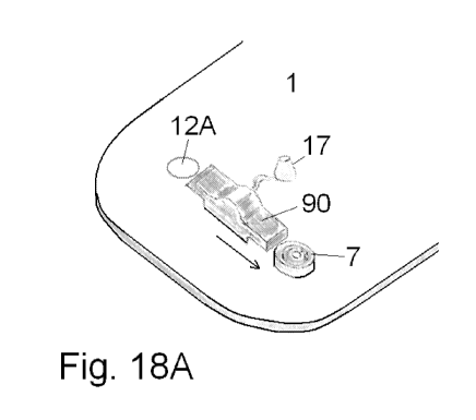
Fig. 17, 18A and 18B shows yet another embodiment of a base part provided with
more than one cannula part. Fig. 17 shows a cut-through side view of the
embodiment, fig. 18A shows an upper view of the embodiment and fig. 18B shows
an enlargement of the coupling part establishing a first fluid path with the
delivery
part.
According to this embodiment of a base part, a second fluid path can be
established
by pushing a moving part 90 to a first or a second position from a central
closed
position which central closed position of the movable part 90 allows for
insertion of
either one of two cannula parts 7. The moving part 90 is an unreleasable part
of the
base part 1 i.e. it cannot be removed from the base part but only be moved
between
different positions. The movable part 90 has an inlet 95 for fluid; the inlet
95 is
unreleasably connected to a flexible tube connecting the movable part 90 to an
inlet
opening 13 for the second fluid path. The inlet opening 13 is the end of a
penetrating
cannula protected by a bubble shaped membrane 17. The membrane 17 is
penetrated by the cannula when a delivery part is pressed against the base
part,
when the delivery part is pressed against the base part a first fluid path is
established between the reservoir of the delivery part and the base part and
fluid
can flow directly to the movable part 90, fluid can only flow from the movable
part 90
to a cannula part 7 if the movable part 90 is pushed to contact with a cannula
part 7
as illustrated with the arrow on fig. 18A.
Common for all the embodiments are that the base part has one inlet for fluid
and
one or more outlets for fluid i.e. the medication enters at one position via
the inlet of
the second fluid path and the second fluid path is then provided with one or
more
outlets to one or more cannula parts. Normally, there is no "reservoir" after
the fluid
has left the especially protected reservoir 6 of the delivery part which is
used to
store the fluid medication before and during use, after the fluid has left
this
designated reservoir 6 the fluid travels in a plug-flow assuring that all
fluid has a
well-defined short residence time inside the base part.